pregnane x receptor-humanized mice recapitulate...
TRANSCRIPT

JPET #224295
1
Pregnane X receptor-humanized mice recapitulate gender differences in ethanol
metabolism but not hepatotoxicity
Krisstonia Spruiell, Afua A. Gyamfi, Susan T. Yeyeodu, Ricardo M. Richardson, Frank J.
Gonzalez, and Maxwell A. Gyamfi
Julius L. Chambers Biomedical Biotechnology Research Institute, North Carolina
Central University, Durham, NC 27707, USA (KS, AAG, STY, RMR and MAG).
Laboratory of Metabolism, Center for Cancer Research, National Cancer Institute,
Building 37, Room 3106, Bethesda, MD 20892, USA (FJG).
This article has not been copyedited and formatted. The final version may differ from this version.JPET Fast Forward. Published on July 9, 2015 as DOI: 10.1124/jpet.115.224295
at ASPE
T Journals on M
ay 22, 2018jpet.aspetjournals.org
Dow
nloaded from

JPET #224295
2
(a) Running Title: hPXR mice and acute ethanol hepatotoxicity
(b) Corresponding author: Maxwell A. Gyamfi, Ph.D. Cardio-metabolic Research Program JLC-Biomedical/ Biotechnology Research Institute North Carolina Central University 700 George St, Durham NC 27707 Phone: 919-530-7832; Fax: 919-530-6815 E-mail: [email protected]
(c) Content:
Text pages: Number of text pages: 38 Number of tables: 1 Number of figures: 8 Number of references: 55 Abstract: 250 words Introduction: 738 words Discussion: 1587 words
(d) Abbreviations: ACC-1α, acetyl-CoA carboxylase 1α; ACOX-1, acyl CoA oxidase 1; ADH or Adh, alcohol dehydrogenase; ALDH or Aldh, aldehyde dehydrogenase; ALT, alanine aminotransferase; AST, aspartate aminotransferase; BAC, bacterial artificial chromosome; BEC, blood ethanol concentration; CPT1, carnitine palmitoyltransferase 1; CYP2E1, cytochrome P450 2E1; CYP3A11, cytochrome P450 3A11; ERα, estrogen receptor α; EtOH, ethanol; ERK1/2, extracellular regulated kinases 1/2; FAT/CD36, fatty acid translocase; FAS, fatty acid synthase; GRP78, glucose regulated protein 78; H & E, hematoxylin and eosin; LBD, ligand binding domain; L-FABP-1, liver fatty acid binding protein 1; MAPK, mitogen activated protein kinase; MTTP, microsomal triglyceride transfer protein; NEFA, nonesterified fatty acid; PCN, pregnenolone 16α-carbonitrile; PCNA, proliferating cell nuclear antigen; PEMT, phosphatidylethanolamine N-methyltransferase; PPARs, peroxisome proliferator-activated receptors; PXR, pregnane X receptor; hPXR, PXR-humanized; SCD1, stearoyl-CoA desaturase 1; SREBP-1c, sterol regulatory element binding protein 1c; WT, wild-type.
This article has not been copyedited and formatted. The final version may differ from this version.JPET Fast Forward. Published on July 9, 2015 as DOI: 10.1124/jpet.115.224295
at ASPE
T Journals on M
ay 22, 2018jpet.aspetjournals.org
Dow
nloaded from

JPET #224295
3
Abstract
Both human and rodent females are more susceptible to developing alcoholic liver
disease (ALD) following chronic ethanol (EtOH) ingestion. However, little is known about
the relative effects of acute EtOH exposure on hepatotoxicity in female vs. male mice.
The nuclear receptor pregnane X receptor (PXR, NR1I2) is a broad-specificity sensor
with species-specific responses to toxic agents. To examine the effects of the human
PXR on acute EtOH toxicity, the responses of male and female PXR-humanized (hPXR)
transgenic mice administered oral binge EtOH (4.5 g/kg) were analyzed. Basal
differences were observed between hPXR males and females in which females
expressed higher levels of two principal enzymes responsible for EtOH metabolism,
alcohol dehydrogenase 1 and aldehyde dehydrogenase 2 and two key mediators of
hepatocyte replication and repair, cyclin D1 and proliferating cell nuclear antigen. EtOH
ingestion upregulated hepatic estrogen receptor α, cyclin D1, and cytochrome P450 2E1
in both genders, but differentially altered lipid and EtOH metabolism. Consistent with
higher basal levels of EtOH metabolizing enzymes, blood EtOH was more rapidly
cleared in hPXR females. These factors combined to provide greater protection against
EtOH-induced liver injury in female hPXR mice, as revealed by markers for liver
damage, lipid peroxidation, and endoplasmic reticulum stress. These results indicate
that female hPXR mice are less susceptible to acute binge EtOH-induced hepatotoxicity
than their male counterparts, due at least in part to the relative suppression of cellular
stress and enhanced expression of enzymes involved in both EtOH metabolism and
hepatocyte proliferation and repair in hPXR females.
This article has not been copyedited and formatted. The final version may differ from this version.JPET Fast Forward. Published on July 9, 2015 as DOI: 10.1124/jpet.115.224295
at ASPE
T Journals on M
ay 22, 2018jpet.aspetjournals.org
Dow
nloaded from

JPET #224295
4
Introduction
Gender is a major factor that impacts susceptibility to alcoholic liver disease
(ALD); epidemiologic studies suggest that for any given level of alcohol consumption
women have a higher likelihood of developing liver cirrhosis than men (Pares et al.,
1986). Additionally, liver injury progresses faster in women with alcoholic hepatitis who
stop or reduce drinking (Pares et al., 1986). Several theories have been proposed to
explain this disparity based on gender differences in ethanol (EtOH) pharmacokinetics,
estrogen levels, and alcohol elimination rates (Sato et al., 2001).
Ethanol is metabolized predominantly in the liver by enzymes located in different
subcellular compartments of the hepatocyte (Zakhari and Li, 2007). Alcohol
dehydrogenase (ADH), a cytosolic NAD+-dependent enzyme, is the main enzyme that
catalyzes the conversion of EtOH to acetaldehyde, a potent toxicant that accounts for
most of the toxic effects of EtOH (Zakhari and Li, 2007). Acetaldehyde produced from
EtOH is further converted to the nontoxic acetate by a mitochondrial aldehyde
dehydrogenase (ALDH or Aldh) (Zakhari and Li, 2007). In addition to ADH, the
endoplasmic reticulum enzyme cytochrome P450 2E1 (CYP2E1), which is induced by
EtOH can metabolize EtOH to acetaldehyde at high alcohol doses (Zakhari and Li,
2007). Catalase, located in the peroxisomes, is also capable of oxidizing EtOH to
acetaldehyde (Zakhari and Li, 2007).
The effect of gender on ADH and ALDH activity is controversial. Pharmacokinetic
studies revealed that women have lower levels of gastric and hepatic ADH, resulting in
increased blood alcohol levels and EtOH toxicity (Chrostek et al., 2003; Frezza et al.,
1990). However, Maly and Sasse (1991), found that hepatic ADH activity is enhanced
This article has not been copyedited and formatted. The final version may differ from this version.JPET Fast Forward. Published on July 9, 2015 as DOI: 10.1124/jpet.115.224295
at ASPE
T Journals on M
ay 22, 2018jpet.aspetjournals.org
Dow
nloaded from

JPET #224295
5
in women (Maly and Sasse, 1991). While only minor variations in ALDH activity between
men and women have been reported, ALDH activity is higher in male hamsters than
females (Chrostek et al., 2003; Lee et al., 2001; Maly and Sasse, 1991).
We have previously shown that nuclear receptors, a class of intracellular
transcription factors activated by ligands are involved in the pathogenesis of ALD
(Gyamfi et al., 2008; Gyamfi et al., 2006). Notably, the nuclear hormone receptor
pregnane X receptor [PXR; NR1I2) expressed primarily in the liver and intestine, was
originally characterized as a xenobiotic receptor important for defense against toxic
agents and for eliminating drugs and other xenobiotics (Kliewer et al., 2002).
Interestingly, PXR activation induces hepatic triglyceride accumulation, a characteristic
feature of ALD, suggesting the possibility that PXR is associated with ALD
pathogenesis (Zhou et al., 2006). However, it is not known whether PXR is involved in
the gender dimorphism of EtOH hepatotoxicity.
Due to differences between the mouse and human ligand binding domain (LBD)
sequences, species-specific responses to ligand activation of human and mouse PXR
have been reported (Lehmann et al., 1998). Thus, some chemical ligands like
rifampicin and rifaximin that activate human PXR usually have little effect on the mouse
form of this receptor and vice versa (Ma et al., 2007a; Ma et al., 2007b). To this end,
PXR-humanized (hPXR) transgenic mice were developed to provide a more valid in
vivo model of human xenobiotic responses (Ma et al., 2007a). Interestingly, recent
reports indicate that transgenic mice expressing the human PXR gene are more prone
to high-fat diet (HFD)-induced hyperglycemia in both genders compared to their
respective wild-type (WT) counterparts (Spruiell et al., 2014a; Spruiell et al., 2014b).
This article has not been copyedited and formatted. The final version may differ from this version.JPET Fast Forward. Published on July 9, 2015 as DOI: 10.1124/jpet.115.224295
at ASPE
T Journals on M
ay 22, 2018jpet.aspetjournals.org
Dow
nloaded from

JPET #224295
6
Further, HFD-fed hPXR females express lower basal protein levels of both hepatic and
white adipose tissue (WAT) estrogen receptor α (ERα) (Spruiell et al., 2014a). Given
that higher estrogen levels and/or signaling in women have been implicated in EtOH-
induced liver injury, we reasoned that the human PXR gene may play a role in sex
differences in EtOH-induced liver injury (Eagon, 2010).
Most rodent studies investigating sex-specific differences in EtOH hepatotoxicity
have used the chronic EtOH ingestion model (Colantoni et al., 2002; Kono et al., 2000;
Nanji et al., 2001; Ronis et al., 2004). In comparison, relatively few studies have
investigated the effects of acute EtOH exposure on hepatotoxicity in male and female
mice (Wagnerberger et al., 2013). While some studies have addressed gender
dimorphisms in EtOH hepatotoxicity using rodents, extrapolation of the results to
humans can be difficult. In this study, binge EtOH was administered to male and female
hPXR mice revealing that the basal protein expression levels of both ADH1 and ALDH2
are higher in the liver of female hPXR mice. As a result, female hPXR mice eliminate
EtOH more effectively than their male counterparts, thereby mitigating acute EtOH
hepatotoxicity.
This article has not been copyedited and formatted. The final version may differ from this version.JPET Fast Forward. Published on July 9, 2015 as DOI: 10.1124/jpet.115.224295
at ASPE
T Journals on M
ay 22, 2018jpet.aspetjournals.org
Dow
nloaded from

JPET #224295
7
Materials and Methods
Animal care and treatment. Breeding pairs of male and female hPXR mice on a
C57BL/6 background were transferred from a colony housed at the National Cancer
Institute (NIH, Bethesda, Maryland) (Ma et al., 2007a). The hPXR mice were generated
by bacterial artificial chromosome (BAC) transgenesis, in which the transgene contains
the complete human PXR gene and the 5’- and 3’-flanking sequences and then bred
with Pxr-null mice to produce hPXR mice carrying a C57BL/6 genetic background (Ma
et al., 2007a). Human PXR was co-expressed with CYP3A in hPXR mice in liver,
duodenum, jejunum, and ileum, matching the gene expression pattern in humans and
“humanizing” mouse liver and intestine with respect to PXR (Ma et al., 2007a).
Treatment with PXR ligands revealed a clear species difference between WT and hPXR
mice in their response to xenobiotics, suggesting that this BAC transgenic hPXR mouse
model is useful for investigating human PXR function in vivo (Ma et al., 2007a). Mice (3-
5 mice/cage) were housed in polycarbonate cages on racks directly vented via the
facility’s exhaust system at 22°C with a 12/12-h light/dark cycle at the Animal Resources
Complex at North Carolina Central University. Age-matched (10-12 weeks of age) male
and female hPXR mice, were each randomly separated into two groups (n = 8-9 for
each group) and were gavaged with 3 doses of 4.5 g/kg 50% (vol/vol) EtOH or saline
solution every 12 hours at 9:00 AM, 9:00 PM, 9:00 AM the next day as previously reported
(Kirpich et al., 2012; Wang et al., 2013a). Four (4) hours after the final dose at 1:00 PM,
mice were anesthetized with isoflurane and killed. Sections of liver were rapidly
dissected, weighed, snap-frozen in liquid nitrogen and kept at –80°C. Blood samples
collected by cardiac puncture from anesthetized mice were centrifuged at 3000 rpm for
This article has not been copyedited and formatted. The final version may differ from this version.JPET Fast Forward. Published on July 9, 2015 as DOI: 10.1124/jpet.115.224295
at ASPE
T Journals on M
ay 22, 2018jpet.aspetjournals.org
Dow
nloaded from

JPET #224295
8
15 min to collect serum and stored at -80oC to determine EtOH concentration, liver
enzymes, triglycerides, and nonesterfied fatty acid (NEFA) concentrations. All
procedures conducted in accordance with the NIH Guidelines for the Care and Use of
Laboratory Animals were approved by the North Carolina Central University Institutional
Animal Care and Use Committee.
H & E Staining of Liver Sections. Liver slices were fixed in 10% formalin/phosphate-
buffered saline, and then stained with hematoxylin and eosin (H & E) for histological
examination.
Serum alanine aminotransferase (ALT), aspartate aminotransferase (AST), triglyceride,
and NEFA measurements. Serum was processed from blood and stored at -80oC.
Serum ALT and AST activity were determined using the Cholestech LDX analyzer
(Cholestech Corporation, Hayward, CA) as reported previously (Spruiell et al., 2014a;
Spruiell et al., 2014b). Serum triglyceride and NEFA levels were quantified using a
Triglyceride and NEFA-HR (2) test kits (Wako Pure Chemical Industries, Richmond, VA).
Determination of serum alcohol concentration. Blood samples were collected after three
doses of binge EtOH administration and centrifuged at 3000 rpm for 15 min to collect
serum for blood EtOH concentration (BEC). In a separate study, male and female hPXR
mice were administered a single dose of 50% (vol/vol) EtOH (4.5 g/kg) by gastric
intubation after overnight starvation. Mice (n = 4-5) were sacrificed 1, 2, 4, 6 and 8 h
after EtOH administration and blood samples were collected to prepare serum. The
This article has not been copyedited and formatted. The final version may differ from this version.JPET Fast Forward. Published on July 9, 2015 as DOI: 10.1124/jpet.115.224295
at ASPE
T Journals on M
ay 22, 2018jpet.aspetjournals.org
Dow
nloaded from

JPET #224295
9
EtOH L3K assay kit for quantitative measurement of EtOH concentration (Sekisui
Diagnostics P.E.I Inc, Charlottetown, Canada) was used according to the
manufacturer’s instructions as reported previously (Perides et al., 2005). The reaction is
based on the enzymatic conversion of EtOH by ADH to acetaldehyde and NADH. EtOH
concentration in the serum was quantified as the rate of increase in NADH absorbance
due to the reduction of NAD+ at 340 nm.
Hepatic triglyceride and cholesterol levels. Total liver lipids were extracted from 100 mg
of liver homogenate using methanol and chloroform as previously described (Gyamfi et
al., 2008). Hepatic triglyceride and cholesterol levels were quantified using Triglyceride
and Cholesterol test kits (Wako Pure Chemical Industries, Richmond, VA).
Measurements of lipid peroxidation (LPO) in liver tissues. The extent of LPO in liver
homogenates were quantitatively determined by measuring the concentration of the
thiobarbituric acid (TBA)-reactive product, malondialdehyde (MDA) using the TBA
reactive substances (TBARS) kit (ZeptoMetrix, Buffalo, NY) as we previously reported
(Tanaka et al., 2008). Protein contents in liver homogenates were determined by the
BCATM protein assay kit (Thermo Scientific, Rockford, IL).
Preparation of liver extracts for Western blot analyses. Frozen livers, were homogenized
at 4oC and Western blot analysis performed on extracts as described previously (Gyamfi
et al., 2006). Protein contents in liver homogenates were determined by the BCATM
protein assay kit (Thermo Scientific, Rockford, IL). Liver homogenate (40 μg/lane), were
This article has not been copyedited and formatted. The final version may differ from this version.JPET Fast Forward. Published on July 9, 2015 as DOI: 10.1124/jpet.115.224295
at ASPE
T Journals on M
ay 22, 2018jpet.aspetjournals.org
Dow
nloaded from

JPET #224295
10
mixed in Laemmli loading buffer containing β-mercaptoethanol, boiled for 5 min,
separated on 10 or 15% SDS-PAGE gels, and transferred to polyvinylidene difluoride
(PVDF) membrane. The membranes were probed with one or more of the following
antibodies according to the manufacturers’ recommendations: anti-CYP2E1 and anti-
catalase (Abcam, Cambridge, MA), anti-ERα, anti-CYP3A, anti-glucose-regulated
protein 78 (GRP78), anti-ALDH2, and anti-ADH1 (Santa Cruz Biotechnology, Santa
Cruz, CA), anti-phospho-p38 mitogen activated protein kinase (p38 MAPK)
(Thr180/Tyr182), anti-p38 MAPK, anti-phospho-P44/42 MAPK (i.e. extracellular
regulated kinases 1/2, p-ERK1/2) (Thr202/Tyr204), anti-ERK1/2, anti-caspase 12, anti-
cyclin D1 and anti-phosphorylated-elF2α (Ser51) (Cell Signaling Technology, Boston,
MA), or anti-proliferating cell nuclear antigen (PCNA) (Sigma, St. Louis, MO). Blots
were then incubated with the appropriate peroxidase-conjugated anti-rabbit IgG
secondary antibodies (Santa Cruz Biotechnology, Santa Cruz, CA) diluted in TBST plus
1% milk for 60 min at room temperature. Following initial probing, blots were stripped
and reprobed with anti-α-tubulin antibody (Cell Signaling Technology, Boston, MA).
Proteins were visualized using enhanced chemiluminescence and band intensity was
quantified using ImageJ software (U. S. National Institutes of Health, Bethesda, MD).
Quantification of mRNA levels using real-time polymerase chain reaction (real-time
PCR). Total RNA was isolated from frozen liver tissues using the Trizol reagent
according to the manufacturer’s protocol (Invitrogen, Carlsbard, CA). Total RNA (5 µg)
was reversed transcribed into cDNA with random hexamer primers using Tetro cDNA
Synthesis Kit (Bioline, Taunton, MA) as we previously described (Spruiell et al., 2014b).
This article has not been copyedited and formatted. The final version may differ from this version.JPET Fast Forward. Published on July 9, 2015 as DOI: 10.1124/jpet.115.224295
at ASPE
T Journals on M
ay 22, 2018jpet.aspetjournals.org
Dow
nloaded from

JPET #224295
11
The cDNA was then diluted 20-fold with water and and subjected to real-time
quantitative PCR by the SensiFast SYBR Hi-ROX Kit (Bioline, Taunton, MA) to quantify
the mRNA levels of Peroxisome proliferator-activated receptor α (PPARα), PPARγ,
sterol regulatory element binding protein 1c (SREBP-1c), carnitine palmitoyltransferase
1 (CPT1), acyl CoA oxidase 1 (ACOX-1), liver fatty acid binding protein 1 (L-FABP-1),
microsomal triglyceride transfer protein (MTTP), fatty acid translocase (FAT/CD36),
acetyl-CoA carboxylase 1α (ACC-1α), fatty acid synthase (FAS), stearoyl-CoA
desaturase 1 (SCD1), and phosphatidylethanolamine N-methyltransferase (PEMT). The
primers (Table 1) for mRNA encoding PPARα, PPARγ, SREBP-1c, CPT1, ACOX-1, L-
FABP, MTTP, CD36, ACC-1α, FAS, SCD1, and GAPDH were designed using Primer
Express 2.0 (Applied Biosystems, Foster City, CA). Furthermore, the following
proprietary Taqman Gene Expression Assays for isoforms of Adh and Aldh2 were
purchased from Applied Biosystems/Life Technologies (Grand Island, NY) and used for
real-time quantitative PCR: Adh1 (class I Adh, No. 00507711_m1), Adh4 (class II Adh,
No. 00478838_m1), Adh5 (class III Adh, No. 00475804_g1), Aldh2 (No. 00477463_m1)
and Gapdh (house keeping gene; No. 99999915_g1). The amplification reactions were
carried out in the ABI 7900HT Fast Real-Time PCR System (Applied Biosystems, Foster
City, CA) as previously described (Gyamfi et al., 2008). Results were presented as
levels of expression relative to that of controls after normalizing with Gapdh mRNA
using the comparative CT method.
Statistical Analysis. Data are presented as means ± SEM (n = 8-9). Statistical analysis
was performed using one-way ANOVA followed by Tukey HSD post-hoc test. A P value
This article has not been copyedited and formatted. The final version may differ from this version.JPET Fast Forward. Published on July 9, 2015 as DOI: 10.1124/jpet.115.224295
at ASPE
T Journals on M
ay 22, 2018jpet.aspetjournals.org
Dow
nloaded from

JPET #224295
12
of < 0.05 was considered statistically significant. Statistical analyses were performed
using IBM SPSS Statistics 20 software (Armonk, NY).
This article has not been copyedited and formatted. The final version may differ from this version.JPET Fast Forward. Published on July 9, 2015 as DOI: 10.1124/jpet.115.224295
at ASPE
T Journals on M
ay 22, 2018jpet.aspetjournals.org
Dow
nloaded from
JPET #224295
13
Results
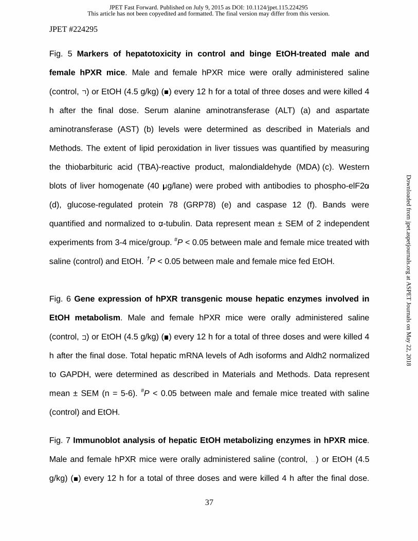
EtOH induces lipid accumulation in both male and female hPXR mice
Body and liver weights were significantly higher in male hPXR mice than hPXR
females; however, binge EtOH ingestion had no effect on either body, liver weight or
liver-to-body weight ratios of either sex (Fig. 1a-c). H & E staining revealed that hepatic
lipid droplets in the control-fed mice of both genders were less abundant than their
EtOH-fed counterparts (Fig. 2a). Furthermore, EtOH-induced accumulation of lipid
droplets correlated with increased hepatic triglyceride levels in both male and female
hPXR mice (Fig. 2a and 2b). However, EtOH treatment did not significantly impact
hepatic cholesterol levels in either male or female hPXR mice (Fig. 2c). Furthermore,
neither serum triglycerides nor NEFA levels differed between genders or treatments
(Fig. 2d and 2e).
Ethanol ingestion upregulates hepatic estrogen receptor α (ERα) and its target gene
cyclin D1 in both male and female hPXR mice
In humans and in rodents, EtOH ingestion was found to alter circulating sex
steroid levels and increase hepatic estrogen receptor (ER) expression in males
(Colantoni et al., 2002). Furthermore, a link between 1) gender dependent recovery
from liver injury, and 2) upregulation of cell cycle genes including the ERα target gene,
cyclin D1 and the cell proliferation marker, PCNA, was observed (Balasenthil et al.,
2004; Wang et al., 2013b). Furthermore, MAPKs have been implicated in EtOH-induced
cyclin D1 expression, cell cycle inhibition, and cell survival (Stepniak et al., 2006). To
investigate the effect of sex steroid levels in male and female hPXR mice on gender
This article has not been copyedited and formatted. The final version may differ from this version.JPET Fast Forward. Published on July 9, 2015 as DOI: 10.1124/jpet.115.224295
at ASPE
T Journals on M
ay 22, 2018jpet.aspetjournals.org
Dow
nloaded from

JPET #224295
14
differences in ALD, we used Western blot analysis to determine hepatic levels of ERα,
cyclin D1, PCNA and two MAPK family members ERK and p38 and their activated
phosphorylated forms. As expected, the basal hepatic protein levels of ERα were higher
in control female hPXR mice compared to their male counterparts (Fig. 3a). EtOH
ingestion induced hepatic expression of ERα protein in male hPXR mice (Fig. 3a).
Hepatic ERα protein levels were also enhanced 3.5-fold in female hPXR mice by EtOH
ingestion (Fig. 3a). Similarly, basal hepatic protein levels of the ERα target gene cyclin
D1 (Fig. 3b) and PCNA (Fig. 3c) involved in liver regeneration were significantly higher
2.5- and 2.1-fold, respectively, in female hPXR mice than hPXR males (Fig. 3b and c)
(Balasenthil et al., 2004). EtOH significantly increased cyclin D1 protein levels in both
male (2.2-fold) and female hPXR (1.3-fold) mice (Fig. 3b and 3c). Cyclin D1 levels were
subject to modest upregulation by binge EtOH treatment in males and females, whereas
PCNA levels were not (Fig. 3b and 3c). EtOH did not significantly alter hepatic ERK1/2
and p38 protein expression (Fig. 3d and e). However, EtOH ingestion inhibited ERK1/2
activation in the livers of male and female hPXR mice by 71% and 53%, respectively, in
comparison to their untreated controls (Fig. 3d). Furthermore, EtOH also suppressed
p38 activation in both male hPXR by 81% and female hPXR mice by 69% (Fig 3e).
Ethanol effect on hepatic lipid synthesis, uptake, and oxidation genes are more
pronounced in female hPXR mice than males.
There are several regulators of hepatic lipid synthesis, metabolism, oxidation,
storage and transport. PPARs are ligand-activated transcription factors that primarily
regulate genes involved in lipid metabolism; PPAR isoforms varies in fatty livers
This article has not been copyedited and formatted. The final version may differ from this version.JPET Fast Forward. Published on July 9, 2015 as DOI: 10.1124/jpet.115.224295
at ASPE
T Journals on M
ay 22, 2018jpet.aspetjournals.org
Dow
nloaded from

JPET #224295
15
(Memon et al., 2000). SREBP-1c, a transcription factor known to play important role in
de novo fatty acid and triglyceride synthesis may also contribute to lipid accumulation in
the liver (Horton et al., 2002). PEMT is the only enzyme in liver that converts
phosphatidylethanolamine into phosphatidylcholine, a process that is required for very
low density lipoprotein (VLDL) assembly and lipid export from hepatocytes (Vance,
2014). Furthermore, PEMT deficiency or inhibition promotes steatosis and liver
damage (Vance, 2014). Because acute EtOH exposure resulted in increased lipid
accumulation in both genders (Fig. 2a and 2b), changes in the expression of these
regulatory factors and their target genes were explored. Basal hepatic Pparα mRNA
levels were comparable between control male and female hPXR mice (Fig. 4a). EtOH
decreased Pparα mRNA levels in both male and female hPXR mice; however, the
decrease was not statistically significant compared to their respective controls (Fig. 4a).
Similarly, basal Cpt-1 and Acox-1 mRNA levels did not vary between the two hPXR
genders and their mRNA levels were not different after EtOH treatment (Fig. 4a). While
constitutive Lfabp-1 mRNA levels did not vary between the two hPXR genders and
EtOH did not have any significant effects on Lfabp-1 mRNA levels in male hPXR mice,
EtOH induced a 1.5-fold increase in Lfabp-1 mRNA levels in female hPXR mice (Fig.
4a). Similarly, constitutive Mttp mRNA levels did not differ between male and female
hPXR mice whereas EtOH significantly induced the Mttp gene 1.7-fold only in female
hPXR mice (Fig. 4a). The basal levels of Srebp-1c mRNA and SREBP-1c target gene
Acc-1α, Fas, and Scd1 mRNAs were comparable between male and female hPXR mice
(Fig. 4b). However, EtOH administration diminished Srebp-1c mRNA levels by 60% only
in male hPXR mice (Fig. 4b). EtOH had no significant effect on Acc-1α mRNA in males,
This article has not been copyedited and formatted. The final version may differ from this version.JPET Fast Forward. Published on July 9, 2015 as DOI: 10.1124/jpet.115.224295
at ASPE
T Journals on M
ay 22, 2018jpet.aspetjournals.org
Dow
nloaded from

JPET #224295
16
but increased Acc-1α mRNA levels 1.6-fold in female hPXR mice (Fig. 4b). Fas mRNA
was the only gene to be upregulated by EtOH in male hPXR mice (1.6-fold), but this
increase did not reach statistical significance and merely matched basal Fas mRNA
levels in control hPXR females (Fig. 4b). In contrast, EtOH significantly increased Fas
mRNA levels (1.9-fold) in female hPXR mice (Fig. 4b). Unexpectedly, EtOH ingestion
significantly decreased Scd-1 mRNA levels (64%) in female hPXR mice, but not in male
hPXR mice (Fig. 4b). Basal hepatic Pparγ mRNA levels did not vary between male and
female hPXR mice treated with saline, even though basal mRNA levels of the Pparγ
downstream target Cd36 were 2.7-fold higher in female hPXR mice compared to male
hPXR mice (Fig. 4c) (Tontonoz et al., 1998). While EtOH ingestion did not significantly
affect Pparγ and Cd36 mRNA levels in male hPXR mice, it induced an increase in
hepatic Pparγ and Cd36 expression by 1.5- and 1.8-fold, respectively, in hPXR females
(Fig. 4c). In contrast, hepatic Pemt mRNA levels did not vary significantly between male
and female hPXR control mice and between control and EtOH treated hPXR females
(Fig. 4d). Pemt gene expression was suppressed by EtOH (47%) in male hPXR mice.
Ethanol-induced hepatotoxicity is greater in male hPXR mice
Serum ALT and AST activities were measured as indices of hepatocyte/organ injury.
EtOH increased both ALT and AST levels in male hPXR mice by 1.8- and 2.1-fold,
respectively (Fig. 5a and 5b) and in female mice by 1.6-fold and 1.5-fold, respectively
(Fig. 5a and 5b). However, the increases in ALT and AST activity in females were not
statistically significant, suggesting that EtOH-induced hepatotoxicity is more severe in
male hPXR mice (Fig. 5a and 5b). LPO, an early biochemical feature of EtOH toxicity
This article has not been copyedited and formatted. The final version may differ from this version.JPET Fast Forward. Published on July 9, 2015 as DOI: 10.1124/jpet.115.224295
at ASPE
T Journals on M
ay 22, 2018jpet.aspetjournals.org
Dow
nloaded from
JPET #224295
17
and a major indicator of oxidative stress was quantified by measuring the thiobarbituric
acid-reactive product, MDA. Levels of MDA were significantly increased by EtOH only in
the livers of male hPXR (7.3-fold) mice (Fig. 5c). Moreover, EtOH induced increases in
MDA levels were significantly higher in hPXR males compared to EtOH-fed hPXR
females (Fig. 5c). Expression of hepatic endoplasmic reticulum stress markers
phospho-elF2α and GRP78 was also upregulated 11.1 and 1.4-fold, respectively, in
EtOH-treated vs. control males (Fig. 5d and 5e). Phospho-elF2α protein levels were
elevated in the livers of control fed female hPXR mice by 2.6-fold compared to their
male counterparts (Fig. 5d). Intriguingly, EtOH exposure elevated hepatic phospho-
elF2α protein levels (4.3-fold) but not GRP78 in hPXR females (Fig. 5d and 5e). Hepatic
protein levels of active caspase 12, the endoplasmic reticulum stress-specific caspase,
was increased only in EtOH-fed hPXR males (2.5-fold) and this increase was higher
than in EtOH-fed female hPXR mice (Fig. 5f).
Basal ADH and ALDH2 protein but not gene expression is greater in female hPXR mice
Alcohol metabolism is considered a major factor in alcohol-related liver damage
(Ronis et al., 2010; Zakhari and Li, 2007). The hepatotoxicity data (Fig. 5a and 5b)
prompted further examination of ADH, ALDH2, catalase, CYP2E1, and CYP3A enzymes
known to be involved in EtOH metabolism in male and female hPXR mice (Zakhari and
Li, 2007). The basal hepatic Adh1 (class1 Adh) mRNA levels did not vary significantly
between genders or treatments (Fig. 6a). However, constitutive Adh4 (class II Adh)
mRNA levels tended to be lower in female hPXR mice (59%) compared to control male
hPXR mice, but the difference was not statistically significant (P = 0.052) (Fig. 6b).
This article has not been copyedited and formatted. The final version may differ from this version.JPET Fast Forward. Published on July 9, 2015 as DOI: 10.1124/jpet.115.224295
at ASPE
T Journals on M
ay 22, 2018jpet.aspetjournals.org
Dow
nloaded from

JPET #224295
18
While EtOH did not have any significant effects on Adh4 mRNA levels in female hPXR
mice, it significantly decreased the levels in male hPXR mice (54%) compared to control
males (Fig. 6b). The hepatic Adh5 (class III Adh) mRNA levels did not vary significantly
between male and female hPXR control mice (Fig. 6c). While EtOH treatment did not
alter hepatic Adh5 mRNA levels in female hPXR mice, it tended to inhibit Adh5 gene
expression in male hPXR mice (Fig. 6c). In contrast, the basal mRNA levels of Aldh2
were somewhat (133%) but not significantly higher in female hPXR mice compared to
hPXR males (Fig. 6d). EtOH treatment significantly decreased Aldh2 mRNA levels only
in female hPXR mice (Fig. 6d). Unexpectedly, immunoblot analysis indicated a dramatic
increase in basal hepatic ADH1 protein in female hPXR (4.3-fold) compared to male
hPXR mice (Fig. 7a), even though, mRNA levels did not change significantly (Fig. 6a).
EtOH ingestion did not affect hepatic ADH1 protein expression in male hPXR mice,
however, it inhibited ADH1 protein levels somewhat in female hPXR mice compared to
control hPXR females (Fig. 7a). Furthermore, after EtOH treatment, hepatic ADH1
protein levels in female hPXR mice were significantly higher than in EtOH-fed males
(Fig. 7a). Similarly, while the mRNA levels did not vary, like ADH1, the basal hepatic
ALDH2 protein levels were significantly higher in female hPXR mice (3.0-fold) than in
males (Fig. 7b). Also as with ADH1, ALDH2 protein levels were not altered by EtOH
treatment in male hPXR mice (Fig. 7b). In contrast, EtOH treatment inhibited ALDH2
protein levels by 37% in female hPXR mice compared to controls consistent with the
EtOH inhibition of mRNA expression of this gene (Fig. 6d and 7b). Even so, ALDH2
protein levels in EtOH-treated females remained about 1.5-fold higher than in EtOH-fed
males (Fig. 7b). By contrast, basal hepatic catalase protein levels did not vary between
This article has not been copyedited and formatted. The final version may differ from this version.JPET Fast Forward. Published on July 9, 2015 as DOI: 10.1124/jpet.115.224295
at ASPE
T Journals on M
ay 22, 2018jpet.aspetjournals.org
Dow
nloaded from

JPET #224295
19
male and female hPXR mice or between saline and EtOH treatments (Fig. 7c). CYP2E1
(Fig. 7d) and CYP3A11 (Fig. 7e) protein expression in the liver did not differ between
control male and female mice. However, CYP2E1 but not CYP3A11 protein expression
was upregulated (2.0-fold) by EtOH treatment in both male and female hPXR mice (Fig.
7d).
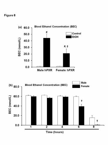
Female hPXR mice eliminate blood EtOH more efficiently than males
To determine the impact on EtOH metabolism of elevated levels of ADH1 and
ALDH2 protein expression observed in female hPXR mice (Fig. 7a and 7b), residual
concentrations of EtOH were measured after administration of three doses of binge
EtOH. Elevated levels of alcohol metabolizing enzymes in female hPXR mice resulted in
lower BEC in EtOH-fed females compared to EtOH-fed hPXR males (Fig. 8a). Gender
differences in EtOH oxidation in the gut, or “first-pass” metabolism by gastric ADH may
explain the sex-related differences in alcohol bioavailability and toxicity (DiPadova et al.,
1987). Fasting eliminates “first-pass” metabolism and diminishes the contribution of
gastric ADH to EtOH oxidation (DiPadova et al., 1987). Therefore, to study the impact of
the high hepatic ADH1 protein levels in female hPXR mice, EtOH elimination rates were
determined in fasting hPXR mice, after a single dose of EtOH by gastric intubation.
BECs were measured at various time points. While BECs remained high in both male
and female hPXR mice up to 4 hours after EtOH challenge, female hPXR mice were
able to clear blood EtOH more efficiently than males by 6 hours, and by 8 hours BEC
was virtually eliminated in females (Fig. 8b).
This article has not been copyedited and formatted. The final version may differ from this version.JPET Fast Forward. Published on July 9, 2015 as DOI: 10.1124/jpet.115.224295
at ASPE
T Journals on M
ay 22, 2018jpet.aspetjournals.org
Dow
nloaded from

JPET #224295
20
Discussion
Despite numerous studies investigating chronic EtOH responses in rodents, sex
differences in acute EtOH hepatotoxicity have not been thoroughly investigated. In this
study, binge EtOH was administered to male and female hPXR mice and sex
differences in the gene and protein expression of enzymes associated with EtOH and
lipid metabolism, EtOH clearance, and hepatotoxicity, and markers of ER status,
proliferation and endoplasmic reticulum stress were examined. Surprisingly, female
hPXR mice were less susceptible to acute binge EtOH-induced liver injury than their
male counterparts. Once EtOH is absorbed and distributed, sex differences could occur
in hepatic EtOH metabolism leading to liver damage (Ronis et al., 2010; Zakhari and Li,
2007).
We first determined the basal gene and protein expression levels of ADH1, the
major enzyme responsible for EtOH catabolism, and the ADH4 and ADH5 isoforms
active at high EtOH concentrations (Zakhari and Li, 2007). In the current study, basal
Adh1, Adh4, and Adh5 mRNA levels did not differ between male and female hPXR
mice. EtOH significantly downregulated the expression of Adh4 mRNA only in male
hPXR mice. Contrary to gene expression, basal hepatic protein expression of ADH1
was higher in female hPXR mice than males. These findings are consistent with
previous reports showing that hepatic ADH regulation is post-transcriptional and that
ADH protein expression increases without a corresponding increase in Adh mRNA
levels (Gyamfi et al., 2006; Mezey et al., 2005; Tussey and Felder, 1989). The current
findings are also consistent with several reports that hepatic ADH is more highly
This article has not been copyedited and formatted. The final version may differ from this version.JPET Fast Forward. Published on July 9, 2015 as DOI: 10.1124/jpet.115.224295
at ASPE
T Journals on M
ay 22, 2018jpet.aspetjournals.org
Dow
nloaded from

JPET #224295
21
expressed in female rodents and women (Aasmoe and Aarbakke, 1999; Harada et al.,
1998; Kishimoto et al., 2002; Maly and Sasse, 1991).
Variations in EtOH metabolizing enzyme expression and activity not only
influence EtOH metabolism, but also EtOH clearance and EtOH-induced liver injury
(Gyamfi et al., 2008; Gyamfi et al., 2006; Zakhari and Li, 2007). Besides ADH1, the
basal hepatic protein levels of ALDH2 were also upregulated in female hPXR mice
suggesting that this metabolic pathway converting EtOH to acetate via acetaldehyde is
more efficient in female hPXR animals. Correspondingly, measurements of BEC, the
EtOH concentration in the blood after metabolism of consumed EtOH in hPXR males
and females treated with or without binge EtOH were lower in females relative to males.
Consistent with this, pharmacological activation of ALDH2 reversed alcoholic steatosis
and apoptosis through accelerating acetaldehyde clearance, suggesting that higher
ALDH2 levels in hPXR females may provide enhanced protection against alcohol
hepatotoxicity (Zhong et al., 2015).
EtOH metabolism and elimination may vary depending on gender, age, body
weight, stomach content, and medication use (Jones and Sternebring, 1992). Gastric
ADH activity is higher in males than in females likely resulting in differences in first pass
EtOH metabolism and BEC (Frezza et al., 1990). With evidence that shows fasting
increases EtOH absorption and eliminates the contribution of gastric ADH to BEC, a
fasting model was employed to focus exclusively on the role of hepatic ADH and other
EtOH detoxifying enzymes in the liver (DiPadova et al., 1987). While peak BEC levels
were similar between fasting male and female hPXR mice after a single EtOH dose, the
rate of EtOH elimination was enhanced in female hPXR mice compared to males.
This article has not been copyedited and formatted. The final version may differ from this version.JPET Fast Forward. Published on July 9, 2015 as DOI: 10.1124/jpet.115.224295
at ASPE
T Journals on M
ay 22, 2018jpet.aspetjournals.org
Dow
nloaded from

JPET #224295
22
Similarly, serum EtOH levels were lower in female C3H/HeNCrj (C3H/He) mice after
acute EtOH administration, with females exhibiting enhanced ADH and ALDH activity
(Kishimoto et al., 2002). The phenomenon that women metabolize EtOH more rapidly
than men has also been observed consistently in human subjects (Jones, 2010; Mishra
et al., 1989). The similarity in EtOH elimination between our hPXR mice and humans
suggest that hPXR mice may be an appropriate model to further explore the effect of
gender, EtOH concentration, and hepatic metabolic capacity on EtOH catabolism.
Estrogen and its receptors impacts the gender differences we observed in EtOH
metabolism in hPXR mice, either directly or indirectly. First, ADH is hormonally,
nutritionally, and developmentally regulated (Boleda et al., 1992; Rao et al., 1997). The
emergence of gender differences in ADH activity coincide with sexual development: a
relative increase in ADH activity in females is not detected until the sixth week of age,
the age at which rodents reach sexual maturity, and remains elevated thereafter (Rao et
al., 1997). Second, estrogen increases hepatic ADH activity, while testosterone
decreases it (Harada et al., 1998; Rachamin et al., 1980). Similar, to ADH, estrogen also
increases mitochondrial ALDH activity, consistent with the sexual dimorphism in both
ADH and ALDH expression seen in hPXR females in the current study (Kishimoto et al.,
2002). Third, in humans and in Sprague/Dawley rats, EtOH ingestion leads to increased
ER expression in males not females. (Colantoni et al., 2002). Curiously, we observed an
increase in hepatic ERα protein levels not only in EtOH-fed male hPXR mice, but also
their female counterparts, unlike the rat model where ERα levels in females did not
increase significantly upon chronic EtOH exposure (Colantoni et al., 2002). Taken
together, our data suggests that a functional relationship exists between the human
This article has not been copyedited and formatted. The final version may differ from this version.JPET Fast Forward. Published on July 9, 2015 as DOI: 10.1124/jpet.115.224295
at ASPE
T Journals on M
ay 22, 2018jpet.aspetjournals.org
Dow
nloaded from

JPET #224295
23
PXR and ERα. Further indirect evidence gleaned from the literature supports this
possibility. 1) PXR expression is enhanced in ERα-negative breast cancer cells
(Dotzlaw et al., 1999), 2) Expression of both PXR and ERα is upregulated in an in vitro
reporter system by the organochlorine insecticide chlordecone (Lee et al., 2008).
Earlier studies indicate that feminization of the liver protects males but not
females against EtOH-induced liver injury (Colantoni et al., 2002). In contrast, we found
a significant increase in levels of two markers of liver damage, ALT and AST in EtOH-
fed hPXR males, whereas females showed only a modest (statistically insignificant)
increase in serum ALT and AST levels. Thus, male hPXR mice appear to be more
susceptible to acute EtOH-induced hepatotoxicity than hPXR females contrary to
previous findings in female mice were more prone to acute EtOH-induced steatosis
(Wagnerberger et al., 2013). However, similar to our hepatotoxicity results, serum ALT
levels were significantly increased only in male C57BL/6J mice, but not females, after
acute EtOH (6 g/kg) administration (Wagnerberger et al., 2013). CYP2E1 activity is
induced after EtOH exposure and has been implicated as the source of oxidative stress,
which further exacerbates liver injury (Zakhari and Li, 2007). In the current study,
CYP2E1 was induced about 2-fold by EtOH in livers of both male and female hPXR
mice, however, LPO, a marker of oxidative stress was significantly increased in EtOH-
fed hPXR males, but not in hPXR females. Consistent with an increase in the serum
levels of liver damage markers ALT and AST in EtOH-fed male hPXR mice,
endoplasmic reticulum stress markers Caspase 12, GRP78 and phospho-elF2α protein
levels were higher in male hPXR mice than in females (Dara et al., 2011).
Compensatory tissue repair also influences the final outcome of hepatotoxicity
This article has not been copyedited and formatted. The final version may differ from this version.JPET Fast Forward. Published on July 9, 2015 as DOI: 10.1124/jpet.115.224295
at ASPE
T Journals on M
ay 22, 2018jpet.aspetjournals.org
Dow
nloaded from
JPET #224295
24
(Chanda and Mehendale, 1996). Cyclin D1 signals hepatocyte commitment to division
and liver regeneration (Apte et al., 2004). p38 MAPK regulates cyclin D1 expression
(Stepniak et al., 2006). In the current study, basal levels and EtOH-induced up-
regulation of regenerative cyclin D1 were elevated in female hPXR mice. However,
while cyclin D1 was upregulated by EtOH in male hPXR mice, the cell proliferation
marker, PCNA was not, even though hepatic PCNA protein levels were elevated in
control and EtOH-treated hPXR females, consistent with the possibility of increased
DNA repair after liver damage as reported by others (Essers et al., 2005). It is possible
that the suppression of p38 and ERK activation by EtOH in both male and female hPXR
mice was involved in D1 upregulation seen in the current study as previously reported
(Stepniak et al., 2006).
A surprising observation of the current study was that while EtOH-induced
steatosis and triglyceride accumulation were observed in both genders, the effect of
EtOH on hepatic genes involved in lipid metabolism was strongly influenced by gender.
Importantly, mRNA levels of Lfabp-1 (involved in the uptake of fatty acids and fatty acid
oxidation) and Mttp (involved in the assembly and secretion of VLDL from hepatocytes)
were induced by EtOH only in hPXR females (Desvergne et al., 1998; Letteron et al.,
2003). Moreover, EtOH also induced genes involved in lipid synthesis and uptake,
including Acc-1α, Fas, Pparγ, and Cd36, but did not affect levels of Srebp-1c in female
hPXR mice. While male hPXR were resistant to EtOH-induced modulation of genes
involved in fatty acid synthesis, uptake, and oxidation, they were more prone to
inhibition of the phosphatidylcholine biosynthesis enzyme PEMT, which may lead to
decreased VLDL formation and steatosis as seen in the hPXR males. Considered
This article has not been copyedited and formatted. The final version may differ from this version.JPET Fast Forward. Published on July 9, 2015 as DOI: 10.1124/jpet.115.224295
at ASPE
T Journals on M
ay 22, 2018jpet.aspetjournals.org
Dow
nloaded from

JPET #224295
25
together, the current results suggest different mechanisms for EtOH-induced steatosis in
male and female hPXR mice; while EtOH-induced inhibition of Pemt gene expression is
the major mechanism for steatosis in males, upregulation of fatty acid synthesis and
uptake genes may account for steatosis in females. Our data provide evidence for the
contribution of the human PXR gene to sexual dimorphism of lipid metabolism in
response to EtOH ingestion. However, the detailed mechanisms remain to be
determined.
In conclusion, the present results demonstrate gender differences in EtOH-
induced expression of lipid metabolic genes, EtOH elimination and hepatotoxicity.
Surprisingly, female hPXR mice are less susceptible to acute EtOH-induced liver injury
compared to their male counterparts and are more efficient in reducing BEC, at least in
part because of increased expression of 1) ADH1 and ALDH2 involved in metabolism of
EtOH, and 2) cyclin D1 and PCNA important for repair of injured hepatocytes and 3)
suppression of EtOH-induced activation of LPO and endoplasmic reticulum stress. To
our knowledge, this is the first report of sexual dimorphism in ADH1 and ALDH2 protein
expression in hPXR mice.
This article has not been copyedited and formatted. The final version may differ from this version.JPET Fast Forward. Published on July 9, 2015 as DOI: 10.1124/jpet.115.224295
at ASPE
T Journals on M
ay 22, 2018jpet.aspetjournals.org
Dow
nloaded from
JPET #224295
26
Acknowledgements
Author Contribution: Conceived and designed experiments: Maxwell A. Gyamfi and Frank J. Gonzalez
Conducted experiments: Krisstonia Spruiell, Afua A. Gyamfi, and Maxwell A. Gyamfi
Contributed new reagents and materials: Maxwell A. Gyamfi, Ricardo M
Richardson, and Frank J. Gonzalez
Performed data analysis: Maxwell A. Gyamfi and Susan Yeyeodu
Wrote or contributed to the writing of the manuscript: Maxwell A. Gyamfi, Susan
Yeyeodu, Ricardo M. Richardson, and Frank J. Gonzalez
This article has not been copyedited and formatted. The final version may differ from this version.JPET Fast Forward. Published on July 9, 2015 as DOI: 10.1124/jpet.115.224295
at ASPE
T Journals on M
ay 22, 2018jpet.aspetjournals.org
Dow
nloaded from

JPET #224295
27
References
Aasmoe, L., and Aarbakke, J. (1999). Sex-dependent induction of alcohol dehydrogenase activity
in rats. Biochem Pharmacol 57, 1067-1072.
Apte, U.M., McRee, R., and Ramaiah, S.K. (2004). Hepatocyte proliferation is the possible
mechanism for the transient decrease in liver injury during steatosis stage of alcoholic
liver disease. Toxicol Pathol 32, 567-576.
Balasenthil, S., Barnes, C.J., Rayala, S.K., and Kumar, R. (2004). Estrogen receptor activation at
serine 305 is sufficient to upregulate cyclin D1 in breast cancer cells. FEBS Lett 567,
243-247.
Boleda, M.D., Farres, J., Guerri, C., and Pares, X. (1992). Alcohol dehydrogenase isoenzymes in
rat development. Effect of maternal ethanol consumption. Biochem Pharmacol 43, 1555-
1561.
Chanda, S., and Mehendale, H.M. (1996). Hepatic cell division and tissue repair: a key to
survival after liver injury. Molecular medicine today 2, 82-89.
Chrostek, L., Jelski, W., Szmitkowski, M., and Puchalski, Z. (2003). Gender-related differences
in hepatic activity of alcohol dehydrogenase isoenzymes and aldehyde dehydrogenase in
humans. J Clin Lab Anal 17, 93-96.
Colantoni, A., Emanuele, M.A., Kovacs, E.J., Villa, E., and Van Thiel, D.H. (2002). Hepatic
estrogen receptors and alcohol intake. Mol Cell Endocrinol 193, 101-104.
Dara, L., Ji, C., and Kaplowitz, N. (2011). The contribution of endoplasmic reticulum stress to
liver diseases. Hepatology 53, 1752-1763.
Desvergne, B., A, I.J., Devchand, P.R., and Wahli, W. (1998). The peroxisome proliferator-
activated receptors at the cross-road of diet and hormonal signalling. J Steroid Biochem
This article has not been copyedited and formatted. The final version may differ from this version.JPET Fast Forward. Published on July 9, 2015 as DOI: 10.1124/jpet.115.224295
at ASPE
T Journals on M
ay 22, 2018jpet.aspetjournals.org
Dow
nloaded from

JPET #224295
28
Mol Biol 65, 65-74.
DiPadova, C., Worner, T.M., Julkunen, R.J., and Lieber, C.S. (1987). Effects of fasting and
chronic alcohol consumption on the first-pass metabolism of ethanol. Gastroenterology
92, 1169-1173.
Dotzlaw, H., Leygue, E., Watson, P., and Murphy, L.C. (1999). The human orphan receptor PXR
messenger RNA is expressed in both normal and neoplastic breast tissue. Clin Cancer Res
5, 2103-2107.
Eagon, P.K. (2010). Alcoholic liver injury: influence of gender and hormones. World J
Gastroenterol 16, 1377-1384.
Essers, J., Theil, A.F., Baldeyron, C., van Cappellen, W.A., Houtsmuller, A.B., Kanaar, R., and
Vermeulen, W. (2005). Nuclear dynamics of PCNA in DNA replication and repair. Mol
Cell Biol 25, 9350-9359.
Frezza, M., di Padova, C., Pozzato, G., Terpin, M., Baraona, E., and Lieber, C.S. (1990). High
blood alcohol levels in women. The role of decreased gastric alcohol dehydrogenase
activity and first-pass metabolism. N Engl J Med 322, 95-99.
Gyamfi, M.A., He, L., French, S.W., Damjanov, I., and Wan, Y.J. (2008). Hepatocyte retinoid X
receptor alpha-dependent regulation of lipid homeostasis and inflammatory cytokine
expression contributes to alcohol-induced liver injury. J Pharmacol Exp Ther 324, 443-
453.
Gyamfi, M.A., Kocsis, M.G., He, L., Dai, G., Mendy, A.J., and Wan, Y.J. (2006). The role of
retinoid X receptor alpha in regulating alcohol metabolism. The Journal of pharmacology
and experimental therapeutics 319, 360-368.
Harada, S., Tachiyashiki, K., and Imaizumi, K. (1998). Effect of sex hormones on rat liver
This article has not been copyedited and formatted. The final version may differ from this version.JPET Fast Forward. Published on July 9, 2015 as DOI: 10.1124/jpet.115.224295
at ASPE
T Journals on M
ay 22, 2018jpet.aspetjournals.org
Dow
nloaded from

JPET #224295
29
cytosolic alcohol dehydrogenase activity. J Nutr Sci Vitaminol (Tokyo) 44, 625-639.
Horton, J.D., Goldstein, J.L., and Brown, M.S. (2002). SREBPs: activators of the complete
program of cholesterol and fatty acid synthesis in the liver. J Clin Invest 109, 1125-1131.
Jones, A.W. (2010). Evidence-based survey of the elimination rates of ethanol from blood with
applications in forensic casework. Forensic science international 200, 1-20.
Jones, A.W., and Sternebring, B. (1992). Kinetics of ethanol and methanol in alcoholics during
detoxification. Alcohol Alcohol 27, 641-647.
Kirpich, I., Ghare, S., Zhang, J., Gobejishvili, L., Kharebava, G., Barve, S.J., Barker, D., Moghe,
A., McClain, C.J., and Barve, S. (2012). Binge alcohol-induced microvesicular liver
steatosis and injury are associated with down-regulation of hepatic Hdac 1, 7, 9, 10, 11
and up-regulation of Hdac 3. Alcohol Clin Exp Res 36, 1578-1586.
Kishimoto, R., Ogishi, Y., Ueda, M., Matsusaki, M., Amako, K., Goda, K., and Park, S.S. (2002).
Gender-related differences in mouse hepatic ethanol metabolism. J Nutr Sci Vitaminol
(Tokyo) 48, 216-224.
Kliewer, S.A., Goodwin, B., and Willson, T.M. (2002). The nuclear pregnane X receptor: a key
regulator of xenobiotic metabolism. Endocr Rev 23, 687-702.
Kono, H., Wheeler, M.D., Rusyn, I., Lin, M., Seabra, V., Rivera, C.A., Bradford, B.U., Forman,
D.T., and Thurman, R.G. (2000). Gender differences in early alcohol-induced liver injury:
role of CD14, NF-kappaB, and TNF-alpha. Am J Physiol Gastrointest Liver Physiol 278,
G652-661.
Lee, J., Scheri, R.C., Zhang, Y., and Curtis, L.R. (2008). Chlordecone, a mixed pregnane X
receptor (PXR) and estrogen receptor alpha (ERalpha) agonist, alters cholesterol
homeostasis and lipoprotein metabolism in C57BL/6 mice. Toxicol Appl Pharmacol 233,
This article has not been copyedited and formatted. The final version may differ from this version.JPET Fast Forward. Published on July 9, 2015 as DOI: 10.1124/jpet.115.224295
at ASPE
T Journals on M
ay 22, 2018jpet.aspetjournals.org
Dow
nloaded from

JPET #224295
30
193-202.
Lee, S.F., Chen, Z.Y., and Fong, W.P. (2001). Gender difference in enzymes related with alcohol
consumption in hamster, an avid consumer of alcohol. Comparative biochemistry and
physiology. Toxicology & pharmacology : CBP 129, 285-293.
Lehmann, J.M., McKee, D.D., Watson, M.A., Willson, T.M., Moore, J.T., and Kliewer, S.A.
(1998). The human orphan nuclear receptor PXR is activated by compounds that regulate
CYP3A4 gene expression and cause drug interactions. J Clin Invest 102, 1016-1023.
Letteron, P., Sutton, A., Mansouri, A., Fromenty, B., and Pessayre, D. (2003). Inhibition of
microsomal triglyceride transfer protein: another mechanism for drug-induced steatosis in
mice. Hepatology 38, 133-140.
Ma, X., Shah, Y., Cheung, C., Guo, G.L., Feigenbaum, L., Krausz, K.W., Idle, J.R., and
Gonzalez, F.J. (2007a). The PREgnane X receptor gene-humanized mouse: a model for
investigating drug-drug interactions mediated by cytochromes P450 3A. Drug Metab
Dispos 35, 194-200.
Ma, X., Shah, Y.M., Guo, G.L., Wang, T., Krausz, K.W., Idle, J.R., and Gonzalez, F.J. (2007b).
Rifaximin is a gut-specific human pregnane X receptor activator. J Pharmacol Exp Ther
322, 391-398.
Maly, I.P., and Sasse, D. (1991). Intraacinar profiles of alcohol dehydrogenase and aldehyde
dehydrogenase activities in human liver. Gastroenterology 101, 1716-1723.
Memon, R.A., Tecott, L.H., Nonogaki, K., Beigneux, A., Moser, A.H., Grunfeld, C., and
Feingold, K.R. (2000). Up-regulation of peroxisome proliferator-activated receptors
(PPAR-alpha) and PPAR-gamma messenger ribonucleic acid expression in the liver in
murine obesity: troglitazone induces expression of PPAR-gamma-responsive adipose
This article has not been copyedited and formatted. The final version may differ from this version.JPET Fast Forward. Published on July 9, 2015 as DOI: 10.1124/jpet.115.224295
at ASPE
T Journals on M
ay 22, 2018jpet.aspetjournals.org
Dow
nloaded from

JPET #224295
31
tissue-specific genes in the liver of obese diabetic mice. Endocrinology 141, 4021-4031.
Mezey, E., Rennie-Tankersley, L., and Potter, J.J. (2005). Effect of leptin on liver alcohol
dehydrogenase. Biochem Biophys Res Commun 337, 1324-1329.
Mishra, L., Sharma, S., Potter, J.J., and Mezey, E. (1989). More rapid elimination of alcohol in
women as compared to their male siblings. Alcohol Clin Exp Res 13, 752-754.
Nanji, A.A., Jokelainen, K., Fotouhinia, M., Rahemtulla, A., Thomas, P., Tipoe, G.L., Su, G.L.,
and Dannenberg, A.J. (2001). Increased severity of alcoholic liver injury in female rats:
role of oxidative stress, endotoxin, and chemokines. Am J Physiol Gastrointest Liver
Physiol 281, G1348-1356.
Pares, A., Caballeria, J., Bruguera, M., Torres, M., and Rodes, J. (1986). Histological course of
alcoholic hepatitis. Influence of abstinence, sex and extent of hepatic damage. J Hepatol
2, 33-42.
Perides, G., Tao, X., West, N., Sharma, A., and Steer, M.L. (2005). A mouse model of ethanol
dependent pancreatic fibrosis. Gut 54, 1461-1467.
Rachamin, G., MacDonald, J.A., Wahid, S., Clapp, J.J., Khanna, J.M., and Israel, Y. (1980).
Modulation of alcohol dehydrogenase and ethanol metabolism by sex hormones in the
spontaneously hypertensive rat. Effect of chronic ethanol administration. Biochem J 186,
483-490.
Rao, U.N., Aravindakshan, M., Satyanarayan, V., and Chauhan, P.S. (1997). Genotype- and
gender-dependent hepatic alcohol dehydrogenase (ADH) activity in developing mice.
Alcohol 14, 527-531.
Ronis, M.J., Korourian, S., Blackburn, M.L., Badeaux, J., and Badger, T.M. (2010). The role of
ethanol metabolism in development of alcoholic steatohepatitis in the rat. Alcohol 44,
This article has not been copyedited and formatted. The final version may differ from this version.JPET Fast Forward. Published on July 9, 2015 as DOI: 10.1124/jpet.115.224295
at ASPE
T Journals on M
ay 22, 2018jpet.aspetjournals.org
Dow
nloaded from

JPET #224295
32
157-169.
Ronis, M.J., Korourian, S., Yoon, S., Ingelman-Sundberg, M., Albano, E., Lindros, K.O., and
Badger, T.M. (2004). Lack of sexual dimorphism in alcohol-induced liver damage (ALD)
in rats treated chronically with ethanol-containing low carbohydrate diets: The role of
ethanol metabolism and endotoxin. Life Sci 75, 469-483.
Sato, N., Lindros, K.O., Baraona, E., Ikejima, K., Mezey, E., Jarvelainen, H.A., and
Ramchandani, V.A. (2001). Sex difference in alcohol-related organ injury. Alcohol Clin
Exp Res 25, 40S-45S.
Spruiell, K., Jones, D.Z., Cullen, J.M., Awumey, E.M., Gonzalez, F.J., and Gyamfi, M.A.
(2014a). Role of human pregnane X receptor in high fat diet-induced obesity in pre-
menopausal female mice. Biochem Pharmacol 89, 399-412.
Spruiell, K., Richardson, R.M., Cullen, J.M., Awumey, E.M., Gonzalez, F.J., and Gyamfi, M.A.
(2014b). Role of pregnane X receptor in obesity and glucose homeostasis in male mice. J
Biol Chem 289, 3244-3261.
Stepniak, E., Ricci, R., Eferl, R., Sumara, G., Sumara, I., Rath, M., Hui, L., and Wagner, E.F.
(2006). c-Jun/AP-1 controls liver regeneration by repressing p53/p21 and p38 MAPK
activity. Genes Dev 20, 2306-2314.
Tanaka, Y., Aleksunes, L.M., Yeager, R.L., Gyamfi, M.A., Esterly, N., Guo, G.L., and Klaassen,
C.D. (2008). NF-E2-related factor 2 inhibits lipid accumulation and oxidative stress in
mice fed a high-fat diet. J Pharmacol Exp Ther 325, 655-664.
Tontonoz, P., Nagy, L., Alvarez, J.G., Thomazy, V.A., and Evans, R.M. (1998). PPARgamma
promotes monocyte/macrophage differentiation and uptake of oxidized LDL. Cell 93,
241-252.
This article has not been copyedited and formatted. The final version may differ from this version.JPET Fast Forward. Published on July 9, 2015 as DOI: 10.1124/jpet.115.224295
at ASPE
T Journals on M
ay 22, 2018jpet.aspetjournals.org
Dow
nloaded from

JPET #224295
33
Tussey, L., and Felder, M.R. (1989). Tissue-specific genetic variation in the level of mouse
alcohol dehydrogenase is controlled transcriptionally in kidney and posttranscriptionally
in liver. Proc Natl Acad Sci U S A 86, 5903-5907.
Vance, D.E. (2014). Phospholipid methylation in mammals: from biochemistry to physiological
function. Biochim Biophys Acta 1838, 1477-1487.
Wagnerberger, S., Fiederlein, L., Kanuri, G., Stahl, C., Millonig, G., Mueller, S., Bischoff, S.C.,
and Bergheim, I. (2013). Sex-specific differences in the development of acute alcohol-
induced liver steatosis in mice. Alcohol Alcohol 48, 648-656.
Wang, T., Yang, P., Zhan, Y., Xia, L., Hua, Z., and Zhang, J. (2013a). Deletion of circadian gene
Per1 alleviates acute ethanol-induced hepatotoxicity in mice. Toxicology 314, 193-201.
Wang, Y., Ye, F., Ke, Q., Wu, Q., Yang, R., and Bu, H. (2013b). Gender-dependent histone
deacetylases injury may contribute to differences in liver recovery rates of male and
female mice. Transplantation proceedings 45, 463-473.
Zakhari, S., and Li, T.K. (2007). Determinants of alcohol use and abuse: Impact of quantity and
frequency patterns on liver disease. Hepatology 46, 2032-2039.
Zhong, W., Zhang, W., Li, Q., Xie, G., Sun, Q., Sun, X., Tan, X., Sun, X., Jia, W., and Zhou, Z.
(2015). Pharmacological activation of aldehyde dehydrogenase 2 by Alda-1 reverses
alcohol-induced hepatic steatosis and cell death in mice. J Hepatol 62, 1375-1381.
Zhou, J., Zhai, Y., Mu, Y., Gong, H., Uppal, H., Toma, D., Ren, S., Evans, R.M., and Xie, W.
(2006). A novel pregnane X receptor-mediated and sterol regulatory element-binding
protein-independent lipogenic pathway. J Biol Chem 281, 15013-15020.
This article has not been copyedited and formatted. The final version may differ from this version.JPET Fast Forward. Published on July 9, 2015 as DOI: 10.1124/jpet.115.224295
at ASPE
T Journals on M
ay 22, 2018jpet.aspetjournals.org
Dow
nloaded from

JPET #224295
34
Footnotes
This work was supported by the National Institutes of Health National Institute on
Alcohol Abuse and Alcoholism [Grant AA019765], National Institutes of Health
National Institute on Minority Health and Health Disparities [Grant MD000175], and
National Institutes of Health National Cancer Institute [Grant CA92077]. This
research was also supported in part by the National Cancer Institute Intramural
Research Program, NIH.
This article has not been copyedited and formatted. The final version may differ from this version.JPET Fast Forward. Published on July 9, 2015 as DOI: 10.1124/jpet.115.224295
at ASPE
T Journals on M
ay 22, 2018jpet.aspetjournals.org
Dow
nloaded from

JPET #224295
35
Legends for Figures
Fig. 1 Body weight, liver weight, and liver-to-body weight ratio in male and female
hPXR mice. Male and female PXR-humanized (hPXR) mice were orally administered
saline (control, □) or EtOH (4.5 g/kg) (■) every 12 h for a total of three doses and killed 4
h after the final dose. Body weight (a), liver weight (b), and liver-to-body weight ratios (c)
were determined. Data represent mean ± SEM (n = 8-9). *P < 0.05 between male and
female mice treated with saline (control). †P < 0.05 between male and female mice
treated with EtOH.
Fig. 2 Characterization of hepatic and serum lipids in control and binge EtOH-fed
male and female hPXR mice. Male and female hPXR mice were orally administered
saline (control, □) or EtOH (4.5 g/kg) (■) every 12 h for a total of three doses and were
killed 4 h after the final dose. Hematoxylin and eosin (H & E) staining (original
magnification ×400) (a), hepatic triglyceride (b), hepatic cholesterol (c), serum
triglyceride (d) and serum NEFA levels (e) were assayed as described in Materials and
Methods. Steatosis (indicated by arrows) in both male and female hPXR mice fed EtOH.
Data represent mean ± SEM (n = 5-6). #P < 0.05 between male and female mice treated
with saline (control) and EtOH.
Fig. 3 Immunoblot analysis of hepatic estrogen receptor α (ERα), cyclin D1,
proliferating cell nuclear antigen (PCNA), and mitogen activated protein kinases
extracellular signal-related kinase (ERK1/2) and p38 MAPK and in male and female
hPXR mice. Male and female hPXR mice were orally administered saline (control, □) or
This article has not been copyedited and formatted. The final version may differ from this version.JPET Fast Forward. Published on July 9, 2015 as DOI: 10.1124/jpet.115.224295
at ASPE
T Journals on M
ay 22, 2018jpet.aspetjournals.org
Dow
nloaded from

JPET #224295
36
EtOH (4.5 g/kg) (■) every 12 h for a total of three doses and were killed 4 h after the
final dose. Western blots of liver homogenate (40 μg/lane) probed with antibodies to
ERα (a), cyclin D1 (b), PCNA (c), ERK1/2 (d) or p38 (e). Bands were quantified and
normalized to α-tubulin. Data represent mean ± SEM of 2 independent experiments of
3-4 mice/group. *P < 0.05 between male and female mice treated with saline (control).
#P < 0.05 between male and female mice treated with saline (control) and EtOH. †P <
0.05 between male and female mice fed EtOH.
Fig. 4 Gene expression of hPXR mouse hepatic enzymes involved in lipid
metabolism. Male and female hPXR mice were orally administered saline (control, □)
or EtOH (4.5 g/kg) (■) every 12 h for a total of three doses and killed 4 h after the final
dose. Total hepatic mRNA levels of peroxisome proliferator-activated receptor α (Pparα),
Pparγ, sterol regulatory element binding protein 1c (SREBP-1c), carnitine
palmitoyltransferase 1 (CPT1), acyl Coenzyme A oxidase 1 (ACOX-1), liver fatty acid
binding protein 1 (L-FABP-1), microsomal triglyceride transfer protein (MTTP), fatty acid
translocase (FAT/CD36), acetyl-CoA carboxylase 1 (ACC1), fatty acid synthase (FAS),
stearoyl-CoA desaturase 1 (SCD1), phosphatidylethanolamine N-methyltransferase
(PEMT) and GAPDH were quantified by the SensiFast SYBR Hi-ROX Kit (Bioline,
Taunton, MA). Data represent mean ± SEM (n = 5-6). *P < 0.05 between male and
female mice treated with saline (control). #P < 0.05 between male and female mice
treated with saline (control) and EtOH. †P < 0.05 between male and female mice fed
EtOH.
This article has not been copyedited and formatted. The final version may differ from this version.JPET Fast Forward. Published on July 9, 2015 as DOI: 10.1124/jpet.115.224295
at ASPE
T Journals on M
ay 22, 2018jpet.aspetjournals.org
Dow
nloaded from
JPET #224295
37
Fig. 5 Markers of hepatotoxicity in control and binge EtOH-treated male and
female hPXR mice. Male and female hPXR mice were orally administered saline
(control, □) or EtOH (4.5 g/kg) (■) every 12 h for a total of three doses and were killed 4
h after the final dose. Serum alanine aminotransferase (ALT) (a) and aspartate
aminotransferase (AST) (b) levels were determined as described in Materials and
Methods. The extent of lipid peroxidation in liver tissues was quantified by measuring
the thiobarbituric acid (TBA)-reactive product, malondialdehyde (MDA) (c). Western
blots of liver homogenate (40 μg/lane) were probed with antibodies to phospho-elF2α
(d), glucose-regulated protein 78 (GRP78) (e) and caspase 12 (f). Bands were
quantified and normalized to α-tubulin. Data represent mean ± SEM of 2 independent
experiments from 3-4 mice/group. #P < 0.05 between male and female mice treated with
saline (control) and EtOH. †P < 0.05 between male and female mice fed EtOH.
Fig. 6 Gene expression of hPXR transgenic mouse hepatic enzymes involved in
EtOH metabolism. Male and female hPXR mice were orally administered saline
(control, □) or EtOH (4.5 g/kg) (■) every 12 h for a total of three doses and were killed 4
h after the final dose. Total hepatic mRNA levels of Adh isoforms and Aldh2 normalized
to GAPDH, were determined as described in Materials and Methods. Data represent
mean ± SEM (n = 5-6). #P < 0.05 between male and female mice treated with saline
(control) and EtOH.
Fig. 7 Immunoblot analysis of hepatic EtOH metabolizing enzymes in hPXR mice.
Male and female hPXR mice were orally administered saline (control, □) or EtOH (4.5
g/kg) (■) every 12 h for a total of three doses and were killed 4 h after the final dose.
This article has not been copyedited and formatted. The final version may differ from this version.JPET Fast Forward. Published on July 9, 2015 as DOI: 10.1124/jpet.115.224295
at ASPE
T Journals on M
ay 22, 2018jpet.aspetjournals.org
Dow
nloaded from

JPET #224295
38
Western blots of liver homogenate (40 μg/lane) were probed with antibodies to alcohol
dehydrogenase 1 (ADH1) (a), aldehyde dehydrogenase 2 (ALDH2) (b), catalase (c),
cytochrome P450 2E1 (CYP2E1) (d) or CYP3A11 (e). Bands were quantified and
normalized to α-tubulin. Data represent mean ± SEM of 2 independent experiments of
3-4 mice/group. *P < 0.05 between male and female mice treated with saline (control).
#P < 0.05 between male and female mice treated with saline (control) and EtOH. †P <
0.05 between male and female mice fed EtOH.
Fig. 8 Blood ethanol (EtOH) concentration (BEC) and EtOH elimination rates in
male and female mice. a) Male and female hPXR mice were orally administered saline
(control, □) or EtOH (4.5 g/kg) (■) every 12 h for a total of three doses and were killed 4
h after the final dose. Serum was assayed for BEC as described in Materials and
Methods. b) Male and female hPXR mice were orally administered a single dose of 4.5
g/kg EtOH (50%, vol/vol) by gastric intubation after overnight starvation. Mice were
sacrificed 1, 2, 4, 6, and 8 h after EtOH administration and BEC was determined for
each time point. Data represent mean ± SEM (n = 4-6). #P < 0.05 between male and
female mice treated with saline (control) and EtOH. †P < 0.05 between male and female
mice fed EtOH.
This article has not been copyedited and formatted. The final version may differ from this version.JPET Fast Forward. Published on July 9, 2015 as DOI: 10.1124/jpet.115.224295
at ASPE
T Journals on M
ay 22, 2018jpet.aspetjournals.org
Dow
nloaded from

JPET #224295
39
Table 1. Sequences of primers used for real-time quantitative PCR Name Sequence Accession #
PPARα Sense GATTCAGAAGAAGAACCGGAACA NM011144 Antisense TGCTTTTTCAGATCTTGGCATTC PPARγ Sense CCCAATGGTTGCTGATTACAAA NM011146 Antisense GAGGGAGTTAGAAGGTTCTTCATGA SREBP-1c Sense CATGCCATGGGCAAGTACAC NM011480 Antisense TGTTGCCATGGAGATAGCATCT CPT-1 Sense CGATCATCATGACTATGCGCTACT NM013495 Antisense GCCGTGCTCTGCAAACATC ACOX1 Sense TTTGTTGTCCCTATCCGTGAGA NM 015729 Antisense GCCGATATCCCCAACAGTGA L-FABP Sense TGCATGAAGGGAAGAAAATCAAA NM017399 Antisense CCCCCAGGGTGAACTCATT MTTP Sense CCGCTGTGCTTGCAGAAGA NM008642 Antisense TTTGACACTATTTTTCCTGCTATGGT CD36 Sense TCCAGCCAATGCCTTTGC NM007643 Antisense TGGAGATTACTTTTTCAGTGCAGAA ACC-1α Sense ATGTCCGCACTGACTGTAACCA NM133360 Antisense TGCTCCGCACAGATTCTTCA FAS Sense CCCGGAGTCGCTTGAGTATATT NM007988 Antisense GGACCGAGTAATGCCATTCAG SCD1 Sense CGTTCCAGA ATGACGTGTACGA NM009127 Antisense AGGGTCGGCGTGTGTTTC PEMT Sense TCTGCATCCTGCTTTTGAACA NM008819 Antisense TGGGCTGGCTCATCATAGC GAPDH Sense TGTGTCCGTCGTGGATCTGA NM001001303 Antisense CCTGCTTCACCACCTTCTTGA
This article has not been copyedited and formatted. The final version may differ from this version.JPET Fast Forward. Published on July 9, 2015 as DOI: 10.1124/jpet.115.224295
at ASPE
T Journals on M
ay 22, 2018jpet.aspetjournals.org
Dow
nloaded from

This article has not been copyedited and formatted. The final version may differ from this version.JPET Fast Forward. Published on July 9, 2015 as DOI: 10.1124/jpet.115.224295
at ASPE
T Journals on M
ay 22, 2018jpet.aspetjournals.org
Dow
nloaded from
This article has not been copyedited and formatted. The final version may differ from this version.JPET Fast Forward. Published on July 9, 2015 as DOI: 10.1124/jpet.115.224295
at ASPE
T Journals on M
ay 22, 2018jpet.aspetjournals.org
Dow
nloaded from

This article has not been copyedited and formatted. The final version may differ from this version.JPET Fast Forward. Published on July 9, 2015 as DOI: 10.1124/jpet.115.224295
at ASPE
T Journals on M
ay 22, 2018jpet.aspetjournals.org
Dow
nloaded from

This article has not been copyedited and formatted. The final version may differ from this version.JPET Fast Forward. Published on July 9, 2015 as DOI: 10.1124/jpet.115.224295
at ASPE
T Journals on M
ay 22, 2018jpet.aspetjournals.org
Dow
nloaded from

This article has not been copyedited and formatted. The final version may differ from this version.JPET Fast Forward. Published on July 9, 2015 as DOI: 10.1124/jpet.115.224295
at ASPE
T Journals on M
ay 22, 2018jpet.aspetjournals.org
Dow
nloaded from

This article has not been copyedited and formatted. The final version may differ from this version.JPET Fast Forward. Published on July 9, 2015 as DOI: 10.1124/jpet.115.224295
at ASPE
T Journals on M
ay 22, 2018jpet.aspetjournals.org
Dow
nloaded from

This article has not been copyedited and formatted. The final version may differ from this version.JPET Fast Forward. Published on July 9, 2015 as DOI: 10.1124/jpet.115.224295
at ASPE
T Journals on M
ay 22, 2018jpet.aspetjournals.org
Dow
nloaded from
This article has not been copyedited and formatted. The final version may differ from this version.JPET Fast Forward. Published on July 9, 2015 as DOI: 10.1124/jpet.115.224295
at ASPE
T Journals on M
ay 22, 2018jpet.aspetjournals.org
Dow
nloaded from