prenatal diagnosis of fetal aneuploidies using qf-pcr: the...
TRANSCRIPT
Original article
Journal of Prenatal Medicine 2011; 5 (4): 83-89 83
Shereen H. Atef1
Sawsan S. Hafez1
Nermein H. Mahmoud1
Sanaa M. Helmy2
1 Clinical Pathology Department, Ain Shams University,Faculty of Medicine, Egypt2 Prenatal Diagnosis and Fetal Medicine Department,National Research Center, Cairo, Egypt
Corresponding Author: Shereen H. AtefAssistant professor of Clinical Pathology, Ain ShamsUniversity, Faculty of Medicine, Cairo, EgyptMail address: Abu Dhabi, PO Box: 46266, UAEE- mail: [email protected]
Summary
Background: The most common chromosomal ab-normalities identified at birth are aneuploidies ofchromosome 21, 18, 13, X and Y. Prenatal diagnosisof fetal aneuploidies is routinely done by traditionalcytogenetic culture; a major drawback of this tech-nique is the long period of time required to reach adiagnosis. In this study we evaluated the QF-PCR asa rapid technique for prenatal diagnosis of commonaneuploidies. Method: This work was carried out on Sixty amnio-tic fluid samples taken from patients with one or mo-re of the following indications: advanced maternalage (3 case), abnormal biochemical markers (6 ca-ses), abnormal ultrasound (12 cases) or previous hi-story of abnormal child (39 cases). Each sample wastested by QF-PCR and traditional cytogenetic. Aneu-ploidy screenings were performed amplifying fourSTRs on chromosomes 21, 18, 13, two pseudoauto-somal, one X linked, as well as the AMXY and SRY.Markers were distributed in two multiplex QFPCRassays (S1 and S2) in order to reduce the risk ofsample mishandling. Results: All the QF-PCR results were successful,while there were two culture failures, only one ofthem was repeated. No discrepancy was seen bet-ween the results of both techniques. Fifty six sam-ples showed normal patterns, three samples sho-wed trisomy 21, successfully detected by both tech-niques and one sample showed normal pattern byQF-PCR but could not be compared to the cytogene-tic due to culture failure, the pregnancy outcome ofthis case was a normal baby.Conclusion: Our study concluded that QF-PCR is areliable technique for prenatal diagnosis of the com-mon chromosomal aneuploidies. It has the advanta-
ges over the cytogenetic culture of being faster withthe results appearing within 24-48 hours, simpler,doesn’t need a highly qualified staff, less prone tofailure and more cost effective.
Key Words: QF-PCR, aneuploidy, STR.
IntroductionAneuploidies are the most frequent chromosomal ab-normalities at birth. Errors in meiosis result in gametesthat contain abnormal numbers of chromosomes andproduce aneuploidies. Although aneuploidies are notvery common, yet it is still among the most importantcauses of mental handicap, congenital malformation,abnormal sexual development and spontaneous abor-tion. The commonest autosomal trisomies are 21 (Downsyndrome), 18 (Edward syndrome) and 13 (Patau syn -drome). The other group of aneuploidies is the sexaneuploidies which are less sever forms, such as Turnersyndrome (45, X0), Klinefelter (47, XXY)(1).Prenatal diagnosis of chromosomal abnormalities deter-mined by analysis of cultured cells from the amnioticfluid had its beginning in 1966. During the last 30 years,many studies has been aimed at developing of promi-sing rapid method for prenatal diagnosis. As althoughcytogenetic analysis is considered as the gold standardfor prenatal diagnosis, yet it has the major disadvantageof prolonged time to get final report (up to 14 days) (2,3).In the early 1990 s,QF-PCR (quantitative fluorescentpolymerase chain reaction) as a one of the molecularbiological methods started to be used for the detectionof major chromosomal aneuploidies aiming to providerapid diagnosis of such chromosomal abnormalities (4-6). It is based on visualization and quantitation of speci-fic DNA sequences (STR, short tandem repeats) usingfluorescent primers (7).QF-PCR has been confirmed to be highly sensitive andspecific in detection of major chromosomal abnormali-ties (8-14), having the major advantage of highlythroughput of samples at low cost (13,15,16).
Material and Methods
Amniotic fluid samples were collected from sixty pre-gnant women who were referred to the prenatal diagno-sis clinic at the National Research Center, Cairo, Egypt.The selected subjects were estimated to be at high riskof having a fetal chromosomal aberration having one ormore of the following criteria: advanced maternal age,which is at least 35 years old at the expected date of de-livery (17), previous child or pregnancy with chromoso-mal abnormalities involving chromosome 21, 13, 18, Xor Y or multiple congenital malformation (18), ultrasound
Prenatal diagnosis of fetal aneuploidies using QF-PCR: the egyptian study
H.A. Shereen et al.
84 Journal of Prenatal Medicine 2011; 5 (4): 83-89
abnormalities and/or marker of chromosomal aneu-ploidy (19) and abnormal maternal serum biochemicalmarker (AFP and/or β-HCG) (20). At least 20 ml of amniotic fluid were collected from eachpregnant woman. The fresh sample was divided into twoparts; the first part for genomic extraction followed byQF-PCR, the second part is for cytogenetic diagnosis forconfirmation of the results (21). Conventional cytogene-tic analyses were performed on all the prenatal samples,cultured and harvested according to standard procedu-res; the results were issued between 14 and 21days.Genomic DNA extraction was performed on the cell pel-let obtained from 5-10 ml of amniotic fluid after centrifu-gation at 4500 g for 30 minutes at 4°C with the QIAampDNA blood mini kit according to the kit’s user manual(Qiagen, Germany) (22).QF-PCR was done using Aneufast kit; the primers usedin this kit are listed in Table 1.Each sample is subjected to simultaneous analysiswith two sets of markers multiplexes S1 and S2. Aneu-ploidy screenings were performed amplifying fourSTRs on chromosomes 21, 18, 13, two pseudoautoso-mal, one X linked, as well as the AMXY and SRY; mar-
kers were distributed in two multiplex QFPCR assays(S1 and S2) in order to reduce the risk of sample mis-handling (Table 2). Following collection of the productsand simultaneous electrophoretic analysis, agreementbetween results from the two multiplexes allows dia-gnosis to be performed with two independent assayson each sample. Samples with less than two informati-ve markers on each chromosome, were re-testedusing chromosome specific multiplex PCR assays in-cluding up to seven STRs on chromosomes 21 and 18,eight STRs on the X, and six markers on chromosome13 (Table 2). These sets of additional markers were al-so used to confirm sample identity in all aneuploid ca-ses by testing a second aliquot obtained from the ori-ginal sample. The fluorescent QF-PCR products and size standardswere analyzed by capillary electrophoresis on ABI 3100Avant, ABI 3130 and 3130XL automated DNA sequen-cers using Genescan 3.7, GeneMapper 3.7 and 4.0 (Ap-plied Biosystems, Foster City, CA) or Genemarker Soft-ware (SoftGenetics, State College, PA) as previouslydescribed (7, 11, 23, 24). All prenatal samples were pro-cessed and reported within 24-48 h.
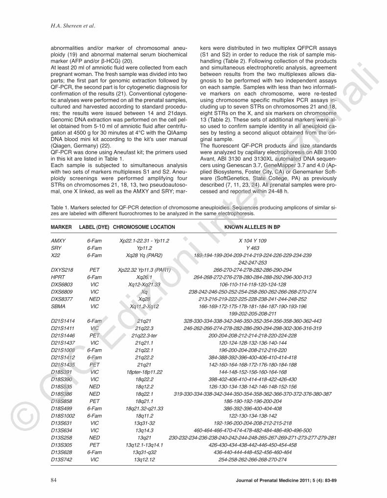
Table 1. Markers selected for QF-PCR detection of chromosome aneuploidies. Sequences producing amplicons of similar si-zes are labeled with different fluorochromes to be analyzed in the same electrophoresis.
MARkER LABEL (dYE) CHROMOSOME LOCATION kNOwN ALLELES IN BP
AMXY 6-Fam Xp22.1-22.31 - Yp11.2 X 104 Y 109
SRY 6-Fam Yp11.2 Y 463
X22 6-Fam Xq28 Yq (PAR2) 189-194-199-204-209-214-219-224-226-229-234-239
242-247-253
DXYS218 PET Xp22.32 Yp11.3 (PAR1) 266-270-274-278-282-286-290-294
HPRT 6-Fam Xq26.1 264-268-272-276-278-280-284-288-292-296-300-313
DXS6803 VIC Xq12-Xq21.33 106-110-114-118-120-124-128
DXS6809 VIC Xq 238-242-246-250-252-254-258-260-262-266-268-270-274
DXS8377 NED Xq28 213-216-219-222-225-228-238-241-244-248-252
SBMA VIC Xq11.2-Xq12 166-169-172-175-178-181-184-187-190-193-196
199-202-205-208-211
D21S1414 6-Fam 21q21 328-330-334-338-342-346-350-352-354-356-358-360-362-443
D21S1411 VIC 21q22.3 246-262-266-274-278-282-286-290-294-298-302-306-316-319
D21S1446 PET 21q22.3-ter 200-204-208-212-214-218-220-224-228
D21S1437 VIC 21q21.1 120-124-128-132-136-140-144
D21S1008 6-Fam 21q22.1 196-200-204-208-212-216-220
D21S1412 6-Fam 21q22.2 384-388-392-396-400-406-410-414-418
D21S1435 PET 21q21 142-160-164-168-172-176-180-184-188
D18S391 VIC 18pter-18p11.22 144-148-152-156-160-164-168
D18S390 VIC 18q22.2 398-402-406-410-414-418-422-426-430
D18S535 NED 18q12.2 126-130-134-138-142-146-148-152-156
D18S386 NED 18q22.1 319-330-334-338-342-344-350-354-358-362-366-370-372-376-380-387
D18S858 PET 18q21.1 186-190-192-196-200-204
D18S499 6-Fam 18q21.32-q21.33 386-392-396-400-404-408
D18S1002 6-Fam 18q11.2 122-130-134-138-142
D13S631 VIC 13q31-32 192-196-200-204-208-212-215-218
D13S634 VIC 13q14.3 460-464-466-470-474-478-482-484-486-490-496-500
D13S258 NED 13q21 230-232-234-236-238-240-242-244-248-265-267-269-271-273-277-279-281
D13S305 PET 13q12.1-13q14.1 426-430-434-438-442-446-450-454-458
D13S628 6-Fam 13q31-q32 436-440-444-448-452-456-460-464
D13S742 VIC 13q12.12 254-258-262-266-268-270-274

Prenatal diagnosis of fetal aneuploidies using QF-PCR: the egyptian study
Results
Sixty amniotic fluid samples were tested in this study byQF-PCR and the results were compared to the cytoge-netic results of the same sample. The maternal age of48 (80%) out of the 60 patients involved in this studywas less than 35 years old. Regarding referral cause,thirty nine patients (65%) complained of a previous hi-story of abnormal child (twenty four patients had historyof trisomy 21, twelve had history of a child with multiplecongenital anomalies and three patients had history ofTurner syndrome), twelve cases (20%) were referreddue to an abnormal ultrasound. Six case had increasednuchal translucency, three case showed fetal bilateralventriculomegaly, three cases showed bilateral clenchedfists and club feet. Six patients (10%) were referred dueto abnormal maternal serum Alfa Fetoprotein (AFP), theirMultiple of Medians (MoM) ranging between 6.1 and 8.4MoM, with highly elevated risk of aneuploidy estimated tobe between 1:50 and 1:27 respectively. Three patients(5%) were referred to our clinic due to advanced mater-nal age (39, 40 and 41 years old) (Fig. 1).The gestational age of the cases ranged between 12and 28 weeks, except one case who presented to ourclinic at 32 weeks of pregnancy. The total culture suc-cess rate was 90% after an average of 3-4 harvests.Two culture failures were met in this study; one of themwas heavily blood stained, another clear sample was re-quested for re-culture with successful result. The other
failed culture was due to late gestational age and mostof the cells were degenerated. The culture time rangedbetween 14-21 days with mean and standard deviationequal to18.5 (±2.12) days.Twenty two samples showed normal female pattern(46XX), thirty four samples showed normal male pattern(46XY), three samples showed a male pattern with tri-somy 21 (47XY +21) and the result of one sample couldnot be obtained due to culture failure (Fig. 2).All the samples were successfully tested by QF-PCR,results was available within 48 hours, and were in con-cordance with the cytogenetic results, with 100% speci-ficity, 100% sensitivity and the diagnostic efficiency offetal aneuploidies was 100%. The ratio between theheight peaks was calculated for each marker, we assi-gned as normal peak ratios between 0.8 and 1.4 and ab-normal ratios greater than 1.8 or less than 0.65. Twopeaks with normal ratio was diagnosed as normal dis-omy, three peaks with ratios between 0.8 and 1.4, or twopeaks with a ratio greater than 1.8 or less than 0.65 we-re diagnosed as trisomy. Single (homozygous) peakswere considered uninformative and were discarded. Aminimum of two informative markers is required to con-fidently diagnose either normality or abnormality (25).
Three patterns were obtained by QF-PCR
1. Normal female pattern: Twenty two samples showednormal female patterns, with one peak appearing at theX specific locus of the AMXY marker (at 104 bp) absen-ce of both the Y specific locus of AMXY (at 109 bp) andthe SRY peaks. The X- specific HPRT marker showednormal heterozygous peaks in all except two samples,while the pseudoautosomal X22 and DXYS218 markersshowed either heterozygous or homozygous patterns.The samples showed at least two normal heterozygousmarkers on each of the chromosomes 21, 18 and 13.2. Normal male pattern: Thirty four samples showed nor-mal male patterns with two peaks appearing at theAMXY for the X (104 bp) and Y (109 bp) specific loci. Asingle peak was present at the SRY, HPRT, while normalheterozygous peaks appearing at X22 and DXY218markers, with the presence of at least two normal hete-
Figure 1. Pie chart showing a comparison between differentcauses of patients’ referral.
Table 2. Multiplex assays included in the AneufastTM QF-PCR Kit. Mix 1 and 2 are used to screen all prenatal samples withfour markers on chromosomes 13, 18 and 21, two pseudoautosomal X and Y and one X-linked marker. AMXY and SRY areused for sexing. Two autosomal markers and sexing sequences are present in both multiplexes, this allow obtaining resultswith two independent assays on each sample.
S1 S2 MXY M21 M18 M13
AMXY SRY SRY D21S1411 D18S386 D13S631
D21S1414 X22 AMXY D21S1437 D18S391 D13S634
D21S1446 DXYS218 HPRT D21S1412* D18S858* D13S742*
D13S631 HPRT SBMA* D21S1435* D18S499* D13S628*
D13S305 D21S1411 DXS6803* D21S1008* D18S1002*
D18S535 D21S1437 DXS6809*
D18S391 D13S634 DXS8377*
D13S258
D18S386
D18S390
Journal of Prenatal Medicine 2011; 5 (4): 83-89 85

H.A. Shereen et al.
86 Journal of Prenatal Medicine 2011; 5 (4): 83-89
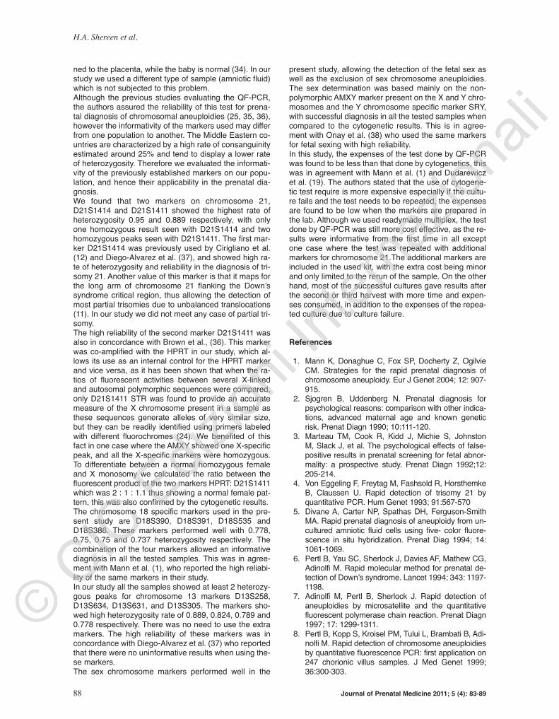
rozygous markers on each of the chromosomes 21, 18and 13.3. Trisomy 21 male pattern: Three sample showed ampli-fication of the X and Y specific product of the AMXY mar-ker, SRY marker was amplified, homozygous peak at theHPRT marker. Markers on chromosome 21 showed triso-mic patterns as follows; three markers D21S1414,D21S1411 and D21S1437 showed triallelic trisomy whileD21S1446 showed diallelic trisomy. The 18 and 13 speci-fic markers showed normal patterns (Figure 3). Inconclusive results: One sample showed inconclusiveresult for chromosome 21 with only one heterozygousmarker D21S1414, one homozygous marker D21S1446,while D21S1411 and D21S1437 failed to be amplified.The other markers on chromosome 18, 13, X and Y sho-wed normal male pattern. The same sample was rete-sted with the chromosome 21 extra markers. The extramarkers (D21S1008 D21S1412, D21S1437 andD21S1411) showed normal heterozygous peaks.
discussion
During the past decades, there has been a considerable
progress in further refining the non-invasive methods forthe prenatal detection of fetal diseases. Biochemicaland ultrasound tests have become standard procedures
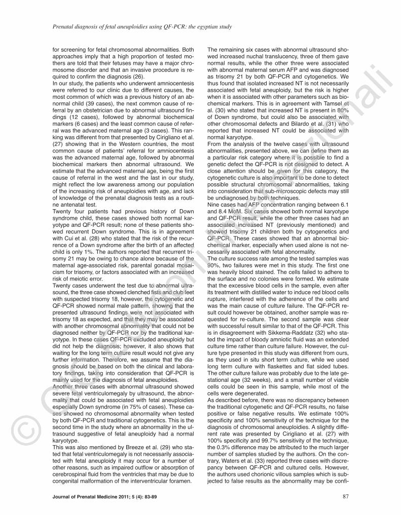
Figure 2. Figure showing a G-banding metaphase of normalfemale.
Figure 3. Trisomy 21 male pattern: the D21S1414, D21S1411 and D21S1437 markers show triallelic trisomic peaks, the D21S1446shows diallelic trisomic peaks. The chromosome 18 and 13 specific markers show normal figures. The X and Y chromosome spe-cific AMXY markers amplified, SRY product present, other sex chromosome markers show heterozygous diallelic figure.
Prenatal diagnosis of fetal aneuploidies using QF-PCR: the egyptian study
Journal of Prenatal Medicine 2011; 5 (4): 83-89 87
for screening for fetal chromosomal abnormalities. Bothapproaches imply that a high proportion of tested mo-thers are told that their fetuses may have a major chro-mosome disorder and that an invasive procedure is re-quired to confirm the diagnosis (26).In our study, the patients who underwent amniocentesiswere referred to our clinic due to different causes, themost common of which was a previous history of an ab-normal child (39 cases), the next common cause of re-ferral by an obstetrician due to abnormal ultrasound fin-dings (12 cases), followed by abnormal biochemicalmarkers (6 cases) and the least common cause of refer-ral was the advanced maternal age (3 cases). This ran-king was different from that presented by Cirigliano et al.(27) showing that in the Western countries, the mostcommon cause of patients’ referral for amniocentesiswas the advanced maternal age, followed by abnormalbiochemical markers then abnormal ultrasound. Weestimate that the advanced maternal age, being the firstcause of referral in the west and the last in our study,might reflect the low awareness among our populationof the increasing risk of aneuploidies with age, and lackof knowledge of the prenatal diagnosis tests as a routi-ne antenatal test.Twenty four patients had previous history of Downsyndrome child, these cases showed both normal kar-yotype and QF-PCR result; none of these patients sho-wed recurrent Down syndrome. This is in agreementwith Cui et al. (28) who stated that the risk of the recur-rence of a Down syndrome after the birth of an affectedchild is only 1%. The authors reported that recurrent tri-somy 21 may be owing to chance alone because of thematernal age-associated risk, parental gonadal mosai-cism for trisomy, or factors associated with an increasedrisk of meiotic error. Twenty cases underwent the test due to abnormal ultra-sound, the three case showed clenched fists and club feetwith suspected trisomy 18, however, the cytogenetic andQF-PCR showed normal male pattern, showing that thepresented ultrasound findings were not associated withtrisomy 18 as expected, and that they may be associatedwith another chromosomal abnormality that could not bediagnosed neither by QF-PCR nor by the traditional kar-yotype. In these cases QF-PCR excluded aneuploidy butdid not help the diagnosis; however, it also shows thatwaiting for the long term culture result would not give anyfurther information. Therefore, we assume that the dia-gnosis should be based on both the clinical and labora-tory findings, taking into consideration that QF-PCR ismainly used for the diagnosis of fetal aneuploidies. Another three cases with abnormal ultrasound showedsevere fetal ventriculomegaly by ultrasound, the abnor-mality that could be associated with fetal aneuploidiesespecially Down syndrome (in 75% of cases). These ca-ses showed no chromosomal abnormality when testedby both QF-PCR and traditional cytogenetics. This is thesecond time in the study where an abnormality in the ul-trasound suggestive of fetal aneuploidy had a normalkaryotype.This was also mentioned by Breeze et al. (29) who sta-ted that fetal ventriculomegaly is not necessarily associa-ted with fetal aneuploidy it may occur for a number ofother reasons, such as impaired outflow or absorption ofcerebrospinal fluid from the ventricles that may be due tocongenital malformation of the interventricular foramen.
The remaining six cases with abnormal ultrasound sho-wed increased nuchal translucency, three of them gavenormal results, while the other three were associatedwith abnormal maternal serum AFP and was diagnosedas trisomy 21 by both QF-PCR and cytogenetics. Wethus found that isolated increased NT is not necessarilyassociated with fetal aneuploidy, but the risk is higherwhen it is associated with other parameters such as bio-chemical markers. This is in agreement with Tamsel etal. (30) who stated that increased NT is present in 80%of Down syndrome, but could also be associated withother chromosomal defects and Bilardo et al. (31) whoreported that increased NT could be associated withnormal karyotype. From the analysis of the twelve cases with ultrasoundabnormalities, presented above, we can define them asa particular risk category where it is possible to find agenetic defect the QF-PCR is not designed to detect. Aclose attention should be given for this category, thecytogenetic culture is also important to be done to detectpossible structural chromosomal abnormalities, takinginto consideration that sub-microscopic defects may stillbe undiagnosed by both techniques. Nine cases had AFP concentration ranging between 6.1and 8.4 MoM. Six cases showed both normal karyotypeand QF-PCR result, while the other three cases had anassociated increased NT (previously mentioned) andshowed trisomy 21 children both by cytogenetics andQF-PCR. These cases showed that an abnormal bio-chemical marker, especially when used alone is not ne-cessarily associated with fetal abnormality.The culture success rate among the tested samples was90%, two failures were met in this study. The first onewas heavily blood stained. The cells failed to adhere tothe surface and no colonies were formed. We estimatethat the excessive blood cells in the sample, even afterits treatment with distilled water to induce red blood cellsrupture, interfered with the adherence of the cells andwas the main cause of culture failure. The QF-PCR re-sult could however be obtained, another sample was re-quested for re-culture. The second sample was clearwith successful result similar to that of the QF-PCR. Thisis in disagreement with Sikkema-Raddatz (32) who sta-ted the impact of bloody amniotic fluid was an extendedculture time rather than culture failure. However, the cul-ture type presented in this study was different from ours,as they used in situ short term culture, while we usedlong term culture with flaskettes and flat sided tubes.The other culture failure was probably due to the late ge-stational age (32 weeks), and a small number of viablecells could be seen in this sample, while most of thecells were degenerated. As described before, there was no discrepancy betweenthe traditional cytogenetic and QF-PCR results, no falsepositive or false negative results. We estimate 100%specificity and 100% sensitivity of the technique for thediagnosis of chromosomal aneuploidies. A slightly diffe-rent rate was presented by Cirigliano et al. (27) with100% specificity and 99.7% sensitivity of the technique,the 0.3% difference may be attributed to the much largernumber of samples studied by the authors. On the con-trary, Waters et al. (33) reported three cases with discre-pancy between QF-PCR and cultured cells. However,the authors used chorionic villous samples which is sub-jected to false results as the abnormality may be confi-
H.A. Shereen et al.
88 Journal of Prenatal Medicine 2011; 5 (4): 83-89
ned to the placenta, while the baby is normal (34). In ourstudy we used a different type of sample (amniotic fluid)which is not subjected to this problem. Although the previous studies evaluating the QF-PCR,the authors assured the reliability of this test for prena-tal diagnosis of chromosomal aneuploidies (25, 35, 36),however the informativity of the markers used may differfrom one population to another. The Middle Eastern co-untries are characterized by a high rate of consanguinityestimated around 25% and tend to display a lower rateof heterozygosity. Therefore we evaluated the informati-vity of the previously established markers on our popu-lation, and hence their applicability in the prenatal dia-gnosis.We found that two markers on chromosome 21,D21S1414 and D21S1411 showed the highest rate ofheterozygosity 0.95 and 0.889 respectively, with onlyone homozygous result seen with D21S1414 and twohomozygous peaks seen with D21S1411. The first mar-ker D21S1414 was previously used by Cirigliano et al.(12) and Diego-Alvarez et al. (37), and showed high ra-te of heterozygosity and reliability in the diagnosis of tri-somy 21. Another value of this marker is that it maps forthe long arm of chromosome 21 flanking the Down’ssyndrome critical region, thus allowing the detection ofmost partial trisomies due to unbalanced translocations(11). In our study we did not meet any case of partial tri-somy.The high reliability of the second marker D21S1411 wasalso in concordance with Brown et al., (36). This markerwas co-amplified with the HPRT in our study, which al-lows its use as an internal control for the HPRT markerand vice versa, as it has been shown that when the ra-tios of fluorescent activities between several X-linkedand autosomal polymorphic sequences were compared,only D21S1411 STR was found to provide an accuratemeasure of the X chromosome present in a sample asthese sequences generate alleles of very similar size,but they can be readily identified using primers labeledwith different fluorochromes (24). We benefited of thisfact in one case where the AMXY showed one X-specificpeak, and all the X-specific markers were homozygous.To differentiate between a normal homozygous femaleand X monosomy we calculated the ratio between thefluorescent product of the two markers HPRT: D21S1411which was 2 : 1 : 1.1 thus showing a normal female pat-tern, this was also confirmed by the cytogenetic results. The chromosome 18 specific markers used in the pre-sent study are D18S390, D18S391, D18S535 andD18S386. These markers performed well with 0.778,0.75, 0.75 and 0.737 heterozygosity respectively. Thecombination of the four markers allowed an informativediagnosis in all the tested samples. This was in agree-ment with Mann et al. (1), who reported the high reliabi-lity of the same markers in their study. In our study all the samples showed at least 2 heterozy-gous peaks for chromosome 13 markers D13S258,D13S634, D13S631, and D13S305. The markers sho-wed high heterozygosity rate of 0.889, 0.824, 0.789 and0.778 respectively. There was no need to use the extramarkers. The high reliability of these markers was inconcordance with Diego-Alvarez et al. (37) who reportedthat there were no uninformative results when using the-se markers. The sex chromosome markers performed well in the
present study, allowing the detection of the fetal sex aswell as the exclusion of sex chromosome aneuploidies.The sex determination was based mainly on the non-polymorphic AMXY marker present on the X and Y chro-mosomes and the Y chromosome specific marker SRY,with successful diagnosis in all the tested samples whencompared to the cytogenetic results. This is in agree-ment with Onay et al. (38) who used the same markersfor fetal sexing with high reliability. In this study, the expenses of the test done by QF-PCRwas found to be less than that done by cytogenetics, thiswas in agreement with Mann et al. (1) and Dudarewiczet al. (19). The authors stated that the use of cytogene-tic test require is more expensive especially if the cultu-re fails and the test needs to be repeated, the expensesare found to be low when the markers are prepared inthe lab. Although we used readymade multiplex, the testdone by QF-PCR was still more cost effective, as the re-sults were informative from the first time in all exceptone case where the test was repeated with additionalmarkers for chromosome 21.The additional markers areincluded in the used kit, with the extra cost being minorand only limited to the rerun of the sample. On the otherhand, most of the successful cultures gave results afterthe second or third harvest with more time and expen-ses consumed, in addition to the expenses of the repea-ted culture due to culture failure.
References
11. Mann K, Donaghue C, Fox SP, Docherty Z, OgilvieCM. Strategies for the rapid prenatal diagnosis ofchromosome aneuploidy. Eur J Genet 2004; 12: 907-915.
12. Sjogren B, Uddenberg N. Prenatal diagnosis forpsychological reasons: comparison with other indica-tions, advanced maternal age and known geneticrisk. Prenat Diagn 1990; 10:111-120.
13. Marteau TM, Cook R, Kidd J, Michie S, JohnstonM, Slack J, et al. The psychological effects of false-positive results in prenatal screening for fetal abnor-mality: a prospective study. Prenat Diagn 1992;12:205-214.
14. Von Eggeling F, Freytag M, Fashsold R, HorsthemkeB, Claussen U. Rapid detection of trisomy 21 byquantitative PCR. Hum Genet 1993; 91:567-570
15. Divane A, Carter NP, Spathas DH, Ferguson-SmithMA. Rapid prenatal diagnosis of aneuploidy from un-cultured amniotic fluid cells using five- color fluore-scence in situ hybridization. Prenat Diag 1994; 14:1061-1069.
16. Pertl B, Yau SC, Sherlock J, Davies AF, Mathew CG,Adinolfi M. Rapid molecular method for prenatal de-tection of Down’s syndrome. Lancet 1994; 343: 1197-1198.
17. Adinolfi M, Pertl B, Sherlock J. Rapid detection ofaneuploidies by microsatellite and the quantitativefluorescent polymerase chain reaction. Prenat Diagn1997; 17: 1299-1311.
18. Pertl B, Kopp S, Kroisel PM, Tului L, Brambati B, Adi-nolfi M. Rapid detection of chromosome aneuploidiesby quantitative fluorescence PCR: first application on247 chorionic villus samples. J Med Genet 1999;36:300-303.
19. Schmidt W, Jenderny J, Hecher K, Hackeloer BJ,Kerber S, Kochhan L and Held KR. Detection ofaneuploidy in chromosomes X, Y, 13, 18 and 21 byQF- PCR in 662 selected pregnancies at risk. MolHum Reprod 2000; 6:855-860.
10. Adinolfi M, Sherlock J. Prenatal detection of chromo-some disorders by QF- PCR. Lancet 2001; 358:1030-1031.
11. Cirigliano V, Lewin P, Szpiro-Tapies S, Fuster C, Adi-nolfi M. Assessment of new markers for the rapid de-tection of aneuploidies by quantitative fluorescentPCR (QF-PCR). Ann Hum Gen 2001; 65:421-427.
12. Cirigliano V, Voglino G, Canadas MP, Marongiu A,Ejarque M, Ordonez E, et al. Rapid prenatal diagno-sis of common chromosome aneuploidies by QF-PCR. Assessment on 18,000 consecutive clinicalsamples. Mol Hum Reprod 2004;10:839-846.
13. Cirigliano V, Voglino G, Marongiu A, Cañadas P, Or-doñez E, Lloveras E, et al. Rapid prenatal diagnosisby QF- PCR: evaluation of 30,000 consecutive clini-cal samples and future applications. Ann N Y AcadSci 2006; 1075: 288-298.
14. Mann K, Fox SP, Abbs SJ, Ghing Yau S, Scriven PN,Docherty Z et al. Development and implementation ofa new rapid aneuploid diagnostic service with the UKNational Health Service and implication for the futureof prenatal diagnosis. Lancet 2001; 358:1057-1061.
15. Adinolfi M, Sherlock J, Cirigliano V, Pertl B. Prenatalscreening of aneuploidies by quantitative fluorescentPCR. Community Genet 2000; 3: 50-60.
16. Cirigliano V, Voglino G, Adonilfini M. Non invasivescreening and rapid QF-PCR assay can greatly redu-ce the need of cytogenetic analysis in prenatal dia-gnosis. Reprod Biomed Online 2005; 11:671-673.
17. Summers AM., Langlois S, Wyatt P, Wilson D. Prena-tal Screening for Fetal Aneuploidy. J Obstet GynaecolCan 2007; 29:146-161.
18. Benn PA, Hsu LYF. Prenatal Diagnosis of Chromoso-mal Abnormalities through Amniocentesis. In: GeneticDisorders and the Fetus. Milunsky A. (ed.), The JohnsHopkins University Press, pp. 2004; 214-296.
19. Dudarewicz L, Holzgreve W, Jeziorowska A, Jakubo-ski L, Zimmermann B. Molecular methods for rapiddetection of aneuploidy. J Appl Genet 2005; 46(2):207-15.
20. Extermann P, Bischofl P, Marguerat P, Mermillod B.Second-trimester maternal serum screening for Dow-n’s syndrome: free b-human chorionic gonadotrophin(HCG) and a-fetoprotein, with or without unconjuga-ted oestriol, compared with total HCG, a-fetoproteinand unconjugated oestriol. Hum Reprod 1998; 13 (1):220 -223.
21. Rooney DE, Czepulkowski BH (Eds). Human Cytoge-netics. A Practical Approach 2001, 2nd ed., IRLPress, Oxford.
22. Rahil H, Solassol J, Philippe C, Lefort G, Jonveaux,P. Rapid detection of common autosomal aneuploi-dies by quantitative fluorescent PCR on unculturedamniocytes. Eur. J. Hum. Genet. 2002; 10: 462-466.
23. Pertl B, Weitgasser U, Fopp S, Kroisel PM, Sher-lock J , Andinolfi M . Rapid detection of trisomies 21and 18 and sexing by quantitative fluorescent mul-tiplex PCR. Hum Gent 1996;98:55-59.
24. Cirigliano V, Ejarque M, Fuster C, Adinolfi M. Xchro mosome dosage by quantitative fluorescent
PCR and rapid prenatal diagnosis of sex chromoso-me aneuploidies. Mol Hum Reprod. 2002; 8:1042-1045.
25. Quaife R, Wong LF, Tan SY, Chua WY, Lim SS,Hammersley C, et al. QF-PCR based prenatal de-tection of aneuploidy in a southeast Asian popula-tion. Prenat Diagn2004; 24:407-413.
26. Speevak MD, Dolling J, Terespolsky D, BlumenthalA, Farrel, S.A. An algorithm for the prenatal detec-tion of chromosome anomalies by QF-PCR and G-banded analysis. Prenat Diagn 2008; 28:1221-1226.
27. Cirigliano V, Voglino G, Ordoñez E, Marongiu A,Paz Cañadas M, Ejarque M, et al. Rapid prenataldiagnosis of common chromosome aneuploidies byOF-PCR, results of 9 years of clinical experience.Prenat Diagn 2009; 29(1): 40-49.
28. Cui YX, Hao LJ, Wang YH, Xia XY, Shi YC, Lu HY,et al. Case report: Second pregnancy of trisomy 21in a mother with mosaicism. Chin Med J 2007;120(14):1295-1296.
29. Breeze AC, Alexander PM, Murdoch EM, Missfel-der-Lobos HH, Hackett GA, Lees CC. Obstetric andneonatal outcomes in severe fetal ventriculome-galy. Prenat Diagn 2007; 27(2):124-129.
30. Tamsel S, Özbek S, Demirpolat G. Ultrasound eva-luation of fetal chromosome Disorders. Diagn IntervRadiol 2007; 13: 97-100.
31. Bilardo C, Muller M, Pajkrt E, Clur S, Van Zalen M,Bijlsma E .Increased nuchal translucency thicknessand normal karyotype: time for praenatal reassu-rance. Ultrasound Obstet Gynecol 2007; 30:11-18.
32. Sikkema-Raddatz B. Quality assessment of prena-tal cytogenetic diagnosis. MD degree thesis, Medi-cal Genetics, University of Groningen, the Nether-lands; 2005.
33. Waters JJ, Mann K, Grimsley L, Ogilvie C, Dona-ghue C, Staples L, et al. Complete discrepancy bet-ween QF-PCR analysis of uncultured villi and kar-yotyping of cultured cells in the prenatal diagnosisof trisomy 21 in three cases. Prenat Diagn 2007;27: 332-339.
34. Lau ET, Tang L, Wong C, Hang LY, Ghosh A, LeungAC, et al. Assessing discrepent findings betweenQF-PCR on uncultured prenatal samples and kar-yotyping on long-term culture. Prenat Diagn 2009;29:252-255.
35. Donaghue C, Mann K, Docherty Z , Ogilive CM. De-tection of mosaicism for primary trisomies in prena-tal samples by QF-PCR and Karyotype analysis. Pre-nat Diagn 2005; 25:65-72.
36. Brown L, Abigania M, Warburton D, Brown S. Valida-tion of QF-PCR for prenatal aneuploidy screening inthe United States. Prenat Diagn 2006; 26:1068-1074.
37. Diego-Alvarez D, Garcia-Hoyos M, Trujillo MJ, Gon-zalez-Gonzalez C, De Alba MR, Ayuso C, et al. Appli-cation of quantitative fluorescent PCR with short tan-dem repeat markers to the study of aneuploidies inspontaneous miscarriages. Hum Reprod 2005;20:1235-1243.
38. Onay H, Timur U, Ayca A, Sacide P, Murat I, SivekarT, et al. Rapid prenatal diagnosis of common aneu-ploidies in amniotic fluid using quantitative fluorescentpolymerase chain reaction. Gynecol Obst Invest2008; 66:104-110.
Prenatal diagnosis of fetal aneuploidies using QF-PCR: the egyptian study
Journal of Prenatal Medicine 2011; 5 (4): 83-89 89