preparation and characterization of poly (l-lactic acid ... · pdf filepreparation and...
TRANSCRIPT
Preparation and Characterization of Poly (L-Lactic Acid) and Poly
(Ethylene Oxide) Blends
R.A. Zoppia*, E.A.R. Duekb, D.C. Coraçac, P.P. Barrosa
aInstituto de Ciências Biológicas e Química, PUC - Campinas, C.P. 1111,13020-904 Campinas - SP, Brazil
bFaculdade de Engenharia Mecânica, Unicamp, 13083-970 Campinas - SP, BrazilcInstituto de Biologia, Unicamp, 13083-970 Campinas - SP, Brazil
Received: November 11, 2000; Revised April 20, 2001
Poly(L-lactic acid) (PLLA) and poly(ethylene oxide) (PEO) blends were prepared by mechani-cal mixture and fusion of homopolymers. Samples were submitted to in vitro degradation tests(immersion in a phosphate buffer solution with pH = 7.4 at 37 °C). Independently of the blendcomposition, PEO was dissolved after 14 days of immersion. As expected, after immersion, scanningelectron microscopy showed that the blends were porous, contrary to the samples, which were notimmersed in the buffer solution. Phase separation was not evident. Using differential scanningcalorimetry, the melting points (Tm) of both PLLA and PEO crystalline fractions were observed andremained practically constant, indicating no miscibility. Thermogravimetry showed that the tem-perature where the main mass loss stage starts (Tonset), depended on the blend composition andperiod of immersion in the buffer. The blends and the PLLA homopolymer were implanted in defectsproduced in the tibias of rats. The blends were as biocompatible as the PLLA.
Keywords: poly(L-lactic acid), poly(ethylene oxide), blends, biomaterials
1. Introduction
Biodegradable polyesters based on poly(L-lactic acid),PLLA, have been used as biomaterials for temporary thera-peutic applications mainly in orthopedic devices, control-led drug release and support for cell culture1-4. The mainadvantage of PLLA is its degradation by simple hydrolysisof the ester backbone in aqueous environments such asbody fluids. This makes it very convenient for devices witha temporary function. The degradation products are finallymetabolized to carbon dioxide and water or excreted viathe kidneys, so is not necessary to remove the device fromthe implantation site after tissue healing1,5. Pure PLLAdevices in general have a long degradation time6-9.
Mainil-Varlet and co-workers have been using poly(L-lactic acid) and poly(DL-lactic acid) in implants in the tibiaof sheep and verified that the absorption of these materialswere not complete even after one year of implantation; themolecular weight, however, decreased from 40000-50000to 500-3000 g/mol8.
PLLA degradation and biocompatibility have been in-vestigated in rats. The histological reaction in PLLA im-plants is slow but, the decrease in the polymer molecularweight is fast. The complete absorption of PLLA has notbeen observed in rats and it has been estimated that thisphenomenon should occur after 3.5 years of implantation6.
Based on the positive results observed for animals,PLLA has been used in the fixation of bone fractures inhumans. After 3.5 years of implantation, the slow polymerdegradation caused a swelling of the implanted region,without however the necessity of extracting the implant10.
An alternative procedure to change degradation time, isthe blending of PLLA with other polymers, which can bedegradable or non-degradable polymers. In general, thiskind of blends exhibits advantageous physical and me-chanical properties5,11. The degradation time of the devicecan be varied from months to years, depending on itsamorphous/crystalline and hydrophilic/hydrophobic pro-perties5,12.
Materials Research, Vol. 4, No. 2, 117-125, 2001. © 2001
*e-mail: [email protected] apresentado no 14° CBECIMAT, Águas de São Pedro, Dezembro 2000.
Most of the investigated systems represent mixtures oftwo amorphous polymers, such as poly(phenylene ox-ide)/polystyrene, or mixtures in which one of the compo-nents is semi-crystalline, such as poly(vinylchloride)/caprolactone12. Semi-crystalline/semi-crystal-line polymer blends, which have been reported in literature,include poly(vinylidene fluoride)/(acrylates)13, poly(ethyl-ene oxide)/poly(hidroxybutyrate)14, poly(vinylidene fluo-ride)/poly(hidroxybutyrate)15,poly(caprolactone)/poly(carbonate)16-18, poly(vinylidenefluoride)/poly(1,4-butylene adipate)19,20. In these systems,the phase behavior and morphological properties have beeninvestigated.
Meikle and co-workers implanted poly(DL-lacticacid)/poly(glycolic acid) blends into the head of rabbits,using as a control spontaneous bone regeneration. By his-tomorphometry analysis they verified that the differencesin bone regeneration were not statistically significant, after1, 2 or 3 months of implantation21, when compared to thecontrol.
Poly(ethylene oxide), PEO, is a hydrophilic non-de-gradable polymer, while PLLA is hydrolytically degrad-able and hydrophobic. Both of these polymers aresemi-crystalline. PEO is also of particular interest inbiomedical applications mainly due to its good biocompati-bility and low toxicity. The glass transition temperatures ofPEO and PLLA are -54 °C and 57 °C, and the meltingtemperatures are 74 °C and 180 °C, respectively11. Blendsof PLLA and poly(ethylene oxide)11,22-24 or poly(ethyleneglycol)25-27 have been described in literature, where theirmiscibility, compatibility and mechanical properties arefocused on. In these cases, the blend preparation was car-ried out from the mixture of the homopolymers which weredissolved in a common solvent.
It has been verified that PLLA/PEO blends, which weresubmitted to the degradation process by immersion into abuffer solution, became porous due to the PEO fractiondissolution, while pure PLLA films remained dense. Thisporous morphology depended on the method employed forthe blend preparation. In a previous research, blends wereprepared from the casting of a solution containing bothPLLA and PEO dissolved homopolymers. In these cases,the pores had a circular shape and their dimension de-pended on the composition of the blend28.
A porous morphology can promote film hydration,which plays an important role in the PLLA degradation ratevia hydrolysis of the ester backbone. The PEO fractiondissolution could also be interesting in cases where solubledrugs in the PEO fraction could be controlled delivered. Aporous morphology can also be very desirable in most ofbiomedical applications. In some cases, the polymeric ma-trix must present a uniform and interconnected porousstructure to allow cell growth to be easily distributed
throughout the device. This way, an organized network ofthe tissue constituents can be formed.
Here, the poly(L-lactic acid)/poly(ethylene oxide)blends, which were prepared by mechanical mixture andfusion of the homopolymers, were investigated. PLLAhomopolymer and PLLA/PEO blends were implanted intodefects produced in the tibia of rats, in order to investigateblend biocompatibility and its potential application in themanufacturing of devices used for bone repair. The first invivo results obtained for 50/50 (w/w) PLLA/PEO blendsafter 2 and 4 weeks of implantation, are presented here. Theresults for longer periods of implantation and for 80/20 and20/80 PLLA/PEO blends are in progress.
2. ExperimentalBlends were prepared by mixing PLLA (Medisorb;
MW = 300000 g/mol) and PEO (Aldrich; MW = 200000g/mol) in a mini injector LMM-2017 Mini Max Molder.Sticks of PLLA and PLLA/PEO of different compositions(80/20, 50/50 and 20/80 w/w) were prepared by the meltingof homopolymers at 190 °C, using a 2.0 mm diameter and9.3 cm high (internal dimensions) mold, which remainedat 120 °C during the processing. The heating of the ho-mopolymer mixture was carried out for 1 min followed by2 min of shearing and mold injection. The mold was cooledat room temperature for 20 min.
In vitro degradation tests were carried out using 80/20,50/50 and 20/80 PLLA/PEO blends. Samples were im-mersed in a buffer solution (KH2PO4 – NaOH; pH = 7.4)at 37 °C, which was changed all week. Tests were per-formed during a 2-week period. These conditions haveoften been referred to in literature as degradation tests27, 29.
Blends were immersed for different periods (7 or 14days). After each period, samples were dried at 50 °C untilthey reached a constant mass, and characterized as de-scribed below. The mass loss percentage was calculatedcomparing the mass values of the samples before and aftersubmitting them to degradation tests. Samples are denotedhere as a function of the degradation time. PLLA/PEO t = 0was used for blends that were not immersed in the buffersolution, and PLLA/PEO t = 7 or t = 14 days was used forblends after the degradation process.
Samples were fractured by immersion into liquid nitro-gen. Surface fracture was covered with gold by sputteringand observed in a JEOL JXA 840 scanning electron micro-scope.
Thermogravimetric analysis was carried out in a TG209Netzsch thermal analyzer from 25 to 800 °C at 10 °C.min-1
under nitrogen.Differential scanning calorimetry was performed in a
DSC200 Netzsch thermal analyzer using the followingtemperature program: rapid cooling from 25 to -100 °C;heating from -100 to 200 °C at 10 °C.min-1; isotherm at200 °C for 5 min; cooling from 200 to -100 °C at
118 Zoppi et al. Materials Research
10 °C.min-1; isotherm at -100 °C for 5 min; heating from-100 to 200 °C at 10 °C.min-1. Samples were analyzedunder nitrogen.
Dynamic-mechanical analysis was performed in a409 DMA Netzsch thermomechanical analyzer from -100to 230 °C, under air, using the following conditions: duocantilever mode, force = 1N, amplitude = 15 µm andfrequency = 1 Hz.
In vivo tests were carried out following a standardmethod described in reference 30. Fifty-two male Wistarrats (200 - 250 g) were used in the study. Prior to surgery,the animals were anaesthetized with a solution of Ketamineplus Xylazine administered by an intramuscular injectionusing a dose of 1.5 mL per kg of body weight. After theasepsis of the site, a longitudinal incision in the skin,approximately 1 cm long, parallel to the tibia, was carriedout. The muscular tissue was parted and moved away untilthe periosteum appeared. With a low rotation mini motorand a 3.0 mm diameter drill, sockets were produced in thetibia in order to allow the introduction of the test bodies inthe shape of a 2 mm diameter and 2 mm long toggle. Thisprocedure was carried out in the legs of all animals, whichwere divided into the following groups: the PLLA/PVCgroup: 20 animals received PLLA in the tibia of their leftleg, and poly(vinyl chloride) PVC in the tibia of their rightleg as a control. The BLEND/PVC group: 20 animalsreceived the 50/50 PLLA/PEO blend in the tibia of theirleft leg and poly(vinyl chloride) PVC in the tibia of theirright leg as a control. The CONTROL group: 12 animalswere submitted to surgery for the insertion of a socket inthe tibia of the left leg, in which test bodies were notimplanted.
Immediately after the insertion of the sockets and/orrank of the test bodies, the muscular tissue was sutured.
During all this process, the area was continuously irri-gated with a physiological solution, so the heating of thetissue did not occur. After the skin suture, the surface waswashed with an anti-septic solution. During the post-surgi-cal periods the animals received doses of an analgesicdiluted in water in a dose 2.5 mL/L, supplied for 2 daysafter the surgery. The animals remained lodged in riversteamers in groups of 3 animals for each streamer, withburst light and ventilation, with solid ration and watersupply without restriction.
After 2, 4, 8 and 16 post-surgical weeks, the animalswere killed by an intraabdominal injection with an over-dose of 10% Chloral Hydrate solution, after which speci-mens of the tissue were removed. Immediately, the bonespecimens were fixed in a 10% formal solution at roomtemperature, for 48 h. Decalcified sections were made andstained with hematoxylin and eosin (HE) for histologicalobservations under a light microscope.
Test bodies of PVC (Aldrich) were prepared from thecasting of a 5wt% polymer/THF solution. Thick films(2 mm of thickness) were cut in a 2 mm diameter diskshape.
3. Results and DiscussionTable 1 shows the mass loss percentage for PLLA/PEO
blends occurred during the period of 14 days of in vitrodegradation. The highest mass loss percentage occurredduring the first week of the in vitro degradation. Thisprocess is related to the water diffusion in the blend fol-lowed by the dissolution of the PEO fraction. Since thePLLA fraction presents a low degradation rate, mass losspractically did not change during the period from 7 to 14days of degradation.
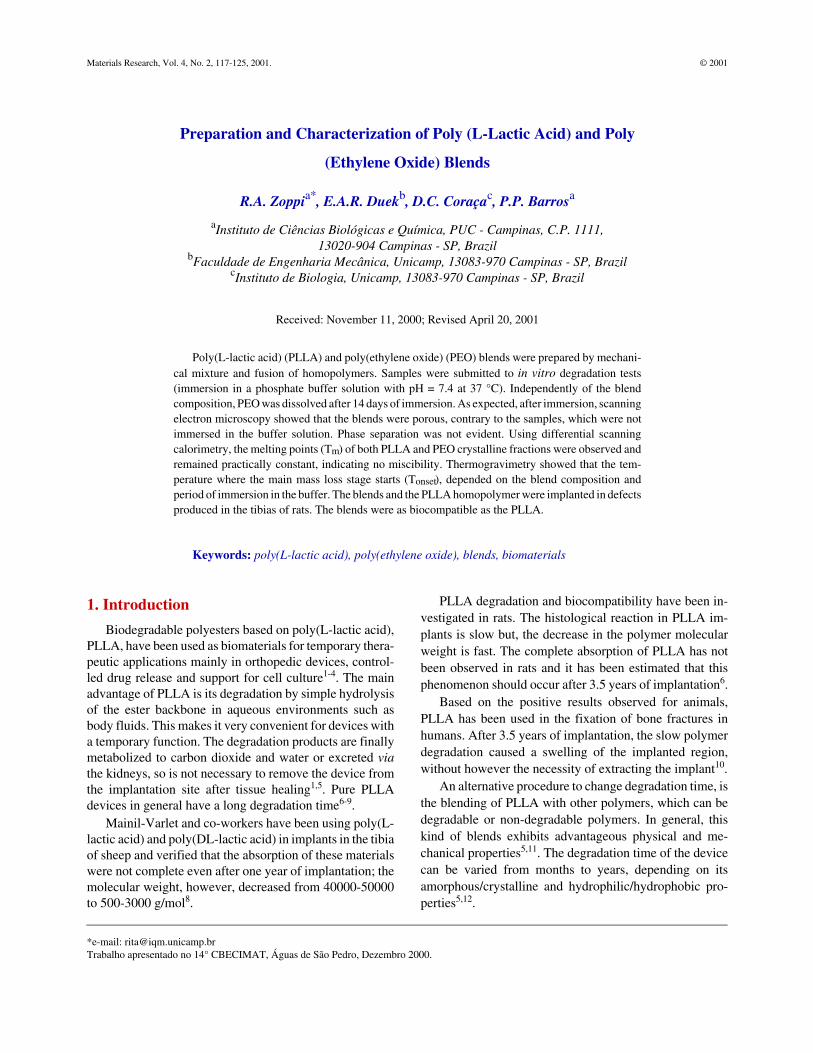
Figure 1 shows the surface fracture of PLLA/PEOblends of different compositions observed by scanningelectron microscopy as a function of the degradation time.For samples which were not immersed into the buffersolution (t = 0), the occurrence of phase separation was notclear. Contrary to 80/20 PLLA/PEO which presented adense morphology, pores with a circular shape and differ-ent dimensions (near 1 to 10 µm), could be observed forblends containing higher PEO contents.
After a period in a buffer, all the channels could beobserved. The morphology of the blends (t = 7days) wassimilar to that shown for blends (t = 14 days). For 80/20PLLA/PEO blends channels with holes in them are distrib-uted in a dense structure, while for 20/80 PLLA/PEOblends, the channels are distributed into a more porousstructure. For 50/50 PLLA/PEO blends, an intermediatesituation was verified, where the dense structure becamecracked and surrounded by channels with holes in them.
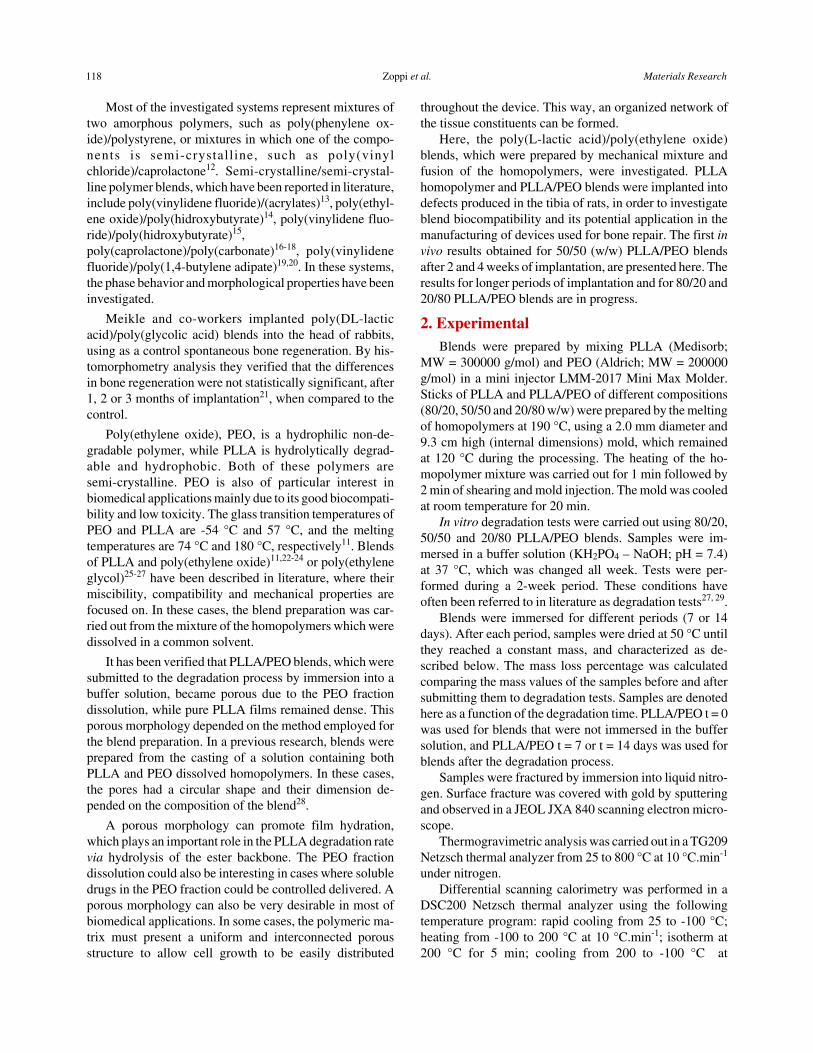
Figure 2a shows the thermal analysis measurements forpure PLLA, pure PEO and PLLA/PEO blends (t = 0). Forblends two main thermal degradation processes were ob-served. The first one was due to the PLLA, and the secondone to the PEO thermal degradation processes, respec-tively. After the immersion in the buffer, one main massloss stage was observed for 80/20 and 50/50 PLLA/PEOblends, Fig. 2b. Considering that the PEO fraction is ex-tracted during the in vitro degradation tests, this mass lossstage was due to the thermal degradation process of the
Vol. 4, No. 2, 2001 Poly (L-Lactic Acid) and Poly (Ethylene Oxide) Blends 119
Table 1. Mass loss percentage obtained from the in vitro degradation testsfor PLLA/PEO blends of different compositions as a function of the periodof immersion in the buffer.
PLLA/PEOMass loss percentage (%)
t = 7 days t = 14 days
80/20 18.7 19.9
50/50 47.0 46.0
20/80 71.0 80.2

PLLA fraction. For 20/80 PLLA/PEO blends (t = 7 or t =14days) a second mass loss stage near 400 °C was alsoobserved. Although the in vitro degradation tests and dif-ferential scanning calorimetry had indicated that PEO isabsent after 14 days of immersion in a buffer, this secondmass loss process could be assigned to one PEO fraction,which had not yet dissolved.
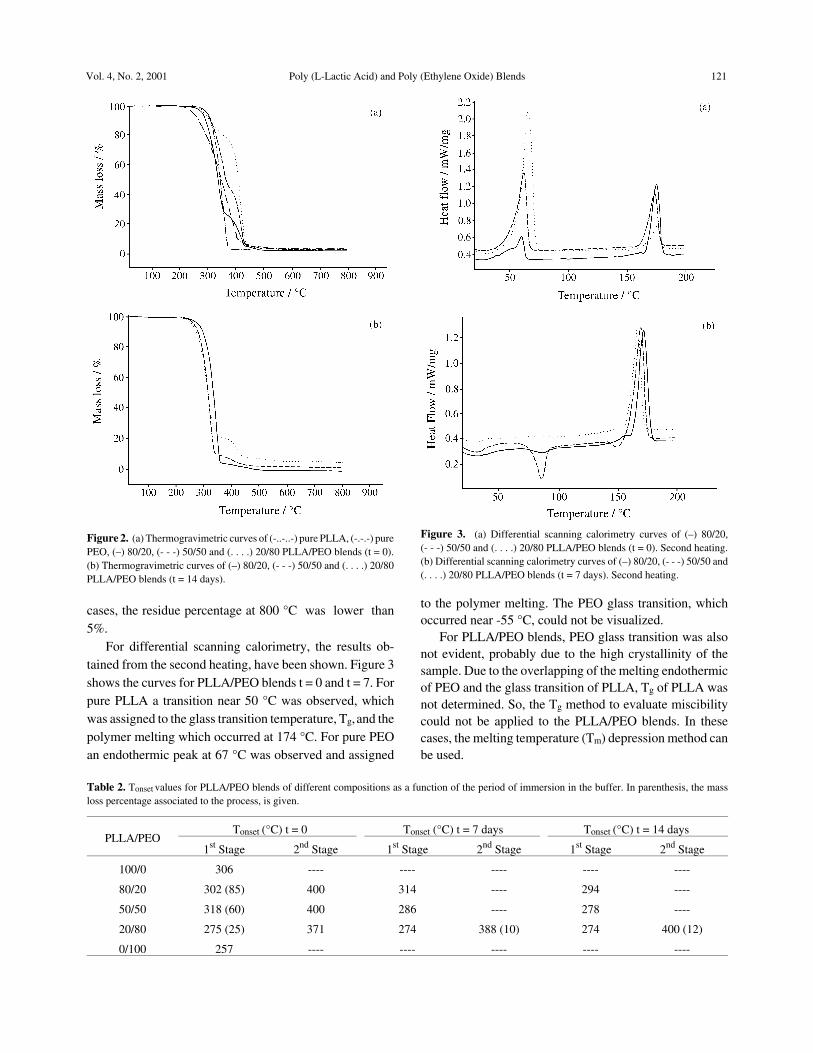
Table 2 shows Tonsed values as a function of the blendcomposition and the immersion time. Tonset represents thetemperature where the main thermal degradation stagestarts. For blends, Tonset, after the immersion in the buffer,was lower than that observed for blends t = 0, except forthe 80/20 PLLA/PEO composition, indicating that the in-corporation of PEO in the PLLA and its posterior extrac-tion, may favor the thermal degradation of PLLA. In all
120 Zoppi et al. Materials Research
Figure 1. Scanning electron microscopy of (a) 80/20, (b) 50/50 and (c) 20/80 PLLA/PEO blends t = 0 (left hand side) and t = 14 days (right hand side).
cases, the residue percentage at 800 °C was lower than5%.
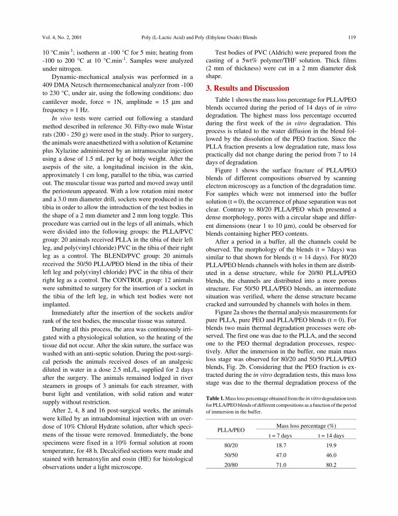
For differential scanning calorimetry, the results ob-tained from the second heating, have been shown. Figure 3shows the curves for PLLA/PEO blends t = 0 and t = 7. Forpure PLLA a transition near 50 °C was observed, whichwas assigned to the glass transition temperature, Tg, and thepolymer melting which occurred at 174 °C. For pure PEOan endothermic peak at 67 °C was observed and assigned
to the polymer melting. The PEO glass transition, whichoccurred near -55 °C, could not be visualized.
For PLLA/PEO blends, PEO glass transition was alsonot evident, probably due to the high crystallinity of thesample. Due to the overlapping of the melting endothermicof PEO and the glass transition of PLLA, Tg of PLLA wasnot determined. So, the Tg method to evaluate miscibilitycould not be applied to the PLLA/PEO blends. In thesecases, the melting temperature (Tm) depression method canbe used.
Vol. 4, No. 2, 2001 Poly (L-Lactic Acid) and Poly (Ethylene Oxide) Blends 121
Figure 2. (a) Thermogravimetric curves of (-..-..-) pure PLLA, (-.-.-) purePEO, (–) 80/20, (- - -) 50/50 and (. . . .) 20/80 PLLA/PEO blends (t = 0).(b) Thermogravimetric curves of (–) 80/20, (- - -) 50/50 and (. . . .) 20/80PLLA/PEO blends (t = 14 days).
Table 2. Tonset values for PLLA/PEO blends of different compositions as a function of the period of immersion in the buffer. In parenthesis, the massloss percentage associated to the process, is given.
PLLA/PEOTonset (°C) t = 0 Tonset (°C) t = 7 days Tonset (°C) t = 14 days
1st Stage 2nd Stage 1st Stage 2nd Stage 1st Stage 2nd Stage
100/0 306 ---- ---- ---- ---- ----
80/20 302 (85) 400 314 ---- 294 ----
50/50 318 (60) 400 286 ---- 278 ----
20/80 275 (25) 371 274 388 (10) 274 400 (12)
0/100 257 ---- ---- ---- ---- ----
Figure 3. (a) Differential scanning calorimetry curves of (–) 80/20,(- - -) 50/50 and (. . . .) 20/80 PLLA/PEO blends (t = 0). Second heating.(b) Differential scanning calorimetry curves of (–) 80/20, (- - -) 50/50 and(. . . .) 20/80 PLLA/PEO blends (t = 7 days). Second heating.
A composition-dependent melting endotherm usuallyindicates a miscible blend, whereas a fully phase separatedimmiscible system will display a constant Tm. Under com-plete immiscibility conditions, each of the crystallizablecomponents of the mixture will exhibit the Tm of thecorresponding pure homopolymer.
Table 3 shows the values of Tm for run 1 (Tm1) and run2 (Tm2) of pure PLLA, pure PEO and for the binary mixturewith different weight fractions. The melting temperature ofPLLA is almost constant for both run 1 and run 2, inde-pendently of the blend composition, suggesting that therewere no interactions between PLLA and PEO molecules inthe mixture as grown from fusion and melt-crystallizedsamples. For PEO, both Tm1 and Tm2 decreased slightlywith the increasing PLLA in the mixture, but this changemay be due to the morphological effects. PEO seems to bemore sensitive to the presence of PLLA chains, while amuch smaller influence is exerted by PEO on the crystallinephase of PLLA. This behavior can be related to the differentdegrees of crystallinity of both polymers, with PEO beingmore crystalline. Therefore, relatively small PLLA con-tents will interfere with the PEO crystalline array, causingshifting of the melting temperature. For PLLA, consider-able amounts of PEO can blend with the amorphous phaseof PLLA, without significantly affecting the crystallinedomains.
Table 4 shows the variation of the fusion enthalpy (∆Hf)of PLLA and PEO, which was correlated with the blendcomposition, for run 1 and run 2. Independent of the blendcomposition, ∆Hf values of PEO for run 2 decreased com-pared with run 1. Crystallization of PLLA is completebefore crystallization of PEO commenced, showing that thetwo polymers crystallize in different and well-separatedtemperature regime. The crystallization of PEO seems tobe severely hampered by PLLA, which may be explainedby the fact that the PLLA component has already com-pletely solidified at the temperatures where PEO crystal-lizes, thus restricting free crystal growth for this polymer.
For PLLA/PEO blends, which were immersed in abuffer, independent of the immersion time or the blendcomposition, only one endothermic process could be ob-
served. Table 5 shows Tm and ∆Hf values for samples whichwere immersed in the buffer solution. Comparing withnon-immersed samples, Tm values for blends t = 7 or t =14days were slightly lower than t = 0. In relation to ∆Hf, thevalues were lower after immersion in a buffer, except forthe 80/20 PLLA/PEO blends, where ∆Hf actually increasedslightly. It is interesting to note that the variation of ∆Hf
was more drastic for the 20/80 PLLA/PEO blends. In thiscase, immersed samples seemed to be much more amor-phous than non-immersed ones. ∆Hf is directly related tothe degree of crystallinity. During the immersion of asemi-crystalline polymer in a buffer, if degradation occurs,it is expected that the amorphous fraction degrades beforethe crystalline one does. So, one would also expect that thecrystallinity of the remaining porous matrix, to increasewith degradation time, since the amorphous phase wasbeing removed. After this process, the degradation of thecrystalline fraction started and consequently a decrease inthe degree of crystallinity was expected. The results shownin Table 5 can indicate that, depending on the blend com-position, it would be possible to control the degradation rateof the material. Using higher contents of PEO the materialseems to be more susceptible to the degradation process.
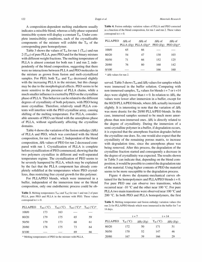
Figure 4 shows the dynamic-mechanical curves ob-tained for the homopolymers and PLLA/PEO blends t = 0.For pure PEO one can observe two transitions, whichoccurred near -10 °C and the other near 100 °C. For purePLLA two main transitions were observed near 100 °C and200 °C. In both PEO and PLLA homopolymers, the first
Table 3. Melting temperatures Tm1 and Tm2 for run 1 and run 2 of purePLLA, pure PEO and PLLA in the mixture with PEO. These valuescorrespond to t = 0.
PLLA/PEO Tm1 (°C) Tm2 (°C) Tm1 (°C)* Tm2 (°C)*
100/0 173 163 ---- ----
80/20 179 175 65 59
50/50 179 173 68 61
20/80 178 175 73 64
0/100 ---- ---- 69 66
* Melting temperatures of PEO in the mixture with PLLA.
Table 4. Fusion enthalpy variation values of PLLA and PEO correctedas a function of the blend composition, for run 1 and run 2. These values
correspond to t = 0.
PLLA/PEO ∆Hf ofPLLA (J/g)
∆Hf ofPLLA (J/g)*
∆Hf ofPEO (J/g)
∆Hf ofPEO (J/g)*
100/0 65 66 ---- ----
80/20 54 47 130 103
50/50 71 66 152 125
20/80 76 80 168 142
0/100 ---- ---- 166 160
* ∆Hf values for run 2.
Table 5. Melting temperature and fusion enthalpy variation values (forrun 2) for PLLA/PEO blends which were immersed in the buffer for 7 or14 days.
t = 7 t = 14
PLLA/PEO Tm (°C) ∆Hf (J/g) Tm (°C) ∆Hf (J/g)
80/20 172 50 171 51
50/50 170 52 147 46
20/80 167 39 166 29
122 Zoppi et al. Materials Research
transition could be due to the glass transition and the secondone to the melting of the polymer. For blends, the mechani-cal properties are determined primarily by the mutual solu-bility of the two homopolymers. If two polymers arecompletely soluble in one another the properties of themixture are nearly the same as those of a random copolymerof the same composition. In these cases, the damping peakfor the mixture occurred at an intermediate temperaturebetween the glass transition temperatures observed for thehomopolymers. If the two polymers in a mixture are insol-uble, they exist as two separated phases, and two glasstransitions are observed instead of one, as can be seen forPLLA/PEO blends. Two damping peaks were observed,which were very close to those in pure PLLA and pure PEO.These results are in accordance with those obtained fromdifferential scanning calorimetry, which showed thatPLLA/PEO is an immiscible system. After immersion in abuffer, samples were far too fragile to allow dynamic-me-chanical tests.
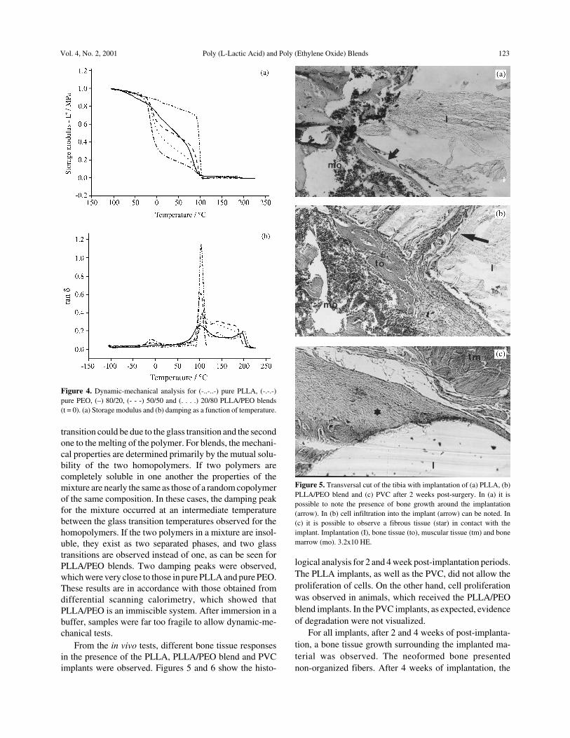
From the in vivo tests, different bone tissue responsesin the presence of the PLLA, PLLA/PEO blend and PVCimplants were observed. Figures 5 and 6 show the histo-
logical analysis for 2 and 4 week post-implantation periods.The PLLA implants, as well as the PVC, did not allow theproliferation of cells. On the other hand, cell proliferationwas observed in animals, which received the PLLA/PEOblend implants. In the PVC implants, as expected, evidenceof degradation were not visualized.
For all implants, after 2 and 4 weeks of post-implanta-tion, a bone tissue growth surrounding the implanted ma-terial was observed. The neoformed bone presentednon-organized fibers. After 4 weeks of implantation, the
Figure 4. Dynamic-mechanical analysis for (-..-..-) pure PLLA, (-.-.-)pure PEO, (–) 80/20, (- - -) 50/50 and (. . . .) 20/80 PLLA/PEO blends(t = 0). (a) Storage modulus and (b) damping as a function of temperature.
Figure 5. Transversal cut of the tibia with implantation of (a) PLLA, (b)PLLA/PEO blend and (c) PVC after 2 weeks post-surgery. In (a) it ispossible to note the presence of bone growth around the implantation(arrow). In (b) cell infiltration into the implant (arrow) can be noted. In(c) it is possible to observe a fibrous tissue (star) in contact with theimplant. Implantation (I), bone tissue (to), muscular tissue (tm) and bonemarrow (mo). 3.2x10 HE.
Vol. 4, No. 2, 2001 Poly (L-Lactic Acid) and Poly (Ethylene Oxide) Blends 123
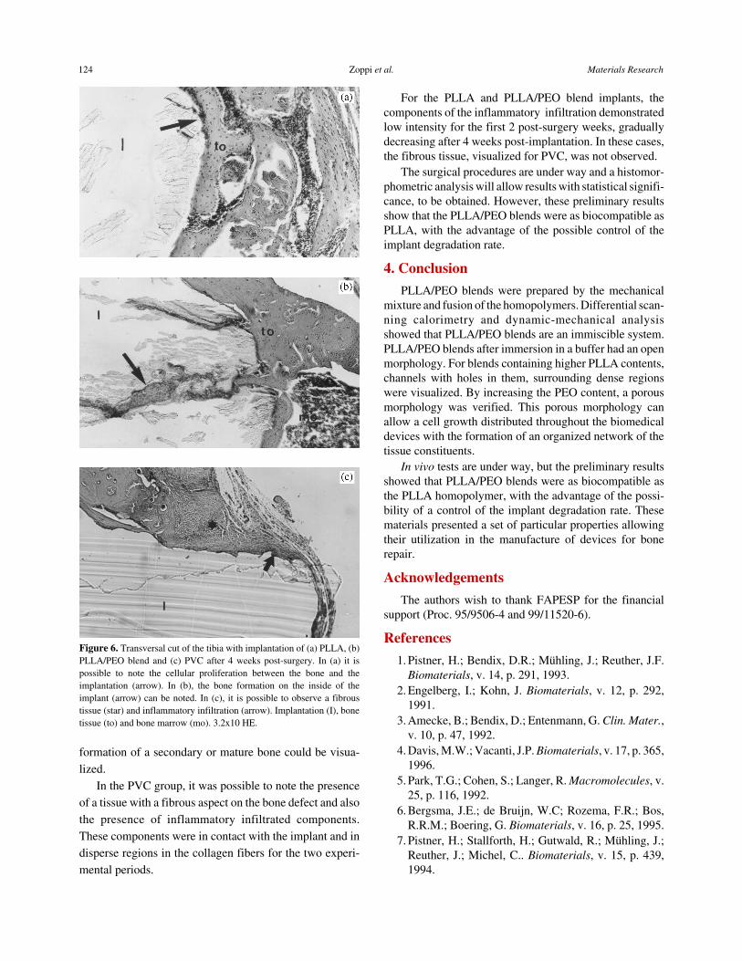
formation of a secondary or mature bone could be visua-lized.
In the PVC group, it was possible to note the presenceof a tissue with a fibrous aspect on the bone defect and alsothe presence of inflammatory infiltrated components.These components were in contact with the implant and indisperse regions in the collagen fibers for the two experi-mental periods.
For the PLLA and PLLA/PEO blend implants, thecomponents of the inflammatory infiltration demonstratedlow intensity for the first 2 post-surgery weeks, graduallydecreasing after 4 weeks post-implantation. In these cases,the fibrous tissue, visualized for PVC, was not observed.
The surgical procedures are under way and a histomor-phometric analysis will allow results with statistical signifi-cance, to be obtained. However, these preliminary resultsshow that the PLLA/PEO blends were as biocompatible asPLLA, with the advantage of the possible control of theimplant degradation rate.
4. Conclusion
PLLA/PEO blends were prepared by the mechanicalmixture and fusion of the homopolymers. Differential scan-ning calorimetry and dynamic-mechanical analysisshowed that PLLA/PEO blends are an immiscible system.PLLA/PEO blends after immersion in a buffer had an openmorphology. For blends containing higher PLLA contents,channels with holes in them, surrounding dense regionswere visualized. By increasing the PEO content, a porousmorphology was verified. This porous morphology canallow a cell growth distributed throughout the biomedicaldevices with the formation of an organized network of thetissue constituents.
In vivo tests are under way, but the preliminary resultsshowed that PLLA/PEO blends were as biocompatible asthe PLLA homopolymer, with the advantage of the possi-bility of a control of the implant degradation rate. Thesematerials presented a set of particular properties allowingtheir utilization in the manufacture of devices for bonerepair.
Acknowledgements
The authors wish to thank FAPESP for the financialsupport (Proc. 95/9506-4 and 99/11520-6).
References
1. Pistner, H.; Bendix, D.R.; Mühling, J.; Reuther, J.F.Biomaterials, v. 14, p. 291, 1993.
2. Engelberg, I.; Kohn, J. Biomaterials, v. 12, p. 292,1991.
3. Amecke, B.; Bendix, D.; Entenmann, G. Clin. Mater.,v. 10, p. 47, 1992.
4. Davis, M.W.; Vacanti, J.P. Biomaterials, v. 17, p. 365,1996.
5. Park, T.G.; Cohen, S.; Langer, R. Macromolecules, v.25, p. 116, 1992.
6. Bergsma, J.E.; de Bruijn, W.C; Rozema, F.R.; Bos,R.R.M.; Boering, G. Biomaterials, v. 16, p. 25, 1995.
7. Pistner, H.; Stallforth, H.; Gutwald, R.; Mühling, J.;Reuther, J.; Michel, C.. Biomaterials, v. 15, p. 439,1994.
Figure 6. Transversal cut of the tibia with implantation of (a) PLLA, (b)PLLA/PEO blend and (c) PVC after 4 weeks post-surgery. In (a) it ispossible to note the cellular proliferation between the bone and theimplantation (arrow). In (b), the bone formation on the inside of theimplant (arrow) can be noted. In (c), it is possible to observe a fibroustissue (star) and inflammatory infiltration (arrow). Implantation (I), bonetissue (to) and bone marrow (mo). 3.2x10 HE.
124 Zoppi et al. Materials Research
8. Mainil-Varlet, P.; Rahn, B.; Gogolewski, S. Biomate-rials, v. 18, p. 257, 1997.
9. Mainil-Varlet, P.; Gogolewski, S.; Nieuwenhuis, P. J.of Mater. Sci.: Mater. in Med., v. 7, p. 713, 1996.
10. Bos, R.R.M.; Rozema, F.R.; Boering, G. J. Oral Max-illofac. Surg., v. 45, p. 751, 1987.
11. Nijenhuis, A.J.; Colstee, E.; Grijpma, D.W.; Pen-nings, A.J. Polymer, v. 37, p. 5849, 1996.
12. Penning, J.P.; Manley, R.St.J. Macromolecules, v. 29,p. 77, 1996.
13. Eshuis, A.; Roerdink, E.; Challa, G. Polymer, v. 23, p.735, 1982.
14. Avella, M.; Martucelli, H. Polymer, v. 29, p.1731,1988.
15. Edie, S.L.; Marand, H. Polym. Prep.(Am. Chem. Soc.,Div. Polym. Chem.), v. 32, p. 329, 1991.
16. Jonza, J.M.; Porter, R.S. Macromolecules, v. 19, p.1946, 1986.
17. Cheung, Y.W.; Stein, R.S. Macromolecules, v. 27, p.2512, 1994.
18. Cheung, Y.W.; Stein, R.S.; Lin, J.S.; Wignall, G.D.Macromolecules, v. 27, p. 2520, 1994.
19. Penning, J.P.; Manley, R.St.J. Macromolecules, v. 29,p. 84, 1996.
20. Fujita, K.; Kyu, T. Macromolecules, v. 29, p. 91, 1996.21. Meikle, M.C.; Papaioannou, S.; Ratledge, T.J. Bioma-
terials, v. 15, p. 513, 1994.22. Nakafuku, C. Polymer Journal, v. 26, p. 680, 1994.23. Nakafuku, C. Polymer Journal, v. 28, p. 568, 1996.24. Nakafuku, C.; Sakoda, M. Polymer Journal, v. 25, p.
909, 1993.25. Younes, H.; Cohn, D. Eur. Polym. J., v. 24, p. 765,
1988.26. Yang, J.-M.; Chen, H.-L.; Hwang, J.C. Polymer Jour-
nal, v. 29, p. 657, 1997.27. Sheth, M.; Kumar, R.A.; Davé, V.; Gross, R.A.;
McCarthy, S.P. Journal of Applied Polymer Science,v. 66, p. 1495, 1997.
28. Zoppi, R.A.; Contant, S.; Duek, E.A.R.; Nunes, S.P.,submitted.
29. Lam, K.L.; Nieuwenhuis, P.; Molenaar, I.; Essel-brugge, H.; Feijen, J.; Dijkstra, P.J.; Schakenraad,J.M. J. Mater. Sci.: Mater. in Med., v. 5, p. 181, 1994.
30. Standard practice for assessment of compatibility ofbiomaterials for surgical implants with respect toeffect of materials on muscle and bone, Annual Bookof ASTM Standards, F 981-93, ASTM, 1973.
FAPESP helped in meeting the publication costs of this article
Vol. 4, No. 2, 2001 Poly (L-Lactic Acid) and Poly (Ethylene Oxide) Blends 125