preparation of loading lactobacillus delbrueckii and ...article.scirea.org/pdf/55003.pdf3 low...
TRANSCRIPT
1
Preparation and in vitro characterization of Lactobacillus
plantarum and Lactobacillus fermentum beads using low
methoxyl pectin
Qingshen Sun
*1, Yanyan Zhang
1, Xiuyan Ma
1, Dequan Han
1, Yue Shi
1, Quan Sun
1,
Xiaoxiong Ma1, Chunhai Yang
1, Bo Pan
1
1Laboratory of Microbiology, College of Life Science, Heilongjiang University, Harbin
150080, China
Corresponding author: Qingshen Sun
Abstract
Lactobacillus plantarum (LP) and Lactobacillus fermentum (LF) were encapsulated into low
methoxyl pectin (LMP) beads by CaCl2 external ionization. The encapsulating efficiency
(EE), in vitro release and stability studies were performed by plate counting. The results
showed that more than 99.99% EE were obtained. The contents of LF and LP were 10 11
cfu/g
beads (dry base). Stability study showed that encapsulation improved the stability under
ambient temperature compared with the unencapsulated microbes. It provides the foundation
for the further development and application of composite Lactobacillus beads in modulation
of intestinal and systematic function.
Keywords: Low methoxyl pectin Bead; Lactobacillus plantarum; Lactobacillus fermentum
INTRODUCTION
SCIREA Journal of Food
http://www.scirea.org/journal/Food
April 26, 2017
Volume 2, Issue 1, February 2017

2
Probiotics is a kind of live microorganisms which can exert the health effects when there are
enough number reaching colon or the lower parts of small intestine [1-4], and have been
studied extensively for their use as new therapy to the treatment of many diseases, including
the cardiovascular disease, obesity and its related syndrome. Studies also found that the
intestinal flora were most abundant in the colon, therefore, it is conceivable that the changes
of colon flora composition for weight loss may have a more direct effect. However, because
of the instable nature of some probiotics during passage into gastrointestinal (GI) system
where extreme pH and bile salt exist, many measures have been taken, of which
microencapsulation technique showed great promise [5-15]. However, because of the
intestinal soluble features of some materials such as alginate [16, 17], more efforts have been
directed to find more sable materials for probiotics encapsulation.
Pectin is plant-derived component which has been used in many areas based on its different
characters [18-22]. According to the methoxyl contents, pectin can be divided into high
methoxyl pectin (HMP) and low methoxyl pectin (LMP). In recent years, LMP has been
studied in drug delivery system as it can form beads under calcium chloride treatment [22-27],
which are widely used as colon delivery carrier [28-30]. In this study, the LMP was used for
encapsulating LF and LP individually or in combination. The in vitro tests were performed to
verify the possibility of using this kind of material as carrier for colon-targeted probiotic
delivery system.
MATERIALS AND METHODS
Strain
Lactobacillus fermentum subsp. bulgaricus (LF, CGMCC1.1880) was purchased from China
General Microbiological Culture Collection Center (CGMCC). Lactobacillus plantarum (LP)
was isolated from Northeast sauerkraut in our lab.
Preparation of cells for encapsulation
The frozen cultures of Lactobacillus fermentum subsp. bulgaricus (LF) and Lactobacillus
plantarum (LP) were transferred independently into MRS broth at 4% (v/v) under aerobic
environment. Then the cultures were harvested after centrifugation at 4000rm (6C type low
speed centrifugator, Labnet, internation. inc) for 15 min. The pellets were subcultured 3
passages and collected at 4000rpm for 15 min.
3
Low methoxyl pectin (food grade, Anhui Yu Ning Biotechnology Co. Ltd.) beads containing
LP and LF alone or in combination at 1:1 (v/v) were prepared by external ionotropic gelation
[20]. In brief, 2% pectin aqueous solution containing LP and LF alone or in combination was
extruded into 300mM anhydrous calcium chloride (food grade, Anhui Yu Ning
Biotechnology Co. Ltd.) solution using 10 mL injector with 7# needle under stirring. After
extrusion, the solution was stirred for another 20 minutes. After that, the beads were collected
by filtration and freeze-dried after mixing with cryoprotectant. The formula for
cryoprotectant was composed of 10.43 g skim milk (Inner Mongolia Yili Industrial Group
Limited by Share Ltd), 8.91g sucrose and 0.23g MnSO4 in 100mL formulation base, which
were mixed well and boiled at 100 ℃ for 10 min, then cooled down to 4 ℃. The beads
were mixed with cryoprotectant at 4:1 (w/v) [31]. Then the mixture was prefrozen at -20 ℃
for 3 h, then freeze dried for 12 hours. The freeze drying products were collected after dried.
For storage, these products were subdivided into three parts, one was stored in desiccator at
room temperature; the other parts were stored at 4 ℃ and -20 ℃.
The supernatants were collected and used for enumeration of the LP and LF by plate counting
as follows. The morphology of the beads was characterized by optical microscopy.
Encapsulation efficiency (EE)
The EE was calculated by plate counting using MRS agar. The MRS agar medium was
prepared according to manufacturer`s guide and autoclaved at 121 ℃ for 20 min using
LDZX-50KB type vertical pressure steam sterilizer (Shanghai Shen An medical instrument
factory). After cool down to 50 ℃, the medium was poured onto 10 cm plates and
continually cool down. Then the collected flora was diluted serially and spread onto the
plates. After anaerobically culture for 40-44h, the LP and LF were counted and then
calculated as cfu/mL (for supernatants) or cfu/g (for beads). For enumeration of the LP and
LF in supernatants, the supernatant was diluted directly and spread on pates.
The EE was calculated according to Reddy et al [32] based on plate counting technique with
some modifications.
EE=log (A-B)/ log A*100%
Where A and B represent the total number of Lactobacillus added (CFU/ mL) and those in
supernatants.
The total number of lactobacillus was counted by plate counting also. In brief, the LP and LF

4
cultures were spread onto MRS plates after collection and dilution into suitable
concentrations.
In vitro release
Simulated gastric fluid (SGF) was prepared according to the U.S. Pharmacopeia [33], with
some minor modifications. In brief, 100mL SGF containing 1.00 g pepsin (Tissue Culture
Grade,AMERCO), the pH was adjusted to 2.0 with 10 N HCl. Simulated bile fluid (SBF): 1%
SBF was prepared by adding 1g No. 7 bile salt into 99 mL sterile water. Simulated intestinal
fluid (SIF): 1g pancreatin (Tissue Culture Grade, AMERCO) was dissolved into 100mL 0.2M
PBS (pH 6.8) before use.
In vitro release tests were performed by putting the beads in SGF for 2h, SBF 20min, SIF 2h,
continually [34]. After each process, the beads were collected and disintegrated using 50mM
Ethylene diamine tetra-acetic acid (EDTA) solution as above, then washed with autoclaved
saline. The collected flora was spread on 10 cm MRS plates for enumeration in duplicate.
Stability study
The freeze drying beads were stored at -20℃, 4℃ and ambient temperature in desiccator,
respectively, samples were tested weekly in triplicate by plate counting as above.
Determination of microbe number in beads
Strain specific primer design Bacterial DNA was extracted using Ezup pillar bacterial
genome DNA extraction kit. The LF and LP were amplified using 16S rDNA sequences, then
subjected to sequence analysis. The individual specific sites were selected for primer design
using Primer 5.0 primer design software from NCBI website.
RT-PCR The quantification of the LP and LF was based on the pure culture standards. A
tenfold dilution series of the strain in MRS was prepared corresponding to 104 to 10
8
CFU/mL LP and LF, respectively. These dilutions were used for RT-PCR analysis.
Real-time PCR was performed by Bio-Rad apparatus. 0.1g Lyophilized composite
Lactobacillus beads were taken out and subjected to 50 mmol/L EDTA solution for thorough
breakdown. RT-PCR quantitative analysis of the LP and LF was performed using
double-stranded chimeric SYBR dye method.
The specific primers of LP and LF were designed as follows:
LF: PrimerF CAACAAGGGAAAAACGGCGA
5
PrimerR GGAATCAGGTAGGGCGTCTG
LP: PrimerF AAGATAAGCGGCCAGTGCTT
PrimerR CACCGCCACCAAAATTACCG
Amplification was carried out in a 20 uL final volume containing 1 uL bacterial DNA as
templates. 2.0 uL 10×PCR buffer, 1.6 uL 2.5mM dNTPs, 1.2 uL 2.5mM Mg2+, 0.2 uL Taq
DNase, 1.0 uL former primer, 1.0 uL reverse primer and 12 uL ddH2O. The thermocycler
programs had denaturation at 94℃ for 5 min, then 35 cycles with the following steps:
denaturation at 94℃ for 1 min, annealing at 53℃ for 30 s (for LP) or 55℃ for 30 s (for LF),
and elongation at 72℃ for 10 min. For each step, the temperature transition rate was 20 ℃.
Fluorescence signal was detected at the end of the annealing step in every cycle.
Statistical analysis
The results were expressed as mean ±std. All the data were analyzed using Origin 8.0
software. Comparisons were analyzed by one-way analysis of ANOVA.
RESULTS AND DISCUSSION
In vitro characterization of the beads
The study is aimed at the characterization of LMP beads loading LP and LF alone or in
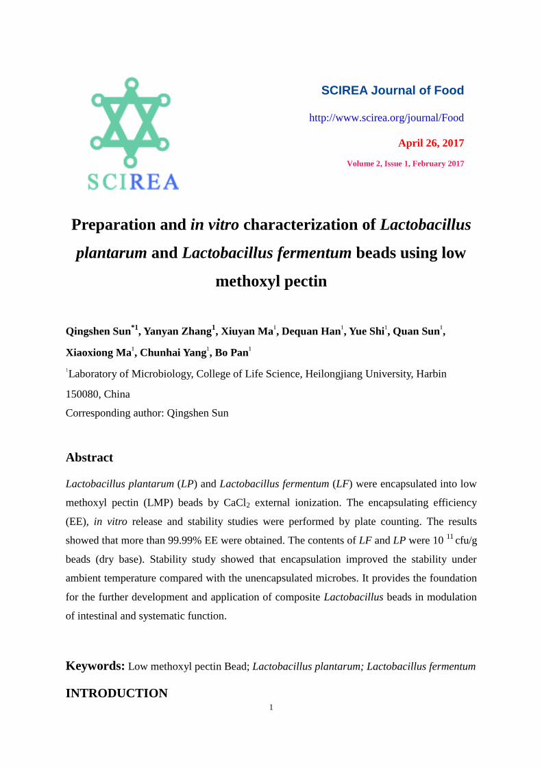
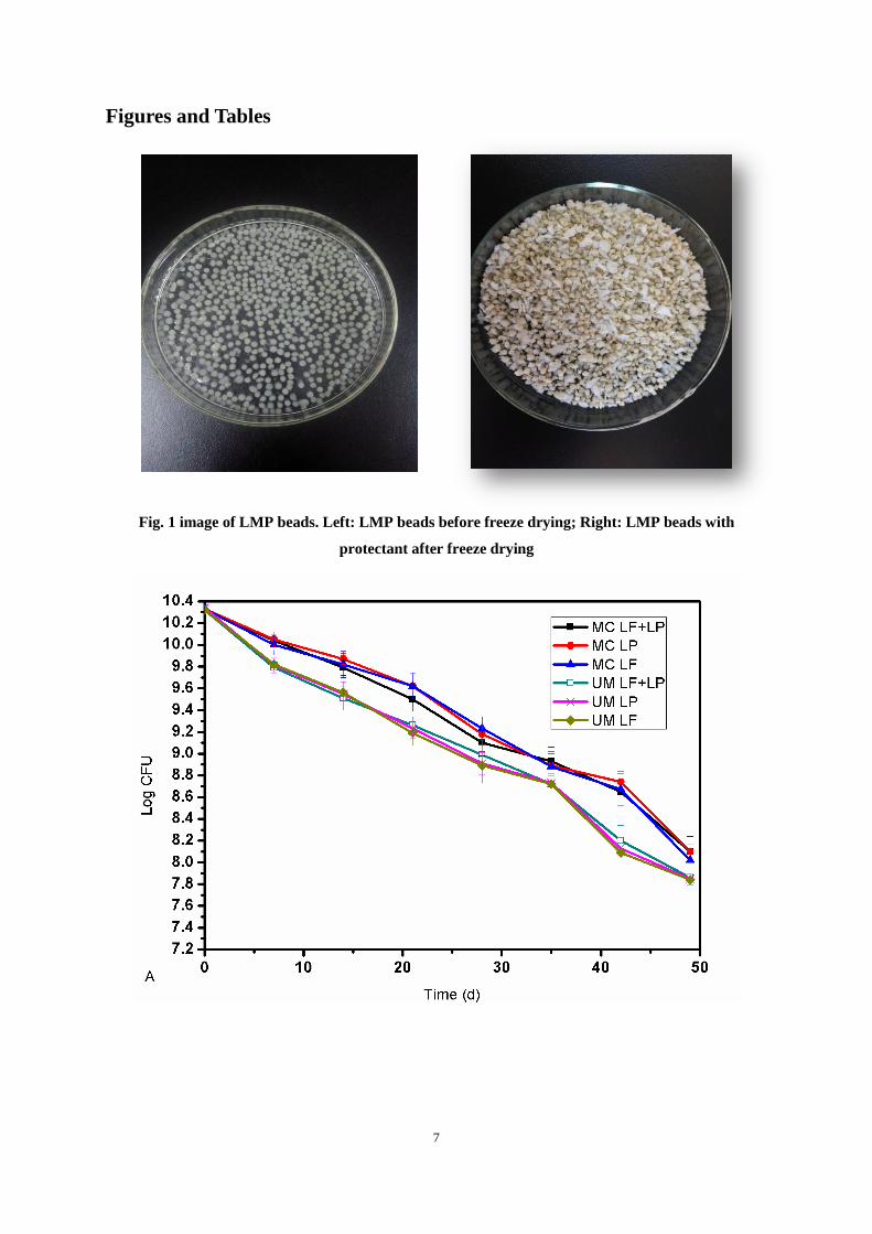
combination in vitro/in vivo. Figure 1 showed the morphology of the beads which were round
and smooth in solutions with excellent disperse character. The average sizes were
2.00±0.02mm. After freeze drying, the beads were light yellow. Our results showed the
spherical morphology with the size which are suitable for animal experiments. The size
distribution may be determined by several factors, such as the internal diameter of syringe
needle, the pectin concentration, the stirring rate. In this study, the optimized protocol
documented by Jung et al was used [35]. There was 3 to 4 log cfu decrease of the LP and LF
in supernatants from beads prepared with LMP encapsulation, indicating that EE was higher
than 99.9%.
Table 1 showed the survivability after in vitro release profile in different simulated fluids. All
the unencapsulated strains showed significantly decrease in activities after subjected to SGF,
with 3.83, 3.95, 3.74 log CFU reduction for LF+LP, LF and LP, respectively. After subjected
to SBF treatment, there is another 1 log CFU reduction, but all were relatively stable in SIF.
6
In comparison, the LF+LP, LF and LP beads showed only 1.10, 1.10, and 0.87 log CFU
reduction after SGF treatment. Another 0.78, 0.69, 1.04 log CFU reduction appeared after
SBF treatment. Similar to unencapsulated strains, all were stable in SIF, indicating that it can
reach small intestine or colon only if the higher survival rate can be obtained upon passage
through gastric acid or bile salt.
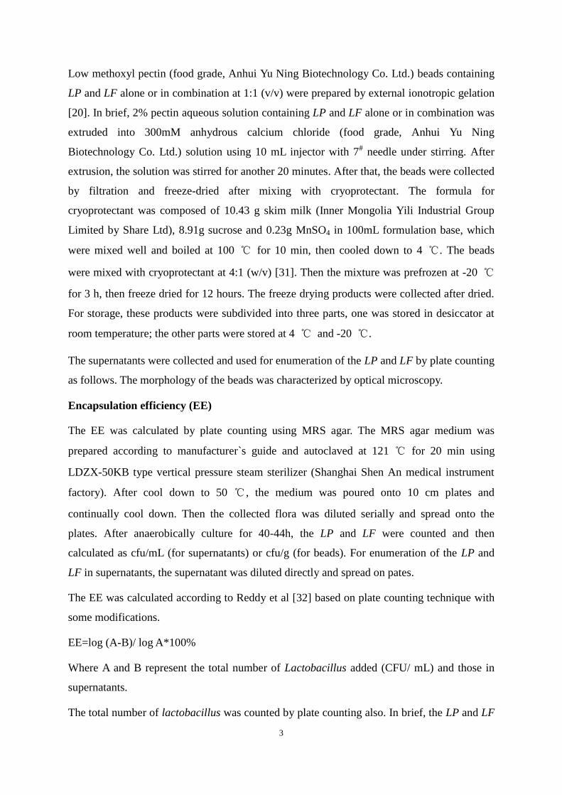
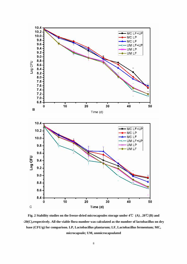
Stability analysis
Stability of the probiotics during storage is another important consideration as this affect the
applicability of the products. Generally, temperature, light, oxygen and water can directly
affect the stability of the encapsulated probiotics [36]. For the beads after freeze drying, the
results have been obtained for the beads prepared in different time, which showed that more
stability was found for those stored at -20 and 4℃. Encapsulation with LMP did not change
the stability of the microbes (Fig. 2).
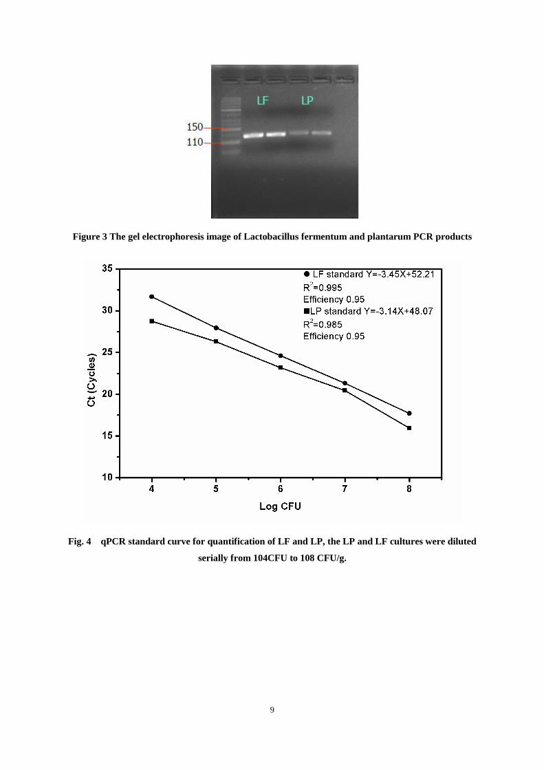
Quantification of the strains in beads
The strain-specific real-time PCR was conducted to quantify the number of the LP and LF in
beads. The PCR products of LF and LP were imaged by agar gel electrophoresis (Fig. 3) ,
then sequenced and checked for the accuracy of the segments using web tool of the National
Center of Biotechnology Information (NCBI) Blast software (data not shown).
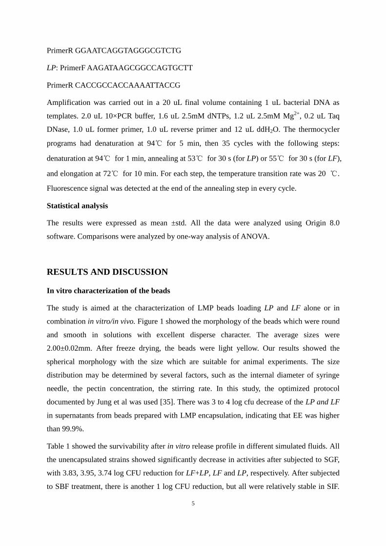
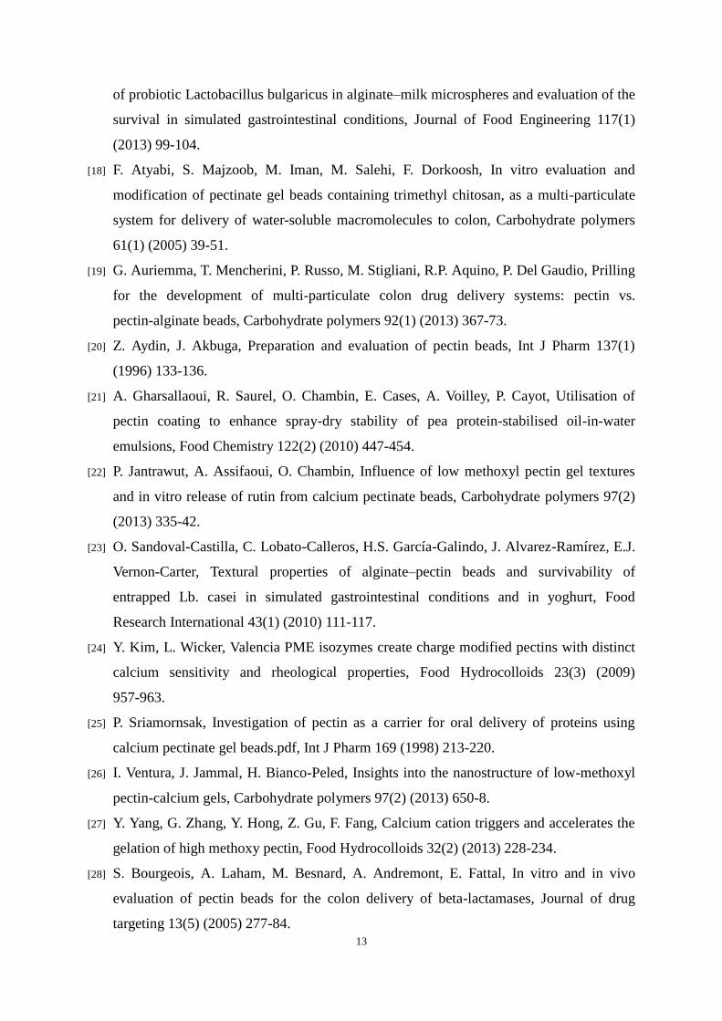
The qPCR standard curve was plotted by diluting the LF and LP cultures from 104
CFU to
108 CFU/g, Amplification efficiency were 95% for both strains (Fig. 4). Using the individual
standard regression equation, the number of LP and LF in beads were absolutely calculated as
1011
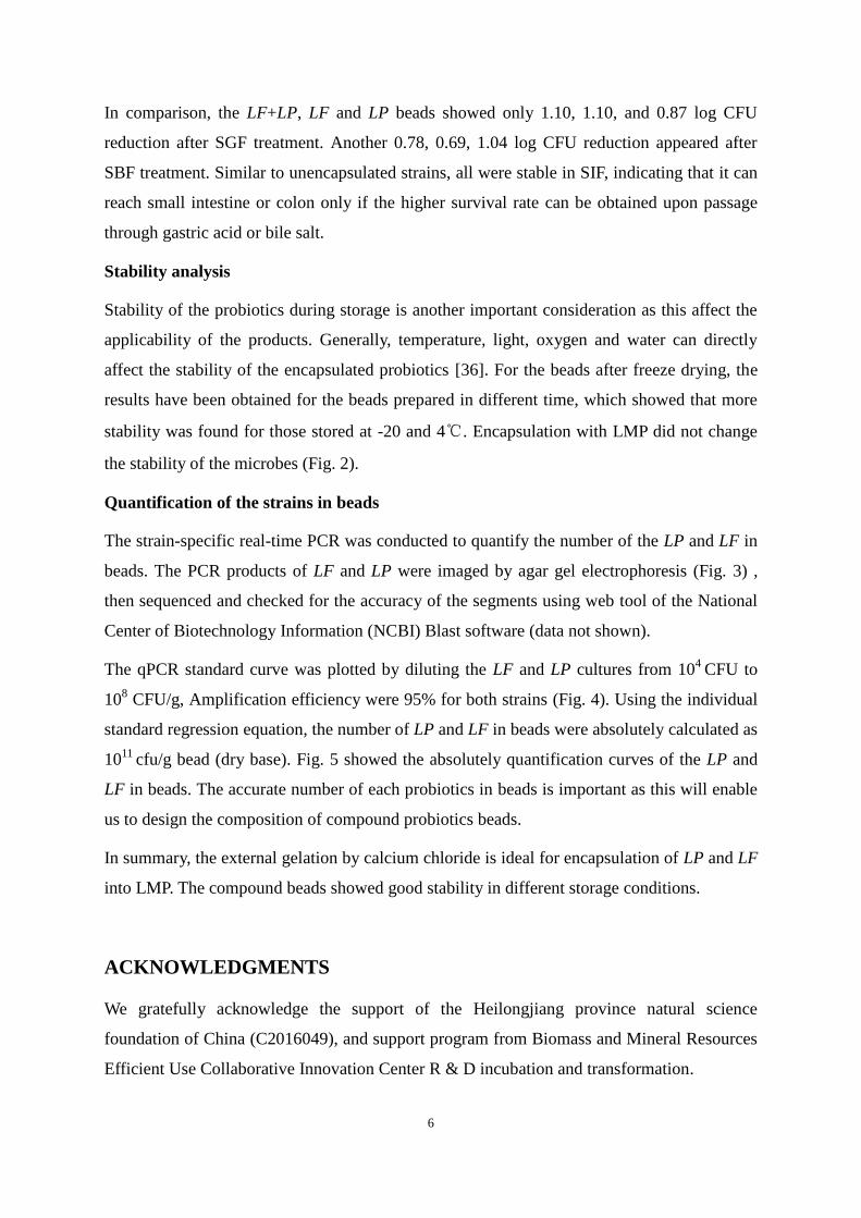
cfu/g bead (dry base). Fig. 5 showed the absolutely quantification curves of the LP and
LF in beads. The accurate number of each probiotics in beads is important as this will enable
us to design the composition of compound probiotics beads.
In summary, the external gelation by calcium chloride is ideal for encapsulation of LP and LF
into LMP. The compound beads showed good stability in different storage conditions.
ACKNOWLEDGMENTS
We gratefully acknowledge the support of the Heilongjiang province natural science
foundation of China (C2016049), and support program from Biomass and Mineral Resources
Efficient Use Collaborative Innovation Center R & D incubation and transformation.
7
Figures and Tables
Fig. 1 image of LMP beads. Left: LMP beads before freeze drying; Right: LMP beads with
protectant after freeze drying
8
Fig. 2 Stability studies on the freeze-dried microcapsules storage under 4℃ (A) , 20℃(B) and
-20(C),respectively. All the viable flora number was calculated as the number of lactobacillus on dry
base (CFU/g) for comparison. LP, Lactobacillus plantarum; LF, Lactobacillus fermentum; MC,
microcapsule; UM, unmicrocapsulated
9
Figure 3 The gel electrophoresis image of Lactobacillus fermentum and plantarum PCR products
Fig. 4 qPCR standard curve for quantification of LF and LP, the LP and LF cultures were diluted
serially from 104CFU to 108 CFU/g.

10
A
B
Fig 5 The absolute quantitative graphs of Lactobacillus fermentium (A) and Lactobacillus
plantarum (B) in microcapsules

11
Table 1 In vitro simulated gastric-intestinal tract test of the beads (log CFU)
Note: SGF, simulated gastric fluid; SBF, simulated bile fluid; SIF, simulated intestine fluid.
SGF-SBF-SIF refers to the LB underwent the SGF treatment for 2h, SBF for 20min and SIF
for 3h. Different lowercases and uppercases represent the significant difference at p<0.05 and
p<0.01 level in the same column, respectively. All the flora were counted on dry lactobacillus
base. LP, Lactobacillus plantarum; LF, Lactobacillus fermentum; MC, microcapsule; UM,
unmicrocapsulated
REFERENCES
[1] R.A. Britton, J. Versalovic, Probiotics and gastrointestinal infections, Interdisciplinary
perspectives on infectious diseases 2008 (2008) 290769.
[2] M.L. Jimenez-Pranteda, D. Poncelet, M.E. Nader-Macias, A. Arcos, M. Aguilera, M.
Monteoliva-Sanchez, A. Ramos-Cormenzana, Stability of lactobacilli encapsulated in
various microbial polymers, Journal of bioscience and bioengineering 113(2) (2012)
179-84.
[3] Y.E. Dommels, R.A. Kemperman, Y.E. Zebregs, R.B. Draaisma, A. Jol, D.A. Wolvers,
E.E. Vaughan, R. Albers, Survival of Lactobacillus reuteri DSM 17938 and Lactobacillus
rhamnosus GG in the human gastrointestinal tract with daily consumption of a low-fat
probiotic spread, Applied and environmental microbiology 75(19) (2009) 6198-204.
[4] D. Dianawati, N.P. Shah, Survival, acid and bile tolerance, and surface hydrophobicity of
microencapsulated B. animalis ssp. lactis Bb12 during storage at room temperature, J
Food Sci 76(9) (2011) M592-9.
[5] A.K. Anal, H. Singh, Recent advances in microencapsulation of probiotics for industrial
applications and targeted delivery, Trends in Food Science & Technology 18(5) (2007)
240-251.
[6] M.T. Cook, G. Tzortzis, D. Charalampopoulos, V.V. Khutoryanskiy, Microencapsulation
LF+LP UM LF+LP MC UM LF MC LF UM LP MC LP
Original 10.54±0.07 10.32±0.06 10.52±0.05 10.31±0.07 10.51±0.04 10.21±0.07
SGF 2h 6.71±0.12 9.22±0.10 6.57±0.10 9.21±0.11 6.73±0.08 9.34±0.09
SBF 20min 5.66±0.21 8.54±0.17 5.59±0.11 8.52±0.18 5.80±0.11 8.30±0.13
SIF 3h 5.60±0.18 8.54±0.18 5.52±0.12 8.56±0.16 5.71±0.12 8.55±0.16

12
of probiotics for gastrointestinal delivery, Journal of controlled release : official journal
of the Controlled Release Society 162(1) (2012) 56-67.
[7] M.R. Corbo, A. Bevilacqua, M. Gallo, B. Speranza, M. Sinigaglia, Immobilization and
microencapsulation of Lactobacillus plantarum: Performances and in vivo applications,
Innovative Food Science & Emerging Technologies 18 (2013) 196-201.
[8] G. Della Porta, F. Castaldo, M. Scognamiglio, L. Paciello, P. Parascandola, E. Reverchon,
Bacteria microencapsulation in PLGA microdevices by supercritical emulsion extraction,
The Journal of Supercritical Fluids 63 (2012) 1-7.
[9] T. Heidebach, P. Först, U. Kulozik, Influence of casein-based microencapsulation on
freeze-drying and storage of probiotic cells, Journal of Food Engineering 98(3) (2010)
309-316.
[10] S.-J. Kim, S.Y. Cho, S.H. Kim, O.-J. Song, I.I.S. Shin, D.S. Cha, H.J. Park, Effect of
microencapsulation on viability and other characteristics in Lactobacillus acidophilus
ATCC 43121, LWT - Food Science and Technology 41(3) (2008) 493-500.
[11] F. Liu, Z. Chen, C.-H. Tang, Microencapsulation properties of protein isolates from three
selected Phaseolus legumes in comparison with soy protein isolate, LWT - Food Science
and Technology 55(1) (2014) 74-82.
[12] A. Nag, K.-S. Han, H. Singh, Microencapsulation of probiotic bacteria using pH-induced
gelation of sodium caseinate and gellan gum, International Dairy Journal 21(4) (2011)
247-253.
[13] A.C. Oliveira, T.S. Moretti, C. Boschini, J.C.C. Baliero, L.A.P. Freitas, O. Freitas, C.S.
Favaro-Trindade, Microencapsulation ofB. lactis(BI 01) andL. acidophilus(LAC 4) by
Complex Coacervation Followed by Spouted-Bed Drying, Drying Technology 25(10)
(2007) 1687-1693.
[14] S. Rathore, P.M. Desai, C.V. Liew, L.W. Chan, P.W.S. Heng, Microencapsulation of
microbial cells, Journal of Food Engineering 116(2) (2013) 369-381.
[15] Q. Sun, F. Wang, D. Han, Y. Zhao, Z. Liu, H. Lei, Y. Song, X. Huang, X. Li, A. Ma, G.
Yuan, X. Li, Z. Yang, Preparation and optimization of soy protein isolate-high methoxy
pectin microcapsules loaded withLactobacillus delbrueckii, International Journal of Food
Science & Technology 49(5) (2013) 1287-1293.
[16] R.R. Mokarram, S.A. Mortazavi, M.B.H. Najafi, F. Shahidi, The influence of multi stage
alginate coating on survivability of potential probiotic bacteria in simulated gastric and
intestinal juice, Food Research International 42(8) (2009) 1040-1045.
[17] L.-E. Shi, Z.-H. Li, D.-T. Li, M. Xu, H.-Y. Chen, Z.-L. Zhang, Z.-X. Tang, Encapsulation
13
of probiotic Lactobacillus bulgaricus in alginate–milk microspheres and evaluation of the
survival in simulated gastrointestinal conditions, Journal of Food Engineering 117(1)
(2013) 99-104.
[18] F. Atyabi, S. Majzoob, M. Iman, M. Salehi, F. Dorkoosh, In vitro evaluation and
modification of pectinate gel beads containing trimethyl chitosan, as a multi-particulate
system for delivery of water-soluble macromolecules to colon, Carbohydrate polymers
61(1) (2005) 39-51.
[19] G. Auriemma, T. Mencherini, P. Russo, M. Stigliani, R.P. Aquino, P. Del Gaudio, Prilling
for the development of multi-particulate colon drug delivery systems: pectin vs.
pectin-alginate beads, Carbohydrate polymers 92(1) (2013) 367-73.
[20] Z. Aydin, J. Akbuga, Preparation and evaluation of pectin beads, Int J Pharm 137(1)
(1996) 133-136.
[21] A. Gharsallaoui, R. Saurel, O. Chambin, E. Cases, A. Voilley, P. Cayot, Utilisation of
pectin coating to enhance spray-dry stability of pea protein-stabilised oil-in-water
emulsions, Food Chemistry 122(2) (2010) 447-454.
[22] P. Jantrawut, A. Assifaoui, O. Chambin, Influence of low methoxyl pectin gel textures
and in vitro release of rutin from calcium pectinate beads, Carbohydrate polymers 97(2)
(2013) 335-42.
[23] O. Sandoval-Castilla, C. Lobato-Calleros, H.S. García-Galindo, J. Alvarez-Ramírez, E.J.
Vernon-Carter, Textural properties of alginate–pectin beads and survivability of
entrapped Lb. casei in simulated gastrointestinal conditions and in yoghurt, Food
Research International 43(1) (2010) 111-117.
[24] Y. Kim, L. Wicker, Valencia PME isozymes create charge modified pectins with distinct
calcium sensitivity and rheological properties, Food Hydrocolloids 23(3) (2009)
957-963.
[25] P. Sriamornsak, Investigation of pectin as a carrier for oral delivery of proteins using
calcium pectinate gel beads.pdf, Int J Pharm 169 (1998) 213-220.
[26] I. Ventura, J. Jammal, H. Bianco-Peled, Insights into the nanostructure of low-methoxyl
pectin-calcium gels, Carbohydrate polymers 97(2) (2013) 650-8.
[27] Y. Yang, G. Zhang, Y. Hong, Z. Gu, F. Fang, Calcium cation triggers and accelerates the
gelation of high methoxy pectin, Food Hydrocolloids 32(2) (2013) 228-234.
[28] S. Bourgeois, A. Laham, M. Besnard, A. Andremont, E. Fattal, In vitro and in vivo
evaluation of pectin beads for the colon delivery of beta-lactamases, Journal of drug
targeting 13(5) (2005) 277-84.

14
[29] S. Das, A. Chaudhury, K.Y. Ng, Polyethyleneimine-modified pectin beads for
colon-specific drug delivery: in vitro and in vivo implications, Journal of
microencapsulation 28(4) (2011) 268-79.
[30] A. Oehme, A. Valotis, G. Krammer, I. Zimmermann, P. Schreier, Preparation and
characterization of shellac-coated anthocyanin pectin beads as dietary colonic delivery
system, Molecular nutrition & food research 55 Suppl 1 (2011) S75-85.
[31] F. Shamekhi, M. Shuhaimi, A. Ariff, Y.A. Manap, Cell viability of microencapsulated
Bifidobacterium animalis subsp. lactis under freeze-drying, storage and gastrointestinal
tract simulation conditions, Folia microbiologica 58(2) (2013) 91-101.
[32] K.B.P.K. Reddy, A.N. Madhu, S.G. Prapulla, Comparative survival and evaluation of
functional probiotic properties of spray-dried lactic acid bacteria, Int J Dairy Technol
62(2) (2009) 240-248.
[33] K. Miura, H. Yamashiro, K. Uotani, S. Kojima, T. Yutsudo, J. Lu, O. Yoshida, Y. Yamano,
H. Maki, H. Arimoto, Mode of action of Van-M-02, a novel glycopeptide inhibitor of
peptidoglycan synthesis, in vancomycin-resistant bacteria, Antimicrobial agents and
chemotherapy 54(2) (2010) 960-2.
[34] F.P. De Castro-Cislaghi, C.D.R.E. Silva, C.B. Fritzen-Freire, J.G. Lorenz, E.S.
Sant’Anna, Bifidobacterium Bb-12 microencapsulated by spray drying with whey:
Survival under simulated gastrointestinal conditions, tolerance to NaCl, and viability
during storage, Journal of Food Engineering 113(2) (2012) 186-193.
[35] J. Jung, R.D. Arnold, L. Wicker, Pectin and charge modified pectin hydrogel beads as a
colon-targeted drug delivery carrier, Colloids and surfaces. B, Biointerfaces 104 (2013)
116-21.
[36] G.M. Maciel, K.S. Chaves, C.R. Grosso, M.L. Gigante, Microencapsulation of
Lactobacillus acidophilus La-5 by spray-drying using sweet whey and skim milk as
encapsulating materials, Journal of dairy science 97(4) (2014) 1991-8.