preparation of titanium dioxide nanoparticles and
TRANSCRIPT

Malaysia Journal of Science 36 (3):132-144 (2017)
132
Preparation of Titanium Dioxide Nanoparticles and PolyVinyl
Pyrrolidone Polymer Films as Antibacterial, Antibiofilm against
Pathogenic Bacteria on Different Surfaces
1Jehan Abdul Sattar Salman,
2Mahasin F. Hadi AL-Kadhemy
*,
3Samara A.
Madhloom
2AL-Mustansiriyah University – College of Science – Department of Biology
2,3AL-Mustansiriyah University – College of Science – Department of Physics
Baghdad, IRAQ *Corresponding Author: [email protected]
Received: 10 February 2017 Revised manuscript received: 22 Nov 2017 Accepted: 27 Nov 2017
ABSTRACT In this paper, the structural properties that included X-ray diffraction
(XRD) of Polyvinyl Pyrrolidone (PVP) and PVP with titanium dioxide nanoparticles
have studied. PVP still appeared amorphous structure in spite of presence of titanium
dioxide nanoparticle with crystalline form. The films of pure PVP and PVP with titanium
dioxide nanoparticle have been prepared by casting method and used in covering glass
plates and plastic to study the effect of these films on growth of bacteria. The
antibacterial effect against Staphylococcus aureus and Escherichia coli has been tested.
In this study the prepared films had antibacterial effect on plastic and glass plates, the
reduction of bacterial growth percentage for PVP films on plastic and glass plates
reached to 60.89% and 62.5% against S. aureus and 49%, 51% against E. coli,
respectively. Whereas the percentage for prepared films from a mixture of PVP with
titanium dioxide nanoparticle on plastic and glass plates reached 89.42% and 86%,
respectively against S. aureus and 100% , 69% against E.coli. Antibiofilm effect of PVP
with titanium dioxide nanoparticle against pathogenic bacteria on catheters was studied
for (4) weeks. PVP with titanium dioxide nanoparticle films had the ability to inhibit
biofilm formation for pathogenic bacteria on catheters, the inhibition of biofilm
percentage reached to 83.97% against S.aureus and 65.3% against E.coli after fourth
week of storage.
Keywords: (PVP) Polymer; TiO2 nanoparticles; Antibacterial Activity; Antibiofilm;
Staphylococcus aureus and Escherichia coli; catheters.
INTRODUCTION
Polyvinyl Pyrrolidone (PVP) is a
polymer with different grades according
to its molecular weight. It applicable to
use as a binder in the tablet formulations.
The wet granulation for PVP with a
molecular weights of 25,000 to 90,000
compared to other binders, generally
gives harder granulates with very good
flow ability, higher binding and low
friability (Chowhan, et al., 1992). Also,
to enhance these properties, PVP

Malaysia Journal of Science 36 (3):132-144 (2017)
133
increases the dissolution of the active
ingredient. The tablets of
Acetaminophen (paracetamol) that
formulate with ratio 4% of PVP with
molecular weight 90,000 used as binder
released the drug more quickly than
tablets with gelatin or hydroxypropyl
cellulose as binder, this mean the
povidone tablets were harder (Jun et al.,
1989).The same results were obtained
with ratio 0.6 or 1.0% of PVP (Mw
90,000) or hydroxypropyl cellulose
(Sinchalpanid, 1993). A lot of the active
substances have poor aqueous solubility
because they have limited
bioavailability. The easy way to enhance
the bioavailability of an active substance
by improving its dissolution by adding
solubilizing agents, such as the soluble
PVP grades. These water-soluble
complexes with many active substances
and increase the bio availability and a
large number of organic solvents; such
as alcohols, amines, acids, chlorinated
hydrocarbons, lactones and amides. In
addition to, polymer is insoluble in
common Esters, hydrocarbons, ethers
and ketones (Basf, 2009). All grades of
povidone can be used as hydrophilic
polymers which physically stabilize
suspensions. The protective colloids is
considered themost important and
primary function in all suspensions,that
hydrophilize the individual solid
particles and sterically separate them.
This led to increase the volume of any
sediment and makes it is very easy to
redisperse by shaking. Also Povidone
prevents dissolved portion of active
substance from crystallizing out by
forming soluble complexes with it
(Kadajji& Mitrevej, 2011).
Titanium dioxide nanoparticles have
become a new generation of advanced
materials because their novel and
interesting optical, dielectric, and photo-
catalytic properties from size
quantization (Alivisatos, 1996).
Titanium dioxide (TiO2) is a photo
catalyst and widely utilize as a self-
disinfecting and self-cleaning material
for surface coating used in many
applications. Titanium dioxide has a
more helpful role in our environmental
purification due to its nontoxicity, photo
induced super-hydrophobicity and
antifogging effect (Fujishima& Honda,
1972).These properties have used to
remove bacteria and harmful organic
materials from water and air. Also in
self-cleaning or self-sterilizing surfaces
for places such as medical centers
(Wong et al., 2008). The aim of this
work, used the TiO2 nanoparticles/PVP
films inhibit bacterial growth and
biofilm formation in different surfaces
like glass and plastic and catheters.
MATERIAL AND METHODS
TiO2 nanoparticles/PVP Films
Pure PVP and TiO2nanoparticles doped
PVP films have been prepared by
employing solution-casting method (Al-
Kadhemy, 2012; Nawaf, 2016). Hot
distilled water (~55°C) (10 ml) was used
to dissolve (0.5 g) from PVP) PVP is a

Malaysia Journal of Science 36 (3):132-144 (2017)
134
granular powder with molecular weight
(Mw=40000 g/mole) obtained from
(Ourchem for Laboratory Use Only) this
solution was magnetically stirred
continuously for (30 min) until mixture
became homogeneous viscous solution.
Then it poured into glass and plastic
petri dish with diameter (10 cm), keeps
under room temperature (~ 30°C) for (5
days) to evaporate all solvent slowly,
and obtained PVP thin film with
thickness about (0.00091 µm).In order to
prepare TiO2nanoparticles/PVP
composite films with two particle sizes
for TiO2nanoparticles (15.7 and 45.7)
nm; the amount of powder for each
particle sizes as used (0.01 g) with (10
ml) hot distilled water. (6 ml) of this
TiO2nanoparticles solution was added to
PVP solution to get TiO2 (15.7 and 45.7)
nm/PVP films. X-Ray Diffraction
instrument used with type (SHIMADZU
XRD – 6000) made in Japan to check
XRD pattern. The instrument has the
following specifications; Target is CuKα,
wavelength is 1.5406 ̊A, Current is 30
(mA) and Voltage is (40 KV).
Coated of Catheters by TiO2
Nanoparticles with (PVP) Polymer
The catheter pieces with length (2 cm)
had coated in solution consisting of
mixture (PVP) polymer with volume (10
ml) and TiO2 with particle size (15.7 nm)
with volume (6 ml). Then put the
catheter pieces in solution and left to dry
for (7 days) and these were stored for (4)
weeks to study the effect of storage on
antibiofilm.
Antibacterial Effect of (PVP), TiO2
Nanoparticles/PVP Films
Antibacterial activity of PVP films
(40000 g/mole) doped with TiO2
nanoparticles was studied against
Staphylococcus aureus and Escherichia
coli (Department of Biology / College of
science / Mustansiriyah University/
Baghdad /Iraq). (PVP) and TiO2
nanoparticles/PVP were coated on
plastic and glass plates, dried for (5)
days. After drying the suspensions of
bacterial isolates 108 cell/ml are poured
onto the film of plastic and glass plates,
the control included plates with bacterial
suspensions without (PVP) and TiO2
nanoparticles/ PVP films. All plates
were incubated at 37°C for 24 h. After
the incubation 0.1 ml of each dilution
was taking, spread on Nutrient agar (Hi
Media), incubated at 37°C for 24h
(Salman et al., 2014). The colonies were
counted and the reduction of bacterial
growth percentage was calculated using
the following equation described by
(Ghosh et al., 2010):
100)(
(%)
A
BAR (1)
R = the reduction rate of bacterial
growth, A = the number of colonies
from control,
B= the number of colonies from coated
plates with (PVP) or TiO2nanoparticles
/PVP films.
Malaysia Journal of Science 36 (3):132-144 (2017)
135
Antibiofilm effect of TiO2
Nanoparticles/PVP in Catheters
The effect of (PVP) on biofilm
formation of pathogenic bacteria in
catheters was examined according to
method of (Namasivayam et al., 2013)
with some modification. Briefly, the
coated pieces were immersed in 10 ml of
nutrient broth that inoculated with S.
aureus and E. coli separately, incubated
at 37oC for 24 h. After incubation, the
broth was decanted then all coated and
uncoated catheter pieces (without any
coated treatment) were stain for 30 min
at room temperature with (0.1 ml)
crystal violet solution. Catheter pieces
were washed with distilled water to
remove the addition stain and washed
three times with (95%) ethanol, then
ethanol was collected for measuring the
absorbance of each piece at wavelength
(570 nm) using spectrophotometer and
inhibition of biofilm formation
percentage was calculated as equation
described as (Namasivayamet al., 2013):
%Inhibition of biofilm formation=
ODincontrol−ODintreatment
ODincontrol100 (2)
Control: uncoated catheters, treatment:
coated catheters.
Measurement (LD50) of Polymer
PolyVinylPyrrolidone (PVP) and TiO2
Nanoparticles
Six group of male Swiss mice (4 Weeks
old), Weight approximately 20 g,
obtained from National Centre for Drug
Control and Research (NCDCR). For
each of (PVP) and TiO2 nanoparticles
solution were daily administered orally
for 10 days with (0.1 ml) with a dose of
(5000, 10000,
15000,20000,25000,30000) mg/kg and
with a dose of (0.05-5.00) mg/kg,
respectively. Additional group of mice
received normal saline (0.1 ml) as a
control group. At the end of dosing, all
mice in all groups were examined, and
the concentration which was killed half
of animals was determined and
considered LD50.
RESULTS AND DISCUSSIONS
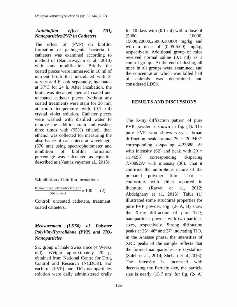
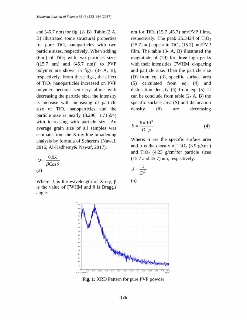
The X-ray diffraction pattern of pure
PVP powder is shown in fig. (1). The
pure PVP scan shows very a broad
diffraction peak around 2θ = 20.9402º
corresponding d-spacing 4.23888 Aº
with intensity (62) and peak with 2θ =
11.4695° corresponding d-spacing
7.70892Aᵒ with intensity (36). That it
confirms the amorphous nature of the
prepared polymer film. That is
conformity with either reported in
literature (Rawat et al., 2012;
Abdelghany et al., 2015). Table (1)
illustrated some structural properties for
pure PVP powder. Fig. (2- A, B) show
the X-ray diffraction of pure TiO2
nanoparticles powder with two particles
sizes, respectively. Strong diffraction
peaks at 25º, 48º and 37º indicating TiO2
in the Anatase phase, the intensities of
XRD peaks of the sample reflects that
the formed nanoparticles are crystalline
(Saleh et al., 2014; Shehap et al.,2016).
The intensity is increased with
decreasing the Particle size, the particle
size is nearly (15.7 nm) for fig. (2- A)
Malaysia Journal of Science 36 (3):132-144 (2017)
136
and (45.7 nm) for fig. (2- B). Table (2 A,
B) illustrated some structural properties
for pure TiO2 nanoparticles with two
particle sizes, respectively. When adding
(6ml) of TiO2 with two particles sizes
((15.7 nm) and (45.7 nm)) to PVP
polymer are shown in figs. (3- A, B),
respectively. From these figs., the effect
of TiO2 nanoparticles increased on PVP
polymer become semi-crystalline with
decreasing the particle size, the intensity
is increase with increasing of particle
size of TiO2 nanoparticles and the
particle size is nearly (8.296, 1.71554)
nm for TiO2 (15.7 ,45.7) nm/PVP films,
respectively. The peak 25.3424ͦ of TiO2
(15.7 nm) appear in TiO2 (15.7) nm/PVP
film. The table (3- A, B) illustrated the
magnitude of (2θ) for three high peaks
with their intensities, FWHM, d-spacing
and particle size. Then the particle size
(D) from eq. (3), specific surface area
(S) calculated from eq. (4) and
dislocation density (δ) from eq. (5). It
can be conclude from table (2- A, B) the
specific surface area (S) and dislocation
density (δ) are decreasing
with increasing with particle size. An
average grain size of all samples was
estimate from the X-ray line broadening
analysis by formula of Scherer's (Nawaf,
2016; Al-Kadhemy& Nawaf, 2017):
CosD
9.0
(3)
Where: λ is the wavelength of X-ray, β
is the value of FWHM and θ is Bragg's
angle.
DS
3106 (4)
Where: S are the specific surface area
and ρ is the density of TiO2 (3.9 g/cm3)
and TiO2 (4.23 g/cm3for particle sizes
(15.7 and 45.7) nm, respectively.
2
1
D
(5)
Fig. 1: XRD Pattern for pure PVP powder

Malaysia Journal of Science 36 (3):132-144 (2017)
137
2θ (degree) Intensity (counts) D( ̊A)
11.4695 36 7.70892
20.0826 52 4.41792
20.3418 52 4.36221
20.9402 63 4.23888
2θ(degree)
FWHM
(degree)
Intensity
(counts)
d (Aͦ)
hkl
D(nm)
Sx106
(m2.g
-1)
δx106 (m
-2)
25.3424 0.54100 285 3.51165 011 15.1 0.1018 4.385
36.9362 0.28000 16 2.43168 013 30.4 0.0505 1.082
37.8804 0.67000 50 2.37321 004 12.6 0.1220 6.298
38.7148 0.50000 12 2.32396 112 16.9 0.0910 3.501
48.0716 0.59500 77 1.89120 020 14.7 0.1046 4.627
53.9815 0.75000 39 1.69727 015 11.9 0.1292 7.061
55.0311 0.73000 39 1.66735 121 12.2 0.1260 6.718
62.0836 0.40000 12 1.49380 123 23.4 0.0657 1.826
62.7034 0.76000 28 1.48052 024 12.3 0.1250 6.609
68.8616 0.76000 10 1.36237 116 12.7 0.1211 6.200
70.3212 0.64000 12 1.33763 220 15.2 0.1011 4.328
75.0799 0.84000 16 1.26421 125 11.9 0.1292 7.061
A B
Table 1: XRD Parameters for Pure PVP Powder
Table 2- A: XRD Parameters for Pure TiO2 (15.7 nm) Powder
Fig. (2) XRD Pattern for Pure TiO2 nanoparticles powder with two particles
sizes A-(15.7 nm) B-(45.7 nm)

Malaysia Journal of Science 36 (3):132-144 (2017)
138
Table 2- B: XRD Parameters for Pure TiO2 (45.7 nm) Powder
2θ
(degree)
FWHM
(degree)
Intensity
(counts)
D
(Aͦ)
hkl D
(nm)
Sx106
(m2.g
-1)
δx105
(m-2
)
25.3712 0.21100 759 3.50773 011 39.5 0.0358 6.409
37.0077 0.19670 46 2.42715 013 43.0 0.0329 5.408
37.8515 0.2010ẟ0 172 2.37496 004 41.9 0.0338 5.696
38.6286 0.18750 48 2.32895 112 45.9 0.0308 4.746
48.0967 0.19930 253 1.89027 020 44.6 0.0317 5.0272
53.9434 0.20180 159 1.69838 015 44.4 0.0319 5.0726
55.1201 0.21570 147 1.66487 121 42.2 0.0336 5.6153
62.1691 0.17900 25 1.49196 123 52.2 0.0271 3.6699
62.7443 0.20810 119 1.47965 024 45.1 0.0314 4.9163
68.8009 0.21860 45 1.36343 116 44.2 0.0320 5.1186
70.3422 0.19800 55 1.33728 220 49.9 0.0284 4.0160
75.0910 0.214200 85 1.26405 125 47.3 0.0299 4.4696
2θ(deg) FWHM(deg) Intensity(counts) d (̊Aͦ) D(nm)
8.9490 1.36 35 9.87369 5.862
10.4163 2.76 72 8.48589 2.891
10.9527 3.18 69 8.07147 2.510
24.9112 0.64 21 3.57145 12.838
A B
Fig. 3: XRD Pattern for A- TiO2 (15.7 nm)/PVP and B- TiO2 (45.7nm)/PVP Films
TiO2
Table 3- A: XRD Parameters for TiO2 (15.7 nm)/PVP Film

Malaysia Journal of Science 36 (3):132-144 (2017)
139
2θ(deg) FWHM(deg) Intensity(counts) d (̊Aͦ) D(nm)
19.3648 4.7 261 4.58003 1.71554
20.5811 0.000 220 4.31203 0.00000
21.8777 0.000 150 4.05931 0.00000
Reduction of S. aureus and E. coli
growth were tested by using PVP (Pure)
and TiO2 nanoparticle (45.7nm)/PVP,
TiO2 nanoparticle (15.7nm)/PVP films
on plastic and glass plates. The best
results about reduction growth of S.
aureus were obtained by PVP (Pure)
film on glass plate reached to (62.5%)
and TiO2 (15.7nm)/PVP film on plastic
reached to (89.42%). And the best
results for reduction growth of E.coli
were observed for TiO2 (45.7nm)/PVP
film reached to (59%) and TiO2
(15.7nm)/PVP film reached to (100%)
on plastic, while the best reduction for
PVP(Pure) film on glass reached to
(51%) (Table4).
Bacterial isolate
Treatment
Type of plates
Reduction of growth (%)
Staphylococcus aureus
PVP ( Pure )
Plastic
60.89
PVP+TiO2(45.7nm) 55.1
PVP+TiO2(15.7nm) 89.42
PVP ( Pure )
Glass
62.5
PVP+TiO2(45.7nm) 69.55
PVP+TiO2(15.7nm) 86
Escherichia coli
PVP ( Pure )
Plastic
49
PVP+TiO2(45.7nm) 59
PVP+TiO2(15.7nm) 100
PVP ( Pure )
Glass
51
PVP+TiO2(45.7nm) 49
PVP+TiO2(15.7nm) 69
Table 5 illustrated the results for
antibiofilm effect of TiO2 (15.7
nm)/PVP nanocomposite was studied
against S. aureus and E. coli on catheters
for different storage time. Results
showed that one week of storage of
coated catheters the biofilm inhibition
ratio was (29%), after two weeks of
storage the inhibition ratio (42.6%), and
after the three weeks the inhibition ratio
reached to (56.21%). The best inhibitory
effect obtained after four weeks of
storage with inhibition ratio
(83.97%).The results for inhibition of
biofilm for E. coli reached to (22%) after
one week, while two weeks of storage of
catheters the inhibition ratio (30.9%),
and after the three week of storage
reached to (39.87%) and (65.3%) after
three and four weeks, respectively.
Table 4: Reduction of bacterial growth for PVP and TiO2 nanoparticles/PVP films
Table 3- B: XRD Parameters for TiO2 (45.7 nm)/PVP Film

Malaysia Journal of Science 36 (3):132-144 (2017)
140
Bacterial isolate
Times
Optical Density (O.D)
Inhibition biofilm
formation (%)
Control Coated
Catheter
Staphylococcus aureus
(1) Week 0.234 0.167 29
(2) Week 1.511 0.867 42.6
(3) Week 1.158 0.507 56.21
(4) Week 1.479 0.237 83.97
Escherichia coli
(1) Week 0.157 0.122 22
(2) Week 0.440 0.304 30.9
(3) Week 1.134 0.455 39.87
(4) Week 1.222 0.424 65.3
The antibacterial activity is increase with
decreasing the particle size of TiO2
nanoparticles with increasing the
specific surface area and dislocation
density. PVP is use as stabilizers; it has
optical purity that authorizes the
exploration of nanoparticle formation.
PVP acts as a copping agent and the
antimicrobial activity of PVP caused
modification of nanoparticles, the
polymer is most effective agent in the
particles stabilization against
aggregation (Jayaprakash et al., 2015).
The metal oxides carry the positive
charge while the bacteria carry negative
charges; this causing electromagnetic
attraction between bacterial surface and
the metal oxides that caused oxidization
and death of bacteria (Zhand& Chen,
2009). They cause holes in the cell walls
of bacteria, increasing permeability and
death of cell (Holt & Bard, 2005). The
opposite charges of nanoparticles and
bacteria are attributing to their
bioactivity and adhesion due to
electrostatic forces. Nanoparticles have
larger surface area, which enhances
bactericidal activity than the large size
particles; they realize cytotoxicity to the
bacteria (Bhupendra et al., 2009).
Fungicidal and bactericidal effects of
TiO2 on Pseudomonas aeruginosa, E.
coli, Salmonella choleraesuis, Vibrio
parahaemolyticus,
Listeriamonocytogenes,S.aureus,Diaport
heactinidiae and Penicilliumexpansum
have reported. The development of
TiO2-coated or incorporated packaging
of food and equipment of food preparing
has also interest. (Chaweng kijwanich &
Hayata, 2008), concluded that the TiO2
coated film could reduce the bacterial
contamination on the surface of food
products and reduce the risks of bacterial
Table 5: Inhibition of Biofilm Formation of TiO2 (15.7 nm)/PVP against S.aureus and E.coli
on Catheter after different times

Malaysia Journal of Science 36 (3):132-144 (2017)
141
growth on fresh-cut products. Inhibition
activity of metallic nanoparticles on
biofilm formation of bacteria has been
importance, as the device-related
infections that cause of morbidity and
mortality in hospitalized patients (Del-
pozo et al., 2009). TiO2 nanoparticle had
inhibitory effect on biofilm formation of
multidrug resistant bacteria (Ibrahem et
al., 2014). (Haghighiet al.2013) showed
that TiO2 nanoparticles could kill
Candida albicans and inhibit the
formation of biofilm. Different shape
and size of TiO2 nanoparticles can be
used for the photo catalytic treatment of
aqueous biofilm, pathogenic bacteria and
multi drug resistant bacteria. Maurer-
Jones et al. (2013) observed, significant
changes in bacterial biofilm after
treatment with TiO2nanoparticles,
nanoparticles caused altered gene
expression relating to growth and
biofilm formation. TiO2 nanoparticles
leads to larger reduction of bacterial
biofilm formation in the glass surface
(Chorianopoulos et al., 2010). The TiO2
nanoparticles efficiently inhibited
bacterial adhesion to acrylic surfaces as
well as have strong antibacterial effect in
the planktonic stage and biofilm
formation (Bahador et al., 2014).
Measurement (LD50) of (PVP) Polymer
and TiO2 Nanoparticles.
Toxicity of (PVP) and TiO2
nanoparticles was detect by
determination the dose that cause death
of 50% of laboratory animals. Results
showed that no effect of (PVP) and TiO2
nanoparticles on the laboratory animals,
LD50 was (> 2000 and> 5) mg\Kg,
respectively.
CONCLUSION
The effect of TiO2 nanoparticles on
crystal structure of PVP polymer has
investigated by x-ray diffraction. There
is some peaks fromTiO2 appeared into
structure of polymer. The TiO2
nanoparticles/ PVP films have
antibacterial effect against bacteria in
plastic and glass plates. In addition, it
has antibiofilm effect in catheters.
REFERENCES
Abdelghany A.M., Meikhail M.S.,
Abdellrazek E.M. &Abond M.M.
(2015). Spectroscopic inquest of
CdS, PbS and ZnS Doped PVP
composite: A Density Functional
Theory Approach.Research
Journal of Pharmaceutical,
Biological and Chemical
Sciences, 6(3): 1686-1697.
Alivisatos A. P. (1996).
SemiconductorClustors,
Nanocrystals and Quantum
Dots.Science. 271(5251): 933-
937.
Al-Kadhemy M. F. H., Hussein R., Al-
Zuky A. A. D.(2012).Analysis of
absorption spectra of styrene-
butadiene in toluene, journal of
physical science, 23(1):1-12.
Al-Kadhemy M. F. H., Nawaf S.
H.(2017). Nonlinear and linear
optical properties of Eosin B
dye- AgNO3-
Malaysia Journal of Science 36 (3):132-144 (2017)
142
Polyvinylprrolidone films,
Material focus ,6(1): 54-62.
Basf, (2009). PVP and more …
Luvitec, Luvicross and Collacral,
Brochure.
Bahador A., Ghorbanzadeh R.
&Kassaee M.Z. (2014). Anti-
microbial Activity of Acrylic
Resins with In-Situ Generated
Nanosilveron Cariogenic
Planktonic and Biofilm
Bacteria.Int. Res. J. Biological
Sci. 3(4): 38-46.
Bhupendra Ch., AnJana K. V., Nidhi A.,
Upadhyay R.V.,& Mehta R.V.
(2009). Enhanced Antibacterial
activity of biofunctional Fe3O4-
Ag Core-Shell nanostructures.
Nano Res. 2: 955-965.
Chawenqkijwaich C. &Hayata Y.(2008).
Development of TiO2 powder-
coated food packing film and its
ability to inactivate Escherichia
coli in vitro and in actual tests.
Int. J. Food Microbiol. 123(3):
288-92.
Chorianopoulos N.G., Tsoukleris D.S.,
Panagou E.Z., Falaras P.
&Nychas G. (2010). Use of
titanium dioxide (TiO2)
photocatalysts as alternative
means for Listeria
monocytogenes biofilm
disinfection in food processing.
Food Microbiol. 28: 164-170.
Chowhan, Z.T., Amaro, A.A., &Ong,
J.T.H., (1992). Punch Geometry
and Formulation Considerations
in Reducing Tablet Friability and
Their Effect on in vitro
Dissolution. J. Pharm. Sci. 81:
290-294.
Del-pozo J.,Crumlish M., Ferguson
H.M. & Turnbull J.F. (2009).
Aretrospective Cross- Sectional
Study on Candidatusarthromitus
associated rainbow trout
gastroeuterities (RTGE) in the
UK. Aquacultrue 290: 22-27.
Fujishima A. & Honda K., (1972).
Electrochemical Photolysis of
Waterata Semiconductor
Electrode.Nature 238: 37.
Ghosh, S., Upadhay,A., Singh,A. &
Kumar, A. (2010). Investigation
of antimicrobial activity of silver
nano particle loaded cotton
fabrics, which may promote
wound healing. International
Journal of Pharma and Bio
Sciences.1 (3):1-10.
Haghighi Mood S, Golfeshan
AH,Tabatabaei M,
SalehiJouzaniGh, NajafiGh,
Gholami M,& Ardjmand M.
(2013). Lignocellulosic biomass
to bioethanol; a comprehensive
review on pretreatment. Renew
Sust. Energ. Rev. 27: 77–93.
Holt K.B.& Bard A. J. (2005)Interaction
of silver (1) ions with the
respiratory chain of Escherichia
coli:an electrochemical and
scanning electrochemical
microscopy of micro molar Ag.
Biochemistry. 44(39):13214-23.
Ibrahem K.H., Salman J.A.S & Ali, F.A.
(2014). Effect of Titanium
Nanoparticles Biosynthesis by
Laactobacilllus Crispatus on
Urease, Hemolysin and Biofilm
Forming by Some Bacteria
Causing Recurrent UTI in Iraqi
Women. European Scientific
Journal 10(9): 324-338.
Malaysia Journal of Science 36 (3):132-144 (2017)
143
Jayaprakash, N., Vijaya,J.J. &Kennedy,
L.J. (2015). Microwave-Assisted
Rapid Facile Synthesis,
Characterization, and Their
Antibacterial Activity of PVP
Capped Silver Nanospheres,
Synthesis and Reactivity.
Inorganic, Metal-Organic, and
Nano-Metal Chemistry. 45(10):
1533-1538. Jun, Y.B., Min, B.H.,
Kim, S.I., Kim& Y.I.J. (1989).
Preparation and Evaluation of
Acetaminophen Tablets. Kor.
Pharm. Sci. 19: 123-128.
Kadajji V. G. &Betageri G. V.
(2011).Water Soluble Polymers
for Pharmaceutical
Applications.Polymers 3: 1972-
2009.
Maurer-Jones M.A., Gunsolus I.L.,
Murphy C.J.& Haynes C.L.
(2013). Toxicity of engineered
nanoparticles in the
environment. Anal Chem.85:
3036–3049.
Nawaf S. H. (2016). Physical
andNonlinear Optical Properties
of PVP Polymer doped by Silver
Nitrate and Nano Silver –Eosin
B, M.S.C. Thesis, College of
Science, Al-Mustansiriyah
University.
Namasivayam, S.K.R. and Roy, E.
A,(2013). Enhanced Antibiofilm
Activity of Chitosan Stabilized
Chemogenic Silver Nanoparticles
Against Escherichia coli Int. J. of
Sci. and Res.Publications. 3(4),
1-9.
Rawat A., Mahavar H.K., Chauhan
S.,Tanwar A. & Singh P.J.
(2012). Optical band gap of
polyvinyl pyrrolidone/
polyacrilamide blend thin films.
Indian Journal of pure & Applied
Physics 50: 100-104.
Sinchalpanid, N.& Mitrevej, A. (1993).
Comparative Evaluation of
Hydroxypropyl Cellulose and
Povidone in Paracetamol Tablet
Formulations. Mahidol J. Pharm.
Sci. 20: 33-39.
Saleh A.F., Jaffar A.M.,Samoom N.A. &
Mahmmod M. W. (2014). Effect
Adding PVA Polymer on
Structural and Optical Properties
of TiO2 Thin Films. Journal of
Al-Nahrain Sci. 17 (2): 116-121.
Salman, J.A.S., Al Kadhemy, M.F.H.,
Jaleel, M.S. &Abdal, A.KH.
(2014). Effect of PVA,
PVA/Biosurfactant on Some
Pathogenic Bacteria in Glass and
Plastic Plates. International
Journal of Current Microbiology
and Applied Science. 3(10): 301-
309.
Shehap A.M. &Akil D.S. (2016).
Structural and optical properties
of TiO2 nanoparticles/PVA for
different composites thin films
Int. J. Nanoelectronics and
Materials. 9:17-36.
Wong M.S, Hsu S.W, Rao K.K&
KumarC.P. (2008).Influence of
crystallinity and carbon content
on visible light photocatalysis of
carbon doped titania thin
films.Journal of Molecular
Catalysis A: Chemical. 279 (1):
20-26.
Zhand. H& Chen G.P. (2009).
Antibacterial activities of
Ag/TiO2nanocomposite powders
synthesized by a one-potsol-gel-
method. Envirron sci. Technol.
34(8): 2905-1

Malaysia Journal of Science 36 (3):132-144 (2017)
144