present summary of - aapm
TRANSCRIPT

1
Mark J. Rivard, Ph.D.
on behalf of the Working Group authors:
Mark Rivard,1 Wayne Butler,2 Larry DeWerd,3 M Saiful Huq,4 Geoffrey Ibbott,5
Christopher Melhus,1 Michael Mitch,6 Ravinder Nath,7 Jeffrey Williamson 8
1 Department of Radiation Oncology, Tufts-New England Medical Center, Boston, MA2 Schiffler Cancer Center, Wheeling Hospital,Wheeling WV3 Radiation Calibration Laboratory, University of Wisconsin, Madison, WI4 Department of Radiation Oncology, University of Pittsburgh Medical Center, Pittsburgh, PA5 Radiological Physics Center, University of Texas, MD Anderson Cancer Center, Houston, TX6 Ionizing Radiation Division, National Institute of Standards and Technology, Gaithersburg, MD7 Department of Therapeutic Radiology, Yale University School of Medicine, New Haven, CT8 Department of Radiation Oncology, Virginia Commonwealth University, Richmond, VA
AAPM TG-43 Update for 2004 and BeyondDr. Rivard has received research funds from the
following brachytherapy source manufacturers:Implant Sciences Corporation
IsoRay Medical, Inc.Mentor Corporation
North American ScientificNucletron Corporation
Theragenics CorporationXoft, Inc.
other Working Group members may also serve asmanufacturer consultants - see AAPM COI website
Ful l Disclosure
Purpose
Present summary of:
– AAPM TG-43U1 brachyt herapy dosimetry protocol,
– fut ure supplements, and
– possibl e future project s.
2004 AAPM TG-43U1 Report
• high level perspective of brachytherapy in U.S.• dosimetry formalism and clinical datasets• revised dosimetry formalism
– 2-D,1-D, and air kerma strength definition• consensus data formulation• clinical implementation recommendations• clarify interpolation / extrapolation methods• recommendations to dosimetry investigators
– experimental measurements & Monte Carlo calculations• formalism errata and published comments

2
Brachytherap y Backg rou nd
1898: Marie Cur ie disco vered radioactivity (210Po),and later 226Ra
1903: awarde d Nobel Prize, same year thatAlexander Graham Bell propos ed brach ythe rapy
226Ra brach ythe rapy domin ated for next 60 years
post -1950s reactor developm ent pro duce d man-made radionuclide s: 60Co, 137Cs, 192Ir, 198Au, etc.
currently wide vari ety of sourc e manufac turers ,radionuclide s, and brach yth erapy source type s CT and radi ograph of clinic al LDR 125I impla nt
Permanent LDR Brac hytherapy
AAPM Comm ittee Structure 101
AAPM BoDRadiation SafetyTherapy ResearchCalibrationsQuality AssuranceTherapy ImagingTreatment PlanningTreatment DeliveryBrachytherapy
Science CouncilImaging Therapy
ASTRO, ABS, ESTRO, NRC, FDA, etc
LIBC LEBD HEBD EBM BSRButler Rivard Li Thomadsen Ibbott
Brachytherap y Data Coordinat ion
AAPM
RPC / NIST / ADCLs
clinics
vendors
patients

3
Purpose of the Revised Protoco l
The goals of the revised protocol (TG-43U1) were:
(a) provide a revised definition of air-kerma strength;
(b) eliminate apparent activity for specification of source strength;
(c) eliminate the anisotropy constant in favor of the distancedependent 1-D anisotropy function;
(d) provide guidance on extrapolating tabulated TG-43 parametersto longer and shorter distances; and
(e) eliminate minor inconsistencies and omissions in the originalprotocol and its implementation.
Brach ytherapy Seeds
realistic modeling of internal components
NASI LDR 125I Source SK Measur ement Requireme nts
accidental manufacture of sources with zero activityor twice the activity
may not trust 3rd party (nuclear pharmacy) assay
measure all sources preceding clinical use
also use film for autoradiograph
required by AAPM TG-40 and TG-56 reports

4
NIST WAFAC NIST WAFAC
Brachytherap y Calculation Geometry Revised AAPM TG-43Brachytherapy Dosim etry Formalism (2-D)
dose rate to water at point P(r,θ)SK air kerma strength
Λ dose rate constantgL(r) radial dose functionGL(r,θ) geometry function (line source approximation)F(r,θ) 2-D anisotropy function
( ) ( )( ) ( ) ( )L
K LL 0 0
G r,D r, S g r F r ,
G r ,
θθ Λ θ
θ
⋅= ⋅ ⋅ ⋅ ⋅
( )D r,θ⋅

5
dose rate to water at point P(r)air kerma strength
Λ dose rate constantgL(r) radial dose function
GL(r,θ) geometry function (line source approximation)1-D anisotropy function
0
0 0
( , )(r) ( )
( , )L
K L anL
G rD(r)= S g r
G r
θ φθ
⋅ Λ ⋅ ⋅ ⋅�
KSD(r)�
( )an rφ
Revised AAPM TG-43Brachythe rapy Dosimetry Formalism (1-D)
air kerma strengthair kerma rate in vacuo at specification
point d with energy cutoff δ, typically 5 keV– low-energy photon cutoff now included, and– measurement conditions are now specified
( )K dδ�
2( )KS K d dδ≡ �
KS
Revised Ai r Kerma Strengt h Definitio n
Manufacturer and source type CONSENSUSΛ[cGy h-1 U-1]
% difference in Λfrom 1999 value
Amersham 6702 125I 1.036 N/AAmersham 6711 125I 0.965 N/ABest Industries 2301 125I 1.018 +3.3%NASI MED3631-A/M 125I 1.036 +1.0%Bebig/Theragenics 125.SO6 125I 1.012 +2.2%Imagyn IS-12501 125I 0.940 +3.5%Theragenics 200 103Pd 0.686 +4.0%NASI MED3633 103Pd 0.693 +4.3%
BAD
BAD
GOOD
BEST
Comparis on of 1-D Formalisms
0
0 0
( , )(r) ( )
( , )L
K L anL
G rD(r)= S g r
G r
θ φθ
⋅Λ ⋅ ⋅ ⋅�
( ) ( )2
0K P an
rD r S g r ( r )
rΛ φ
⋅ = ⋅ ⋅ ⋅ ⋅
0P
0 0
( , )g (r) ( )
( , )L
K anL
G rD(r)= S r
G r
θ φθ
⋅ Λ ⋅ ⋅ ⋅�
( ) ( )2
0K L an
rD r S g r ( r )
rΛ φ
⋅ = ⋅ ⋅ ⋅ ⋅
• comparisons of all candidate datasets
• average MCΛ and average EXPΛ from literature
CONΛ = (MCΛ + EXPΛ)
2
• g(r) and F(r,θ) candidate datasets transformedusing common L, possibly with Leff = ∆S × N
• g(r) and F(r,θ) typically taken from Monte Carlo
• φan(r) calculated from consensus F(r,θ) dataset
• final results tabulated with common mesh
Conse nsus Dataset Formula tion Methodology

6
Consensu s Dataset Formulation Methodology
• Literature review of experimental methods & MonteCarlo dosimetry characterization of 8brachytherapy seeds (2004 AAPM TG-43U1)– Amersham / Oncura model 6733 125I
– DraxImage model LS-1 125I
– Implant Science model 3500 125I
– IBt 1251L 125I
– IsoAid model IAI-125 125I
– Mentor model SL-125/SH-125.SO6 125I
– SourceTech Medical STM1251 125I
– Best Medical model 2335 103Pd
Conse nsus Dataset Formula tion Methodology
• Literature review of experimental methods & MonteCarlo dosimetry results for 8 brachytherapy seeds:
– Amersham Health models 6702 and 6711 125I
– Bebig/Theragenics Corporation model I25.SO6 125I
– Best Industries model 2301 125I
– Imagyn Medical Technologies model IS-12501 125I
– North American Scientific model MED3631-A/M 125I
– Theragenics Corporation model 200 103Pd
– North American Scientific model MED3633 103Pd
Clinic al Impl ementation Recommendat ions
• know your Bx TxP algorithm, deal with limitations
• acceptance testing and commissioning– follow AAPM TG-40, TG-53, & TG-56 recommendations
– compare/validate with Eq.(10) reference dose rates
• well chamber ADCL calibrations, NIST traceability
r (cm)Amersham
6702Amersham
6711Best2301
NASIMED3631-A/M
BebigI25.SO6
ImagynIS-12501
Theragenics200
NASIMED3633
0.5 4.119 3.937 3.978 4.112 3.922 3.426 3.014 3.1841.0 0.995 0.911 1.004 0.986 0.950 0.815 0.587 0.6261.5 0.413 0.368 0.419 0.420 0.398 0.334 0.199 0.2152.0 0.213 0.186 0.217 0.207 0.205 0.169 0.0837 0.09143.0 0.0768 0.0643 0.0783 0.0746 0.0733 0.0582 0.0206 0.02274.0 0.0344 0.0284 0.0347 0.0325 0.0323 0.0246 0.00634 0.006975.0 0.0169 0.0134 0.0171 0.0157 0.0157 0.0118 0.00221 0.002476.0 0.00890 0.00688 0.00908 0.00811 0.00840 0.00592 0.000846 0.0009337.0 0.00490 0.00373 0.00506 0.00429 0.00459 0.00328 0.000342 0.000364
• g(r) presented for dimensionless units, consistencywith investigator g(r), and 5th order polynomial
• explicit contraindication for erroneous 1-D equation
• goodbye Aapp and anisotropy constant
• methodology to extrapolate dose calculations forlarge and small distances
0L
0 0
( , )g (r) ( )
( , )P
K anP
G rD(r)= S r
G r
θ φθ
Λ ⋅ ⋅ ⋅�
Corre ction of Errors and Inconsistencies

7
• apparent activity: Aapp
– choice of (Γδ)x may lead to dosimetric errors
– AAPM solely specifies SK for calibration standard
• anisotropy constant: φan
– not able to accurately reproduce dosimetry data r < 1 cm
– changes may be made to minimize error, but can lead tosignificant errors under specific circumstances
Removal of Previou sly Defined Terms
• NIST-specified source spectra, half-lives, ρ andatomic composition for both air and water
Reference Data
MBq–1MBq–1
Need for Uncertainty Analyses I-125 g(r), Variable ρρρρ and Compo sit ion
0.0001
0.001
0.01
0.1
1
0 5 10 15 20 25 30radius [cm]
125
Irad
iald
ose
func
tion,
g(r)
water, p=1.00tissue, p=1.00tissue, p=1.05tissue, p=1.15tissue, p=1.25tissue, p=1.50

8
Need for Extrapolat ion Methodology
0.0001
0.001
0.01
0.1
1
0 5 10 15 20 25 30radius [cm]
125
Irad
iald
ose
func
tion,
g(r)
water, p=1.00tissue, p=1.00tissue, p=1.05tissue, p=1.15tissue, p=1.25tissue, p=1.50
0.0001
0.001
0.01
0.1
1
0 5 10 15 20 25 30radius [cm]
103 P
dra
dial
dose
func
tion,
g(r)
water, p=1.00tissue, p=1.00tissue, p=1.05tissue, p=1.15tissue, p=1.25tissue, p=1.50
Pd-103 g(r), Variable ρρρρ and Composit ion
0.0001
0.001
0.01
0.1
1
0 5 10 15 20 25 30radius [cm]
103 P
dra
dial
dose
func
tion,
g(r)
water, p=1.00tissue, p=1.00tissue, p=1.05tissue, p=1.15tissue, p=1.25tissue, p=1.50
Need Extrapol ation Metho dology
• experimental measurement descriptors– description of internal and external source geometry– source irradiation geometry, orientation, irradiation timeline– detector calib. technique & energy response function, E(r)– radiation detector and readout system– measurement phantom– phantom dimensions and use of backscatter– estimation volume averaging effect at all detector positions– # of repeated readings with standard deviation, # of sources– NIST SK value and uncertainty for measured source– uncertainty analysis section (statistical and systematic)
Recommendations to Dos imetry Investiga tors
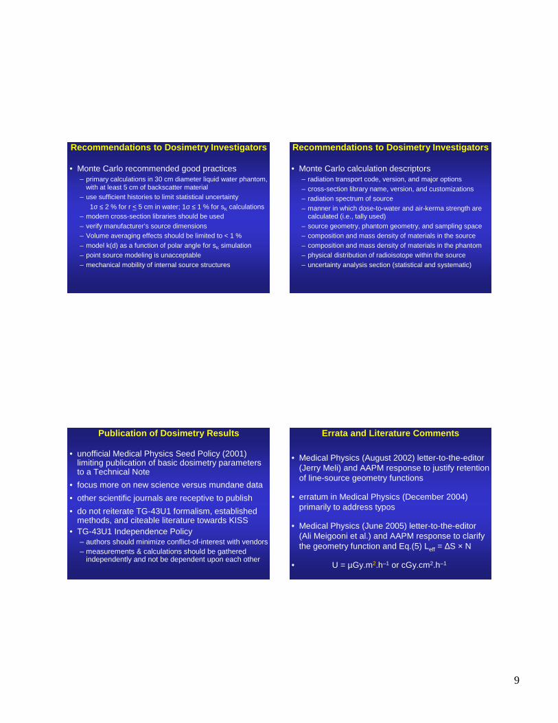
9
• Monte Carlo recommended good practices– primary calculations in 30 cm diameter liquid water phantom,
with at least 5 cm of backscatter material– use sufficient histories to limit statistical uncertainty
1σ ≤ 2 % for r < 5 cm in water; 1σ ≤ 1 % for sK calculations– modern cross-section libraries should be used– verify manufacturer’s source dimensions– Volume averaging effects should be limited to < 1 %– model k(d) as a function of polar angle for sK simulation– point source modeling is unacceptable– mechanical mobility of internal source structures
Recommendations to Dosimetry Investiga tors
• Monte Carlo calculation descriptors– radiation transport code, version, and major options– cross-section library name, version, and customizations– radiation spectrum of source– manner in which dose-to-water and air-kerma strength are
calculated (i.e., tally used)– source geometry, phantom geometry, and sampling space– composition and mass density of materials in the source– composition and mass density of materials in the phantom– physical distribution of radioisotope within the source– uncertainty analysis section (statistical and systematic)
Recommendations to Dos imetry Investiga tors
• unofficial Medical Physics Seed Policy (2001)limiting publication of basic dosimetry parametersto a Technical Note
• focus more on new science versus mundane data
• other scientific journals are receptive to publish
• do not reiterate TG-43U1 formalism, establishedmethods, and citeable literature towards KISS
• TG-43U1 Independence Policy– authors should minimize conflict-of-interest with vendors– measurements & calculations should be gathered
independently and not be dependent upon each other
Publication of Dosimetry Results Errata and Li terature Commen ts
• Medical Physics (August 2002) letter-to-the-editor(Jerry Meli) and AAPM response to justify retentionof line-source geometry functions
• erratum in Medical Physics (December 2004)primarily to address typos
• Medical Physics (June 2005) letter-to-the-editor(Ali Meigooni et al.) and AAPM response to clarifythe geometry function and Eq.(5) Leff = ∆S × N
• U = µGy.m2.h–1 or cGy.cm2.h–1

10
1.E-04
1.E-03
1.E-02
1.E-01
1.E+00
1.E+01
1.E+02
1.E+03
0.0 1.0 2.0 3.0 4.0 5.0 6.0 7.0 8.0
Radius (cm)D
ose
Rat
e(c
Gy/
min
)
50 kV
50 kV MCNP5
Pd 103
I 125
Ir 192
similar dose rates to 10 Ci HDR 192Ir over region of interest
(Gy/
min
ute)
Depth Dose Charact eristi cs
Free Air Atti x Ioni zation Chamber
Pushrod
Guard Tube
SteppingMotor ControlledSlideAssembly#1
Stepping Motor Controlled SlideAssembly#2
CollectingElectrode
Variable PositionPlate
Fixed CenterPosition
High VoltageBias
EntranceWindow
UW-Madison ADCL
Beam DefiningAperture
SensitiveCollectingVolume
Exit WindowX-raybeam
Signal
Free Air Atti x Chamber Schematic

11
252Cf Brachythe rapy ? Radial Dose Functio ns, g(r)
Conclusion
• need to supplement consensus data from theAAPM TG-43U1 (2004) report
• consensus datasets for 8+8 brachytherapysources are provided for consistent clinical use
• guidance provided to physicists and RTP softwarevendors for interpolation / extrapolation methods
• draft report is under review at AAPM, results to bepublished in Medical Physics
• forthcoming supplement for additional sources
• need to advance the field through better algorithms