presents - wpta.org evaluation, treatment and...michael karegeannes, pt, mhsc, lat, mtc, cfc, cctt,...
TRANSCRIPT

Presents
The Evaluation Treatment and Management of Temporomandibular Disorders Craniofacial Pain
And Orofacial Pain
April 20 2017
By Michael Karegeannes
PTMHScLATMTCCFCCCTTCMTPT
Jeff Verhagen PTMBACMTPT
Mike Verplancke DPTCSCSCMTPT
Freedomptcom Treatingtmjcom
Michael Karegeannes PT MHSc LAT MTC CFC CCTT CMTPT is the owner of Freedom Physical Therapy Services 4 clinics in WI since 1997 and a practicing physical therapist since 1989 His specialties in the field of physical therapy include TMD spine and pelvic dysfunctions myofascial pain and orthopedic therapies Michael graduated from the University of Wisconsin-Madison in 1989 with a Bachelorrsquos of Science Degree in Physical Therapy After he graduated he received his athletic training license from the University of Wisconsin-Milwaukee and later Michael completed his manual therapy certification and Masters of Health Science from the University of St Augustine in Florida
In 2005 Michael had the honor of being trained by Dr Mariano Rocabado (in conjunction with the University of St Augustine) who is the leading national expert in the field of craniofacial therapies Michael holds a Craniofacial Certification with the University of St Augustine
In addition Michael has attended an orofacial pain and TMD residency with the University of Minnesota Dental School He is a member of the American Academy of Orofacial Pain (AAOP) a board member with the Physical Therapy Board of Craniofacial and Cervical Therapeutics (PTBCCT) and is one of the few physical therapists in the United States recognized as a certified cervical and temporomandibular therapist with the AAOP
In 2011 Michael received thorough and extensive training in the technique of intramuscular dry needling and is certified through Myopain Seminars the premier post-graduate medical and physical therapy continuing education company in the United States with a focus on myofascial trigger points Michael is also on faculty with Myopain Seminars Michael is also one of the few PTs in the USA trained in the CRAFTA approach to TMD
His diverse experience knowledge and manual skills allow Michael to be highly effective in the evaluation and treatment of his clientele
Michael is a member of the APTA WPTA NATA AAOP PTBCCT AES PAMA and IMS
Jeffrey Verhagen started his career as a Physical Therapist in 1990 after graduating from the University of Wisconsin -Madison He furthered his breadth of knowledge by attending several post-graduate continuing education opportunities with a focus on manual therapy for orthopedic spine and sports injuries He soon learned that he enjoyed treating spine problems and patients with headaches
In order to properly assess the etiology of headaches he expanded his studies to include the evaluation and treatment of TMDcraniofacial pain and in 2013 completed a certification in Intramuscular Dry Needling through Myopain Seminars Dry Needling treats myofascial trigger points and their affect on localized and referred pain He soon learned that he could successfully treat TMD and craniofacial pain for patients that had tried other treatment approaches and still were left with pain and loss of function
Jeff has attended several courses and has reviewed the literature to expand his ability to successfully treat this challenging population Freedom Physical Therapy Services commissioned Steve Kraus PT a well known TMJ therapist and educator to come to the Milwaukee area and teach the latest treatment techniques and research on TMDcraniofacial pain to our therapy staff Jeff looks forward to continuing to expand his knowledge to better serve this patient population
Jeff has been with Freedom Physical Therapy Service since July of 2000 and currently serves as the Clinical Operations Administrator for the practice He earned his Master of Business Administration from Cardinal Stritch University in 1999 He also served 21 years in the Army Reserves and is a Desert Storm veteran
Jeff resides in Brookfield with his wife and two children and enjoys many hobbies to include golf fishing and reading
Michael Verplancke left his roots of St Louis Missouri to both attend and play ice hockey at St Maryrsquos University of Minnesota where Mike graduated in 1998 with a Bachelor of Arts degree in Biology He then attended Finch University of Health SciencesThe Chicago Medical School in North Chicago Illinois to obtain his physical therapy degree He graduated in 2001 with a Doctorate of Physical Therapy Mike has participated in numerous continuing education seminars with a focus on manual therapy techniques evaluation and treatment of the spine and TMJcraniofacial pain mobilization techniques and intramuscular dry needling
In 2002 Mike obtained his credentials as a Certified Strength and Conditioning Specialist (CSCS) which has assisted him with exercise prescription as well as progression of overall health and wellness for patients
In 2014 Mike completed his certification in Intramuscular Dry Needling through Myopain Seminars This treatment approach has provided a technique that improves treatment of Myofascial Trigger Points and their affect on localized and referred pain as well as muscle activation patterns throughout the body In addition Michael has attended an Orofacial Pain and TMD residency with the University of Minnesota Dental School
Mike has been trained in Temporal Mandibular Disorders (TMD) and Craniofacial Pain (CFP) by Dr Mariano Rocabado a world renowned leader in the field of TMD and CFP Mike is currently working towards his Craniofacial Certification in conjunction with Dr Rocabado and the University of St Augustine
Mike has been an employee at Freedom Physical Therapy since July of 2005 and is currently the Lead Physical Therapist and clinic director of the Grafton location
Mike has a wide range of hobbies and interests which include playing ice hockey softball golf and basketball He also enjoys spending quality time with his wife Marion their three daughters Sophia Liliana and Emilia and their Golden Retriever Stan
Green Bay WI April 20 2017 Michael Karegeannes PT wwwtreatingtmjcom 1
Time Ordered Agenda 4-20-2017
900 ndash 945am Introduction and TMJ Anatomy and TMJ Biomechanics
945 ndash 1030am Pathomechanics abnormal mechanics and parafunction etiology imaging
1030 ndash 1100pm Break
1100 ndash 1200pm Epidemiology Lab Evaluation of ROM of TMJ Lab Muscle Palpation Hypermobility Screen muscle treatment
1200 ndash 200pm Lunch
200 - 245 pm TMJ Arthralgia and various Disc related TMJ Dysfunction
245pm ndash 330pm Lab Mobilization Techniques for the TMJ
300 - 345pm Break
345 ndash 415pm TMJ Exercise Rocabado 6 x 6 self help
415 ndash 500pm Cervical spine as it relates to TMJ Ergonomics Lab C spine assessmenttreatment dry needling Demo to facial and selected neck muscles
500pm Adjourn Thank you
Green Bay WI April 20 2017 Michael Karegeannes PT wwwtreatingtmjcom 2
The Evaluation Treatment and Management of Temporomandibular
Disorders Craniofacial Pain and Orofacial Pain
By
Michael KaregeannesPTMHScLATMTCCFCCCTTCMTPT
CRANIO-CERVICO-MANDIBULAR RELATIONSHIP
Cranio-MandibularCranio-Vertebral
ASSIMILATION
Green Bay WI April 20 2017 Michael Karegeannes PT wwwtreatingtmjcom 3
Tragus of the ear
Anatomical Video Clip
The condyle (anterior view) The medial pole (MP) is more prominent that the lateral pole (LP)
Copyright copy2013 by Mosby an imprint of Elsevier Inc
(A) Lateral view and (B) diagram showing the anatomic
components RT retrodiscal tissue SRL superior retrodiscal lamina (elastic) IRL inferior retrodiscal
lamina (collagenous) ACL anterior capsular ligament (collagenous) SLP and ILP superior and inferior
lateral pterygoid muscles AS articular surface SC and IC
superior and inferior joint cavity the discal (collateral) ligament has
not been drawn
Courtesy of Per-Lennart Westeson MD Rochester NY
TMJ ( anterior or coronal view) AD articular disc CL capsular ligament LDL lateral discal ligament MDL medial discal ligament DC superior joint cavity IC inferior joint cavity
Medial and Lateral Discal Collateral Ligament
Green Bay WI April 20 2017 Michael Karegeannes PT wwwtreatingtmjcom 4
TMJ MechanicsDiscTMJ pain primarily originates from tissues in the posterior and lateral aspect of the TMJ ie capsule TMJ ligament Lateral collateral ligament synovium and retrodiscal tissue
Mandibular Biomechanics Osteokinematics
bull Depression (opening) ndash 40 to 50mm normal- 36 mm for most dental procedures
bull Elevation (closing)bull Protrusion- 5 to 7 mm from incisor to incisorbull Retrusion to protrusion 10mm be specific and
consistentbull Lateral excursion ndash 10 mm
41 RatioFor every 1mm of lateral excursion 4mm of opening
TMJ AUTOPSY SPECIMEN(Normal Joint)
TMJ AUTOPSY SPECIMEN(Reducing Disc)
Green Bay WI April 20 2017 Michael Karegeannes PT wwwtreatingtmjcom 5
TMJ AUTOPSY SPECIMEN(Non-Reducing Disc)
Mandibular Opening Patterns
bull Deflection Movement away from midline but it does NOT return to midline
A man with reducing disc displacement of the right joint (a) On mouth opening there is an early transient locking and a slight deflection of the mandibular midline to the right affected side (b) When the condyle slides over the posterior edge of the disc there is a rapid exaggeration of the mandibular shift and the mandibular midline then returns to center At further mouth opening the mandibular movement is symmetrical (c) Lateral excursion to the contralateral side is impaired before the click but not necessarily after (d) Lateral excursion to the ipsilateral or affected side is typically normal (e) Protrusion there is a slight deflection of the mandible to the right affected side after the ldquoclickrdquo the mandible tends to track in the center
Mandibular Opening Patterns (cont)Deviation ldquoSrdquo Movement away from midline but
returns to midline as it gets to end range
Mandibular Opening Patterns (cont)
bull Midline Could be normal or could be bilateral Disc dislocation without reduction depends on
Mandibular ROM (LAB)
Will Cover In Lab
Hypermobility Screen
Green Bay WI April 20 2017 Michael Karegeannes PT wwwtreatingtmjcom 6
Hypermobility
Beighton 9 Point Scoring System Revised 1998 Brighton Diagnostic Criteria For JHS
Chapter 2 Assessment of HypermobilityP Beighton et al Hypermobility of Joints
Springer-Verlag London Limited 2012
Rodney Grahame CBE MD FRCP Joint Hypermobility Syndrome Pain Current Pain and Headache Reports
2009 13427-433
Generalized hypermobility is one of the most important etiological factors in the development of craniomandibular disorders Professor Rocabado presented the following summary of etiological factors and
we can see that clenchingbruxism and mobility rank much higher than history of trauma or orthodontics
The importance of systemic hypermobility is evident when we consider the association between parafunction and hypermobility It has been found that 79 of patients with systemic hypermobility and
clenchinggrinding of teeth (or nail biting) go on to develop a TMJ problem A control with clenchinggrinding of teeth but without systemic hypermobility were found to have only 16 incidence The
implications seem to be that hypermobile individuals do not tolerate the added stress of parafunction So most patients have hypermobility and parafunction going hand in hand
Green Bay WI April 20 2017 Michael Karegeannes PT wwwtreatingtmjcom 7
CWP ndash Chronic Widespread Pain FM - Fibromyalgia CFS - Chronic Fatigue SyndromeJHS - Joint Hypermobility Syndrome EDS - Ehlers Danlos Syndrome MFS ndash Marfan Syndrome
This diagram illustrates that people with CWP CFS and FM can be hypermobile or may have JHS that JHS and EDS may present in similar ways and that the very complex systemic
problems of the bowel lungs heart and blood vessels are features of conditions such as EDS and MFS and not JHS
httphypermobilityorg
What other problems might a person with hypermobility have to suggest there is an underlying medical condition
The things individuals might most often present with beyond joint problems include
bull Easy bruising scarring that is stretched thin and often wrinkled andstretch marks that appeared at a young age and in many places across the body The skin often feels soft and velvety
bull Weakness of the abdominal and pelvic wall muscles that presents as hernias (such as hiatus hernia) or prolapse of the pelvic floor causing problems with bowel and bladder function
bull Unexplained chest pains ndash perhaps the individual has been told they have a heart murmur and mitral valve prolapse
bull Blackouts or near blackouts that may be associated with low bloodpressure or fast heart rate and often triggered by change in posture from lyingsitting to standing or after standing in one position for even just a few minutes httphypermobilityorg
bull Symptoms that sound like Irritable Bowel Syndrome with bloating constipation and cramp-like abdominal pain
bull Shortness of breath perhaps diagnosed as asthma because the symptoms seem the same but not responding to inhalers in the way the doctor might have expected because it is not true asthma
bull Noticing that local anesthetics used for example in dentistrydo not seem to be very effective or require much more than might be expected
bull Severe fatigue Anxiety and phobias
httphypermobilityorg
Cervical Spine Joint Hypermobility a possible predisposing factor for new daily
persistent headache TD Rozen JM Roth and N Deneberg Michigan Head-Pain and
Neurological Institute Ann Arbor MI USA
History
bull 1934 James Costen described a group ofsymptoms centering around the ear and TMJthe term Costen Syndrome was developed
bull While much of what Costen had suggested hasbeen disproved his interest certainly was acatalyst to foster more work andunderstanding in the area of TMD
Green Bay WI April 20 2017 Michael Karegeannes PT wwwtreatingtmjcom 8
Fast forward through various other terms over the years such as TMJ disturbances TMJ dysfunction syndrome Functional TMJ disturbances occlusomadibular disturbance myoarthropathy of the TMJ Craniomandibular disorders to what Bell in 1992 coined
ldquoTemporomandibular Disordersrdquo
ldquoTemporomandibular Disordersrdquo
The term does not merely suggest problems that are isolated to the TMJs but includes all disturbances associated with the function of the masticatory system rather than a single diagnosis
bull Arthrogenous
bull Myogenous
bull Atrhrogenous and Myogenous
The American Dental Association adopted the term TM disorders or Temporomandibular Disorders
In 1993 the AAOP collaborated with the International Headache Society (IHS) to integrate TMD into an already existing medical diagnostic classification system
TMDUncommon Classifications of TMDs
bull Ankylosis
bull Aplasia or hyperplasia
bull Pathology such as an infection fracture or neoplasm ( malignant or benign)
Common SymptomsSigns of TMD
bull Pain in the area of the TMJ and jaw muscles
bull Pain with mouth opening chew and or yawn
bull Joint sounds with jaw movements
bull Intermittent locking closed or open
bull Limited mouth opening
bull Headache
bull Earache or pain
Myogeneous
Masticatory Muscle Pain
Muscle Spasms ICD 10 M791ICD 9 72885
Contracture of muscle unspecified site ICD 10 M6240
Adhesions and ankylosis of temporomandibular joint M2661
Artrhogeneous
Arthralgia ICD 10 M2662ICD 9 52462
Primary osteoarthritis unspecified site M1991
Disc Displacements ICD 10 M2662ICD 9 52463
Common Classifications ICD 10 Coding of TMDs
Green Bay WI April 20 2017 Michael Karegeannes PT wwwtreatingtmjcom 9
other specified disorders of TMJ ICD 10 M2669
Dislocation of jaw initial encounter ICD10 S030XXA
Cervicalgia ICD 10 M542 ICD 9 7231
Myofascial Syndrome ICD 10 M791ICD 9 7291
Headache ICD 10 R51 ICD 9 7840
Chronic tension-type headache intractable G4422133912 CTTH
Treatment ndash Insurance Issues and TMD
Diagnosis
It is about 5050 as far as which insurances will
cover the diagnosis of TMJTMD
If they do sometimes small TMD cap applies (say
only $1250) others fall into same coverage for other
MS issues
Most patients have a combo of cervical and TMD
sxrsquos therefore it is not unreasonable to use a
cervical dx
For Medicare you will need a script from their
medical doctor not the dentist as Medicare does
not cover TMD
This is a graphic model depicting the relationship between various factors that are associated with the onset of TMD The model begins with a normally functioning masticatory system There are five(6) major etiologic factors that may be associated with TMD Whether these factors influence the onset of TMD is determined by the patientrsquos individual adaptability When the significance of these factors is minimal and adaptability is great the patient does not report any TMD symptoms
Per Rocabado must have
centric relation or balance of CV
joints
Craniovertebral Junction
Management of Temoromandibular Disorders and Occlusion by Jeff Okeson 7th edition 2013
Copyright copy2013 by Mosby an imprint of Elsevier Inc
In this graphic model the etiologic factor of occlusion is depicted as being significant (perhaps a newly placed poorly fitting crown) If this factor exceeds the patientrsquos adaptability TMD symptoms may now be reported by the
patient In this instance improvement of the occlusal condition (adjustment of the crown) would reduce this etiologic factor thereby bringing the patient within adaptability and thus resolving the TMD symptoms The same effect can be associated with any of the five etiologic factors and helps explain why data show that treatment of any of these factors may reduce symptoms
Craniovertebral Junction
Management of Temoromandibular Disorders and Occlusion by Jeff Okeson 7th edition 2013
Copyright copy2013 by Mosby an imprint of Elsevier Inc
This graphic model depicts the concept that some patients may present with less adaptability or a reduction in adaptability When this occurs the various etiologic factors that did not originally create symptoms may now lead to symptoms When
adaptability is very limited attempts at reducing any of the five factors may be ineffective
Craniovertebral Junction
Copyright copy2013 by Mosby an imprint of Elsevier Inc
Management of Temoromandibular Disorders and Occlusion by Jeff Okeson 7th edition 2013
Green Bay WI April 20 2017 Michael Karegeannes PT wwwtreatingtmjcom 10
Craniovertebral Junction
Managing this etiologic factorsMay no longer be adequate
This graphic model depicts a much more difficult management problem As symptoms become prolonged the pain condition can move from acute to chronic As pain becomes chronic the central nervous system can be altered making management more complicated Some of these alterations may involve the hypothalamus-pituitary-adrenal axis central sensitization andor a reduction in descending inhibitory control When this occurs more chronic pain conditions may develop which cannot be managed by addressing the five etiologic factors
Management of Temoromandibular Disorders and Occlusion by Jeff Okeson 7th edition 2013
Copyright copy2013 by Mosby an imprint of Elsevier Inc
Parafunctional Activity
Some clinical signs associated with parafunctional activity A Evidence of cheek biting during sleep B Here the lateral borders of the tongue are scalloped conforming to the lingual surfaces of mandibular teeth During sleep a combination of negative intraoral pressure and forcing of the tongue against the teeth produces this altered tongue shape This is a form of parafunctional activity
Copyright copy2013 by Mosby an imprint of Elsevier Inc
Management of Temoromandibular Disorders and Occlusion by Jeff Okeson 7th edition 2013
Masseter hyperplasia secondary to chronic bruxism
TMD Diagnostic Guidelines
bull 1992- Research Diagnostic criteria for TMDs
ndash Dworkin SF LeResche L Research Diagnostic Criteria for Temporomandibular Disorder 1992 6301-355
ndash RDC is a landmark paper providing operational definitions to distinguish TMD patients from controls and to diagnose with reasonable reliable and valid tests and measurements the most common subtypes of TMD
ndash httpwwwrdc-tmdinternationalorgHomeaspx
bull For over 20 years it has been the most widely used and heavily cited publication of diagnostic protocol in clinical and research settings
bull It was never intended to be a final document but rather a work in progress
bull The Journal of Oralfacial Pain 2010 volume 24 Issue 1 offers several articles validating and recommending updates to the 1992 RDC paper
Green Bay WI April 20 2017 Michael Karegeannes PT wwwtreatingtmjcom 11
The new DCTMD protocol is a necessary step toward the ultimate
goal of developing a mechanism and etiology based DCTMD that will more accurately direct clinicians in providing
personalized care for their patients
Steenks M De Wijer A Validity of the Research Diagnostic Criteria for Temporomandibular Disorders Axis in Clinical Research settings Journal of Oralfacial Pain 2009239-16
Summary of RDCTMD diagnostic Guidelines
A To diagnose all common subtypes of TMD involves taking a history and performing a physical examination that is reliable and valid
B Does NOT require
1 Electronic Equipment
a) Sonograph
b) EMG
c) Jaw Tracking Devices
2 Radiographs
-Radiographs are indicated if recent trauma red flags are present patient not responding to conservative care and surgery is being considered for disc replacement
Panoramic X-Raybull Is a two-dimensional dental x-ray that can show the
maxilla and mandible all the teeth including the wisdom teeth the frontal and maxillary sinuses the nasal cavity and the temporomandibular joint and other near by head and neck anatomy
bull Can determine bony changes of the condyle and fractures or severe dislocations of the condyle Does not image soft tissue so the position of the disc cannot be determined by this test
Green Bay WI April 20 2017 Michael Karegeannes PT wwwtreatingtmjcom 12
Computed Tomography
bull The latest advancement in this technology iscalled Cone Beam tomography
bull It allows for viewing the condyle in multipleplanes so that all surfaces can be visualized
bull This technology is capable of reconstructing3D images
bull Cone beam technology can image both hardand soft tissues but MRI GOLD standard forsoft tissue
Patient positioned in a cone beam CT scanner
Computed tomographic scan A A typical CT projection of the TMJ Hard tissue (bone) is visualized better than soft tissue with this technique B A three-
dimensional CT reconstruction of an edentulous mouth
From Wilkinson T Maryniuk G J Craniomandib Pract 137 1983
Green Bay WI April 20 2017 Michael Karegeannes PT wwwtreatingtmjcom 13
A three-dimensional image reconstructed from a cone beam image These three-dimensional images can be rotated on the computer screen so that the clinician can visualize the precise area of interest (Courtesy of Dr Allan Farmer and Dr William Scarf Louisville KY)
MRI
bull Gold Standard for evaluating the soft tissue of the TMJ especially disc position
bull Major advantage of not introducing radiation
bull Disadvantages include expensive not typically available in a dental setting quality of images may very from facility to facility
bull Cine or dynamic MRI on its way
Magnetic resonance image (MRI) A When the mouth is closed the articular disc (dark) is dislocated anterior to
the condyle (arrows)B During opening the disc is recaptured into its normal position on the condyle
Copyright copy2013 by Mosby an imprint of Elsevier Inc
The clinician should note that the presence of a displaced disc in an MRI does not constitute a
pathological finding IT has been demonstrated that between 26 and 38 of normal asymptomatic
subjects are found to have disc position abnormality on MRI Therefore the clinician should rely primarily on history and examination findings to establish the
diagnosis and use imaging information only as contributing data
Green Bay WI April 20 2017 Michael Karegeannes PT wwwtreatingtmjcom 14
This study suggests that disc displacement is relatively common (34) in asymptomatic volunteers and is highly
associated with patients (86) with TMD
Although there was a 33 prevalence of disc displacement in asymptomatic volunteers there was a
highly significant difference in the prevalence of internal derangement in symptomatic subjects Bruxing was
statistically linked to TMJ disc displacement and could explain the anatomic variation in abnormal disc position
In conclusion panoramic radiography had poor reliability and low sensitivity compared with CT for detecting TMJ-related osseous changes These findings suggest that this imaging modality has
limited utility for assessing the TMJ Magnetic resonance imaging had fair reliability and marginal sensitivity in diagnosing osseous
changes compared with CT Therefore MRI is not an ideal imaging technique for detecting osseous changes and CT remains the image
of choice for assessing osseous tissues Regarding soft tissue assessment MRI had excellent reliability for assessing disc position
and good reliability for detecting effusions Overall the criteria proposed in this study for image analysis covered all possible osseous and nonosseous conditions of TMJ in a large group of
participants in the multisite RDCTMD Validation Project17 The image analysis criteria presented in this paper are reliable for
diagnosing osseous and nonosseous components of TMJ using CT and MRI respectively We recommend that they be used in both
clinical and research settings
Sagittal CT views of condyles representing examples of nonosteoarthritic or indeterminateosseous changes observed A-B Rounded condylar head and well-defined cortical margin
C Rounded condylar head and well-defined noncortical margin D-E Indeterminate forOA slight flattening of anterior slope and well-defined cortical margin F Indeterminate forOA flattening of anterior slope and a pointed anterior tip that is not sclerosed well-defined
cortical margin fossa is shallow G Well-defined cortical margin has a notch on thesuperior part a deviation in form fossa is shallow H Narrowed appearance of the condylarhead near medial part close position of the cortical plates gives the impression of sclerosis
a nonosteoarthritic condyle
Axially corrected coronal CT views of condyles representing examples of osseous changes observed and corresponding osteoarthritis (OA) diagnoses A-B Nonosteoarthritic condyles rounded condylar head and well-defined cortical margin C Nonosteoarthritic condyle flattened superior margin and well-defined cortical margin D Nonosteoarthritic condyle flattened lateral slope and well-defined cortical margin E Indeterminate for OA rounded condylar head and subcortical sclerosis F Indeterminate for OA subcortical sclerosis G OA subcortical sclerosis surface erosion H-I OA surface erosion J OA generalized sclerosis and subcortical cysts K Nonosteoarthritic condyle well-defined corticated margin bifid appearance deviation in form L Nonosteoarthritic condyle subcortical sclerosis in nonarticulating surface bifid appearance deviation in form
Green Bay WI April 20 2017 Michael Karegeannes PT wwwtreatingtmjcom 15
Eaglersquos Syndrome
Eaglersquos syndrome A An extremely long and calcified styloid process seen in the panoramic projection This patient was suffering from submandibular neck pain especially with movement
of the head B A very large styloid process that has been fractured is seen in this panoramic projection There is also a large radiolucency in the mandibular molar region secondary to a
gunshot wound (B courtesy of Dr Jay Mackman Radiology and Dental Imaging Center of Wisconsin Milwaukee WI)
Epidemiology of TMDs
bull Cross sectional epidemiologic studies of selected non-patient populations show that 40 to 75 of those populations have at least one sign of joint dysfunction ( eg movement abnormalities joint noise tenderness on palpation)
bull 33 of selected non-patient populations have at least one symptom of dysfunction (eg face pain joint pain)
bull Joint sounds or deviations on mouth opening occur in ~ 50 of non-patient samples
bull Other signs are relatively rare mouth opening limitations occur in less that 5 of non-patient populations
bull Pain in the TM region is reported in ~10 of the population older than 18 years it is primarily a condition of young and middle aged adults
bull WomenMen 21 and as high as 91
bull Only 36 to 7 of these individuals are estimated to be in need of treatment
bull Only 7 of patient population with benign TMJ clicking showed progression to bothersome clicking status over a 1 to 75 year period
bull Most patients with clicking remained stable or showed less or no clicking throughout evaluation period
Management of TMDs
bull Management goals for patients with TMDs are similar to those for other orthopedic or rheumatologic disorders
ndash Decrease pain
ndash Decrease adverse loading
ndash Restore function
ndash Resume normal daily activities
Orofacial Pain Guidelines for Assessment Diagnosis and Management 4th edition AAOP 2008
Green Bay WI April 20 2017 Michael Karegeannes PT wwwtreatingtmjcom 16
Conservative (reversible) Therapy
bull Physical Therapy
bull Self ManagementPatient Education
bull Behavioral modification
bull Medications
bull Orthopedic Appliances
Long term follow up of TMD patients shows that 50 to more than 90 of patients have few or no symptoms after conservative treatment From a retrospective study of 154 patients it was concluded that most TMD patients have minimal recurrent symptoms 7 years after treatment Furthermore 85 To 90 of the patients in 3 longitudinal studies lasting 2 to 10 years had relief of symptoms after conservative treatment and stability was achieved in most cases between 6 and 12 months after start of treatment
Orofacial Pain Guidelines for Assessment Diagnosis and Management 4th edition AAOP 2008
Even a Monkey can do it 3 Main TMD Categories
bull Masticatory Muscle Disorders
bull Arthralgia or Joint Disorders
bull Disc Derangement Disorders
Masticatory Muscle Disorders
Referral of myofascial trigger-point pain to the teeth A The temporalis refers only to the maxillary teeth
The Trigger Point Manual 2nd edition Baltimore Williams amp Wilkins 1999 pp 331 351 398)
B The masseter refers only to the posterior teeth
C The digastric anterior refers only to the mandibular incisors
The Trigger Point Manual 2nd edition Baltimore Williams amp Wilkins 1999 pp 331 351 398)
Green Bay WI April 20 2017 Michael Karegeannes PT wwwtreatingtmjcom 17
The lateral pterygoid muscle refers pain deep into the temporomandibular joint and to the region of the maxillary sinus can cause tinnitus as well
The medial pterygoid muscle refers pain in poorly circumscribed regions related to the mouth (tongue pharynx and hard palate) below and behind the temporomandibular joint (TMJ) including deep in the ear but not to the teeth Stuffiness of the ear may be a symptom of medial pterygoid TrPs
The Trigger Point Manual 2nd edition Baltimore Williams amp Wilkins 1999 pp 331 351 398)
Symptoms related to Masticatory Muscle Disorders
bull Patients commonly report pain that is associated with functional activities such as chewing swallowing and speaking
bull Patient reports pain in the face jaw temple in front of the ear or in the ear in the past month
bull Pain is aggravated by manual palpation of muscle(s)
bull Acute malocclusion (Lateral Pterygoid spasm)
bull Pain can awaken them at night andor is present in AM upon awakening
Ear Symptoms
Ear Symptoms such as ringingfullness in the ears may be related to an increase in activity of
ndash tensor typani
ndash Tensor veli palatini
ndash Levator veli palatini
ldquoit is understood that anatomically they are muscles of the middle ear although they are really muscles of mastication because they are modulated by motorneurons coming from the trigeminal motor nucleusrdquo
Green Bay WI April 20 2017 Michael Karegeannes PT wwwtreatingtmjcom 18
ldquojaw muscular activity may cause ear symptoms resulting from the tensor typani and tensor veli
palatine muscles participationrdquo
RAMIREZ A L M SANDOVAL O G P amp BALLESTEROS L ETheories on Otic Symptoms in Temporomandibular DisordersPast and Present Int J Morphol 23(2)141-156 2005
Stuffiness of the ear may be a symptom of medial pterygoid TrPs In order for the tensor veli palatini muscle to dilate the eustachian tube it must push the adjacent medial pterygoid muscle and interposed fascia
aside In the resting state the presence of the medial pterygoid helps to keep the eustachian tube closed Tense myofascial TrP bands in the
medial pterygoid muscle may block the opening action of the tensor velipalatini on the eustachian tube producing barohypoacusis (ear
stuffiness) Medial pterygoid tenderness was confirmed in all 31 patients who were examined and who had this symptom
The Trigger Point Manual 2nd edition Baltimore Williams amp Wilkins 1999 pp 331 351 398)Drake Grayrsquos Anatomy for Students 2nd Edition
Copyrightcopy 2009 by Churchill Livingstone an imprint of Elsevier Inc All rights reserved
Biting on a tongue blade is being used to determine whether the patientrsquos pain has its
origin in the joint structures or the muscles Biting on the tongue blade on the right side will reduce right intracapsular pain while biting on the tongue blade on the left side will
increase the pain in the right joint
Copyright copy2013 by Mosby an imprint of Elsevier Inc
If symptoms increase on the
side you are biting on may
incriminate muscle on that
side
This illustration shows that when a patient is biting on a tongue blade the tongue blade become a fulcrum with muscles on both sides Therefore biting hard on the right side will reduce the pressure in the ipsilateral joint If the tongue blade is moved to the left side and the patient is asked to bite the pressure in the right joint will increase
Copyright copy2013 by Mosby an imprint of Elsevier Inc
It is concluded that the bite test is of significantvalue for evaluation of TMJ disorders and canbe useful for the indication of complementary
radiological examinations
Konan E Boutault F Wagner A Lopez R Roch Paoli JR Clinical significance of
the Krogh-Poulsen bite test in mandibular dysfunction Rev Stomatol ChirMaxillofac 2003 Oct104(5)253-9
Julsvoll EH Voslashllestad NK Robinson HS Validation of clinical tests for patients
with long-lasting painful temporomandibular disorders with anterior disc
displacement without reduction Man Ther 2016 Feb21109-19
Green Bay WI April 20 2017 Michael Karegeannes PT wwwtreatingtmjcom 19
Diagnosing Masticatory Muscle Pain
Subjective
1 Patient reports having pain in the face jaw temple in front of the ear or in the ear in the past month
2 Patient is asked if the pain
a Increases during the day with chew talk yawn andor with parafunctional activities
b awakens them at night and or present in AM upon awakening
Examiner confirms pain location is a muscle(s)
Objective Finding Plus
1 During digital palpation a minimum of one site is painful in the masseter muscle ortemporalis andor
2 Patient reports having pain with maximum unassisted opening andor
3 Mouth opening is limited (may or may not be painful)
Pain that is reproduced or increased is familiar pain and is located in a muscle
I (Mike) typically find ROM mechanics are normal minimal or no joint noises
I (Mike) like to assess temporalis insertion on coronoid for tendinitis
Muscles of Mastication
Muscle Palpation (LAB)
bull Temporalis
bull Masseter
bull Medial Pterygoid
bull Temporalis Tendon
bull Vicinity of Lateral Pterygoid
bull Palatini Muscles
bull Anterior and Posterior Digastrics
TEMPORALIS
Palpation of the anterior (A) middle (B) and posterior regions (C) of the temporal muscles Copyright copy2013 by Mosby an imprint of Elsevier Inc
MasseterMasseter
Green Bay WI April 20 2017 Michael Karegeannes PT wwwtreatingtmjcom 20
Medial Pterygoid
Medial Pterygoid
Temporalis Tendon
I prefer to use my pinky finger vs Index
Palpation of the tendon of the temporalis The clinicianrsquos finger is moved up the anterior border of the ramus until the coronoid process and the attachment of the tendon of the temporalis are felt Copyright copy2013 by Mosby an imprint of Elsevier Inc
Vicinity of Lateral Pterygoid
Using Pinky medial to coronoid process press superiorly into recess and have patient open into your finger
The intraoral palpability of the inferior caput of the lateral pterygoid muscle is verified The basic requirement for successfully palpating the lateral pterygoid
muscle is the exact knowledge of muscle topography and the intraoral palpation pathway After documented palpation of the muscle belly in
cadaverous preparations MRI and EMG also visualized palpation of the lateral pterygoid muscle in vivo The palpation technique seems to be essential and
basically feasible
Conclusion Considering the lack of validity and reliability associated with the palpation of the lateral pterygoid area
this diagnostic procedure should be discarded
Palpation of the lateral pterygoid region in TMD-where is the
evidence JC Turp S Minagi Journal of Dentistry 29 2001 475-483
Evidence - The intraoral palpability of the lateral pterygoid muscle
ndash A prospective study
Stelzenmueller W Umstadt H Weber D Goenner-Oezkan V Kopp S LissonJAnn Anat 2015 Dec 17
Drake Grayrsquos Anatomy for Students 2nd Edition Copyrightcopy 2009 by Churchill Livingstone an imprint of Elsevier Inc All rights reserved
Tensor and Levator Veli Palatini
Green Bay WI April 20 2017 Michael Karegeannes PT wwwtreatingtmjcom 21
To treat the two palatini muscles insert the index finger or little finger into the mouth and stay just lateral to the midline Feel for the end of the hard palate and do not press upward until you fall off the hard palate Press upward to affect the palatini muscles Sweep laterally with your finger hooked on the posterior part of the soft
palate and continue all the way out to the hamulus
Anterior and Posterior Digastrics
I will also palpate digastrics intraorally
Treatment for Masticatory Muscle Pain (MMP)
bull No Chew Diet
bull Behavioral Modification
bull Modify daily activities that may perpetuate MMP
ndash Discontinue oral parafunctional activity
Modalities
Intraoral Massage
Patient Self Massage
Self Help
Modalities
bull US US with ketoprofen rub or Dexamethasone
bull Iontophoresis
ndash Schiffman E TMJ Iontophoresis A double blind randomized clinical trial JOP1996 102
LLLT
bull LLLT (Low Level Laser Therapy) is the application of red and near infra-red light over injuries or lesions to improve wound and soft tissue healing reduce inflammation and give relief for both acute and chronic pain
Petrucci A Effectiveness of LLLT in TMD A Systematic Review and Meta-Analysis J Orofac Pain 201125298-307
ESTIM
bull Rich-mar Unit
bull Pre Mod IF
bull Estim on Surge Ramp up
bull 10 secs on10 off
bull 2x2 dual lead
bull 2 leads 1R 1B
bull Purpose is to inhibit pain and to relax muscle
Green Bay WI April 20 2017 Michael Karegeannes PT wwwtreatingtmjcom 22
TEMPORALIS SELF MASSAGE
--Place heel of hands on temporal area
--Gently clench teeth and feel muscle contract
under the heel of your hands
--massage using pressure to your tolerance for
2 to 3 minutes several times a day
MASSETER SELF MASSAGE
Using index and thumb finger gently massage
the muscles used for chewing 2 to 3 minutes
several times a day to your tolerance
CONTROLLED MOUTH OPENING
--Place tongue in the ldquoNordquo or ldquoNeverrdquo
position--Tip of tongue should be against the
hard palate NOT pressing against theback of your teeth
--This exercise focuses on the ldquorollingrdquo
movement occurring in the TMJ and isless traumatic
--You should not hear or feel anyclicking
--This controlled amount of openingshould dictate how wide you shouldopen when you yawn and what size bite of food you should take
--Perform this exercise in good posturalalignment tongue remains in contactwith the hard palate
--Perform 10 reps slow and controlled--Every 2 hours during the day
Experiment move tongue further back to limit opening or put tongue along upper back right molars to promote more movement of left condylar head or vice versa put tongue along left upper back molars to promote increase movement of right
condylar head
Temporalis Tendon Ice Massage
I
Occasional Muscle disordersbull Lateral Pterygoid Spasm patient is unable to
bring their back teeth together on the side of the pterygoid spasm( subjectively and objectively)
bull May occur after unexpected resistance when chewing food yawning wearing CPAP appliance wearing ARA
bull Rx dry needling gentle sustained posterior glide of mandible
bull Other causes of why a patient cannot bring back teeth togetherndash TMJ Arthralgia
ndash Prolong use of posterior coverage appliance
bull Trismus- is a tonic contraction of the muscles of mastication In the past this word was often used to describe the effects of tetanus also called ldquolock Jawrdquo The term trismus is now used to describe significant restriction to mouth opening (lt20mm) often related to intraoral injections or trauma
bull Subjective severe limitation of mouth opening an identifiable event
bull Objective MIOlt 20mm firm end feel with passive overpressure
A common cause of trismus often seen in general practice is the limited mouth opening that occurs 2ndash5 days after a mandibular
block has been administered This is usually attributed to inaccurate positioning of the needle when giving the inferior
nerve block Ideally the needle should be placed in the pterygoid space which is bound by the internal oblique ridge of the mandible on the lateral side and pterygomandibular raphe
on the medial side Occasionally the medial pterygoid muscle is accidentally penetrated or a vessel is punctured and a small
bleed follows a haematoma can occur in the muscle bed and subsequently organize causing a fibrosis Trismus due to this
cause can be protracted and quite severe
httpswwwyoutubecomwatchv=kmPqV-dMo98Mandibular Anesthesia - Inferior Alveolar Nerve Block
Green Bay WI April 20 2017 Michael Karegeannes PT wwwtreatingtmjcom 23
Trismus Treatment
Stretching exercises can often be used to regain normal opening movement The patient is instructed to apply stretching force gently and intermittently to the
elevator muscle with the fingers Pain should not be elicited If it is then the force should be decreased or the exercises stopped completely
Have patient read out loud 3 to 5 minutes daily with cork in mouth Cut cork down to a size that applies some stretch but patient is able to attempt to articulate
I have found if I cut down a tongue blade 1 to 2 mm less than the opening measurement I achieve after a session and give to patient they come back each session maintaining some of the motion we achieved at the prior session
This specimen depicts significant osteoarthritic changes Here the articular surface of the condyle and fossa is flattened and the disc (D) is anteriorly dislocated
Courtesy of Dr Frank Dolwick University of Florida Gainesville FL
Bakke M Petersson A Wiesel M Svanholt P Sonnesen LJ Oral Facial Pain Headache 2014 Fall28(4)331-7
A considerable portion (476) of OSA patients without ongoing pain and TMJ complaints had bony deviations with CBCT images These bony changes were not
associated with clinical diagnoses
CBCT images of a TMJ of a patient with a RDCTMD diagnosis of OA and with osteoarthritic changes (erosions and osteophyte) (a) Sagittal (b) Coronal
Radiographic evidence of osteoarthritis A Severely deformed condyle resulting from osteoarthritis (cone beam CT) B A panoramic radiograph depicting left condylar
changes C Significant osteoarthritic changes in the condyle and fossa D Lateral cone beam CT view revealing significant flattening resulting in an osteophyte (arrow)
Copyright copy2013 by Mosby an imprint of Elsevier Inc
Symptoms related to TMD Arthralgia
bull Patient reports having pain in face jaw temple in front of the ear or in the ear the past month
bull Patient is asked if pain increases during the day with chew talk andor yawn
bull Parafunctional activity may also increase pain
bull May follow typical arthritic pattern less ROM in am more during day tightening again at night
Green Bay WI April 20 2017 Michael Karegeannes PT wwwtreatingtmjcom 24
Objective Examination for TMD Arthralgia
Joint Palpation
bull Lateral Pole with back teeth together pain No ___ Yes ___
bull Lateral Pole with mouth open (gt11mm) pain No ___ Yes ___
bull Posterior attachment ldquoinside earrdquo pain No ___ Yes ___
In my opinion palpation here is assessing Lateral Collateral Ligament tenderness and assessing posterior disc attachments informing you of position of condyle as possibly being more Superior and Posterior in the Temporal fossa
Steve Kraus recommends doing back teeth together and apart because some patients may say they have pain on opening and not with back teeth together So if opening is painful it only tells you of the possibility of three tissues that may be involved (later collateral TMJ lig or capsule -pertaining to the joint only but it can also suggest muscle)
Tests primarily provides a base line to reassess the effects of treatment and nothing else
One test tells you nothing multiple tests (when using clinical reasoning) tells you more
Palpation of the TMJ A Lateral aspect of the joint with the mouth closed B Lateral aspect of the joint during opening and closing C With the patientrsquos mouth fully open the clinician moves a finger behind the condyle to palpate the posterior aspect of the joint
I tend to prefer lateral excursion to opposite side
Copyright copy2013 by Mosby an imprint of Elsevier Inc
Biting on a tongue blade is being used to determine whether the patientrsquos pain has its
origin in the joint structures or the muscles Biting on the tongue blade on the right side will reduce right intracapsular pain while biting on the tongue blade on the left side will increase the pain in the right joint
Copyright copy2013 by Mosby an imprint of Elsevier Inc
Joint Pain Map
Synovial Spaces 1 2 5 6
TMD Arthralgia ROMbull Assess ROM as demonstrated earlier
bull Jaw dynamics may or may not be limited
bull I like to use a stethoscope to listen from crepitation type sounds grating grinding vs a discrete ldquoclickrdquo
bull Here is where Condylar asymmetry is important to at least file away in your memory banks
Green Bay WI April 20 2017 Michael Karegeannes PT wwwtreatingtmjcom 25
A Sometimes significant osteoarthritic changes occur in only one joint When this happens rapidly the affected condyle can
collapse resulting in a shifting of the mandible to that side This is referred to as idiopathic condylar resorption B In this patient
there has been a midline shift to the patientrsquos right This shift is evident even in the relationships of the posterior arches This
idiopathic condylar resorption was isolated to the patientrsquos right condyle C A cone beam CT of the right condyle showing the
degenerative changes D The loss of condylar support in the right condyle caused a shift to the right so that only the right second
molar is contacting Copyright 2013 by Mosby an imprint of Elsevier Inc
A This radiograph reveals idiopathic condylar resorption of the left condyle The majority of this bone loss occurred in a 3-month period B As a consequence of this significant and rapid bone loss the mandible was shifted to the left side where only the left second molars contact C As the left masseter and temporal muscles contract the mandible is shifted to the left so that only the second molars contact on the affected side D The heavy contact on the second molar accompanied by the elevator muscle activity has caused a posterior open bite on the patientrsquos right side
Copyright 2013 by Mosby an imprint of Elsevier Inc
Rheumatoid arthritis commonly causes a significant and relatively rapid loss of the articular bone of both condyles With this loss of posterior support to the mandible the posterior teeth begin to contact heavily These teeth act as fulcrums by which the mandible rotates collapsing posteriorly and opening anteriorly The result is an anterior open bite
Copyright 2013 by Mosby an imprint of Elsevier Inc
LAB
Palpation of Lateral Poles
Bite Test
Pain Map ndash we will not cover
Treatment for Arthralgia
bull No Chew Dietbull Gentle Mandibular Mobilizations to gate painflush
jointbull Treat Masticatory pain ( if diagnosed)bull Controlled mouth opening bull Control Yawnbull Avoid elective dental workbull Discuss with Dentist possible NSAIDsbull Modalities
ndash Heat or icendash US (phono)ndash Iontondash LLLT
Hypermobility
bull Patient will report the jaw ldquogoes outrdquo when opening wide
bull At the later stage of opening the condyle will jump forward leaving a small depression in the face behind it
bull A deviation can be present and greater and much closer to the maximally open position that that seen with Disc Derangement disorder
Green Bay WI April 20 2017 Michael Karegeannes PT wwwtreatingtmjcom 26
bull An eminence click may or may not be felt or heard
bull In my opinion loading the mandible with opening brings out subluxation or eminence click so that it is more noticeable
bull S jaw goes out when opening wide joint noises end of mouth opening or beginning of mouth closing jaw catches on closing from fully opened position
bull O A jutter is felt at end of mouth opening and the beginning of mouth closing eminence click may or may not be felt or heard
DEFLECTION
With unilateral hypermobility the mandibular midline shifts to the contralateral side at the very end
of mouth opening when the condyle passes the articulating
eminence and you end up with a deflection
Treatment for hypermobility
bull Patient education
bull Control mouth openingndash Eat smaller bites of food
ndash Limit mouth opening with dental cleanings
ndash No Jimmy John subs
ndash Control yawning (tongue up against palate far back if necessary)
ndash Controlled mouth opening
ndash Neuromuscular reed- hyperboloid training deep masseter and lateral pterygoid retraining
Manipulation after TMJ dislocation (a) The condyle is dislocated anterior to the articular eminence (red
shadow) (b) During manipulation the posterior part of the mandible is distinctly guided downwards-backwards to aid the condyle in passing the articulating eminence
and retrude into the articulating fossa (c) After successful manipulation the condyle is seated in the articulating fossa behind the articulating eminence at
intercuspal position
a b
c
A Clinical appearance of a spontaneous dislocation (open lock) The patient is unable to close the mouth B A panoramic radiograph of a patient experiencing a spontaneous dislocation Here the condyles are bilaterally positioned anterior to the eminences
Green Bay WI April 20 2017 Michael Karegeannes PT wwwtreatingtmjcom 27
Normal Stable DiscCondyle DiscEminence
Disc DisplacementAn anterior or anterior medial disc displacement is the most common form of a disc displacement that potentially effects jaw
range of motion
A In the closed-joint position the pull of the superior lateral pterygoid muscle is in an anteromedial direction (arrows) B When the mandible translates forward into a protrusive position the pull of the superior head is even more medially directed (arrows) Note that in this protruded position the major directional pull of the muscle is medial and not anterior C From a superior view the entire disc may be displaced anteriorly medially D In some instances the lateral portion of the disc may be more displaced than the medial portion E In still another instance the medial portion of the disc may be more displaced than the lateral portion
Parts A B courtesy of Dr Samuel J Higdon Portland OR
A Normal position of the disc on the condyle in the closed-joint position B Functional displacement of the disc Its posterior border has been thinned and the discal and inferior
retrodiscal ligaments are elongated allowing activity of the superior lateral pterygoid to displace the disc anteriorly (and medially C In this specimen the condyle is articulating on the posterior
band of the disc (PB) and not on the intermediate zone (IZ) This depicts an anterior displacement of the disc (Part C courtesy of Dr Julio Turell University of Montevideo Uruguay)
httpwwwpipererccomtmjasp
Piper Classification Rocabado has 4 Phases of Disc Displacement Disorders
Musculoskeletal Approach to Maxillofacial Pain by Rocabado and Iglarsh1991
Green Bay WI April 20 2017 Michael Karegeannes PT wwwtreatingtmjcom 28
bull Phase I- the disc drops medially as the LCL elongates beyond 70 or 80
bull With Phase 1 no joint sounds and there is actually a medial subluxation in opening and a lateral reduction in closing (opposite from phase 2 and 3 in which the disc reduces with opening and subluxes with closing)
bull Causes could be occlusal fulcrum cross bite posterior rotation of cranium
Musculoskeletal Approach to Maxillofacial Pain by Rocabado and Iglarsh1991 Musculoskeletal Approach to Maxillofacial Pain by Rocabado and Iglarsh1991
bull Phase II- click will occur at about 10-20mm of opening an early opening click a late closing click
bull Pattern of dysfunction in Phase II is opposite I In Phase II with opening the click is the disc reducing and the click on closing is the disc subluxating again
bull So the disc is now more anterior and probably medial
bull Frequently accompanied by parafunction ie nocturnal grinding or nail biting
Musculoskeletal Approach to Maxillofacial Pain by Rocabado and Iglarsh1991
bull Phase III- there is significant LCL and posterior ligament overstretching
bull The shape of the disc can become distorted more convex
bull Late opening click at 20-30mm and a click upon closure
bull Can develop capsular tightness and hence mobilizations are performed to address this
Green Bay WI April 20 2017 Michael Karegeannes PT wwwtreatingtmjcom 29
bull Phase IV- The disc may be completely deformed in front of the condyle
bull The condyle will rotate deep within the mandibular fossa but cannot translate forward in the initial stages of disease although later normal motion may return
bull There is no joint sound bull Limitation of the translation phase of opening
may result in a loss of up to 50 of total opening with a deflection of the mandible to the side with limited motion
bull This is called a posterior-superior condylar-disc luxation with an anterior disc-temporal luxation
bull Disc is not reducible
Couple of key pointsbull The further anteriorly the disc is on the temporal
eminence the more displaced it is and the greater opening (or protrusion according to the 411 rule) needed for the condyle to reduce into the disc
bull The disc can also displace laterally and (more commonly) medially
bull Note that the condyle and the disc luxate in opposite directions
bull A therapist who assesses a sound with 40-50mm of opening with loading is usually not subluxated The sound is due to the condyle going over the anterior border of the disc or temporal eminence- this is an over-rotation subluxation due to hypermobility (over-rotation because all the translation has been used up) It is not a phase 4 but may lead to a phase 1 later on
Classification of Disc Displacement
Stage 1
Disc displacement with Reduction (DDWR)
Stage 2
Disc Displacement without Reduction with limited opening (DDWoR WLO)
Stage 3
Disc Displacement without Reduction without limited opening (DDWoR WoLO)
Evaluation and Treatment of TMD by Steve Kraus Jan 2013 course presentation
Factors contributing to a disc displacement
bull Trauma
bull Anteriorly displaced by muscles ( sup Lat Ptyer)
bull Superior retrodiscal lamina becomes elongated
bull Tanaka identified a ligamentous attachment of the medial portion of the condyle-disc complex to the medial wall of the fossa if this ligament was bound down forward movement of the condyle might create a tethering of the disc medially
bull LCL laxityndash Repetitive
ndash Sustained
ndash Excessive joint loading
DDWR- STAGE I
Management of Temoromandibular Disorders and Occlusion by Jeff Okeson 7th edition 2013
Reciprocal click Between positions 2 and 3 a click is felt as the condyle moves across the posterior border of the disc Normal condyle-disc function occurs during the remaining opening and closing movement until the closed-joint position is approached Then a second click is heard as the condyle once again moves from the intermediate zone to the posterior border of the disc (between 8 and 1)
Copyright copy2013 by Mosby an imprint of Elsevier Inc
DDWRbull Subjective
ndash patient reports having a click in their jaw(s) while opening their mouth
ndash Patient may or may not have a prior experience of their jaw locking intermittently so that it would not open all the way
bull Definition- Click or a snap pop or crack is a distinct sound of brief and very limited duration with a clear beginning and end that emanates from the TMJ
Green Bay WI April 20 2017 Michael Karegeannes PT wwwtreatingtmjcom 30
DDWR
bull Objective Findings
ndash A palpable irregularity of a reciprocal clicking is felt and maybe heard
ndash Opening click is louder that the closing click
ndash Opening click at 5 to 10mm or greater of interincisal opening with the closing click less than 5mm
ndash Clicking may occur during lateral excursion or protrusion
ndash Clicking is eliminated on protrusive opening and closing
DDWoR WLO- STAGE II (close lock)
Functional disc dislocation without reduction (Closed lock) The condyle never assumes a normal relationship on the disc but instead causes the disc to move forward ahead of it This condition limits the distance it can translate forward
Copyright copy2013 by Mosby an imprint of Elsevier Inc
DDWoR WLO- STAGE II
bull Subjective-ndash Limited opening that is severe enough to interfere
with eating yawning brushing of teeth flossing singing etc
ndash History of clicking wor wo intermittent locking
ndash Intermittent locking would be when a patient experiences a sudden episode of limited mouth opening Mouth opening returns to normal with spontaneous resolution or resolution in response to force or movement (lateral protrusive andor opening) exerted by patient
Evaluation and Treatment of TMD by Steve Kraus Jan 2013 course presentation
DDWoR WLO- STAGE II
bull Objective-
ndash Maximum unassisted mouth opening lt30mm with deflection to the side of the involved joint
ndash Protrusion limited with deflection to the side of the involved joint
ndash Contralateral excursion is limited (assuming disc is medially displaced vs Just anteriorly displaced)
Evaluation and Treatment of TMD by Steve Kraus Jan 2013 course presentation
A In this specimen there is a functionally anterior dislocated disc and the condyle is totally articulating in the retrodiscal tissues (RT)B The specimen also has an anterior dislocated disc The condyle has moved closer to the fossa as the joint space (JS) has narrowed This disc dislocation is probably chronic
What other view might help you determine if disc was medially or laterally displaced as well
Courtesy of Dr Per-Lennart Westesson University of Rochester Rochester NY
Green Bay WI April 20 2017 Michael Karegeannes PT wwwtreatingtmjcom 31
Do all DDWR progress to a DDWoR WLO
bull Some patients may never progress from Stage I to Stage II
bull Some patients may never have a history of a clicking or reducing discs and go immediately to a non reducing disc problem
bull Some patients may progress from a DDWR to a DDWoR WLO immediately after one click and others may click and make joint noises and never progress
DDWoR WoLO ndashStage III
bull Subjective-
ndash May complain of limited opening but is not severe enough to interfere with eating yawning brushing teeth flossing singing etc
ndash History of clicking wor wo intermittent lockingbull Objective Findings- Active unassisted mouth opening is gt30mm of
MIO Palpable crepitus may or may not be identified
during opening and closing
Evaluation and Treatment of TMD by Steve Kraus Jan 2013 course presentation
TMJ AUTOPSY SPECIMEN(Normal Joint)
TMJ AUTOPSY SPECIMEN(Reducing Disc)
TMJ AUTOPSY SPECIMEN(Non-Reducing Disc)
DDWRSubjective-clickingObjective ndashfeelhear a click during mandibular opening and closing
--elimination of click on protrusive opening and closing
DDWoR WLOSubjectivemdashlimited function
---prior history of popping with or without intermittent lockingObjective ndashmandibular openinglt 30mm
--limited mandibular movement with deflection towards the involved joint during opening and protrusion--a decrease in lateral excursion towards the opposite side of the involved joint
DDWoR WoLOSubjectivendash may complain of limited function
--prior history of popping with or without locking crepitus may or may not be present
Objectivemdashnormal or near normal (gt30mm) mouth opening--palpable irregularities of crepitus may or may not be identified during opening and closing
Evaluation and Treatment of TMD by Steve Kraus Jan 2013 course presentation
Green Bay WI April 20 2017 Michael Karegeannes PT wwwtreatingtmjcom 32
Lab SessionmdashDiagnosing Stage I II IIIPhysical Therapy for DDWR without
Pain
bull Explain and educate patient as to what is going on popping may continue indefinitely they may experience brief moments of locking
bull Reassure what they have is common their condition rarely deteriorates to the level of having chronic pain and loss of oral function
bull Try to ease their fears so as not to create anxiety or somatize their joint condition
Evaluation and Treatment of TMD by Steve Kraus Jan 2013 course presentation
Physical Therapy for DDWR with Pain
bull Treat other sources of symptoms that may be unrelated to the DDwR such as
ndash Arthralgia
ndash Muscle pain
ndash Cervical spine pain
Physical Therapy for DDWR with Intermittent locking
bull Objective is to decrease joint loading which mayndash Decrease intermittent locking andorndash Decrease loudness of the pop andorndash Increase mouth opening before the pop (in essence
increasing translation by probably moving the disc more anterior)
ndash Improving opening while decreasing loudness or intensity of pop maybe eliminating click all together
Decrease joint loading by ndash Decreasing jaw muscle tensionndash Decreasing neck muscle tension
Evaluation and Treatment of TMD by Steve Kraus Jan 2013 course presentation
Manual Techniques
bull IntraoralExtraoral Massage of muscles identified on evaluation
bull Mobilization techniques to TMJ
ndash Long axis distraction
ndash Laterally mobilize condyle for medial joint distraction
ndash Stabilize condyle and mobilize Cranium laterally for lateral joint distraction (superior compartment per Rocabado)
bull Then I will follow up all mobilizations with controlled mouth opening
bull Then I have patient perform lateral excursion with 2 tongue depressors or hyperboloid
ndash Active activeassistive passive
ndash I do not do protrusive movements until pain and inflammation are significantly down
ndash Protrusion or translation is irritating to joint if you regain lateral excursion which is less irritating to joint protrusion will come as will opening ROM
Green Bay WI April 20 2017 Michael Karegeannes PT wwwtreatingtmjcom 33
bull I always finish Jaw techniques with a courtesy suboccipital distraction
bull I then proceed to slowly progress patient thru postural correction program
bull I will typically show the rest position of the jaw and add jaw wiggle
bull I will add chin nod to begin decompressing suboccipital area
bull If necessary I will finish with modalities laser ice etc for pain relief
bull Over time add stabilization exercise with Hyperboloid deep masseter (laterally) and Lateral Pterygoid (medially) to the disc
TMJ MOBILIZATION LAB
Physical Therapy for DDWoR WLObull Increase condyledisc translation
bull Manual massage techniques
bull Mobilization techniques Mobilization with guided movement into translation or lateral excursion
ndash Gentle techniques
ndash Min to no pain
ndash Add cervical extension with mobilization techniques if needed
bull Controlled mouth opening (tongue on left or right maxillary molars)
bull Lateral excursions
bull Mandibular isometrics
bull Tongue depressor stretch
bull Cervical treatment
bull Modalities as needed
Green Bay WI April 20 2017 Michael Karegeannes PT wwwtreatingtmjcom 34
neutral left lateral excursion right lateral excrusion
Protrusion Tongue depressor stretch
Finger Spread Technique
Self Long Axis Distraction Technique
Home Program
bull Handouts
bull Postural correction exercises
bull Measuring tongue depressor so patient knows what their opening was when they left treatment Goal is to maintain opening between sessions but not uncommon to lose a few mm between sessions
bull I like to gain 3 to 5mm each session with the goal being maintaining about 5 mm range each week
Physical Therapy for DDWoR WoLO
bull Requires no treatment other than explain what they are experiencing
bull Crepitus may continue indefinitely if present
bull What they have is common
bull If pain is present then Treatndash Arthralgia
ndash Masticatory Muscle Pain
ndash Referred pain from Cervical spine
Evaluation and Treatment of TMD by Steve Kraus Jan 2013 course presentation
Characteristics of 511 patients with temporomandibular disorders referred for physical therapyby Steven L Kraus PT
Oral Surg Oral Med Oral Pathol Oral Radiol 2014118432-439
Concurrent diagnostic subsets identified in this study are similar to those of other studies which show that a 1-
category diagnostic subset of TMD is scarce in the clinical environment The largest subset of TMD is myofascial pain
and the most common concurrent diagnostic subset identified in group II and group III is myofascial pain The most prudent thing to do therefore in the clinical setting would be
to manage myofascial pain Managing myofascial painoften reduces pain and dysfunction associated with group II
and III diagnoses Treating only myofascial pain may work well for the majority of patients For other patients it may be
necessary that their concurrent diagnostic subsets be dealt with simultaneously
Characteristics of 511 patients with temporomandibular disorders referred for physical therapyby Steven L Kraus PT
Oral Surg Oral Med Oral Pathol Oral Radiol 2014118432-439
Green Bay WI April 20 2017 Michael Karegeannes PT wwwtreatingtmjcom 35
Rocabado 6 x 6
Hyperboloid Exercises for ROM and Neuromuscular Reeducation
Effectiveness of Manual Therapy and Therapeutic Exercise for
Temporomandibular Disorders
Systematic Review and Meta-AnalysisSusan Armijo-Olivo Laurent Pitance Vandana Singh Francisco Neto
Norman Thie Ambra MichelottiPhys Ther 2016 Jan96(1)9-25
Although the overall level of evidence is low exercises and MT are safe and simple interventions that could potentially be beneficial for patients with
TMD Active and passive exercise for the jaw postural exercises and neck exercises appear to have
favorable effects for patients with TMD Manual therapy alone or in combination with exercises shows
promising effects Exercises did not show clear superiority over other conservative treatments for TMD
Here is the group I like to work with There are so many kids with headaches
parafunction mouth breathers poor posture This is the group to me that we have the greatest chance of helping and
not allowing early degeneration of the TM joints and cervical spine
Cervical Spine Evaluation
Green Bay WI April 20 2017 Michael Karegeannes PT wwwtreatingtmjcom 36
Jules Hesse fysiotherapistAmsterdamhttpwwwjuleshessenl
Characteristics of 511 patients with temporomandibular disorders referred for physical therapy
by Steven L Kraus PTOral Surg Oral Med Oral Pathol Oral Radiol 2014118432-439
Neck pain was the second most common symptom in the present study reported by 68 of the 511 patients The coexistence of neck pain and TMD has been reported previously and its clinical implications should
not be underestimated
CONCLUSIONSSubjects with TMD had signs of upper cervical spine movement impairment greater in those with headache Only subjects with TMD and headache had impairment of cervical spine sagittal plane mobility This study provides evidence for the importance of examination of upper cervical mobility determined by the FRT in patients who suffer from TMD
A single blind examiner evaluated cervical range of motion (ROM) measures including axial rotation during the flexion-rotation test (FRT) and sagittal plane ROM Twenty asymptomatic subjects were compared with 37 subjects with pain attributed to TMD confirmed by the Revised Research Diagnostic Criteria Subjects with TMD were divided according to the presence of headache (26 without headache TMDNHA 11 with headache TMDHA) One-way analysis of variance and planned orthogonal comparisons were used to determine differences in cervical mobility between groups All subjects with TMD were positive on the FRT with restricted ROM while none were in the control group
Green Bay WI April 20 2017 Michael Karegeannes PT wwwtreatingtmjcom 37
POSTURE FACIAL ASSYMETRY
Cervical Spine Treatment
101
C2-7V
65
65
5
SAGITAL CRANIOCERVICAL from ROCABADO
Rocabado
DEGENERATION ISNOT A PROBLEM
OF AGE
ADULTPEDIATRIC
Rocabado
Green Bay WI April 20 2017 Michael Karegeannes PT wwwtreatingtmjcom 38
Metal Mayhem
Convergence of the trigeminal and cervical nerves is an anatomic and
physiologic explanation for referred pain from the cervical
region to the trigeminal
The muscles of the jaw tongue face throat and neck work
synergistically to execute multiple orofacial functions but pain in
these areas alters the movementsNeck or shoulder pain may result in impaired jaw movement and
vice versa
A Injury to the trapezius muscle results in the tissue damage Nociception arising in this cervical region is transmitted to the second-order neuron and relayed on to the higher centers for interruption B As this input becomes protracted note that the adjacent converging neuron is also centrally excited which relays additional nociception on to the higher centers The sensory cortex now perceives two locations of pain One area is the trapezius region which represents a true source of nociception (primary pain) The second area of perceived pain is felt in the TMJ area which is only a site of pain not a source of it This pain is heterotopic (referred) (Adapted from Okeson JP Bellrsquos Orofacial Pains 5th ed Chicago Quintessence 199566)
Perspective The current study showed the existence of multiple active muscle TrPsin the masticatory and neck-shoulder muscles in women with myofascial TMD pain The local and referred pain elicited from active TrPs reproduced pain complaints in these patients Further referred pain areas were larger in TMD pain patients than in healthy controls The results are also in accordance with the notion of peripheral and central sensitization mechanisms in patients with myofascial TMD
Combined treatment approach of Mobilization Manipulation and
Exercise provides relief
journal of orthopaedic amp sports physical therapy | volume 39 | number 5 | may 2009 |
Key Points
Mechanical neck disorders are common costly and can be disabling This systematic review of 33 trials did not favor mobilizations andor manipulations done alone or combined with other treatments like heat for relieving acute or persistent pain and improving function when compared to no treatment Mobilization andor manipulation when used with exercise are effective for alleviating persistent neck pain and improving function when compared to those who received no treatment When compared to one another neither mobilization nor manipulation was superior There was insufficient evidence available to draw conclusions for mechanical neck disorder with radicular findings
Green Bay WI April 20 2017 Michael Karegeannes PT wwwtreatingtmjcom 39
Individuals with neck pain who received a combination of thoracic spine thrust manipulation and cervical spine nonthrust manipulation plus exercise demonstrated better overall short-term outcomes on
the numeric pain rating scale the Neck Disability Index and the global rating of change
journal of orthopaedic amp sports physical therapy | volume 43 | number 3 | march 2013
Low to Moderate quality evidence supports the use of specific cervical and scapular stretching and strengthening exercise for chronic neck pain
immediately post treatment and intermediate term and
cervicogenic headaches in the long term
Functional and Structural Changes in Muscles related to pain(adapted by Falla and Farina 2007)
THE ROLE OF THE PHYSICAL THERAPIST IS TO BE A
ldquoSTUDENT OF MOTION SCULPTOR OF STRUCTURE
AND FACILITATOR OF FUNCTIONrdquo
As quoted from a 1991 article
By
Mannheimer and Rosenthal
Treatment Approach Cervical Spine
Myofascial Mobilization
Joint Manipulationamp
Mobilization
Patient directed exercises to maintain myofascial mobility
And Joint mobility
Neuromuscular reeducation and strengthening of supporting
musculature
Ergonomics and Bad Habits Healing Hands by Joseph Ventura
Green Bay WI April 20 2017 Michael Karegeannes PT wwwtreatingtmjcom 40
Soft tissue massage
bull Massage induces acute antinociceptive effects involving opioid and oxytocin interaction(hug)
bull Massage may alter the sensitization state of the trigeminocervical nucleus
bull Massage may influence the potential to change the neck muscle tone through alteration of the trigeminalreticular pathway
Dr Kathleen C Light a professor at the University of North Carolina
Department of Psychiatry
Effects of partner support on resting oxytocin cortisol norepinephrine and blood pressure before and after warm partner contact Psychosom Med 2005 Jul-
Aug67(4)531-8 Grewen KM Girdler SS Amico J Light KC
ANTERIOR CRANIAL LUXATIONDENTAL CASE LACK OF DIAGNOSIS
PTOT RESPONSIBILTY
Green Bay WI April 20 2017 Michael Karegeannes PT wwwtreatingtmjcom 41
3 Primary Conditions That Could Contribute to Disruption of Strength
and Integrity of These Ligaments
bull Downrsquos Syndrome
bull Rheumatoid Arthritis
bull Cervical Spine Trauma
ndash MVA or CAD Injuries
S3 Seminar manual by Stanley Paris 2000 edition
Occult hypermobility of the craniocervical junction a case report and reviewMathers KS Schneider M Timko M J Orthop Sports Phys Ther 2011 Jun41(6)444-57
3 Quick Screens
bull Alar Odontoid Integrity Test
bull Transverse Ligament TestAnterior Shear Test
ndash SC Flexion or chin nod
ndash Sharp Purser Test
S3 Seminar manual by Stanley Paris 2000 edition
Occult hypermobility of the craniocervical junction a case report and reviewMathers KS Schneider M Timko M J Orthop Sports Phys Ther 2011 Jun41(6)444-57
Suboccipital Release
More aggressive suboccipital release with head pinning
DownslideDistraction
Gentle Relaxation and Mobilization While Assessing
Green Bay WI April 20 2017 Michael Karegeannes PT wwwtreatingtmjcom 42
C0-C2 DistractionStabilize C2 with
lumbrical grip not pincher
Distract base of occiput with other
hand and head pinning
I will palpate transverse process of Atlas bilaterally either sitting or supine or both to assess which one feels more prominent which might suggest a rotational issue of C1 usually the one more prominent tends to be more tender Then use FRT to confirm If you have a hard time finding the TP of C1 have patient protruded their mandible to expose TPs
ASSESSING C1C2 ROTATION RESTRICTIONSWith Flexion-Rotation Test
MUSCLE ENERGY TECHNIQUE TO CORRECT C1C2 ROTATION RESTRICTION
Muscle Energy Technique for Restricted Rotation to the Right at C1-2
Green Bay WI April 20 2017 Michael Karegeannes PT wwwtreatingtmjcom 43
Muscle Energy Technique for Restricted Rotation to the Right at C1-2
1The patient is supine and the operator sits or stands at the patients head2With the palms of both hands (andor belly) the operator supports the patients head and flexes the neck as far as it easily goes usually approximately 45 degrees or I will do chin to chest3 Monitoring on the posterior lateral aspect of C2 the operator turns the patients head to the right until C2 on the left just begins to move or you engage barrier and then backs off the barrier by de-rotating slightly4 The patient is asked to turn his or her head gently to the left (or donrsquot let me
turn you right) or simply to move his or her eyes to the left The effort is maintained for 3-5 seconds while the operator resists any movement 5 On relaxation the operator rotates the patients head further to the right and the patient is asked to look to the right to help facilitate further right rotation 6 Steps 4 and 5 are repeated two or more times7 Retest to see if right rotation has been fully restored at this segment
Do the opposite for restricted rotation to the left at C1C2
MANUAL TOWEL TRACTION
Block cervical spine and
occiput from going into too
much extension
Lean back on back leg to use
your body vs too much arms
PA scooping motion with your fingers to upper thoracic spine
OSTEOPATHIC MANIPULATION
bull High VelocityLow Amplitude
bull Create a barrier
ndash A cumulative end of range vs anatomical end of range
ndash You ldquocreaterdquo this barrier by combining many components flexion extension sidebending sideshifting rotation and donrsquot forget compression
Effects of manipulation
bull Mechanicalndash Stretch out tight capsulesndash Stretch out adhesionsndash Snap adhesions ldquopoprdquondash Alter positional relationships
bull Psychologicalndash Laying on of intelligent and
skilled hands provides confidence and assurance something good will happen
ndash Hearing or feeling the ldquopoprdquo
bull Neurophysiologicalndash Firing type III inhibitory
receptors reducing muscle holding
bull Chemical effectsndash Possible release of endorphins
Healing Hands by Joseph Ventura
OA SIDEBENDING MANIPULATION
Green Bay WI April 20 2017 Michael Karegeannes PT wwwtreatingtmjcom 44
The present study shows that the dry needling
treatment is effective in relieving the pain and in
improving the quality of life of patients with MPS
Trapezius
SCMFinger placement for photographic reasons
Levator
Anterior scalene
Oblique Capitas Inferior directed towards opposite eye (fingers removed for photographic reasons)
Start with back of head and spine against corner of wall
Nod your chin down about 15 degrees The back of your head should remain in
contact with the wall Your eyes should remain level-imagine a
string at the top of your head pulling up
Do 15 reps x 5 secs each5 times a day
Chin Nod
Green Bay WI April 20 2017 Michael Karegeannes PT wwwtreatingtmjcom 45
CERVICAL JOINT LIBERATION (Rocabado)
Stabilize C2
C1-C2 self-sustained natural apophyseal glide (SNAG) for cervical right rotation Force is applied to the C1 level via horizontal
pressure from the strap At the same time the subject actively turns hisher head to the right
The best available studies indicate that the C2C3 zygapophysial joints are the most common source of
cervicogenic headache
Cooper G Bailey B Bogduk N Cervical zygapophysial joint pain maps Pain Medicine 2007 8 344ndash53
Lord S Barnsley L Wallis B Bogduk N Third occipital headache a prevalence study J Neurol Neurosurg Psychiatr 1994 57 1187ndash90
Bogduk N Marsland A On the concept of third occipital headache J Neurol Neurosurg Psychiatr 1986 49 775ndash80
Bogduk N Marsland A The cervical zygapophysial joints as a source of neck pain Spine 1988 13 610ndash17
Make sure arms are just above 90 degrees Elbows should be bent at 90 degrees Hold position for 3 minutes increase time
as tolerated
Do exercise 1 to 2 times a day
To enhance stretch nod chin down and pullyour navel into your spine
Foam Roller Chest Stretch
Sidebend your head to one side slightly off the foam roller gently grab your head to add in the sidebendingcomponent
Place your arm in the position shown experiment with arm straight or slightly bent
Now rotate your head up and back to feel stretch in area as shown below
Do each side twice 2 reps x 30 to 60 seconds
Do 1 to 2 times a day
Green Bay WI April 20 2017 Michael Karegeannes PT wwwtreatingtmjcom 46
Edge of table should be about mid chest Arms at your side
PRONE CHIN NOD
Nod chin in as in standing wall exercise Imagine string at top of head pulling you tall
Perform 15 reps x 5 secs eachPerform exercise 1 to 2 times a day
Thumb is up
Start position
Finish Position
Sitting with excellent posture Be sure to reach way back Perform 2 sets of 10-15 reps
each arm Perform 3x a week
DIAGNOL THERABAND SCAPULAR STRENGTHENING
EXERCISE
Thumb is pointing
back
Start Position
Finish Position
Sitting with excellent posture Be sure to reach way back Perform 2 sets of 10-15 reps
each arm Perform 3x a week
HORIZONTAL ABDUCTION THERABAND SCAPULAR
STRENGTHENING EXERCISE
LATISSIMUS THERABAND STRENGTHENING EXERCISE
Palms
facing out
Lower only to
shoulder height
Theraband should
be behind head
Sitting with excellent posture Be sure to reach way back Perform 2 sets of 10-15 reps each arm Perform 3x a week
Start Position Finish Position
Alternate View
ALL-IN-ONE SCAPULARNECK STABILIZATION EXERCISE
Nod your chin down 15 degrees Keep band pressed between your head and
the wall Straighten arms without your head leaving
wall Perform 2 sets x 10 reps each 3 to 4 times a week
Lower Trapezius
Mid Trapezius
Green Bay WI April 20 2017 Michael Karegeannes PT wwwtreatingtmjcom 47
Rhomboid Latissimus Dorsi
Serratus Anterior
Magnetic resonance imaging study of the morphometry ofcervical extensor muscles in chronic tension-type headache
C Fernaacutendez-de-las-Pentildeas A Bueno J Ferrando JM Elliott ML Cuadrado amp JA Pareja
Rectus capitis posterior minor left side is image of control group right side is image of CTTH group
In conclusion RCPmin and RCPmaj muscles showed reduced rCSA in CTTH patients comparedwith healthy controls Headache intensity frequency and duration were greater in those CTTH
patients with more reduced rCSA in both RCPmin and RCPmaj muscles
Craniocervical FlexionStart with pressure biofeedback inflated to 20 mmHg Make sure your chin and forehead are lined up Nod your head keeping the large neck muscles soft and
bringing the reading up to 22 mmHg Work up to ten 10-second holds Then progress to 24 26 and 28 mmHg
5 to 10 reps 5 to10 second hold times increase as tolerated
The Change in Deep Cervical Flexor Activity After Training Is Associated With the Degree of Pain Reduction in Patients With Chronic Neck Pain Deborah Falla PhD Shaun OrsquoLeary PhD
Dario Farina PhD and Gwendolen Jull PhD (Clin J Pain 201228628ndash634)
YAMUNA BODY ROLLING
Supportive Therapiesamp
Ergonomics
Green Bay WI April 20 2017 Michael Karegeannes PT wwwtreatingtmjcom 48
httpwwwrapidforce-phscom
Correlations between Posture and Jaw Relations Danner Jakstat and Ahlers Journal of
Craniomandibular Function 20091(2)149-163
Green Bay WI April 20 2017 Michael Karegeannes PT wwwtreatingtmjcom 49
Better Reading posture
Green Bay WI April 20 2017 Michael Karegeannes PT wwwtreatingtmjcom 50
wwwlumobodytechcom
LUMOback the first posture sensor and mobile
application to support healthy backsNow introducing the LumoLift
wwwvarideskcom
Green Bay WI April 20 2017 Michael Karegeannes PT wwwtreatingtmjcom 51
ERGONOMICSBAD HABBITS
So even though there is vast evidence showing the functional coupling between the
musculoskeletal structures of the cervical spine and the masticatory system there is only weak evidence for a direct biomechanical mechanism (such as the effect of poor head posture on the
masticatory system) as a cause for TMDs
The association between the cervical spine the stomatognathic system and craniofacial pain a critical review Armijo Olivo S1 Magee DJ Parfitt M Major P Thie NM J Orofac Pain 2006 Fall20(4)271-87
The association between head and cervical posture and temporomandibular disorders a systematic review Olivo SA1 Bravo J Magee DJ Thie NM Major PW Flores-Mir C J Orofac Pain 2006 Winter20(1)9-23
ldquoPOSTURE IS A REFLECTION
OF ONES ATTITUDE TO LIFErdquo
Dr Stanley Paris
Occlusal Appliance Therapy
Maxillary Occlusal Appliance
Copyright copy2013 by Mosby an imprint of Elsevier Inc
bull An occlusal appliance is a removable device usually made of hard acrylic which fits over the occlusal and incisal surfaces of the teeth in one archndash Occlusal Splint
ndash Bite guard
ndash Night guard
ndash Interocclusal appliance
ndash Orthopedic device
Green Bay WI April 20 2017 Michael Karegeannes PT wwwtreatingtmjcom 52
Not enough evidence about whether stabilisation splints can reduce pain caused by painful temporomandibular (jaw) disorders Pain
dysfunction syndrome (PDS) is the most common TMD (temporomandibular disorder from the joint between the lower jaw
and base of the skull) PDS is also called facial arthromyalgia myofacialpain dysfunction syndrome and craniomandibular dysfunction One
option is a splint (a type of bite plate) at night when people otherwise may grind their teeth more The stabilisation splint (SS) is one type also known as the Tanner appliance the Fox appliance the Michigan splint or the centric relation appliance The review found there is not enough
evidence from trials to show whether or not stabilisation splints can reduce PDS
J Evid Base Dent Pract 2006648-52
Conclusions that can be drawn from this evidence-basedreview include
1 There is insufficient evidence to suggest that any occlusaltreatment as reviewed here is more or less effective thanplacebo in treating TMJD pain
2 There is also insufficient evidence to suggest that anyocclusal treatment is as or more effective than otherrehabilitation treatments in treating TMJD pain
3 There is also insufficient evidence to support thegeneralized preventive influence of occlusal adjustmentand orthodontic correction of malocclusion on TMJDdevelopment
Since occlusal treatments are typically irreversible treatments and the evidence on its therapeutic or preventive effects
on TMJD is insufficient it is recommended that reversible treatment
such as self-care splints physical therapy and cognitive-behavioral
therapy be used to initially manage signs and symptoms of TMJD
Indications for an oral appliance per Kraus
bull To reduce AM jaw muscle pain and or HA if symptoms are triggered by sleep bruxism induced by mayalgia or arthralgia
bull To reduce AM lockingcatching associated with a disc displacement related to nocturnal bruxism activity
bull To protect occlusal surfaces of teeth and dental restorations from sleep bruxism forces
Evaluation and Treatment of TMD by Steve Kraus Jan 2013 course presentation
The nociceptive reflex is activated by unexpectedly biting on a hard object The noxious stimulus is initiated when the tooth and periodontal ligament is stressed Afferent nerve fibers carry the impulse to the trigeminal spinal tract nucleus The afferent neurons stimulate both excitatory and inhibitory interneurons The interneurons synapse with the efferent neurons in the trigeminal motor nucleus Inhibitory interneurons synapse with efferent fibers leading to the elevator muscles The message carried is to discontinue contraction The excitatory interneurons synapse with the efferent neurons that innervate the jaw depressing muscles The message sent is to contract which brings the teeth away from the noxious stimulus
Copyright copy2013 by Mosby an imprint of Elsevier Inc
Green Bay WI April 20 2017 Michael Karegeannes PT wwwtreatingtmjcom 53
Bruxism vs Clenching
bull Bruxing is an oral parafunctional activity that can occur Diurnally or nocturnally Bruxing is classified as a series of rhythmic contractions tooth grinding ~ 20 to 38 of children brux and 25 to 50 of adults brux
bull Clenching is more of a single contraction episode and this can occur as well during the day or night
The purpose of an anterior positioning appliance is to temporarily bring the mandible forward in an attempt to improve the condyle-disc relationship A This specimen reveals
a disc dislocation B The MRI shows the disc to be dislocated anterior to the condyle C When the mandible is brought forward the condyle is repositioned on the
disc D The MRI shows the disc in its normal position on the condyle This is a temporary therapeutic position that will encourage adaptation of the retrodiscal tissues
(Courtesy of Dr Per-Lennart Westesson University of Rochester Rochester NY)
What is the best occlusal appliance design
bull All appliances have the potential to be therapeutic for reasons that are not known
bull All Appliances have the potential of causing adverse side effects
ndash Increase pain
ndash Movement of teeth extrusion or intrusion
ndash Potentially could lead to orthognathic surgery
bull The best appliance design is one that causes the least amount of adverse side effects while providing for the most potential therapeutic benefit
ndash Reduce AM muscle painHA
ndash Reduce AM locking
ndash Protect occlusal surfaces
Appropriate features of an oral appliance
bull Maxillary or mandibular full coverage Maxillary is preferred when possible
bull Hard Acrylic vs soft
bull Thin as possible thickness of appliance is determined by looking at thickness of the appliance in the posterior molar region
bull Even centric stops on the appliance- ask the patient if they hit evenly on the appliance ask the patient to ldquotap-tap-taprdquo on the appliance and if they hit evenly on the appliance
bull Shallow inclines of the acrylic leading into the centric stops
bull Anterior guidance during protrusive and lateral excursions- anterior guidance is the relationship of any of the lower anterior 6 teeth maintaining contact with any of the upper 6 teeth during protrusive and lateral excursions resulting in disclussion of the posterior teeth during such movement
Green Bay WI April 20 2017 Michael Karegeannes PT wwwtreatingtmjcom 54
Why should a TMD specialist assess the design and understand the purpose of an oral appliance
bull Gain insight of the appropriateness of the oralappliance design
bull Communicate to the dentist necessary changes in oral appliance design-possibly as a result of your work on posture cervical spine alignment etchellip
bull Not uncommon for a dentist to ask for your advise on design of an oral appliance
bull Be satisfied that the patient has the best oralappliance design (according to the evidence) toachieve the best possible outcomes
James Fricton DDS MSJohn O Look DDS PhDEdward Wright DDS MSFrancisco G P Alencar Jr DDS MSHong Chen DDS MSMaureen Lang DDS MSWei Ouyang DDS PhDAna Miriam Velly DDS PhD Summer 2010 Volume 24 Issue 3pages 237-254
Conclusion Hard stabilization appliances when adjusted properly have good evidence of modest efficacy in the treatment of TMJD pain compared to non-occluding appliances and no treatment Other types of appliances including soft stabilization appliances anterior positioning appliances and anterior bite appliances have some RCT evidence of efficacy in reducing TMJD pain However the potential for adverse events with these appliances is higher and suggests the need for close monitoring in their use
PTDDS
Management and Treatment of Temporomandibular Disorders A Clinical PerspectiveEDWARD F WRIGHT DDS MS SARAH L NORTH PT MPT THE JOURNAL OF MANUAL amp
MANIPULATIVE THERAPY VOLUME 17 NUMBER 4 pg 247-254
Green Bay WI April 20 2017 Michael Karegeannes PT wwwtreatingtmjcom 55
Itrsquos not about the nail
THANK YOU Michael Karegeannes--OwnerPT MHSc LAT MTC CFC CCTT CMTPT
Jeff Verhagen -PT MBA CMTPT
Mike Verplancke ndash
DPT CSCS CMTPT
4 locationsFox Point WIBrookfield WIGrafton WIMukwonago WI
414-352-2082 work414-352-5279 fax
mkaregeannesfreedomptcom jverhagenfreedomptcom mlverplanckefreedomptcom
wwwfreedomptcomwwwtreatingtmjcom
Green Bay WI April 20 2017 Michael Karegeannes PT wwwtreatingtmjcom 56
copy 2015 Freedom Physical Therapy Services SC
TMD EVALUATION FORM TMD HISTORY
Health Professionals seen for current symptom(s) ____________________________________
Diagnostic procedure(s) done _____________________________________________________
Onset traumainsidioussurgeryother _____________________________________________
Previous treatment(s) ____________________________________________________________ Medications anti-inflammatorymuscle relaxernarcoticanti-depressantanti-
anxiety
Progression of symptoms better worse no change ________________________________
Do you have jaw pain No ___ Yes ___ R ___ L ___
Is your jaw pain constant ___ daily ___ weekly ___
What increases your jaw pain ______________________________________________
What decreases your jaw pain ______________________________________________
Does your jaw click pop grind No ___ Yes ___ R ___ L ___
Do you have limited mouth opening No ___ Yes ___ R ___ L ___
Has your jaw ever locked open ___ or close ___ No ___ Yes ___ R ___ L ___
Do you clench ___ grind ___ brace ___ No ___ Yes ___ R ___ L ___
Do you have ear symptoms No ___ Yes ___ R ___ L ___
Is your ear symptom (_____________________) constant ___ daily ____ weekly ___
What increases your ear symptom __________________________________________
What decreases your ear symptom __________________________________________
Do you have headaches No ___ Yes ___
Location ________________________________________________________________
Is your HA constant ___ daily ___ weekly ___
Green Bay WI April 20 2017 Michael Karegeannes PT wwwtreatingtmjcom 57
copy 2015 Freedom Physical Therapy Services SC
Do you wake with Headaches No___ Yes___ What increases your HA __________________________________________________
What decreases your HA __________________________________________________
Do you have neck shoulder pain No ___ Yes ___ R ___ L ___
Is your NS pain constant ___ daily ___ weekly ___
What increases your N S pain _____________________________________________
What decreases your N S pain _____________________________________________
Other symptoms _______________________________________________________________
______________________________________________________________________________
A Mandibular Dynamics Key 1= Mild Pain 1 Vertical Opening 2 = Moderate Pain
a Opening wo pain ___mm 3 = Sever Pain RIGHT LEFT
b Maximum opening wpain ___mm 1 2 3 1 2 3 c Opening Pattern wmandibular loading a Straight _______
b Deflection R____ L____c Deviation R to L ___ L to R____
2 Excursionsa Right lateral Excursion Right Left
limited No ___ Yes___ Pain No___ Yes___ 1 2 3 1 2 3
b Left lateral excursionlimited No ___ Yes ___ Pain No ___ Yes ___ 1 2 3 1 2 3
c Protrusionlimited No ___ Yes ___ Deflection No___ Yes ___
Pain No ___ Yes ___ 1 2 3 1 2 3
Green Bay WI April 20 2017 Michael Karegeannes PT wwwtreatingtmjcom 58
copy 2015 Freedom Physical Therapy Services SC
B Muscle Palpation Right Left 1 Temporalis pain tight No ___ Yes ___ 1 2 3 1 2 3 2 Masseter pain tight No ___ Yes ___ 1 2 3 1 2 3 3 Medial Ptyergoid pain tight No ___ Yes ___ 1 2 3 1 2 3 4 Temporalis Tendon pain tight No ___ Yes ___ 1 2 3 1 2 3 5 Vicinity of Lat Pty pain tight No ___ Yes ___ 1 2 3 1 2 3 6 AntPost Digastrics pain tight No ___ Yes ___ 1 2 3 1 2 3 7 SCM paintight No ___ Yes ___ 1 2 3 1 2 3 8 Subocc paintight No ___ Yes ___ 1 2 3 1 2 3 9 Levator pain tight No ___ Yes ___ 1 2 3 1 2 3 10 Trapezius paintight No ___ Yes ___ 1 2 3 1 2 3
C Joint Palpation1 Lateral Pole with back teeth together pain No ___ Yes ___ 1 2 3 1 2 3 2 Lateral Pole with mouth open (gt11mm) pain No ___ Yes ___ 1 2 3 1 2 3 3 Posterior attachment ldquoinside earrdquo pain No ___ Yes ___ 1 2 3 1 2 3
D Pain Map
E Joint Loading Right Left 1 Biting of fulcrum right molars pain No ___ Yes ___ 1 2 3 1 2 3
If painful response was joint (L) ___ muscle (R) ___ both ___ 2 Biting on fulcrum left molars pain No ___ Yes ___ 1 2 3 1 2 3
If painful response was joint(R) ___ muscle(L) ___ both ___ F Patient can bring back teeth together No ___ Yes ___G Hypermobile No ___ Yes ___ Identified by ldquojutterrdquo RL andor eminence click RLH Beighton Scale ___9
Green Bay WI April 20 2017 Michael Karegeannes PT wwwtreatingtmjcom 59
copy 2015 Freedom Physical Therapy Services SC
I Stage I (Disc Displacement with Reduction) Summary No __ Yes __ R __ L __ 1 Palpable opening click No ___ Yes ___ R___ L ___2 Late closing click No ___ Yes ___ R ___ L ___ Click too soft to ID ___ 3 Opening click eliminated on protrusive opening and closing No R _ L _ Yes R _ L _4 Patient reports having had intermittent locking No ___ Yes ___
J Stage II (Disc Displacement without Reduction with Limited Mouth Opening) Summary No __Yes __ R _ L_
1 Active Unassisted Mouth Opening is lt 30mm (see mandibular dynamics) No _ Yes _2 Prior history of clicking with or without intermittent locking No __ Yes __ R _ L _
K Stage III (Disc Displacement without Reduction without Limited Mouth Opening) Summary No_Yes _ R _ L _
1 Active Unassisted Mouth Opening is gt 30mm (See mandibular mechanics) No __ Yes__
2 Prior History of Clicking with or without Intermittent Locking No __Yes __ R __ L __
L Crepitus No __ Yes __ R __ L __M Occlusion Anterior Open Bite __ Posterior Open Bite __ Cross Bite YesNo R or LN Parafunctions Identified No ___ Yes ___ nail biting scalloped tongue cheek biting etcO Lip or Tongue Frenulum tightness No____ Yes____
P Cranial Nerve Testsa Trigeminal
i Jaw Reflexii Corneal Reflex
iii Sensory testing (light touch and pin prick)iv Isometric tests
1 Direction of movementsa Depressionb Elevationc Lateral excursion
b Faciali Muscle testing
ii Corneal reflexiii Sense of tasteiv Salivary glandv Tear gland
Green Bay WI April 20 2017 Michael Karegeannes PT wwwtreatingtmjcom 60
copy 2015 Freedom Physical Therapy Services SC
Oral Appliance Evaluation
1 Maxillary Full Coverage yes no Maxillary Anterior Coverage yes no Mandibular Anterior Coverage yes no Mandibular Full Coverage yes no Mandibular Posterior Coverage yes no Anterior Repositioning Appliance yes no
2 Hard_____ Soft____ 3 Thin_____ Thick___ 4 Centric Stops on a full coverage hard appliance ( taptaptap)
NA ___ even all around ___ front___back right___back left ___
5 Inclines leading into the centric stops Shallow ___ Steep ___
6 Anterior Guidance ___ balancing interference(s) were not present during protrusiveAnd lateral movements
___ balancing interference (s) were present during
Protrusive movement R L no R lateral movement yes(left) no L lateral movement yes(right) no
_____________________________________________________________________________________
Frequency in use of the oral appliance 24 hours a day yes no 24 hours a day except for eating yes no As needed during the day yes no At night yes no
Did your dentist tell you the purpose of the intraoral appliance yes no
If yes what is the purpose _______________________________________________________
______________________________________________________________________________
Green Bay WI April 20 2017 Michael Karegeannes PT wwwtreatingtmjcom 61
copy 2015 Freedom Physical Therapy Services SC
ROCABADO 6 X 6
The therapist should always implement a home treatment program to reinforce the clinical treatment
plan of care and modify the patients lifestyle and how the patientrsquos body functions in its working and
home environment As the craniomandibular and craniocervical environment is altered it must be
maintained Exercises remove parafunctional habits and reinforce new postures and functions The
home program the 6 x 6 programrdquo is a sequence of exercises for the patient in dysfunction This 6 x 6
exercise program complements the active clinical program performed by the therapist during the
patients acute and submiddotacute dysfunctional stages The home program exercises modify or reinforce the
postural relationships of
the cranium to the upper cervical spine
the cervical spine(anterior posterior and lateral aspects) to the shoulder girdle and
the mandible to the maxilla
These inter-relationships make up the elements that impact on the orthostatic equilibrium of the entire
upper body Consequently the practitioner must consider each of these components the relationship of
one component to another and the impact of each component on the entire body Therefore the
therapist treating temporomandibular pain must not restrict their evaluation and treatment to the
structures or the face and jaw but look at the entire body The objectives of the home self-mobilization
program are
I To learn a neutral postural position and rest position of the mandible
2 To fight the ldquosoft tissue memory of the old position
3 To restore the muscle to its original functional length
4 To restore normal joint play and mobility
5 To restore normal balance among the body parts
6 To give the patient an ongoing exercise program to incorporate into their life activities
This program should be initiated as the patients pain begins to diminish At this point the exercises will not irritate an acute condition and a therapist-patient relationship is established to some degree These factors will maximize effective patient compliance with the exercise program Factors that promote patient compliance are
Green Bay WI April 20 2017 Michael Karegeannes PT wwwtreatingtmjcom 62
copy 2015 Freedom Physical Therapy Services SC
I The exercises should not produce nor increase pain2 The patient should be taught the exercises gradually one to two per session3 The exercises should not take more than a few minutes to perform4 The performance of the exercises should be performed regularly and often so they become aroutine act that contributes to the consistent compliance of the patient to the program5 The exercise performance should be spaced through the day (every 2-3 hours)6 Performance expectations should not exceed the what a patient can truly accomplishbetween work and home responsibilities7 Visual cues wilt help patients remember to perform the exercises ie sticky note oncomputer phone desk mirror in bathroom a timer or any helpful reminder
The 6 x 6 program is not meant to be a time consuming regime These exercises should be able to be performed in any position and not last more than a few minutes The program is termed 6 x6rdquo because
1 There should be no more than six instructions2 They should be repeated six times each and3 They should be performed six times a day
Although each exercise program should be individually designed to the patients complaints there are six fundamental exercises commonly used in the treatment of head-neck-mandibular dysfunction for these patients These components are as noted below
1 Rest position of the tongue To establish a correct position of the tongue during rest it isnecessary to teach the patient the correct position of the tongue against the hard palate The practitioner asks the patient to place their tongue against the hard palate (never directly behind the upper incisors) and make a cluck-likerdquo sound or say the word ldquonordquo or ldquoneverrdquo This action will position the tip of the tongue in the same position that it assumes during swallowing and the correct mandibular rest position After identifying this position the patient attempts to maintain the anterior one-third of their tongue against the palate with a slight pressure key word is slight This position will contribute to the creation of negative pressure in Donders space to hold the tongue with little or no muscle action against the hard palate the normal rest position of the tongue With the tongue in this position the patient should attempt to breathe through their nose and use the diaphragm muscles for respiration rather than the accessory respiratory muscle (ie pectoralis scalenes scm and intercostals) The use of the accessory muscles promotes abnormal forward head and rounded shoulder posture and could lead to a host of other musculoskeletal problems
Green Bay WI April 20 2017 Michael Karegeannes PT wwwtreatingtmjcom 63
copy 2015 Freedom Physical Therapy Services SC
2 Control temporomandibular or joint rotation The temporomandibular joint rotation exercise limitsearly translation of the temporomandibular joint and promotes the normal repositioning of themandibular head in the fossa Rotation also allows the disc to maintain a healthy position over themandibular head rotating anteriorly or posteriorly during opening and closing of the mandible Duringthe phase of rotation in opening the upper head of the lateral pterygoid is relaxed and the lower head iscontracted This creates a pumping effect of the intracapsular musculature which is necessary tomaintain a healthy synovial joint The practitioner instructs the patient to place and hold the anteriorone-third of their tongue flat against the palate as the patient opens their mouth This tongue positionlimits the range of opening to rotation and reduces the patients tendency to protrude the mandible Inaddition this exercise will minimize joint sounds and therefore reduce the abnormal wear on thestructures of the temporomandibular joint This can be done by the patient monitoring joint rotation byplacing hisher finger over the temporomandibular joint the anterior lateral capsule After the patient isable to perform this exercise adequately they should attempt to chew with limited translation Thisamount of opening with minimal to no pain can also help limit or determine the bite size of food theyare allowed to chew The patient can more easily control translation if he practices chewing in front of amirror while palpating the temporomandibular joint The major emphasis of treatment of thehypermobile joints are to limit translation of the mandibular head control rotation stabilize and realignthe connective tissue ligaments capsule and joint and maintain the orthostatic head neck andshoulder girdle positions As the patient learns to control temporomandibular rotation and implementrhythmic stabilization techniques they will be treating the hypermobility of the TM joints
Green Bay WI April 20 2017 Michael Karegeannes PT wwwtreatingtmjcom 64
copy 2015 Freedom Physical Therapy Services SC
3 Mandibular rhythmic stabilization technique (RST) This technique is adapted from proprioceptiveneuromuscular facilitation techniques It attempts to increase the individuals muscular control bystimulating the proprioceptive capabilities of the neuromuscular system RST is a series of isometriccontractions of the muscles of mastication to resist opening closing and lateral excursion - the threeplanes of motion The patient should begin with their mandible in its rest position They should notpermit movement of the temporomandibular joint in response to the resistance applied Excessiveforces should not be used because only the force of one or two fingers is required to stimulate themuscles of mastication Initially the patient may palpate the specific muscle of mastication beingstimulated to provide feedback that the exercise is being done correctly Excessive force may inducemandibular motion and potentially damage or irritate the joint These exercises re-educate the patientsneuromuscular system to avoid unconscious abnormal positioning or posturing of the structures of themaxillofacial region while maintaining an orthostatic posture
Green Bay WI April 20 2017 Michael Karegeannes PT wwwtreatingtmjcom 65
copy 2015 Freedom Physical Therapy Services SC
4 Cervical joint liberation This technique creates a distraction force on the upper cervical vertebraeand relieves mechanical compression by elongating the posterior and suboccipital cervical muscles Inthe patient with maxillo-oral facial pain mechanical compressions occur between the occiput-atlas atlasaxis and axis-C3 joints The patient can distract their craniovertebral joints by holding both hands withfingers interlaced behind their neck to stabilize the C-2 to C-7 region Then the patient nods their headforward six times while stabilizing the cervical spine in only 15 degrees of anterior cranial rotation Thiscontrolled flexion relieves neurovascular compression in the upper cervical region The application ofthis technique distracts the occiput from the atlas to counteract the dysfunctional effect of extension ofthe cervical spine produced by a forward head posture The patient should recognize that this exercisedoes not include flexion of the neck but flexion of the head on the cervical spine
Stabilize C2
Green Bay WI April 20 2017 Michael Karegeannes PT wwwtreatingtmjcom 66
copy 2015 Freedom Physical Therapy Services SC
5 Axial extension of the cervical spine This exercise improves the functional and mechanicalrelationship of the head to the cervical spine Before learning axial extension the patient must learntoposition their head forward to the level of the sternum aligning the malar bone The patient createsa distraction force on the cervical vertebrae as he combines flexion of the occiput on the upper cervicalspine and extension of the lower cervical spine relative to the thoracic region These movementsposition the head in an ideal orthostatic position In this ideal position the scm muscle shifts from itsvertical relationship in the dysfunctional posture to its normal posterior angulation in a posteriordirection in axial extension The realignment of the scm limits the unnecessary cervical muscle activityrequired to maintain the abnormal forward head posture the levator scapulae and trapezius relaxes byshortening The practitioner may remind the patient that the muscles of the posterior cervical spinemust work twice as hard 10 hold the head upright in the forward head posture than in the morenormal head on neck posture
Start with back of head and spine against corner of wall
--Nod your chin down about 15 degrees --The back of your head should remain in contact with the wall --Your eyes should remain level-imagine a string at the top of your head pulling up
Green Bay WI April 20 2017 Michael Karegeannes PT wwwtreatingtmjcom 67
copy 2015 Freedom Physical Therapy Services SC
6 Shoulder girdle retraction These exercises restore the shoulder girdle to a more ideal and stablepostural relationship of the head-neck- shoulder complex The therapist instructs the patient to retractand depress the shoulder girdle relative to the rib cage These actions correct the abnormal scapularabduction reduce tension in the acromioclavicular joint relieve compression in the sternoclavicularjoint and promote elevation of the sternum The therapist in time must instruct the patient inexercises to strengthen the upper larger back muscles ie rhomboids mid trapezius and the inferiortrapezius to maintain the shoulder girdle in this corrected position and prevent postural relapse(Antoniotti and Rocabado)
Perform chin nod and roll shoulders back and downward
Green Bay WI April 20 2017 Michael Karegeannes PT wwwtreatingtmjcom 68
copy 2015 Freedom Physical Therapy Services SC
Additional Scapular exercise per Rocabado
Reference Musculoskeletal Approach to Maxillofacial Pain by Rocabado and Iglarsh1991
Green Bay WI April 20 2017 Michael Karegeannes PT wwwtreatingtmjcom 69
copy 2015 Freedom Physical Therapy Services SC
HYPERBOLOID EXERCISES
Move hyperboloid for 2 minutes from molar to molar
1 to 2 x a day
Laterally deviate your mandible right Laterally deviate your mandible left
10 reps 2 to 3 times a day 10 reps 2 to 3 times a day
Protrude Mandible forward teeth to teeth
10 reps 2 to 3 times a day
Green Bay WI April 20 2017 Michael Karegeannes PT wwwtreatingtmjcom 70
copy 2015 Freedom Physical Therapy Services SC
Guidelines To Minimize TMJ Pain
In general avoid
Large bites ldquobigrdquo food
Repetitive chewing
Forceful bites
Using tongue to remove food from teeth
Specifically avoid these foods
Gum
Jaw breakers
Popcorn
Caramel
Steak
Pizza
Bagels
Chips
Nuts
Ice
Beef jerky
Crunchy fruits and vegetables
French bread
Hard cereal
Lettuce
Other things to avoid
Resting chin on hand
Holding phone with shoulder
Sleeping on stomach
ldquoBigrdquo yawns
Singing
Yelling
Nail biting
Chewing on inside of cheek
Biting lip
Green Bay WI April 20 2017 Michael Karegeannes PT wwwtreatingtmjcom 71
- WPTA TMD coverpage final
- Time ordered Agenda for WPTA conference
- WPTA Spring conference TMD powerpoint for 3-31-17 final
- WPTA handout for back of manual 3-31-17
-

TMJ AUTOPSY SPECIMEN(Non-Reducing Disc)
Mandibular Opening Patterns
bull Deflection Movement away from midline but it does NOT return to midline
A man with reducing disc displacement of the right joint (a) On mouth opening there is an early transient locking and a slight deflection of the mandibular midline to the right affected side (b) When the condyle slides over the posterior edge of the disc there is a rapid exaggeration of the mandibular shift and the mandibular midline then returns to center At further mouth opening the mandibular movement is symmetrical (c) Lateral excursion to the contralateral side is impaired before the click but not necessarily after (d) Lateral excursion to the ipsilateral or affected side is typically normal (e) Protrusion there is a slight deflection of the mandible to the right affected side after the ldquoclickrdquo the mandible tends to track in the center
Mandibular Opening Patterns (cont)Deviation ldquoSrdquo Movement away from midline but
returns to midline as it gets to end range
Mandibular Opening Patterns (cont)
bull Midline Could be normal or could be bilateral Disc dislocation without reduction depends on
Mandibular ROM (LAB)
Will Cover In Lab
Hypermobility Screen
Green Bay WI April 20 2017 Michael Karegeannes PT wwwtreatingtmjcom 6
Hypermobility
Beighton 9 Point Scoring System Revised 1998 Brighton Diagnostic Criteria For JHS
Chapter 2 Assessment of HypermobilityP Beighton et al Hypermobility of Joints
Springer-Verlag London Limited 2012
Rodney Grahame CBE MD FRCP Joint Hypermobility Syndrome Pain Current Pain and Headache Reports
2009 13427-433
Generalized hypermobility is one of the most important etiological factors in the development of craniomandibular disorders Professor Rocabado presented the following summary of etiological factors and
we can see that clenchingbruxism and mobility rank much higher than history of trauma or orthodontics
The importance of systemic hypermobility is evident when we consider the association between parafunction and hypermobility It has been found that 79 of patients with systemic hypermobility and
clenchinggrinding of teeth (or nail biting) go on to develop a TMJ problem A control with clenchinggrinding of teeth but without systemic hypermobility were found to have only 16 incidence The
implications seem to be that hypermobile individuals do not tolerate the added stress of parafunction So most patients have hypermobility and parafunction going hand in hand
Green Bay WI April 20 2017 Michael Karegeannes PT wwwtreatingtmjcom 7
CWP ndash Chronic Widespread Pain FM - Fibromyalgia CFS - Chronic Fatigue SyndromeJHS - Joint Hypermobility Syndrome EDS - Ehlers Danlos Syndrome MFS ndash Marfan Syndrome
This diagram illustrates that people with CWP CFS and FM can be hypermobile or may have JHS that JHS and EDS may present in similar ways and that the very complex systemic
problems of the bowel lungs heart and blood vessels are features of conditions such as EDS and MFS and not JHS
httphypermobilityorg
What other problems might a person with hypermobility have to suggest there is an underlying medical condition
The things individuals might most often present with beyond joint problems include
bull Easy bruising scarring that is stretched thin and often wrinkled andstretch marks that appeared at a young age and in many places across the body The skin often feels soft and velvety
bull Weakness of the abdominal and pelvic wall muscles that presents as hernias (such as hiatus hernia) or prolapse of the pelvic floor causing problems with bowel and bladder function
bull Unexplained chest pains ndash perhaps the individual has been told they have a heart murmur and mitral valve prolapse
bull Blackouts or near blackouts that may be associated with low bloodpressure or fast heart rate and often triggered by change in posture from lyingsitting to standing or after standing in one position for even just a few minutes httphypermobilityorg
bull Symptoms that sound like Irritable Bowel Syndrome with bloating constipation and cramp-like abdominal pain
bull Shortness of breath perhaps diagnosed as asthma because the symptoms seem the same but not responding to inhalers in the way the doctor might have expected because it is not true asthma
bull Noticing that local anesthetics used for example in dentistrydo not seem to be very effective or require much more than might be expected
bull Severe fatigue Anxiety and phobias
httphypermobilityorg
Cervical Spine Joint Hypermobility a possible predisposing factor for new daily
persistent headache TD Rozen JM Roth and N Deneberg Michigan Head-Pain and
Neurological Institute Ann Arbor MI USA
History
bull 1934 James Costen described a group ofsymptoms centering around the ear and TMJthe term Costen Syndrome was developed
bull While much of what Costen had suggested hasbeen disproved his interest certainly was acatalyst to foster more work andunderstanding in the area of TMD
Green Bay WI April 20 2017 Michael Karegeannes PT wwwtreatingtmjcom 8
Fast forward through various other terms over the years such as TMJ disturbances TMJ dysfunction syndrome Functional TMJ disturbances occlusomadibular disturbance myoarthropathy of the TMJ Craniomandibular disorders to what Bell in 1992 coined
ldquoTemporomandibular Disordersrdquo
ldquoTemporomandibular Disordersrdquo
The term does not merely suggest problems that are isolated to the TMJs but includes all disturbances associated with the function of the masticatory system rather than a single diagnosis
bull Arthrogenous
bull Myogenous
bull Atrhrogenous and Myogenous
The American Dental Association adopted the term TM disorders or Temporomandibular Disorders
In 1993 the AAOP collaborated with the International Headache Society (IHS) to integrate TMD into an already existing medical diagnostic classification system
TMDUncommon Classifications of TMDs
bull Ankylosis
bull Aplasia or hyperplasia
bull Pathology such as an infection fracture or neoplasm ( malignant or benign)
Common SymptomsSigns of TMD
bull Pain in the area of the TMJ and jaw muscles
bull Pain with mouth opening chew and or yawn
bull Joint sounds with jaw movements
bull Intermittent locking closed or open
bull Limited mouth opening
bull Headache
bull Earache or pain
Myogeneous
Masticatory Muscle Pain
Muscle Spasms ICD 10 M791ICD 9 72885
Contracture of muscle unspecified site ICD 10 M6240
Adhesions and ankylosis of temporomandibular joint M2661
Artrhogeneous
Arthralgia ICD 10 M2662ICD 9 52462
Primary osteoarthritis unspecified site M1991
Disc Displacements ICD 10 M2662ICD 9 52463
Common Classifications ICD 10 Coding of TMDs
Green Bay WI April 20 2017 Michael Karegeannes PT wwwtreatingtmjcom 9
other specified disorders of TMJ ICD 10 M2669
Dislocation of jaw initial encounter ICD10 S030XXA
Cervicalgia ICD 10 M542 ICD 9 7231
Myofascial Syndrome ICD 10 M791ICD 9 7291
Headache ICD 10 R51 ICD 9 7840
Chronic tension-type headache intractable G4422133912 CTTH
Treatment ndash Insurance Issues and TMD
Diagnosis
It is about 5050 as far as which insurances will
cover the diagnosis of TMJTMD
If they do sometimes small TMD cap applies (say
only $1250) others fall into same coverage for other
MS issues
Most patients have a combo of cervical and TMD
sxrsquos therefore it is not unreasonable to use a
cervical dx
For Medicare you will need a script from their
medical doctor not the dentist as Medicare does
not cover TMD
This is a graphic model depicting the relationship between various factors that are associated with the onset of TMD The model begins with a normally functioning masticatory system There are five(6) major etiologic factors that may be associated with TMD Whether these factors influence the onset of TMD is determined by the patientrsquos individual adaptability When the significance of these factors is minimal and adaptability is great the patient does not report any TMD symptoms
Per Rocabado must have
centric relation or balance of CV
joints
Craniovertebral Junction
Management of Temoromandibular Disorders and Occlusion by Jeff Okeson 7th edition 2013
Copyright copy2013 by Mosby an imprint of Elsevier Inc
In this graphic model the etiologic factor of occlusion is depicted as being significant (perhaps a newly placed poorly fitting crown) If this factor exceeds the patientrsquos adaptability TMD symptoms may now be reported by the
patient In this instance improvement of the occlusal condition (adjustment of the crown) would reduce this etiologic factor thereby bringing the patient within adaptability and thus resolving the TMD symptoms The same effect can be associated with any of the five etiologic factors and helps explain why data show that treatment of any of these factors may reduce symptoms
Craniovertebral Junction
Management of Temoromandibular Disorders and Occlusion by Jeff Okeson 7th edition 2013
Copyright copy2013 by Mosby an imprint of Elsevier Inc
This graphic model depicts the concept that some patients may present with less adaptability or a reduction in adaptability When this occurs the various etiologic factors that did not originally create symptoms may now lead to symptoms When
adaptability is very limited attempts at reducing any of the five factors may be ineffective
Craniovertebral Junction
Copyright copy2013 by Mosby an imprint of Elsevier Inc
Management of Temoromandibular Disorders and Occlusion by Jeff Okeson 7th edition 2013
Green Bay WI April 20 2017 Michael Karegeannes PT wwwtreatingtmjcom 10
Craniovertebral Junction
Managing this etiologic factorsMay no longer be adequate
This graphic model depicts a much more difficult management problem As symptoms become prolonged the pain condition can move from acute to chronic As pain becomes chronic the central nervous system can be altered making management more complicated Some of these alterations may involve the hypothalamus-pituitary-adrenal axis central sensitization andor a reduction in descending inhibitory control When this occurs more chronic pain conditions may develop which cannot be managed by addressing the five etiologic factors
Management of Temoromandibular Disorders and Occlusion by Jeff Okeson 7th edition 2013
Copyright copy2013 by Mosby an imprint of Elsevier Inc
Parafunctional Activity
Some clinical signs associated with parafunctional activity A Evidence of cheek biting during sleep B Here the lateral borders of the tongue are scalloped conforming to the lingual surfaces of mandibular teeth During sleep a combination of negative intraoral pressure and forcing of the tongue against the teeth produces this altered tongue shape This is a form of parafunctional activity
Copyright copy2013 by Mosby an imprint of Elsevier Inc
Management of Temoromandibular Disorders and Occlusion by Jeff Okeson 7th edition 2013
Masseter hyperplasia secondary to chronic bruxism
TMD Diagnostic Guidelines
bull 1992- Research Diagnostic criteria for TMDs
ndash Dworkin SF LeResche L Research Diagnostic Criteria for Temporomandibular Disorder 1992 6301-355
ndash RDC is a landmark paper providing operational definitions to distinguish TMD patients from controls and to diagnose with reasonable reliable and valid tests and measurements the most common subtypes of TMD
ndash httpwwwrdc-tmdinternationalorgHomeaspx
bull For over 20 years it has been the most widely used and heavily cited publication of diagnostic protocol in clinical and research settings
bull It was never intended to be a final document but rather a work in progress
bull The Journal of Oralfacial Pain 2010 volume 24 Issue 1 offers several articles validating and recommending updates to the 1992 RDC paper
Green Bay WI April 20 2017 Michael Karegeannes PT wwwtreatingtmjcom 11
The new DCTMD protocol is a necessary step toward the ultimate
goal of developing a mechanism and etiology based DCTMD that will more accurately direct clinicians in providing
personalized care for their patients
Steenks M De Wijer A Validity of the Research Diagnostic Criteria for Temporomandibular Disorders Axis in Clinical Research settings Journal of Oralfacial Pain 2009239-16
Summary of RDCTMD diagnostic Guidelines
A To diagnose all common subtypes of TMD involves taking a history and performing a physical examination that is reliable and valid
B Does NOT require
1 Electronic Equipment
a) Sonograph
b) EMG
c) Jaw Tracking Devices
2 Radiographs
-Radiographs are indicated if recent trauma red flags are present patient not responding to conservative care and surgery is being considered for disc replacement
Panoramic X-Raybull Is a two-dimensional dental x-ray that can show the
maxilla and mandible all the teeth including the wisdom teeth the frontal and maxillary sinuses the nasal cavity and the temporomandibular joint and other near by head and neck anatomy
bull Can determine bony changes of the condyle and fractures or severe dislocations of the condyle Does not image soft tissue so the position of the disc cannot be determined by this test
Green Bay WI April 20 2017 Michael Karegeannes PT wwwtreatingtmjcom 12
Computed Tomography
bull The latest advancement in this technology iscalled Cone Beam tomography
bull It allows for viewing the condyle in multipleplanes so that all surfaces can be visualized
bull This technology is capable of reconstructing3D images
bull Cone beam technology can image both hardand soft tissues but MRI GOLD standard forsoft tissue
Patient positioned in a cone beam CT scanner
Computed tomographic scan A A typical CT projection of the TMJ Hard tissue (bone) is visualized better than soft tissue with this technique B A three-
dimensional CT reconstruction of an edentulous mouth
From Wilkinson T Maryniuk G J Craniomandib Pract 137 1983
Green Bay WI April 20 2017 Michael Karegeannes PT wwwtreatingtmjcom 13
A three-dimensional image reconstructed from a cone beam image These three-dimensional images can be rotated on the computer screen so that the clinician can visualize the precise area of interest (Courtesy of Dr Allan Farmer and Dr William Scarf Louisville KY)
MRI
bull Gold Standard for evaluating the soft tissue of the TMJ especially disc position
bull Major advantage of not introducing radiation
bull Disadvantages include expensive not typically available in a dental setting quality of images may very from facility to facility
bull Cine or dynamic MRI on its way
Magnetic resonance image (MRI) A When the mouth is closed the articular disc (dark) is dislocated anterior to
the condyle (arrows)B During opening the disc is recaptured into its normal position on the condyle
Copyright copy2013 by Mosby an imprint of Elsevier Inc
The clinician should note that the presence of a displaced disc in an MRI does not constitute a
pathological finding IT has been demonstrated that between 26 and 38 of normal asymptomatic
subjects are found to have disc position abnormality on MRI Therefore the clinician should rely primarily on history and examination findings to establish the
diagnosis and use imaging information only as contributing data
Green Bay WI April 20 2017 Michael Karegeannes PT wwwtreatingtmjcom 14
This study suggests that disc displacement is relatively common (34) in asymptomatic volunteers and is highly
associated with patients (86) with TMD
Although there was a 33 prevalence of disc displacement in asymptomatic volunteers there was a
highly significant difference in the prevalence of internal derangement in symptomatic subjects Bruxing was
statistically linked to TMJ disc displacement and could explain the anatomic variation in abnormal disc position
In conclusion panoramic radiography had poor reliability and low sensitivity compared with CT for detecting TMJ-related osseous changes These findings suggest that this imaging modality has
limited utility for assessing the TMJ Magnetic resonance imaging had fair reliability and marginal sensitivity in diagnosing osseous
changes compared with CT Therefore MRI is not an ideal imaging technique for detecting osseous changes and CT remains the image
of choice for assessing osseous tissues Regarding soft tissue assessment MRI had excellent reliability for assessing disc position
and good reliability for detecting effusions Overall the criteria proposed in this study for image analysis covered all possible osseous and nonosseous conditions of TMJ in a large group of
participants in the multisite RDCTMD Validation Project17 The image analysis criteria presented in this paper are reliable for
diagnosing osseous and nonosseous components of TMJ using CT and MRI respectively We recommend that they be used in both
clinical and research settings
Sagittal CT views of condyles representing examples of nonosteoarthritic or indeterminateosseous changes observed A-B Rounded condylar head and well-defined cortical margin
C Rounded condylar head and well-defined noncortical margin D-E Indeterminate forOA slight flattening of anterior slope and well-defined cortical margin F Indeterminate forOA flattening of anterior slope and a pointed anterior tip that is not sclerosed well-defined
cortical margin fossa is shallow G Well-defined cortical margin has a notch on thesuperior part a deviation in form fossa is shallow H Narrowed appearance of the condylarhead near medial part close position of the cortical plates gives the impression of sclerosis
a nonosteoarthritic condyle
Axially corrected coronal CT views of condyles representing examples of osseous changes observed and corresponding osteoarthritis (OA) diagnoses A-B Nonosteoarthritic condyles rounded condylar head and well-defined cortical margin C Nonosteoarthritic condyle flattened superior margin and well-defined cortical margin D Nonosteoarthritic condyle flattened lateral slope and well-defined cortical margin E Indeterminate for OA rounded condylar head and subcortical sclerosis F Indeterminate for OA subcortical sclerosis G OA subcortical sclerosis surface erosion H-I OA surface erosion J OA generalized sclerosis and subcortical cysts K Nonosteoarthritic condyle well-defined corticated margin bifid appearance deviation in form L Nonosteoarthritic condyle subcortical sclerosis in nonarticulating surface bifid appearance deviation in form
Green Bay WI April 20 2017 Michael Karegeannes PT wwwtreatingtmjcom 15
Eaglersquos Syndrome
Eaglersquos syndrome A An extremely long and calcified styloid process seen in the panoramic projection This patient was suffering from submandibular neck pain especially with movement
of the head B A very large styloid process that has been fractured is seen in this panoramic projection There is also a large radiolucency in the mandibular molar region secondary to a
gunshot wound (B courtesy of Dr Jay Mackman Radiology and Dental Imaging Center of Wisconsin Milwaukee WI)
Epidemiology of TMDs
bull Cross sectional epidemiologic studies of selected non-patient populations show that 40 to 75 of those populations have at least one sign of joint dysfunction ( eg movement abnormalities joint noise tenderness on palpation)
bull 33 of selected non-patient populations have at least one symptom of dysfunction (eg face pain joint pain)
bull Joint sounds or deviations on mouth opening occur in ~ 50 of non-patient samples
bull Other signs are relatively rare mouth opening limitations occur in less that 5 of non-patient populations
bull Pain in the TM region is reported in ~10 of the population older than 18 years it is primarily a condition of young and middle aged adults
bull WomenMen 21 and as high as 91
bull Only 36 to 7 of these individuals are estimated to be in need of treatment
bull Only 7 of patient population with benign TMJ clicking showed progression to bothersome clicking status over a 1 to 75 year period
bull Most patients with clicking remained stable or showed less or no clicking throughout evaluation period
Management of TMDs
bull Management goals for patients with TMDs are similar to those for other orthopedic or rheumatologic disorders
ndash Decrease pain
ndash Decrease adverse loading
ndash Restore function
ndash Resume normal daily activities
Orofacial Pain Guidelines for Assessment Diagnosis and Management 4th edition AAOP 2008
Green Bay WI April 20 2017 Michael Karegeannes PT wwwtreatingtmjcom 16
Conservative (reversible) Therapy
bull Physical Therapy
bull Self ManagementPatient Education
bull Behavioral modification
bull Medications
bull Orthopedic Appliances
Long term follow up of TMD patients shows that 50 to more than 90 of patients have few or no symptoms after conservative treatment From a retrospective study of 154 patients it was concluded that most TMD patients have minimal recurrent symptoms 7 years after treatment Furthermore 85 To 90 of the patients in 3 longitudinal studies lasting 2 to 10 years had relief of symptoms after conservative treatment and stability was achieved in most cases between 6 and 12 months after start of treatment
Orofacial Pain Guidelines for Assessment Diagnosis and Management 4th edition AAOP 2008
Even a Monkey can do it 3 Main TMD Categories
bull Masticatory Muscle Disorders
bull Arthralgia or Joint Disorders
bull Disc Derangement Disorders
Masticatory Muscle Disorders
Referral of myofascial trigger-point pain to the teeth A The temporalis refers only to the maxillary teeth
The Trigger Point Manual 2nd edition Baltimore Williams amp Wilkins 1999 pp 331 351 398)
B The masseter refers only to the posterior teeth
C The digastric anterior refers only to the mandibular incisors
The Trigger Point Manual 2nd edition Baltimore Williams amp Wilkins 1999 pp 331 351 398)
Green Bay WI April 20 2017 Michael Karegeannes PT wwwtreatingtmjcom 17
The lateral pterygoid muscle refers pain deep into the temporomandibular joint and to the region of the maxillary sinus can cause tinnitus as well
The medial pterygoid muscle refers pain in poorly circumscribed regions related to the mouth (tongue pharynx and hard palate) below and behind the temporomandibular joint (TMJ) including deep in the ear but not to the teeth Stuffiness of the ear may be a symptom of medial pterygoid TrPs
The Trigger Point Manual 2nd edition Baltimore Williams amp Wilkins 1999 pp 331 351 398)
Symptoms related to Masticatory Muscle Disorders
bull Patients commonly report pain that is associated with functional activities such as chewing swallowing and speaking
bull Patient reports pain in the face jaw temple in front of the ear or in the ear in the past month
bull Pain is aggravated by manual palpation of muscle(s)
bull Acute malocclusion (Lateral Pterygoid spasm)
bull Pain can awaken them at night andor is present in AM upon awakening
Ear Symptoms
Ear Symptoms such as ringingfullness in the ears may be related to an increase in activity of
ndash tensor typani
ndash Tensor veli palatini
ndash Levator veli palatini
ldquoit is understood that anatomically they are muscles of the middle ear although they are really muscles of mastication because they are modulated by motorneurons coming from the trigeminal motor nucleusrdquo
Green Bay WI April 20 2017 Michael Karegeannes PT wwwtreatingtmjcom 18
ldquojaw muscular activity may cause ear symptoms resulting from the tensor typani and tensor veli
palatine muscles participationrdquo
RAMIREZ A L M SANDOVAL O G P amp BALLESTEROS L ETheories on Otic Symptoms in Temporomandibular DisordersPast and Present Int J Morphol 23(2)141-156 2005
Stuffiness of the ear may be a symptom of medial pterygoid TrPs In order for the tensor veli palatini muscle to dilate the eustachian tube it must push the adjacent medial pterygoid muscle and interposed fascia
aside In the resting state the presence of the medial pterygoid helps to keep the eustachian tube closed Tense myofascial TrP bands in the
medial pterygoid muscle may block the opening action of the tensor velipalatini on the eustachian tube producing barohypoacusis (ear
stuffiness) Medial pterygoid tenderness was confirmed in all 31 patients who were examined and who had this symptom
The Trigger Point Manual 2nd edition Baltimore Williams amp Wilkins 1999 pp 331 351 398)Drake Grayrsquos Anatomy for Students 2nd Edition
Copyrightcopy 2009 by Churchill Livingstone an imprint of Elsevier Inc All rights reserved
Biting on a tongue blade is being used to determine whether the patientrsquos pain has its
origin in the joint structures or the muscles Biting on the tongue blade on the right side will reduce right intracapsular pain while biting on the tongue blade on the left side will
increase the pain in the right joint
Copyright copy2013 by Mosby an imprint of Elsevier Inc
If symptoms increase on the
side you are biting on may
incriminate muscle on that
side
This illustration shows that when a patient is biting on a tongue blade the tongue blade become a fulcrum with muscles on both sides Therefore biting hard on the right side will reduce the pressure in the ipsilateral joint If the tongue blade is moved to the left side and the patient is asked to bite the pressure in the right joint will increase
Copyright copy2013 by Mosby an imprint of Elsevier Inc
It is concluded that the bite test is of significantvalue for evaluation of TMJ disorders and canbe useful for the indication of complementary
radiological examinations
Konan E Boutault F Wagner A Lopez R Roch Paoli JR Clinical significance of
the Krogh-Poulsen bite test in mandibular dysfunction Rev Stomatol ChirMaxillofac 2003 Oct104(5)253-9
Julsvoll EH Voslashllestad NK Robinson HS Validation of clinical tests for patients
with long-lasting painful temporomandibular disorders with anterior disc
displacement without reduction Man Ther 2016 Feb21109-19
Green Bay WI April 20 2017 Michael Karegeannes PT wwwtreatingtmjcom 19
Diagnosing Masticatory Muscle Pain
Subjective
1 Patient reports having pain in the face jaw temple in front of the ear or in the ear in the past month
2 Patient is asked if the pain
a Increases during the day with chew talk yawn andor with parafunctional activities
b awakens them at night and or present in AM upon awakening
Examiner confirms pain location is a muscle(s)
Objective Finding Plus
1 During digital palpation a minimum of one site is painful in the masseter muscle ortemporalis andor
2 Patient reports having pain with maximum unassisted opening andor
3 Mouth opening is limited (may or may not be painful)
Pain that is reproduced or increased is familiar pain and is located in a muscle
I (Mike) typically find ROM mechanics are normal minimal or no joint noises
I (Mike) like to assess temporalis insertion on coronoid for tendinitis
Muscles of Mastication
Muscle Palpation (LAB)
bull Temporalis
bull Masseter
bull Medial Pterygoid
bull Temporalis Tendon
bull Vicinity of Lateral Pterygoid
bull Palatini Muscles
bull Anterior and Posterior Digastrics
TEMPORALIS
Palpation of the anterior (A) middle (B) and posterior regions (C) of the temporal muscles Copyright copy2013 by Mosby an imprint of Elsevier Inc
MasseterMasseter
Green Bay WI April 20 2017 Michael Karegeannes PT wwwtreatingtmjcom 20
Medial Pterygoid
Medial Pterygoid
Temporalis Tendon
I prefer to use my pinky finger vs Index
Palpation of the tendon of the temporalis The clinicianrsquos finger is moved up the anterior border of the ramus until the coronoid process and the attachment of the tendon of the temporalis are felt Copyright copy2013 by Mosby an imprint of Elsevier Inc
Vicinity of Lateral Pterygoid
Using Pinky medial to coronoid process press superiorly into recess and have patient open into your finger
The intraoral palpability of the inferior caput of the lateral pterygoid muscle is verified The basic requirement for successfully palpating the lateral pterygoid
muscle is the exact knowledge of muscle topography and the intraoral palpation pathway After documented palpation of the muscle belly in
cadaverous preparations MRI and EMG also visualized palpation of the lateral pterygoid muscle in vivo The palpation technique seems to be essential and
basically feasible
Conclusion Considering the lack of validity and reliability associated with the palpation of the lateral pterygoid area
this diagnostic procedure should be discarded
Palpation of the lateral pterygoid region in TMD-where is the
evidence JC Turp S Minagi Journal of Dentistry 29 2001 475-483
Evidence - The intraoral palpability of the lateral pterygoid muscle
ndash A prospective study
Stelzenmueller W Umstadt H Weber D Goenner-Oezkan V Kopp S LissonJAnn Anat 2015 Dec 17
Drake Grayrsquos Anatomy for Students 2nd Edition Copyrightcopy 2009 by Churchill Livingstone an imprint of Elsevier Inc All rights reserved
Tensor and Levator Veli Palatini
Green Bay WI April 20 2017 Michael Karegeannes PT wwwtreatingtmjcom 21
To treat the two palatini muscles insert the index finger or little finger into the mouth and stay just lateral to the midline Feel for the end of the hard palate and do not press upward until you fall off the hard palate Press upward to affect the palatini muscles Sweep laterally with your finger hooked on the posterior part of the soft
palate and continue all the way out to the hamulus
Anterior and Posterior Digastrics
I will also palpate digastrics intraorally
Treatment for Masticatory Muscle Pain (MMP)
bull No Chew Diet
bull Behavioral Modification
bull Modify daily activities that may perpetuate MMP
ndash Discontinue oral parafunctional activity
Modalities
Intraoral Massage
Patient Self Massage
Self Help
Modalities
bull US US with ketoprofen rub or Dexamethasone
bull Iontophoresis
ndash Schiffman E TMJ Iontophoresis A double blind randomized clinical trial JOP1996 102
LLLT
bull LLLT (Low Level Laser Therapy) is the application of red and near infra-red light over injuries or lesions to improve wound and soft tissue healing reduce inflammation and give relief for both acute and chronic pain
Petrucci A Effectiveness of LLLT in TMD A Systematic Review and Meta-Analysis J Orofac Pain 201125298-307
ESTIM
bull Rich-mar Unit
bull Pre Mod IF
bull Estim on Surge Ramp up
bull 10 secs on10 off
bull 2x2 dual lead
bull 2 leads 1R 1B
bull Purpose is to inhibit pain and to relax muscle
Green Bay WI April 20 2017 Michael Karegeannes PT wwwtreatingtmjcom 22
TEMPORALIS SELF MASSAGE
--Place heel of hands on temporal area
--Gently clench teeth and feel muscle contract
under the heel of your hands
--massage using pressure to your tolerance for
2 to 3 minutes several times a day
MASSETER SELF MASSAGE
Using index and thumb finger gently massage
the muscles used for chewing 2 to 3 minutes
several times a day to your tolerance
CONTROLLED MOUTH OPENING
--Place tongue in the ldquoNordquo or ldquoNeverrdquo
position--Tip of tongue should be against the
hard palate NOT pressing against theback of your teeth
--This exercise focuses on the ldquorollingrdquo
movement occurring in the TMJ and isless traumatic
--You should not hear or feel anyclicking
--This controlled amount of openingshould dictate how wide you shouldopen when you yawn and what size bite of food you should take
--Perform this exercise in good posturalalignment tongue remains in contactwith the hard palate
--Perform 10 reps slow and controlled--Every 2 hours during the day
Experiment move tongue further back to limit opening or put tongue along upper back right molars to promote more movement of left condylar head or vice versa put tongue along left upper back molars to promote increase movement of right
condylar head
Temporalis Tendon Ice Massage
I
Occasional Muscle disordersbull Lateral Pterygoid Spasm patient is unable to
bring their back teeth together on the side of the pterygoid spasm( subjectively and objectively)
bull May occur after unexpected resistance when chewing food yawning wearing CPAP appliance wearing ARA
bull Rx dry needling gentle sustained posterior glide of mandible
bull Other causes of why a patient cannot bring back teeth togetherndash TMJ Arthralgia
ndash Prolong use of posterior coverage appliance
bull Trismus- is a tonic contraction of the muscles of mastication In the past this word was often used to describe the effects of tetanus also called ldquolock Jawrdquo The term trismus is now used to describe significant restriction to mouth opening (lt20mm) often related to intraoral injections or trauma
bull Subjective severe limitation of mouth opening an identifiable event
bull Objective MIOlt 20mm firm end feel with passive overpressure
A common cause of trismus often seen in general practice is the limited mouth opening that occurs 2ndash5 days after a mandibular
block has been administered This is usually attributed to inaccurate positioning of the needle when giving the inferior
nerve block Ideally the needle should be placed in the pterygoid space which is bound by the internal oblique ridge of the mandible on the lateral side and pterygomandibular raphe
on the medial side Occasionally the medial pterygoid muscle is accidentally penetrated or a vessel is punctured and a small
bleed follows a haematoma can occur in the muscle bed and subsequently organize causing a fibrosis Trismus due to this
cause can be protracted and quite severe
httpswwwyoutubecomwatchv=kmPqV-dMo98Mandibular Anesthesia - Inferior Alveolar Nerve Block
Green Bay WI April 20 2017 Michael Karegeannes PT wwwtreatingtmjcom 23
Trismus Treatment
Stretching exercises can often be used to regain normal opening movement The patient is instructed to apply stretching force gently and intermittently to the
elevator muscle with the fingers Pain should not be elicited If it is then the force should be decreased or the exercises stopped completely
Have patient read out loud 3 to 5 minutes daily with cork in mouth Cut cork down to a size that applies some stretch but patient is able to attempt to articulate
I have found if I cut down a tongue blade 1 to 2 mm less than the opening measurement I achieve after a session and give to patient they come back each session maintaining some of the motion we achieved at the prior session
This specimen depicts significant osteoarthritic changes Here the articular surface of the condyle and fossa is flattened and the disc (D) is anteriorly dislocated
Courtesy of Dr Frank Dolwick University of Florida Gainesville FL
Bakke M Petersson A Wiesel M Svanholt P Sonnesen LJ Oral Facial Pain Headache 2014 Fall28(4)331-7
A considerable portion (476) of OSA patients without ongoing pain and TMJ complaints had bony deviations with CBCT images These bony changes were not
associated with clinical diagnoses
CBCT images of a TMJ of a patient with a RDCTMD diagnosis of OA and with osteoarthritic changes (erosions and osteophyte) (a) Sagittal (b) Coronal
Radiographic evidence of osteoarthritis A Severely deformed condyle resulting from osteoarthritis (cone beam CT) B A panoramic radiograph depicting left condylar
changes C Significant osteoarthritic changes in the condyle and fossa D Lateral cone beam CT view revealing significant flattening resulting in an osteophyte (arrow)
Copyright copy2013 by Mosby an imprint of Elsevier Inc
Symptoms related to TMD Arthralgia
bull Patient reports having pain in face jaw temple in front of the ear or in the ear the past month
bull Patient is asked if pain increases during the day with chew talk andor yawn
bull Parafunctional activity may also increase pain
bull May follow typical arthritic pattern less ROM in am more during day tightening again at night
Green Bay WI April 20 2017 Michael Karegeannes PT wwwtreatingtmjcom 24
Objective Examination for TMD Arthralgia
Joint Palpation
bull Lateral Pole with back teeth together pain No ___ Yes ___
bull Lateral Pole with mouth open (gt11mm) pain No ___ Yes ___
bull Posterior attachment ldquoinside earrdquo pain No ___ Yes ___
In my opinion palpation here is assessing Lateral Collateral Ligament tenderness and assessing posterior disc attachments informing you of position of condyle as possibly being more Superior and Posterior in the Temporal fossa
Steve Kraus recommends doing back teeth together and apart because some patients may say they have pain on opening and not with back teeth together So if opening is painful it only tells you of the possibility of three tissues that may be involved (later collateral TMJ lig or capsule -pertaining to the joint only but it can also suggest muscle)
Tests primarily provides a base line to reassess the effects of treatment and nothing else
One test tells you nothing multiple tests (when using clinical reasoning) tells you more
Palpation of the TMJ A Lateral aspect of the joint with the mouth closed B Lateral aspect of the joint during opening and closing C With the patientrsquos mouth fully open the clinician moves a finger behind the condyle to palpate the posterior aspect of the joint
I tend to prefer lateral excursion to opposite side
Copyright copy2013 by Mosby an imprint of Elsevier Inc
Biting on a tongue blade is being used to determine whether the patientrsquos pain has its
origin in the joint structures or the muscles Biting on the tongue blade on the right side will reduce right intracapsular pain while biting on the tongue blade on the left side will increase the pain in the right joint
Copyright copy2013 by Mosby an imprint of Elsevier Inc
Joint Pain Map
Synovial Spaces 1 2 5 6
TMD Arthralgia ROMbull Assess ROM as demonstrated earlier
bull Jaw dynamics may or may not be limited
bull I like to use a stethoscope to listen from crepitation type sounds grating grinding vs a discrete ldquoclickrdquo
bull Here is where Condylar asymmetry is important to at least file away in your memory banks
Green Bay WI April 20 2017 Michael Karegeannes PT wwwtreatingtmjcom 25
A Sometimes significant osteoarthritic changes occur in only one joint When this happens rapidly the affected condyle can
collapse resulting in a shifting of the mandible to that side This is referred to as idiopathic condylar resorption B In this patient
there has been a midline shift to the patientrsquos right This shift is evident even in the relationships of the posterior arches This
idiopathic condylar resorption was isolated to the patientrsquos right condyle C A cone beam CT of the right condyle showing the
degenerative changes D The loss of condylar support in the right condyle caused a shift to the right so that only the right second
molar is contacting Copyright 2013 by Mosby an imprint of Elsevier Inc
A This radiograph reveals idiopathic condylar resorption of the left condyle The majority of this bone loss occurred in a 3-month period B As a consequence of this significant and rapid bone loss the mandible was shifted to the left side where only the left second molars contact C As the left masseter and temporal muscles contract the mandible is shifted to the left so that only the second molars contact on the affected side D The heavy contact on the second molar accompanied by the elevator muscle activity has caused a posterior open bite on the patientrsquos right side
Copyright 2013 by Mosby an imprint of Elsevier Inc
Rheumatoid arthritis commonly causes a significant and relatively rapid loss of the articular bone of both condyles With this loss of posterior support to the mandible the posterior teeth begin to contact heavily These teeth act as fulcrums by which the mandible rotates collapsing posteriorly and opening anteriorly The result is an anterior open bite
Copyright 2013 by Mosby an imprint of Elsevier Inc
LAB
Palpation of Lateral Poles
Bite Test
Pain Map ndash we will not cover
Treatment for Arthralgia
bull No Chew Dietbull Gentle Mandibular Mobilizations to gate painflush
jointbull Treat Masticatory pain ( if diagnosed)bull Controlled mouth opening bull Control Yawnbull Avoid elective dental workbull Discuss with Dentist possible NSAIDsbull Modalities
ndash Heat or icendash US (phono)ndash Iontondash LLLT
Hypermobility
bull Patient will report the jaw ldquogoes outrdquo when opening wide
bull At the later stage of opening the condyle will jump forward leaving a small depression in the face behind it
bull A deviation can be present and greater and much closer to the maximally open position that that seen with Disc Derangement disorder
Green Bay WI April 20 2017 Michael Karegeannes PT wwwtreatingtmjcom 26
bull An eminence click may or may not be felt or heard
bull In my opinion loading the mandible with opening brings out subluxation or eminence click so that it is more noticeable
bull S jaw goes out when opening wide joint noises end of mouth opening or beginning of mouth closing jaw catches on closing from fully opened position
bull O A jutter is felt at end of mouth opening and the beginning of mouth closing eminence click may or may not be felt or heard
DEFLECTION
With unilateral hypermobility the mandibular midline shifts to the contralateral side at the very end
of mouth opening when the condyle passes the articulating
eminence and you end up with a deflection
Treatment for hypermobility
bull Patient education
bull Control mouth openingndash Eat smaller bites of food
ndash Limit mouth opening with dental cleanings
ndash No Jimmy John subs
ndash Control yawning (tongue up against palate far back if necessary)
ndash Controlled mouth opening
ndash Neuromuscular reed- hyperboloid training deep masseter and lateral pterygoid retraining
Manipulation after TMJ dislocation (a) The condyle is dislocated anterior to the articular eminence (red
shadow) (b) During manipulation the posterior part of the mandible is distinctly guided downwards-backwards to aid the condyle in passing the articulating eminence
and retrude into the articulating fossa (c) After successful manipulation the condyle is seated in the articulating fossa behind the articulating eminence at
intercuspal position
a b
c
A Clinical appearance of a spontaneous dislocation (open lock) The patient is unable to close the mouth B A panoramic radiograph of a patient experiencing a spontaneous dislocation Here the condyles are bilaterally positioned anterior to the eminences
Green Bay WI April 20 2017 Michael Karegeannes PT wwwtreatingtmjcom 27
Normal Stable DiscCondyle DiscEminence
Disc DisplacementAn anterior or anterior medial disc displacement is the most common form of a disc displacement that potentially effects jaw
range of motion
A In the closed-joint position the pull of the superior lateral pterygoid muscle is in an anteromedial direction (arrows) B When the mandible translates forward into a protrusive position the pull of the superior head is even more medially directed (arrows) Note that in this protruded position the major directional pull of the muscle is medial and not anterior C From a superior view the entire disc may be displaced anteriorly medially D In some instances the lateral portion of the disc may be more displaced than the medial portion E In still another instance the medial portion of the disc may be more displaced than the lateral portion
Parts A B courtesy of Dr Samuel J Higdon Portland OR
A Normal position of the disc on the condyle in the closed-joint position B Functional displacement of the disc Its posterior border has been thinned and the discal and inferior
retrodiscal ligaments are elongated allowing activity of the superior lateral pterygoid to displace the disc anteriorly (and medially C In this specimen the condyle is articulating on the posterior
band of the disc (PB) and not on the intermediate zone (IZ) This depicts an anterior displacement of the disc (Part C courtesy of Dr Julio Turell University of Montevideo Uruguay)
httpwwwpipererccomtmjasp
Piper Classification Rocabado has 4 Phases of Disc Displacement Disorders
Musculoskeletal Approach to Maxillofacial Pain by Rocabado and Iglarsh1991
Green Bay WI April 20 2017 Michael Karegeannes PT wwwtreatingtmjcom 28
bull Phase I- the disc drops medially as the LCL elongates beyond 70 or 80
bull With Phase 1 no joint sounds and there is actually a medial subluxation in opening and a lateral reduction in closing (opposite from phase 2 and 3 in which the disc reduces with opening and subluxes with closing)
bull Causes could be occlusal fulcrum cross bite posterior rotation of cranium
Musculoskeletal Approach to Maxillofacial Pain by Rocabado and Iglarsh1991 Musculoskeletal Approach to Maxillofacial Pain by Rocabado and Iglarsh1991
bull Phase II- click will occur at about 10-20mm of opening an early opening click a late closing click
bull Pattern of dysfunction in Phase II is opposite I In Phase II with opening the click is the disc reducing and the click on closing is the disc subluxating again
bull So the disc is now more anterior and probably medial
bull Frequently accompanied by parafunction ie nocturnal grinding or nail biting
Musculoskeletal Approach to Maxillofacial Pain by Rocabado and Iglarsh1991
bull Phase III- there is significant LCL and posterior ligament overstretching
bull The shape of the disc can become distorted more convex
bull Late opening click at 20-30mm and a click upon closure
bull Can develop capsular tightness and hence mobilizations are performed to address this
Green Bay WI April 20 2017 Michael Karegeannes PT wwwtreatingtmjcom 29
bull Phase IV- The disc may be completely deformed in front of the condyle
bull The condyle will rotate deep within the mandibular fossa but cannot translate forward in the initial stages of disease although later normal motion may return
bull There is no joint sound bull Limitation of the translation phase of opening
may result in a loss of up to 50 of total opening with a deflection of the mandible to the side with limited motion
bull This is called a posterior-superior condylar-disc luxation with an anterior disc-temporal luxation
bull Disc is not reducible
Couple of key pointsbull The further anteriorly the disc is on the temporal
eminence the more displaced it is and the greater opening (or protrusion according to the 411 rule) needed for the condyle to reduce into the disc
bull The disc can also displace laterally and (more commonly) medially
bull Note that the condyle and the disc luxate in opposite directions
bull A therapist who assesses a sound with 40-50mm of opening with loading is usually not subluxated The sound is due to the condyle going over the anterior border of the disc or temporal eminence- this is an over-rotation subluxation due to hypermobility (over-rotation because all the translation has been used up) It is not a phase 4 but may lead to a phase 1 later on
Classification of Disc Displacement
Stage 1
Disc displacement with Reduction (DDWR)
Stage 2
Disc Displacement without Reduction with limited opening (DDWoR WLO)
Stage 3
Disc Displacement without Reduction without limited opening (DDWoR WoLO)
Evaluation and Treatment of TMD by Steve Kraus Jan 2013 course presentation
Factors contributing to a disc displacement
bull Trauma
bull Anteriorly displaced by muscles ( sup Lat Ptyer)
bull Superior retrodiscal lamina becomes elongated
bull Tanaka identified a ligamentous attachment of the medial portion of the condyle-disc complex to the medial wall of the fossa if this ligament was bound down forward movement of the condyle might create a tethering of the disc medially
bull LCL laxityndash Repetitive
ndash Sustained
ndash Excessive joint loading
DDWR- STAGE I
Management of Temoromandibular Disorders and Occlusion by Jeff Okeson 7th edition 2013
Reciprocal click Between positions 2 and 3 a click is felt as the condyle moves across the posterior border of the disc Normal condyle-disc function occurs during the remaining opening and closing movement until the closed-joint position is approached Then a second click is heard as the condyle once again moves from the intermediate zone to the posterior border of the disc (between 8 and 1)
Copyright copy2013 by Mosby an imprint of Elsevier Inc
DDWRbull Subjective
ndash patient reports having a click in their jaw(s) while opening their mouth
ndash Patient may or may not have a prior experience of their jaw locking intermittently so that it would not open all the way
bull Definition- Click or a snap pop or crack is a distinct sound of brief and very limited duration with a clear beginning and end that emanates from the TMJ
Green Bay WI April 20 2017 Michael Karegeannes PT wwwtreatingtmjcom 30
DDWR
bull Objective Findings
ndash A palpable irregularity of a reciprocal clicking is felt and maybe heard
ndash Opening click is louder that the closing click
ndash Opening click at 5 to 10mm or greater of interincisal opening with the closing click less than 5mm
ndash Clicking may occur during lateral excursion or protrusion
ndash Clicking is eliminated on protrusive opening and closing
DDWoR WLO- STAGE II (close lock)
Functional disc dislocation without reduction (Closed lock) The condyle never assumes a normal relationship on the disc but instead causes the disc to move forward ahead of it This condition limits the distance it can translate forward
Copyright copy2013 by Mosby an imprint of Elsevier Inc
DDWoR WLO- STAGE II
bull Subjective-ndash Limited opening that is severe enough to interfere
with eating yawning brushing of teeth flossing singing etc
ndash History of clicking wor wo intermittent locking
ndash Intermittent locking would be when a patient experiences a sudden episode of limited mouth opening Mouth opening returns to normal with spontaneous resolution or resolution in response to force or movement (lateral protrusive andor opening) exerted by patient
Evaluation and Treatment of TMD by Steve Kraus Jan 2013 course presentation
DDWoR WLO- STAGE II
bull Objective-
ndash Maximum unassisted mouth opening lt30mm with deflection to the side of the involved joint
ndash Protrusion limited with deflection to the side of the involved joint
ndash Contralateral excursion is limited (assuming disc is medially displaced vs Just anteriorly displaced)
Evaluation and Treatment of TMD by Steve Kraus Jan 2013 course presentation
A In this specimen there is a functionally anterior dislocated disc and the condyle is totally articulating in the retrodiscal tissues (RT)B The specimen also has an anterior dislocated disc The condyle has moved closer to the fossa as the joint space (JS) has narrowed This disc dislocation is probably chronic
What other view might help you determine if disc was medially or laterally displaced as well
Courtesy of Dr Per-Lennart Westesson University of Rochester Rochester NY
Green Bay WI April 20 2017 Michael Karegeannes PT wwwtreatingtmjcom 31
Do all DDWR progress to a DDWoR WLO
bull Some patients may never progress from Stage I to Stage II
bull Some patients may never have a history of a clicking or reducing discs and go immediately to a non reducing disc problem
bull Some patients may progress from a DDWR to a DDWoR WLO immediately after one click and others may click and make joint noises and never progress
DDWoR WoLO ndashStage III
bull Subjective-
ndash May complain of limited opening but is not severe enough to interfere with eating yawning brushing teeth flossing singing etc
ndash History of clicking wor wo intermittent lockingbull Objective Findings- Active unassisted mouth opening is gt30mm of
MIO Palpable crepitus may or may not be identified
during opening and closing
Evaluation and Treatment of TMD by Steve Kraus Jan 2013 course presentation
TMJ AUTOPSY SPECIMEN(Normal Joint)
TMJ AUTOPSY SPECIMEN(Reducing Disc)
TMJ AUTOPSY SPECIMEN(Non-Reducing Disc)
DDWRSubjective-clickingObjective ndashfeelhear a click during mandibular opening and closing
--elimination of click on protrusive opening and closing
DDWoR WLOSubjectivemdashlimited function
---prior history of popping with or without intermittent lockingObjective ndashmandibular openinglt 30mm
--limited mandibular movement with deflection towards the involved joint during opening and protrusion--a decrease in lateral excursion towards the opposite side of the involved joint
DDWoR WoLOSubjectivendash may complain of limited function
--prior history of popping with or without locking crepitus may or may not be present
Objectivemdashnormal or near normal (gt30mm) mouth opening--palpable irregularities of crepitus may or may not be identified during opening and closing
Evaluation and Treatment of TMD by Steve Kraus Jan 2013 course presentation
Green Bay WI April 20 2017 Michael Karegeannes PT wwwtreatingtmjcom 32
Lab SessionmdashDiagnosing Stage I II IIIPhysical Therapy for DDWR without
Pain
bull Explain and educate patient as to what is going on popping may continue indefinitely they may experience brief moments of locking
bull Reassure what they have is common their condition rarely deteriorates to the level of having chronic pain and loss of oral function
bull Try to ease their fears so as not to create anxiety or somatize their joint condition
Evaluation and Treatment of TMD by Steve Kraus Jan 2013 course presentation
Physical Therapy for DDWR with Pain
bull Treat other sources of symptoms that may be unrelated to the DDwR such as
ndash Arthralgia
ndash Muscle pain
ndash Cervical spine pain
Physical Therapy for DDWR with Intermittent locking
bull Objective is to decrease joint loading which mayndash Decrease intermittent locking andorndash Decrease loudness of the pop andorndash Increase mouth opening before the pop (in essence
increasing translation by probably moving the disc more anterior)
ndash Improving opening while decreasing loudness or intensity of pop maybe eliminating click all together
Decrease joint loading by ndash Decreasing jaw muscle tensionndash Decreasing neck muscle tension
Evaluation and Treatment of TMD by Steve Kraus Jan 2013 course presentation
Manual Techniques
bull IntraoralExtraoral Massage of muscles identified on evaluation
bull Mobilization techniques to TMJ
ndash Long axis distraction
ndash Laterally mobilize condyle for medial joint distraction
ndash Stabilize condyle and mobilize Cranium laterally for lateral joint distraction (superior compartment per Rocabado)
bull Then I will follow up all mobilizations with controlled mouth opening
bull Then I have patient perform lateral excursion with 2 tongue depressors or hyperboloid
ndash Active activeassistive passive
ndash I do not do protrusive movements until pain and inflammation are significantly down
ndash Protrusion or translation is irritating to joint if you regain lateral excursion which is less irritating to joint protrusion will come as will opening ROM
Green Bay WI April 20 2017 Michael Karegeannes PT wwwtreatingtmjcom 33
bull I always finish Jaw techniques with a courtesy suboccipital distraction
bull I then proceed to slowly progress patient thru postural correction program
bull I will typically show the rest position of the jaw and add jaw wiggle
bull I will add chin nod to begin decompressing suboccipital area
bull If necessary I will finish with modalities laser ice etc for pain relief
bull Over time add stabilization exercise with Hyperboloid deep masseter (laterally) and Lateral Pterygoid (medially) to the disc
TMJ MOBILIZATION LAB
Physical Therapy for DDWoR WLObull Increase condyledisc translation
bull Manual massage techniques
bull Mobilization techniques Mobilization with guided movement into translation or lateral excursion
ndash Gentle techniques
ndash Min to no pain
ndash Add cervical extension with mobilization techniques if needed
bull Controlled mouth opening (tongue on left or right maxillary molars)
bull Lateral excursions
bull Mandibular isometrics
bull Tongue depressor stretch
bull Cervical treatment
bull Modalities as needed
Green Bay WI April 20 2017 Michael Karegeannes PT wwwtreatingtmjcom 34
neutral left lateral excursion right lateral excrusion
Protrusion Tongue depressor stretch
Finger Spread Technique
Self Long Axis Distraction Technique
Home Program
bull Handouts
bull Postural correction exercises
bull Measuring tongue depressor so patient knows what their opening was when they left treatment Goal is to maintain opening between sessions but not uncommon to lose a few mm between sessions
bull I like to gain 3 to 5mm each session with the goal being maintaining about 5 mm range each week
Physical Therapy for DDWoR WoLO
bull Requires no treatment other than explain what they are experiencing
bull Crepitus may continue indefinitely if present
bull What they have is common
bull If pain is present then Treatndash Arthralgia
ndash Masticatory Muscle Pain
ndash Referred pain from Cervical spine
Evaluation and Treatment of TMD by Steve Kraus Jan 2013 course presentation
Characteristics of 511 patients with temporomandibular disorders referred for physical therapyby Steven L Kraus PT
Oral Surg Oral Med Oral Pathol Oral Radiol 2014118432-439
Concurrent diagnostic subsets identified in this study are similar to those of other studies which show that a 1-
category diagnostic subset of TMD is scarce in the clinical environment The largest subset of TMD is myofascial pain
and the most common concurrent diagnostic subset identified in group II and group III is myofascial pain The most prudent thing to do therefore in the clinical setting would be
to manage myofascial pain Managing myofascial painoften reduces pain and dysfunction associated with group II
and III diagnoses Treating only myofascial pain may work well for the majority of patients For other patients it may be
necessary that their concurrent diagnostic subsets be dealt with simultaneously
Characteristics of 511 patients with temporomandibular disorders referred for physical therapyby Steven L Kraus PT
Oral Surg Oral Med Oral Pathol Oral Radiol 2014118432-439
Green Bay WI April 20 2017 Michael Karegeannes PT wwwtreatingtmjcom 35
Rocabado 6 x 6
Hyperboloid Exercises for ROM and Neuromuscular Reeducation
Effectiveness of Manual Therapy and Therapeutic Exercise for
Temporomandibular Disorders
Systematic Review and Meta-AnalysisSusan Armijo-Olivo Laurent Pitance Vandana Singh Francisco Neto
Norman Thie Ambra MichelottiPhys Ther 2016 Jan96(1)9-25
Although the overall level of evidence is low exercises and MT are safe and simple interventions that could potentially be beneficial for patients with
TMD Active and passive exercise for the jaw postural exercises and neck exercises appear to have
favorable effects for patients with TMD Manual therapy alone or in combination with exercises shows
promising effects Exercises did not show clear superiority over other conservative treatments for TMD
Here is the group I like to work with There are so many kids with headaches
parafunction mouth breathers poor posture This is the group to me that we have the greatest chance of helping and
not allowing early degeneration of the TM joints and cervical spine
Cervical Spine Evaluation
Green Bay WI April 20 2017 Michael Karegeannes PT wwwtreatingtmjcom 36
Jules Hesse fysiotherapistAmsterdamhttpwwwjuleshessenl
Characteristics of 511 patients with temporomandibular disorders referred for physical therapy
by Steven L Kraus PTOral Surg Oral Med Oral Pathol Oral Radiol 2014118432-439
Neck pain was the second most common symptom in the present study reported by 68 of the 511 patients The coexistence of neck pain and TMD has been reported previously and its clinical implications should
not be underestimated
CONCLUSIONSSubjects with TMD had signs of upper cervical spine movement impairment greater in those with headache Only subjects with TMD and headache had impairment of cervical spine sagittal plane mobility This study provides evidence for the importance of examination of upper cervical mobility determined by the FRT in patients who suffer from TMD
A single blind examiner evaluated cervical range of motion (ROM) measures including axial rotation during the flexion-rotation test (FRT) and sagittal plane ROM Twenty asymptomatic subjects were compared with 37 subjects with pain attributed to TMD confirmed by the Revised Research Diagnostic Criteria Subjects with TMD were divided according to the presence of headache (26 without headache TMDNHA 11 with headache TMDHA) One-way analysis of variance and planned orthogonal comparisons were used to determine differences in cervical mobility between groups All subjects with TMD were positive on the FRT with restricted ROM while none were in the control group
Green Bay WI April 20 2017 Michael Karegeannes PT wwwtreatingtmjcom 37
POSTURE FACIAL ASSYMETRY
Cervical Spine Treatment
101
C2-7V
65
65
5
SAGITAL CRANIOCERVICAL from ROCABADO
Rocabado
DEGENERATION ISNOT A PROBLEM
OF AGE
ADULTPEDIATRIC
Rocabado
Green Bay WI April 20 2017 Michael Karegeannes PT wwwtreatingtmjcom 38
Metal Mayhem
Convergence of the trigeminal and cervical nerves is an anatomic and
physiologic explanation for referred pain from the cervical
region to the trigeminal
The muscles of the jaw tongue face throat and neck work
synergistically to execute multiple orofacial functions but pain in
these areas alters the movementsNeck or shoulder pain may result in impaired jaw movement and
vice versa
A Injury to the trapezius muscle results in the tissue damage Nociception arising in this cervical region is transmitted to the second-order neuron and relayed on to the higher centers for interruption B As this input becomes protracted note that the adjacent converging neuron is also centrally excited which relays additional nociception on to the higher centers The sensory cortex now perceives two locations of pain One area is the trapezius region which represents a true source of nociception (primary pain) The second area of perceived pain is felt in the TMJ area which is only a site of pain not a source of it This pain is heterotopic (referred) (Adapted from Okeson JP Bellrsquos Orofacial Pains 5th ed Chicago Quintessence 199566)
Perspective The current study showed the existence of multiple active muscle TrPsin the masticatory and neck-shoulder muscles in women with myofascial TMD pain The local and referred pain elicited from active TrPs reproduced pain complaints in these patients Further referred pain areas were larger in TMD pain patients than in healthy controls The results are also in accordance with the notion of peripheral and central sensitization mechanisms in patients with myofascial TMD
Combined treatment approach of Mobilization Manipulation and
Exercise provides relief
journal of orthopaedic amp sports physical therapy | volume 39 | number 5 | may 2009 |
Key Points
Mechanical neck disorders are common costly and can be disabling This systematic review of 33 trials did not favor mobilizations andor manipulations done alone or combined with other treatments like heat for relieving acute or persistent pain and improving function when compared to no treatment Mobilization andor manipulation when used with exercise are effective for alleviating persistent neck pain and improving function when compared to those who received no treatment When compared to one another neither mobilization nor manipulation was superior There was insufficient evidence available to draw conclusions for mechanical neck disorder with radicular findings
Green Bay WI April 20 2017 Michael Karegeannes PT wwwtreatingtmjcom 39
Individuals with neck pain who received a combination of thoracic spine thrust manipulation and cervical spine nonthrust manipulation plus exercise demonstrated better overall short-term outcomes on
the numeric pain rating scale the Neck Disability Index and the global rating of change
journal of orthopaedic amp sports physical therapy | volume 43 | number 3 | march 2013
Low to Moderate quality evidence supports the use of specific cervical and scapular stretching and strengthening exercise for chronic neck pain
immediately post treatment and intermediate term and
cervicogenic headaches in the long term
Functional and Structural Changes in Muscles related to pain(adapted by Falla and Farina 2007)
THE ROLE OF THE PHYSICAL THERAPIST IS TO BE A
ldquoSTUDENT OF MOTION SCULPTOR OF STRUCTURE
AND FACILITATOR OF FUNCTIONrdquo
As quoted from a 1991 article
By
Mannheimer and Rosenthal
Treatment Approach Cervical Spine
Myofascial Mobilization
Joint Manipulationamp
Mobilization
Patient directed exercises to maintain myofascial mobility
And Joint mobility
Neuromuscular reeducation and strengthening of supporting
musculature
Ergonomics and Bad Habits Healing Hands by Joseph Ventura
Green Bay WI April 20 2017 Michael Karegeannes PT wwwtreatingtmjcom 40
Soft tissue massage
bull Massage induces acute antinociceptive effects involving opioid and oxytocin interaction(hug)
bull Massage may alter the sensitization state of the trigeminocervical nucleus
bull Massage may influence the potential to change the neck muscle tone through alteration of the trigeminalreticular pathway
Dr Kathleen C Light a professor at the University of North Carolina
Department of Psychiatry
Effects of partner support on resting oxytocin cortisol norepinephrine and blood pressure before and after warm partner contact Psychosom Med 2005 Jul-
Aug67(4)531-8 Grewen KM Girdler SS Amico J Light KC
ANTERIOR CRANIAL LUXATIONDENTAL CASE LACK OF DIAGNOSIS
PTOT RESPONSIBILTY
Green Bay WI April 20 2017 Michael Karegeannes PT wwwtreatingtmjcom 41
3 Primary Conditions That Could Contribute to Disruption of Strength
and Integrity of These Ligaments
bull Downrsquos Syndrome
bull Rheumatoid Arthritis
bull Cervical Spine Trauma
ndash MVA or CAD Injuries
S3 Seminar manual by Stanley Paris 2000 edition
Occult hypermobility of the craniocervical junction a case report and reviewMathers KS Schneider M Timko M J Orthop Sports Phys Ther 2011 Jun41(6)444-57
3 Quick Screens
bull Alar Odontoid Integrity Test
bull Transverse Ligament TestAnterior Shear Test
ndash SC Flexion or chin nod
ndash Sharp Purser Test
S3 Seminar manual by Stanley Paris 2000 edition
Occult hypermobility of the craniocervical junction a case report and reviewMathers KS Schneider M Timko M J Orthop Sports Phys Ther 2011 Jun41(6)444-57
Suboccipital Release
More aggressive suboccipital release with head pinning
DownslideDistraction
Gentle Relaxation and Mobilization While Assessing
Green Bay WI April 20 2017 Michael Karegeannes PT wwwtreatingtmjcom 42
C0-C2 DistractionStabilize C2 with
lumbrical grip not pincher
Distract base of occiput with other
hand and head pinning
I will palpate transverse process of Atlas bilaterally either sitting or supine or both to assess which one feels more prominent which might suggest a rotational issue of C1 usually the one more prominent tends to be more tender Then use FRT to confirm If you have a hard time finding the TP of C1 have patient protruded their mandible to expose TPs
ASSESSING C1C2 ROTATION RESTRICTIONSWith Flexion-Rotation Test
MUSCLE ENERGY TECHNIQUE TO CORRECT C1C2 ROTATION RESTRICTION
Muscle Energy Technique for Restricted Rotation to the Right at C1-2
Green Bay WI April 20 2017 Michael Karegeannes PT wwwtreatingtmjcom 43
Muscle Energy Technique for Restricted Rotation to the Right at C1-2
1The patient is supine and the operator sits or stands at the patients head2With the palms of both hands (andor belly) the operator supports the patients head and flexes the neck as far as it easily goes usually approximately 45 degrees or I will do chin to chest3 Monitoring on the posterior lateral aspect of C2 the operator turns the patients head to the right until C2 on the left just begins to move or you engage barrier and then backs off the barrier by de-rotating slightly4 The patient is asked to turn his or her head gently to the left (or donrsquot let me
turn you right) or simply to move his or her eyes to the left The effort is maintained for 3-5 seconds while the operator resists any movement 5 On relaxation the operator rotates the patients head further to the right and the patient is asked to look to the right to help facilitate further right rotation 6 Steps 4 and 5 are repeated two or more times7 Retest to see if right rotation has been fully restored at this segment
Do the opposite for restricted rotation to the left at C1C2
MANUAL TOWEL TRACTION
Block cervical spine and
occiput from going into too
much extension
Lean back on back leg to use
your body vs too much arms
PA scooping motion with your fingers to upper thoracic spine
OSTEOPATHIC MANIPULATION
bull High VelocityLow Amplitude
bull Create a barrier
ndash A cumulative end of range vs anatomical end of range
ndash You ldquocreaterdquo this barrier by combining many components flexion extension sidebending sideshifting rotation and donrsquot forget compression
Effects of manipulation
bull Mechanicalndash Stretch out tight capsulesndash Stretch out adhesionsndash Snap adhesions ldquopoprdquondash Alter positional relationships
bull Psychologicalndash Laying on of intelligent and
skilled hands provides confidence and assurance something good will happen
ndash Hearing or feeling the ldquopoprdquo
bull Neurophysiologicalndash Firing type III inhibitory
receptors reducing muscle holding
bull Chemical effectsndash Possible release of endorphins
Healing Hands by Joseph Ventura
OA SIDEBENDING MANIPULATION
Green Bay WI April 20 2017 Michael Karegeannes PT wwwtreatingtmjcom 44
The present study shows that the dry needling
treatment is effective in relieving the pain and in
improving the quality of life of patients with MPS
Trapezius
SCMFinger placement for photographic reasons
Levator
Anterior scalene
Oblique Capitas Inferior directed towards opposite eye (fingers removed for photographic reasons)
Start with back of head and spine against corner of wall
Nod your chin down about 15 degrees The back of your head should remain in
contact with the wall Your eyes should remain level-imagine a
string at the top of your head pulling up
Do 15 reps x 5 secs each5 times a day
Chin Nod
Green Bay WI April 20 2017 Michael Karegeannes PT wwwtreatingtmjcom 45
CERVICAL JOINT LIBERATION (Rocabado)
Stabilize C2
C1-C2 self-sustained natural apophyseal glide (SNAG) for cervical right rotation Force is applied to the C1 level via horizontal
pressure from the strap At the same time the subject actively turns hisher head to the right
The best available studies indicate that the C2C3 zygapophysial joints are the most common source of
cervicogenic headache
Cooper G Bailey B Bogduk N Cervical zygapophysial joint pain maps Pain Medicine 2007 8 344ndash53
Lord S Barnsley L Wallis B Bogduk N Third occipital headache a prevalence study J Neurol Neurosurg Psychiatr 1994 57 1187ndash90
Bogduk N Marsland A On the concept of third occipital headache J Neurol Neurosurg Psychiatr 1986 49 775ndash80
Bogduk N Marsland A The cervical zygapophysial joints as a source of neck pain Spine 1988 13 610ndash17
Make sure arms are just above 90 degrees Elbows should be bent at 90 degrees Hold position for 3 minutes increase time
as tolerated
Do exercise 1 to 2 times a day
To enhance stretch nod chin down and pullyour navel into your spine
Foam Roller Chest Stretch
Sidebend your head to one side slightly off the foam roller gently grab your head to add in the sidebendingcomponent
Place your arm in the position shown experiment with arm straight or slightly bent
Now rotate your head up and back to feel stretch in area as shown below
Do each side twice 2 reps x 30 to 60 seconds
Do 1 to 2 times a day
Green Bay WI April 20 2017 Michael Karegeannes PT wwwtreatingtmjcom 46
Edge of table should be about mid chest Arms at your side
PRONE CHIN NOD
Nod chin in as in standing wall exercise Imagine string at top of head pulling you tall
Perform 15 reps x 5 secs eachPerform exercise 1 to 2 times a day
Thumb is up
Start position
Finish Position
Sitting with excellent posture Be sure to reach way back Perform 2 sets of 10-15 reps
each arm Perform 3x a week
DIAGNOL THERABAND SCAPULAR STRENGTHENING
EXERCISE
Thumb is pointing
back
Start Position
Finish Position
Sitting with excellent posture Be sure to reach way back Perform 2 sets of 10-15 reps
each arm Perform 3x a week
HORIZONTAL ABDUCTION THERABAND SCAPULAR
STRENGTHENING EXERCISE
LATISSIMUS THERABAND STRENGTHENING EXERCISE
Palms
facing out
Lower only to
shoulder height
Theraband should
be behind head
Sitting with excellent posture Be sure to reach way back Perform 2 sets of 10-15 reps each arm Perform 3x a week
Start Position Finish Position
Alternate View
ALL-IN-ONE SCAPULARNECK STABILIZATION EXERCISE
Nod your chin down 15 degrees Keep band pressed between your head and
the wall Straighten arms without your head leaving
wall Perform 2 sets x 10 reps each 3 to 4 times a week
Lower Trapezius
Mid Trapezius
Green Bay WI April 20 2017 Michael Karegeannes PT wwwtreatingtmjcom 47
Rhomboid Latissimus Dorsi
Serratus Anterior
Magnetic resonance imaging study of the morphometry ofcervical extensor muscles in chronic tension-type headache
C Fernaacutendez-de-las-Pentildeas A Bueno J Ferrando JM Elliott ML Cuadrado amp JA Pareja
Rectus capitis posterior minor left side is image of control group right side is image of CTTH group
In conclusion RCPmin and RCPmaj muscles showed reduced rCSA in CTTH patients comparedwith healthy controls Headache intensity frequency and duration were greater in those CTTH
patients with more reduced rCSA in both RCPmin and RCPmaj muscles
Craniocervical FlexionStart with pressure biofeedback inflated to 20 mmHg Make sure your chin and forehead are lined up Nod your head keeping the large neck muscles soft and
bringing the reading up to 22 mmHg Work up to ten 10-second holds Then progress to 24 26 and 28 mmHg
5 to 10 reps 5 to10 second hold times increase as tolerated
The Change in Deep Cervical Flexor Activity After Training Is Associated With the Degree of Pain Reduction in Patients With Chronic Neck Pain Deborah Falla PhD Shaun OrsquoLeary PhD
Dario Farina PhD and Gwendolen Jull PhD (Clin J Pain 201228628ndash634)
YAMUNA BODY ROLLING
Supportive Therapiesamp
Ergonomics
Green Bay WI April 20 2017 Michael Karegeannes PT wwwtreatingtmjcom 48
httpwwwrapidforce-phscom
Correlations between Posture and Jaw Relations Danner Jakstat and Ahlers Journal of
Craniomandibular Function 20091(2)149-163
Green Bay WI April 20 2017 Michael Karegeannes PT wwwtreatingtmjcom 49
Better Reading posture
Green Bay WI April 20 2017 Michael Karegeannes PT wwwtreatingtmjcom 50
wwwlumobodytechcom
LUMOback the first posture sensor and mobile
application to support healthy backsNow introducing the LumoLift
wwwvarideskcom
Green Bay WI April 20 2017 Michael Karegeannes PT wwwtreatingtmjcom 51
ERGONOMICSBAD HABBITS
So even though there is vast evidence showing the functional coupling between the
musculoskeletal structures of the cervical spine and the masticatory system there is only weak evidence for a direct biomechanical mechanism (such as the effect of poor head posture on the
masticatory system) as a cause for TMDs
The association between the cervical spine the stomatognathic system and craniofacial pain a critical review Armijo Olivo S1 Magee DJ Parfitt M Major P Thie NM J Orofac Pain 2006 Fall20(4)271-87
The association between head and cervical posture and temporomandibular disorders a systematic review Olivo SA1 Bravo J Magee DJ Thie NM Major PW Flores-Mir C J Orofac Pain 2006 Winter20(1)9-23
ldquoPOSTURE IS A REFLECTION
OF ONES ATTITUDE TO LIFErdquo
Dr Stanley Paris
Occlusal Appliance Therapy
Maxillary Occlusal Appliance
Copyright copy2013 by Mosby an imprint of Elsevier Inc
bull An occlusal appliance is a removable device usually made of hard acrylic which fits over the occlusal and incisal surfaces of the teeth in one archndash Occlusal Splint
ndash Bite guard
ndash Night guard
ndash Interocclusal appliance
ndash Orthopedic device
Green Bay WI April 20 2017 Michael Karegeannes PT wwwtreatingtmjcom 52
Not enough evidence about whether stabilisation splints can reduce pain caused by painful temporomandibular (jaw) disorders Pain
dysfunction syndrome (PDS) is the most common TMD (temporomandibular disorder from the joint between the lower jaw
and base of the skull) PDS is also called facial arthromyalgia myofacialpain dysfunction syndrome and craniomandibular dysfunction One
option is a splint (a type of bite plate) at night when people otherwise may grind their teeth more The stabilisation splint (SS) is one type also known as the Tanner appliance the Fox appliance the Michigan splint or the centric relation appliance The review found there is not enough
evidence from trials to show whether or not stabilisation splints can reduce PDS
J Evid Base Dent Pract 2006648-52
Conclusions that can be drawn from this evidence-basedreview include
1 There is insufficient evidence to suggest that any occlusaltreatment as reviewed here is more or less effective thanplacebo in treating TMJD pain
2 There is also insufficient evidence to suggest that anyocclusal treatment is as or more effective than otherrehabilitation treatments in treating TMJD pain
3 There is also insufficient evidence to support thegeneralized preventive influence of occlusal adjustmentand orthodontic correction of malocclusion on TMJDdevelopment
Since occlusal treatments are typically irreversible treatments and the evidence on its therapeutic or preventive effects
on TMJD is insufficient it is recommended that reversible treatment
such as self-care splints physical therapy and cognitive-behavioral
therapy be used to initially manage signs and symptoms of TMJD
Indications for an oral appliance per Kraus
bull To reduce AM jaw muscle pain and or HA if symptoms are triggered by sleep bruxism induced by mayalgia or arthralgia
bull To reduce AM lockingcatching associated with a disc displacement related to nocturnal bruxism activity
bull To protect occlusal surfaces of teeth and dental restorations from sleep bruxism forces
Evaluation and Treatment of TMD by Steve Kraus Jan 2013 course presentation
The nociceptive reflex is activated by unexpectedly biting on a hard object The noxious stimulus is initiated when the tooth and periodontal ligament is stressed Afferent nerve fibers carry the impulse to the trigeminal spinal tract nucleus The afferent neurons stimulate both excitatory and inhibitory interneurons The interneurons synapse with the efferent neurons in the trigeminal motor nucleus Inhibitory interneurons synapse with efferent fibers leading to the elevator muscles The message carried is to discontinue contraction The excitatory interneurons synapse with the efferent neurons that innervate the jaw depressing muscles The message sent is to contract which brings the teeth away from the noxious stimulus
Copyright copy2013 by Mosby an imprint of Elsevier Inc
Green Bay WI April 20 2017 Michael Karegeannes PT wwwtreatingtmjcom 53
Bruxism vs Clenching
bull Bruxing is an oral parafunctional activity that can occur Diurnally or nocturnally Bruxing is classified as a series of rhythmic contractions tooth grinding ~ 20 to 38 of children brux and 25 to 50 of adults brux
bull Clenching is more of a single contraction episode and this can occur as well during the day or night
The purpose of an anterior positioning appliance is to temporarily bring the mandible forward in an attempt to improve the condyle-disc relationship A This specimen reveals
a disc dislocation B The MRI shows the disc to be dislocated anterior to the condyle C When the mandible is brought forward the condyle is repositioned on the
disc D The MRI shows the disc in its normal position on the condyle This is a temporary therapeutic position that will encourage adaptation of the retrodiscal tissues
(Courtesy of Dr Per-Lennart Westesson University of Rochester Rochester NY)
What is the best occlusal appliance design
bull All appliances have the potential to be therapeutic for reasons that are not known
bull All Appliances have the potential of causing adverse side effects
ndash Increase pain
ndash Movement of teeth extrusion or intrusion
ndash Potentially could lead to orthognathic surgery
bull The best appliance design is one that causes the least amount of adverse side effects while providing for the most potential therapeutic benefit
ndash Reduce AM muscle painHA
ndash Reduce AM locking
ndash Protect occlusal surfaces
Appropriate features of an oral appliance
bull Maxillary or mandibular full coverage Maxillary is preferred when possible
bull Hard Acrylic vs soft
bull Thin as possible thickness of appliance is determined by looking at thickness of the appliance in the posterior molar region
bull Even centric stops on the appliance- ask the patient if they hit evenly on the appliance ask the patient to ldquotap-tap-taprdquo on the appliance and if they hit evenly on the appliance
bull Shallow inclines of the acrylic leading into the centric stops
bull Anterior guidance during protrusive and lateral excursions- anterior guidance is the relationship of any of the lower anterior 6 teeth maintaining contact with any of the upper 6 teeth during protrusive and lateral excursions resulting in disclussion of the posterior teeth during such movement
Green Bay WI April 20 2017 Michael Karegeannes PT wwwtreatingtmjcom 54
Why should a TMD specialist assess the design and understand the purpose of an oral appliance
bull Gain insight of the appropriateness of the oralappliance design
bull Communicate to the dentist necessary changes in oral appliance design-possibly as a result of your work on posture cervical spine alignment etchellip
bull Not uncommon for a dentist to ask for your advise on design of an oral appliance
bull Be satisfied that the patient has the best oralappliance design (according to the evidence) toachieve the best possible outcomes
James Fricton DDS MSJohn O Look DDS PhDEdward Wright DDS MSFrancisco G P Alencar Jr DDS MSHong Chen DDS MSMaureen Lang DDS MSWei Ouyang DDS PhDAna Miriam Velly DDS PhD Summer 2010 Volume 24 Issue 3pages 237-254
Conclusion Hard stabilization appliances when adjusted properly have good evidence of modest efficacy in the treatment of TMJD pain compared to non-occluding appliances and no treatment Other types of appliances including soft stabilization appliances anterior positioning appliances and anterior bite appliances have some RCT evidence of efficacy in reducing TMJD pain However the potential for adverse events with these appliances is higher and suggests the need for close monitoring in their use
PTDDS
Management and Treatment of Temporomandibular Disorders A Clinical PerspectiveEDWARD F WRIGHT DDS MS SARAH L NORTH PT MPT THE JOURNAL OF MANUAL amp
MANIPULATIVE THERAPY VOLUME 17 NUMBER 4 pg 247-254
Green Bay WI April 20 2017 Michael Karegeannes PT wwwtreatingtmjcom 55
Itrsquos not about the nail
THANK YOU Michael Karegeannes--OwnerPT MHSc LAT MTC CFC CCTT CMTPT
Jeff Verhagen -PT MBA CMTPT
Mike Verplancke ndash
DPT CSCS CMTPT
4 locationsFox Point WIBrookfield WIGrafton WIMukwonago WI
414-352-2082 work414-352-5279 fax
mkaregeannesfreedomptcom jverhagenfreedomptcom mlverplanckefreedomptcom
wwwfreedomptcomwwwtreatingtmjcom
Green Bay WI April 20 2017 Michael Karegeannes PT wwwtreatingtmjcom 56
copy 2015 Freedom Physical Therapy Services SC
TMD EVALUATION FORM TMD HISTORY
Health Professionals seen for current symptom(s) ____________________________________
Diagnostic procedure(s) done _____________________________________________________
Onset traumainsidioussurgeryother _____________________________________________
Previous treatment(s) ____________________________________________________________ Medications anti-inflammatorymuscle relaxernarcoticanti-depressantanti-
anxiety
Progression of symptoms better worse no change ________________________________
Do you have jaw pain No ___ Yes ___ R ___ L ___
Is your jaw pain constant ___ daily ___ weekly ___
What increases your jaw pain ______________________________________________
What decreases your jaw pain ______________________________________________
Does your jaw click pop grind No ___ Yes ___ R ___ L ___
Do you have limited mouth opening No ___ Yes ___ R ___ L ___
Has your jaw ever locked open ___ or close ___ No ___ Yes ___ R ___ L ___
Do you clench ___ grind ___ brace ___ No ___ Yes ___ R ___ L ___
Do you have ear symptoms No ___ Yes ___ R ___ L ___
Is your ear symptom (_____________________) constant ___ daily ____ weekly ___
What increases your ear symptom __________________________________________
What decreases your ear symptom __________________________________________
Do you have headaches No ___ Yes ___
Location ________________________________________________________________
Is your HA constant ___ daily ___ weekly ___
Green Bay WI April 20 2017 Michael Karegeannes PT wwwtreatingtmjcom 57
copy 2015 Freedom Physical Therapy Services SC
Do you wake with Headaches No___ Yes___ What increases your HA __________________________________________________
What decreases your HA __________________________________________________
Do you have neck shoulder pain No ___ Yes ___ R ___ L ___
Is your NS pain constant ___ daily ___ weekly ___
What increases your N S pain _____________________________________________
What decreases your N S pain _____________________________________________
Other symptoms _______________________________________________________________
______________________________________________________________________________
A Mandibular Dynamics Key 1= Mild Pain 1 Vertical Opening 2 = Moderate Pain
a Opening wo pain ___mm 3 = Sever Pain RIGHT LEFT
b Maximum opening wpain ___mm 1 2 3 1 2 3 c Opening Pattern wmandibular loading a Straight _______
b Deflection R____ L____c Deviation R to L ___ L to R____
2 Excursionsa Right lateral Excursion Right Left
limited No ___ Yes___ Pain No___ Yes___ 1 2 3 1 2 3
b Left lateral excursionlimited No ___ Yes ___ Pain No ___ Yes ___ 1 2 3 1 2 3
c Protrusionlimited No ___ Yes ___ Deflection No___ Yes ___
Pain No ___ Yes ___ 1 2 3 1 2 3
Green Bay WI April 20 2017 Michael Karegeannes PT wwwtreatingtmjcom 58
copy 2015 Freedom Physical Therapy Services SC
B Muscle Palpation Right Left 1 Temporalis pain tight No ___ Yes ___ 1 2 3 1 2 3 2 Masseter pain tight No ___ Yes ___ 1 2 3 1 2 3 3 Medial Ptyergoid pain tight No ___ Yes ___ 1 2 3 1 2 3 4 Temporalis Tendon pain tight No ___ Yes ___ 1 2 3 1 2 3 5 Vicinity of Lat Pty pain tight No ___ Yes ___ 1 2 3 1 2 3 6 AntPost Digastrics pain tight No ___ Yes ___ 1 2 3 1 2 3 7 SCM paintight No ___ Yes ___ 1 2 3 1 2 3 8 Subocc paintight No ___ Yes ___ 1 2 3 1 2 3 9 Levator pain tight No ___ Yes ___ 1 2 3 1 2 3 10 Trapezius paintight No ___ Yes ___ 1 2 3 1 2 3
C Joint Palpation1 Lateral Pole with back teeth together pain No ___ Yes ___ 1 2 3 1 2 3 2 Lateral Pole with mouth open (gt11mm) pain No ___ Yes ___ 1 2 3 1 2 3 3 Posterior attachment ldquoinside earrdquo pain No ___ Yes ___ 1 2 3 1 2 3
D Pain Map
E Joint Loading Right Left 1 Biting of fulcrum right molars pain No ___ Yes ___ 1 2 3 1 2 3
If painful response was joint (L) ___ muscle (R) ___ both ___ 2 Biting on fulcrum left molars pain No ___ Yes ___ 1 2 3 1 2 3
If painful response was joint(R) ___ muscle(L) ___ both ___ F Patient can bring back teeth together No ___ Yes ___G Hypermobile No ___ Yes ___ Identified by ldquojutterrdquo RL andor eminence click RLH Beighton Scale ___9
Green Bay WI April 20 2017 Michael Karegeannes PT wwwtreatingtmjcom 59
copy 2015 Freedom Physical Therapy Services SC
I Stage I (Disc Displacement with Reduction) Summary No __ Yes __ R __ L __ 1 Palpable opening click No ___ Yes ___ R___ L ___2 Late closing click No ___ Yes ___ R ___ L ___ Click too soft to ID ___ 3 Opening click eliminated on protrusive opening and closing No R _ L _ Yes R _ L _4 Patient reports having had intermittent locking No ___ Yes ___
J Stage II (Disc Displacement without Reduction with Limited Mouth Opening) Summary No __Yes __ R _ L_
1 Active Unassisted Mouth Opening is lt 30mm (see mandibular dynamics) No _ Yes _2 Prior history of clicking with or without intermittent locking No __ Yes __ R _ L _
K Stage III (Disc Displacement without Reduction without Limited Mouth Opening) Summary No_Yes _ R _ L _
1 Active Unassisted Mouth Opening is gt 30mm (See mandibular mechanics) No __ Yes__
2 Prior History of Clicking with or without Intermittent Locking No __Yes __ R __ L __
L Crepitus No __ Yes __ R __ L __M Occlusion Anterior Open Bite __ Posterior Open Bite __ Cross Bite YesNo R or LN Parafunctions Identified No ___ Yes ___ nail biting scalloped tongue cheek biting etcO Lip or Tongue Frenulum tightness No____ Yes____
P Cranial Nerve Testsa Trigeminal
i Jaw Reflexii Corneal Reflex
iii Sensory testing (light touch and pin prick)iv Isometric tests
1 Direction of movementsa Depressionb Elevationc Lateral excursion
b Faciali Muscle testing
ii Corneal reflexiii Sense of tasteiv Salivary glandv Tear gland
Green Bay WI April 20 2017 Michael Karegeannes PT wwwtreatingtmjcom 60
copy 2015 Freedom Physical Therapy Services SC
Oral Appliance Evaluation
1 Maxillary Full Coverage yes no Maxillary Anterior Coverage yes no Mandibular Anterior Coverage yes no Mandibular Full Coverage yes no Mandibular Posterior Coverage yes no Anterior Repositioning Appliance yes no
2 Hard_____ Soft____ 3 Thin_____ Thick___ 4 Centric Stops on a full coverage hard appliance ( taptaptap)
NA ___ even all around ___ front___back right___back left ___
5 Inclines leading into the centric stops Shallow ___ Steep ___
6 Anterior Guidance ___ balancing interference(s) were not present during protrusiveAnd lateral movements
___ balancing interference (s) were present during
Protrusive movement R L no R lateral movement yes(left) no L lateral movement yes(right) no
_____________________________________________________________________________________
Frequency in use of the oral appliance 24 hours a day yes no 24 hours a day except for eating yes no As needed during the day yes no At night yes no
Did your dentist tell you the purpose of the intraoral appliance yes no
If yes what is the purpose _______________________________________________________
______________________________________________________________________________
Green Bay WI April 20 2017 Michael Karegeannes PT wwwtreatingtmjcom 61
copy 2015 Freedom Physical Therapy Services SC
ROCABADO 6 X 6
The therapist should always implement a home treatment program to reinforce the clinical treatment
plan of care and modify the patients lifestyle and how the patientrsquos body functions in its working and
home environment As the craniomandibular and craniocervical environment is altered it must be
maintained Exercises remove parafunctional habits and reinforce new postures and functions The
home program the 6 x 6 programrdquo is a sequence of exercises for the patient in dysfunction This 6 x 6
exercise program complements the active clinical program performed by the therapist during the
patients acute and submiddotacute dysfunctional stages The home program exercises modify or reinforce the
postural relationships of
the cranium to the upper cervical spine
the cervical spine(anterior posterior and lateral aspects) to the shoulder girdle and
the mandible to the maxilla
These inter-relationships make up the elements that impact on the orthostatic equilibrium of the entire
upper body Consequently the practitioner must consider each of these components the relationship of
one component to another and the impact of each component on the entire body Therefore the
therapist treating temporomandibular pain must not restrict their evaluation and treatment to the
structures or the face and jaw but look at the entire body The objectives of the home self-mobilization
program are
I To learn a neutral postural position and rest position of the mandible
2 To fight the ldquosoft tissue memory of the old position
3 To restore the muscle to its original functional length
4 To restore normal joint play and mobility
5 To restore normal balance among the body parts
6 To give the patient an ongoing exercise program to incorporate into their life activities
This program should be initiated as the patients pain begins to diminish At this point the exercises will not irritate an acute condition and a therapist-patient relationship is established to some degree These factors will maximize effective patient compliance with the exercise program Factors that promote patient compliance are
Green Bay WI April 20 2017 Michael Karegeannes PT wwwtreatingtmjcom 62
copy 2015 Freedom Physical Therapy Services SC
I The exercises should not produce nor increase pain2 The patient should be taught the exercises gradually one to two per session3 The exercises should not take more than a few minutes to perform4 The performance of the exercises should be performed regularly and often so they become aroutine act that contributes to the consistent compliance of the patient to the program5 The exercise performance should be spaced through the day (every 2-3 hours)6 Performance expectations should not exceed the what a patient can truly accomplishbetween work and home responsibilities7 Visual cues wilt help patients remember to perform the exercises ie sticky note oncomputer phone desk mirror in bathroom a timer or any helpful reminder
The 6 x 6 program is not meant to be a time consuming regime These exercises should be able to be performed in any position and not last more than a few minutes The program is termed 6 x6rdquo because
1 There should be no more than six instructions2 They should be repeated six times each and3 They should be performed six times a day
Although each exercise program should be individually designed to the patients complaints there are six fundamental exercises commonly used in the treatment of head-neck-mandibular dysfunction for these patients These components are as noted below
1 Rest position of the tongue To establish a correct position of the tongue during rest it isnecessary to teach the patient the correct position of the tongue against the hard palate The practitioner asks the patient to place their tongue against the hard palate (never directly behind the upper incisors) and make a cluck-likerdquo sound or say the word ldquonordquo or ldquoneverrdquo This action will position the tip of the tongue in the same position that it assumes during swallowing and the correct mandibular rest position After identifying this position the patient attempts to maintain the anterior one-third of their tongue against the palate with a slight pressure key word is slight This position will contribute to the creation of negative pressure in Donders space to hold the tongue with little or no muscle action against the hard palate the normal rest position of the tongue With the tongue in this position the patient should attempt to breathe through their nose and use the diaphragm muscles for respiration rather than the accessory respiratory muscle (ie pectoralis scalenes scm and intercostals) The use of the accessory muscles promotes abnormal forward head and rounded shoulder posture and could lead to a host of other musculoskeletal problems
Green Bay WI April 20 2017 Michael Karegeannes PT wwwtreatingtmjcom 63
copy 2015 Freedom Physical Therapy Services SC
2 Control temporomandibular or joint rotation The temporomandibular joint rotation exercise limitsearly translation of the temporomandibular joint and promotes the normal repositioning of themandibular head in the fossa Rotation also allows the disc to maintain a healthy position over themandibular head rotating anteriorly or posteriorly during opening and closing of the mandible Duringthe phase of rotation in opening the upper head of the lateral pterygoid is relaxed and the lower head iscontracted This creates a pumping effect of the intracapsular musculature which is necessary tomaintain a healthy synovial joint The practitioner instructs the patient to place and hold the anteriorone-third of their tongue flat against the palate as the patient opens their mouth This tongue positionlimits the range of opening to rotation and reduces the patients tendency to protrude the mandible Inaddition this exercise will minimize joint sounds and therefore reduce the abnormal wear on thestructures of the temporomandibular joint This can be done by the patient monitoring joint rotation byplacing hisher finger over the temporomandibular joint the anterior lateral capsule After the patient isable to perform this exercise adequately they should attempt to chew with limited translation Thisamount of opening with minimal to no pain can also help limit or determine the bite size of food theyare allowed to chew The patient can more easily control translation if he practices chewing in front of amirror while palpating the temporomandibular joint The major emphasis of treatment of thehypermobile joints are to limit translation of the mandibular head control rotation stabilize and realignthe connective tissue ligaments capsule and joint and maintain the orthostatic head neck andshoulder girdle positions As the patient learns to control temporomandibular rotation and implementrhythmic stabilization techniques they will be treating the hypermobility of the TM joints
Green Bay WI April 20 2017 Michael Karegeannes PT wwwtreatingtmjcom 64
copy 2015 Freedom Physical Therapy Services SC
3 Mandibular rhythmic stabilization technique (RST) This technique is adapted from proprioceptiveneuromuscular facilitation techniques It attempts to increase the individuals muscular control bystimulating the proprioceptive capabilities of the neuromuscular system RST is a series of isometriccontractions of the muscles of mastication to resist opening closing and lateral excursion - the threeplanes of motion The patient should begin with their mandible in its rest position They should notpermit movement of the temporomandibular joint in response to the resistance applied Excessiveforces should not be used because only the force of one or two fingers is required to stimulate themuscles of mastication Initially the patient may palpate the specific muscle of mastication beingstimulated to provide feedback that the exercise is being done correctly Excessive force may inducemandibular motion and potentially damage or irritate the joint These exercises re-educate the patientsneuromuscular system to avoid unconscious abnormal positioning or posturing of the structures of themaxillofacial region while maintaining an orthostatic posture
Green Bay WI April 20 2017 Michael Karegeannes PT wwwtreatingtmjcom 65
copy 2015 Freedom Physical Therapy Services SC
4 Cervical joint liberation This technique creates a distraction force on the upper cervical vertebraeand relieves mechanical compression by elongating the posterior and suboccipital cervical muscles Inthe patient with maxillo-oral facial pain mechanical compressions occur between the occiput-atlas atlasaxis and axis-C3 joints The patient can distract their craniovertebral joints by holding both hands withfingers interlaced behind their neck to stabilize the C-2 to C-7 region Then the patient nods their headforward six times while stabilizing the cervical spine in only 15 degrees of anterior cranial rotation Thiscontrolled flexion relieves neurovascular compression in the upper cervical region The application ofthis technique distracts the occiput from the atlas to counteract the dysfunctional effect of extension ofthe cervical spine produced by a forward head posture The patient should recognize that this exercisedoes not include flexion of the neck but flexion of the head on the cervical spine
Stabilize C2
Green Bay WI April 20 2017 Michael Karegeannes PT wwwtreatingtmjcom 66
copy 2015 Freedom Physical Therapy Services SC
5 Axial extension of the cervical spine This exercise improves the functional and mechanicalrelationship of the head to the cervical spine Before learning axial extension the patient must learntoposition their head forward to the level of the sternum aligning the malar bone The patient createsa distraction force on the cervical vertebrae as he combines flexion of the occiput on the upper cervicalspine and extension of the lower cervical spine relative to the thoracic region These movementsposition the head in an ideal orthostatic position In this ideal position the scm muscle shifts from itsvertical relationship in the dysfunctional posture to its normal posterior angulation in a posteriordirection in axial extension The realignment of the scm limits the unnecessary cervical muscle activityrequired to maintain the abnormal forward head posture the levator scapulae and trapezius relaxes byshortening The practitioner may remind the patient that the muscles of the posterior cervical spinemust work twice as hard 10 hold the head upright in the forward head posture than in the morenormal head on neck posture
Start with back of head and spine against corner of wall
--Nod your chin down about 15 degrees --The back of your head should remain in contact with the wall --Your eyes should remain level-imagine a string at the top of your head pulling up
Green Bay WI April 20 2017 Michael Karegeannes PT wwwtreatingtmjcom 67
copy 2015 Freedom Physical Therapy Services SC
6 Shoulder girdle retraction These exercises restore the shoulder girdle to a more ideal and stablepostural relationship of the head-neck- shoulder complex The therapist instructs the patient to retractand depress the shoulder girdle relative to the rib cage These actions correct the abnormal scapularabduction reduce tension in the acromioclavicular joint relieve compression in the sternoclavicularjoint and promote elevation of the sternum The therapist in time must instruct the patient inexercises to strengthen the upper larger back muscles ie rhomboids mid trapezius and the inferiortrapezius to maintain the shoulder girdle in this corrected position and prevent postural relapse(Antoniotti and Rocabado)
Perform chin nod and roll shoulders back and downward
Green Bay WI April 20 2017 Michael Karegeannes PT wwwtreatingtmjcom 68
copy 2015 Freedom Physical Therapy Services SC
Additional Scapular exercise per Rocabado
Reference Musculoskeletal Approach to Maxillofacial Pain by Rocabado and Iglarsh1991
Green Bay WI April 20 2017 Michael Karegeannes PT wwwtreatingtmjcom 69
copy 2015 Freedom Physical Therapy Services SC
HYPERBOLOID EXERCISES
Move hyperboloid for 2 minutes from molar to molar
1 to 2 x a day
Laterally deviate your mandible right Laterally deviate your mandible left
10 reps 2 to 3 times a day 10 reps 2 to 3 times a day
Protrude Mandible forward teeth to teeth
10 reps 2 to 3 times a day
Green Bay WI April 20 2017 Michael Karegeannes PT wwwtreatingtmjcom 70
copy 2015 Freedom Physical Therapy Services SC
Guidelines To Minimize TMJ Pain
In general avoid
Large bites ldquobigrdquo food
Repetitive chewing
Forceful bites
Using tongue to remove food from teeth
Specifically avoid these foods
Gum
Jaw breakers
Popcorn
Caramel
Steak
Pizza
Bagels
Chips
Nuts
Ice
Beef jerky
Crunchy fruits and vegetables
French bread
Hard cereal
Lettuce
Other things to avoid
Resting chin on hand
Holding phone with shoulder
Sleeping on stomach
ldquoBigrdquo yawns
Singing
Yelling
Nail biting
Chewing on inside of cheek
Biting lip
Green Bay WI April 20 2017 Michael Karegeannes PT wwwtreatingtmjcom 71
- WPTA TMD coverpage final
- Time ordered Agenda for WPTA conference
- WPTA Spring conference TMD powerpoint for 3-31-17 final
- WPTA handout for back of manual 3-31-17
-

Hypermobility
Beighton 9 Point Scoring System Revised 1998 Brighton Diagnostic Criteria For JHS
Chapter 2 Assessment of HypermobilityP Beighton et al Hypermobility of Joints
Springer-Verlag London Limited 2012
Rodney Grahame CBE MD FRCP Joint Hypermobility Syndrome Pain Current Pain and Headache Reports
2009 13427-433
Generalized hypermobility is one of the most important etiological factors in the development of craniomandibular disorders Professor Rocabado presented the following summary of etiological factors and
we can see that clenchingbruxism and mobility rank much higher than history of trauma or orthodontics
The importance of systemic hypermobility is evident when we consider the association between parafunction and hypermobility It has been found that 79 of patients with systemic hypermobility and
clenchinggrinding of teeth (or nail biting) go on to develop a TMJ problem A control with clenchinggrinding of teeth but without systemic hypermobility were found to have only 16 incidence The
implications seem to be that hypermobile individuals do not tolerate the added stress of parafunction So most patients have hypermobility and parafunction going hand in hand
Green Bay WI April 20 2017 Michael Karegeannes PT wwwtreatingtmjcom 7
CWP ndash Chronic Widespread Pain FM - Fibromyalgia CFS - Chronic Fatigue SyndromeJHS - Joint Hypermobility Syndrome EDS - Ehlers Danlos Syndrome MFS ndash Marfan Syndrome
This diagram illustrates that people with CWP CFS and FM can be hypermobile or may have JHS that JHS and EDS may present in similar ways and that the very complex systemic
problems of the bowel lungs heart and blood vessels are features of conditions such as EDS and MFS and not JHS
httphypermobilityorg
What other problems might a person with hypermobility have to suggest there is an underlying medical condition
The things individuals might most often present with beyond joint problems include
bull Easy bruising scarring that is stretched thin and often wrinkled andstretch marks that appeared at a young age and in many places across the body The skin often feels soft and velvety
bull Weakness of the abdominal and pelvic wall muscles that presents as hernias (such as hiatus hernia) or prolapse of the pelvic floor causing problems with bowel and bladder function
bull Unexplained chest pains ndash perhaps the individual has been told they have a heart murmur and mitral valve prolapse
bull Blackouts or near blackouts that may be associated with low bloodpressure or fast heart rate and often triggered by change in posture from lyingsitting to standing or after standing in one position for even just a few minutes httphypermobilityorg
bull Symptoms that sound like Irritable Bowel Syndrome with bloating constipation and cramp-like abdominal pain
bull Shortness of breath perhaps diagnosed as asthma because the symptoms seem the same but not responding to inhalers in the way the doctor might have expected because it is not true asthma
bull Noticing that local anesthetics used for example in dentistrydo not seem to be very effective or require much more than might be expected
bull Severe fatigue Anxiety and phobias
httphypermobilityorg
Cervical Spine Joint Hypermobility a possible predisposing factor for new daily
persistent headache TD Rozen JM Roth and N Deneberg Michigan Head-Pain and
Neurological Institute Ann Arbor MI USA
History
bull 1934 James Costen described a group ofsymptoms centering around the ear and TMJthe term Costen Syndrome was developed
bull While much of what Costen had suggested hasbeen disproved his interest certainly was acatalyst to foster more work andunderstanding in the area of TMD
Green Bay WI April 20 2017 Michael Karegeannes PT wwwtreatingtmjcom 8
Fast forward through various other terms over the years such as TMJ disturbances TMJ dysfunction syndrome Functional TMJ disturbances occlusomadibular disturbance myoarthropathy of the TMJ Craniomandibular disorders to what Bell in 1992 coined
ldquoTemporomandibular Disordersrdquo
ldquoTemporomandibular Disordersrdquo
The term does not merely suggest problems that are isolated to the TMJs but includes all disturbances associated with the function of the masticatory system rather than a single diagnosis
bull Arthrogenous
bull Myogenous
bull Atrhrogenous and Myogenous
The American Dental Association adopted the term TM disorders or Temporomandibular Disorders
In 1993 the AAOP collaborated with the International Headache Society (IHS) to integrate TMD into an already existing medical diagnostic classification system
TMDUncommon Classifications of TMDs
bull Ankylosis
bull Aplasia or hyperplasia
bull Pathology such as an infection fracture or neoplasm ( malignant or benign)
Common SymptomsSigns of TMD
bull Pain in the area of the TMJ and jaw muscles
bull Pain with mouth opening chew and or yawn
bull Joint sounds with jaw movements
bull Intermittent locking closed or open
bull Limited mouth opening
bull Headache
bull Earache or pain
Myogeneous
Masticatory Muscle Pain
Muscle Spasms ICD 10 M791ICD 9 72885
Contracture of muscle unspecified site ICD 10 M6240
Adhesions and ankylosis of temporomandibular joint M2661
Artrhogeneous
Arthralgia ICD 10 M2662ICD 9 52462
Primary osteoarthritis unspecified site M1991
Disc Displacements ICD 10 M2662ICD 9 52463
Common Classifications ICD 10 Coding of TMDs
Green Bay WI April 20 2017 Michael Karegeannes PT wwwtreatingtmjcom 9
other specified disorders of TMJ ICD 10 M2669
Dislocation of jaw initial encounter ICD10 S030XXA
Cervicalgia ICD 10 M542 ICD 9 7231
Myofascial Syndrome ICD 10 M791ICD 9 7291
Headache ICD 10 R51 ICD 9 7840
Chronic tension-type headache intractable G4422133912 CTTH
Treatment ndash Insurance Issues and TMD
Diagnosis
It is about 5050 as far as which insurances will
cover the diagnosis of TMJTMD
If they do sometimes small TMD cap applies (say
only $1250) others fall into same coverage for other
MS issues
Most patients have a combo of cervical and TMD
sxrsquos therefore it is not unreasonable to use a
cervical dx
For Medicare you will need a script from their
medical doctor not the dentist as Medicare does
not cover TMD
This is a graphic model depicting the relationship between various factors that are associated with the onset of TMD The model begins with a normally functioning masticatory system There are five(6) major etiologic factors that may be associated with TMD Whether these factors influence the onset of TMD is determined by the patientrsquos individual adaptability When the significance of these factors is minimal and adaptability is great the patient does not report any TMD symptoms
Per Rocabado must have
centric relation or balance of CV
joints
Craniovertebral Junction
Management of Temoromandibular Disorders and Occlusion by Jeff Okeson 7th edition 2013
Copyright copy2013 by Mosby an imprint of Elsevier Inc
In this graphic model the etiologic factor of occlusion is depicted as being significant (perhaps a newly placed poorly fitting crown) If this factor exceeds the patientrsquos adaptability TMD symptoms may now be reported by the
patient In this instance improvement of the occlusal condition (adjustment of the crown) would reduce this etiologic factor thereby bringing the patient within adaptability and thus resolving the TMD symptoms The same effect can be associated with any of the five etiologic factors and helps explain why data show that treatment of any of these factors may reduce symptoms
Craniovertebral Junction
Management of Temoromandibular Disorders and Occlusion by Jeff Okeson 7th edition 2013
Copyright copy2013 by Mosby an imprint of Elsevier Inc
This graphic model depicts the concept that some patients may present with less adaptability or a reduction in adaptability When this occurs the various etiologic factors that did not originally create symptoms may now lead to symptoms When
adaptability is very limited attempts at reducing any of the five factors may be ineffective
Craniovertebral Junction
Copyright copy2013 by Mosby an imprint of Elsevier Inc
Management of Temoromandibular Disorders and Occlusion by Jeff Okeson 7th edition 2013
Green Bay WI April 20 2017 Michael Karegeannes PT wwwtreatingtmjcom 10
Craniovertebral Junction
Managing this etiologic factorsMay no longer be adequate
This graphic model depicts a much more difficult management problem As symptoms become prolonged the pain condition can move from acute to chronic As pain becomes chronic the central nervous system can be altered making management more complicated Some of these alterations may involve the hypothalamus-pituitary-adrenal axis central sensitization andor a reduction in descending inhibitory control When this occurs more chronic pain conditions may develop which cannot be managed by addressing the five etiologic factors
Management of Temoromandibular Disorders and Occlusion by Jeff Okeson 7th edition 2013
Copyright copy2013 by Mosby an imprint of Elsevier Inc
Parafunctional Activity
Some clinical signs associated with parafunctional activity A Evidence of cheek biting during sleep B Here the lateral borders of the tongue are scalloped conforming to the lingual surfaces of mandibular teeth During sleep a combination of negative intraoral pressure and forcing of the tongue against the teeth produces this altered tongue shape This is a form of parafunctional activity
Copyright copy2013 by Mosby an imprint of Elsevier Inc
Management of Temoromandibular Disorders and Occlusion by Jeff Okeson 7th edition 2013
Masseter hyperplasia secondary to chronic bruxism
TMD Diagnostic Guidelines
bull 1992- Research Diagnostic criteria for TMDs
ndash Dworkin SF LeResche L Research Diagnostic Criteria for Temporomandibular Disorder 1992 6301-355
ndash RDC is a landmark paper providing operational definitions to distinguish TMD patients from controls and to diagnose with reasonable reliable and valid tests and measurements the most common subtypes of TMD
ndash httpwwwrdc-tmdinternationalorgHomeaspx
bull For over 20 years it has been the most widely used and heavily cited publication of diagnostic protocol in clinical and research settings
bull It was never intended to be a final document but rather a work in progress
bull The Journal of Oralfacial Pain 2010 volume 24 Issue 1 offers several articles validating and recommending updates to the 1992 RDC paper
Green Bay WI April 20 2017 Michael Karegeannes PT wwwtreatingtmjcom 11
The new DCTMD protocol is a necessary step toward the ultimate
goal of developing a mechanism and etiology based DCTMD that will more accurately direct clinicians in providing
personalized care for their patients
Steenks M De Wijer A Validity of the Research Diagnostic Criteria for Temporomandibular Disorders Axis in Clinical Research settings Journal of Oralfacial Pain 2009239-16
Summary of RDCTMD diagnostic Guidelines
A To diagnose all common subtypes of TMD involves taking a history and performing a physical examination that is reliable and valid
B Does NOT require
1 Electronic Equipment
a) Sonograph
b) EMG
c) Jaw Tracking Devices
2 Radiographs
-Radiographs are indicated if recent trauma red flags are present patient not responding to conservative care and surgery is being considered for disc replacement
Panoramic X-Raybull Is a two-dimensional dental x-ray that can show the
maxilla and mandible all the teeth including the wisdom teeth the frontal and maxillary sinuses the nasal cavity and the temporomandibular joint and other near by head and neck anatomy
bull Can determine bony changes of the condyle and fractures or severe dislocations of the condyle Does not image soft tissue so the position of the disc cannot be determined by this test
Green Bay WI April 20 2017 Michael Karegeannes PT wwwtreatingtmjcom 12
Computed Tomography
bull The latest advancement in this technology iscalled Cone Beam tomography
bull It allows for viewing the condyle in multipleplanes so that all surfaces can be visualized
bull This technology is capable of reconstructing3D images
bull Cone beam technology can image both hardand soft tissues but MRI GOLD standard forsoft tissue
Patient positioned in a cone beam CT scanner
Computed tomographic scan A A typical CT projection of the TMJ Hard tissue (bone) is visualized better than soft tissue with this technique B A three-
dimensional CT reconstruction of an edentulous mouth
From Wilkinson T Maryniuk G J Craniomandib Pract 137 1983
Green Bay WI April 20 2017 Michael Karegeannes PT wwwtreatingtmjcom 13
A three-dimensional image reconstructed from a cone beam image These three-dimensional images can be rotated on the computer screen so that the clinician can visualize the precise area of interest (Courtesy of Dr Allan Farmer and Dr William Scarf Louisville KY)
MRI
bull Gold Standard for evaluating the soft tissue of the TMJ especially disc position
bull Major advantage of not introducing radiation
bull Disadvantages include expensive not typically available in a dental setting quality of images may very from facility to facility
bull Cine or dynamic MRI on its way
Magnetic resonance image (MRI) A When the mouth is closed the articular disc (dark) is dislocated anterior to
the condyle (arrows)B During opening the disc is recaptured into its normal position on the condyle
Copyright copy2013 by Mosby an imprint of Elsevier Inc
The clinician should note that the presence of a displaced disc in an MRI does not constitute a
pathological finding IT has been demonstrated that between 26 and 38 of normal asymptomatic
subjects are found to have disc position abnormality on MRI Therefore the clinician should rely primarily on history and examination findings to establish the
diagnosis and use imaging information only as contributing data
Green Bay WI April 20 2017 Michael Karegeannes PT wwwtreatingtmjcom 14
This study suggests that disc displacement is relatively common (34) in asymptomatic volunteers and is highly
associated with patients (86) with TMD
Although there was a 33 prevalence of disc displacement in asymptomatic volunteers there was a
highly significant difference in the prevalence of internal derangement in symptomatic subjects Bruxing was
statistically linked to TMJ disc displacement and could explain the anatomic variation in abnormal disc position
In conclusion panoramic radiography had poor reliability and low sensitivity compared with CT for detecting TMJ-related osseous changes These findings suggest that this imaging modality has
limited utility for assessing the TMJ Magnetic resonance imaging had fair reliability and marginal sensitivity in diagnosing osseous
changes compared with CT Therefore MRI is not an ideal imaging technique for detecting osseous changes and CT remains the image
of choice for assessing osseous tissues Regarding soft tissue assessment MRI had excellent reliability for assessing disc position
and good reliability for detecting effusions Overall the criteria proposed in this study for image analysis covered all possible osseous and nonosseous conditions of TMJ in a large group of
participants in the multisite RDCTMD Validation Project17 The image analysis criteria presented in this paper are reliable for
diagnosing osseous and nonosseous components of TMJ using CT and MRI respectively We recommend that they be used in both
clinical and research settings
Sagittal CT views of condyles representing examples of nonosteoarthritic or indeterminateosseous changes observed A-B Rounded condylar head and well-defined cortical margin
C Rounded condylar head and well-defined noncortical margin D-E Indeterminate forOA slight flattening of anterior slope and well-defined cortical margin F Indeterminate forOA flattening of anterior slope and a pointed anterior tip that is not sclerosed well-defined
cortical margin fossa is shallow G Well-defined cortical margin has a notch on thesuperior part a deviation in form fossa is shallow H Narrowed appearance of the condylarhead near medial part close position of the cortical plates gives the impression of sclerosis
a nonosteoarthritic condyle
Axially corrected coronal CT views of condyles representing examples of osseous changes observed and corresponding osteoarthritis (OA) diagnoses A-B Nonosteoarthritic condyles rounded condylar head and well-defined cortical margin C Nonosteoarthritic condyle flattened superior margin and well-defined cortical margin D Nonosteoarthritic condyle flattened lateral slope and well-defined cortical margin E Indeterminate for OA rounded condylar head and subcortical sclerosis F Indeterminate for OA subcortical sclerosis G OA subcortical sclerosis surface erosion H-I OA surface erosion J OA generalized sclerosis and subcortical cysts K Nonosteoarthritic condyle well-defined corticated margin bifid appearance deviation in form L Nonosteoarthritic condyle subcortical sclerosis in nonarticulating surface bifid appearance deviation in form
Green Bay WI April 20 2017 Michael Karegeannes PT wwwtreatingtmjcom 15
Eaglersquos Syndrome
Eaglersquos syndrome A An extremely long and calcified styloid process seen in the panoramic projection This patient was suffering from submandibular neck pain especially with movement
of the head B A very large styloid process that has been fractured is seen in this panoramic projection There is also a large radiolucency in the mandibular molar region secondary to a
gunshot wound (B courtesy of Dr Jay Mackman Radiology and Dental Imaging Center of Wisconsin Milwaukee WI)
Epidemiology of TMDs
bull Cross sectional epidemiologic studies of selected non-patient populations show that 40 to 75 of those populations have at least one sign of joint dysfunction ( eg movement abnormalities joint noise tenderness on palpation)
bull 33 of selected non-patient populations have at least one symptom of dysfunction (eg face pain joint pain)
bull Joint sounds or deviations on mouth opening occur in ~ 50 of non-patient samples
bull Other signs are relatively rare mouth opening limitations occur in less that 5 of non-patient populations
bull Pain in the TM region is reported in ~10 of the population older than 18 years it is primarily a condition of young and middle aged adults
bull WomenMen 21 and as high as 91
bull Only 36 to 7 of these individuals are estimated to be in need of treatment
bull Only 7 of patient population with benign TMJ clicking showed progression to bothersome clicking status over a 1 to 75 year period
bull Most patients with clicking remained stable or showed less or no clicking throughout evaluation period
Management of TMDs
bull Management goals for patients with TMDs are similar to those for other orthopedic or rheumatologic disorders
ndash Decrease pain
ndash Decrease adverse loading
ndash Restore function
ndash Resume normal daily activities
Orofacial Pain Guidelines for Assessment Diagnosis and Management 4th edition AAOP 2008
Green Bay WI April 20 2017 Michael Karegeannes PT wwwtreatingtmjcom 16
Conservative (reversible) Therapy
bull Physical Therapy
bull Self ManagementPatient Education
bull Behavioral modification
bull Medications
bull Orthopedic Appliances
Long term follow up of TMD patients shows that 50 to more than 90 of patients have few or no symptoms after conservative treatment From a retrospective study of 154 patients it was concluded that most TMD patients have minimal recurrent symptoms 7 years after treatment Furthermore 85 To 90 of the patients in 3 longitudinal studies lasting 2 to 10 years had relief of symptoms after conservative treatment and stability was achieved in most cases between 6 and 12 months after start of treatment
Orofacial Pain Guidelines for Assessment Diagnosis and Management 4th edition AAOP 2008
Even a Monkey can do it 3 Main TMD Categories
bull Masticatory Muscle Disorders
bull Arthralgia or Joint Disorders
bull Disc Derangement Disorders
Masticatory Muscle Disorders
Referral of myofascial trigger-point pain to the teeth A The temporalis refers only to the maxillary teeth
The Trigger Point Manual 2nd edition Baltimore Williams amp Wilkins 1999 pp 331 351 398)
B The masseter refers only to the posterior teeth
C The digastric anterior refers only to the mandibular incisors
The Trigger Point Manual 2nd edition Baltimore Williams amp Wilkins 1999 pp 331 351 398)
Green Bay WI April 20 2017 Michael Karegeannes PT wwwtreatingtmjcom 17
The lateral pterygoid muscle refers pain deep into the temporomandibular joint and to the region of the maxillary sinus can cause tinnitus as well
The medial pterygoid muscle refers pain in poorly circumscribed regions related to the mouth (tongue pharynx and hard palate) below and behind the temporomandibular joint (TMJ) including deep in the ear but not to the teeth Stuffiness of the ear may be a symptom of medial pterygoid TrPs
The Trigger Point Manual 2nd edition Baltimore Williams amp Wilkins 1999 pp 331 351 398)
Symptoms related to Masticatory Muscle Disorders
bull Patients commonly report pain that is associated with functional activities such as chewing swallowing and speaking
bull Patient reports pain in the face jaw temple in front of the ear or in the ear in the past month
bull Pain is aggravated by manual palpation of muscle(s)
bull Acute malocclusion (Lateral Pterygoid spasm)
bull Pain can awaken them at night andor is present in AM upon awakening
Ear Symptoms
Ear Symptoms such as ringingfullness in the ears may be related to an increase in activity of
ndash tensor typani
ndash Tensor veli palatini
ndash Levator veli palatini
ldquoit is understood that anatomically they are muscles of the middle ear although they are really muscles of mastication because they are modulated by motorneurons coming from the trigeminal motor nucleusrdquo
Green Bay WI April 20 2017 Michael Karegeannes PT wwwtreatingtmjcom 18
ldquojaw muscular activity may cause ear symptoms resulting from the tensor typani and tensor veli
palatine muscles participationrdquo
RAMIREZ A L M SANDOVAL O G P amp BALLESTEROS L ETheories on Otic Symptoms in Temporomandibular DisordersPast and Present Int J Morphol 23(2)141-156 2005
Stuffiness of the ear may be a symptom of medial pterygoid TrPs In order for the tensor veli palatini muscle to dilate the eustachian tube it must push the adjacent medial pterygoid muscle and interposed fascia
aside In the resting state the presence of the medial pterygoid helps to keep the eustachian tube closed Tense myofascial TrP bands in the
medial pterygoid muscle may block the opening action of the tensor velipalatini on the eustachian tube producing barohypoacusis (ear
stuffiness) Medial pterygoid tenderness was confirmed in all 31 patients who were examined and who had this symptom
The Trigger Point Manual 2nd edition Baltimore Williams amp Wilkins 1999 pp 331 351 398)Drake Grayrsquos Anatomy for Students 2nd Edition
Copyrightcopy 2009 by Churchill Livingstone an imprint of Elsevier Inc All rights reserved
Biting on a tongue blade is being used to determine whether the patientrsquos pain has its
origin in the joint structures or the muscles Biting on the tongue blade on the right side will reduce right intracapsular pain while biting on the tongue blade on the left side will
increase the pain in the right joint
Copyright copy2013 by Mosby an imprint of Elsevier Inc
If symptoms increase on the
side you are biting on may
incriminate muscle on that
side
This illustration shows that when a patient is biting on a tongue blade the tongue blade become a fulcrum with muscles on both sides Therefore biting hard on the right side will reduce the pressure in the ipsilateral joint If the tongue blade is moved to the left side and the patient is asked to bite the pressure in the right joint will increase
Copyright copy2013 by Mosby an imprint of Elsevier Inc
It is concluded that the bite test is of significantvalue for evaluation of TMJ disorders and canbe useful for the indication of complementary
radiological examinations
Konan E Boutault F Wagner A Lopez R Roch Paoli JR Clinical significance of
the Krogh-Poulsen bite test in mandibular dysfunction Rev Stomatol ChirMaxillofac 2003 Oct104(5)253-9
Julsvoll EH Voslashllestad NK Robinson HS Validation of clinical tests for patients
with long-lasting painful temporomandibular disorders with anterior disc
displacement without reduction Man Ther 2016 Feb21109-19
Green Bay WI April 20 2017 Michael Karegeannes PT wwwtreatingtmjcom 19
Diagnosing Masticatory Muscle Pain
Subjective
1 Patient reports having pain in the face jaw temple in front of the ear or in the ear in the past month
2 Patient is asked if the pain
a Increases during the day with chew talk yawn andor with parafunctional activities
b awakens them at night and or present in AM upon awakening
Examiner confirms pain location is a muscle(s)
Objective Finding Plus
1 During digital palpation a minimum of one site is painful in the masseter muscle ortemporalis andor
2 Patient reports having pain with maximum unassisted opening andor
3 Mouth opening is limited (may or may not be painful)
Pain that is reproduced or increased is familiar pain and is located in a muscle
I (Mike) typically find ROM mechanics are normal minimal or no joint noises
I (Mike) like to assess temporalis insertion on coronoid for tendinitis
Muscles of Mastication
Muscle Palpation (LAB)
bull Temporalis
bull Masseter
bull Medial Pterygoid
bull Temporalis Tendon
bull Vicinity of Lateral Pterygoid
bull Palatini Muscles
bull Anterior and Posterior Digastrics
TEMPORALIS
Palpation of the anterior (A) middle (B) and posterior regions (C) of the temporal muscles Copyright copy2013 by Mosby an imprint of Elsevier Inc
MasseterMasseter
Green Bay WI April 20 2017 Michael Karegeannes PT wwwtreatingtmjcom 20
Medial Pterygoid
Medial Pterygoid
Temporalis Tendon
I prefer to use my pinky finger vs Index
Palpation of the tendon of the temporalis The clinicianrsquos finger is moved up the anterior border of the ramus until the coronoid process and the attachment of the tendon of the temporalis are felt Copyright copy2013 by Mosby an imprint of Elsevier Inc
Vicinity of Lateral Pterygoid
Using Pinky medial to coronoid process press superiorly into recess and have patient open into your finger
The intraoral palpability of the inferior caput of the lateral pterygoid muscle is verified The basic requirement for successfully palpating the lateral pterygoid
muscle is the exact knowledge of muscle topography and the intraoral palpation pathway After documented palpation of the muscle belly in
cadaverous preparations MRI and EMG also visualized palpation of the lateral pterygoid muscle in vivo The palpation technique seems to be essential and
basically feasible
Conclusion Considering the lack of validity and reliability associated with the palpation of the lateral pterygoid area
this diagnostic procedure should be discarded
Palpation of the lateral pterygoid region in TMD-where is the
evidence JC Turp S Minagi Journal of Dentistry 29 2001 475-483
Evidence - The intraoral palpability of the lateral pterygoid muscle
ndash A prospective study
Stelzenmueller W Umstadt H Weber D Goenner-Oezkan V Kopp S LissonJAnn Anat 2015 Dec 17
Drake Grayrsquos Anatomy for Students 2nd Edition Copyrightcopy 2009 by Churchill Livingstone an imprint of Elsevier Inc All rights reserved
Tensor and Levator Veli Palatini
Green Bay WI April 20 2017 Michael Karegeannes PT wwwtreatingtmjcom 21
To treat the two palatini muscles insert the index finger or little finger into the mouth and stay just lateral to the midline Feel for the end of the hard palate and do not press upward until you fall off the hard palate Press upward to affect the palatini muscles Sweep laterally with your finger hooked on the posterior part of the soft
palate and continue all the way out to the hamulus
Anterior and Posterior Digastrics
I will also palpate digastrics intraorally
Treatment for Masticatory Muscle Pain (MMP)
bull No Chew Diet
bull Behavioral Modification
bull Modify daily activities that may perpetuate MMP
ndash Discontinue oral parafunctional activity
Modalities
Intraoral Massage
Patient Self Massage
Self Help
Modalities
bull US US with ketoprofen rub or Dexamethasone
bull Iontophoresis
ndash Schiffman E TMJ Iontophoresis A double blind randomized clinical trial JOP1996 102
LLLT
bull LLLT (Low Level Laser Therapy) is the application of red and near infra-red light over injuries or lesions to improve wound and soft tissue healing reduce inflammation and give relief for both acute and chronic pain
Petrucci A Effectiveness of LLLT in TMD A Systematic Review and Meta-Analysis J Orofac Pain 201125298-307
ESTIM
bull Rich-mar Unit
bull Pre Mod IF
bull Estim on Surge Ramp up
bull 10 secs on10 off
bull 2x2 dual lead
bull 2 leads 1R 1B
bull Purpose is to inhibit pain and to relax muscle
Green Bay WI April 20 2017 Michael Karegeannes PT wwwtreatingtmjcom 22
TEMPORALIS SELF MASSAGE
--Place heel of hands on temporal area
--Gently clench teeth and feel muscle contract
under the heel of your hands
--massage using pressure to your tolerance for
2 to 3 minutes several times a day
MASSETER SELF MASSAGE
Using index and thumb finger gently massage
the muscles used for chewing 2 to 3 minutes
several times a day to your tolerance
CONTROLLED MOUTH OPENING
--Place tongue in the ldquoNordquo or ldquoNeverrdquo
position--Tip of tongue should be against the
hard palate NOT pressing against theback of your teeth
--This exercise focuses on the ldquorollingrdquo
movement occurring in the TMJ and isless traumatic
--You should not hear or feel anyclicking
--This controlled amount of openingshould dictate how wide you shouldopen when you yawn and what size bite of food you should take
--Perform this exercise in good posturalalignment tongue remains in contactwith the hard palate
--Perform 10 reps slow and controlled--Every 2 hours during the day
Experiment move tongue further back to limit opening or put tongue along upper back right molars to promote more movement of left condylar head or vice versa put tongue along left upper back molars to promote increase movement of right
condylar head
Temporalis Tendon Ice Massage
I
Occasional Muscle disordersbull Lateral Pterygoid Spasm patient is unable to
bring their back teeth together on the side of the pterygoid spasm( subjectively and objectively)
bull May occur after unexpected resistance when chewing food yawning wearing CPAP appliance wearing ARA
bull Rx dry needling gentle sustained posterior glide of mandible
bull Other causes of why a patient cannot bring back teeth togetherndash TMJ Arthralgia
ndash Prolong use of posterior coverage appliance
bull Trismus- is a tonic contraction of the muscles of mastication In the past this word was often used to describe the effects of tetanus also called ldquolock Jawrdquo The term trismus is now used to describe significant restriction to mouth opening (lt20mm) often related to intraoral injections or trauma
bull Subjective severe limitation of mouth opening an identifiable event
bull Objective MIOlt 20mm firm end feel with passive overpressure
A common cause of trismus often seen in general practice is the limited mouth opening that occurs 2ndash5 days after a mandibular
block has been administered This is usually attributed to inaccurate positioning of the needle when giving the inferior
nerve block Ideally the needle should be placed in the pterygoid space which is bound by the internal oblique ridge of the mandible on the lateral side and pterygomandibular raphe
on the medial side Occasionally the medial pterygoid muscle is accidentally penetrated or a vessel is punctured and a small
bleed follows a haematoma can occur in the muscle bed and subsequently organize causing a fibrosis Trismus due to this
cause can be protracted and quite severe
httpswwwyoutubecomwatchv=kmPqV-dMo98Mandibular Anesthesia - Inferior Alveolar Nerve Block
Green Bay WI April 20 2017 Michael Karegeannes PT wwwtreatingtmjcom 23
Trismus Treatment
Stretching exercises can often be used to regain normal opening movement The patient is instructed to apply stretching force gently and intermittently to the
elevator muscle with the fingers Pain should not be elicited If it is then the force should be decreased or the exercises stopped completely
Have patient read out loud 3 to 5 minutes daily with cork in mouth Cut cork down to a size that applies some stretch but patient is able to attempt to articulate
I have found if I cut down a tongue blade 1 to 2 mm less than the opening measurement I achieve after a session and give to patient they come back each session maintaining some of the motion we achieved at the prior session
This specimen depicts significant osteoarthritic changes Here the articular surface of the condyle and fossa is flattened and the disc (D) is anteriorly dislocated
Courtesy of Dr Frank Dolwick University of Florida Gainesville FL
Bakke M Petersson A Wiesel M Svanholt P Sonnesen LJ Oral Facial Pain Headache 2014 Fall28(4)331-7
A considerable portion (476) of OSA patients without ongoing pain and TMJ complaints had bony deviations with CBCT images These bony changes were not
associated with clinical diagnoses
CBCT images of a TMJ of a patient with a RDCTMD diagnosis of OA and with osteoarthritic changes (erosions and osteophyte) (a) Sagittal (b) Coronal
Radiographic evidence of osteoarthritis A Severely deformed condyle resulting from osteoarthritis (cone beam CT) B A panoramic radiograph depicting left condylar
changes C Significant osteoarthritic changes in the condyle and fossa D Lateral cone beam CT view revealing significant flattening resulting in an osteophyte (arrow)
Copyright copy2013 by Mosby an imprint of Elsevier Inc
Symptoms related to TMD Arthralgia
bull Patient reports having pain in face jaw temple in front of the ear or in the ear the past month
bull Patient is asked if pain increases during the day with chew talk andor yawn
bull Parafunctional activity may also increase pain
bull May follow typical arthritic pattern less ROM in am more during day tightening again at night
Green Bay WI April 20 2017 Michael Karegeannes PT wwwtreatingtmjcom 24
Objective Examination for TMD Arthralgia
Joint Palpation
bull Lateral Pole with back teeth together pain No ___ Yes ___
bull Lateral Pole with mouth open (gt11mm) pain No ___ Yes ___
bull Posterior attachment ldquoinside earrdquo pain No ___ Yes ___
In my opinion palpation here is assessing Lateral Collateral Ligament tenderness and assessing posterior disc attachments informing you of position of condyle as possibly being more Superior and Posterior in the Temporal fossa
Steve Kraus recommends doing back teeth together and apart because some patients may say they have pain on opening and not with back teeth together So if opening is painful it only tells you of the possibility of three tissues that may be involved (later collateral TMJ lig or capsule -pertaining to the joint only but it can also suggest muscle)
Tests primarily provides a base line to reassess the effects of treatment and nothing else
One test tells you nothing multiple tests (when using clinical reasoning) tells you more
Palpation of the TMJ A Lateral aspect of the joint with the mouth closed B Lateral aspect of the joint during opening and closing C With the patientrsquos mouth fully open the clinician moves a finger behind the condyle to palpate the posterior aspect of the joint
I tend to prefer lateral excursion to opposite side
Copyright copy2013 by Mosby an imprint of Elsevier Inc
Biting on a tongue blade is being used to determine whether the patientrsquos pain has its
origin in the joint structures or the muscles Biting on the tongue blade on the right side will reduce right intracapsular pain while biting on the tongue blade on the left side will increase the pain in the right joint
Copyright copy2013 by Mosby an imprint of Elsevier Inc
Joint Pain Map
Synovial Spaces 1 2 5 6
TMD Arthralgia ROMbull Assess ROM as demonstrated earlier
bull Jaw dynamics may or may not be limited
bull I like to use a stethoscope to listen from crepitation type sounds grating grinding vs a discrete ldquoclickrdquo
bull Here is where Condylar asymmetry is important to at least file away in your memory banks
Green Bay WI April 20 2017 Michael Karegeannes PT wwwtreatingtmjcom 25
A Sometimes significant osteoarthritic changes occur in only one joint When this happens rapidly the affected condyle can
collapse resulting in a shifting of the mandible to that side This is referred to as idiopathic condylar resorption B In this patient
there has been a midline shift to the patientrsquos right This shift is evident even in the relationships of the posterior arches This
idiopathic condylar resorption was isolated to the patientrsquos right condyle C A cone beam CT of the right condyle showing the
degenerative changes D The loss of condylar support in the right condyle caused a shift to the right so that only the right second
molar is contacting Copyright 2013 by Mosby an imprint of Elsevier Inc
A This radiograph reveals idiopathic condylar resorption of the left condyle The majority of this bone loss occurred in a 3-month period B As a consequence of this significant and rapid bone loss the mandible was shifted to the left side where only the left second molars contact C As the left masseter and temporal muscles contract the mandible is shifted to the left so that only the second molars contact on the affected side D The heavy contact on the second molar accompanied by the elevator muscle activity has caused a posterior open bite on the patientrsquos right side
Copyright 2013 by Mosby an imprint of Elsevier Inc
Rheumatoid arthritis commonly causes a significant and relatively rapid loss of the articular bone of both condyles With this loss of posterior support to the mandible the posterior teeth begin to contact heavily These teeth act as fulcrums by which the mandible rotates collapsing posteriorly and opening anteriorly The result is an anterior open bite
Copyright 2013 by Mosby an imprint of Elsevier Inc
LAB
Palpation of Lateral Poles
Bite Test
Pain Map ndash we will not cover
Treatment for Arthralgia
bull No Chew Dietbull Gentle Mandibular Mobilizations to gate painflush
jointbull Treat Masticatory pain ( if diagnosed)bull Controlled mouth opening bull Control Yawnbull Avoid elective dental workbull Discuss with Dentist possible NSAIDsbull Modalities
ndash Heat or icendash US (phono)ndash Iontondash LLLT
Hypermobility
bull Patient will report the jaw ldquogoes outrdquo when opening wide
bull At the later stage of opening the condyle will jump forward leaving a small depression in the face behind it
bull A deviation can be present and greater and much closer to the maximally open position that that seen with Disc Derangement disorder
Green Bay WI April 20 2017 Michael Karegeannes PT wwwtreatingtmjcom 26
bull An eminence click may or may not be felt or heard
bull In my opinion loading the mandible with opening brings out subluxation or eminence click so that it is more noticeable
bull S jaw goes out when opening wide joint noises end of mouth opening or beginning of mouth closing jaw catches on closing from fully opened position
bull O A jutter is felt at end of mouth opening and the beginning of mouth closing eminence click may or may not be felt or heard
DEFLECTION
With unilateral hypermobility the mandibular midline shifts to the contralateral side at the very end
of mouth opening when the condyle passes the articulating
eminence and you end up with a deflection
Treatment for hypermobility
bull Patient education
bull Control mouth openingndash Eat smaller bites of food
ndash Limit mouth opening with dental cleanings
ndash No Jimmy John subs
ndash Control yawning (tongue up against palate far back if necessary)
ndash Controlled mouth opening
ndash Neuromuscular reed- hyperboloid training deep masseter and lateral pterygoid retraining
Manipulation after TMJ dislocation (a) The condyle is dislocated anterior to the articular eminence (red
shadow) (b) During manipulation the posterior part of the mandible is distinctly guided downwards-backwards to aid the condyle in passing the articulating eminence
and retrude into the articulating fossa (c) After successful manipulation the condyle is seated in the articulating fossa behind the articulating eminence at
intercuspal position
a b
c
A Clinical appearance of a spontaneous dislocation (open lock) The patient is unable to close the mouth B A panoramic radiograph of a patient experiencing a spontaneous dislocation Here the condyles are bilaterally positioned anterior to the eminences
Green Bay WI April 20 2017 Michael Karegeannes PT wwwtreatingtmjcom 27
Normal Stable DiscCondyle DiscEminence
Disc DisplacementAn anterior or anterior medial disc displacement is the most common form of a disc displacement that potentially effects jaw
range of motion
A In the closed-joint position the pull of the superior lateral pterygoid muscle is in an anteromedial direction (arrows) B When the mandible translates forward into a protrusive position the pull of the superior head is even more medially directed (arrows) Note that in this protruded position the major directional pull of the muscle is medial and not anterior C From a superior view the entire disc may be displaced anteriorly medially D In some instances the lateral portion of the disc may be more displaced than the medial portion E In still another instance the medial portion of the disc may be more displaced than the lateral portion
Parts A B courtesy of Dr Samuel J Higdon Portland OR
A Normal position of the disc on the condyle in the closed-joint position B Functional displacement of the disc Its posterior border has been thinned and the discal and inferior
retrodiscal ligaments are elongated allowing activity of the superior lateral pterygoid to displace the disc anteriorly (and medially C In this specimen the condyle is articulating on the posterior
band of the disc (PB) and not on the intermediate zone (IZ) This depicts an anterior displacement of the disc (Part C courtesy of Dr Julio Turell University of Montevideo Uruguay)
httpwwwpipererccomtmjasp
Piper Classification Rocabado has 4 Phases of Disc Displacement Disorders
Musculoskeletal Approach to Maxillofacial Pain by Rocabado and Iglarsh1991
Green Bay WI April 20 2017 Michael Karegeannes PT wwwtreatingtmjcom 28
bull Phase I- the disc drops medially as the LCL elongates beyond 70 or 80
bull With Phase 1 no joint sounds and there is actually a medial subluxation in opening and a lateral reduction in closing (opposite from phase 2 and 3 in which the disc reduces with opening and subluxes with closing)
bull Causes could be occlusal fulcrum cross bite posterior rotation of cranium
Musculoskeletal Approach to Maxillofacial Pain by Rocabado and Iglarsh1991 Musculoskeletal Approach to Maxillofacial Pain by Rocabado and Iglarsh1991
bull Phase II- click will occur at about 10-20mm of opening an early opening click a late closing click
bull Pattern of dysfunction in Phase II is opposite I In Phase II with opening the click is the disc reducing and the click on closing is the disc subluxating again
bull So the disc is now more anterior and probably medial
bull Frequently accompanied by parafunction ie nocturnal grinding or nail biting
Musculoskeletal Approach to Maxillofacial Pain by Rocabado and Iglarsh1991
bull Phase III- there is significant LCL and posterior ligament overstretching
bull The shape of the disc can become distorted more convex
bull Late opening click at 20-30mm and a click upon closure
bull Can develop capsular tightness and hence mobilizations are performed to address this
Green Bay WI April 20 2017 Michael Karegeannes PT wwwtreatingtmjcom 29
bull Phase IV- The disc may be completely deformed in front of the condyle
bull The condyle will rotate deep within the mandibular fossa but cannot translate forward in the initial stages of disease although later normal motion may return
bull There is no joint sound bull Limitation of the translation phase of opening
may result in a loss of up to 50 of total opening with a deflection of the mandible to the side with limited motion
bull This is called a posterior-superior condylar-disc luxation with an anterior disc-temporal luxation
bull Disc is not reducible
Couple of key pointsbull The further anteriorly the disc is on the temporal
eminence the more displaced it is and the greater opening (or protrusion according to the 411 rule) needed for the condyle to reduce into the disc
bull The disc can also displace laterally and (more commonly) medially
bull Note that the condyle and the disc luxate in opposite directions
bull A therapist who assesses a sound with 40-50mm of opening with loading is usually not subluxated The sound is due to the condyle going over the anterior border of the disc or temporal eminence- this is an over-rotation subluxation due to hypermobility (over-rotation because all the translation has been used up) It is not a phase 4 but may lead to a phase 1 later on
Classification of Disc Displacement
Stage 1
Disc displacement with Reduction (DDWR)
Stage 2
Disc Displacement without Reduction with limited opening (DDWoR WLO)
Stage 3
Disc Displacement without Reduction without limited opening (DDWoR WoLO)
Evaluation and Treatment of TMD by Steve Kraus Jan 2013 course presentation
Factors contributing to a disc displacement
bull Trauma
bull Anteriorly displaced by muscles ( sup Lat Ptyer)
bull Superior retrodiscal lamina becomes elongated
bull Tanaka identified a ligamentous attachment of the medial portion of the condyle-disc complex to the medial wall of the fossa if this ligament was bound down forward movement of the condyle might create a tethering of the disc medially
bull LCL laxityndash Repetitive
ndash Sustained
ndash Excessive joint loading
DDWR- STAGE I
Management of Temoromandibular Disorders and Occlusion by Jeff Okeson 7th edition 2013
Reciprocal click Between positions 2 and 3 a click is felt as the condyle moves across the posterior border of the disc Normal condyle-disc function occurs during the remaining opening and closing movement until the closed-joint position is approached Then a second click is heard as the condyle once again moves from the intermediate zone to the posterior border of the disc (between 8 and 1)
Copyright copy2013 by Mosby an imprint of Elsevier Inc
DDWRbull Subjective
ndash patient reports having a click in their jaw(s) while opening their mouth
ndash Patient may or may not have a prior experience of their jaw locking intermittently so that it would not open all the way
bull Definition- Click or a snap pop or crack is a distinct sound of brief and very limited duration with a clear beginning and end that emanates from the TMJ
Green Bay WI April 20 2017 Michael Karegeannes PT wwwtreatingtmjcom 30
DDWR
bull Objective Findings
ndash A palpable irregularity of a reciprocal clicking is felt and maybe heard
ndash Opening click is louder that the closing click
ndash Opening click at 5 to 10mm or greater of interincisal opening with the closing click less than 5mm
ndash Clicking may occur during lateral excursion or protrusion
ndash Clicking is eliminated on protrusive opening and closing
DDWoR WLO- STAGE II (close lock)
Functional disc dislocation without reduction (Closed lock) The condyle never assumes a normal relationship on the disc but instead causes the disc to move forward ahead of it This condition limits the distance it can translate forward
Copyright copy2013 by Mosby an imprint of Elsevier Inc
DDWoR WLO- STAGE II
bull Subjective-ndash Limited opening that is severe enough to interfere
with eating yawning brushing of teeth flossing singing etc
ndash History of clicking wor wo intermittent locking
ndash Intermittent locking would be when a patient experiences a sudden episode of limited mouth opening Mouth opening returns to normal with spontaneous resolution or resolution in response to force or movement (lateral protrusive andor opening) exerted by patient
Evaluation and Treatment of TMD by Steve Kraus Jan 2013 course presentation
DDWoR WLO- STAGE II
bull Objective-
ndash Maximum unassisted mouth opening lt30mm with deflection to the side of the involved joint
ndash Protrusion limited with deflection to the side of the involved joint
ndash Contralateral excursion is limited (assuming disc is medially displaced vs Just anteriorly displaced)
Evaluation and Treatment of TMD by Steve Kraus Jan 2013 course presentation
A In this specimen there is a functionally anterior dislocated disc and the condyle is totally articulating in the retrodiscal tissues (RT)B The specimen also has an anterior dislocated disc The condyle has moved closer to the fossa as the joint space (JS) has narrowed This disc dislocation is probably chronic
What other view might help you determine if disc was medially or laterally displaced as well
Courtesy of Dr Per-Lennart Westesson University of Rochester Rochester NY
Green Bay WI April 20 2017 Michael Karegeannes PT wwwtreatingtmjcom 31
Do all DDWR progress to a DDWoR WLO
bull Some patients may never progress from Stage I to Stage II
bull Some patients may never have a history of a clicking or reducing discs and go immediately to a non reducing disc problem
bull Some patients may progress from a DDWR to a DDWoR WLO immediately after one click and others may click and make joint noises and never progress
DDWoR WoLO ndashStage III
bull Subjective-
ndash May complain of limited opening but is not severe enough to interfere with eating yawning brushing teeth flossing singing etc
ndash History of clicking wor wo intermittent lockingbull Objective Findings- Active unassisted mouth opening is gt30mm of
MIO Palpable crepitus may or may not be identified
during opening and closing
Evaluation and Treatment of TMD by Steve Kraus Jan 2013 course presentation
TMJ AUTOPSY SPECIMEN(Normal Joint)
TMJ AUTOPSY SPECIMEN(Reducing Disc)
TMJ AUTOPSY SPECIMEN(Non-Reducing Disc)
DDWRSubjective-clickingObjective ndashfeelhear a click during mandibular opening and closing
--elimination of click on protrusive opening and closing
DDWoR WLOSubjectivemdashlimited function
---prior history of popping with or without intermittent lockingObjective ndashmandibular openinglt 30mm
--limited mandibular movement with deflection towards the involved joint during opening and protrusion--a decrease in lateral excursion towards the opposite side of the involved joint
DDWoR WoLOSubjectivendash may complain of limited function
--prior history of popping with or without locking crepitus may or may not be present
Objectivemdashnormal or near normal (gt30mm) mouth opening--palpable irregularities of crepitus may or may not be identified during opening and closing
Evaluation and Treatment of TMD by Steve Kraus Jan 2013 course presentation
Green Bay WI April 20 2017 Michael Karegeannes PT wwwtreatingtmjcom 32
Lab SessionmdashDiagnosing Stage I II IIIPhysical Therapy for DDWR without
Pain
bull Explain and educate patient as to what is going on popping may continue indefinitely they may experience brief moments of locking
bull Reassure what they have is common their condition rarely deteriorates to the level of having chronic pain and loss of oral function
bull Try to ease their fears so as not to create anxiety or somatize their joint condition
Evaluation and Treatment of TMD by Steve Kraus Jan 2013 course presentation
Physical Therapy for DDWR with Pain
bull Treat other sources of symptoms that may be unrelated to the DDwR such as
ndash Arthralgia
ndash Muscle pain
ndash Cervical spine pain
Physical Therapy for DDWR with Intermittent locking
bull Objective is to decrease joint loading which mayndash Decrease intermittent locking andorndash Decrease loudness of the pop andorndash Increase mouth opening before the pop (in essence
increasing translation by probably moving the disc more anterior)
ndash Improving opening while decreasing loudness or intensity of pop maybe eliminating click all together
Decrease joint loading by ndash Decreasing jaw muscle tensionndash Decreasing neck muscle tension
Evaluation and Treatment of TMD by Steve Kraus Jan 2013 course presentation
Manual Techniques
bull IntraoralExtraoral Massage of muscles identified on evaluation
bull Mobilization techniques to TMJ
ndash Long axis distraction
ndash Laterally mobilize condyle for medial joint distraction
ndash Stabilize condyle and mobilize Cranium laterally for lateral joint distraction (superior compartment per Rocabado)
bull Then I will follow up all mobilizations with controlled mouth opening
bull Then I have patient perform lateral excursion with 2 tongue depressors or hyperboloid
ndash Active activeassistive passive
ndash I do not do protrusive movements until pain and inflammation are significantly down
ndash Protrusion or translation is irritating to joint if you regain lateral excursion which is less irritating to joint protrusion will come as will opening ROM
Green Bay WI April 20 2017 Michael Karegeannes PT wwwtreatingtmjcom 33
bull I always finish Jaw techniques with a courtesy suboccipital distraction
bull I then proceed to slowly progress patient thru postural correction program
bull I will typically show the rest position of the jaw and add jaw wiggle
bull I will add chin nod to begin decompressing suboccipital area
bull If necessary I will finish with modalities laser ice etc for pain relief
bull Over time add stabilization exercise with Hyperboloid deep masseter (laterally) and Lateral Pterygoid (medially) to the disc
TMJ MOBILIZATION LAB
Physical Therapy for DDWoR WLObull Increase condyledisc translation
bull Manual massage techniques
bull Mobilization techniques Mobilization with guided movement into translation or lateral excursion
ndash Gentle techniques
ndash Min to no pain
ndash Add cervical extension with mobilization techniques if needed
bull Controlled mouth opening (tongue on left or right maxillary molars)
bull Lateral excursions
bull Mandibular isometrics
bull Tongue depressor stretch
bull Cervical treatment
bull Modalities as needed
Green Bay WI April 20 2017 Michael Karegeannes PT wwwtreatingtmjcom 34
neutral left lateral excursion right lateral excrusion
Protrusion Tongue depressor stretch
Finger Spread Technique
Self Long Axis Distraction Technique
Home Program
bull Handouts
bull Postural correction exercises
bull Measuring tongue depressor so patient knows what their opening was when they left treatment Goal is to maintain opening between sessions but not uncommon to lose a few mm between sessions
bull I like to gain 3 to 5mm each session with the goal being maintaining about 5 mm range each week
Physical Therapy for DDWoR WoLO
bull Requires no treatment other than explain what they are experiencing
bull Crepitus may continue indefinitely if present
bull What they have is common
bull If pain is present then Treatndash Arthralgia
ndash Masticatory Muscle Pain
ndash Referred pain from Cervical spine
Evaluation and Treatment of TMD by Steve Kraus Jan 2013 course presentation
Characteristics of 511 patients with temporomandibular disorders referred for physical therapyby Steven L Kraus PT
Oral Surg Oral Med Oral Pathol Oral Radiol 2014118432-439
Concurrent diagnostic subsets identified in this study are similar to those of other studies which show that a 1-
category diagnostic subset of TMD is scarce in the clinical environment The largest subset of TMD is myofascial pain
and the most common concurrent diagnostic subset identified in group II and group III is myofascial pain The most prudent thing to do therefore in the clinical setting would be
to manage myofascial pain Managing myofascial painoften reduces pain and dysfunction associated with group II
and III diagnoses Treating only myofascial pain may work well for the majority of patients For other patients it may be
necessary that their concurrent diagnostic subsets be dealt with simultaneously
Characteristics of 511 patients with temporomandibular disorders referred for physical therapyby Steven L Kraus PT
Oral Surg Oral Med Oral Pathol Oral Radiol 2014118432-439
Green Bay WI April 20 2017 Michael Karegeannes PT wwwtreatingtmjcom 35
Rocabado 6 x 6
Hyperboloid Exercises for ROM and Neuromuscular Reeducation
Effectiveness of Manual Therapy and Therapeutic Exercise for
Temporomandibular Disorders
Systematic Review and Meta-AnalysisSusan Armijo-Olivo Laurent Pitance Vandana Singh Francisco Neto
Norman Thie Ambra MichelottiPhys Ther 2016 Jan96(1)9-25
Although the overall level of evidence is low exercises and MT are safe and simple interventions that could potentially be beneficial for patients with
TMD Active and passive exercise for the jaw postural exercises and neck exercises appear to have
favorable effects for patients with TMD Manual therapy alone or in combination with exercises shows
promising effects Exercises did not show clear superiority over other conservative treatments for TMD
Here is the group I like to work with There are so many kids with headaches
parafunction mouth breathers poor posture This is the group to me that we have the greatest chance of helping and
not allowing early degeneration of the TM joints and cervical spine
Cervical Spine Evaluation
Green Bay WI April 20 2017 Michael Karegeannes PT wwwtreatingtmjcom 36
Jules Hesse fysiotherapistAmsterdamhttpwwwjuleshessenl
Characteristics of 511 patients with temporomandibular disorders referred for physical therapy
by Steven L Kraus PTOral Surg Oral Med Oral Pathol Oral Radiol 2014118432-439
Neck pain was the second most common symptom in the present study reported by 68 of the 511 patients The coexistence of neck pain and TMD has been reported previously and its clinical implications should
not be underestimated
CONCLUSIONSSubjects with TMD had signs of upper cervical spine movement impairment greater in those with headache Only subjects with TMD and headache had impairment of cervical spine sagittal plane mobility This study provides evidence for the importance of examination of upper cervical mobility determined by the FRT in patients who suffer from TMD
A single blind examiner evaluated cervical range of motion (ROM) measures including axial rotation during the flexion-rotation test (FRT) and sagittal plane ROM Twenty asymptomatic subjects were compared with 37 subjects with pain attributed to TMD confirmed by the Revised Research Diagnostic Criteria Subjects with TMD were divided according to the presence of headache (26 without headache TMDNHA 11 with headache TMDHA) One-way analysis of variance and planned orthogonal comparisons were used to determine differences in cervical mobility between groups All subjects with TMD were positive on the FRT with restricted ROM while none were in the control group
Green Bay WI April 20 2017 Michael Karegeannes PT wwwtreatingtmjcom 37
POSTURE FACIAL ASSYMETRY
Cervical Spine Treatment
101
C2-7V
65
65
5
SAGITAL CRANIOCERVICAL from ROCABADO
Rocabado
DEGENERATION ISNOT A PROBLEM
OF AGE
ADULTPEDIATRIC
Rocabado
Green Bay WI April 20 2017 Michael Karegeannes PT wwwtreatingtmjcom 38
Metal Mayhem
Convergence of the trigeminal and cervical nerves is an anatomic and
physiologic explanation for referred pain from the cervical
region to the trigeminal
The muscles of the jaw tongue face throat and neck work
synergistically to execute multiple orofacial functions but pain in
these areas alters the movementsNeck or shoulder pain may result in impaired jaw movement and
vice versa
A Injury to the trapezius muscle results in the tissue damage Nociception arising in this cervical region is transmitted to the second-order neuron and relayed on to the higher centers for interruption B As this input becomes protracted note that the adjacent converging neuron is also centrally excited which relays additional nociception on to the higher centers The sensory cortex now perceives two locations of pain One area is the trapezius region which represents a true source of nociception (primary pain) The second area of perceived pain is felt in the TMJ area which is only a site of pain not a source of it This pain is heterotopic (referred) (Adapted from Okeson JP Bellrsquos Orofacial Pains 5th ed Chicago Quintessence 199566)
Perspective The current study showed the existence of multiple active muscle TrPsin the masticatory and neck-shoulder muscles in women with myofascial TMD pain The local and referred pain elicited from active TrPs reproduced pain complaints in these patients Further referred pain areas were larger in TMD pain patients than in healthy controls The results are also in accordance with the notion of peripheral and central sensitization mechanisms in patients with myofascial TMD
Combined treatment approach of Mobilization Manipulation and
Exercise provides relief
journal of orthopaedic amp sports physical therapy | volume 39 | number 5 | may 2009 |
Key Points
Mechanical neck disorders are common costly and can be disabling This systematic review of 33 trials did not favor mobilizations andor manipulations done alone or combined with other treatments like heat for relieving acute or persistent pain and improving function when compared to no treatment Mobilization andor manipulation when used with exercise are effective for alleviating persistent neck pain and improving function when compared to those who received no treatment When compared to one another neither mobilization nor manipulation was superior There was insufficient evidence available to draw conclusions for mechanical neck disorder with radicular findings
Green Bay WI April 20 2017 Michael Karegeannes PT wwwtreatingtmjcom 39
Individuals with neck pain who received a combination of thoracic spine thrust manipulation and cervical spine nonthrust manipulation plus exercise demonstrated better overall short-term outcomes on
the numeric pain rating scale the Neck Disability Index and the global rating of change
journal of orthopaedic amp sports physical therapy | volume 43 | number 3 | march 2013
Low to Moderate quality evidence supports the use of specific cervical and scapular stretching and strengthening exercise for chronic neck pain
immediately post treatment and intermediate term and
cervicogenic headaches in the long term
Functional and Structural Changes in Muscles related to pain(adapted by Falla and Farina 2007)
THE ROLE OF THE PHYSICAL THERAPIST IS TO BE A
ldquoSTUDENT OF MOTION SCULPTOR OF STRUCTURE
AND FACILITATOR OF FUNCTIONrdquo
As quoted from a 1991 article
By
Mannheimer and Rosenthal
Treatment Approach Cervical Spine
Myofascial Mobilization
Joint Manipulationamp
Mobilization
Patient directed exercises to maintain myofascial mobility
And Joint mobility
Neuromuscular reeducation and strengthening of supporting
musculature
Ergonomics and Bad Habits Healing Hands by Joseph Ventura
Green Bay WI April 20 2017 Michael Karegeannes PT wwwtreatingtmjcom 40
Soft tissue massage
bull Massage induces acute antinociceptive effects involving opioid and oxytocin interaction(hug)
bull Massage may alter the sensitization state of the trigeminocervical nucleus
bull Massage may influence the potential to change the neck muscle tone through alteration of the trigeminalreticular pathway
Dr Kathleen C Light a professor at the University of North Carolina
Department of Psychiatry
Effects of partner support on resting oxytocin cortisol norepinephrine and blood pressure before and after warm partner contact Psychosom Med 2005 Jul-
Aug67(4)531-8 Grewen KM Girdler SS Amico J Light KC
ANTERIOR CRANIAL LUXATIONDENTAL CASE LACK OF DIAGNOSIS
PTOT RESPONSIBILTY
Green Bay WI April 20 2017 Michael Karegeannes PT wwwtreatingtmjcom 41
3 Primary Conditions That Could Contribute to Disruption of Strength
and Integrity of These Ligaments
bull Downrsquos Syndrome
bull Rheumatoid Arthritis
bull Cervical Spine Trauma
ndash MVA or CAD Injuries
S3 Seminar manual by Stanley Paris 2000 edition
Occult hypermobility of the craniocervical junction a case report and reviewMathers KS Schneider M Timko M J Orthop Sports Phys Ther 2011 Jun41(6)444-57
3 Quick Screens
bull Alar Odontoid Integrity Test
bull Transverse Ligament TestAnterior Shear Test
ndash SC Flexion or chin nod
ndash Sharp Purser Test
S3 Seminar manual by Stanley Paris 2000 edition
Occult hypermobility of the craniocervical junction a case report and reviewMathers KS Schneider M Timko M J Orthop Sports Phys Ther 2011 Jun41(6)444-57
Suboccipital Release
More aggressive suboccipital release with head pinning
DownslideDistraction
Gentle Relaxation and Mobilization While Assessing
Green Bay WI April 20 2017 Michael Karegeannes PT wwwtreatingtmjcom 42
C0-C2 DistractionStabilize C2 with
lumbrical grip not pincher
Distract base of occiput with other
hand and head pinning
I will palpate transverse process of Atlas bilaterally either sitting or supine or both to assess which one feels more prominent which might suggest a rotational issue of C1 usually the one more prominent tends to be more tender Then use FRT to confirm If you have a hard time finding the TP of C1 have patient protruded their mandible to expose TPs
ASSESSING C1C2 ROTATION RESTRICTIONSWith Flexion-Rotation Test
MUSCLE ENERGY TECHNIQUE TO CORRECT C1C2 ROTATION RESTRICTION
Muscle Energy Technique for Restricted Rotation to the Right at C1-2
Green Bay WI April 20 2017 Michael Karegeannes PT wwwtreatingtmjcom 43
Muscle Energy Technique for Restricted Rotation to the Right at C1-2
1The patient is supine and the operator sits or stands at the patients head2With the palms of both hands (andor belly) the operator supports the patients head and flexes the neck as far as it easily goes usually approximately 45 degrees or I will do chin to chest3 Monitoring on the posterior lateral aspect of C2 the operator turns the patients head to the right until C2 on the left just begins to move or you engage barrier and then backs off the barrier by de-rotating slightly4 The patient is asked to turn his or her head gently to the left (or donrsquot let me
turn you right) or simply to move his or her eyes to the left The effort is maintained for 3-5 seconds while the operator resists any movement 5 On relaxation the operator rotates the patients head further to the right and the patient is asked to look to the right to help facilitate further right rotation 6 Steps 4 and 5 are repeated two or more times7 Retest to see if right rotation has been fully restored at this segment
Do the opposite for restricted rotation to the left at C1C2
MANUAL TOWEL TRACTION
Block cervical spine and
occiput from going into too
much extension
Lean back on back leg to use
your body vs too much arms
PA scooping motion with your fingers to upper thoracic spine
OSTEOPATHIC MANIPULATION
bull High VelocityLow Amplitude
bull Create a barrier
ndash A cumulative end of range vs anatomical end of range
ndash You ldquocreaterdquo this barrier by combining many components flexion extension sidebending sideshifting rotation and donrsquot forget compression
Effects of manipulation
bull Mechanicalndash Stretch out tight capsulesndash Stretch out adhesionsndash Snap adhesions ldquopoprdquondash Alter positional relationships
bull Psychologicalndash Laying on of intelligent and
skilled hands provides confidence and assurance something good will happen
ndash Hearing or feeling the ldquopoprdquo
bull Neurophysiologicalndash Firing type III inhibitory
receptors reducing muscle holding
bull Chemical effectsndash Possible release of endorphins
Healing Hands by Joseph Ventura
OA SIDEBENDING MANIPULATION
Green Bay WI April 20 2017 Michael Karegeannes PT wwwtreatingtmjcom 44
The present study shows that the dry needling
treatment is effective in relieving the pain and in
improving the quality of life of patients with MPS
Trapezius
SCMFinger placement for photographic reasons
Levator
Anterior scalene
Oblique Capitas Inferior directed towards opposite eye (fingers removed for photographic reasons)
Start with back of head and spine against corner of wall
Nod your chin down about 15 degrees The back of your head should remain in
contact with the wall Your eyes should remain level-imagine a
string at the top of your head pulling up
Do 15 reps x 5 secs each5 times a day
Chin Nod
Green Bay WI April 20 2017 Michael Karegeannes PT wwwtreatingtmjcom 45
CERVICAL JOINT LIBERATION (Rocabado)
Stabilize C2
C1-C2 self-sustained natural apophyseal glide (SNAG) for cervical right rotation Force is applied to the C1 level via horizontal
pressure from the strap At the same time the subject actively turns hisher head to the right
The best available studies indicate that the C2C3 zygapophysial joints are the most common source of
cervicogenic headache
Cooper G Bailey B Bogduk N Cervical zygapophysial joint pain maps Pain Medicine 2007 8 344ndash53
Lord S Barnsley L Wallis B Bogduk N Third occipital headache a prevalence study J Neurol Neurosurg Psychiatr 1994 57 1187ndash90
Bogduk N Marsland A On the concept of third occipital headache J Neurol Neurosurg Psychiatr 1986 49 775ndash80
Bogduk N Marsland A The cervical zygapophysial joints as a source of neck pain Spine 1988 13 610ndash17
Make sure arms are just above 90 degrees Elbows should be bent at 90 degrees Hold position for 3 minutes increase time
as tolerated
Do exercise 1 to 2 times a day
To enhance stretch nod chin down and pullyour navel into your spine
Foam Roller Chest Stretch
Sidebend your head to one side slightly off the foam roller gently grab your head to add in the sidebendingcomponent
Place your arm in the position shown experiment with arm straight or slightly bent
Now rotate your head up and back to feel stretch in area as shown below
Do each side twice 2 reps x 30 to 60 seconds
Do 1 to 2 times a day
Green Bay WI April 20 2017 Michael Karegeannes PT wwwtreatingtmjcom 46
Edge of table should be about mid chest Arms at your side
PRONE CHIN NOD
Nod chin in as in standing wall exercise Imagine string at top of head pulling you tall
Perform 15 reps x 5 secs eachPerform exercise 1 to 2 times a day
Thumb is up
Start position
Finish Position
Sitting with excellent posture Be sure to reach way back Perform 2 sets of 10-15 reps
each arm Perform 3x a week
DIAGNOL THERABAND SCAPULAR STRENGTHENING
EXERCISE
Thumb is pointing
back
Start Position
Finish Position
Sitting with excellent posture Be sure to reach way back Perform 2 sets of 10-15 reps
each arm Perform 3x a week
HORIZONTAL ABDUCTION THERABAND SCAPULAR
STRENGTHENING EXERCISE
LATISSIMUS THERABAND STRENGTHENING EXERCISE
Palms
facing out
Lower only to
shoulder height
Theraband should
be behind head
Sitting with excellent posture Be sure to reach way back Perform 2 sets of 10-15 reps each arm Perform 3x a week
Start Position Finish Position
Alternate View
ALL-IN-ONE SCAPULARNECK STABILIZATION EXERCISE
Nod your chin down 15 degrees Keep band pressed between your head and
the wall Straighten arms without your head leaving
wall Perform 2 sets x 10 reps each 3 to 4 times a week
Lower Trapezius
Mid Trapezius
Green Bay WI April 20 2017 Michael Karegeannes PT wwwtreatingtmjcom 47
Rhomboid Latissimus Dorsi
Serratus Anterior
Magnetic resonance imaging study of the morphometry ofcervical extensor muscles in chronic tension-type headache
C Fernaacutendez-de-las-Pentildeas A Bueno J Ferrando JM Elliott ML Cuadrado amp JA Pareja
Rectus capitis posterior minor left side is image of control group right side is image of CTTH group
In conclusion RCPmin and RCPmaj muscles showed reduced rCSA in CTTH patients comparedwith healthy controls Headache intensity frequency and duration were greater in those CTTH
patients with more reduced rCSA in both RCPmin and RCPmaj muscles
Craniocervical FlexionStart with pressure biofeedback inflated to 20 mmHg Make sure your chin and forehead are lined up Nod your head keeping the large neck muscles soft and
bringing the reading up to 22 mmHg Work up to ten 10-second holds Then progress to 24 26 and 28 mmHg
5 to 10 reps 5 to10 second hold times increase as tolerated
The Change in Deep Cervical Flexor Activity After Training Is Associated With the Degree of Pain Reduction in Patients With Chronic Neck Pain Deborah Falla PhD Shaun OrsquoLeary PhD
Dario Farina PhD and Gwendolen Jull PhD (Clin J Pain 201228628ndash634)
YAMUNA BODY ROLLING
Supportive Therapiesamp
Ergonomics
Green Bay WI April 20 2017 Michael Karegeannes PT wwwtreatingtmjcom 48
httpwwwrapidforce-phscom
Correlations between Posture and Jaw Relations Danner Jakstat and Ahlers Journal of
Craniomandibular Function 20091(2)149-163
Green Bay WI April 20 2017 Michael Karegeannes PT wwwtreatingtmjcom 49
Better Reading posture
Green Bay WI April 20 2017 Michael Karegeannes PT wwwtreatingtmjcom 50
wwwlumobodytechcom
LUMOback the first posture sensor and mobile
application to support healthy backsNow introducing the LumoLift
wwwvarideskcom
Green Bay WI April 20 2017 Michael Karegeannes PT wwwtreatingtmjcom 51
ERGONOMICSBAD HABBITS
So even though there is vast evidence showing the functional coupling between the
musculoskeletal structures of the cervical spine and the masticatory system there is only weak evidence for a direct biomechanical mechanism (such as the effect of poor head posture on the
masticatory system) as a cause for TMDs
The association between the cervical spine the stomatognathic system and craniofacial pain a critical review Armijo Olivo S1 Magee DJ Parfitt M Major P Thie NM J Orofac Pain 2006 Fall20(4)271-87
The association between head and cervical posture and temporomandibular disorders a systematic review Olivo SA1 Bravo J Magee DJ Thie NM Major PW Flores-Mir C J Orofac Pain 2006 Winter20(1)9-23
ldquoPOSTURE IS A REFLECTION
OF ONES ATTITUDE TO LIFErdquo
Dr Stanley Paris
Occlusal Appliance Therapy
Maxillary Occlusal Appliance
Copyright copy2013 by Mosby an imprint of Elsevier Inc
bull An occlusal appliance is a removable device usually made of hard acrylic which fits over the occlusal and incisal surfaces of the teeth in one archndash Occlusal Splint
ndash Bite guard
ndash Night guard
ndash Interocclusal appliance
ndash Orthopedic device
Green Bay WI April 20 2017 Michael Karegeannes PT wwwtreatingtmjcom 52
Not enough evidence about whether stabilisation splints can reduce pain caused by painful temporomandibular (jaw) disorders Pain
dysfunction syndrome (PDS) is the most common TMD (temporomandibular disorder from the joint between the lower jaw
and base of the skull) PDS is also called facial arthromyalgia myofacialpain dysfunction syndrome and craniomandibular dysfunction One
option is a splint (a type of bite plate) at night when people otherwise may grind their teeth more The stabilisation splint (SS) is one type also known as the Tanner appliance the Fox appliance the Michigan splint or the centric relation appliance The review found there is not enough
evidence from trials to show whether or not stabilisation splints can reduce PDS
J Evid Base Dent Pract 2006648-52
Conclusions that can be drawn from this evidence-basedreview include
1 There is insufficient evidence to suggest that any occlusaltreatment as reviewed here is more or less effective thanplacebo in treating TMJD pain
2 There is also insufficient evidence to suggest that anyocclusal treatment is as or more effective than otherrehabilitation treatments in treating TMJD pain
3 There is also insufficient evidence to support thegeneralized preventive influence of occlusal adjustmentand orthodontic correction of malocclusion on TMJDdevelopment
Since occlusal treatments are typically irreversible treatments and the evidence on its therapeutic or preventive effects
on TMJD is insufficient it is recommended that reversible treatment
such as self-care splints physical therapy and cognitive-behavioral
therapy be used to initially manage signs and symptoms of TMJD
Indications for an oral appliance per Kraus
bull To reduce AM jaw muscle pain and or HA if symptoms are triggered by sleep bruxism induced by mayalgia or arthralgia
bull To reduce AM lockingcatching associated with a disc displacement related to nocturnal bruxism activity
bull To protect occlusal surfaces of teeth and dental restorations from sleep bruxism forces
Evaluation and Treatment of TMD by Steve Kraus Jan 2013 course presentation
The nociceptive reflex is activated by unexpectedly biting on a hard object The noxious stimulus is initiated when the tooth and periodontal ligament is stressed Afferent nerve fibers carry the impulse to the trigeminal spinal tract nucleus The afferent neurons stimulate both excitatory and inhibitory interneurons The interneurons synapse with the efferent neurons in the trigeminal motor nucleus Inhibitory interneurons synapse with efferent fibers leading to the elevator muscles The message carried is to discontinue contraction The excitatory interneurons synapse with the efferent neurons that innervate the jaw depressing muscles The message sent is to contract which brings the teeth away from the noxious stimulus
Copyright copy2013 by Mosby an imprint of Elsevier Inc
Green Bay WI April 20 2017 Michael Karegeannes PT wwwtreatingtmjcom 53
Bruxism vs Clenching
bull Bruxing is an oral parafunctional activity that can occur Diurnally or nocturnally Bruxing is classified as a series of rhythmic contractions tooth grinding ~ 20 to 38 of children brux and 25 to 50 of adults brux
bull Clenching is more of a single contraction episode and this can occur as well during the day or night
The purpose of an anterior positioning appliance is to temporarily bring the mandible forward in an attempt to improve the condyle-disc relationship A This specimen reveals
a disc dislocation B The MRI shows the disc to be dislocated anterior to the condyle C When the mandible is brought forward the condyle is repositioned on the
disc D The MRI shows the disc in its normal position on the condyle This is a temporary therapeutic position that will encourage adaptation of the retrodiscal tissues
(Courtesy of Dr Per-Lennart Westesson University of Rochester Rochester NY)
What is the best occlusal appliance design
bull All appliances have the potential to be therapeutic for reasons that are not known
bull All Appliances have the potential of causing adverse side effects
ndash Increase pain
ndash Movement of teeth extrusion or intrusion
ndash Potentially could lead to orthognathic surgery
bull The best appliance design is one that causes the least amount of adverse side effects while providing for the most potential therapeutic benefit
ndash Reduce AM muscle painHA
ndash Reduce AM locking
ndash Protect occlusal surfaces
Appropriate features of an oral appliance
bull Maxillary or mandibular full coverage Maxillary is preferred when possible
bull Hard Acrylic vs soft
bull Thin as possible thickness of appliance is determined by looking at thickness of the appliance in the posterior molar region
bull Even centric stops on the appliance- ask the patient if they hit evenly on the appliance ask the patient to ldquotap-tap-taprdquo on the appliance and if they hit evenly on the appliance
bull Shallow inclines of the acrylic leading into the centric stops
bull Anterior guidance during protrusive and lateral excursions- anterior guidance is the relationship of any of the lower anterior 6 teeth maintaining contact with any of the upper 6 teeth during protrusive and lateral excursions resulting in disclussion of the posterior teeth during such movement
Green Bay WI April 20 2017 Michael Karegeannes PT wwwtreatingtmjcom 54
Why should a TMD specialist assess the design and understand the purpose of an oral appliance
bull Gain insight of the appropriateness of the oralappliance design
bull Communicate to the dentist necessary changes in oral appliance design-possibly as a result of your work on posture cervical spine alignment etchellip
bull Not uncommon for a dentist to ask for your advise on design of an oral appliance
bull Be satisfied that the patient has the best oralappliance design (according to the evidence) toachieve the best possible outcomes
James Fricton DDS MSJohn O Look DDS PhDEdward Wright DDS MSFrancisco G P Alencar Jr DDS MSHong Chen DDS MSMaureen Lang DDS MSWei Ouyang DDS PhDAna Miriam Velly DDS PhD Summer 2010 Volume 24 Issue 3pages 237-254
Conclusion Hard stabilization appliances when adjusted properly have good evidence of modest efficacy in the treatment of TMJD pain compared to non-occluding appliances and no treatment Other types of appliances including soft stabilization appliances anterior positioning appliances and anterior bite appliances have some RCT evidence of efficacy in reducing TMJD pain However the potential for adverse events with these appliances is higher and suggests the need for close monitoring in their use
PTDDS
Management and Treatment of Temporomandibular Disorders A Clinical PerspectiveEDWARD F WRIGHT DDS MS SARAH L NORTH PT MPT THE JOURNAL OF MANUAL amp
MANIPULATIVE THERAPY VOLUME 17 NUMBER 4 pg 247-254
Green Bay WI April 20 2017 Michael Karegeannes PT wwwtreatingtmjcom 55
Itrsquos not about the nail
THANK YOU Michael Karegeannes--OwnerPT MHSc LAT MTC CFC CCTT CMTPT
Jeff Verhagen -PT MBA CMTPT
Mike Verplancke ndash
DPT CSCS CMTPT
4 locationsFox Point WIBrookfield WIGrafton WIMukwonago WI
414-352-2082 work414-352-5279 fax
mkaregeannesfreedomptcom jverhagenfreedomptcom mlverplanckefreedomptcom
wwwfreedomptcomwwwtreatingtmjcom
Green Bay WI April 20 2017 Michael Karegeannes PT wwwtreatingtmjcom 56
copy 2015 Freedom Physical Therapy Services SC
TMD EVALUATION FORM TMD HISTORY
Health Professionals seen for current symptom(s) ____________________________________
Diagnostic procedure(s) done _____________________________________________________
Onset traumainsidioussurgeryother _____________________________________________
Previous treatment(s) ____________________________________________________________ Medications anti-inflammatorymuscle relaxernarcoticanti-depressantanti-
anxiety
Progression of symptoms better worse no change ________________________________
Do you have jaw pain No ___ Yes ___ R ___ L ___
Is your jaw pain constant ___ daily ___ weekly ___
What increases your jaw pain ______________________________________________
What decreases your jaw pain ______________________________________________
Does your jaw click pop grind No ___ Yes ___ R ___ L ___
Do you have limited mouth opening No ___ Yes ___ R ___ L ___
Has your jaw ever locked open ___ or close ___ No ___ Yes ___ R ___ L ___
Do you clench ___ grind ___ brace ___ No ___ Yes ___ R ___ L ___
Do you have ear symptoms No ___ Yes ___ R ___ L ___
Is your ear symptom (_____________________) constant ___ daily ____ weekly ___
What increases your ear symptom __________________________________________
What decreases your ear symptom __________________________________________
Do you have headaches No ___ Yes ___
Location ________________________________________________________________
Is your HA constant ___ daily ___ weekly ___
Green Bay WI April 20 2017 Michael Karegeannes PT wwwtreatingtmjcom 57
copy 2015 Freedom Physical Therapy Services SC
Do you wake with Headaches No___ Yes___ What increases your HA __________________________________________________
What decreases your HA __________________________________________________
Do you have neck shoulder pain No ___ Yes ___ R ___ L ___
Is your NS pain constant ___ daily ___ weekly ___
What increases your N S pain _____________________________________________
What decreases your N S pain _____________________________________________
Other symptoms _______________________________________________________________
______________________________________________________________________________
A Mandibular Dynamics Key 1= Mild Pain 1 Vertical Opening 2 = Moderate Pain
a Opening wo pain ___mm 3 = Sever Pain RIGHT LEFT
b Maximum opening wpain ___mm 1 2 3 1 2 3 c Opening Pattern wmandibular loading a Straight _______
b Deflection R____ L____c Deviation R to L ___ L to R____
2 Excursionsa Right lateral Excursion Right Left
limited No ___ Yes___ Pain No___ Yes___ 1 2 3 1 2 3
b Left lateral excursionlimited No ___ Yes ___ Pain No ___ Yes ___ 1 2 3 1 2 3
c Protrusionlimited No ___ Yes ___ Deflection No___ Yes ___
Pain No ___ Yes ___ 1 2 3 1 2 3
Green Bay WI April 20 2017 Michael Karegeannes PT wwwtreatingtmjcom 58
copy 2015 Freedom Physical Therapy Services SC
B Muscle Palpation Right Left 1 Temporalis pain tight No ___ Yes ___ 1 2 3 1 2 3 2 Masseter pain tight No ___ Yes ___ 1 2 3 1 2 3 3 Medial Ptyergoid pain tight No ___ Yes ___ 1 2 3 1 2 3 4 Temporalis Tendon pain tight No ___ Yes ___ 1 2 3 1 2 3 5 Vicinity of Lat Pty pain tight No ___ Yes ___ 1 2 3 1 2 3 6 AntPost Digastrics pain tight No ___ Yes ___ 1 2 3 1 2 3 7 SCM paintight No ___ Yes ___ 1 2 3 1 2 3 8 Subocc paintight No ___ Yes ___ 1 2 3 1 2 3 9 Levator pain tight No ___ Yes ___ 1 2 3 1 2 3 10 Trapezius paintight No ___ Yes ___ 1 2 3 1 2 3
C Joint Palpation1 Lateral Pole with back teeth together pain No ___ Yes ___ 1 2 3 1 2 3 2 Lateral Pole with mouth open (gt11mm) pain No ___ Yes ___ 1 2 3 1 2 3 3 Posterior attachment ldquoinside earrdquo pain No ___ Yes ___ 1 2 3 1 2 3
D Pain Map
E Joint Loading Right Left 1 Biting of fulcrum right molars pain No ___ Yes ___ 1 2 3 1 2 3
If painful response was joint (L) ___ muscle (R) ___ both ___ 2 Biting on fulcrum left molars pain No ___ Yes ___ 1 2 3 1 2 3
If painful response was joint(R) ___ muscle(L) ___ both ___ F Patient can bring back teeth together No ___ Yes ___G Hypermobile No ___ Yes ___ Identified by ldquojutterrdquo RL andor eminence click RLH Beighton Scale ___9
Green Bay WI April 20 2017 Michael Karegeannes PT wwwtreatingtmjcom 59
copy 2015 Freedom Physical Therapy Services SC
I Stage I (Disc Displacement with Reduction) Summary No __ Yes __ R __ L __ 1 Palpable opening click No ___ Yes ___ R___ L ___2 Late closing click No ___ Yes ___ R ___ L ___ Click too soft to ID ___ 3 Opening click eliminated on protrusive opening and closing No R _ L _ Yes R _ L _4 Patient reports having had intermittent locking No ___ Yes ___
J Stage II (Disc Displacement without Reduction with Limited Mouth Opening) Summary No __Yes __ R _ L_
1 Active Unassisted Mouth Opening is lt 30mm (see mandibular dynamics) No _ Yes _2 Prior history of clicking with or without intermittent locking No __ Yes __ R _ L _
K Stage III (Disc Displacement without Reduction without Limited Mouth Opening) Summary No_Yes _ R _ L _
1 Active Unassisted Mouth Opening is gt 30mm (See mandibular mechanics) No __ Yes__
2 Prior History of Clicking with or without Intermittent Locking No __Yes __ R __ L __
L Crepitus No __ Yes __ R __ L __M Occlusion Anterior Open Bite __ Posterior Open Bite __ Cross Bite YesNo R or LN Parafunctions Identified No ___ Yes ___ nail biting scalloped tongue cheek biting etcO Lip or Tongue Frenulum tightness No____ Yes____
P Cranial Nerve Testsa Trigeminal
i Jaw Reflexii Corneal Reflex
iii Sensory testing (light touch and pin prick)iv Isometric tests
1 Direction of movementsa Depressionb Elevationc Lateral excursion
b Faciali Muscle testing
ii Corneal reflexiii Sense of tasteiv Salivary glandv Tear gland
Green Bay WI April 20 2017 Michael Karegeannes PT wwwtreatingtmjcom 60
copy 2015 Freedom Physical Therapy Services SC
Oral Appliance Evaluation
1 Maxillary Full Coverage yes no Maxillary Anterior Coverage yes no Mandibular Anterior Coverage yes no Mandibular Full Coverage yes no Mandibular Posterior Coverage yes no Anterior Repositioning Appliance yes no
2 Hard_____ Soft____ 3 Thin_____ Thick___ 4 Centric Stops on a full coverage hard appliance ( taptaptap)
NA ___ even all around ___ front___back right___back left ___
5 Inclines leading into the centric stops Shallow ___ Steep ___
6 Anterior Guidance ___ balancing interference(s) were not present during protrusiveAnd lateral movements
___ balancing interference (s) were present during
Protrusive movement R L no R lateral movement yes(left) no L lateral movement yes(right) no
_____________________________________________________________________________________
Frequency in use of the oral appliance 24 hours a day yes no 24 hours a day except for eating yes no As needed during the day yes no At night yes no
Did your dentist tell you the purpose of the intraoral appliance yes no
If yes what is the purpose _______________________________________________________
______________________________________________________________________________
Green Bay WI April 20 2017 Michael Karegeannes PT wwwtreatingtmjcom 61
copy 2015 Freedom Physical Therapy Services SC
ROCABADO 6 X 6
The therapist should always implement a home treatment program to reinforce the clinical treatment
plan of care and modify the patients lifestyle and how the patientrsquos body functions in its working and
home environment As the craniomandibular and craniocervical environment is altered it must be
maintained Exercises remove parafunctional habits and reinforce new postures and functions The
home program the 6 x 6 programrdquo is a sequence of exercises for the patient in dysfunction This 6 x 6
exercise program complements the active clinical program performed by the therapist during the
patients acute and submiddotacute dysfunctional stages The home program exercises modify or reinforce the
postural relationships of
the cranium to the upper cervical spine
the cervical spine(anterior posterior and lateral aspects) to the shoulder girdle and
the mandible to the maxilla
These inter-relationships make up the elements that impact on the orthostatic equilibrium of the entire
upper body Consequently the practitioner must consider each of these components the relationship of
one component to another and the impact of each component on the entire body Therefore the
therapist treating temporomandibular pain must not restrict their evaluation and treatment to the
structures or the face and jaw but look at the entire body The objectives of the home self-mobilization
program are
I To learn a neutral postural position and rest position of the mandible
2 To fight the ldquosoft tissue memory of the old position
3 To restore the muscle to its original functional length
4 To restore normal joint play and mobility
5 To restore normal balance among the body parts
6 To give the patient an ongoing exercise program to incorporate into their life activities
This program should be initiated as the patients pain begins to diminish At this point the exercises will not irritate an acute condition and a therapist-patient relationship is established to some degree These factors will maximize effective patient compliance with the exercise program Factors that promote patient compliance are
Green Bay WI April 20 2017 Michael Karegeannes PT wwwtreatingtmjcom 62
copy 2015 Freedom Physical Therapy Services SC
I The exercises should not produce nor increase pain2 The patient should be taught the exercises gradually one to two per session3 The exercises should not take more than a few minutes to perform4 The performance of the exercises should be performed regularly and often so they become aroutine act that contributes to the consistent compliance of the patient to the program5 The exercise performance should be spaced through the day (every 2-3 hours)6 Performance expectations should not exceed the what a patient can truly accomplishbetween work and home responsibilities7 Visual cues wilt help patients remember to perform the exercises ie sticky note oncomputer phone desk mirror in bathroom a timer or any helpful reminder
The 6 x 6 program is not meant to be a time consuming regime These exercises should be able to be performed in any position and not last more than a few minutes The program is termed 6 x6rdquo because
1 There should be no more than six instructions2 They should be repeated six times each and3 They should be performed six times a day
Although each exercise program should be individually designed to the patients complaints there are six fundamental exercises commonly used in the treatment of head-neck-mandibular dysfunction for these patients These components are as noted below
1 Rest position of the tongue To establish a correct position of the tongue during rest it isnecessary to teach the patient the correct position of the tongue against the hard palate The practitioner asks the patient to place their tongue against the hard palate (never directly behind the upper incisors) and make a cluck-likerdquo sound or say the word ldquonordquo or ldquoneverrdquo This action will position the tip of the tongue in the same position that it assumes during swallowing and the correct mandibular rest position After identifying this position the patient attempts to maintain the anterior one-third of their tongue against the palate with a slight pressure key word is slight This position will contribute to the creation of negative pressure in Donders space to hold the tongue with little or no muscle action against the hard palate the normal rest position of the tongue With the tongue in this position the patient should attempt to breathe through their nose and use the diaphragm muscles for respiration rather than the accessory respiratory muscle (ie pectoralis scalenes scm and intercostals) The use of the accessory muscles promotes abnormal forward head and rounded shoulder posture and could lead to a host of other musculoskeletal problems
Green Bay WI April 20 2017 Michael Karegeannes PT wwwtreatingtmjcom 63
copy 2015 Freedom Physical Therapy Services SC
2 Control temporomandibular or joint rotation The temporomandibular joint rotation exercise limitsearly translation of the temporomandibular joint and promotes the normal repositioning of themandibular head in the fossa Rotation also allows the disc to maintain a healthy position over themandibular head rotating anteriorly or posteriorly during opening and closing of the mandible Duringthe phase of rotation in opening the upper head of the lateral pterygoid is relaxed and the lower head iscontracted This creates a pumping effect of the intracapsular musculature which is necessary tomaintain a healthy synovial joint The practitioner instructs the patient to place and hold the anteriorone-third of their tongue flat against the palate as the patient opens their mouth This tongue positionlimits the range of opening to rotation and reduces the patients tendency to protrude the mandible Inaddition this exercise will minimize joint sounds and therefore reduce the abnormal wear on thestructures of the temporomandibular joint This can be done by the patient monitoring joint rotation byplacing hisher finger over the temporomandibular joint the anterior lateral capsule After the patient isable to perform this exercise adequately they should attempt to chew with limited translation Thisamount of opening with minimal to no pain can also help limit or determine the bite size of food theyare allowed to chew The patient can more easily control translation if he practices chewing in front of amirror while palpating the temporomandibular joint The major emphasis of treatment of thehypermobile joints are to limit translation of the mandibular head control rotation stabilize and realignthe connective tissue ligaments capsule and joint and maintain the orthostatic head neck andshoulder girdle positions As the patient learns to control temporomandibular rotation and implementrhythmic stabilization techniques they will be treating the hypermobility of the TM joints
Green Bay WI April 20 2017 Michael Karegeannes PT wwwtreatingtmjcom 64
copy 2015 Freedom Physical Therapy Services SC
3 Mandibular rhythmic stabilization technique (RST) This technique is adapted from proprioceptiveneuromuscular facilitation techniques It attempts to increase the individuals muscular control bystimulating the proprioceptive capabilities of the neuromuscular system RST is a series of isometriccontractions of the muscles of mastication to resist opening closing and lateral excursion - the threeplanes of motion The patient should begin with their mandible in its rest position They should notpermit movement of the temporomandibular joint in response to the resistance applied Excessiveforces should not be used because only the force of one or two fingers is required to stimulate themuscles of mastication Initially the patient may palpate the specific muscle of mastication beingstimulated to provide feedback that the exercise is being done correctly Excessive force may inducemandibular motion and potentially damage or irritate the joint These exercises re-educate the patientsneuromuscular system to avoid unconscious abnormal positioning or posturing of the structures of themaxillofacial region while maintaining an orthostatic posture
Green Bay WI April 20 2017 Michael Karegeannes PT wwwtreatingtmjcom 65
copy 2015 Freedom Physical Therapy Services SC
4 Cervical joint liberation This technique creates a distraction force on the upper cervical vertebraeand relieves mechanical compression by elongating the posterior and suboccipital cervical muscles Inthe patient with maxillo-oral facial pain mechanical compressions occur between the occiput-atlas atlasaxis and axis-C3 joints The patient can distract their craniovertebral joints by holding both hands withfingers interlaced behind their neck to stabilize the C-2 to C-7 region Then the patient nods their headforward six times while stabilizing the cervical spine in only 15 degrees of anterior cranial rotation Thiscontrolled flexion relieves neurovascular compression in the upper cervical region The application ofthis technique distracts the occiput from the atlas to counteract the dysfunctional effect of extension ofthe cervical spine produced by a forward head posture The patient should recognize that this exercisedoes not include flexion of the neck but flexion of the head on the cervical spine
Stabilize C2
Green Bay WI April 20 2017 Michael Karegeannes PT wwwtreatingtmjcom 66
copy 2015 Freedom Physical Therapy Services SC
5 Axial extension of the cervical spine This exercise improves the functional and mechanicalrelationship of the head to the cervical spine Before learning axial extension the patient must learntoposition their head forward to the level of the sternum aligning the malar bone The patient createsa distraction force on the cervical vertebrae as he combines flexion of the occiput on the upper cervicalspine and extension of the lower cervical spine relative to the thoracic region These movementsposition the head in an ideal orthostatic position In this ideal position the scm muscle shifts from itsvertical relationship in the dysfunctional posture to its normal posterior angulation in a posteriordirection in axial extension The realignment of the scm limits the unnecessary cervical muscle activityrequired to maintain the abnormal forward head posture the levator scapulae and trapezius relaxes byshortening The practitioner may remind the patient that the muscles of the posterior cervical spinemust work twice as hard 10 hold the head upright in the forward head posture than in the morenormal head on neck posture
Start with back of head and spine against corner of wall
--Nod your chin down about 15 degrees --The back of your head should remain in contact with the wall --Your eyes should remain level-imagine a string at the top of your head pulling up
Green Bay WI April 20 2017 Michael Karegeannes PT wwwtreatingtmjcom 67
copy 2015 Freedom Physical Therapy Services SC
6 Shoulder girdle retraction These exercises restore the shoulder girdle to a more ideal and stablepostural relationship of the head-neck- shoulder complex The therapist instructs the patient to retractand depress the shoulder girdle relative to the rib cage These actions correct the abnormal scapularabduction reduce tension in the acromioclavicular joint relieve compression in the sternoclavicularjoint and promote elevation of the sternum The therapist in time must instruct the patient inexercises to strengthen the upper larger back muscles ie rhomboids mid trapezius and the inferiortrapezius to maintain the shoulder girdle in this corrected position and prevent postural relapse(Antoniotti and Rocabado)
Perform chin nod and roll shoulders back and downward
Green Bay WI April 20 2017 Michael Karegeannes PT wwwtreatingtmjcom 68
copy 2015 Freedom Physical Therapy Services SC
Additional Scapular exercise per Rocabado
Reference Musculoskeletal Approach to Maxillofacial Pain by Rocabado and Iglarsh1991
Green Bay WI April 20 2017 Michael Karegeannes PT wwwtreatingtmjcom 69
copy 2015 Freedom Physical Therapy Services SC
HYPERBOLOID EXERCISES
Move hyperboloid for 2 minutes from molar to molar
1 to 2 x a day
Laterally deviate your mandible right Laterally deviate your mandible left
10 reps 2 to 3 times a day 10 reps 2 to 3 times a day
Protrude Mandible forward teeth to teeth
10 reps 2 to 3 times a day
Green Bay WI April 20 2017 Michael Karegeannes PT wwwtreatingtmjcom 70
copy 2015 Freedom Physical Therapy Services SC
Guidelines To Minimize TMJ Pain
In general avoid
Large bites ldquobigrdquo food
Repetitive chewing
Forceful bites
Using tongue to remove food from teeth
Specifically avoid these foods
Gum
Jaw breakers
Popcorn
Caramel
Steak
Pizza
Bagels
Chips
Nuts
Ice
Beef jerky
Crunchy fruits and vegetables
French bread
Hard cereal
Lettuce
Other things to avoid
Resting chin on hand
Holding phone with shoulder
Sleeping on stomach
ldquoBigrdquo yawns
Singing
Yelling
Nail biting
Chewing on inside of cheek
Biting lip
Green Bay WI April 20 2017 Michael Karegeannes PT wwwtreatingtmjcom 71
- WPTA TMD coverpage final
- Time ordered Agenda for WPTA conference
- WPTA Spring conference TMD powerpoint for 3-31-17 final
- WPTA handout for back of manual 3-31-17
-

Medial Pterygoid
Medial Pterygoid
Temporalis Tendon
I prefer to use my pinky finger vs Index
Palpation of the tendon of the temporalis The clinicianrsquos finger is moved up the anterior border of the ramus until the coronoid process and the attachment of the tendon of the temporalis are felt Copyright copy2013 by Mosby an imprint of Elsevier Inc
Vicinity of Lateral Pterygoid
Using Pinky medial to coronoid process press superiorly into recess and have patient open into your finger
The intraoral palpability of the inferior caput of the lateral pterygoid muscle is verified The basic requirement for successfully palpating the lateral pterygoid
muscle is the exact knowledge of muscle topography and the intraoral palpation pathway After documented palpation of the muscle belly in
cadaverous preparations MRI and EMG also visualized palpation of the lateral pterygoid muscle in vivo The palpation technique seems to be essential and
basically feasible
Conclusion Considering the lack of validity and reliability associated with the palpation of the lateral pterygoid area
this diagnostic procedure should be discarded
Palpation of the lateral pterygoid region in TMD-where is the
evidence JC Turp S Minagi Journal of Dentistry 29 2001 475-483
Evidence - The intraoral palpability of the lateral pterygoid muscle
ndash A prospective study
Stelzenmueller W Umstadt H Weber D Goenner-Oezkan V Kopp S LissonJAnn Anat 2015 Dec 17
Drake Grayrsquos Anatomy for Students 2nd Edition Copyrightcopy 2009 by Churchill Livingstone an imprint of Elsevier Inc All rights reserved
Tensor and Levator Veli Palatini
Green Bay WI April 20 2017 Michael Karegeannes PT wwwtreatingtmjcom 21
To treat the two palatini muscles insert the index finger or little finger into the mouth and stay just lateral to the midline Feel for the end of the hard palate and do not press upward until you fall off the hard palate Press upward to affect the palatini muscles Sweep laterally with your finger hooked on the posterior part of the soft
palate and continue all the way out to the hamulus
Anterior and Posterior Digastrics
I will also palpate digastrics intraorally
Treatment for Masticatory Muscle Pain (MMP)
bull No Chew Diet
bull Behavioral Modification
bull Modify daily activities that may perpetuate MMP
ndash Discontinue oral parafunctional activity
Modalities
Intraoral Massage
Patient Self Massage
Self Help
Modalities
bull US US with ketoprofen rub or Dexamethasone
bull Iontophoresis
ndash Schiffman E TMJ Iontophoresis A double blind randomized clinical trial JOP1996 102
LLLT
bull LLLT (Low Level Laser Therapy) is the application of red and near infra-red light over injuries or lesions to improve wound and soft tissue healing reduce inflammation and give relief for both acute and chronic pain
Petrucci A Effectiveness of LLLT in TMD A Systematic Review and Meta-Analysis J Orofac Pain 201125298-307
ESTIM
bull Rich-mar Unit
bull Pre Mod IF
bull Estim on Surge Ramp up
bull 10 secs on10 off
bull 2x2 dual lead
bull 2 leads 1R 1B
bull Purpose is to inhibit pain and to relax muscle
Green Bay WI April 20 2017 Michael Karegeannes PT wwwtreatingtmjcom 22
TEMPORALIS SELF MASSAGE
--Place heel of hands on temporal area
--Gently clench teeth and feel muscle contract
under the heel of your hands
--massage using pressure to your tolerance for
2 to 3 minutes several times a day
MASSETER SELF MASSAGE
Using index and thumb finger gently massage
the muscles used for chewing 2 to 3 minutes
several times a day to your tolerance
CONTROLLED MOUTH OPENING
--Place tongue in the ldquoNordquo or ldquoNeverrdquo
position--Tip of tongue should be against the
hard palate NOT pressing against theback of your teeth
--This exercise focuses on the ldquorollingrdquo
movement occurring in the TMJ and isless traumatic
--You should not hear or feel anyclicking
--This controlled amount of openingshould dictate how wide you shouldopen when you yawn and what size bite of food you should take
--Perform this exercise in good posturalalignment tongue remains in contactwith the hard palate
--Perform 10 reps slow and controlled--Every 2 hours during the day
Experiment move tongue further back to limit opening or put tongue along upper back right molars to promote more movement of left condylar head or vice versa put tongue along left upper back molars to promote increase movement of right
condylar head
Temporalis Tendon Ice Massage
I
Occasional Muscle disordersbull Lateral Pterygoid Spasm patient is unable to
bring their back teeth together on the side of the pterygoid spasm( subjectively and objectively)
bull May occur after unexpected resistance when chewing food yawning wearing CPAP appliance wearing ARA
bull Rx dry needling gentle sustained posterior glide of mandible
bull Other causes of why a patient cannot bring back teeth togetherndash TMJ Arthralgia
ndash Prolong use of posterior coverage appliance
bull Trismus- is a tonic contraction of the muscles of mastication In the past this word was often used to describe the effects of tetanus also called ldquolock Jawrdquo The term trismus is now used to describe significant restriction to mouth opening (lt20mm) often related to intraoral injections or trauma
bull Subjective severe limitation of mouth opening an identifiable event
bull Objective MIOlt 20mm firm end feel with passive overpressure
A common cause of trismus often seen in general practice is the limited mouth opening that occurs 2ndash5 days after a mandibular
block has been administered This is usually attributed to inaccurate positioning of the needle when giving the inferior
nerve block Ideally the needle should be placed in the pterygoid space which is bound by the internal oblique ridge of the mandible on the lateral side and pterygomandibular raphe
on the medial side Occasionally the medial pterygoid muscle is accidentally penetrated or a vessel is punctured and a small
bleed follows a haematoma can occur in the muscle bed and subsequently organize causing a fibrosis Trismus due to this
cause can be protracted and quite severe
httpswwwyoutubecomwatchv=kmPqV-dMo98Mandibular Anesthesia - Inferior Alveolar Nerve Block
Green Bay WI April 20 2017 Michael Karegeannes PT wwwtreatingtmjcom 23
Trismus Treatment
Stretching exercises can often be used to regain normal opening movement The patient is instructed to apply stretching force gently and intermittently to the
elevator muscle with the fingers Pain should not be elicited If it is then the force should be decreased or the exercises stopped completely
Have patient read out loud 3 to 5 minutes daily with cork in mouth Cut cork down to a size that applies some stretch but patient is able to attempt to articulate
I have found if I cut down a tongue blade 1 to 2 mm less than the opening measurement I achieve after a session and give to patient they come back each session maintaining some of the motion we achieved at the prior session
This specimen depicts significant osteoarthritic changes Here the articular surface of the condyle and fossa is flattened and the disc (D) is anteriorly dislocated
Courtesy of Dr Frank Dolwick University of Florida Gainesville FL
Bakke M Petersson A Wiesel M Svanholt P Sonnesen LJ Oral Facial Pain Headache 2014 Fall28(4)331-7
A considerable portion (476) of OSA patients without ongoing pain and TMJ complaints had bony deviations with CBCT images These bony changes were not
associated with clinical diagnoses
CBCT images of a TMJ of a patient with a RDCTMD diagnosis of OA and with osteoarthritic changes (erosions and osteophyte) (a) Sagittal (b) Coronal
Radiographic evidence of osteoarthritis A Severely deformed condyle resulting from osteoarthritis (cone beam CT) B A panoramic radiograph depicting left condylar
changes C Significant osteoarthritic changes in the condyle and fossa D Lateral cone beam CT view revealing significant flattening resulting in an osteophyte (arrow)
Copyright copy2013 by Mosby an imprint of Elsevier Inc
Symptoms related to TMD Arthralgia
bull Patient reports having pain in face jaw temple in front of the ear or in the ear the past month
bull Patient is asked if pain increases during the day with chew talk andor yawn
bull Parafunctional activity may also increase pain
bull May follow typical arthritic pattern less ROM in am more during day tightening again at night
Green Bay WI April 20 2017 Michael Karegeannes PT wwwtreatingtmjcom 24
Objective Examination for TMD Arthralgia
Joint Palpation
bull Lateral Pole with back teeth together pain No ___ Yes ___
bull Lateral Pole with mouth open (gt11mm) pain No ___ Yes ___
bull Posterior attachment ldquoinside earrdquo pain No ___ Yes ___
In my opinion palpation here is assessing Lateral Collateral Ligament tenderness and assessing posterior disc attachments informing you of position of condyle as possibly being more Superior and Posterior in the Temporal fossa
Steve Kraus recommends doing back teeth together and apart because some patients may say they have pain on opening and not with back teeth together So if opening is painful it only tells you of the possibility of three tissues that may be involved (later collateral TMJ lig or capsule -pertaining to the joint only but it can also suggest muscle)
Tests primarily provides a base line to reassess the effects of treatment and nothing else
One test tells you nothing multiple tests (when using clinical reasoning) tells you more
Palpation of the TMJ A Lateral aspect of the joint with the mouth closed B Lateral aspect of the joint during opening and closing C With the patientrsquos mouth fully open the clinician moves a finger behind the condyle to palpate the posterior aspect of the joint
I tend to prefer lateral excursion to opposite side
Copyright copy2013 by Mosby an imprint of Elsevier Inc
Biting on a tongue blade is being used to determine whether the patientrsquos pain has its
origin in the joint structures or the muscles Biting on the tongue blade on the right side will reduce right intracapsular pain while biting on the tongue blade on the left side will increase the pain in the right joint
Copyright copy2013 by Mosby an imprint of Elsevier Inc
Joint Pain Map
Synovial Spaces 1 2 5 6
TMD Arthralgia ROMbull Assess ROM as demonstrated earlier
bull Jaw dynamics may or may not be limited
bull I like to use a stethoscope to listen from crepitation type sounds grating grinding vs a discrete ldquoclickrdquo
bull Here is where Condylar asymmetry is important to at least file away in your memory banks
Green Bay WI April 20 2017 Michael Karegeannes PT wwwtreatingtmjcom 25
A Sometimes significant osteoarthritic changes occur in only one joint When this happens rapidly the affected condyle can
collapse resulting in a shifting of the mandible to that side This is referred to as idiopathic condylar resorption B In this patient
there has been a midline shift to the patientrsquos right This shift is evident even in the relationships of the posterior arches This
idiopathic condylar resorption was isolated to the patientrsquos right condyle C A cone beam CT of the right condyle showing the
degenerative changes D The loss of condylar support in the right condyle caused a shift to the right so that only the right second
molar is contacting Copyright 2013 by Mosby an imprint of Elsevier Inc
A This radiograph reveals idiopathic condylar resorption of the left condyle The majority of this bone loss occurred in a 3-month period B As a consequence of this significant and rapid bone loss the mandible was shifted to the left side where only the left second molars contact C As the left masseter and temporal muscles contract the mandible is shifted to the left so that only the second molars contact on the affected side D The heavy contact on the second molar accompanied by the elevator muscle activity has caused a posterior open bite on the patientrsquos right side
Copyright 2013 by Mosby an imprint of Elsevier Inc
Rheumatoid arthritis commonly causes a significant and relatively rapid loss of the articular bone of both condyles With this loss of posterior support to the mandible the posterior teeth begin to contact heavily These teeth act as fulcrums by which the mandible rotates collapsing posteriorly and opening anteriorly The result is an anterior open bite
Copyright 2013 by Mosby an imprint of Elsevier Inc
LAB
Palpation of Lateral Poles
Bite Test
Pain Map ndash we will not cover
Treatment for Arthralgia
bull No Chew Dietbull Gentle Mandibular Mobilizations to gate painflush
jointbull Treat Masticatory pain ( if diagnosed)bull Controlled mouth opening bull Control Yawnbull Avoid elective dental workbull Discuss with Dentist possible NSAIDsbull Modalities
ndash Heat or icendash US (phono)ndash Iontondash LLLT
Hypermobility
bull Patient will report the jaw ldquogoes outrdquo when opening wide
bull At the later stage of opening the condyle will jump forward leaving a small depression in the face behind it
bull A deviation can be present and greater and much closer to the maximally open position that that seen with Disc Derangement disorder
Green Bay WI April 20 2017 Michael Karegeannes PT wwwtreatingtmjcom 26
bull An eminence click may or may not be felt or heard
bull In my opinion loading the mandible with opening brings out subluxation or eminence click so that it is more noticeable
bull S jaw goes out when opening wide joint noises end of mouth opening or beginning of mouth closing jaw catches on closing from fully opened position
bull O A jutter is felt at end of mouth opening and the beginning of mouth closing eminence click may or may not be felt or heard
DEFLECTION
With unilateral hypermobility the mandibular midline shifts to the contralateral side at the very end
of mouth opening when the condyle passes the articulating
eminence and you end up with a deflection
Treatment for hypermobility
bull Patient education
bull Control mouth openingndash Eat smaller bites of food
ndash Limit mouth opening with dental cleanings
ndash No Jimmy John subs
ndash Control yawning (tongue up against palate far back if necessary)
ndash Controlled mouth opening
ndash Neuromuscular reed- hyperboloid training deep masseter and lateral pterygoid retraining
Manipulation after TMJ dislocation (a) The condyle is dislocated anterior to the articular eminence (red
shadow) (b) During manipulation the posterior part of the mandible is distinctly guided downwards-backwards to aid the condyle in passing the articulating eminence
and retrude into the articulating fossa (c) After successful manipulation the condyle is seated in the articulating fossa behind the articulating eminence at
intercuspal position
a b
c
A Clinical appearance of a spontaneous dislocation (open lock) The patient is unable to close the mouth B A panoramic radiograph of a patient experiencing a spontaneous dislocation Here the condyles are bilaterally positioned anterior to the eminences
Green Bay WI April 20 2017 Michael Karegeannes PT wwwtreatingtmjcom 27
Normal Stable DiscCondyle DiscEminence
Disc DisplacementAn anterior or anterior medial disc displacement is the most common form of a disc displacement that potentially effects jaw
range of motion
A In the closed-joint position the pull of the superior lateral pterygoid muscle is in an anteromedial direction (arrows) B When the mandible translates forward into a protrusive position the pull of the superior head is even more medially directed (arrows) Note that in this protruded position the major directional pull of the muscle is medial and not anterior C From a superior view the entire disc may be displaced anteriorly medially D In some instances the lateral portion of the disc may be more displaced than the medial portion E In still another instance the medial portion of the disc may be more displaced than the lateral portion
Parts A B courtesy of Dr Samuel J Higdon Portland OR
A Normal position of the disc on the condyle in the closed-joint position B Functional displacement of the disc Its posterior border has been thinned and the discal and inferior
retrodiscal ligaments are elongated allowing activity of the superior lateral pterygoid to displace the disc anteriorly (and medially C In this specimen the condyle is articulating on the posterior
band of the disc (PB) and not on the intermediate zone (IZ) This depicts an anterior displacement of the disc (Part C courtesy of Dr Julio Turell University of Montevideo Uruguay)
httpwwwpipererccomtmjasp
Piper Classification Rocabado has 4 Phases of Disc Displacement Disorders
Musculoskeletal Approach to Maxillofacial Pain by Rocabado and Iglarsh1991
Green Bay WI April 20 2017 Michael Karegeannes PT wwwtreatingtmjcom 28
bull Phase I- the disc drops medially as the LCL elongates beyond 70 or 80
bull With Phase 1 no joint sounds and there is actually a medial subluxation in opening and a lateral reduction in closing (opposite from phase 2 and 3 in which the disc reduces with opening and subluxes with closing)
bull Causes could be occlusal fulcrum cross bite posterior rotation of cranium
Musculoskeletal Approach to Maxillofacial Pain by Rocabado and Iglarsh1991 Musculoskeletal Approach to Maxillofacial Pain by Rocabado and Iglarsh1991
bull Phase II- click will occur at about 10-20mm of opening an early opening click a late closing click
bull Pattern of dysfunction in Phase II is opposite I In Phase II with opening the click is the disc reducing and the click on closing is the disc subluxating again
bull So the disc is now more anterior and probably medial
bull Frequently accompanied by parafunction ie nocturnal grinding or nail biting
Musculoskeletal Approach to Maxillofacial Pain by Rocabado and Iglarsh1991
bull Phase III- there is significant LCL and posterior ligament overstretching
bull The shape of the disc can become distorted more convex
bull Late opening click at 20-30mm and a click upon closure
bull Can develop capsular tightness and hence mobilizations are performed to address this
Green Bay WI April 20 2017 Michael Karegeannes PT wwwtreatingtmjcom 29
bull Phase IV- The disc may be completely deformed in front of the condyle
bull The condyle will rotate deep within the mandibular fossa but cannot translate forward in the initial stages of disease although later normal motion may return
bull There is no joint sound bull Limitation of the translation phase of opening
may result in a loss of up to 50 of total opening with a deflection of the mandible to the side with limited motion
bull This is called a posterior-superior condylar-disc luxation with an anterior disc-temporal luxation
bull Disc is not reducible
Couple of key pointsbull The further anteriorly the disc is on the temporal
eminence the more displaced it is and the greater opening (or protrusion according to the 411 rule) needed for the condyle to reduce into the disc
bull The disc can also displace laterally and (more commonly) medially
bull Note that the condyle and the disc luxate in opposite directions
bull A therapist who assesses a sound with 40-50mm of opening with loading is usually not subluxated The sound is due to the condyle going over the anterior border of the disc or temporal eminence- this is an over-rotation subluxation due to hypermobility (over-rotation because all the translation has been used up) It is not a phase 4 but may lead to a phase 1 later on
Classification of Disc Displacement
Stage 1
Disc displacement with Reduction (DDWR)
Stage 2
Disc Displacement without Reduction with limited opening (DDWoR WLO)
Stage 3
Disc Displacement without Reduction without limited opening (DDWoR WoLO)
Evaluation and Treatment of TMD by Steve Kraus Jan 2013 course presentation
Factors contributing to a disc displacement
bull Trauma
bull Anteriorly displaced by muscles ( sup Lat Ptyer)
bull Superior retrodiscal lamina becomes elongated
bull Tanaka identified a ligamentous attachment of the medial portion of the condyle-disc complex to the medial wall of the fossa if this ligament was bound down forward movement of the condyle might create a tethering of the disc medially
bull LCL laxityndash Repetitive
ndash Sustained
ndash Excessive joint loading
DDWR- STAGE I
Management of Temoromandibular Disorders and Occlusion by Jeff Okeson 7th edition 2013
Reciprocal click Between positions 2 and 3 a click is felt as the condyle moves across the posterior border of the disc Normal condyle-disc function occurs during the remaining opening and closing movement until the closed-joint position is approached Then a second click is heard as the condyle once again moves from the intermediate zone to the posterior border of the disc (between 8 and 1)
Copyright copy2013 by Mosby an imprint of Elsevier Inc
DDWRbull Subjective
ndash patient reports having a click in their jaw(s) while opening their mouth
ndash Patient may or may not have a prior experience of their jaw locking intermittently so that it would not open all the way
bull Definition- Click or a snap pop or crack is a distinct sound of brief and very limited duration with a clear beginning and end that emanates from the TMJ
Green Bay WI April 20 2017 Michael Karegeannes PT wwwtreatingtmjcom 30
DDWR
bull Objective Findings
ndash A palpable irregularity of a reciprocal clicking is felt and maybe heard
ndash Opening click is louder that the closing click
ndash Opening click at 5 to 10mm or greater of interincisal opening with the closing click less than 5mm
ndash Clicking may occur during lateral excursion or protrusion
ndash Clicking is eliminated on protrusive opening and closing
DDWoR WLO- STAGE II (close lock)
Functional disc dislocation without reduction (Closed lock) The condyle never assumes a normal relationship on the disc but instead causes the disc to move forward ahead of it This condition limits the distance it can translate forward
Copyright copy2013 by Mosby an imprint of Elsevier Inc
DDWoR WLO- STAGE II
bull Subjective-ndash Limited opening that is severe enough to interfere
with eating yawning brushing of teeth flossing singing etc
ndash History of clicking wor wo intermittent locking
ndash Intermittent locking would be when a patient experiences a sudden episode of limited mouth opening Mouth opening returns to normal with spontaneous resolution or resolution in response to force or movement (lateral protrusive andor opening) exerted by patient
Evaluation and Treatment of TMD by Steve Kraus Jan 2013 course presentation
DDWoR WLO- STAGE II
bull Objective-
ndash Maximum unassisted mouth opening lt30mm with deflection to the side of the involved joint
ndash Protrusion limited with deflection to the side of the involved joint
ndash Contralateral excursion is limited (assuming disc is medially displaced vs Just anteriorly displaced)
Evaluation and Treatment of TMD by Steve Kraus Jan 2013 course presentation
A In this specimen there is a functionally anterior dislocated disc and the condyle is totally articulating in the retrodiscal tissues (RT)B The specimen also has an anterior dislocated disc The condyle has moved closer to the fossa as the joint space (JS) has narrowed This disc dislocation is probably chronic
What other view might help you determine if disc was medially or laterally displaced as well
Courtesy of Dr Per-Lennart Westesson University of Rochester Rochester NY
Green Bay WI April 20 2017 Michael Karegeannes PT wwwtreatingtmjcom 31
Do all DDWR progress to a DDWoR WLO
bull Some patients may never progress from Stage I to Stage II
bull Some patients may never have a history of a clicking or reducing discs and go immediately to a non reducing disc problem
bull Some patients may progress from a DDWR to a DDWoR WLO immediately after one click and others may click and make joint noises and never progress
DDWoR WoLO ndashStage III
bull Subjective-
ndash May complain of limited opening but is not severe enough to interfere with eating yawning brushing teeth flossing singing etc
ndash History of clicking wor wo intermittent lockingbull Objective Findings- Active unassisted mouth opening is gt30mm of
MIO Palpable crepitus may or may not be identified
during opening and closing
Evaluation and Treatment of TMD by Steve Kraus Jan 2013 course presentation
TMJ AUTOPSY SPECIMEN(Normal Joint)
TMJ AUTOPSY SPECIMEN(Reducing Disc)
TMJ AUTOPSY SPECIMEN(Non-Reducing Disc)
DDWRSubjective-clickingObjective ndashfeelhear a click during mandibular opening and closing
--elimination of click on protrusive opening and closing
DDWoR WLOSubjectivemdashlimited function
---prior history of popping with or without intermittent lockingObjective ndashmandibular openinglt 30mm
--limited mandibular movement with deflection towards the involved joint during opening and protrusion--a decrease in lateral excursion towards the opposite side of the involved joint
DDWoR WoLOSubjectivendash may complain of limited function
--prior history of popping with or without locking crepitus may or may not be present
Objectivemdashnormal or near normal (gt30mm) mouth opening--palpable irregularities of crepitus may or may not be identified during opening and closing
Evaluation and Treatment of TMD by Steve Kraus Jan 2013 course presentation
Green Bay WI April 20 2017 Michael Karegeannes PT wwwtreatingtmjcom 32
Lab SessionmdashDiagnosing Stage I II IIIPhysical Therapy for DDWR without
Pain
bull Explain and educate patient as to what is going on popping may continue indefinitely they may experience brief moments of locking
bull Reassure what they have is common their condition rarely deteriorates to the level of having chronic pain and loss of oral function
bull Try to ease their fears so as not to create anxiety or somatize their joint condition
Evaluation and Treatment of TMD by Steve Kraus Jan 2013 course presentation
Physical Therapy for DDWR with Pain
bull Treat other sources of symptoms that may be unrelated to the DDwR such as
ndash Arthralgia
ndash Muscle pain
ndash Cervical spine pain
Physical Therapy for DDWR with Intermittent locking
bull Objective is to decrease joint loading which mayndash Decrease intermittent locking andorndash Decrease loudness of the pop andorndash Increase mouth opening before the pop (in essence
increasing translation by probably moving the disc more anterior)
ndash Improving opening while decreasing loudness or intensity of pop maybe eliminating click all together
Decrease joint loading by ndash Decreasing jaw muscle tensionndash Decreasing neck muscle tension
Evaluation and Treatment of TMD by Steve Kraus Jan 2013 course presentation
Manual Techniques
bull IntraoralExtraoral Massage of muscles identified on evaluation
bull Mobilization techniques to TMJ
ndash Long axis distraction
ndash Laterally mobilize condyle for medial joint distraction
ndash Stabilize condyle and mobilize Cranium laterally for lateral joint distraction (superior compartment per Rocabado)
bull Then I will follow up all mobilizations with controlled mouth opening
bull Then I have patient perform lateral excursion with 2 tongue depressors or hyperboloid
ndash Active activeassistive passive
ndash I do not do protrusive movements until pain and inflammation are significantly down
ndash Protrusion or translation is irritating to joint if you regain lateral excursion which is less irritating to joint protrusion will come as will opening ROM
Green Bay WI April 20 2017 Michael Karegeannes PT wwwtreatingtmjcom 33
bull I always finish Jaw techniques with a courtesy suboccipital distraction
bull I then proceed to slowly progress patient thru postural correction program
bull I will typically show the rest position of the jaw and add jaw wiggle
bull I will add chin nod to begin decompressing suboccipital area
bull If necessary I will finish with modalities laser ice etc for pain relief
bull Over time add stabilization exercise with Hyperboloid deep masseter (laterally) and Lateral Pterygoid (medially) to the disc
TMJ MOBILIZATION LAB
Physical Therapy for DDWoR WLObull Increase condyledisc translation
bull Manual massage techniques
bull Mobilization techniques Mobilization with guided movement into translation or lateral excursion
ndash Gentle techniques
ndash Min to no pain
ndash Add cervical extension with mobilization techniques if needed
bull Controlled mouth opening (tongue on left or right maxillary molars)
bull Lateral excursions
bull Mandibular isometrics
bull Tongue depressor stretch
bull Cervical treatment
bull Modalities as needed
Green Bay WI April 20 2017 Michael Karegeannes PT wwwtreatingtmjcom 34
neutral left lateral excursion right lateral excrusion
Protrusion Tongue depressor stretch
Finger Spread Technique
Self Long Axis Distraction Technique
Home Program
bull Handouts
bull Postural correction exercises
bull Measuring tongue depressor so patient knows what their opening was when they left treatment Goal is to maintain opening between sessions but not uncommon to lose a few mm between sessions
bull I like to gain 3 to 5mm each session with the goal being maintaining about 5 mm range each week
Physical Therapy for DDWoR WoLO
bull Requires no treatment other than explain what they are experiencing
bull Crepitus may continue indefinitely if present
bull What they have is common
bull If pain is present then Treatndash Arthralgia
ndash Masticatory Muscle Pain
ndash Referred pain from Cervical spine
Evaluation and Treatment of TMD by Steve Kraus Jan 2013 course presentation
Characteristics of 511 patients with temporomandibular disorders referred for physical therapyby Steven L Kraus PT
Oral Surg Oral Med Oral Pathol Oral Radiol 2014118432-439
Concurrent diagnostic subsets identified in this study are similar to those of other studies which show that a 1-
category diagnostic subset of TMD is scarce in the clinical environment The largest subset of TMD is myofascial pain
and the most common concurrent diagnostic subset identified in group II and group III is myofascial pain The most prudent thing to do therefore in the clinical setting would be
to manage myofascial pain Managing myofascial painoften reduces pain and dysfunction associated with group II
and III diagnoses Treating only myofascial pain may work well for the majority of patients For other patients it may be
necessary that their concurrent diagnostic subsets be dealt with simultaneously
Characteristics of 511 patients with temporomandibular disorders referred for physical therapyby Steven L Kraus PT
Oral Surg Oral Med Oral Pathol Oral Radiol 2014118432-439
Green Bay WI April 20 2017 Michael Karegeannes PT wwwtreatingtmjcom 35
Rocabado 6 x 6
Hyperboloid Exercises for ROM and Neuromuscular Reeducation
Effectiveness of Manual Therapy and Therapeutic Exercise for
Temporomandibular Disorders
Systematic Review and Meta-AnalysisSusan Armijo-Olivo Laurent Pitance Vandana Singh Francisco Neto
Norman Thie Ambra MichelottiPhys Ther 2016 Jan96(1)9-25
Although the overall level of evidence is low exercises and MT are safe and simple interventions that could potentially be beneficial for patients with
TMD Active and passive exercise for the jaw postural exercises and neck exercises appear to have
favorable effects for patients with TMD Manual therapy alone or in combination with exercises shows
promising effects Exercises did not show clear superiority over other conservative treatments for TMD
Here is the group I like to work with There are so many kids with headaches
parafunction mouth breathers poor posture This is the group to me that we have the greatest chance of helping and
not allowing early degeneration of the TM joints and cervical spine
Cervical Spine Evaluation
Green Bay WI April 20 2017 Michael Karegeannes PT wwwtreatingtmjcom 36
Jules Hesse fysiotherapistAmsterdamhttpwwwjuleshessenl
Characteristics of 511 patients with temporomandibular disorders referred for physical therapy
by Steven L Kraus PTOral Surg Oral Med Oral Pathol Oral Radiol 2014118432-439
Neck pain was the second most common symptom in the present study reported by 68 of the 511 patients The coexistence of neck pain and TMD has been reported previously and its clinical implications should
not be underestimated
CONCLUSIONSSubjects with TMD had signs of upper cervical spine movement impairment greater in those with headache Only subjects with TMD and headache had impairment of cervical spine sagittal plane mobility This study provides evidence for the importance of examination of upper cervical mobility determined by the FRT in patients who suffer from TMD
A single blind examiner evaluated cervical range of motion (ROM) measures including axial rotation during the flexion-rotation test (FRT) and sagittal plane ROM Twenty asymptomatic subjects were compared with 37 subjects with pain attributed to TMD confirmed by the Revised Research Diagnostic Criteria Subjects with TMD were divided according to the presence of headache (26 without headache TMDNHA 11 with headache TMDHA) One-way analysis of variance and planned orthogonal comparisons were used to determine differences in cervical mobility between groups All subjects with TMD were positive on the FRT with restricted ROM while none were in the control group
Green Bay WI April 20 2017 Michael Karegeannes PT wwwtreatingtmjcom 37
POSTURE FACIAL ASSYMETRY
Cervical Spine Treatment
101
C2-7V
65
65
5
SAGITAL CRANIOCERVICAL from ROCABADO
Rocabado
DEGENERATION ISNOT A PROBLEM
OF AGE
ADULTPEDIATRIC
Rocabado
Green Bay WI April 20 2017 Michael Karegeannes PT wwwtreatingtmjcom 38
Metal Mayhem
Convergence of the trigeminal and cervical nerves is an anatomic and
physiologic explanation for referred pain from the cervical
region to the trigeminal
The muscles of the jaw tongue face throat and neck work
synergistically to execute multiple orofacial functions but pain in
these areas alters the movementsNeck or shoulder pain may result in impaired jaw movement and
vice versa
A Injury to the trapezius muscle results in the tissue damage Nociception arising in this cervical region is transmitted to the second-order neuron and relayed on to the higher centers for interruption B As this input becomes protracted note that the adjacent converging neuron is also centrally excited which relays additional nociception on to the higher centers The sensory cortex now perceives two locations of pain One area is the trapezius region which represents a true source of nociception (primary pain) The second area of perceived pain is felt in the TMJ area which is only a site of pain not a source of it This pain is heterotopic (referred) (Adapted from Okeson JP Bellrsquos Orofacial Pains 5th ed Chicago Quintessence 199566)
Perspective The current study showed the existence of multiple active muscle TrPsin the masticatory and neck-shoulder muscles in women with myofascial TMD pain The local and referred pain elicited from active TrPs reproduced pain complaints in these patients Further referred pain areas were larger in TMD pain patients than in healthy controls The results are also in accordance with the notion of peripheral and central sensitization mechanisms in patients with myofascial TMD
Combined treatment approach of Mobilization Manipulation and
Exercise provides relief
journal of orthopaedic amp sports physical therapy | volume 39 | number 5 | may 2009 |
Key Points
Mechanical neck disorders are common costly and can be disabling This systematic review of 33 trials did not favor mobilizations andor manipulations done alone or combined with other treatments like heat for relieving acute or persistent pain and improving function when compared to no treatment Mobilization andor manipulation when used with exercise are effective for alleviating persistent neck pain and improving function when compared to those who received no treatment When compared to one another neither mobilization nor manipulation was superior There was insufficient evidence available to draw conclusions for mechanical neck disorder with radicular findings
Green Bay WI April 20 2017 Michael Karegeannes PT wwwtreatingtmjcom 39
Individuals with neck pain who received a combination of thoracic spine thrust manipulation and cervical spine nonthrust manipulation plus exercise demonstrated better overall short-term outcomes on
the numeric pain rating scale the Neck Disability Index and the global rating of change
journal of orthopaedic amp sports physical therapy | volume 43 | number 3 | march 2013
Low to Moderate quality evidence supports the use of specific cervical and scapular stretching and strengthening exercise for chronic neck pain
immediately post treatment and intermediate term and
cervicogenic headaches in the long term
Functional and Structural Changes in Muscles related to pain(adapted by Falla and Farina 2007)
THE ROLE OF THE PHYSICAL THERAPIST IS TO BE A
ldquoSTUDENT OF MOTION SCULPTOR OF STRUCTURE
AND FACILITATOR OF FUNCTIONrdquo
As quoted from a 1991 article
By
Mannheimer and Rosenthal
Treatment Approach Cervical Spine
Myofascial Mobilization
Joint Manipulationamp
Mobilization
Patient directed exercises to maintain myofascial mobility
And Joint mobility
Neuromuscular reeducation and strengthening of supporting
musculature
Ergonomics and Bad Habits Healing Hands by Joseph Ventura
Green Bay WI April 20 2017 Michael Karegeannes PT wwwtreatingtmjcom 40
Soft tissue massage
bull Massage induces acute antinociceptive effects involving opioid and oxytocin interaction(hug)
bull Massage may alter the sensitization state of the trigeminocervical nucleus
bull Massage may influence the potential to change the neck muscle tone through alteration of the trigeminalreticular pathway
Dr Kathleen C Light a professor at the University of North Carolina
Department of Psychiatry
Effects of partner support on resting oxytocin cortisol norepinephrine and blood pressure before and after warm partner contact Psychosom Med 2005 Jul-
Aug67(4)531-8 Grewen KM Girdler SS Amico J Light KC
ANTERIOR CRANIAL LUXATIONDENTAL CASE LACK OF DIAGNOSIS
PTOT RESPONSIBILTY
Green Bay WI April 20 2017 Michael Karegeannes PT wwwtreatingtmjcom 41
3 Primary Conditions That Could Contribute to Disruption of Strength
and Integrity of These Ligaments
bull Downrsquos Syndrome
bull Rheumatoid Arthritis
bull Cervical Spine Trauma
ndash MVA or CAD Injuries
S3 Seminar manual by Stanley Paris 2000 edition
Occult hypermobility of the craniocervical junction a case report and reviewMathers KS Schneider M Timko M J Orthop Sports Phys Ther 2011 Jun41(6)444-57
3 Quick Screens
bull Alar Odontoid Integrity Test
bull Transverse Ligament TestAnterior Shear Test
ndash SC Flexion or chin nod
ndash Sharp Purser Test
S3 Seminar manual by Stanley Paris 2000 edition
Occult hypermobility of the craniocervical junction a case report and reviewMathers KS Schneider M Timko M J Orthop Sports Phys Ther 2011 Jun41(6)444-57
Suboccipital Release
More aggressive suboccipital release with head pinning
DownslideDistraction
Gentle Relaxation and Mobilization While Assessing
Green Bay WI April 20 2017 Michael Karegeannes PT wwwtreatingtmjcom 42
C0-C2 DistractionStabilize C2 with
lumbrical grip not pincher
Distract base of occiput with other
hand and head pinning
I will palpate transverse process of Atlas bilaterally either sitting or supine or both to assess which one feels more prominent which might suggest a rotational issue of C1 usually the one more prominent tends to be more tender Then use FRT to confirm If you have a hard time finding the TP of C1 have patient protruded their mandible to expose TPs
ASSESSING C1C2 ROTATION RESTRICTIONSWith Flexion-Rotation Test
MUSCLE ENERGY TECHNIQUE TO CORRECT C1C2 ROTATION RESTRICTION
Muscle Energy Technique for Restricted Rotation to the Right at C1-2
Green Bay WI April 20 2017 Michael Karegeannes PT wwwtreatingtmjcom 43
Muscle Energy Technique for Restricted Rotation to the Right at C1-2
1The patient is supine and the operator sits or stands at the patients head2With the palms of both hands (andor belly) the operator supports the patients head and flexes the neck as far as it easily goes usually approximately 45 degrees or I will do chin to chest3 Monitoring on the posterior lateral aspect of C2 the operator turns the patients head to the right until C2 on the left just begins to move or you engage barrier and then backs off the barrier by de-rotating slightly4 The patient is asked to turn his or her head gently to the left (or donrsquot let me
turn you right) or simply to move his or her eyes to the left The effort is maintained for 3-5 seconds while the operator resists any movement 5 On relaxation the operator rotates the patients head further to the right and the patient is asked to look to the right to help facilitate further right rotation 6 Steps 4 and 5 are repeated two or more times7 Retest to see if right rotation has been fully restored at this segment
Do the opposite for restricted rotation to the left at C1C2
MANUAL TOWEL TRACTION
Block cervical spine and
occiput from going into too
much extension
Lean back on back leg to use
your body vs too much arms
PA scooping motion with your fingers to upper thoracic spine
OSTEOPATHIC MANIPULATION
bull High VelocityLow Amplitude
bull Create a barrier
ndash A cumulative end of range vs anatomical end of range
ndash You ldquocreaterdquo this barrier by combining many components flexion extension sidebending sideshifting rotation and donrsquot forget compression
Effects of manipulation
bull Mechanicalndash Stretch out tight capsulesndash Stretch out adhesionsndash Snap adhesions ldquopoprdquondash Alter positional relationships
bull Psychologicalndash Laying on of intelligent and
skilled hands provides confidence and assurance something good will happen
ndash Hearing or feeling the ldquopoprdquo
bull Neurophysiologicalndash Firing type III inhibitory
receptors reducing muscle holding
bull Chemical effectsndash Possible release of endorphins
Healing Hands by Joseph Ventura
OA SIDEBENDING MANIPULATION
Green Bay WI April 20 2017 Michael Karegeannes PT wwwtreatingtmjcom 44
The present study shows that the dry needling
treatment is effective in relieving the pain and in
improving the quality of life of patients with MPS
Trapezius
SCMFinger placement for photographic reasons
Levator
Anterior scalene
Oblique Capitas Inferior directed towards opposite eye (fingers removed for photographic reasons)
Start with back of head and spine against corner of wall
Nod your chin down about 15 degrees The back of your head should remain in
contact with the wall Your eyes should remain level-imagine a
string at the top of your head pulling up
Do 15 reps x 5 secs each5 times a day
Chin Nod
Green Bay WI April 20 2017 Michael Karegeannes PT wwwtreatingtmjcom 45
CERVICAL JOINT LIBERATION (Rocabado)
Stabilize C2
C1-C2 self-sustained natural apophyseal glide (SNAG) for cervical right rotation Force is applied to the C1 level via horizontal
pressure from the strap At the same time the subject actively turns hisher head to the right
The best available studies indicate that the C2C3 zygapophysial joints are the most common source of
cervicogenic headache
Cooper G Bailey B Bogduk N Cervical zygapophysial joint pain maps Pain Medicine 2007 8 344ndash53
Lord S Barnsley L Wallis B Bogduk N Third occipital headache a prevalence study J Neurol Neurosurg Psychiatr 1994 57 1187ndash90
Bogduk N Marsland A On the concept of third occipital headache J Neurol Neurosurg Psychiatr 1986 49 775ndash80
Bogduk N Marsland A The cervical zygapophysial joints as a source of neck pain Spine 1988 13 610ndash17
Make sure arms are just above 90 degrees Elbows should be bent at 90 degrees Hold position for 3 minutes increase time
as tolerated
Do exercise 1 to 2 times a day
To enhance stretch nod chin down and pullyour navel into your spine
Foam Roller Chest Stretch
Sidebend your head to one side slightly off the foam roller gently grab your head to add in the sidebendingcomponent
Place your arm in the position shown experiment with arm straight or slightly bent
Now rotate your head up and back to feel stretch in area as shown below
Do each side twice 2 reps x 30 to 60 seconds
Do 1 to 2 times a day
Green Bay WI April 20 2017 Michael Karegeannes PT wwwtreatingtmjcom 46
Edge of table should be about mid chest Arms at your side
PRONE CHIN NOD
Nod chin in as in standing wall exercise Imagine string at top of head pulling you tall
Perform 15 reps x 5 secs eachPerform exercise 1 to 2 times a day
Thumb is up
Start position
Finish Position
Sitting with excellent posture Be sure to reach way back Perform 2 sets of 10-15 reps
each arm Perform 3x a week
DIAGNOL THERABAND SCAPULAR STRENGTHENING
EXERCISE
Thumb is pointing
back
Start Position
Finish Position
Sitting with excellent posture Be sure to reach way back Perform 2 sets of 10-15 reps
each arm Perform 3x a week
HORIZONTAL ABDUCTION THERABAND SCAPULAR
STRENGTHENING EXERCISE
LATISSIMUS THERABAND STRENGTHENING EXERCISE
Palms
facing out
Lower only to
shoulder height
Theraband should
be behind head
Sitting with excellent posture Be sure to reach way back Perform 2 sets of 10-15 reps each arm Perform 3x a week
Start Position Finish Position
Alternate View
ALL-IN-ONE SCAPULARNECK STABILIZATION EXERCISE
Nod your chin down 15 degrees Keep band pressed between your head and
the wall Straighten arms without your head leaving
wall Perform 2 sets x 10 reps each 3 to 4 times a week
Lower Trapezius
Mid Trapezius
Green Bay WI April 20 2017 Michael Karegeannes PT wwwtreatingtmjcom 47
Rhomboid Latissimus Dorsi
Serratus Anterior
Magnetic resonance imaging study of the morphometry ofcervical extensor muscles in chronic tension-type headache
C Fernaacutendez-de-las-Pentildeas A Bueno J Ferrando JM Elliott ML Cuadrado amp JA Pareja
Rectus capitis posterior minor left side is image of control group right side is image of CTTH group
In conclusion RCPmin and RCPmaj muscles showed reduced rCSA in CTTH patients comparedwith healthy controls Headache intensity frequency and duration were greater in those CTTH
patients with more reduced rCSA in both RCPmin and RCPmaj muscles
Craniocervical FlexionStart with pressure biofeedback inflated to 20 mmHg Make sure your chin and forehead are lined up Nod your head keeping the large neck muscles soft and
bringing the reading up to 22 mmHg Work up to ten 10-second holds Then progress to 24 26 and 28 mmHg
5 to 10 reps 5 to10 second hold times increase as tolerated
The Change in Deep Cervical Flexor Activity After Training Is Associated With the Degree of Pain Reduction in Patients With Chronic Neck Pain Deborah Falla PhD Shaun OrsquoLeary PhD
Dario Farina PhD and Gwendolen Jull PhD (Clin J Pain 201228628ndash634)
YAMUNA BODY ROLLING
Supportive Therapiesamp
Ergonomics
Green Bay WI April 20 2017 Michael Karegeannes PT wwwtreatingtmjcom 48
httpwwwrapidforce-phscom
Correlations between Posture and Jaw Relations Danner Jakstat and Ahlers Journal of
Craniomandibular Function 20091(2)149-163
Green Bay WI April 20 2017 Michael Karegeannes PT wwwtreatingtmjcom 49
Better Reading posture
Green Bay WI April 20 2017 Michael Karegeannes PT wwwtreatingtmjcom 50
wwwlumobodytechcom
LUMOback the first posture sensor and mobile
application to support healthy backsNow introducing the LumoLift
wwwvarideskcom
Green Bay WI April 20 2017 Michael Karegeannes PT wwwtreatingtmjcom 51
ERGONOMICSBAD HABBITS
So even though there is vast evidence showing the functional coupling between the
musculoskeletal structures of the cervical spine and the masticatory system there is only weak evidence for a direct biomechanical mechanism (such as the effect of poor head posture on the
masticatory system) as a cause for TMDs
The association between the cervical spine the stomatognathic system and craniofacial pain a critical review Armijo Olivo S1 Magee DJ Parfitt M Major P Thie NM J Orofac Pain 2006 Fall20(4)271-87
The association between head and cervical posture and temporomandibular disorders a systematic review Olivo SA1 Bravo J Magee DJ Thie NM Major PW Flores-Mir C J Orofac Pain 2006 Winter20(1)9-23
ldquoPOSTURE IS A REFLECTION
OF ONES ATTITUDE TO LIFErdquo
Dr Stanley Paris
Occlusal Appliance Therapy
Maxillary Occlusal Appliance
Copyright copy2013 by Mosby an imprint of Elsevier Inc
bull An occlusal appliance is a removable device usually made of hard acrylic which fits over the occlusal and incisal surfaces of the teeth in one archndash Occlusal Splint
ndash Bite guard
ndash Night guard
ndash Interocclusal appliance
ndash Orthopedic device
Green Bay WI April 20 2017 Michael Karegeannes PT wwwtreatingtmjcom 52
Not enough evidence about whether stabilisation splints can reduce pain caused by painful temporomandibular (jaw) disorders Pain
dysfunction syndrome (PDS) is the most common TMD (temporomandibular disorder from the joint between the lower jaw
and base of the skull) PDS is also called facial arthromyalgia myofacialpain dysfunction syndrome and craniomandibular dysfunction One
option is a splint (a type of bite plate) at night when people otherwise may grind their teeth more The stabilisation splint (SS) is one type also known as the Tanner appliance the Fox appliance the Michigan splint or the centric relation appliance The review found there is not enough
evidence from trials to show whether or not stabilisation splints can reduce PDS
J Evid Base Dent Pract 2006648-52
Conclusions that can be drawn from this evidence-basedreview include
1 There is insufficient evidence to suggest that any occlusaltreatment as reviewed here is more or less effective thanplacebo in treating TMJD pain
2 There is also insufficient evidence to suggest that anyocclusal treatment is as or more effective than otherrehabilitation treatments in treating TMJD pain
3 There is also insufficient evidence to support thegeneralized preventive influence of occlusal adjustmentand orthodontic correction of malocclusion on TMJDdevelopment
Since occlusal treatments are typically irreversible treatments and the evidence on its therapeutic or preventive effects
on TMJD is insufficient it is recommended that reversible treatment
such as self-care splints physical therapy and cognitive-behavioral
therapy be used to initially manage signs and symptoms of TMJD
Indications for an oral appliance per Kraus
bull To reduce AM jaw muscle pain and or HA if symptoms are triggered by sleep bruxism induced by mayalgia or arthralgia
bull To reduce AM lockingcatching associated with a disc displacement related to nocturnal bruxism activity
bull To protect occlusal surfaces of teeth and dental restorations from sleep bruxism forces
Evaluation and Treatment of TMD by Steve Kraus Jan 2013 course presentation
The nociceptive reflex is activated by unexpectedly biting on a hard object The noxious stimulus is initiated when the tooth and periodontal ligament is stressed Afferent nerve fibers carry the impulse to the trigeminal spinal tract nucleus The afferent neurons stimulate both excitatory and inhibitory interneurons The interneurons synapse with the efferent neurons in the trigeminal motor nucleus Inhibitory interneurons synapse with efferent fibers leading to the elevator muscles The message carried is to discontinue contraction The excitatory interneurons synapse with the efferent neurons that innervate the jaw depressing muscles The message sent is to contract which brings the teeth away from the noxious stimulus
Copyright copy2013 by Mosby an imprint of Elsevier Inc
Green Bay WI April 20 2017 Michael Karegeannes PT wwwtreatingtmjcom 53
Bruxism vs Clenching
bull Bruxing is an oral parafunctional activity that can occur Diurnally or nocturnally Bruxing is classified as a series of rhythmic contractions tooth grinding ~ 20 to 38 of children brux and 25 to 50 of adults brux
bull Clenching is more of a single contraction episode and this can occur as well during the day or night
The purpose of an anterior positioning appliance is to temporarily bring the mandible forward in an attempt to improve the condyle-disc relationship A This specimen reveals
a disc dislocation B The MRI shows the disc to be dislocated anterior to the condyle C When the mandible is brought forward the condyle is repositioned on the
disc D The MRI shows the disc in its normal position on the condyle This is a temporary therapeutic position that will encourage adaptation of the retrodiscal tissues
(Courtesy of Dr Per-Lennart Westesson University of Rochester Rochester NY)
What is the best occlusal appliance design
bull All appliances have the potential to be therapeutic for reasons that are not known
bull All Appliances have the potential of causing adverse side effects
ndash Increase pain
ndash Movement of teeth extrusion or intrusion
ndash Potentially could lead to orthognathic surgery
bull The best appliance design is one that causes the least amount of adverse side effects while providing for the most potential therapeutic benefit
ndash Reduce AM muscle painHA
ndash Reduce AM locking
ndash Protect occlusal surfaces
Appropriate features of an oral appliance
bull Maxillary or mandibular full coverage Maxillary is preferred when possible
bull Hard Acrylic vs soft
bull Thin as possible thickness of appliance is determined by looking at thickness of the appliance in the posterior molar region
bull Even centric stops on the appliance- ask the patient if they hit evenly on the appliance ask the patient to ldquotap-tap-taprdquo on the appliance and if they hit evenly on the appliance
bull Shallow inclines of the acrylic leading into the centric stops
bull Anterior guidance during protrusive and lateral excursions- anterior guidance is the relationship of any of the lower anterior 6 teeth maintaining contact with any of the upper 6 teeth during protrusive and lateral excursions resulting in disclussion of the posterior teeth during such movement
Green Bay WI April 20 2017 Michael Karegeannes PT wwwtreatingtmjcom 54
Why should a TMD specialist assess the design and understand the purpose of an oral appliance
bull Gain insight of the appropriateness of the oralappliance design
bull Communicate to the dentist necessary changes in oral appliance design-possibly as a result of your work on posture cervical spine alignment etchellip
bull Not uncommon for a dentist to ask for your advise on design of an oral appliance
bull Be satisfied that the patient has the best oralappliance design (according to the evidence) toachieve the best possible outcomes
James Fricton DDS MSJohn O Look DDS PhDEdward Wright DDS MSFrancisco G P Alencar Jr DDS MSHong Chen DDS MSMaureen Lang DDS MSWei Ouyang DDS PhDAna Miriam Velly DDS PhD Summer 2010 Volume 24 Issue 3pages 237-254
Conclusion Hard stabilization appliances when adjusted properly have good evidence of modest efficacy in the treatment of TMJD pain compared to non-occluding appliances and no treatment Other types of appliances including soft stabilization appliances anterior positioning appliances and anterior bite appliances have some RCT evidence of efficacy in reducing TMJD pain However the potential for adverse events with these appliances is higher and suggests the need for close monitoring in their use
PTDDS
Management and Treatment of Temporomandibular Disorders A Clinical PerspectiveEDWARD F WRIGHT DDS MS SARAH L NORTH PT MPT THE JOURNAL OF MANUAL amp
MANIPULATIVE THERAPY VOLUME 17 NUMBER 4 pg 247-254
Green Bay WI April 20 2017 Michael Karegeannes PT wwwtreatingtmjcom 55
Itrsquos not about the nail
THANK YOU Michael Karegeannes--OwnerPT MHSc LAT MTC CFC CCTT CMTPT
Jeff Verhagen -PT MBA CMTPT
Mike Verplancke ndash
DPT CSCS CMTPT
4 locationsFox Point WIBrookfield WIGrafton WIMukwonago WI
414-352-2082 work414-352-5279 fax
mkaregeannesfreedomptcom jverhagenfreedomptcom mlverplanckefreedomptcom
wwwfreedomptcomwwwtreatingtmjcom
Green Bay WI April 20 2017 Michael Karegeannes PT wwwtreatingtmjcom 56
copy 2015 Freedom Physical Therapy Services SC
TMD EVALUATION FORM TMD HISTORY
Health Professionals seen for current symptom(s) ____________________________________
Diagnostic procedure(s) done _____________________________________________________
Onset traumainsidioussurgeryother _____________________________________________
Previous treatment(s) ____________________________________________________________ Medications anti-inflammatorymuscle relaxernarcoticanti-depressantanti-
anxiety
Progression of symptoms better worse no change ________________________________
Do you have jaw pain No ___ Yes ___ R ___ L ___
Is your jaw pain constant ___ daily ___ weekly ___
What increases your jaw pain ______________________________________________
What decreases your jaw pain ______________________________________________
Does your jaw click pop grind No ___ Yes ___ R ___ L ___
Do you have limited mouth opening No ___ Yes ___ R ___ L ___
Has your jaw ever locked open ___ or close ___ No ___ Yes ___ R ___ L ___
Do you clench ___ grind ___ brace ___ No ___ Yes ___ R ___ L ___
Do you have ear symptoms No ___ Yes ___ R ___ L ___
Is your ear symptom (_____________________) constant ___ daily ____ weekly ___
What increases your ear symptom __________________________________________
What decreases your ear symptom __________________________________________
Do you have headaches No ___ Yes ___
Location ________________________________________________________________
Is your HA constant ___ daily ___ weekly ___
Green Bay WI April 20 2017 Michael Karegeannes PT wwwtreatingtmjcom 57
copy 2015 Freedom Physical Therapy Services SC
Do you wake with Headaches No___ Yes___ What increases your HA __________________________________________________
What decreases your HA __________________________________________________
Do you have neck shoulder pain No ___ Yes ___ R ___ L ___
Is your NS pain constant ___ daily ___ weekly ___
What increases your N S pain _____________________________________________
What decreases your N S pain _____________________________________________
Other symptoms _______________________________________________________________
______________________________________________________________________________
A Mandibular Dynamics Key 1= Mild Pain 1 Vertical Opening 2 = Moderate Pain
a Opening wo pain ___mm 3 = Sever Pain RIGHT LEFT
b Maximum opening wpain ___mm 1 2 3 1 2 3 c Opening Pattern wmandibular loading a Straight _______
b Deflection R____ L____c Deviation R to L ___ L to R____
2 Excursionsa Right lateral Excursion Right Left
limited No ___ Yes___ Pain No___ Yes___ 1 2 3 1 2 3
b Left lateral excursionlimited No ___ Yes ___ Pain No ___ Yes ___ 1 2 3 1 2 3
c Protrusionlimited No ___ Yes ___ Deflection No___ Yes ___
Pain No ___ Yes ___ 1 2 3 1 2 3
Green Bay WI April 20 2017 Michael Karegeannes PT wwwtreatingtmjcom 58
copy 2015 Freedom Physical Therapy Services SC
B Muscle Palpation Right Left 1 Temporalis pain tight No ___ Yes ___ 1 2 3 1 2 3 2 Masseter pain tight No ___ Yes ___ 1 2 3 1 2 3 3 Medial Ptyergoid pain tight No ___ Yes ___ 1 2 3 1 2 3 4 Temporalis Tendon pain tight No ___ Yes ___ 1 2 3 1 2 3 5 Vicinity of Lat Pty pain tight No ___ Yes ___ 1 2 3 1 2 3 6 AntPost Digastrics pain tight No ___ Yes ___ 1 2 3 1 2 3 7 SCM paintight No ___ Yes ___ 1 2 3 1 2 3 8 Subocc paintight No ___ Yes ___ 1 2 3 1 2 3 9 Levator pain tight No ___ Yes ___ 1 2 3 1 2 3 10 Trapezius paintight No ___ Yes ___ 1 2 3 1 2 3
C Joint Palpation1 Lateral Pole with back teeth together pain No ___ Yes ___ 1 2 3 1 2 3 2 Lateral Pole with mouth open (gt11mm) pain No ___ Yes ___ 1 2 3 1 2 3 3 Posterior attachment ldquoinside earrdquo pain No ___ Yes ___ 1 2 3 1 2 3
D Pain Map
E Joint Loading Right Left 1 Biting of fulcrum right molars pain No ___ Yes ___ 1 2 3 1 2 3
If painful response was joint (L) ___ muscle (R) ___ both ___ 2 Biting on fulcrum left molars pain No ___ Yes ___ 1 2 3 1 2 3
If painful response was joint(R) ___ muscle(L) ___ both ___ F Patient can bring back teeth together No ___ Yes ___G Hypermobile No ___ Yes ___ Identified by ldquojutterrdquo RL andor eminence click RLH Beighton Scale ___9
Green Bay WI April 20 2017 Michael Karegeannes PT wwwtreatingtmjcom 59
copy 2015 Freedom Physical Therapy Services SC
I Stage I (Disc Displacement with Reduction) Summary No __ Yes __ R __ L __ 1 Palpable opening click No ___ Yes ___ R___ L ___2 Late closing click No ___ Yes ___ R ___ L ___ Click too soft to ID ___ 3 Opening click eliminated on protrusive opening and closing No R _ L _ Yes R _ L _4 Patient reports having had intermittent locking No ___ Yes ___
J Stage II (Disc Displacement without Reduction with Limited Mouth Opening) Summary No __Yes __ R _ L_
1 Active Unassisted Mouth Opening is lt 30mm (see mandibular dynamics) No _ Yes _2 Prior history of clicking with or without intermittent locking No __ Yes __ R _ L _
K Stage III (Disc Displacement without Reduction without Limited Mouth Opening) Summary No_Yes _ R _ L _
1 Active Unassisted Mouth Opening is gt 30mm (See mandibular mechanics) No __ Yes__
2 Prior History of Clicking with or without Intermittent Locking No __Yes __ R __ L __
L Crepitus No __ Yes __ R __ L __M Occlusion Anterior Open Bite __ Posterior Open Bite __ Cross Bite YesNo R or LN Parafunctions Identified No ___ Yes ___ nail biting scalloped tongue cheek biting etcO Lip or Tongue Frenulum tightness No____ Yes____
P Cranial Nerve Testsa Trigeminal
i Jaw Reflexii Corneal Reflex
iii Sensory testing (light touch and pin prick)iv Isometric tests
1 Direction of movementsa Depressionb Elevationc Lateral excursion
b Faciali Muscle testing
ii Corneal reflexiii Sense of tasteiv Salivary glandv Tear gland
Green Bay WI April 20 2017 Michael Karegeannes PT wwwtreatingtmjcom 60
copy 2015 Freedom Physical Therapy Services SC
Oral Appliance Evaluation
1 Maxillary Full Coverage yes no Maxillary Anterior Coverage yes no Mandibular Anterior Coverage yes no Mandibular Full Coverage yes no Mandibular Posterior Coverage yes no Anterior Repositioning Appliance yes no
2 Hard_____ Soft____ 3 Thin_____ Thick___ 4 Centric Stops on a full coverage hard appliance ( taptaptap)
NA ___ even all around ___ front___back right___back left ___
5 Inclines leading into the centric stops Shallow ___ Steep ___
6 Anterior Guidance ___ balancing interference(s) were not present during protrusiveAnd lateral movements
___ balancing interference (s) were present during
Protrusive movement R L no R lateral movement yes(left) no L lateral movement yes(right) no
_____________________________________________________________________________________
Frequency in use of the oral appliance 24 hours a day yes no 24 hours a day except for eating yes no As needed during the day yes no At night yes no
Did your dentist tell you the purpose of the intraoral appliance yes no
If yes what is the purpose _______________________________________________________
______________________________________________________________________________
Green Bay WI April 20 2017 Michael Karegeannes PT wwwtreatingtmjcom 61
copy 2015 Freedom Physical Therapy Services SC
ROCABADO 6 X 6
The therapist should always implement a home treatment program to reinforce the clinical treatment
plan of care and modify the patients lifestyle and how the patientrsquos body functions in its working and
home environment As the craniomandibular and craniocervical environment is altered it must be
maintained Exercises remove parafunctional habits and reinforce new postures and functions The
home program the 6 x 6 programrdquo is a sequence of exercises for the patient in dysfunction This 6 x 6
exercise program complements the active clinical program performed by the therapist during the
patients acute and submiddotacute dysfunctional stages The home program exercises modify or reinforce the
postural relationships of
the cranium to the upper cervical spine
the cervical spine(anterior posterior and lateral aspects) to the shoulder girdle and
the mandible to the maxilla
These inter-relationships make up the elements that impact on the orthostatic equilibrium of the entire
upper body Consequently the practitioner must consider each of these components the relationship of
one component to another and the impact of each component on the entire body Therefore the
therapist treating temporomandibular pain must not restrict their evaluation and treatment to the
structures or the face and jaw but look at the entire body The objectives of the home self-mobilization
program are
I To learn a neutral postural position and rest position of the mandible
2 To fight the ldquosoft tissue memory of the old position
3 To restore the muscle to its original functional length
4 To restore normal joint play and mobility
5 To restore normal balance among the body parts
6 To give the patient an ongoing exercise program to incorporate into their life activities
This program should be initiated as the patients pain begins to diminish At this point the exercises will not irritate an acute condition and a therapist-patient relationship is established to some degree These factors will maximize effective patient compliance with the exercise program Factors that promote patient compliance are
Green Bay WI April 20 2017 Michael Karegeannes PT wwwtreatingtmjcom 62
copy 2015 Freedom Physical Therapy Services SC
I The exercises should not produce nor increase pain2 The patient should be taught the exercises gradually one to two per session3 The exercises should not take more than a few minutes to perform4 The performance of the exercises should be performed regularly and often so they become aroutine act that contributes to the consistent compliance of the patient to the program5 The exercise performance should be spaced through the day (every 2-3 hours)6 Performance expectations should not exceed the what a patient can truly accomplishbetween work and home responsibilities7 Visual cues wilt help patients remember to perform the exercises ie sticky note oncomputer phone desk mirror in bathroom a timer or any helpful reminder
The 6 x 6 program is not meant to be a time consuming regime These exercises should be able to be performed in any position and not last more than a few minutes The program is termed 6 x6rdquo because
1 There should be no more than six instructions2 They should be repeated six times each and3 They should be performed six times a day
Although each exercise program should be individually designed to the patients complaints there are six fundamental exercises commonly used in the treatment of head-neck-mandibular dysfunction for these patients These components are as noted below
1 Rest position of the tongue To establish a correct position of the tongue during rest it isnecessary to teach the patient the correct position of the tongue against the hard palate The practitioner asks the patient to place their tongue against the hard palate (never directly behind the upper incisors) and make a cluck-likerdquo sound or say the word ldquonordquo or ldquoneverrdquo This action will position the tip of the tongue in the same position that it assumes during swallowing and the correct mandibular rest position After identifying this position the patient attempts to maintain the anterior one-third of their tongue against the palate with a slight pressure key word is slight This position will contribute to the creation of negative pressure in Donders space to hold the tongue with little or no muscle action against the hard palate the normal rest position of the tongue With the tongue in this position the patient should attempt to breathe through their nose and use the diaphragm muscles for respiration rather than the accessory respiratory muscle (ie pectoralis scalenes scm and intercostals) The use of the accessory muscles promotes abnormal forward head and rounded shoulder posture and could lead to a host of other musculoskeletal problems
Green Bay WI April 20 2017 Michael Karegeannes PT wwwtreatingtmjcom 63
copy 2015 Freedom Physical Therapy Services SC
2 Control temporomandibular or joint rotation The temporomandibular joint rotation exercise limitsearly translation of the temporomandibular joint and promotes the normal repositioning of themandibular head in the fossa Rotation also allows the disc to maintain a healthy position over themandibular head rotating anteriorly or posteriorly during opening and closing of the mandible Duringthe phase of rotation in opening the upper head of the lateral pterygoid is relaxed and the lower head iscontracted This creates a pumping effect of the intracapsular musculature which is necessary tomaintain a healthy synovial joint The practitioner instructs the patient to place and hold the anteriorone-third of their tongue flat against the palate as the patient opens their mouth This tongue positionlimits the range of opening to rotation and reduces the patients tendency to protrude the mandible Inaddition this exercise will minimize joint sounds and therefore reduce the abnormal wear on thestructures of the temporomandibular joint This can be done by the patient monitoring joint rotation byplacing hisher finger over the temporomandibular joint the anterior lateral capsule After the patient isable to perform this exercise adequately they should attempt to chew with limited translation Thisamount of opening with minimal to no pain can also help limit or determine the bite size of food theyare allowed to chew The patient can more easily control translation if he practices chewing in front of amirror while palpating the temporomandibular joint The major emphasis of treatment of thehypermobile joints are to limit translation of the mandibular head control rotation stabilize and realignthe connective tissue ligaments capsule and joint and maintain the orthostatic head neck andshoulder girdle positions As the patient learns to control temporomandibular rotation and implementrhythmic stabilization techniques they will be treating the hypermobility of the TM joints
Green Bay WI April 20 2017 Michael Karegeannes PT wwwtreatingtmjcom 64
copy 2015 Freedom Physical Therapy Services SC
3 Mandibular rhythmic stabilization technique (RST) This technique is adapted from proprioceptiveneuromuscular facilitation techniques It attempts to increase the individuals muscular control bystimulating the proprioceptive capabilities of the neuromuscular system RST is a series of isometriccontractions of the muscles of mastication to resist opening closing and lateral excursion - the threeplanes of motion The patient should begin with their mandible in its rest position They should notpermit movement of the temporomandibular joint in response to the resistance applied Excessiveforces should not be used because only the force of one or two fingers is required to stimulate themuscles of mastication Initially the patient may palpate the specific muscle of mastication beingstimulated to provide feedback that the exercise is being done correctly Excessive force may inducemandibular motion and potentially damage or irritate the joint These exercises re-educate the patientsneuromuscular system to avoid unconscious abnormal positioning or posturing of the structures of themaxillofacial region while maintaining an orthostatic posture
Green Bay WI April 20 2017 Michael Karegeannes PT wwwtreatingtmjcom 65
copy 2015 Freedom Physical Therapy Services SC
4 Cervical joint liberation This technique creates a distraction force on the upper cervical vertebraeand relieves mechanical compression by elongating the posterior and suboccipital cervical muscles Inthe patient with maxillo-oral facial pain mechanical compressions occur between the occiput-atlas atlasaxis and axis-C3 joints The patient can distract their craniovertebral joints by holding both hands withfingers interlaced behind their neck to stabilize the C-2 to C-7 region Then the patient nods their headforward six times while stabilizing the cervical spine in only 15 degrees of anterior cranial rotation Thiscontrolled flexion relieves neurovascular compression in the upper cervical region The application ofthis technique distracts the occiput from the atlas to counteract the dysfunctional effect of extension ofthe cervical spine produced by a forward head posture The patient should recognize that this exercisedoes not include flexion of the neck but flexion of the head on the cervical spine
Stabilize C2
Green Bay WI April 20 2017 Michael Karegeannes PT wwwtreatingtmjcom 66
copy 2015 Freedom Physical Therapy Services SC
5 Axial extension of the cervical spine This exercise improves the functional and mechanicalrelationship of the head to the cervical spine Before learning axial extension the patient must learntoposition their head forward to the level of the sternum aligning the malar bone The patient createsa distraction force on the cervical vertebrae as he combines flexion of the occiput on the upper cervicalspine and extension of the lower cervical spine relative to the thoracic region These movementsposition the head in an ideal orthostatic position In this ideal position the scm muscle shifts from itsvertical relationship in the dysfunctional posture to its normal posterior angulation in a posteriordirection in axial extension The realignment of the scm limits the unnecessary cervical muscle activityrequired to maintain the abnormal forward head posture the levator scapulae and trapezius relaxes byshortening The practitioner may remind the patient that the muscles of the posterior cervical spinemust work twice as hard 10 hold the head upright in the forward head posture than in the morenormal head on neck posture
Start with back of head and spine against corner of wall
--Nod your chin down about 15 degrees --The back of your head should remain in contact with the wall --Your eyes should remain level-imagine a string at the top of your head pulling up
Green Bay WI April 20 2017 Michael Karegeannes PT wwwtreatingtmjcom 67
copy 2015 Freedom Physical Therapy Services SC
6 Shoulder girdle retraction These exercises restore the shoulder girdle to a more ideal and stablepostural relationship of the head-neck- shoulder complex The therapist instructs the patient to retractand depress the shoulder girdle relative to the rib cage These actions correct the abnormal scapularabduction reduce tension in the acromioclavicular joint relieve compression in the sternoclavicularjoint and promote elevation of the sternum The therapist in time must instruct the patient inexercises to strengthen the upper larger back muscles ie rhomboids mid trapezius and the inferiortrapezius to maintain the shoulder girdle in this corrected position and prevent postural relapse(Antoniotti and Rocabado)
Perform chin nod and roll shoulders back and downward
Green Bay WI April 20 2017 Michael Karegeannes PT wwwtreatingtmjcom 68
copy 2015 Freedom Physical Therapy Services SC
Additional Scapular exercise per Rocabado
Reference Musculoskeletal Approach to Maxillofacial Pain by Rocabado and Iglarsh1991
Green Bay WI April 20 2017 Michael Karegeannes PT wwwtreatingtmjcom 69
copy 2015 Freedom Physical Therapy Services SC
HYPERBOLOID EXERCISES
Move hyperboloid for 2 minutes from molar to molar
1 to 2 x a day
Laterally deviate your mandible right Laterally deviate your mandible left
10 reps 2 to 3 times a day 10 reps 2 to 3 times a day
Protrude Mandible forward teeth to teeth
10 reps 2 to 3 times a day
Green Bay WI April 20 2017 Michael Karegeannes PT wwwtreatingtmjcom 70
copy 2015 Freedom Physical Therapy Services SC
Guidelines To Minimize TMJ Pain
In general avoid
Large bites ldquobigrdquo food
Repetitive chewing
Forceful bites
Using tongue to remove food from teeth
Specifically avoid these foods
Gum
Jaw breakers
Popcorn
Caramel
Steak
Pizza
Bagels
Chips
Nuts
Ice
Beef jerky
Crunchy fruits and vegetables
French bread
Hard cereal
Lettuce
Other things to avoid
Resting chin on hand
Holding phone with shoulder
Sleeping on stomach
ldquoBigrdquo yawns
Singing
Yelling
Nail biting
Chewing on inside of cheek
Biting lip
Green Bay WI April 20 2017 Michael Karegeannes PT wwwtreatingtmjcom 71
- WPTA TMD coverpage final
- Time ordered Agenda for WPTA conference
- WPTA Spring conference TMD powerpoint for 3-31-17 final
- WPTA handout for back of manual 3-31-17
-
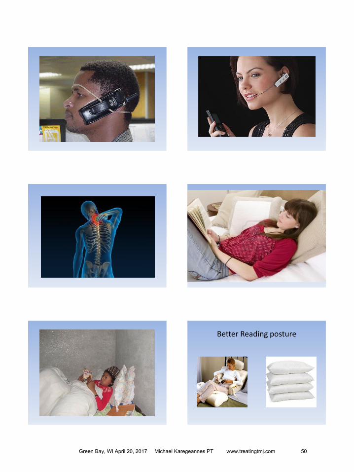

copy 2015 Freedom Physical Therapy Services SC
Guidelines To Minimize TMJ Pain
In general avoid
Large bites ldquobigrdquo food
Repetitive chewing
Forceful bites
Using tongue to remove food from teeth
Specifically avoid these foods
Gum
Jaw breakers
Popcorn
Caramel
Steak
Pizza
Bagels
Chips
Nuts
Ice
Beef jerky
Crunchy fruits and vegetables
French bread
Hard cereal
Lettuce
Other things to avoid
Resting chin on hand
Holding phone with shoulder
Sleeping on stomach
ldquoBigrdquo yawns
Singing
Yelling
Nail biting
Chewing on inside of cheek
Biting lip
Green Bay WI April 20 2017 Michael Karegeannes PT wwwtreatingtmjcom 71
- WPTA TMD coverpage final
- Time ordered Agenda for WPTA conference
- WPTA Spring conference TMD powerpoint for 3-31-17 final
- WPTA handout for back of manual 3-31-17
-