prezentace aplikace powerpoint -...
TRANSCRIPT

Cytogenetics• Postnatal -peripheral lymphocytes• Prenatal - amniocytes, fibroblasts from
amniotis fluid, chorium, placental tissue, umbilical cord blood
• Preimplantation – germinal cells embryonic cells
• Oncological -bone marrow, perph.lymphocytes, tumor cells

Indications for chromosomal examination
• Infertility, dysfertility• Growth failure• Psychomotoric retardation in children• Sexual development failure • Dysmorfic features in children• Congenital malformation in newborns• Exposition to mutagenic agens

MutagenesisDetection of changes ingenom(karyotype) after exposition to various mutagen agens
Searching for breakages, gaps, double minutes, ring chromosomes
Sister chromatids exchange examination
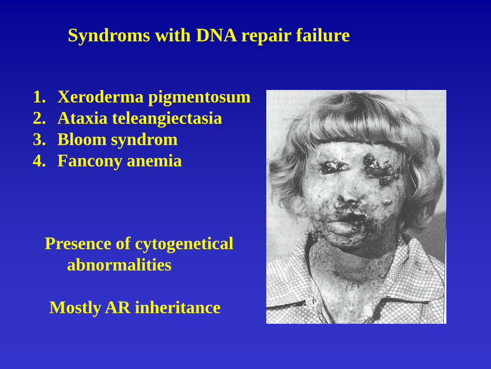
Syndroms with DNA repair failure
1. Xeroderma pigmentosum2. Ataxia teleangiectasia3. Bloom syndrom4. Fancony anemia
Presence of cytogeneticalabnormalities
Mostly AR inheritance

Cultivation of peripheral blood lymphocytes1. Mixing the sample with
medium+ serum+ PHA
2. Cultivation
3. Mitotic division termin.
4. Hypotonic sol.
5. Fixation sol. 2x
6. Slides aging
7. Staining

Chromosomal aberrations• Numerical - changes in number of
chromosomes - no morfological changes
• Structural - alternations in chromosomalstructure
• Physiol.variations - no pathological impact
Numerical chromosomal aberrations
1. Polyploidy (triploidy 69,XXX, tetraploidy…)
2. Aneuploidy - trisomy (47,XXY;47,XX,+21)- monosomy (45,X)
3. Marker chromosomes
4. Mosaics

Genesis of polyploidy

Structural chromosomal aberrations
Inolving one chromosome: deletionduplicationring chromosomeinversion isochromosome
Involving more chromosomes: translocation-Robertsonian- reciprocal

Deletion Duplication Ring chromosome Insertion

IsochromosomInversionParacentric Pericentric

Reciprocaltranslocation
Robertsonien translocation

Down syndrom: 1. Free trisomy – meiotic nondisjunction
2. Translocation
3. Mozaic – mitototic nondisjunction
Incidence
1 : 800

Obr.č.1: Detection of trisomy 21 by interphase FISH technique on uncultivated amniocytes
proved by G- banding on cultivated fibroblasts

Meiosis
Fertilization

Meiosis
Fertilization

Trisomy 18 – Edwards syndrom
90 % meiotic nondisjunction10 % mozaicismPartial trisomy - rare
microcephalyshort sternumnuchal translucencypedes equinovarusflexion finger deformitiesCardiovascular defectsSevere MRother defects
Incidence: 1:8000

Trisomy 13 – Patau syndrom

Deletions syndroms
Cri-du-chat 5p-
80-85 % de novo deletion10-15 % result of translocation in
parents- typical crying-- severe MR, hyperactivity- microcephaly- dysmorfic features, etc…
Incidence: 1:50000

Chromosomal examination in family with balanced chromosomal translocation
Parents:46,XX46,XY,t(5;18)(p13.3;q21.3)Proband:46,XX,del(5)(p13.3pter.)

Unbalanced chromosomal finding in fetusder(18)t(5;18)(p13.3;q21.3)

Wolf-Hirschorn syndrom
Typical facies: „Greek helmet face“Growth retardationMental retardation
90 % de novo deletion 4p1610 % result of translocation in
parents
Incidence: 1:50000

Microdeletion syndromsdetectable by FISH
• DiGeorge• Prader Willi- Angelman • Williams Beuren • Smith-Magenis• Wolf Hirschhorn• Miller-Dieker• Cri-du-Chat• FraX
and others…
• 22q11.2• 15q11-13• 7q11.23• 17p11.2• 4p16.3• 17p13.3• 5p15.2-15.3• Xq27.3

The most frequent gonosomal aberations: 45,XO 47,XXY
results of - meiotic nondisjunction- mitotic nondisjunction - mozaics

Prenatal diagnosis
• Benefit• Techniques• Prenatal cytogenetics, placental mosaicism,
uniparental disomy (UPD)• Preimplantation diagnosis• Modern method in cytogenetics - molecular
cytogenetics

Techniques for prenatal diagnosis
• Invasive:
• Non-invasive:
• Amniocentesis• Chorionic villus sampling• Fetal blood sampling• Fetoscopy
• Ultrasound• Other types of imaging• Fetal cells in the maternal
circulation

Identification of at-risk pregnancies prior pregnancyCHR chromosomal,SGD single gene, MCM multiple
congenital disorders+ Associated, - Not asssociated, (+) May be associated
• Elevated maternal age• Parental consanguinity• Ethnic origin• Positive family history• Maternal illness or
medication• Population carrier
screening
• CHR SGD MCM + - -- + + - + (+) + + + - - +
• - + -

Identification of at-risk pregnancies during a pregnancy
CHR chromosomal,SGD single gene, MCM multiple congenital disorders
• Abnornal ultrasound• AFP screening• Other bioch.
Screening• Maternal exposure to
teratogens
• CHR SGD MCM (+) (+) + (+) (+) (+)
• (+) - -
• - - +

Indications for AC
• Elevated maternal age (35 year and older)• Biochemical screening(AFP, HCG, E3)• Abnormal ultrasound• Positive family history (balanced
translocation in family, single-gene disord. in family, previous child with abnormal chrom. constitution)


Test on amniotic fluid cells and supernatant
• Fetal karyotyping• Fetal enzyme assay
(CVS may be preferred to AC)
• Fetal DNA diagnosis• (CVS is usually preferred
to AC)• FISH on interphase
nuclei

Indications for fetal blood sampling
• Haemophilia A and B *• Severe combined immunodeficiency• Sicle cell disease and beta thalassaemia*• Fetal infections• Suspected mosaicism • In utero transfusion for Rh isoimmunization• Unexplained hydrops• Failed amniotic cell culture or late booking• Abnormal ultrasound appearance - cong. malformation• Unexplained severe fetal growth retardation
* In cases where DNA diagnosis is not possible

Chromosomal examination of chorionic villus
• Advantage: early chromosomal examination(1st trimestr of pregnancy)
• Disadvantage: frequent placental mosaicism risk of spontaneous abortion after CVS rather high


Methods of prenatal cytogenetic
Methods Risk of Advantages Disadvantages fetal miscaridge---------------------------------------------------------------------------------AC 0,5% - contamination with - laborious maternal celss low - time consuming - risk of culture failure--------------------------------------------------------------------------------------------------FBS 1% - result available - higher risk of miscaridge within week - risk of contamination with - failure of culture maternal blood is rare------------------------------------------------------------------------------------------------CVS 1-2% -result available - laborious within 48 h - risk of placental mosaicism -1st trimestr diagnosis

Proportion of particular methods in our department
97-99(1/2)
AC FBS CVS FISH

Result of examinations of patient
• US
• CVS direct• CVS culture
• FISH result
• Abnormal NT 6.6-10 mm at 12th week pregnancy
• 46,XX• 46,XX,-18,+mar
• 46,XX,-18, idic(18)

Karyotype of the fetus with hygroma colli
cysticum

Result of FISH method with probe
for chromosome 18

FISH on interphase nuclei
• result within 24 h• culture is not
neccessary• is used as additional
rapid method for detection of common aneuploidies to minimize stressful waiting for result of AC

Mosaicism
• A general• B CPM (both
clones are present in placenta)
• C CPM(only one different clone is present in placenta)
• D embryonicKalousek, D. K., (1992)


Prader-Willi Syndrome
• hypotonia• initial failure to thrive• distinctive facial features• developmental delay• hypogonadism• eating disorder• deletion of sequences
of paternal copy of chr.15

Angelman Syndrome• Hypotonia• seizures• jerky, ucoordinated
movements• unprovoked smiling• lack of speech• severe developmental
delay• deletion of sequences
from the maternal copy of chr. 15

Microdeletion in patient with the syndrome Prader-Willi
SNRPN/ Tel 15q

Methyl sensitive analyse

child
mother
father
Isodisomy in patient with Angelman syndrome

Pathological outcomes of uniparental disomy
• Recessive disorder from one parent (see picture)
• pathological effect in consequence of mosaicism (IUGR, abortion)
• imprinting (Prader-Willi Syndrome)


Usage of FISH method
• Clinical genetics : postnatal • prenatal• Oncogenetics: haematooncology• tumor genetics• Preimplantation genetics• Mapping of human genom

Origin ofmarker chromosome
TranslocationsMicrodeletions
FISH in prenatal diagnoses
Fast detection of aneuploidies
Mosaics

Detection of gonosomes in patient with
Klinefelter syndrome

Patient with Williems – Beuren syndrome

Detection of microdeletion 7q11.23 in patient with syndrome Williams Beuren

Comparative genomic hybridization (CGH)

Usage of CGH
• Detection of origin of marker chromosome
• Detection of origin of extra chromosomal material
• Detection of the large deletions
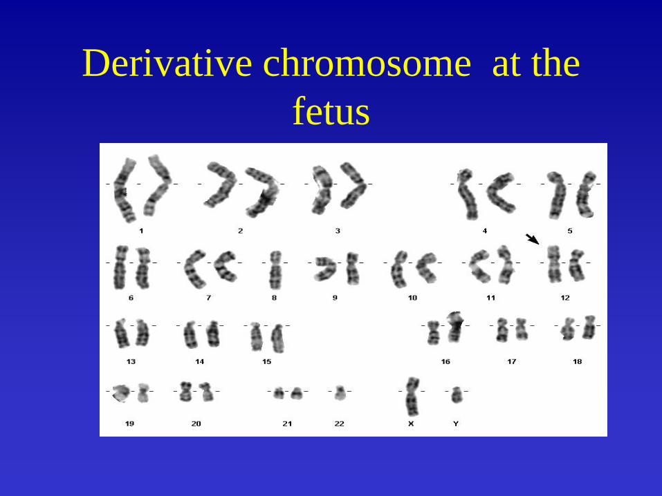
Derivative chromosome at the fetus

Comparative genomic hybridization
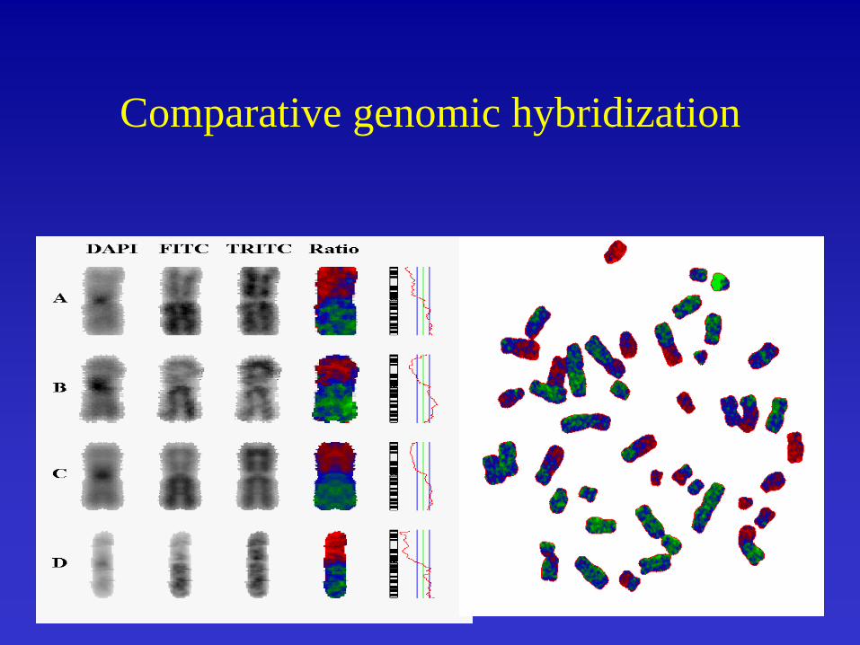
Comparative genomic hybridization

M- FISH

Preimplantation genetic diagnostics
•Examination of : germinal cellsI. and II. polar bodiesblastomeresblastocysts
•Techniques: without X with DNA amplification-----------------------------------------------FISH X PCR+DNA analysis

Eight-cells embryo -optimal stage for blastomere biopsy

Preimplantation cytogenetic diagnostics
1. Biopsie of 1 or 2 blastomers from 6-8 cells embryo
2. Fixation of the cell on slide
3. Single –cell FISH detection of - aneuploidy
- sex - translocation
4. Transfer of unaffected embryos into maternal uterus

FISH on two-cells embryo
Detection of chromosomes 21( CEP 21 Vysis)