pro-resolving lipid mediator resolvin e1 mitigates the
TRANSCRIPT

International Journal of
Molecular Sciences
Article
Pro-Resolving Lipid Mediator Resolvin E1 Mitigatesthe Progress of Diethylnitrosamine-Induced LiverFibrosis in Sprague-Dawley Rats by AttenuatingFibrogenesis and Restricting Proliferation
Maria José Rodríguez 1,2, Francisca Herrera 1, Wendy Donoso 3, Iván Castillo 4,5, Roxana Orrego 6,Daniel R. González 1 and Jessica Zúñiga-Hernández 1,*
1 Departamento de Ciencias Básicas Biomédicas, Facultad de Ciencias de la Salud, Universidad de Talca,Talca 3460000, Chile; [email protected] (M.J.R.); [email protected] (F.H.);[email protected] (D.R.G.)
2 Programa de Doctorado en Ciencias Mención Investigación y Desarrollo de Productos Bioactivos,Instituto de Química de los Recursos Naturales, Universidad de Talca, Talca 3460000, Chile
3 Departamento de Estomatología, Facultad de Ciencias de la Salud, Universidad de Talca, Talca 3460000,Chile; [email protected]
4 Unidad de Anatomía Patológica, Hospital Regional de Talca, Talca 3460001, Chile; [email protected] Centro Oncológico, Facultad de Medicina, Universidad Católica del Maule, Talca 3466706, Chile6 Departamento de Bioquímica Clínica e Inmunohematología, Facultad Ciencias de la Salud,
Universidad de Talca, Talca 3460000, Chile; [email protected]* Correspondence: [email protected]; Tel.: +56-71-241-8855
Received: 1 July 2020; Accepted: 11 August 2020; Published: 22 November 2020�����������������
Abstract: Liver fibrosis is a complex process associated to most types of chronic liver disease,which is characterized by a disturbance of hepatic tissue architecture and the excessive accumulationof extracellular matrix. Resolvin E1 (RvE1) is a representative member of the eicosapentaenoicomega-3 lipid derivatives, and is a drug candidate of the growing family of endogenous resolvins.Considering the aforementioned, the main objective of this study was to analyze the hepatoprotectiveeffect of RvE1 in a rat model of liver fibrosis. Male Sprague-Dawley rats received diethylnitrosamine(DEN, 70 mg/mg body weight intraperitoneally (i.p)) as an inductor of liver fibrosis once weeklyand RvE1(100 ng/body weight i.p) twice weekly for four weeks. RvE1 suppressed the alterationsinduced by DEN, normalizing the levels of alanine aminotransferase (ALT), albumin, and lactatedehydrogenase (LDH), and ameliorated DEN injury by decreasing the architecture distortion,inflammatory infiltration, necrotic areas, and microsteatosis. RvE1 also limited DEN-inducedproliferation through a decrease in Ki67-positive cells and cyclin D1 protein expression, which isrelated to an increase of the levels of cleaved caspase-3. Interestingly, we found that RvE1 promoteshigher nuclear translocation of nuclear factor κB (NF-κB)p65 than DEN. RvE1 also increased thelevels of nuclear the nuclear factor erythroid 2–related factor 2 (Nrf2), but with no antioxidant effect,measured as an increase in glutathione disulfide (GSSG) and a decrease in the ratio of glutathione(GSH)/GSSG. Taken together, these results suggest that RvE1 modulates the fibrogenesis, steatosis,and cell proliferation in a model of DEN induced fibrosis.
Keywords: omega-3 derivatives; eicosanoids; liver fibrosis; apoptosis; microsteatosis
1. Introduction
Liver fibrosis is a dynamic response to chronic liver injury caused by various agents, such asviruses, alcohol, metabolic and autoimmune disorders [1]. Liver fibrosis is a complex process that
Int. J. Mol. Sci. 2020, 21, 8827; doi:10.3390/ijms21228827 www.mdpi.com/journal/ijms

Int. J. Mol. Sci. 2020, 21, 8827 2 of 14
involves several cells of the hepatic sinusoid, which is characterized by a disturbance of the architectureand composition of extracellular matrix (ECM) [2]. Under certain circumstances, the acute inflammationcan lead to persistent chronic inflammation, which when unresolved may promote organ fibrosis anddysfunction, ultimately leading to cirrhosis and its consequences: portal hypertension, hepatocellularcarcinoma (HCC), and liver failure [3]. Hepatic inflammation is a hallmark of the early stage fibrosis,which can progress to extensive fibrosis and cirrhosis [4]. According to the last report from the Centersfor Disease Control and prevention (CDC), cirrhosis and chronical liver disease (CLD) are the eleventhcause of death affecting millions of patients worldwide [5]. Globally, the age-standardized incidencerate of cirrhosis and CLD was 20.7 per 100,000 inhabitants, and the estimated incidence of cirrhosis inEurope is 26.0 per 100,000 [6].
The pro-resolving lipid mediators, such as lipoxins and other specialized pro-resolving mediators(SPM), are crucial for the active resolution of inflammatory processes [3]. SPM were coined as lipoxins,resolvins, protectins and maresins [7–10]. These SPM are potent inhibitors of polymorphonuclear(PMN) leukocyte transendothelial migration and infiltration in vivo [7]. Resolvin E1 (RvE1) is arepresentative member of the SPM omega-3 lipid autacoids, and is a drug candidate of the growingfamily of endogenous resolvins [11]. At very low concentrations, RvE1 promotes the resolution of acuteinflammation by regulating leukocyte infiltration, increasing the macrophage ingestion of apoptoticneutrophils by macrophage, and enhancing the clearance of phagocytes in the lymph nodes [12], spleen,and heart [13]. Also, RvE1 mediated survival and reinforced the shift toward apoptosis [14]. In relationto CLD, RvE1 reduces the levels of serum indicators of liver fibrosis, such as laminin, hyaluronic acid,pro-collagen type III, and type IV collagen [15]. Also, plasma RvE1 increased in patients undergoingmajor hepatobiliary resection when EPA was previously administrated (immunonutrition) and thelevels of RvE1 were correlated with plasma IL-6 after operation [16]. Considering the previousbeneficial results in CLD, the main objective of this study was to analyze the hepatoprotective effect ofRvE1 on a rat model of liver fibrosis.
Animal models to investigate the potential hepatoprotective drugs that have a positive impactin liver fibrosis have been well defined and diethylnitrosamine (DEN) is considered one of the mosttoxic agents that can cause several necroses and consequent fibrosis [17,18]. Also, DEN administrationcauses excessive deposition of extracellular matrix proteins (collagen) in the rat liver and appears to beappropriate for the study of the early events associated with the development of hepatic fibrosis [19].Interesting, DEN is a carcinogenic substance that forms DNA adducts through alkylating metabolitesthat are generated through cytochrome P450 enzymes [20]. Also DEN induces irreversible hepatocellularcarcinogenesis through the overexpression of G1/S phase regulatory proteins through the promotionof changes in the expression of cell cycle regulatory factors [21]. Due to the carcinogenic properties,DEN has become a highly attractive experimental model to study liver tumorigenesis [20] and themechanism of fibrosis associated to cell cycle, inflammation, and hepatitis towards liver carcinogenesis.
2. Results
To investigate the efficacy of RvE1 to attenuate the liver fibrosis or reverse it, rats were submittedto chemically induced liver fibrosis, and the impact of RvE1 was studied (growth chart (in grams) isavailable in Supplementary Figure S1). Biochemical parameters analysis show that DEN induced liveralterations related to increases in ALT, aspartate transaminase (AST), and LDH values, and reductionsof albumin levels (Table 1), with insignifficant alterations in the values of Alkaline phosphatase (ALP)or γ-glutamyl transferase (γGT) in relation to control. The RvE1 administration concomitant to DENnormalized the levels of ALT, albumin and LDH (1.6, 1.7 and 1.7 folds respectively p < 0.05) and showa slight decrease of the levels of AST (p = 0.1780) and LDH (p = 0.2904).
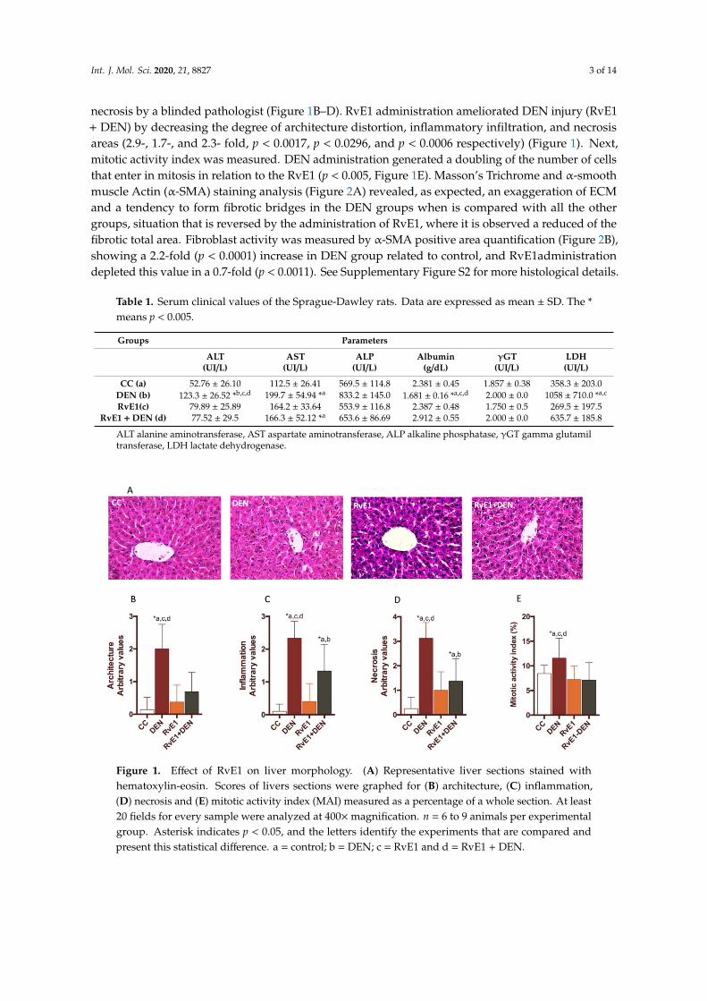
The histological assessment of the livers showed that DEN administration generates architecturaldisorder, with dilatation of the sinusoid (peliosis), substantial confluent and multiacinar liver necrosis,with moderate to severe portal inflammation and degenerative changes in comparison with controlgroup (Figure 1A). Also, the DEN damage was quantified as cytoarquitecture, inflammation and
Int. J. Mol. Sci. 2020, 21, 8827 3 of 14
necrosis by a blinded pathologist (Figure 1B–D). RvE1 administration ameliorated DEN injury (RvE1+ DEN) by decreasing the degree of architecture distortion, inflammatory infiltration, and necrosisareas (2.9-, 1.7-, and 2.3- fold, p < 0.0017, p < 0.0296, and p < 0.0006 respectively) (Figure 1). Next,mitotic activity index was measured. DEN administration generated a doubling of the number of cellsthat enter in mitosis in relation to the RvE1 (p < 0.005, Figure 1E). Masson’s Trichrome and α-smoothmuscle Actin (α-SMA) staining analysis (Figure 2A) revealed, as expected, an exaggeration of ECMand a tendency to form fibrotic bridges in the DEN groups when is compared with all the othergroups, situation that is reversed by the administration of RvE1, where it is observed a reduced of thefibrotic total area. Fibroblast activity was measured by α-SMA positive area quantification (Figure 2B),showing a 2.2-fold (p < 0.0001) increase in DEN group related to control, and RvE1administrationdepleted this value in a 0.7-fold (p < 0.0011). See Supplementary Figure S2 for more histological details.
Table 1. Serum clinical values of the Sprague-Dawley rats. Data are expressed as mean ± SD. The *means p < 0.005.
Groups Parameters
ALT(UI/L)
AST(UI/L)
ALP(UI/L)
Albumin(g/dL)
γGT(UI/L)
LDH(UI/L)
CC (a) 52.76 ± 26.10 112.5 ± 26.41 569.5 ± 114.8 2.381 ± 0.45 1.857 ± 0.38 358.3 ± 203.0DEN (b) 123.3 ± 26.52 *b,c,d 199.7 ± 54.94 *a 833.2 ± 145.0 1.681 ± 0.16 *a,c,d 2.000 ± 0.0 1058 ± 710.0 *a,c
RvE1(c) 79.89 ± 25.89 164.2 ± 33.64 553.9 ± 116.8 2.387 ± 0.48 1.750 ± 0.5 269.5 ± 197.5RvE1 +DEN (d) 77.52 ± 29.5 166.3 ± 52.12 *a 653.6 ± 86.69 2.912 ± 0.55 2.000 ± 0.0 635.7 ± 185.8
ALT alanine aminotransferase, AST aspartate aminotransferase, ALP alkaline phosphatase, γGT gamma glutamiltransferase, LDH lactate dehydrogenase.
Figure 1. Effect of RvE1 on liver morphology. (A) Representative liver sections stained withhematoxylin-eosin. Scores of livers sections were graphed for (B) architecture, (C) inflammation,(D) necrosis and (E) mitotic activity index (MAI) measured as a percentage of a whole section. At least20 fields for every sample were analyzed at 400×magnification. n = 6 to 9 animals per experimentalgroup. Asterisk indicates p < 0.05, and the letters identify the experiments that are compared andpresent this statistical difference. a = control; b = DEN; c = RvE1 and d = RvE1 + DEN.

Int. J. Mol. Sci. 2020, 21, 8827 4 of 14
Figure 2. Effect of RvE1 on matrix deposit. (A) Representative histopathological microphotography ofliver. Upper panel: Representative Masson´s trichrome. Middle panel: Representative images ofα-SMAstaining at 100×. Lowe panel: Representative images of α-SMA staining at 400×. (B) Quantification ofα-SMA positive areas. At least 20 fields for every sample were analyzed at 400×magnification. n = 6animals per experimental group. Asterisk indicates p < 0.05, and the letters identify the experimentsthat are compared and present this statistical difference.
Pathological analysis of fat droplets deposits (microsteatosis) was measures by Oil Red O stainingin the DEN induced fibrosis model (Figure 3A,B). Total lipid area quantified in DEN represent an 9%when is compared with controls groups (CC and RvE1), which shows values less than or near to 1% ofthe total area of analysis (fields). RvE1 + DEN presented a significative decrease in the total percentageof Oil Red O staining when compared to DEN (65% less staining, p < 0.05).
Figure 3. Effect of RvE1 on lipid deposit. (A) Representative histopathological microphotography ofliver. Upper panel: Oil Red O plus counterstain Gill III modified hematoxylin. Lower Panel: Oil RedO without counterstain. (B) Quantification of Oil Red O percentage per field. Magnification 400×.At least 20 fields for every sample were analyzed. n = 6 animals per experimental group. Asteriskindicates p < 0.05, and the letters identify the experiments that are compared and present this statisticaldifference. Arrow indicates the high areas of Oil Red O cumulation.
The effects of RvE1 administration on cell proliferation was measured in hepatocytes using theproliferation marker Ki67 (Figure 4A). Whereas few Ki67- positive hepatocytes were observed incontrol livers (2.1 ± 1.9% per field of view), many were observed in DEN–treated liver (13.4 ± 5.3%per field of view) (Figure 4B) and the RvE1 + DEN evidence a decrease in the number of positiveKi67 hepatocytes (5.2 ± 2.3% per field of view) compared to DEN (p < 0.05). In order to determineswhether RvE1 has a role on hepatocyte apoptosis in the DEN-induced liver fibrosis, the presence of

Int. J. Mol. Sci. 2020, 21, 8827 5 of 14
cleaved-caspase 3 was studied. Figure 4C shows that, in DEN-induced liver fibrosis, there was a highercleaved-caspase 3 expression compared to control (1.7-fold, p < 0.05), while RvE1 group has an increaseof 3.44 and 1.9 folds when compared to control and DEN respectively (p < 0.05). In concordance withthese values, DEN presents an increase in Cyclin D1 protein expression (3.3-fold, p < 0.05), whileRvE1 did not increase the levels of Cyclin D1 compared to controls (0.4397 ± 0.1658 vs 0.5066 ± 0.1115,respectively) (Figure 4D). When the B-cell lymphoma 2 (Bcl-2) protein, an anti-apoptotic protein, wasdetermined (Figure 4E) it was observed that DEN and RvE1 + DEN decreased by 1.5-fold and 1.7-foldcompared to control, respectively. RvE1 do not show any statistic variations respects to DEN in Bcl-2values (p = 0.666). Next, we evaluated the activity of the transcription factors Nrf2 and NF-κBp65, astheir translocation to the nucleus (Figure 5A–D). As expected, DEN promotes the nuclear translocationof NF-κBp65 with a diminution in cytoplasmic protein presence (a median of 0.7663 ± 0.2303 to 1.076 ±0.1203, from cytoplasm to nucleus) compared to control group. Interestingly, the supplement of RvE1promotes higher nuclear translocation of NF-κBp65 compared to control or DEN (2.7-fold and 2.1-foldrespectively, p < 0.05). Since these transcription factors are targets of the cytokines, we evaluated theserum levels of tumor necrosis factor (TNF)-α and interleukin (IL)-10 (Figure 5E,F). TNF-α is increased3.1- and 1-fold in DEN and RvE1 + DEN relative to control, and with non-statistically differencesamong them (2.066 ± 1.114 and 2.137 ± 0.4848 for DEN and RvE1 + DEN respectively). Further, IL-10did not show any significant variation among the groups. Then, we analyzed the nuclear translocationof Nrf2 (Figure 5C,D), where DEN groups show a lost signal in nuclear samples compared to all theother groups (p < 0.05). Nrf2 was incremented in the groups RvE1 and RvE1 + DEN (1.7- and 1.4-fold)with respect to control. Because Nrf2 is a regulator of oxidative balance, GSH, GSSG, and GSH/GSSGratio were measured (Figure 5G–I). We observed that DEN group presents a reduction in GSH levelscompared to control and was restored in the RvE1-treated groups, while DEN group not present anincrease in the levels of GSSG nor GSH/GSSG (p > 0.05), probably due to a loss of functional tissue.RvE1 shows an increase in GSSG (1.6- and 1.3-fold respect to control and DEN respectively, p < 0.05),and a decrease in the ratio GSH/GSSG (22% and 10% regard to control and DEN respectively).
Figure 4. Effect of RvE1 on cell cycle and apoptosis. (A) Representative histopathologicalmicrophotography tissue Ki67 at 400×magnification. (B) Quantification of Ki67 positive cell. Western blotanalysis of (C) Cleaved caspase-3, (D) Cyclin D1 and (E) BCL-2. The levels were normalized to GAPDHor β-actin as housekeeping. n = 6–9 rats per experimental group. Asterisk indicates p < 0.05, and theletters identify the experiments that are compared and present this statistical difference.

Int. J. Mol. Sci. 2020, 21, 8827 6 of 14
Figure 5. Effect of RvE1 on tissue levels of NF-κB and Nrf2, and molecules targets. Western blot analysisof (A) cytoplasmic and (B) nuclear NF-κB; and (C) cytoplasmic and (D) nuclear Nrf2. The cytoplasmiclevels were normalized to GAPDH as housekeeping and nuclear levels were normalized to histoneH1 as housekeeping. (E,F) serum levels of TNF-α and IL-10. (G–I) tissue levels of GSH, GSSG andGSH:GSSG ratio. n = 6–9 rats per experimental group. Asterisk indicates p < 0.05, and the lettersidentify the experiments that are compared and present this statistical difference.
3. Discussion
The CLD is characterized by fibrosis, when fibrosis is self-limited, resulting in a balanced,protective, reparative, and non-tissue injury response. When the regulatory response related to fibrosisbecome chronic and there is a dominance of the repetition of the injury, the benefits of the fibrosisbalance is lost. The end-stage of chronic liver fibrosis results in cirrhosis and is a limiting factor for thedevelopment of HCC. There is important to mention that the only therapeutic approved approaches toremoval the injury related to the describes CLD is the liver trasplantation [22,23]. Because the scarcityof donors is a serious limitation, new therapeutic approaches are urgently required and antifibrotictherapies. The present study aimed to investigate whether RvE1 could reduce liver fibrosis andstimulate the resolution of the damaged fibrotic liver.
There is animal models and human interventions that showed the beneficial effect of the EPAand DHA administration on CLD, in particular when these fatty acids are administrated improvedthe biochemical parameters, inflammation, ameliorates fibrosis, fatty acid cumulation, improvingesteatosis [24–26]. Interestingly, when EPA is compared with DHA in a model of non-alcoholic fattyacid liver disease (NAFLD), EPA is better in controlling hepatic triglyceride cumulation and steatosis,but is DHA the fatty acids who is in charge of controlling the inflammation and oxidative stress,and both are important in the control of fibrosis, acting sinergistically [27]. The above is interesting,since resolvins are derived from EPA or DHA. Thus, here, we present the results obtained from theRvE1, an EPA derived bioactive SPM. In concordance with the above, Gonzalez-Periz et al. reportedthat the protective effect of omega-3 fatty acids observed in their model (ex-vivo and in vivo) wasmediated by protectin D1 and RvE1. RvE1 administration (1.2 ng/g) conferred protection againsthepatic steatosis, decrease of liver injury serum markers, and macrophage infiltration [28]. Qiu et al.
Int. J. Mol. Sci. 2020, 21, 8827 7 of 14
demonstrated that RvE1 (100 ng for 70 days) improves the liver fibrosis caused by the infection ofSchistosoma japonicum to mice. RvE1reduces the levels of transaminases, TNF-α and INF-γ, and lowerthe levels of fibrotic markers such as laminin, hyaluronic acids and pro-collagen III among otherparameters [15]. In our study, RvE1 normalized liver parameters and demonstrated potent antifibroticactivities. Also, recently, it was shown that the RvE1 concentration was decreased during NAFLDprogression [29], and this could be related with the increase in the omega-6/omega-3 ratio which isassociated with lipogenesis and the lipid oxidation promotion of NAFLD [30].
Even though the model of DEN is not the ideal system to study the phenomena of liver steatosis,it has been described that it is a support model to study metabolic and nutritional changes in the fattyacids deposits [31–33]. Histologically, the liver is considered steatotic when >5% of hepatocytes in atissue section stained with H&E contain macrovesicular steatosis [34–36]. Interestingly, H&E oftenunderestimate steatosis by the difficulties in detecting lipid microvesicles after paraffin embedding,and underestimation of the degree of fatty changes [37,38]. On the other hand, Oil-Red O is fast andsensitive and improve the assessment of liver steatosis [38,39]. The fact that RvE1 reduced the levels offatty droplets in our experiments could be interesting to introduce future studies of SPM in the contextof steatosis-related liver fibrosis and even HCC. The aforementioned becomes more interesting if weconsider that Kuang et al. demonstrated that RvE1 and RvD1 can reverse the development of livercancer cells in a long-term concavalin-A induced injury [40].
The steatotic hepatocyte is associated with dysregulated lipolysis resulting in excessive deliveryof fatty acids to the liver, de novo lipogenesis, impaired post-receptor signaling by insulin(i.e., insulin resistance) [41]. It has been proposed that the downregulation of steroyl-CoA responseelement binding protein-1c (SREBP-1c), a de novo lipogenesis related transcriptional regulator couldimproves the non-alcoholic stetatohepatitis (NASH) [41,42]. EPA administration decrease the expressionof SREBP-2 in NAFLD [43] and SREBP-1c in hepatocyte cell culture [44,45]. Among resolvins, only RvD1has been assayed by their role on SREBP-1 [46], here the authors found that RvD1 protect endoplasmicreticulum stress in HepG2 cells, this protection was related to a decrease in caspase activity andSREBP-1 expression [46]. Previously, Neuhofer et al., demonstrated that the deficiency of protectin D1and 17-HDHA (the precursor of RvD1) is linked to the development and perpetuation of obesity-drivenadipose tissue inflammation that promotes type 2 diabetes, and this may be corrected by the additionof 17-HDHA resulting in increased levels of peroxisome proliferator-activated (PPAR)γ, PPARα andadiponect [47]. The mentioned factors are involved with the regulation of fatty acids metabolism, insulinsensitivity, prevention of hepatic steatosis, and interestingly they contribute to the anti-inflammatorymilieu in NASH/NALFD [48,49]. As previously was reported by Gonzalez-Periz, RvE1 protectsagainst liver steatosis by improving insulin tolerance and induction of PPARγ and adiponectin [28].More recently, Barden et al., found that RvE1 was produced in large amounts in patients with metabolicsyndrome who have lost weight [50]. On the other hand, Maciejewska analyzed the relationshipof eicosanoid with steatosis progression in animal model, finding a moderate correlation of RvE1decreasing with NAFLD progression, but in liver tissue the association is weak and results difficultto associated to results observed in serum with the tissue liver observed [29]. The previous resultssuggest that this SPM is sensitive to fat changes in the tissues. The fact that RvE1 administrationdepleted the microsteatotic deposits in our model (fibrosis) confirms the role of lipid metabolismindependently of the model of study, and it will be of interest to study the role of RvE1 on SREBPs andPPARs transcription factors.
In CLD, it is commonly believed that simple steatosis is the benign, while NASH is progressivewith marked increase in liver-related mortality due to cirrhosis or HCC and HCC is common in patientswith fibrotic NASH and NASH related cirrhosis [51]. Also, metabolic factors, such as diabetes andobesity, can contribute to HCC [52]. It has been confirmed that the production of reactive oxygen species(ROS) and inflammatory infiltration promotes cellular proliferation and HCC induction [53]. Liver cellproliferation and its relation with tumor growth can be evaluated by Ki67 expression [54,55]. Previously,El-Kebir et al. confirmed that RvE1 enhances phagocytosis and induces neutrophil apoptosis in isolated
Int. J. Mol. Sci. 2020, 21, 8827 8 of 14
neutrophils, mediated by a caspase-8 and caspase-3 activation [56]. In our model, we described thatRvE1 decreased the mitotic activity index, with Ki67 positive cell staining enhancing the activityof caspase-3 and limiting the expression of cyclin D1 and BCL-2, all of these related to a potentialinhibition of damaged tissue proliferation.
Previously, it has been demonstrated that nanomolar concentration of RvE1 can blocks PMNinfiltration and attenuated dendritic cell migration, activity that is related to his bound to ChemR23 aG-protein-coupled receptor [57]. The Chem23 selectively union blocks TNF-α signaling, also RvE1binds to leukotriene B4 (LTB4) receptor on human PMN and the union to these receptor apparentlymediated the counter regulation of RvE1-ChemR23 to promote acute inflammation resolution [58].The up-regulation of TNF-α is associated with the enhancement of DNA binding activity of NF-κBactivity which is downregulated and modulated by EPA and DHA administration, with the consequenceof anti-inflammatory and ROS reduction [59,60]. Also, RvD1 and RvE1 significantly downregulateCD4+ and CD8+ liver infiltration and the related injury, throughout inhibition of pro-inflammatorycytokine discharge and NF-κB/AP-1 nuclear activity [40]. Based in this information, it is expectedthat the administration of RvE1 downregulates the levels of TNF-α related to an enhance of nucleartranslocation of NF-κB, but in our model, we do not observe molecular anti-inflammatory behavior,we only detected a decrease in the inflammatory infiltration in the liver tissue. The fact that neitherTNF-α nor IL-10 show changes in our model is presumably related to the crosstalk among NF-κB andoxidative stress [61]. We presume that higher doses of RvE1 can block the movement to NF-κB atthe nucleus and promote the expected anti-inflammatory phenotype. In relation with this, it is welldescribed that the activation of Nrf2 antagonizes the inflammatory pathways of transforming growthfactor (TGF)-β1 and NF-κB, and plays a cytoprotective role in cell damage [62], where the disruptionor loss of Nrf2 signaling causes enhanced susceptibility not only to oxidative and electrophilic stresses,but also to inflammatory tissue injuries [63]. Nrf2 regulates apoptosis and fibrotic process throughactivation of antiapoptotic Bcl-2 protein [4]. It should be mentioned that exists a crosstalk betweenNrf2 and NF-κB, where the absence of the first can exacerbate cytokine production, whereas NF-κB canmodulate Nrf2 transcription and activity. Both Nrf2 and NF-κB are regulated by redox sensitive factors,but NF-κB is more readily activated in oxidative environments, another interesting fact is that Nrf2contains several κB sites in its proximal promoter, which are subject to be binding p65, suggesting thathigh activation of NF-κB signaling inhibits/repress Nrf2-ARE pathway [64–66]. This can explain whythe enhancement of nuclear Nrf2 was not reflected in an improvement in ROS an inflammatory status.Finally, it is necessary to mention that Pohl et al. did not find positive changes in liver NASH or fibroticchanges when RvE1 was administrated (1.2 ng/g body for four days) [67].
The data reported here support the role of RvE1 as hepatoprotective by controlling the fibrogenesis,steatosis, and cell proliferation in a model of DEN induced fibrosis. Nevertheless, it has to be consideredthat some of the experiment results does not resemble the expected literature. Thus, further studies,eventually using higher doses of RvE1, decreasing the time between the doses, or increasing the time ofexposition (weeks), could show the described anti-inflammatory effect. Also, we propose to compare itwith other resolvins and SPMs should be assayed to clarify the activity on liver fibrosis.
4. Materials and Methods
4.1. Animals
Male Sprague-Dawley rats (70–110 g) were obtained from Bioterio Central, Dirección deInvestigación, Universidad de Talca, Chile. Animals were allowed free access to food (ChampionS.A., Santiago, Chile) and water, and were housed in a temperature-controlled room on a 15 hlight/dark cycle. All animals received humane care in compliance with the University’s guidelinesand the ethics statement, experimental animal protocol and animal procedures in this project wascomplied with the “Guide for the Care and Use of Laboratory Animals” (National Academy ofSciences, NIH Publication 6-23, revised 1985). All experiments were approved by the Bioethical
Int. J. Mol. Sci. 2020, 21, 8827 9 of 14
Committee (CIECUAL), Folio number 2016-06B-C, Dirección de Investigación, Universidad de Talca.All the experiments with animals proposed in this project was made under the supervision of aveterinarian expert.
4.2. Model of Liver Fibrosis
To induce liver fibrosis induction, DEN (Cat No. 73861, Sigma-Aldrich, Merck KGaA, Darmstadt,Germany) was administrated intraperitoneally (i.p) at doses of 70 mg/g body weight (in 0.9% NaCl),once a week for a period of 4 weeks according to the model of DEN-induced liver fibrosis [18]. RvE1(Cat No. 10007848, Cayman chemical, Ann Arbor, MI, USA) was administrated i.p twice a week forthe same 4 weeks of DEN treatment (100 ng/body weight [15]). Animals were randomly assignedto one of the following groups: (i) vehicle DEN + 0.025% ethanol in 0.9% NaCl (vehicle of RvE1)(group control-control/CC); (ii) DEN + vehicle RvE1 (group DEN); (iii) vehicle DEN + RvE1 (groupRvE1) and (iv) DEN + RvE1. At the end of 4 weeks, the animals were anaesthetized with (1 mL/kg) ofzolazepam chlorhydrate (25 mg/mL) and tiletamine chlorhydrate (25 mg/mL) (Zoletil 50™; Virbac S/A,Carros, France). Blood and liver samples was taken from the medial lobes for experimental analysis.Animal number per experimental group: n = 6–9.
4.3. Measurement of Biochemical Parameters
ALT, AST, Albumin, ALP, LDH, γ-GT were measured using specific diagnostic kit (LiquidColorHuman™, Wiesbaden, Germany). To control the measurements, adequate 2-level controls, normal andpathological, were used. ELISA kits were used for assessment of serum levels (pg/mL) of TNF-α andIL-10 (Thermo Fischer Scientific, Rockford, IL, USA).
4.4. Liver Glutathione Assay
Livers were perfused in situ with washing solution (159 mM KCl and 5mM Tris, pH 7.4) toremove blood and measured liver content of reduced (GSH) and oxidized (GSSG) were measured indeproteinated tissue homogenates samples using a colorimetric Glutathione assay Kit (Cat No. 703002,Cayman Chemicals, Ann Arbor, MI, USA).
4.5. Histopathological Staining
For Hematoxylin & Eosin (H&E) and Masson´s Trichrome stain (Merck KGaA, Darmstadt,Germany), a third part of the lobes were fixed in 10% buffered formalin, embedded in paraffin,and sectioned in 5 µm, before being stained and analyzed for morphology, cell infiltration, collagendeposition, and mitotic index activity (MAI). Analysis was performed blind by a pathologist (I.C)according to the procedure reported previously by our group [68], following the Koroukian score [69]and Goodman’s adapted Ishack score [70,71]. For Oil Red O (0.5%) (Sigma-Aldrich, Merck KGaA,Darmstadt, Germany), fresh liver samples were frozen and cut in cryostat microtome (5 µm thickness,−20 ◦C). Gill´s III modifies Haematoxylin (Merck) was used as counterstain. Mitotic activity index(MAI) was analysed according to Al-Janabi [72]. All the histological analyses were performed in aNikon Eclipse 50i Optic microscope (Nikon, Tokyo, Japan), while the posterior analysis and photographwere developed in Micrometrics SE PremiumTM software (Opticstar, Manchester, UK).
4.6. Inmunohistochemitry Staining
Liver sections were immunostained for Ki67 (1/300, mouse monoclonal, Merck Millipore,Burlington, MA, USA) and α-SMA (1/25, mouse monoclonal, Leica biosystem, Weszlar, Germany).Immunohistochemistry was performed on sequential paraffin liver sections (4-µm thick) incubatedwith specific monoclonal antibodies. Briefly, after microwave antigen retrieval with 0.01 mol/L citratebuffer, pH 6.0, primary antibodies were labeled using VectorStain® Elite® ABC kit peroxidase HRP (CatNo. PK6200, Vector Laboratories, Maravai LifeSciences, CA, USA). Biotinilated secondary antibodies
Int. J. Mol. Sci. 2020, 21, 8827 10 of 14
directed against mouse antigens and visualized by Avidin plus Biotinylated ABC reagents (Vector).Negative controls were performed by replacing the respective primary antibodies by isotype andconcentrations matched irrelevant antibody (Human oral pyogenic granuloma). The images wereobtained using the Leica Microsystems Limited Software v 3.4.0 (Stereo and Macroscope Systems,Heerbrugg, Switzerland) and the images were quantified by ImageJ software v 2.0.0-rc-69/1.53a (NIH,Bethesda, MD, USA, https://imagej.nih.gov/ij/docs/examples/stained-sections/index.html).
4.7. Western Blot Analysis
Cytoplasmic and nuclear samples were obtained from the frozen hepatic tissue samples(200 mg) from the adapted protocol of Deryckere and Gannon [73] and described in Soto et al. [68].Briefly, frozen liver was homogenized and suspended in buffer solution pH 7.9 (10 mM4-(2-hydroxyethyl)-1-piperazineethanesulfonic acid (HEPES), 1 mM ethylenediaminetetraacetic (EDTA),0.6% nonidet P (NP)-40, 150 mM NaCl, and 0.5 mM phenyl methyl sulphonyl fluoride (PMSF)), followedby centrifugation at 3.020× g for 15 s at 4 ◦C. The supernatant corresponds to cytoplasmic fractions.The precipitate was resuspended in 200 µL of nuclear buffer solution pH 7.9 (20 mM HEPES, 0.2 mMEDTA, 25% glycerol, 420 mM NaCl, 1.2 mM MgCl2, 0.5 mM di-thio threitol DTT, 0.5 mM PMSF, 2 mMbenzamidine, an Pierce protease inhibitors cocktail mini tablest® (Pierce, Thermo Fischer Scientific,Rockford, IL, USA)), followed by centrifugation at 13,000× g for 60 s, and the supernatant was incubatedfor 20 min in ice. Then, the supernatant was centrifugated at 13,000× g for 30 s at 4 ◦C to eliminatenuclear debris (precipitate). Cytoplasmic and nuclear protein fractions (50 µg) were separated on 12%polyacrylamide gels using sodium dodecyl sulfate polyacrylamide gel electrophoresis (SDS-PAGEby using Mini-Protean® and Protean II® systems (Bio-Rad Laboratories, Hercules, CA, USA)) andtransferred to nitrocellulose membranes, which were blocked for 1 h at 22 ◦C with TBS containing5% skim milk. The blots were washed with TBS containing 0.1% Tween 20, hybridized with rabbitpolyclonal primary antibodies, for either Nrf2 (1:500), NF-κBp65 (1:1000), active caspase-3 (1:200),and histone H1 (1:350) as a nuclear housekeeping protein. Mouse monoclonal primary antibodiesused were for cyclin D1 (1:1000), BCL-2 (1:500), β-actin (1:2000), and GAPDH (1:2000), with theselast two used as a cytoplasmic housekeeping protein (Nrf2, NF-κB, active caspase-3, histone H1,cyclin D1, GAPDH and secondary antibodies were purchased from Merck Millipore, Burlington,MA, USA; BCL-2 from Thermo Fischer Scientific, Rockford, IL, USA; and β-actin from Santa CruzBiotechnology, Dallas, TX, USA). The antibodies were incubated overnight at 4 ◦C. After extensivewashing, the antigen antibody complexes were detected using horseradish peroxidase-labeled goatanti-rabbit IgG/anti-mouse or rabbit, and the protein was detected with the protein detection kit usingAmersham enhanced chemioluminicense (ECL) Prime Western Blotting Detection Reagent (GeneralElectric Healthcare, Hammersmith, UK). The chemiluminescent signals were analyzed in the OmegaLum™ System (Aplegen, San Francisco, CA, USA), and the quantification of luminescent images wasmade in ImageJ (NIH). For all protocolos general reagents and molecular standars were purchasedfrom Winlker LTDA (Curicó, Maule, Chile).
4.8. Statistical Analysis
Values are presented as the mean ± standard deviation (SD). The number of samples are indicatedin each figure. Student´s t-test for unpaired data or one-way analysis of variance (ANOVA) with theTukey’s test as a post-hoc test was used to assess differences between means of the different groups.For non-parametric data, the Kruskal–Wallis or Mann–Whitney test were used. A p-value of less than0.05 was considered significant. The analyses were performed using the GraphPad Prism 6.0 software(GraphPad software, Inc. San Diego, CA, USA).
Supplementary Materials: Supplementary materials can be found at http://www.mdpi.com/1422-0067/21/22/8827/s1.
Author Contributions: Conceptualization: J.Z.-H.; Data curation: M.J.R., F.H., W.D., I.C., R.O., and J.Z.-H.; Formalanalysis; M.J.R., I.C., and J.Z.-H.; Funding acquisition; M.J.R., D.R.G., and J.Z.-H.; Investigation; M.J.R., F.H., and

Int. J. Mol. Sci. 2020, 21, 8827 11 of 14
J.Z.-H.; Methodology: M.J.R., F.H., R.O., and W.D.; Resources; M.J.R., D.R.G., and J.Z.-H.; Supervision, D.R.G. andJ.Z.-H.; Validation, I.C. and R.O.; Writing—original draft, J.Z.-H.; Writing—review & editing: M.J.R., F.H., W.D.,R.O., D.R.G., and J.Z.-H. All authors have read and agreed to the published version of the manuscript.
Funding: N 21151622 Beca Conicyt (MJR), PiE Quimbio Universidad de Talca.
Acknowledgments: We would to thank Nicole Cespedes and Fernando Guerrero from Laboratorio de AnatomíaPatologica, Escuela de Medicina, Universidad de Talca, for their support in histological techniques and theirhelpful comments.
Conflicts of Interest: The authors declare no conflict of interest.
References
1. Liu, H.; Wei, W.; Sun, W.; Li, X. Protective effects of astragaloside IV on porcine-serum-induced hepaticfibrosis in rats and in vitro effects on hepatic stellate cells. J. Ethnopharmacol. 2009, 122, 502–508. [CrossRef][PubMed]
2. Kolios, G.; Valatas, V.; Kouroumalis, E. Role of Kupffer cells in the pathogenesis of liver disease. World J.Gastroenterol. 2006, 12, 7413–7420. [CrossRef] [PubMed]
3. Spite, M.; Claria, J.; Serhan, C.N. Resolvins, specialized proresolving lipid mediators, and their potentialroles in metabolic diseases. Cell Metab. 2013, 19, 21–36. [CrossRef] [PubMed]
4. Ma, J.-Q.; Ding, J.; Zhang, L.; Liu, C.-M. Protective effects of ursolic acid in an experimental model of liverfibrosis through Nrf2/ARE pathway. Clin. Res. Hepatol. Gastroenterol. 2015, 39, 188–197. [CrossRef]
5. Kenneth, D.; Kochanek, M.A.; Sherry, L.; Murphy, B.S.; Xu, J.Q.; Elizabeth, A.M.D. National Vital StatisticsReports Deaths 2017. Clin. Res. Hepatol. Gastroenterol. 2017, 68, 9–19.
6. Moon, A.M.; Singal, A.G.; Tapper, E.B. Contemporary Epidemiology of Chronic Liver Disease and Cirrhosis.Clin. Gastroenterol. Hepatol. 2019. [CrossRef]
7. Serhan, C.N.; Clish, C.B.; Brannon, J.; Colgan, S.P.; Chiang, N.; Gronert, K. Novel Functional Sets ofLipid-Derived Mediators with Antiinflammatory Actions Generated from Omega-3 Fatty Acids viaCyclooxygenase 2–Nonsteroidal Antiinflammatory Drugs and Transcellular Processing. J. Exp. Med.2000, 192, 1197–1204. [CrossRef]
8. Dalli, J.; Zhu, M.; Vlasenko, N.A.; Deng, B.; Haeggström, J.Z.; Petasis, N.A.; Serhan, C.N. The novel 13 S,14 S-epoxy-maresin is converted by human macrophages to maresin 1 (MaR1), inhibits leukotriene A 4hydrolase (LTA 4 H), and shifts macrophage phenotype. FASEB J. 2013, 27, 2573–2583. [CrossRef]
9. Giera, M.; Ioan-Facsinay, A.; Toes, R.E.; Gao, F.; Dalli, J.; Deelder, A.M.; Serhan, C.N.; Mayboroda, O. Lipidand lipid mediator profiling of human synovial fluid in rheumatoid arthritis patients by means of LC-MS/MS.Biochim. Biophys. Acta BBA Bioenerg. 2012, 1821, 1415–1424. [CrossRef]
10. Serhan, C.N.; Yang, R.; Martinod, K.; Kasuga, K.; Pillai, P.S.; Porter, T.F.; Oh, S.F.; Spite, M. Maresins: Novelmacrophage mediators with potent antiinflammatory and proresolving actions. J. Exp. Med. 2008, 206, 15–23.[CrossRef]
11. Serhan, C.N. Controlling the Resolution of Acute Inflammation: A New Genus of Dual Anti-Inflammatoryand Proresolving Mediators. J. Periodontol. 2008, 79, 1520–1526. [CrossRef] [PubMed]
12. Schwab, J.M.; Chiang, N.; Arita, M.; Serhan, C.N. Resolvin E1 and protectin D1 activateinflammation-resolution programmes. Nature 2007, 447, 869–874. [CrossRef] [PubMed]
13. Zhang, J.; Wang, M.; Ye, J.; Liu, J.; Xu, Y.; Wang, Z.; Ye, D.; Zhao, M.; Wan, J. The Anti-inflammatory MediatorResolvin E1 Protects Mice Against Lipopolysaccharide-Induced Heart Injury. Front. Pharmacol. 2020, 11.[CrossRef] [PubMed]
14. El Kebir, D.; Filep, J.G. Modulation of Neutrophil Apoptosis and the Resolution of Inflammation through β2Integrins. Front. Immunol. 2013, 4. [CrossRef] [PubMed]
15. Qiu, W.; Guo, K.; Yi, L.; Gong, Y.; Huang, L.; Zhong, W. Resolvin E1 reduces hepatic fibrosis in mice withSchistosoma japonicum infection. Exp. Ther. Med. 2014, 7, 1481–1485. [CrossRef] [PubMed]
16. Uno, H.; Furukawa, K.; Suzuki, D.; Shimizu, H.; Ohtsuka, M.; Kato, A.; Yoshitomi, H.; Miyazaki, M.Immunonutrition suppresses acute inflammatory responses through modulation of resolvin E1 in patientsundergoing major hepatobiliary resection. Surgery 2016, 160, 228–236. [CrossRef]
17. Starkel, P.; Leclercq, I. Animal models for the study of hepatic fibrosis. Best Pract. Res. Clin. Gastroenterol.2011, 25, 319–333. [CrossRef]

Int. J. Mol. Sci. 2020, 21, 8827 12 of 14
18. Kim, N.-H.; Heo, J.-D.; Kim, T.B.; Rho, J.-R.; Yang, M.H.; Jeong, E.J. Protective Effects of Ethyl Acetate SolubleFraction of Limonium tetragonum on Diethylnitrosamine-Induced Liver Fibrosis in Rats. Boil. Pharm. Bull.2016, 39, 1022–1028. [CrossRef]
19. George, J.; Chandrakasan, G. Molecular characteristics of dimethylnitrosamine induced fibrotic liver collagen.Biochim. Biophys. Acta BBA Protein Struct. Mol. Enzym. 1996, 1292, 215–222. [CrossRef]
20. Tolba, R.; Kraus, T.; Liedtke, C.; Schwarz, M.; Weiskirchen, R. Diethylnitrosamine (DEN)-induced carcinogenicliver injury in mice. Lab. Anim. 2015, 49, 59–69. [CrossRef]
21. Park, D.-H.; Shin, J.W.; Park, S.-K.; Seo, J.-N.; Li, L.; Jang, J.-J.; Lee, M.-J. Diethylnitrosamine (DEN) inducesirreversible hepatocellular carcinogenesis through overexpression of G1/S-phase regulatory proteins in rat.Toxicol. Lett. 2009, 191, 321–326. [CrossRef] [PubMed]
22. Pinter, M.; Trauner, M.; Peck-Radosavljevic, M.; Sieghart, W. Cancer and liver cirrhosis: Implications onprognosis and management. ESMO Open 2016, 1, e000042. [CrossRef] [PubMed]
23. Campana, L.; Iredale, J.P. Regression of Liver Fibrosis. In Seminars in Liver Disease; Thieme Medical Publisher:New York, NY, USA, 2017; Volume 37, pp. 1–10. [CrossRef]
24. Tanaka, N.; Sano, K.; Horiuchi, A.; Tanaka, E.; Kiyosawa, K.; Aoyama, T. Highly Purified EicosapentaenoicAcid Treatment Improves Nonalcoholic Steatohepatitis. J. Clin. Gastroenterol. 2008, 42, 413–418. [CrossRef]
25. Kajikawa, S.; Imada, K.; Takeuchi, T.; Shimizu, Y.; Kawashima, A.; Harada, T.; Mizuguchi, K. EicosapentaenoicAcid Attenuates Progression of Hepatic Fibrosis with Inhibition of Reactive Oxygen Species Production inRats Fed Methionine- and Choline-Deficient Diet. Dig. Dis. Sci. 2010, 56, 1065–1074. [CrossRef] [PubMed]
26. Popescu, L.A.; Vîrgolici, B.; Lixandru, D.; Miricescu, D.; Condrut, E.; Timnea, O.; Ranetti, E.A.; Militaru, M.;Mohora, M.; Zăgrean, L. Effect of diet and omega-3 fatty acids in NAFLD. Rom. J. Morphol. Embryol. 2013, 54,785–790. [PubMed]
27. Suzuki-Kemuriyama, N.; Matsuzaka, T.; Kuba, M.; Ohno, H.; Han, S.-I.; Takeuchi, Y.; Isaka, M.; Kobayashi, K.;Iwasaki, H.; Yatoh, S.; et al. Different Effects of Eicosapentaenoic and Docosahexaenoic Acids on AtherogenicHigh-Fat Diet-Induced Non-Alcoholic Fatty Liver Disease in Mice. PLoS ONE 2016, 11, e0157580. [CrossRef]
28. González-Périz, A.; Horrillo, R.; Ferré, N.; Gronert, K.; Dong, B.; Morán-Salvador, E.; Titos, E.;Martínez-Clemente, M.; López-Parra, M.; Arroyo, V.; et al. Obesity-induced insulin resistance and hepaticsteatosis are alleviated byω-3 fatty acids: A role for resolvins and protectins. FASEB J. 2009, 23, 1946–1957.[CrossRef]
29. Maciejewska, D.; Drozd, A.; Skonieczna-Zydecka, K.; Skórka-Majewicz, M.; Dec, K.; Jakubczyk, K.; Pilutin, A.;Stachowska, E. Eicosanoids in Nonalcoholic Fatty Liver Disease (NAFLD) Progression. Do Serum EicosanoidsProfile Correspond with Liver Eicosanoids Content during NAFLD Development and Progression? Molecules2020, 25, 2026. [CrossRef]
30. Shang, T.; Liu, L.; Zhou, J.; Zhang, M.; Hu, Q.; Fang, M.; Wu, Y.; Yao, P.; Gong, Z. Protective effects of variousratios of DHA/EPA supplementation on high-fat diet-induced liver damage in mice. Lipids Health Dis. 2017,16, 65. [CrossRef]
31. Van Herck, M.A.; Vonghia, L.; Francque, S. Animal Models of Nonalcoholic Fatty Liver Disease—A Starter’sGuide. Nutrients 2017, 9, 1072. [CrossRef]
32. Shimizu, M.; Yasuda, Y.; Sakai, H.; Kubota, M.; Terakura, D.; Baba, A.; Ohno, T.; Kochi, T.; Tsurumi, H.;Tanaka, T.; et al. Pitavastatin suppresses diethylnitrosamine-induced liver preneoplasms in maleC57BL/KsJ-db/db obese mice. BMC Cancer 2011, 11, 281. [CrossRef] [PubMed]
33. Darvin, S.S.; Toppo, E.; Esakkimuthu, S.; Krishna, T.A.; Ceasar, S.A.; Stalin, A.; Balakrishna, K.; Muniappan, N.;Pazhanivel, N.; Mahaprabhu, R.; et al. Hepatoprotective effect of bisbenzylisoquinoline alkaloid tiliamosinefrom Tiliacora racemosa in high-fat diet/diethylnitrosamine-induced non-alcoholic steatohepatitis. Biomed.Pharmacother. 2018, 108, 963–973. [CrossRef] [PubMed]
34. Petäjä, E.M.; Yki-Järvinen, H. Definitions of Normal Liver Fat and the Association of Insulin Sensitivity withAcquired and Genetic NAFLD—A Systematic Review. Int. J. Mol. Sci. 2016, 17, 633. [CrossRef] [PubMed]
35. Kleiner, D.E.; Brunt, E.M.; Van Natta, M.; Behling, C.; Contos, M.J.; Cummings, O.W.; Ferrell, L.D.; Liu, Y.-C.;Torbenson, M.S.; Unalp-Arida, A.; et al. Design and validation of a histological scoring system for nonalcoholicfatty liver disease. Hepatology 2005, 41, 1313–1321. [CrossRef] [PubMed]
36. Bedossa, P.; Poitou, C.; Veyrie, N.; Bouillot, J.-L.; Basdevant, A.; Paradis, V.; Tordjman, J.; Clément, K.Histopathological algorithm and scoring system for evaluation of liver lesions in morbidly obese patients.Hepatology 2012, 56, 1751–1759. [CrossRef] [PubMed]

Int. J. Mol. Sci. 2020, 21, 8827 13 of 14
37. Catta-Preta, M.; Mendonca, L.S.; Fraulob-Aquino, J.; Aguila, M.B.; Mandarim-De-Lacerda, C.A. A criticalanalysis of three quantitative methods of assessment of hepatic steatosis in liver biopsies. Virchows Arch.2011, 459, 477–485. [CrossRef]
38. Riva, G.; Villanova, M.; Cima, L.; Ghimenton, C.; Bronzoni, C.; Colombari, R.; Crestani, M.; Sina, S.;Brunelli, M.; D’Errico, A.; et al. Oil Red O Is a Useful Tool to Assess Donor Liver Steatosis on Frozen SectionsDuring Transplantation. Transplant. Proc. 2018, 50, 3539–3543. [CrossRef]
39. D’Alessandro, E.; Calabrese, F.; Gringeri, E.; Valente, M. Frozen-Section Diagnosis in Donor Livers: ErrorRate Estimation of Steatosis Degree. Transplant. Proc. 2010, 42, 2226–2228. [CrossRef]
40. Kuang, H.; Hua, X.; Zhou, J.; Yang, R. Resolvin D1 and E1 alleviate the progress of hepatitis toward livercancer in long-term concanavalin A-induced mice through inhibition of NF-κB activity. Oncol. Rep. 2015, 35,307–317. [CrossRef]
41. Friedman, S.L.; Neuschwander-Tetri, B.A.; Rinella, M.; Sanyal, A.J. Mechanisms of NAFLD developmentand therapeutic strategies. Nat. Med. 2018, 24, 908–922. [CrossRef]
42. Benhamed, F.; Denechaud, P.-D.; Lemoine, M.; Robichon, C.; Moldes, M.; Bertrand-Michel, J.; Ratziu, V.;Serfaty, L.; Housset, C.; Capeau, J.; et al. The lipogenic transcription factor ChREBP dissociates hepaticsteatosis from insulin resistance in mice and humans. J. Clin. Investig. 2012, 122, 2176–2194. [CrossRef][PubMed]
43. Chang, M.; Zhang, T.-T.; Han, X.; Tang, Q.-J.; Yanagita, T.; Xu, J.; Xue, C.; Wang, Y. Comparative Analysisof EPA/DHA-PL Forage and Liposomes in Orotic Acid-Induced Nonalcoholic Fatty Liver Rats and TheirRelated Mechanisms. J. Agric. Food Chem. 2018, 66, 1408–1418. [CrossRef] [PubMed]
44. Nanthirudjanar, T.; Furumoto, H.; Hirata, T.; Sugawara, T. Oxidized eicosapentaenoic acids more potentlyreduce LXRα-induced cellular triacylglycerol via suppression of SREBP-1c, PGC-1β and GPA than its intactform. Lipids Health Dis. 2013, 12, 73. [CrossRef] [PubMed]
45. Tajima-Shirasaki, N.; Ishii, K.-A.; Takayama, H.; Shirasaki, T.; Iwama, H.; Chikamoto, K.; Saito, Y.; Iwasaki, Y.;Teraguchi, A.; Lan, F.; et al. Eicosapentaenoic acid down-regulates expression of the selenoprotein P geneby inhibiting SREBP-1c protein independently of the AMP-activated protein kinase pathway in H4IIEC3hepatocytes. J. Boil. Chem. 2017, 292, 10791–10800. [CrossRef]
46. Jung, T.W.; Hwang, H.-J.; Hong, H.C.; Choi, H.Y.; Yoo, H.J.; Baik, S.H.; Choi, K.M. Resolvin D1 reduces ERstress-induced apoptosis and triglyceride accumulation through JNK pathway in HepG2 cells. Mol. Cell.Endocrinol. 2014, 391, 30–40. [CrossRef]
47. Neuhofer, A.; Zeyda, M.; Mascher, D.; Itariu, B.; Murano, I.; Leitner, L.; Hochbrugger, E.E.; Fraisl, P.; Cinti, S.;Serhan, C.N.; et al. Impaired Local Production of Proresolving Lipid Mediators in Obesity and 17-HDHA asa Potential Treatment for Obesity-Associated Inflammation. Diabetes 2013, 62, 1945–1956. [CrossRef]
48. Finelli, C.; Tarantino, G. What is the role of adiponectin in obesity related non-alcoholic fatty liver disease?World J. Gastroenterol. 2013, 19, 802–812. [CrossRef]
49. Liss, K.H.; Finck, B.N. PPARs and nonalcoholic fatty liver disease. Biochimie 2016, 136, 65–74. [CrossRef]50. Barden, A.; Shinde, S.; Tsai, I.-J.; Croft, K.D.; Beilin, L.; Puddey, I.; Mori, T. Effect of weight loss on neutrophil
resolvins in the metabolic syndrome. Prostaglandins Leukot Essent Fat Acids 2019, 148, 25–29. [CrossRef]51. De, A.; Duseja, A. Natural History of Simple Steatosis or Nonalcoholic Fatty Liver. J. Clin. Exp. Hepatol. 2020,
10, 255–262. [CrossRef]52. El-Serag, H.B.; Rudolph, K.L. Hepatocellular Carcinoma: Epidemiology and Molecular Carcinogenesis.
Gastroenterology 2007, 132, 2557–2576. [CrossRef] [PubMed]53. Sakurai, T.; He, G.; Matsuzawa, A.; Yu, G.-Y.; Maeda, S.; Hardiman, G.; Karin, M. Hepatocyte Necrosis
Induced by Oxidative Stress and IL-1α Release Mediate Carcinogen-Induced Compensatory Proliferationand Liver Tumorigenesis. Cancer Cell 2008, 14, 156–165. [CrossRef] [PubMed]
54. Tsurusaki, M.; Sofue, K.; Isoda, H.; Okada, M.; Kitajima, K.; Murakami, T. Comparison ofgadoxetic acid-enhanced magnetic resonance imaging and contrast-enhanced computed tomographywith histopathological examinations for the identification of hepatocellular carcinoma: A multicenter phaseIII study. J. Gastroenterol. 2015, 51, 71–79. [CrossRef]
55. Fuentes-Hernández, S.; Alarcón-Sánchez, B.R.; Guerrero-Escalera, D.; Montes-Aparicio, A.V.; Castro-Gil, M.P.;Idelfonso-García, O.G.; Rosas-Madrigal, S.; Aparicio-Bautista, D.I.; Pérez-Hernández, J.L.; Reyes-Gordillo, K.; et al.Chronic administration of diethylnitrosamine to induce hepatocarcinogenesis and to evaluate its synergisticeffect with other hepatotoxins in mice. Toxicol. Appl. Pharmacol. 2019, 378, 114611. [CrossRef]
Int. J. Mol. Sci. 2020, 21, 8827 14 of 14
56. El Kebir, D.; Gjorstrup, P.; Filep, J.G. Resolvin E1 promotes phagocytosis-induced neutrophil apoptosisand accelerates resolution of pulmonary inflammation. Proc. Natl. Acad. Sci. USA 2012, 109, 14983–14988.[CrossRef] [PubMed]
57. Arita, M.; Bianchini, F.; Aliberti, J.; Sher, A.; Chiang, N.; Hong, S.; Yang, R.; Petasis, N.A.; Serhan, C.N.Stereochemical assignment, antiinflammatory properties, and receptor for the omega-3 lipid mediatorresolvin E1. J. Exp. Med. 2005, 201, 713–722. [CrossRef]
58. Arita, M.; Ohira, T.; Sun, Y.-P.; Elangovan, S.; Chiang, N.; Serhan, C.N. Resolvin E1 selectively interacts withleukotriene B4 receptor BLT1 and ChemR23 to regulate inflammation. J. Immunol. 2007, 178, 3912–3917.[CrossRef]
59. Zúñiga, J.; Venegas, F.; Villarreal, M.; Núñez, D.; Chandía, M.; Valenzuela, R.; Tapia, G.; Varela, P.; Videla, L.A.;Fernández, V. Protection againstin vivoliver ischemia-reperfusion injury byn-3long-chain polyunsaturatedfatty acids in the rat. Free Radic. Res. 2010, 44, 854–863. [CrossRef]
60. Zúñiga, J.; Cancino, M.; Medina, F.; Varela, P.; Vargas, R.; Tapia, G.; Videla, L.A.; Fernández, V.N-3 PUFA Supplementation Triggers PPAR-α Activation and PPAR-α/NF-κB Interaction: Anti-InflammatoryImplications in Liver Ischemia-Reperfusion Injury. PLoS ONE 2011, 6, e28502. [CrossRef]
61. Kadry, M.O.; Abdel-Megeed, R.M.; El-Meliegy, E.; Abdel-Hamid, A.-H.Z. Crosstalk between GSK-3, c-Fos,NFκB and TNF-α signaling pathways play an ambitious role in Chitosan Nanoparticles Cancer Therapy.Toxicol. Rep. 2018, 5, 723–727. [CrossRef]
62. Tang, C.-K.; Mo, Z.-C.; Yin, K.; Zhao, G.-J.; Lv, Y.-C.; Ouyang, X.-P.; Jiang, J.; Fu, Y. Epigallocatechin-3-gallateprevents TNF-?-induced NF-?B activation thereby upregulating ABCA1 via the Nrf2/Keap1 pathway inmacrophage foam cells. Int. J. Mol. Med. 2012, 29. [CrossRef] [PubMed]
63. Yang, J.-J.; Tao, H.; Huang, C.; Li, J. Nuclear erythroid 2-related factor 2: A novel potential therapeutic targetfor liver fibrosis. Food Chem. Toxicol. 2013, 59, 421–427. [CrossRef] [PubMed]
64. Yu, M.; Li, H.; Liu, Q.; Liu, F.; Tang, L.; Li, C.; Yuan, Y.; Zhan, Y.; Xu, W.; Li, W.; et al. Nuclear factor p65interacts with Keap1 to repress the Nrf2-ARE pathway. Cell. Signal. 2011, 23, 883–892. [CrossRef] [PubMed]
65. Rushworth, S.A.; Zaitseva, L.; Murray, M.Y.; Shah, N.M.; Bowles, K.M.; MacEwan, D.J. The high Nrf2expression in human acute myeloid leukemia is driven by NF-κB and underlies its chemo-resistance. Blood2012, 120, 5188–5198. [CrossRef] [PubMed]
66. Wardyn, J.D.; Ponsford, A.H.; Sanderson, C.M. Dissecting molecular cross-talk between Nrf2 and NF-κBresponse pathways. Biochem. Soc. Trans. 2015, 43, 621–626. [CrossRef]
67. Pohl, R.; Rein-Fischboeck, L.; Meier, E.M.; Eisinger, K.; Krautbauer, S.; Buechler, C. Resolvin E1 and chemerinC15 peptide do not improve rodent non-alcoholic steatohepatitis. Exp. Mol. Pathol. 2015, 98, 295–299.[CrossRef]
68. Soto, G.; Rodríguez, M.J.; Fuentealba, R.; Treuer, A.V.; Castillo, I.; Gonzalez, D.R.; Zúñiga-Hernández, J.Maresin 1, a Proresolving Lipid Mediator, Ameliorates Liver Ischemia-Reperfusion Injury and StimulatesHepatocyte Proliferation in Sprague-Dawley Rats. Int. J. Mol. Sci. 2020, 21, 540. [CrossRef]
69. Korourian, S.; Hakkak, R.; Ronis, M.J.J.; Shelnutt, S.R.; Waldron, J.; Ingelman-Sundberg, M.; Badger, T.M.Diet and risk of ethanol-induced hepatotoxicity: Carbohydrate-fat relationships in rats. Toxicol. Sci. 1999, 47,110–117. [CrossRef]
70. Goodman, Z.D. Grading and staging systems for inflammation and fibrosis in chronic liver diseases. J. Hepatol.2007, 47, 598–607. [CrossRef]
71. Ishak, K.; Baptista, A.; Bianchi, L.; Callea, F.; De Groote, J.; Gudat, F.; Denk, H.; Desmet, V.; Korb, G.;Macsween, R.N.; et al. Histological grading and staging of chronic hepatitis. J. Hepatol. 1995, 22, 696–699.[CrossRef]
72. Al-Janabi, S.; Van Slooten, H.-J.; Visser, M.; Van Der Ploeg, T.; Van Diest, P.J.; Jiwa, M. Evaluation of MitoticActivity Index in Breast Cancer Using Whole Slide Digital Images. PLoS ONE 2013, 8, e82576. [CrossRef][PubMed]
73. DeRyckere, F.; Gannon, F. A one-hour minipreparation technique for extraction of DNA-binding proteinsfrom animal tissues. Biotechniques 1994, 16, 405. [PubMed]
© 2020 by the authors. Licensee MDPI, Basel, Switzerland. This article is an open accessarticle distributed under the terms and conditions of the Creative Commons Attribution(CC BY) license (http://creativecommons.org/licenses/by/4.0/).