proceedings of the international workshop on the …knee cap against a hard surface, an x-ray photo...
TRANSCRIPT
Proceedings of the International Workshop on the Scientific approach to the Acheiropoietos Images, ENEA Frascati, Italy, 4‐6 May 2010
Medical and forensic aspects of the Man depicted on the Turin Shroud
N. Svensson
Kalvemosevej 4, DK-4930 Maribo, Denmark, [email protected]
Abstract Selected medical problems of the crucified Turin Shroud Man are endeavored solved by help of disciplines within traumatology and forensic medicine. Traumata of nose, knee and chest are pointed out in order to describe the injuries from an empirical background. The possible sequelae of flogging the thorax are investigated. Additionally, the red color of blood and the formation of the specific "ε"-shaped blood flow are described by experiments. Keyword: Shroud, nose displacement, hemarthrosis, flogging, blood color. 1. INTRODUCTION
Several medical specialists have described the injuries
of the Turin Shroud Man (TSM) depicted on The Turin Shroud (TS) since it was first photographed in 1898.
The specialties count forensic medicine, pathology, hematology and several other disciplines. Additionally, the blood has been analyzed by chemists. Direct observation on the cloth, blood sample analyses, Shroud photo scrutinizing, experiments with bodies and living persons fixed on crosses have let to important physiological results relating to the sufferings and cause of death of the man on the Shroud. However, the interpretation of results and subsequent conclusions in some cases point in different directions.
The author has as a general practitioner treated many acute human body injuries. The experience obtained from these cases has special interest for some areas of TSM research, i.e. nose, knee, chest injuries and blood flow from wounds. Relating to the bright red blood color the claimed cause of redness of the TSM blood has been tested.
In the following there will be dealt with nose, knee, chest and blood of the TSM. 2. EXPERIMENTAL
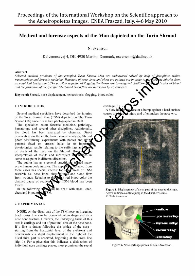
NOSE. At the distal part of the TSM nose an irregular, black cross line can be observed, often diagnosed as a nose bone fracture. However, the underlying tissue of this area is cartilage and out of proximal area of the nose bone. If a line is drawn following the bridge of the nose - starting from the horizontal level of the eyebrows and downwards - a slight displacement to the right of the distal third part is observed, beginning at the cross line (fig. 1). For a physician this indicates a dislocation of individual nose cartilage pieces, most prominent the septal
cartilage (fig. 2). A blow hitting the nose or a bump against a hard surface
causes this kind of injury and often makes the nose wry.
Figure 1. Displacement of distal part of the nose to the right. Arrow indicates outline jump at the distal cross line. © Niels Svensson.
Figure 2. Nose cartilage pieces. © Niels Svensson.
www.ache
iropo
ietos
.info
Proceedings of the International Workshop on the Scientific approach to the Acheiropoietos Images, ENEA Frascati, Italy, 4‐6 May 2010
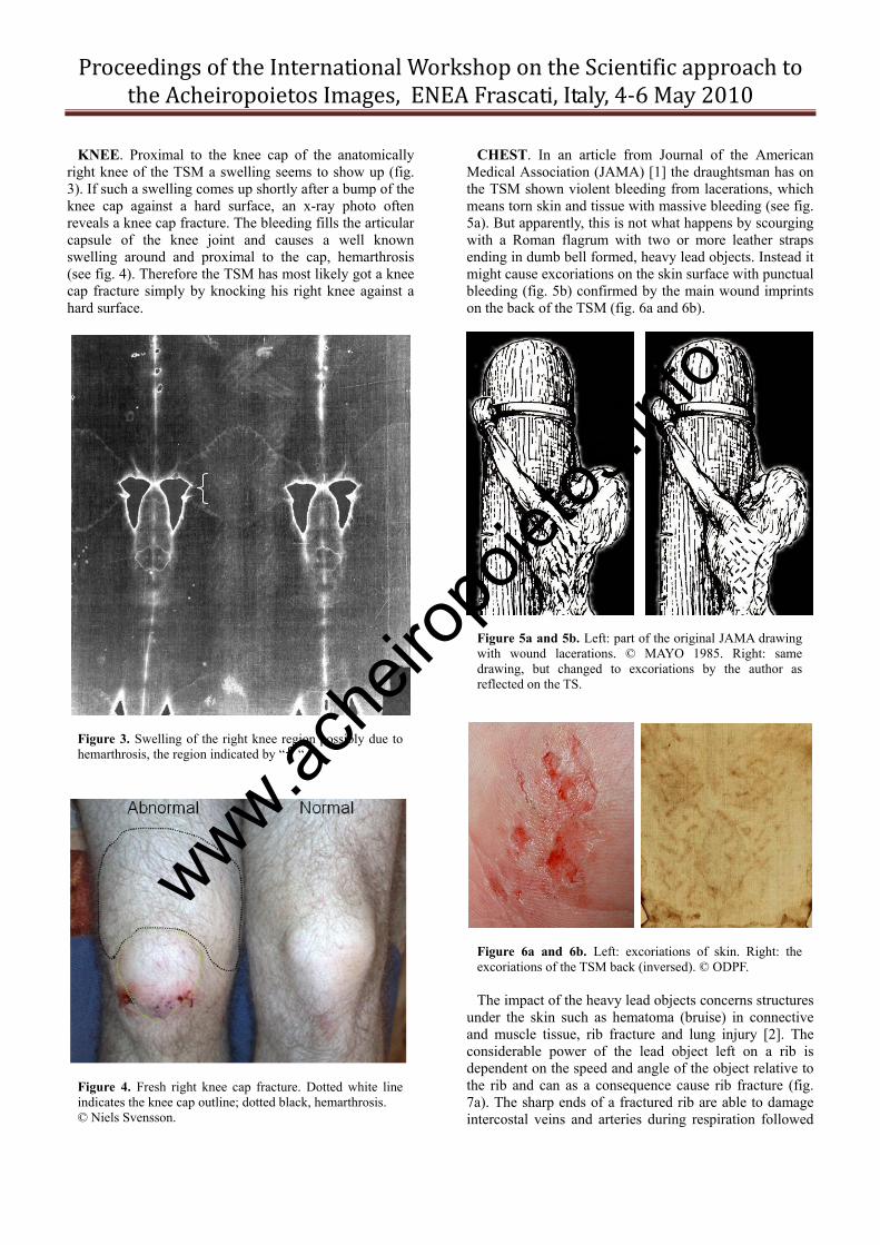
KNEE. Proximal to the knee cap of the anatomically right knee of the TSM a swelling seems to show up (fig. 3). If such a swelling comes up shortly after a bump of the knee cap against a hard surface, an x-ray photo often reveals a knee cap fracture. The bleeding fills the articular capsule of the knee joint and causes a well known swelling around and proximal to the cap, hemarthrosis (see fig. 4). Therefore the TSM has most likely got a knee cap fracture simply by knocking his right knee against a hard surface.
Figure 3. Swelling of the right knee region possibly due to hemarthrosis, the region indicated by “ “.
Figure 4. Fresh right knee cap fracture. Dotted white line indicates the knee cap outline; dotted black, hemarthrosis. © Niels Svensson.
CHEST. In an article from Journal of the American Medical Association (JAMA) [1] the draughtsman has on the TSM shown violent bleeding from lacerations, which means torn skin and tissue with massive bleeding (see fig. 5a). But apparently, this is not what happens by scourging with a Roman flagrum with two or more leather straps ending in dumb bell formed, heavy lead objects. Instead it might cause excoriations on the skin surface with punctual bleeding (fig. 5b) confirmed by the main wound imprints on the back of the TSM (fig. 6a and 6b).
Figure 5a and 5b. Left: part of the original JAMA drawing with wound lacerations. © MAYO 1985. Right: same drawing, but changed to excoriations by the author as reflected on the TS.
Figure 6a and 6b. Left: excoriations of skin. Right: the excoriations of the TSM back (inversed). © ODPF.
The impact of the heavy lead objects concerns structures
under the skin such as hematoma (bruise) in connective and muscle tissue, rib fracture and lung injury [2]. The considerable power of the lead object left on a rib is dependent on the speed and angle of the object relative to the rib and can as a consequence cause rib fracture (fig. 7a). The sharp ends of a fractured rib are able to damage intercostal veins and arteries during respiration followed
www.ache
iropo
ietos
.info

Proceedings of the International Workshop on the Scientific approach to the Acheiropoietos Images, ENEA Frascati, Italy, 4‐6 May 2010
Figure 7a. Back view of the thorax. White arrow points to the intercostal vein and artery, the black to a rib fracture. The lung sac (green) covers the inside of the thorax. © Niels Svensson.
by bleeding into a damaged lung sac (fig. 7b), a common condition in emergency practice known as hemothorax (figures 8a and 8b). One part of the lung sac is widely outstretched behind the chest ribs, the other covers the lungs. In between is vacuum. The sharp fractured ribs may partly cause insufflation of air and bleeding into the sac. Both conditions may cause full or partly lung collapse.
If the individual dies, the postmortem blood in the lung sac will during approximately half to one hour - due to gravity - separate into two layers, i.e. the serum layer and the underlying dense blood corpuscle layer. This sequence
Figure 8a. Left: a test tube with blood one hour after sampling. The blood has separated into serum and blood corpuscle layers. This separation of whole blood also takes place after death in the lung sac, if the body is placed upright (right). © Niels Svensson.
Figure 7b. Front view of the thorax (ribs removed). White arrow marks lower border of the right lung sac, black the lower border of the lung and grey arrow the heart sac. © Niels Svensson.
can be demonstrated by fresh taken blood in a test tube, where the blood separates into two layers (fig. 8a). Apparently, this was what happened to Jesus after his death on the cross as the Gospel of John underlines by eyewitness (John 20.34). When a lance is thrusted into the lower area of the lung sac (fig. 8b), it is expected that the blood and serum pour out of the wound like “blood and water” - and in this succession running downwards. The large side wound of the TSM with blood running out in cascades to the right side of the chest and to the small of the back points to the lung sac as its origin.
Figure 8b. The side wound of the TSM (inversed) has been superimposed figure 8a in order to show the anatomic place of the wound. A lancea shows the place and direction of the thrust. The blood will pour out in cascades. © Niels Svensson.
www.ache
iropo
ietos
.info
Proceedings of the International Workshop on the Scientific approach to the Acheiropoietos Images, ENEA Frascati, Italy, 4‐6 May 2010
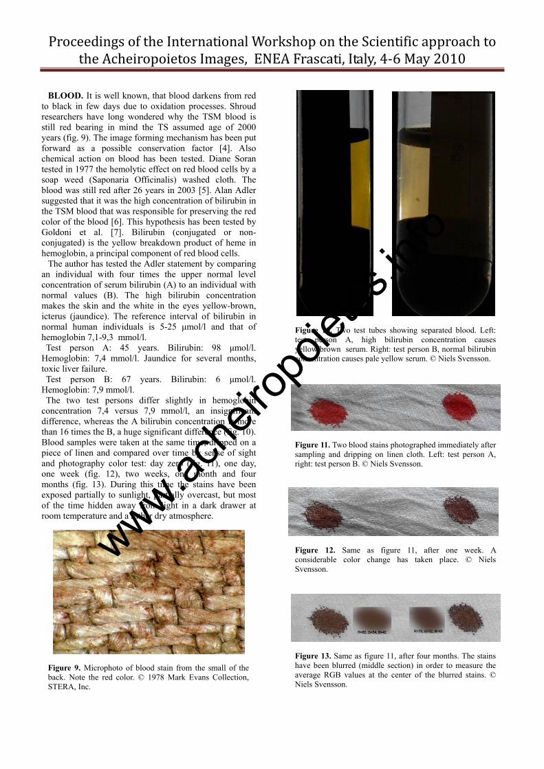
BLOOD. It is well known, that blood darkens from red to black in few days due to oxidation processes. Shroud researchers have long wondered why the TSM blood is still red bearing in mind the TS assumed age of 2000 years (fig. 9). The image forming mechanism has been put forward as a possible conservation factor [4]. Also chemical action on blood has been tested. Diane Soran tested in 1977 the hemolytic effect on red blood cells by a soap weed (Saponaria Officinalis) washed cloth. The blood was still red after 26 years in 2003 [5]. Alan Adler suggested that it was the high concentration of bilirubin in the TSM blood that was responsible for preserving the red color of the blood [6]. This hypothesis has been tested by Goldoni et al. [7]. Bilirubin (conjugated or non-conjugated) is the yellow breakdown product of heme in hemoglobin, a principal component of red blood cells.
The author has tested the Adler statement by comparing an individual with four times the upper normal level concentration of serum bilirubin (A) to an individual with normal values (B). The high bilirubin concentration makes the skin and the white in the eyes yellow-brown, icterus (jaundice). The reference interval of bilirubin in normal human individuals is 5-25 μmol/l and that of hemoglobin 7,1-9,3 mmol/l. Test person A: 45 years. Bilirubin: 98 μmol/l. Hemoglobin: 7,4 mmol/l. Jaundice for several months, toxic liver failure. Test person B: 67 years. Bilirubin: 6 μmol/l. Hemoglobin: 7,9 mmol/l. The two test persons differ slightly in hemoglobin concentration 7,4 versus 7,9 mmol/l, an insignificant difference, whereas the A bilirubin concentration is more than 16 times the B, a huge significant difference (fig. 10). Blood samples were taken at the same time, dripped on a piece of linen and compared over time by sense of sight and photography color test: day zero (fig. 11), one day, one week (fig. 12), two weeks, one month and four months (fig. 13). During this time the stains have been exposed partially to sunlight, partially overcast, but most of the time hidden away from light in a dark drawer at room temperature and a rather dry atmosphere.
Figure 9. Microphoto of blood stain from the small of the back. Note the red color. © 1978 Mark Evans Collection, STERA, Inc.
Figure 10. Two test tubes showing separated blood. Left: test person A, high bilirubin concentration causes yellow/brown serum. Right: test person B, normal bilirubin concentration causes pale yellow serum. © Niels Svensson.
Figure 11. Two blood stains photographed immediately after sampling and dripping on linen cloth. Left: test person A, right: test person B. © Niels Svensson.
Figure 12. Same as figure 11, after one week. A considerable color change has taken place. © Niels Svensson.
Figure 13. Same as figure 11, after four months. The stains have been blurred (middle section) in order to measure the average RGB values at the center of the blurred stains. © Niels Svensson.
www.ache
iropo
ietos
.info

Proceedings of the International Workshop on the Scientific approach to the Acheiropoietos Images, ENEA Frascati, Italy, 4‐6 May 2010
After four months no significant color difference of A and B was observed. The red/green/blue (RGB) values of the photos of the blood were measured by blurring the stains and afterwards measuring in the center of the stains (fig. 13 and table 1).
TABLE 1.
Red Green Blue
Person A 80 54 42
Person B 79 52 43 From the interpretation of the figures it follows that in
the short run (months) high bilirubin concentration has no significant influence on the color of human blood on linen cloth, and therefore, presumably, neither in the long run (years).
It must be added, that the high level of bilirubin (test person A) is mainly composed of conjugated bilirubin while in Adler's hypothesis the main part of the bilirubin might be non-conjugated or free bilirubin. It is certainly very difficult to find blood samples with high levels of free bilirubin. If possible, future experiments should be done with free bilirubin to detect potential differences.
THE ”ε"-SHAPED BLOOD TRICKLE. The TSM
has a characteristic blood trickle above the anatomical left eyebrow (fig. 14), the so called epsilon, ”ε” (the photographical positive image) or figure “3” (the photographical negative image). Shroud literature often claims that the curved (meandering) run of this rivulet is caused by wrinkles/furrows of the forehead. The furrows come up because the man is in agony. The author tested this hypothesis by dripping some drops of his own blood on his furrowed forehead - the head in vertical position - and found by several repetitions that the blood ran straight down to the bony projection of the eyebrow without curving (fig. 15).
Figure 14. The characteristic figure “3” above the left eyebrow of the TSM (white arrow).
Figure 15. Fresh blood dripped above the left eyebrow, the head still. The blood runs down in a straight line. © Niels Svensson.
Figure 16. By tilting the head slowly to the right and left the blood takes a curved, meandering run. © Niels Svensson.
If the head instead is tilted to the left and right during
the blood run, the curved, meandering blood rivulet shows up (fig. 16). Once the rivulet is first formed then the outpouring blood continues to follow the path. Of course the individual furrow pattern influences the flow pattern, but surprisingly the head movement seems to be an additional and substantial curving factor. 3. CONCLUSIONS
From experience of emergency medicine and performed experiments the following can be concluded concerning selected injuries on the Turin Shroud Man:
NOSE. The nose is slightly displaced to the right due to nose cartilage displacement.
KNEE. The swelling of the right knee is most likely due to a knee cap fracture.
CHEST. The cascades of blood from the right side wound emerge from postmortem, separated blood in the lung sac.
BLOOD. The red color of the TSM blood seems not due to high bilirubin concentration.
THE ”ε"-SHAPED BLOOD TRICKLE. The “ε” or figure “3” blood rivulet formation seems, apart from forehead furrows, due to different positions of the head.
www.ache
iropo
ietos
.info
Proceedings of the International Workshop on the Scientific approach to the Acheiropoietos Images, ENEA Frascati, Italy, 4‐6 May 2010
ACKNOWLEDGMENTS
I wish to thank Giulio Fanti, Department of Mechanical Engineering, University of Padua for blood color analysis and Barrie Schwortz, STERA, Inc. for use of Mark Evans Collection. REFERENCES 1. W.D. Edwards, W. J. Gabel, F. E. Hosmer: On the Physical Death of Jesus Christ, JAMA, 255, 1455-1463 (1986)
2. F.T. Zugibe: The Crucifixion of Jesus: A Forensic Inquiry, M. Evans and Company, 21-24 (2005)
3. N. Svensson: Det sande ansigt, Gyldendal, 61-62, 69-78 (2007)
4. J. Jackson: Is the Image on the Shroud Due to a Process Heretofore Unknown to Modern Science? Shroud Spectrum International 34, 19 (1990)
5. R.N. Rogers, A. Arnoldi: Scientific method applied to the Shroud of Turin, www.shroud.com/pdfs/rogers2.pdf (2002)
6. A.D. Adler: The Orphaned Manuscript, Effeta Editrice, 60-61 (2002)
7. C. Goldoni: The Shroud of Turin and the bilirubin blood stains, www.ohioshroudconference.com/papers/p04.pdf (2008)
www.ache
iropo
ietos
.info