proliferation extent of cd34+ cells as a key parameter to maximize megakaryocytic differentiation of...
TRANSCRIPT

Biotechnology Reports 4 (2014) 50–55
Proliferation extent of CD34+ cells as a key parameter to maximizemegakaryocytic differentiation of umbilical cord blood-derivedhematopoietic stem/progenitor cells in a two-stage culture protocol$
Javad Hatami a, Pedro Z. Andrade a, Denise Bacalhau b, Fernando Cirurgião b,Frederico Castelo Ferreira a,*, Joaquim M.S. Cabral a, Cláudia L. da Silva a
aDepartment of Bioengineering and IBB – Institute for Biotechnology and Bioengineering, Instituto Superior Técnico, Universidade de Lisboa, Av. Rovisco Pais,Nr. 1, 1049-001 Lisbon, PortugalbDepartment of Obstetrics, S. Francisco Xavier Hospital, Estrada do Forte do Alto do Duque, 1449-005 Lisbon, Portugal
A R T I C L E I N F O
Article history:Received 19 March 2014Accepted 14 July 2014Available online 16 July 2014
Keywords:Megakaryocytic differentiationPlateletsMegakaryocytesHematopoietic stem/progenitor cellsUmbilical cord blood
A B S T R A C T
Co-infusion of ex-vivo generated megakaryocytic progenitors with hematopoietic stem/progenitor cells(HSC/HPC) may contribute to a faster platelet recovery upon umbilical cord blood (UCB) transplantation.A two stage protocol containing cell expansion and megakaryocyte (Mk) differentiation was establishedusing human UCB CD34+-enriched cells. The expansion stage used a pre-established protocol supportedby a human bone marrow mesenchymal stem cells (MSC) feeder layer and the differentiation stageused TPO (100 ng/mL) and IL-3 (10 ng/mL).18% of culture-derived Mks had higher DNA content (>4 N) andwere able to produce platelet-like particles. The proliferation extent of CD34+ cells obtained in theexpansion stage (FI-CD34+), rather than expansion duration, determined as a key parameter for efficientmegakaryocytic differentiation. A maximum efficiency yield (EY) of 48 � 7.7 Mks/input CD34+ cells wasobtained for a FI-CD34+ of 17 � 2.5, where a higher FI-CD34+ of 42 � 13 resulted in a less efficientmegakaryocytic differentiation (EY of 22 � 6.7 and 19 � 4.6 %CD41).ã 2014 The Authors. Published by Elsevier B.V. This is an open access article under the CC BY license
(http://creativecommons.org/licenses/by/3.0/).
Contents lists available at ScienceDirect
Biotechnology Reports
journal homepage: www.elsevier .com/ locate /btre
1. Introduction
Allogeneic transplantation of CD34+-enriched cells from humanumbilical cord blood (UCB) as a source of hematopoieticstem/progenitor cells (HSC/HPC) is a potential therapy for treatinghemato-oncological diseases and other blood disorders in adultpatients [1,2]. However, a delayed platelet recovery is typicallyassociated to the transplantation of HSC/HPC from UCB, whencompared to adult sources (bone marrow (BM) and mobilizedperipheral blood (mPB)) [3]. Administration of ex-vivo generatedmegakaryocytic progenitor cells and megakaryocytes (Mks) aloneor co-infusion with UCB HSC/HPC can be a promising strategy toreduce the prolonged period of platelet recovery [4,5].
Mks are rare, large and polyploid myeloid cells, which resideprimary in the BM region adjacent to sinusoidal walls [6]. Plateletbiogenesis from Mks occurs through nuclear polyploidization,
$ This is an open-access article distributed under the terms of the CreativeCommons Attribution-NonCommercial-No Derivative Works License, whichpermits non-commercial use, distribution, and reproduction in any medium,provided the original author and source are credited.* Corresponding author. Tel.: +351 218419598; fax: +351 218419062.E-mail address: [email protected] (F.C. Ferreira).
http://dx.doi.org/10.1016/j.btre.2014.07.0022215-017X/ã 2014 The Authors. Published by Elsevier B.V. This is an open access artic
cellular enlargement, cytoplasmic maturation and platelet release.The production of Mks and platelets from different sources of cellssuch as UCB, BM or mPB, as well as embryonic stem cells andinduced pluripotent stem cells has been studied over the lastdecades [7]. In this context, different biological, chemical andphysical factors have been studied in order to establish an optimalprotocol to enhance megakaryocytic differentiation from primitivecell populations [8–11].
The main objective of this study was to test if an optimizedexpansion stage followed by a megakaryocytic differentiationstage would be an effective strategy to maximize Mk productionfrom UCB HSC/HPC. Specifically, we aimed at systematicallyidentifying a relation between proliferation extent of CD34+ cellsand effective megakaryocytic differentiation.
2. Material and methods
2.1. Cell culture
hUCB and hMSC samples were obtained from healthy donorsafter maternal donor and donor informed consent, respectively.CD34+-enriched cells from UCB were expanded using a previouslyoptimized protocol [12]. Briefly, low density mononuclear cells
le under the CC BY license (http://creativecommons.org/licenses/by/3.0/).
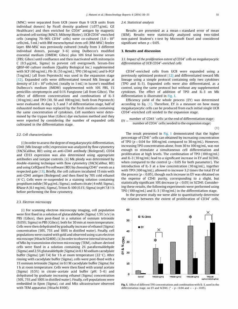
Fig. 1. Effect of different TPO concentrations and combination with IL-3, used in thedifferentiation stage, on EY and %CD41. (* – p < 0.04 and � – p < 0.05).
J. Hatami et al. / Biotechnology Reports 4 (2014) 50–55 51
(MNC) were separated from UCB (more than 9 UCB units fromindividual donors) by Ficoll density gradient (1.077 g/mL; GEHealthcare) and then enriched for CD34+ antigen by magneticactivated cell sorting (MACS; Miltenyi Biotec). UCB CD34+-enrichedcells (ranging 70–90% CD34+ cells) were co-cultured (3.0 � 103
cells/mL, 5 mL) with BM mesenchymal stem cell (BM-MSC) feederlayer. BM-MSC was previously cultured (totally from 3 differentindividual donors, passage 3–6) using Dulbecco's modifiedessential medium (DMEM; Gibco) plus 10% fetal bovine serum(FBS; Gibco) until confluence and then inactivated with mitomycinC (0.5 mg/mL, Sigma) to prevent cell overgrowth. Serum-freeQBSF-60 culture medium (Quality Biological Inc.) supplementedwith SCF (60 ng/mL), Flt-3L (55 ng/mL), TPO (50 ng/mL) and b-FGF(5 ng/mL) (all from Peprotech) was used in the expansion stage[12]. Expanded cells were differentiated toward Mk lineage atdensity of 2.0 � 105 cells/mL (totally in 1 mL) in Iscove's modifiedDulbecco's medium (IMDM) supplemented with 10% FBS, 1%penicillin–streptomycin and 0.1% Fungizone (all from Gibco). Theeffect of different concentrations and combinations of IL-3(10 ng/mL) and TPO (30, 50 and 100 ng/mL; both from Peprotech)were evaluated. At days 3, 5 and 7 of differentiation stage, half ofexhausted medium was replaced by the fresh medium containingthe same concentration of cytokines. Cell numbers were deter-mined by the trypan blue (Gibco) dye exclusion method and theywere reported by considering the number of expanded cellscultivated in the differentiation stage.
2.2. Cell characterization
(i) In order to assess the degree of megakaryocytic differentiation,CD41 (Mk lineage cells) expression was analyzed by flow cytometry(FACSCalibur, BD) using an anti-CD41 antibody (Biolegend). CD34and CD33 expression was also determined using appropriateantibodies and isotype controls. (ii) Mk ploidy was determined bydouble-staining technique with flow cytometry (FACSCalibur, BD)and using CellQuest Pro software (BD) by choosing CD41+ events as arespected gate [13]. Briefly, the cell cultures incubated 15 min withanti-CD41 antigen (Biolegend) and then fixed by 70% cold ethanol(4 �C). Cells were re-suspended in a staining solution containingpropidium iodide (50 mg/mL; Sigma), sodium citrate (4 mM; Sigma),RNase A (0.1 mg/mL; Sigma), Triton X-100 (0.1%; Sigma) in pH 7.8 1 hbefore performing the flow cytometry.
2.3. Electron microscopy
(i) For scanning electron microscopy imaging, cell populationwere first fixed in a solution of glutaraldehyde (Sigma) 1.5% (v/v) inPBS (Gibco), then post-fixed in a solution of osmium tetroxide(0.05%; Sigma) in PBS (Gibco); both for 30 min at room temperature.Cells were then dehydrated by gradually increase of ethanol (Sigma)concentration (50%, 75% and 100% in distilled water). Finally, cellpopulations were coated with gold and observed using scan electronmicroscope(HitachiS2400). (ii) Inordertoobserve internalstructureof Mks by transmission electron microscopy (TEM), culture-derivedcells were fixed in a solution containing 2% paraformaldehyde(Sigma) and 2.5% glutaraldehyde (Sigma) in 0.1 M sodium cacodylatebuffer (Sigma) (pH 7.4) for 1 h at room temperature (22 �C). Afterrinsing with cacodylate buffer (Sigma), cells were post-fixed with a1% osmium tetroxide (Sigma) in 0.1 M cacodylate buffer (Sigma) for1 h at room temperature. Cells were then fixed with uranyl acetate(Sigma) (0.5%) in citrate–acetate acid buffer (pH: 5–6) anddehydrated by graduate increasing ethanol (Sigma) concentration(50%, 75% and 100% in distilled water). Finally, cell populations wereembedded in Epon (Sigma), cut and Mks ultrastructure observedwith TEM apparatus (Hitachi 8100).
2.4. Statistical analysis
Results are presented as a mean � standard error of mean(SEM). Results were statistically analyzed using two-sidednon-paired Student's t-test by Microsoft Excel and consideredsignificant when p < 0.05.
3. Results and discussion
3.1. Impact of the proliferation extent of CD34+ cells on megakaryocyticdifferentiation of UCB CD34+-enriched cells
CD34+-enriched cells from UCB were expanded using apreviously optimized protocol [12] and differentiated toward Mklineage using a simple protocol containing only two cytokines(TPO and IL-3). Expanded cells were also differentiated, as acontrol, using the same protocol but without any supplementedcytokines. The effect of addition of TPO and IL-3 on Mkdifferentiation is illustrated in Fig. 1.
Efficiency yield of the whole process (EY) was determinedaccording to Eq. (1). Therefore, EY is a measure on how manymegakaryocytic cells can be produced from each initial single UCBCD34+-enriched cell seeded to the expansion stage.
EY ¼ number of CD41þcells ðat the end of differentiation stageÞnumber of CD34þcellsðseeded to the expansion stageÞ
(1)
The result presented in Fig. 1 demonstrated that the higherpercentage of CD41+ cells can obtained by increasing concentrationof TPO (p < 0.04 for 100 ng/mL compared to 30 ng/mL). However,increasing TPO concentration alone, from 30 to 100 ng/mL, was notenough to stimulate a simultaneous cell differentiation andproliferation at high levels. The combination of TPO (100 ng/mL)and IL-3 (10 ng/mL) lead to a significant increase in EY and %CD41,when compared to the control (p < 0.05 for both parameter). Theintroduction of IL-3 at a low concentration (10 ng/mL), togetherwith TPO (100 ng/mL), allowed to increase 3.2 times the total EY ofthe process (p < 0.05), though such increase in EY was obtained onthe expense of CD41 purity, corresponding to a slight, butstatistically significant 10% decrease (p < 0.05) in %CD41. Consider-ing these results, the following experiments were performed usingTPO (100 ng/mL) and IL-3 (10 ng/mL) in the differentiation stage.
In the present study we were able to quantitatively determinethe relation between the extent of proliferation of CD34+ cells,
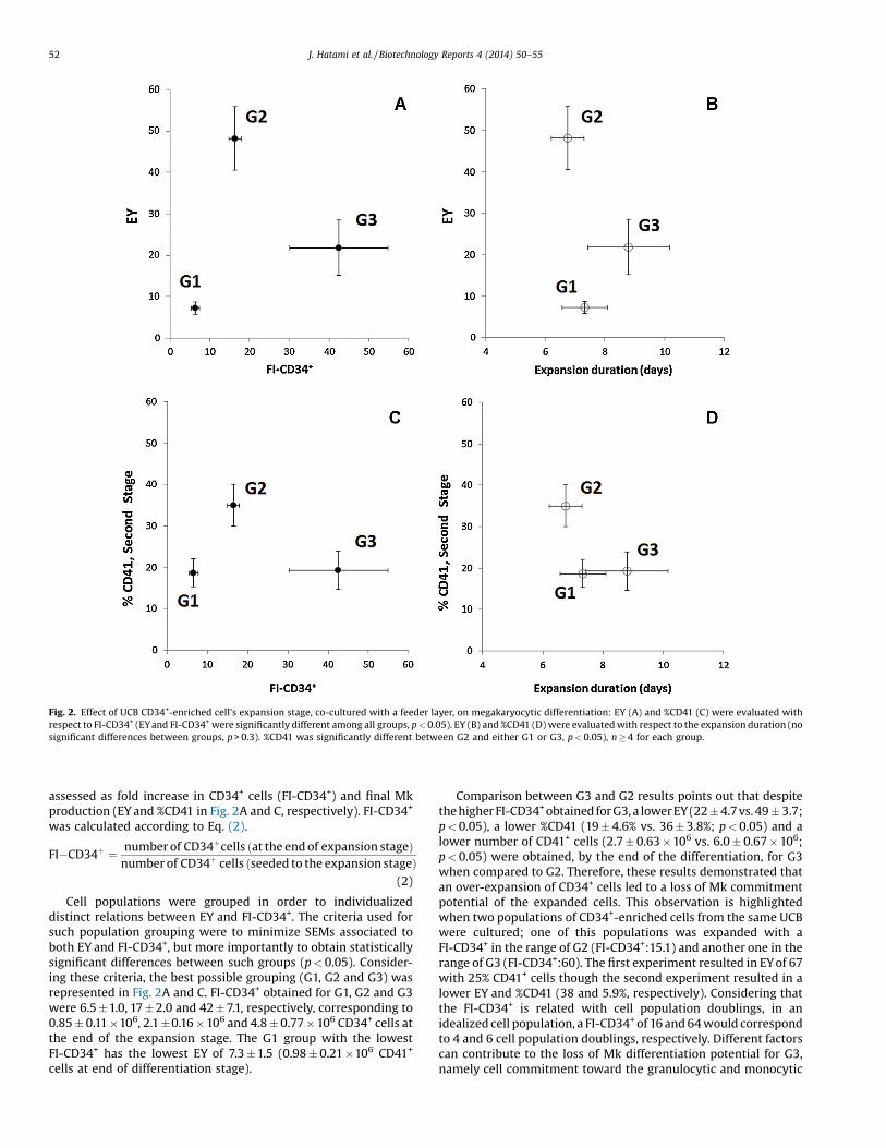
Fig. 2. Effect of UCB CD34+-enriched cell's expansion stage, co-cultured with a feeder layer, on megakaryocytic differentiation: EY (A) and %CD41 (C) were evaluated withrespect to FI-CD34+ (EYand FI-CD34+were significantly different among all groups, p < 0.05). EY (B) and %CD41 (D) were evaluated with respect to the expansion duration (nosignificant differences between groups, p > 0.3). %CD41 was significantly different between G2 and either G1 or G3, p < 0.05), n � 4 for each group.
52 J. Hatami et al. / Biotechnology Reports 4 (2014) 50–55
assessed as fold increase in CD34+ cells (FI-CD34+) and final Mkproduction (EY and %CD41 in Fig. 2A and C, respectively). FI-CD34+
was calculated according to Eq. (2).
FI�CD34þ ¼ number of CD34þcells ðat the end of expansion stageÞnumber of CD34þ cells ðseeded to the expansion stageÞ
(2)
Cell populations were grouped in order to individualizeddistinct relations between EY and FI-CD34+. The criteria used forsuch population grouping were to minimize SEMs associated toboth EY and FI-CD34+, but more importantly to obtain statisticallysignificant differences between such groups (p < 0.05). Consider-ing these criteria, the best possible grouping (G1, G2 and G3) wasrepresented in Fig. 2A and C. FI-CD34+ obtained for G1, G2 and G3were 6.5 �1.0, 17 � 2.0 and 42 � 7.1, respectively, corresponding to0.85 � 0.11 �106, 2.1 �0.16 � 106 and 4.8 � 0.77 � 106 CD34+ cells atthe end of the expansion stage. The G1 group with the lowestFI-CD34+ has the lowest EY of 7.3 � 1.5 (0.98 � 0.21 �106 CD41+
cells at end of differentiation stage).
Comparison between G3 and G2 results points out that despitethe higher FI-CD34+obtained for G3, a lower EY (22 � 4.7 vs. 49 � 3.7;p < 0.05), a lower %CD41 (19 � 4.6% vs. 36 � 3.8%; p < 0.05) and alower number of CD41+ cells (2.7 � 0.63 � 106 vs. 6.0 � 0.67 � 106;p < 0.05) were obtained, by the end of the differentiation, for G3when compared to G2. Therefore, these results demonstrated thatan over-expansion of CD34+ cells led to a loss of Mk commitmentpotential of the expanded cells. This observation is highlightedwhen two populations of CD34+-enriched cells from the same UCBwere cultured; one of this populations was expanded with aFI-CD34+ in the range of G2 (FI-CD34+:15.1) and another one in therange of G3 (FI-CD34+:60). The first experiment resulted in EY of 67with 25% CD41+ cells though the second experiment resulted in alower EY and %CD41 (38 and 5.9%, respectively). Considering thatthe FI-CD34+ is related with cell population doublings, in anidealized cell population, a FI-CD34+ of 16 and 64 would correspondto 4 and 6 cell population doublings, respectively. Different factorscan contribute to the loss of Mk differentiation potential for G3,namely cell commitment toward the granulocytic and monocytic

Fig. 3. Phenotypic characterization of the UCB CD34+-enriched population; %CD33 (A), %CD 34 (B) and %CD41 (C) of each group at the start of expansion (start), the end ofexpansion (Exp.) and differentiation (Diff.) stages. Results presented as mean � SEM (n � 4) and p < 0.05 between * and �.
J. Hatami et al. / Biotechnology Reports 4 (2014) 50–55 53
lineage (24.0 � 4.3% CD14+ cells) [12], or neutrophil lineages(64.0 � 12.1% HLA-DR++ CD117++) [14].
As a control, UCB CD34+-enriched cells were expanded in thesame culture conditions, but in absence of feeder layer; regardlessthe different conditions tested, both FI-CD34+ and EY were
Fig. 4. Microscopy imaging: representative scanning electron microscopy images of huplatelet-like particles (right) (A), transmission electron microscopy (TEM) images of cultare indicated in the image (B). Morphology of culture-derived cells by inverted micros
maintained at low levels (2.7 � 0.91 and 7.0 � 1.2, respectively;n = 3). It has been previously reported that FI-CD34+ was consis-tently lower in the absence of feeder [12,15]. Therefore, this resulthighlighted the positive effect of presence of feeder layer, in theexpansion stage, when targeting an efficient Mk differentiation.
man peripheral blood-derived platelets (left) and ex-vivo generated UCB-derivedure-derived Mk. The nucleus (N), demarcation membrane (dm) and a-granules (G)copy (C). All images were drawn from G2 group.

Fig. 5. Ploidy analysis of culture-derived Mks (A), forward (FSC) and side (SCC) scatter properties of CD41+ cells with 2 N, 4 N and >4 N DNA content (light-yellow) (B).Background scatters (dark-grey) belong to all CD41+ cell population. (For interpretation of the references to color in this figure legend, the reader is referred to the web versionof this article.)
54 J. Hatami et al. / Biotechnology Reports 4 (2014) 50–55
Boyer and colleagues have previously suggested a 5-dayexpansion period as optimal for the increased production ofMks from UCB CD34+ cells (>95% enriched) in a two-phaseprotocol. However, using FI-CD34+ in the expansion stage as anoperational parameter, rather than the expansion duration, hasmore advantages such as considering the intrinsic biologicalvariability of UCB samples and the impact of initial CD34+
enrichment. The current study thus demonstrated that by usingFI-CD34+, as a key parameter, we were able to determine theeffectiveness of megakaryocyte differentiation of UCB cells,identifying different groups with statistical significance (G1, G2and G3 in Fig. 2A and C in terms of FI-CD34+, p < 0.05). Indeed, suchidentification would not be statistically significant if expansionduration was used instead (Fig. 2B and D; p > 0.3 between G1, G2and G3 in terms of expansion duration).
In the current study, the initial population consisted of 1.5 �105
cells with similar cell population compositions (Fig. 3). At the end ofthe expansion, the total numbers of cells were 1.7 � 0.40 � 106,4.2 � 0.30 � 106 and 20 � 9.1 �106 for G1, G2 and G3, respectively. Inthe expansion stage, the reduction in %CD34 (from 90 to 65% for G1,from 83% to 51% for G2 and from 77% to 36% for G3) was accompaniedbyan increase in %CD33 (early myeloid cells), from 56% to 83% for G1,from 52% to 91% for G2 and from 53% to 92% for G3.
A significant decrease in %CD34 was observed during thedifferentiation stage (from 65% to 2.9% for G1, 51–2.5% for G2 and36–5% for G3, Fig. 3A and B). Such decrease in %CD34 wasconcomitant to an increase in %CD41 indicating differentiationtoward Mk lineage. Comparing with the expansion stage, %CD41increased during differentiation stage from 13% to 19% for G1, whilefor G2 raised from 13% to 35%, but only from 17% to 19% for G3(Fig. 3C). Over differentiation stage, the total number of cellsincreased about 3.7 folds for G1 (corresponding to 6.3 �1.0 � 106
total cells), and 4.4 folds for G2 (corresponding to 19 � 4.2 �106
total cells), but only about 1.3 for G3 (corresponding to26 � 13 � 106 total cells).
3.2. Characterization of cultured-derived Mks and platelets-likeparticles
Scanning electron microscopy analysis showed similar mor-phology of culture-derived platelet-like particles and human
PB-derived platelets (Fig. 4A, right and left, respectively),demonstrating the ability of the current protocol to support thein vitro production of platelet-like particles. Likewise, transmis-sion electron microscopy (TEM) analysis of culture-derived Mk(Fig. 4B) showed normal features of a mature Mks withdemarcation membrane (dm) system, nucleus (N) and a-granulescharacteristic of such mature Mk. Electron microscopy (SEM andTEM) imaging was performed on 3 different populations from G2and for each culture, platelet-like particles (similar to the Fig. 4A)was identified in more than 10 microscopy images.
Ploidy analysis (Fig. 5A) revealed that about 18% of culture-derived Mks have higher ploidy (>4 N). Moreover, forward (FCS)and side scatter (SCC) properties of such population are highercompared to the CD41+ cells with 2 N and 4 N DNA content(Fig. 5B). Mks generated from UCB, compared to human PB, weredescribed to be smaller and have less ploidy; however, as reportedpreviously [13] and confirmed in the current study, these are stillable to produce platelets-like particles.
4. Conclusions
The current study presents a two-stage protocol aiming ateffective megakaryocytic differentiation of UCB CD34+-enrichedcells. The results identified distinct individual groups whichelucidate the relation between FI-CD34+ and efficiency of Mkproduction. This information is valuable to balance the proliferationand differentiation potential of CD34+ cells, when targeting efficientMk production. The underlying phenomena for such balance shouldbe actually based in cell population doublings, but FI-CD34+ is atangible parameter easier to quantify. Several studies have reportedproduction of Mk cells and platelets from HSC/HCP. For example,using UCB progenitors, a perfusion system was used to produceenough number of platelets in vitro for clinical transfusion (300–600 � 109) [16]. However, the drawback of aforementioned work wasmost of culture-derived platelets were activated in the absence ofany agonists. Another study reported producing 44 � 8.1 Mks/inputHSC/HPC using human mPB cells through a complex 3-step culture;includes a cocktail comprised by 17 different cytokines and changesin pH and O2 tension during experiment [17]. However, by using onlytwo cytokines (TPO and IL-3), in the differentiation stage, our simplerdifferentiation protocol was able to produce 48 � 7.7 Mks/input

J. Hatami et al. / Biotechnology Reports 4 (2014) 50–55 55
CD34+ cells using a 17 � 2.5 FI-CD34+ and benefits from using UCBprogenitors which are largely available and usually discarded afterdelivery involving a non-invasive collection procedure. This workquantitatively demonstrates that the FI-CD34+, rather than expan-sion duration, can be used as a key parameter to maximize Mk cellgeneration from CD34+-enriched cells. When adapted to fullydefined, GMP-compliant culture reagents and conditions, thisprotocol has the potential to impact cellular therapies within thehemato-oncological field.
Conflict of interest
The authors declare no commercial or financial conflict ofinterest.
Acknowledgements
This work was financially supported by the Fundação para aCiência e Tecnologia (FCT), Portugal through Project PTDC/EQU-EQU/114231/2009, MIT-Portugal Program, PhD scholarshipSFRH/BD/61450/2009 (J. Hatami) and the research contractIF/00442/2012 (F. Ferreira). The authors thank Isabel Nogueira(MicroLab IST), Dr. Patrícia Carvalho (NanoLab IST) and Dr. AntónioPedro Matos (Hospital Curry Cabral, Lisbon, Portugal) for thecontribution with electron microscopy analysis. The authors alsothank Dr. Ana Paula Sousa (Instituto Português do Sangue, Lisbon,Portugal) for donation of PB-derived platelets.
References
[1] C. Cutler, K.K. Ballen, Improving outcomes in umbilical cord bloodtransplantation: state of the art, Blood Rev. 26 (6) (2012) 241–246.
[2] J. Kurtzberg, Update on umbilical cord blood transplantation, Curr. Opin.Pediatr. 21 (1) (2009) 22–29.
[3] P. Ramirez, et al., Delayed platelet recovery after allogeneic transplantation: apredictor of increased treatment-related mortality and poorer survival, BoneMarrow Transplant. 46 (7) (2011) 981–986.
[4] D. Decaudin, et al., Ex vivo expansion of megakaryocyte precursor cells inautologous stem cell transplantation for relapsed malignant lymphoma, BoneMarrow Transplant. 34 (12) (2004) 1089–1093.
[5] H. Emond, et al., Cotransplantation of ex vivo expanded progenitors withnonexpanded cord blood cells improves platelet recovery, Stem Cells Dev. 21(17) (2012) 3209–3219.
[6] S.R. Patel, J.H. Hartwig, J.E. Italiano Jr., The biogenesis of platelets frommegakaryocyte proplatelets, J. Clin. Invest. 115 (12) (2005) 3348–3354.
[7] J.-A. Reems, N. Pineault, S. Sun, In vitro megakaryocyte production and plateletbiogenesis: state of the art, Transfus. Med. Rev. 24 (1) (2010) 33–43.
[8] F. Norol, et al., Effects of cytokines on platelet production from blood andmarrow CD34+ cells, Blood 91 (3) (1998) 830–843.
[9] N. Pineault, et al., Characterization of the effects and potential mechanismsleading to increased megakaryocytic differentiation under mild hyperthermia,Stem Cells Dev. 17 (3) (2008) 483–493.
[10] S.S. Mostafa, W.M. Miller, E.T. Papoutsakis, Oxygen tension influences thedifferentiation, maturation and apoptosis of human megakaryocytes, Br. J.Haematol. 111 (3) (2000) 879–889.
[11] H. Yang, W.M. Miller, E.T. Papoutsakis, Higher pH promotes megakaryocyticmaturation and apoptosis, Stem Cells 20 (4) (2002) 320–328.
[12] P.Z. Andrade, et al., Systematic delineation of optimal cytokine concentrationsto expand hematopoietic stem/progenitor cells in co-culture with mesenchy-mal stem cells, Mol. Biosyst. 6 (7) (2010) 1207–1215.
[13] G. Mattia, et al., Different ploidy levels of megakaryocytes generated fromperipheral or cord blood CD34(+) cells are correlated with different levels ofplatelet release, Blood 99 (3) (2002) 888–897.
[14] P.Z. Andrade, et al., Ex vivo expansion of cord blood haematopoietic stem/progenitor cells under physiological oxygen tensions: clear-cut effects on cellproliferation, differentiation and metabolism, J. Tissue Eng. Regen. Med.(2013), doi:http://dx.doi.org/10.1002/term.1731.
[15] C.L. da Silva, et al., A human stromal-based serum-free culture system supportsthe ex vivo expansion/maintenance of bone marrow and cord bloodhematopoietic stem/progenitor cells, Exp. Hematol. 33 (7) (2005) 828–835.
[16] B. Sullenbarger, et al., Prolonged continuous in vitro human plateletproduction using three-dimensional scaffolds, Exp. Hematol. 37 (1) (2009)101–110.
[17] S. Panuganti, et al., Three-stage ex vivo expansion of high-ploidy megakaryo-cytic cells: toward large-scale platelet production, Tissue Eng. Part A 19 (7–8)(2013) 998–1014.