proof of concept: design and initial evaluation of a new...
TRANSCRIPT
1
ProofofConcept:DesignandInitialEvaluationofaNewDevicetoMeasureGastrointestinalTransitTimeRobert H. Wagner1, Bital Savir‐Baruch1, James R. Halama1, Mukund Venu2, Medhat S. Gabriel1 and
Davide Bova1
1 ‐ Department of Radiology, Section of Nuclear Medicine, Loyola University Medical Center, Maywood,
Illinois 60153
2 – Department of Internal Medicine, Section of Gastroenterology, Loyola University Medical Center,
Maywood, Illinois 60153
Corresponding Author: Robert H. Wagner, MD Professor of Radiology and Director of Nuclear Medicine 2160 S. First Ave. Maywood, Illinois 60153 [email protected] Phone: 708‐216‐8667 FAX: 708‐216‐4206
Financial Support: A portion of this study was supported by a grant from Trinity Healthcare Disclosure: The device was submitted for patent by Loyola University of Chicago. Publication date June 30, 2016. Publication Number: US20160184466 A1. Inventor: Wagner RH Word count – 4,064
Short running title: New Device to Measure GI Transit Time
J of Nuclear Medicine Technology, first published online July 13, 2017 as doi:10.2967/jnmt.117.192377by on May 1, 2020. For personal use only. tech.snmjournals.org Downloaded from
2
AbstractChronic constipation and gastrointestinal motility disorders constitute a large part of a gastroenterology
practice and represent a significant impact on patient quality of life and lifestyle. In most cases,
medications are prescribed to alleviate symptoms without an objective measurement of response.
Commonly used current investigations of gastrointestinal transit times are limited to radiopaque
markers or electronic capsules. Repeated use of these techniques is limited due to radiation exposure
or the significant cost of the devices. We present the proof of concept of a new device to measure the
gastrointestinal transit time using commonly available and inexpensive materials with only a small
amount of radiotracer.
Methods: A gelatin capsule containing paraffin and radiolabeled rice was assembled using 67Ga‐citrate
as the tracer material. This point source transit device (PSTD) was tested for stability in‐vitro and
subsequently given to 4 normal volunteers and 10 patients with symptoms of constipation or diarrhea.
Imaging was performed at regular intervals until the PSTD was excreted.
Results: The PSTD remained intact and visible as a point source in all subjects until excretion. Used in
combination with a diary of bowel movement times and dates, the total transit time could be
determined. The PSTD could be visualized either alone, in combination with a barium small bowel
follow through study, or in conjunction with a gastric emptying study.
Conclusions: The use of a PSTD for the determination of GI transit time is a feasible alternative to other
methods. The PSTD is inexpensive, easy to assemble, requires only a small amount of radiotracer and
remains inert through the GI tract allowing for accurate determination of gastrointestinal transit time.
Further investigation with this device is required to establish optimum imaging parameters and normal
values. The use of gastrointestinal transit time measurements may be useful in the management of
patients with dysmotility and to help select the appropriate pharmaceutical management.
Key Words: whole gut transit scintigraphy; gastrointestinal scintigraphy; small‐bowel transit; colon
transit; constipation
by on May 1, 2020. For personal use only. tech.snmjournals.org Downloaded from
3
IntroductionThe investigation of gastrointestinal function and motility can be traced to the very early days of
radiology. Shortly after the discovery of X‐rays by Roentgen in 1895, stomach motility was explored by
Lindeman (1897) and the physiology of swallowing and peristalsis was described by Cannon (1898)
(1,2,3). Bismuth was used as an early contrast by Rieder in 1904 to explore the stomach and bowel
motility and was later replaced by the less toxic barium in 1910 (4,5). For decades a variety of barium
studies became the standard techniques to investigate the anatomy and physiology of the GI system.
Some previously described methods to determine GI transit time include the use of glass beads (6), dyes
(7), and other non‐digestible materials (8) and noting the time that they first appear in the stool.
Radiopaque markers have also been used successfully, but require serial radiographs repeated radiation
exposure (9). More recently a wireless capsule has been used to measure transit time (10). This
technique however is expensive and the capsule is sometimes difficult to swallow due to its size.
Early scintigraphic techniques to evaluate gastric emptying utilized 51Cr (11), 113mIn‐DTPA or 99mTc‐DTPA
in saline (12). In 1976 Meyer described the use of 99mTc‐labelled chicken liver as a marker of solid food
emptying from the stomach (13). Today the Society of Nuclear Medicine and Molecular Imaging
procedure guideline for adult solid‐meal gastric‐emptying version 3.0 recommends 4 oz. of cooked liquid
egg white, two slices of toasted white bread, 30 gm of jelly or jam and 120 ml. of water as the standard
meal (14). A technique using 111In in activated charcoal in a capsule coated with pH sensitive
methacrylate has also been described to investigate GI transit time (15). Once the methacrylate
dissolves in the terminal ileum, the contents of the capsule are released and mix with the contents of
the large bowel. The geometric center of the activity then needs to be determined. A scintigraphic
technique using liquid tracer sealed in plastic tubing has also been described (16). More recently,
scintigraphy has been recognized as an effective method of measuring gastrointestinal motility and new
Current Procedural Terminology codes are available. The description of these studies can be found in a
joint practice guideline from the Society of Nuclear Medicine and Molecular Imaging and the European
Association of Nuclear Medicine (17). This is an area of current evolution and the application of these
studies in various pathologies has been recently described. (18)
Patients with motility disorders represent a significant part of a gastroenterologists practice. Irritable
bowel disease has a significant impact on patient lifestyle and quality of life. There is an estimated 2.5
million US visits to the physician annually for constipation (19) and over $800 million in laxative use (20).
This is likely underestimated due to use of other over the counter preparations.
Dysmotility may have several origins or be the result of medications taken by the patient, an underlying
allergies, inadequate hydration, organic etiologies or psychogenic origins. The etiologies are often
difficult to identify and a variety of attempts are made to correct the symptoms with little objective
evidence of results. In current practice, subjective improvement of symptoms often dictates therapy
introduction and modification. This “trial and error” approach leads to prolonged exposure to multiple
different medications which may not be without consequence. Indeed, even if transit time is
normalized, patients may continue to have complaints. An inexpensive, safe and easy to perform
procedure with a low radiation burden to objectively measure GI transit time would be beneficial for
by on May 1, 2020. For personal use only. tech.snmjournals.org Downloaded from

4
this patient group. These results could help identify the effect of the interventional medication,
document normalization of transit and perhaps direct the physician to other causes of the symptoms.
Our goal in this project was to develop a point source transit device (PSTD) for the measurement of GI
transit time. With the exception of the isotope, the PSTD would ideally be constructed using
inexpensive and commonly available materials.
Methods
CapsuleDesignWhen considering the design of a device to be followed through the GI tract, we followed the principles
of radiopharmacy (21). The device needed to reflect the underlying physiology without the possibility of
changing it. The device also had to be biologically inert, non‐toxic, not absorbed, and move readily with
the GI contents. To evaluate the movement of solids as they pass through the GI tract, it needed to
have no appreciable bulk and be easily detected and measurable for many days in patients with slower
GI motility.
Our first consideration was to select the ideal isotope to work with. The readily available isotopes that
have longer half‐lives and are amenable to gamma camera imaging are 201Tl, 67Ga and 111In. Thallium
was discounted due to its lower energy. If the PSTD were ever to be used in combination with a 99mTc
gastric emptying study, the 201Tl activity might be difficult to detect. Indium and gallium would both be
effective isotopes for long term imaging and could be easily distinguished from background technetium
activity. We chose 67Ga‐citrate for this investigation primarily due to its lower cost when compared to 111In‐chloride.
The next consideration was to identify the vehicle to carry the isotope through the GI tract. For this, a
non‐digestible material was necessary. Most plastics are biologically inert but empty plastic capsules to
contain the tracer were not easily found. Paraffin wax is a non‐reactive material that is impervious to
both acid and alkali. On further investigation, we also found that paraffin remains solid at physiologic
temperatures with a melting point of approximately 58‐62o C (22). An additional benefit is that paraffin
is an established non‐toxic material that is FDA approved and widely used in food preparation and
storage (23). Commonly available and inexpensive, it is an ideal material that would remain unaltered as
it passes through the GI tract. Unfortunately the isotope and paraffin are immiscible and could not be
easily combined to form a single source. A third component would be needed to contain the isotope
within the paraffin.
The majority of any radiopharmaceutical as dispensed from a radiopharmacy is water with relatively few
atoms of radioactive material. Evaporation of the liquid phase from the radiopharmaceutical would
leave the radioactive atoms behind, but these would be impossible to manipulate. A porous or
hygroscopic material could absorb the radiotracer and allow for easier manipulation. While a variety of
porous materials were considered, we continued to pursue the use of materials that are commonly
available and non‐toxic. Grains of food grade rice are hygroscopic, small, and easy to manipulate. We
by on May 1, 2020. For personal use only. tech.snmjournals.org Downloaded from
5
investigated and developed a technique to evaporate the liquid phase of radiopharmaceutical and
concentrate the activity within the grain of rice.
Finally, we required a mechanism to protect the grain of rice with paraffin to prevent digestion as it
passes through the GI tract. Simply dipping the grain of rice in melted paraffin was insufficient to create
a uniform coating and might not protect the grain sufficiently. A method to easily and uniformly contain
the grain of rice within the paraffin was required. We chose to use a standard gelatin capsule as a
container. After filling the gelatin capsule with melted paraffin, the grain of rice could be placed into the
center and more liquid paraffin added to completely encase the grain of rice. Using this design at least
1‐2 mm of paraffin coating would surround the grain of rice (Figure 1). We felt this to be sufficient for
protection and proceeded with testing the PSTD.
LaboratoryEvaluationEarliest testing of the device integrity was performed using blue food dye to check for leakage.
Subsequently, to investigate in‐vitro stability, five PSTD devices were constructed using 99mTc‐
pertechnetate as the tracer. The activity of each device ranged between 17.2 MBq (464 µCi) and 22.6
MBq (610 µCi) with an average of 18.8 MBq (509 µCi). These five devices were placed in a 1 liter beaker
water bath and warmed to a temperature of 45o C (113o F) for three hours with continuous stirring by a
magnetic stir rod. A 3 ml sample of the water was obtained prior to beginning testing and at 1, 2 and 3
hours. These samples were each measured in a well counter for three minutes to evaluate for leakage
from the devices.
In a standard 67Ga‐citrate study using 185 MBq (5 mCi), approximately 10% (18.5 MBq) of the
intravenous administered dose is excreted through the GI tract (24). Since the PSTD uses approximately
20% of the above activity expected in the GI tract, dose estimates for the stomach, small intestine,
upper large intestine and lower large intestine are 2 mGy, 3.6 mGy, 5.6 mGy and 9 mGy respectively.
In the unlikely event that the PSTD would become lodged in the appendix or a diverticulum and deliver
the entire dose of radiation at that location, we estimated the dose at 0.97 Gy. Such a dose is
significantly less than a single fraction of radiotherapy and would be delivered over a much longer
period of time. Even in this worst case scenario, the dose is unlikely to result in an acute local reaction
of the appendix or diverticulum.
ClinicalEvaluation
PopulationThe study populations consisted of 4 normal volunteers and an additional 10 symptomatic patients. All
subjects (14/14) were greater than 18 years of age. The normal volunteers had no gastrointestinal
complaints. The ten additional symptomatic subjects with GI complaints were referred by a
gastroenterologist, and needed to be scheduled for either a nuclear medicine gastric emptying study or
small bowel follow through study. GI complaints consisted of constipation (9/10) and diarrhea (1/10).
None of the volunteers or symptomatic subjects had prior GI surgery (appendectomy and
cholecystectomy were permitted). All volunteers and symptomatic subjects fasted after midnight.
by on May 1, 2020. For personal use only. tech.snmjournals.org Downloaded from

6
Symptomatic subjects stopped any medication relating to their GI motility for 24 hours prior to the
study. All human investigation was approved by the Loyola University Medical Center institutional
review board and all subjects signed an informed consent form. The Loyola University Medical Center
radiation control committee also approved the investigational use of the device.
DoseThe first of the 4 normal volunteers received the PSTD containing 6.62 MBq (179 µCi) of 67Ga‐citrate.
Based on the intensity seen on the images, the activity in all subsequent PSTDs was decreased and
ranged from 2.39 – 3.55 MBq (64.7 – 96.0 µCi). Normal volunteers swallowed the PSTD with 8 oz. of
water and then were allowed to resume their normal diet.
ImagingFor the initial validation phase of the study, 4 normal volunteers underwent periodic imaging after
swallowing the PSTD until it was no longer visualized on gamma camera. No other imaging was
performed concurrently on the normal volunteers. For the 10 symptomatic subjects, imaging was
performed immediately, at 15, 30, 45 and 60 minutes after swallowing the PSTD. Subsequent images
were obtained every hour thereafter until 6 hours at which point subjects could resume their normal
diet and GI medications. Symptomatic subjects were asked to return for images on subsequent days to
identify excretion of the PSTD. Of the symptomatic subjects 3/10 had a barium small bowel follow
through study performed concurrently and took the PSTD with 500 cc of barium. The remaining 7/10
symptomatic subjects had a nuclear medicine gastric emptying study performed concurrently and
swallowed the PSTD during the standard radiolabeled meal. In subjects who had the gastric emptying
study, a dual isotope acquisition was performed.
Two minute anterior projection images were obtained using a gamma camera (Forte – Philips Medical
Systems (Cleveland), Inc.) with a medium energy collimator. Normal volunteers (4/4) and those
symptomatic subjects that had a concurrent barium small bowel follow through study (3/10) were
imaged with a 57Co sheet source behind them to help identify the anatomical location of the PSTD.
Those symptomatic patients who took the PSTD in conjunction with a gastric emptying study (7/10) did
not require a sheet source behind them during imaging to assist in determining anatomical location.
StabilityThe PSTD was considered stable if the final image with activity prior to excretion of the device
demonstrated a point source on scan. If the PSTD integrity failed, presumably the leaked activity would
be seen as a “blush” and be dispersed within the bowel contents.
TotalGastrointestinalTransitTimeAll volunteers and symptomatic subjects kept a diary of the dates and times of their bowel movements.
The PSTD excretion time was defined as the first bowel movement that occurred between the time of
the last image demonstrating device activity and the subsequent image with no activity. Total
Gastrointestinal Transit time (TGIT) was calculated as the time difference between swallowing the PSTD
and the determined PSTD excretion time point. The PSTD was not recovered as part of this study.
by on May 1, 2020. For personal use only. tech.snmjournals.org Downloaded from

7
Results
StabilityInitial stability testing in the laboratory found the PSTD to be stable when warmed to 45o C (113o F) for
three hours in an agitated water bath. None of the water bath samples demonstrated activity above
background. Although the gelatin capsule covering dissolved, the central paraffin‐rice combination
remained intact. The use of blue food dye in early leak testing was found to be useful in the later
construction of the device. The darker color of the grain of rice made for easier visualization and
subsequent manipulation when inserting the grain into the melted paraffin. Based on initial stability
findings and the non‐significant risk nature of the device, the Loyola University Medical Center
Investigational Review Board and the Radiation Control Committee approved the use of the PSTD. We
report here only on the stability and detectability of the PSTD to measure gastrointestinal transit times.
In all normal and symptomatic subjects (14/14), the PSTD remained visible without apparent leakage
(blush) until the patient either withdrew from the study or the imaging was completed.
TotalGastrointestinalTransitTimeAll normal volunteer subjects (4/4) excreted the PSTD by 24 hours. Average TGIT for the 4/4 normal
volunteers was 15 hours and 57 minutes (15’57”; Std. Dev. 7’18”, Range 7’00” – 22’18”). One volunteer
(1/4) excreted the PSTD at 7 hours and on questioning was found to have consumed several large cups
of coffee subsequent to taking the PSTD. Despite drinking hot coffee, the PSTD remained intact and
visible on images obtained at 3 and 5 hours.
One (1/10) symptomatic subject with constipation who received the PSTD in conjunction with the
barium small bowel follow through study refused further imaging after 4 hours. Another subject (1/10)
with constipation declined to return for imaging after the 24 hour images. The remaining 8/10
continued with imaging until the PSTD was excreted. Average TGIT for the 7/10 symptomatic subjects
who had complaints of constipation was 40 hours and 45 minutes (40’45”; Std. Dev. 18:13, Range 9’50”
– 67’43”). A summary of TGIT for all patients is summarized it table 1.
Although the symptomatic complaint for patient number 9 was constipation, this subject had three
bowel movements between the 6 hour image and the next day delayed image. Excretion may have
occurred at any of these times. As per our definition above, the TGIT we determined was based on the
first bowel movement that occurred after the 6 hour image resulting in a transit time of 9’50”. The two
subsequent bowel movements prior to the final image without activity occurred at 22’ and 24’30” post
administration. It is unknown if excretion occurred at one of these time points instead.
LocationDeterminationDetermination of the location of the PSTD is important if small bowel transit time is to be determined.
In the 4 normal subjects and the 3 symptomatic patients who took the PSTD along with barium for a
small bowel follow through study, imaging was performed using a sheet source behind the patient
(Figure 2). This technique allows a general localization of the device (upper abdomen, mid abdomen,
lateral abdomen or pelvis) but doesn’t with confidence identify the time that the PSTD enters the
by on May 1, 2020. For personal use only. tech.snmjournals.org Downloaded from

8
ascending colon. In the 7/10 symptomatic subjects that swallowed the PSTD in combination with a
gastric emptying study, digital fusion of the planar images obtained using dual isotope acquisition of 99mTc and 67Ga windows (Hermes Medical Solutions. London. UK) allowed the greatest confidence in
determination of the device location within the stomach, small bowel or large bowel (Figure 3).
DiscussionThe current available non‐scintigraphic techniques to measure GI transit times are either expensive
(wireless capsule) or result in multiple x‐rays of the abdomen and greater radiation exposure
(radiopaque markers). Barium and other liquids mix with the GI contents and may not reflect the
physiologic motility of solids making transit time calculations difficult. Marker methods using
indigestible foods or dyes have similar limitations.
The described PSTD is considered by the FDA as a device rather than a radiopharmaceutical (25). The
nature of the device is such that it falls into the classification of non‐significant risk (26). No similar
device is currently available on the market so that near term future use and clinical investigations will
still require IRB approval. It is inexpensive, easy to make and composed of commonly available and non‐
toxic food grade materials. The PSTD was visible in‐vivo for several days and although the number of
subjects in this study is admittedly small, device stability was proven in all subjects.
Using the PSTD, the evaluation of TGIT is simple to perform and only requires a few additional imaging
time points and for the patient to record the timing of their bowel movements. In this investigation, the
bowel movement diary was only for the duration of the exam. To obtain a more complete clinical
picture of the GI motility, a diary of bowel movements for a week prior and a week after the exam may
be of use. In addition, any medication the patient was taking for their GI symptoms was only
discontinued for a short time prior to entering this study. A better evaluation of the transit times might
be derived while the patient is discontinued from medication for a longer period of time. Due to the
frequency of bowel movements seen in patients with diarrhea, this protocol is unlikely to capture the
true TGIT. Further, the frequency of bowel movements would need to be addressed regardless of the
transit time. The PSTD is therefore more likely to be beneficial in patients with constipation.
Used in combination with a gastric emptying study or other scintigraphic techniques, the TGIT time for a
solid can be estimated in addition to gastric emptying. There is potential to use the PSTD in combination
with a gastric emptying study to estimate the small bowel transit time. The protocol and timing of the
images however needs to be optimized to derive these results and further investigation in this
technique is ongoing. Fusion of planar images obtained using dual isotope imaging allows for the
greatest ability to identify the anatomic location of the PSTD.
Further investigation with this device is required to explore its utility and identify the optimum timing of
imaging. The current continuous 6 hours spent in the department can be demanding for the patient.
When used in conjunction with a gastric emptying study, the standard imaging every 15 minutes for the
first hour and every hour thereafter for four hours would normally be performed. If the PSTD is not
seen at the ileocecal junction, additional imaging could be performed at 6 and/or 8 hours. The
by on May 1, 2020. For personal use only. tech.snmjournals.org Downloaded from

9
scheduling of delayed imaging at 24 or 48 hours or even later can be more flexible for the patient since
the image is only used to correlate with bowel movements and estimate the time of excretion.
Objective measurements of bowel transit may be useful to clinicians in evaluating the therapeutic
response since some of the medications used to treat chronic constipation can be quite expensive.
Documentation of baseline transit time and any change to transit time due to medication can then be
objectively used to guide therapy. Should the transit time normalize after intervention and symptoms
persists, an alternative cause such as dietary, allergic, or psychogenic etiologies can be considered. A
larger study of normal patients is also required to more firmly establish the normal range for this device.
We continue to accrue symptomatic subjects into our ongoing study. Finally, the knowledge of normal
or abnormal transit times may be reassuring for the patient and allow both patients and physicians to
pursue other solutions for symptoms.
ConclusionWe have described the proof of concept for a new device for the measurement of GI transit time.
Inexpensive and easy to produce from commonly available materials, it has proved to be stable in‐vivo
and effective in estimating total gastrointestinal transit times for solids using only a small amount of
tracer. Such a device may prove to be useful in combination with other described scintigraphic
techniques. Additional investigation is required to establish the optimum dose, imaging times, and
techniques as well as normal and pathological values. A larger study is also required to determine if this
technique is a useful complement to patient management and to help select the proper pharmaceutical
therapy for individuals with chronic constipation.
DisclosureA portion of this study was supported by a grant from Trinity Healthcare The device was submitted for patent by Loyola University of Chicago. Publication date June 30, 2016. Publication Number: US20160184466 A1. Inventor: Wagner RH
by on May 1, 2020. For personal use only. tech.snmjournals.org Downloaded from

10
Figure1A diagram of the device design (A) illustrates the central core composed of a grain of radiolabeled rice
surrounded by paraffin wax. The final assembled device (B) is small and easily swallowed. Note that the
grain of rice is stained with blue food dye to assist in construction.
by on May 1, 2020. For personal use only. tech.snmjournals.org Downloaded from
11
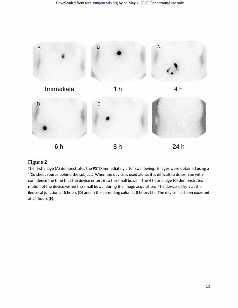
Figure2The first image (A) demonstrates the PSTD immediately after swallowing. Images were obtained using a 57Co sheet source behind the subject. When the device is used alone, it is difficult to determine with
confidence the time that the device enters into the small bowel. The 4 hour image (C) demonstrates
motion of the device within the small bowel during the image acquisition. The device is likely at the
ileocecal junction at 6 hours (D) and in the ascending colon at 8 hours (E). The device has been excreted
at 24 hours (F).
by on May 1, 2020. For personal use only. tech.snmjournals.org Downloaded from
12
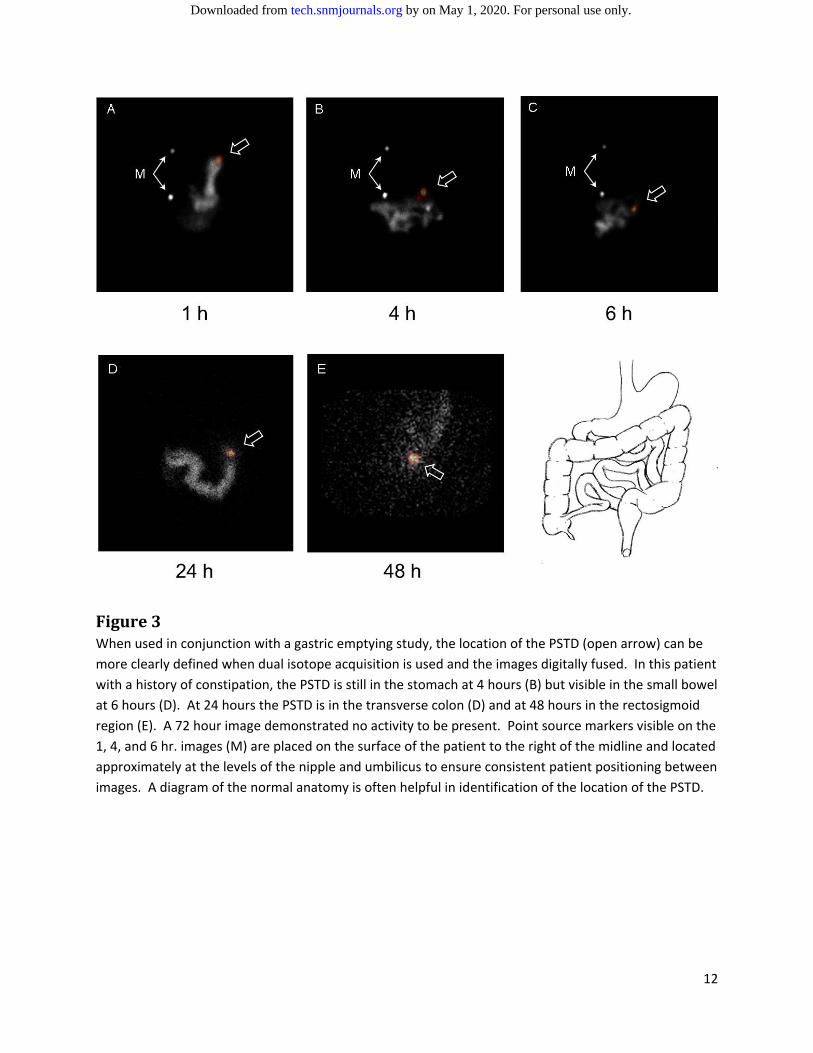
Figure3When used in conjunction with a gastric emptying study, the location of the PSTD (open arrow) can be
more clearly defined when dual isotope acquisition is used and the images digitally fused. In this patient
with a history of constipation, the PSTD is still in the stomach at 4 hours (B) but visible in the small bowel
at 6 hours (D). At 24 hours the PSTD is in the transverse colon (D) and at 48 hours in the rectosigmoid
region (E). A 72 hour image demonstrated no activity to be present. Point source markers visible on the
1, 4, and 6 hr. images (M) are placed on the surface of the patient to the right of the midline and located
approximately at the levels of the nipple and umbilicus to ensure consistent patient positioning between
images. A diagram of the normal anatomy is often helpful in identification of the location of the PSTD.
by on May 1, 2020. For personal use only. tech.snmjournals.org Downloaded from
13
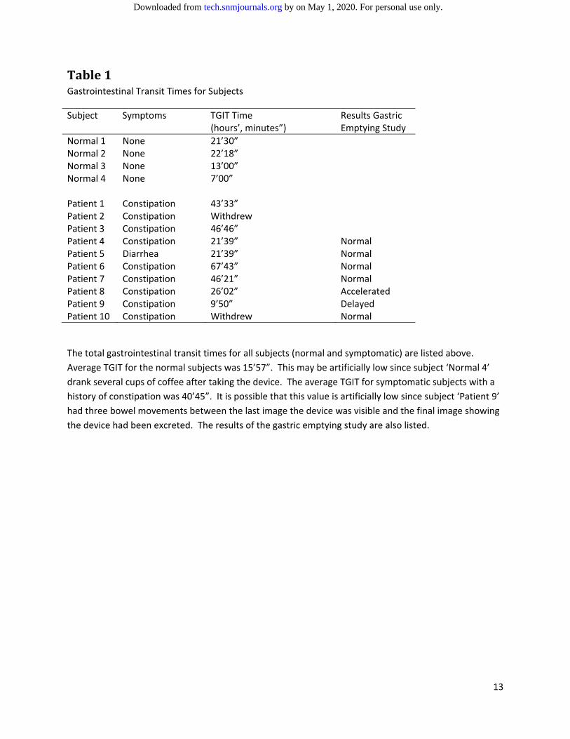
Table1Gastrointestinal Transit Times for Subjects
Subject Symptoms TGIT Time (hours’, minutes”)
Results Gastric Emptying Study
Normal 1 None 21’30” Normal 2 None 22’18” Normal 3 None 13’00” Normal 4 None 7’00” Patient 1 Constipation 43’33” Patient 2 Constipation Withdrew Patient 3 Constipation 46’46” Patient 4 Constipation 21’39” Normal Patient 5 Diarrhea 21’39” Normal Patient 6 Constipation 67’43” Normal Patient 7 Constipation 46’21” Normal Patient 8 Constipation 26’02” Accelerated Patient 9 Constipation 9’50” Delayed Patient 10 Constipation Withdrew Normal
The total gastrointestinal transit times for all subjects (normal and symptomatic) are listed above.
Average TGIT for the normal subjects was 15’57”. This may be artificially low since subject ‘Normal 4’
drank several cups of coffee after taking the device. The average TGIT for symptomatic subjects with a
history of constipation was 40’45”. It is possible that this value is artificially low since subject ‘Patient 9’
had three bowel movements between the last image the device was visible and the final image showing
the device had been excreted. The results of the gastric emptying study are also listed.
by on May 1, 2020. For personal use only. tech.snmjournals.org Downloaded from
14
References 1. Lindemann E. Demonstration of roentgen pictures of the normal and distended stomach. Dtsch Med Wochenschr. 1897;23:266‐267. 2. Cannon WB, Moser A. The movements of the food in the œsophagus. Am J Physiol. 1898;1:435‐444. 3. Cannon WB. The movements of the stomach studied by means of the roentgen rays. Am J Physiol. 1898;1:358‐382. 4. Eisenberg R, Margulis A. Brief history of gastrointestinal radiology. Radiographics. 1991;11:121‐132 5. Carman Rd, Miller A. The roentgenologic determination of gastric motility: with a comparison of the results obtained in a series of cases examined both by the roentgen ray and the test‐meal. Arch Intern Med (Chic). 1915;XVI(3):406‐428. 6. Alvarez WC, Freedlander BL. The rate of progress of food residues through the bowel. JAMA. 1924;83:576‐580. 7. Dimson SB. Carmine as an index of transit time in children with simple constipation. Archives of Disease in Childhood. 1970;45:232‐235. 8. Hoelzel F. The rate of passage of inert materials through the digestive tract. Am J Physiol. 1930;92:466‐497. 9. Hinton JM, Lennard‐Jones JE, Young AC. A new method for studying gut transit times using radiopaque markers. Gut. 1969;10:842–847. 10. Sarosiek I, Selover KH, Katz LA, et al. The assessment of regional gut transit times in healthy controls and patients with gastroparesis using wireless motility technology. Aliment Pharmacol Ther. 2010;31:313‐322. 11. Griffith, G. H., Owen, G. M., Kirkman, S., and Shields, R. Measurement of rate of gastric emptying using chromium‐51. Lancet. 1966;1:1244‐1245. 12. Chaudhuri TK. Use of 99mTc‐DTPA for measuring gastric emptying time. J Nucl Med. 1974;6:391‐5. 13. Meyer JH, MacGregor IL, Gueller R, Martin P, Cavalieri R:99mTc‐tagged chicken liver as a marker of solid food in the human stomach. Am J Dig Dis. 1976;21:296–304 14. Donohoe KJ, Maurer A, Ziessman H, Urbain J, Royal H, Martin‐Comin J. Procedure Guideline for Adult Solid‐Meal Gastric‐Emptying Study 3.0. J Nucl Med Technol. 2009;37:196–200. 15. Burton DD, Camilleri M, Mullan BP, Forstrom LA, Hung JC. Colonic transit scintigraphy labeled activated charcoal compared with ion exchange pellets. J Nucl Med. 1997;38:1807–1810 16. Kekilli E, Yagmur C, Isik B, Aydin OM. Calculating colon transit time with radionuclide‐filled capsules in constipated patients: a new method for colon transit study. Abdom Imaging. 2005;30:593‐597. 17. Maurer A, Camilleri M, Donohoe K, et al. The SNMMI and EANM practice guideline for small‐bowel and colon transit 1.0. J Nucl Med. 2013;54:2004‐2013. 18. Maurer A. Gastrointestinal motility, part 2: small‐bowel and colon transit. J Nucl Med Technol. 2016; 44:12‐18. 19. Sonnenberg A, Koch TR. Physician visits in the United States for constipation: 1958 to 1986. Dig Dis Sci. 1989 34(4):606‐11. 20. Pinto Sanchez MI, Bercik P. Epidemiology and burden of chronic constipation. Canadian Journal of Gastroenterology. 2011;25(Suppl B):11B‐15B. 21. Karesh, Stephen M. Principles of Radiopharmacy. In: Henkin RE, Bova D, Dillehay GL, Karesh SM, Halama JR, Wagner RH, Zimmer AM, eds. Nuclear Medicine. Philadelphia, PA: Mosby; 2006:332‐49. 22. Material Data Safety Sheet: Paraffin Wax. Indo Gulf Company Website. http://indogulfgroup.com/MSDS/PARAFFIN%20wax.pdf Revision #3 date March 18, 2003. Accessed February 20, 2017. 23. Paraffin (synthetic) 21 CFR 175.250. U.S Government Publishing Office Website. https://www.gpo.gov/fdsys/pkg/CFR‐2016‐title21‐vol3/pdf/CFR‐2016‐title21‐vol3‐sec175‐250.pdf Published April 21, 2016. Accessed February 20, 2017. 24. Gallium Citrate GA‐67: Package Insert and Label Information Mallinckrodt Inc. http://druginserts.com/lib/rx/meds/gallium‐citrate‐ga‐67‐1/ Revision date July 2, 2013. Accessed May 22, 2017. 25. FDA‐Division of Industry and Consumer Education (DICE), personal communication, January 2014. 26. Information Sheet Guidance for IRBs, Clinical Investigators, and Sponsors. Significant Risk and Nonsignificant Risk Medical Device Studies. U.S. Food and Drug Administration Website. www.fda.gov/downloads/RegulatoryInformation/Guidances/UCM126418.pdf Updated January 2006. Accessed February 20, 2017.
by on May 1, 2020. For personal use only. tech.snmjournals.org Downloaded from
Doi: 10.2967/jnmt.117.192377Published online: July 13, 2017.J. Nucl. Med. Technol. Robert H Wagner, Bital Savir-Baruch, James Halama, Mukund Venu, Medhat Gabriel and Davide Bova Gastrointestinal Transit TimeProof of Concept: Design and Initial Evaluation of a New Device to Measure
http://tech.snmjournals.org/content/early/2017/07/12/jnmt.117.192377This article and updated information are available at:
http://tech.snmjournals.org/site/subscriptions/online.xhtml
Information about subscriptions to JNMT can be found at:
http://tech.snmjournals.org/site/misc/permission.xhtmlInformation about reproducing figures, tables, or other portions of this article can be found online at:
and the final, published version.proofreading, and author review. This process may lead to differences between the accepted version of the manuscript
ahead of print area, they will be prepared for print and online publication, which includes copyediting, typesetting,JNMTcopyedited, nor have they appeared in a print or online issue of the journal. Once the accepted manuscripts appear in the
. They have not beenJNMT ahead of print articles have been peer reviewed and accepted for publication in JNMT
(Print ISSN: 0091-4916, Online ISSN: 1535-5675)1850 Samuel Morse Drive, Reston, VA 20190.SNMMI | Society of Nuclear Medicine and Molecular Imaging
is published quarterly.Journal of Nuclear Medicine Technology
© Copyright 2017 SNMMI; all rights reserved.
by on May 1, 2020. For personal use only. tech.snmjournals.org Downloaded from