prospective observational study of 200 cases scrotal
TRANSCRIPT

Page 1/22
Feasibility and Safety of Canine Orchiectomy UsingScrotal Approach and a Vessel-Sealing Device: AProspective Observational Study of 200 CasesChristos Yiapanis
1Cyvets Veterinary Centre, Tassou Papadopoulou AvenueCiprian Ober ( [email protected] )
University of Agricultural Sciences and Veterinary MedicineMaria Potamopoulou
Grove Veterinary Hospital, Grove HouseTheodoros Vasilakis
1Cyvets Veterinary Centre, Tassou Papadopoulou AvenueJoshua Milgram
The Hebrew University of Jerusalem
Research Article
Keywords: Canine orchiectomy, scrotal approach, monopolar electrosurgery, automutilation (AM)
Posted Date: September 28th, 2021
DOI: https://doi.org/10.21203/rs.3.rs-818635/v1
License: This work is licensed under a Creative Commons Attribution 4.0 International License. ReadFull License

Page 2/22
AbstractBackground: Canine orchiectomy involves making an incision in the prescrotal area, exteriorizing both testesvia the same incision, ligating the blood vessels and spermatic cord, removing the testes, and suturing theincision. A briefer durations of anesthesia and surgery and decrease of postoperative morbidity could beobtain using a vessel sealing device. The aim of this study was to determine the feasibility and safety oforchiectomy in dogs by a scrotal approach with the use of a vessel-sealing device. Scrotal orchiectomy wasperformed with the use of monopolar electrosurgery device in pure cutting mode and a vessel-sealing device.Data were collected prospectively for the following categorical variables: breed, age, body weight, lifestyle,surgical time, indications for surgery and complications.
Results: No complications were reported in 187 of 200 (93.5%). The complications documented wereautomutilation (AM), incisional complications (IC) and scrotal complications (SC). AM complications (11/200[5.5%]) and SC complications (2/200 [1%]) were treated without additional surgery and resolved by day 10after the surgery. Procedure duration (2,1 minutes± 0.4 minutes) was briefer than traditional castrationduration reported in literature (3.5 ± 0.4 minutes).
Conclusions: Results suggested that canine orchiectomy by scrotal approach with the use of a vessel-sealingdevice was feasible and safe. Furthermore, it was associated with a low complication rate and had thebene�t of reduced surgical time and postoperative morbidity. This technique is promising for widespreadapplication in veterinary surgery to help with haemostasis in canine orchiectomy.
BackgroundCanine orchiectomy is one of the most commonly performed surgical procedures in �rst opinion small animalpractice worldwide [1–4]. The most common indication for orchiectomy is surgical sterilization (neutering).Other indications are the reduction of androgen-related disorders (anxiety and aggressiveness), the tendencyto roam, undesirable urination, prostatic disease, perianal adenomas, perineal hernias, the prevention ofhereditary diseases, testicular abnormalities, scrotal urethrostomy, inguinal-scrotal herniorrhaphy andendocrine abnormalities [5].
There is a wide variety in the techniques used to perform orchiectomy in dogs. The technique is in�uenced bygeographic areas, the age and the size of the patient, �nances, and the purpose of the procedure [4]. Commonapproaches used in adult dogs are via a prescrotal incision, scrotal ablation, and a perineal approach whenperforming perineal surgery. A scrotal approach has been proposed in prepuberal dogs [6, 7], and forcastrating adult dogs of any age [8–10]. This approach may offer advantages that include improvedcosmesis, decreases in anesthetic and surgical times, smaller incision length and less surgical trauma,decreased postoperative discomfort, less self-trauma and decreased likelihood of formation of scrotalhematoma [8–10].
Severe complications of orchiectomy are rare, but may include scrotal swelling and bruising, hemorrhage,scrotal hematoma, abscess, granuloma, incisional problems (swelling, seroma formation, cellulitis, infection,automutilation, dehiscence), urinary incontinence, endocrine alopecia, behavioral changes and eunuchoid

Page 3/22
syndrome [11]. Most complications associated with orchiectomy can be prevented by using good surgicaltechnique, including gentle tissue handling, good hemostasis, and aseptic technique [12].
Hemorrhage from poorly ligated vascular pedicles can result in scrotal hemorrhage or intraabdominalbleeding due to the retraction of the bleeding vascular pedicle through the inguinal canal [7]. Traditionallyhaemostasis is achieved with the use of ligatures but their reported disadvantages are occasional ligatureslippage, introduction of foreign matter, di�cult management, high cost, size limitation and application time[13]. For these reasons the use of electrosurgical devices with enhanced safety characteristics have gainedpopularity in small animal surgery [14]. Vessel sealing devices (VSD) achieve haemostasis by the fusion ofelastin and collagen in the vessel walls and surrounding connective tissue using moderate compression at atemperature less than 100°C (212°F). The low energy used in theses devices results in less collateral thermalspread and tissue damage when compared to the traditional electrocoagulation devices [15, 16]. Ligasure is afeedback- controlled bipolar vessel sealing device that is now well established in human and equine surgeryand has been shown to be effective in sealing vessels up to 7 mm in diameter [17].
The aim of this study was to evaluate the feasibility and safety of Ligasure vessel-sealing device for closedorchiectomy via scrotal approach. We hypothesized that the technique will be fast and the complication rateswould be low.
ResultsThe descriptive statistics are reported at Table 1. The majority of the dogs enrolled were privately owned(79.0%, 158/200) and the most common indication for this operation was neutering (92.0% 184/200). 84dogs (42.0%) weighted less than 10kg while the remaining 116 (58%) weighted more than 10kg at the day ofthe surgery. The mean duration of canine orchiectomy by scrotal approach using a vessel-sealing device was2.1 with a SD (standard deviation) of 0.5. Complications were noted in 13 dogs (6.5%) and all of them werereported during the �rst 24 hours post-operatively (Table 2).

Page 4/22
Table 1Characteristics of the study dogs underwent
orchiectomy by scrotal approach using avessel-sealing device
Characteristic Dogs (%)
(N = 200)
Age in years
Mean 3.7
Standard deviation 2.8
Breed
Pedigree 51 (25.5)
Crossbreed 149 (74.5)
Weight
<10kg 84 (42.0)
11-20kg 68 (34.0)
>20kg 48 (24.0)
Lifestyle
Private 158 (79.0)
Shelter 42 (21.0)
Indication for surgery
Neutering 184 (92.0)
Prostatic disease 7 (3.5)
Testicular disease 4 (2.0)
Other 5 (2.5)
Duration of surgery in mins
Mean 2.1
Standard deviation 0.5
Complication
None 187 (93.5)
AMIC 11 (5.5)
AMICSC 2 (1.0)

Page 5/22
Table 2Summary of the dogs that developed complications and associated treatment
Case Breed Age
(Year)
Weight(kg)
Origin:private/shelter
Dayaftersurgery
Complication Treatment
13 Pomeranian 4 5 Private 1 AM/IC BC/IP
21 Chihuahua 3 3 Private 1 AM/IC BC
46 Labrador 3 27 Private 1 AM/IC/SC BC/IP/ANG/ATB
50 Cross breed 3 5 Private 1 AM/IC BC/IP
75 Poodle 5 7 Private 1 AM/IC BC/IP
114 Pointer 2 16 Private 1 AM/IC BC/IP
118 Poodle 1 6 Private 1 AM/IC/SC BC/ANG/ATB
136 Cross breed 1 5 Private 1 AM/IC BC/IP
141 Pomeranian 1 4 Private 1 AM/IC BC/IP
152 Husky 1 25 Private 1 AM/IC BC/IP
163 Pincher 2 7 Private 1 AM/IC BC
183 Hound 1 10 Shelter 1 AM/IC BC/ANG/ATB
197 Spaniel 10 13 Private 1 Am/IC BC/ANG/ATB
The results from univariable analysis on continues and categorical variable are shown in Tables 3 and 4respectively. The only variable that was signi�cantly associated with complications (including both AMIC andAMICSC) following orchiectomy by scrotal approach using a vessel-sealing device was the weight of the dog.Dogs that were less than 10 kg were 3 times (CI: 0.9–11.3, P = 0.040) more likely to develop complicationscompared to the dogs that weighted more than 10 kg. There was no variable that showed a trend towards tosigni�cance thus the multivariable logistic regression did not yield any additional �ndings.

Page 6/22
Table 3Univariable analysis for associations between continues variables and complications in dogs that
underwent orchiectomy by scrotal approach using a vessel-sealing device.Variable No. of dogs
(N = 200)
Complications OR 95% CI for OR P-value
Yes (%) No (% )
Breed
Pedigree 51 5 (9.8) 46 (90.2) 1.9 0.6–6.1 0.267
Crossbreed 149 8 (5.4) 141 (94.6)
Weight
< 10kg 84 9 (10.7) 75 (89.3) 3.4 0.9–11.3 0.040
> 10kg 116 4 (3.4) 112 (96.6)
Lifestyle
Private 158 11 (7.0) 147 (93.0) 0.6 0.1–3.1 0.607
Shelter 42 2 (4.8) 40 (95.2)
Indication for surgery
Neutering 184 12 (6.5) 172 (93.5) 0.9 0.1–7.8 0.966
Non-neutering causes 16 1 (6.3) 15 (93.8)
Table 4Univariable analysis for associations between categorical variables and
complications in dogs that underwent orchiectomy by scrotal approach usinga vessel-sealing device. Signi�cant P-values ≤ 0.05 are shown in bold
Variable Complications
(N = 200)
Mean SD P-value
Age in year Yes 13 (6.5)
No 187 (93.5)
4.1
3.6
4.1
2.6
0.516
Duration of surgery (minutes) Yes 13 (6.5)
No 187 (93.5)
1.9
2.1
0.5
0.4
0.239
DiscussionWe hypothesized that the technique will be fast and the complication rates would be low in the studypopulation. The �rst hypothesis was supported in that procedure durations was briefer than those reportedfor traditional prescrotal castration as mentioned by Miller et al (2018) [18] (1,9 minutes ± 0.5 minutes incases with complications and 2,1 minutes ± 0.4 minutes in cases without complications in this studycompared with 3.5 ± 0.4 minutes reported by Miller et al.). Based on this, we concluded that the technique in
Page 7/22
this study provides a rapid technique for castration. Shorter duration of castration in male dogs usingLigasure were reported also by Faluvégi et al. 2018 [19], but they used a prescrotal approach.
Another hypothesis for the present study was that the complication rate would be low. This hypothesis wassupported in that presented technique resulted in no haemorrhagic complications for any of the 200 surgeriesperformed, and minor complications observed were self- limiting and only few. Based on these results, weconcluded that the Ligasure scrotal technique provides a rapid and safe approach for canine orchiectomy.
Major hemorrhage associated with orchiectomy is uncommon in dogs [20], and most complications involvinghemostasis result in scrotal hematomas, likely due to oozing from the vaginal tunic with open orchiectomy [1,20]. According to Miller’s et al. experience [18], large dogs with large gonads will generally have a largergonadal blood supply than small dogs with small gonads. Interestingly, dogs that were less than 10 kg were 3times (CI: 0.9–11.3, P = 0.040) more likely to develop complications compared to the dogs that weightedmore than 10 kg in our study. Anyway, the complications were not due to hemorrhage. No hemostaticcomplications were seen after Ligasure application in our study. The weight of the dog was the only variablethat was signi�cantly associated with complications (including both AMIC and AMICSC) followingorchiectomy by scrotal approach using a vessel-sealing device in this study. We consider the behaviour andtemperament of the small dogs to be mostly responsible for incisional complications.
All complications were identi�ed at 24 hours post-surgery assessment and resolved without additionalsurgery. All incisional complications were managed by telephone communication with the owners. None ofthe owners with incisional complications felt that their dog required additional analgesia. The cases thatdeveloped incisional and scrotal complications (dog 46 and 118) were admitted for examination andadditional analgesia with NSAIDs and antibiotics were administered. None of the cases requiredhospitalization or additional surgery. Both complications were encountered in client owned animals. In case118, the owners were unable to apply the buster collar as soon as automutilation was noticed due toaggression of the dog. In case 46 that both incisional and scrotal complications were also observed, theapplication of the collar was delayed because the dog was left unattended for several hours. Based on this,we can assume that automutilation is an important factor in the appearance and progression ofcomplications. In the authors’ experience, shelter animals had less complications, assuming that thedistractions in the environment prevented automutilation or complications passed unnoticed.
In the uncomplicated cases contraction of the scrotum and elimination of the incisional gap was observed byday 3. In all cases that the complications were observed, su�cient regression of the scrotum with noincisional gap was observed by day 10. In the cases that both incisional and scrotal complications wereobserved (46, 118), some skin discoloration remained in the scrotal region by day 10. Nevertheless, themajority of the animals in the study did not require buster collar, and strict exercise restriction was limited to24 hours post discharge. Interestingly, the limited postoperative restrictions and the avoidance of the bustercollar were factors that were strongly appreciated by the owners when they were questioned regarding ownerssatisfaction. Assessment at 10 days after surgery is routinely performed by several practices but the value ofassessment at this time point is questionable due to the fact that the majority of the complications arise the�rst days after the procedure [5]. Due to the uncomplicated healing and the rapid elimination of the incisionalgap, most owners felt that further assessment was not necessary.
Page 8/22
Activation of the vessel sealing system uses electrosurgical radiofrequency energy that causes the collagenand elastin in the blood vessel walls to reorganize within the tissue thereby forming a permanent seal [21].Monopolar electrosurgery was used to incise the scrotal skin, and to control minor bleeding from septalvessels. Even though skin incisions made with monopolar electrosurgery were associated with decreasedhealing compared to scalpel made incisions [21], su�cient regression of the scrotum and no incisional gapwas evident by day 10 in all cases. Clearly bene�ts in hemostasis improvements, and overall anaesthesia andsurgery time reduction were demonstrated in many studies [22–27]. Even though there are reasonableconcerns regarding the high cost of using VSD in routine procedures, appropriate re-sterilisation of the handpiece allows a signi�cant reduction in the costs by increasing the number of procedures without decreasingsafety characteristics [13, 28].
Several limitations of the study should be considered when interpreting the results. Some incisionalcomplications could have developed beyond the 24-hour postoperative. Anyway, the owners were instructedto report any complication with the surgery. Similar to our study, in a previous report [5], most complicationsfollowing canine orchiectomy were discovered at the 24-hour recheck examination and no complicationswere found at the next recheck examination, performed 10 days after surgery. The authors of the study alsoadvocated for rechecking of patients in the immediate postoperative period and questioned the value ofrechecking the patients 10 days after surgery.
We found only one study of postoperative complications after Ligasure orchiectomy in the peer-reviewedveterinary literature, thus, direct comparisons with the �ndings of the present study is limited [19]. Moreover,the authors of the study used prescrotal approach, thereby precluding any meaningful comparison with ourdata.
Another limitation of the study include the use of semiobjective methods for documenting each complicationand the fact that the assessment of the majority of the cases was based on owners perception. For thesereasons, long-term results should be evaluated in a larger randomized prospective studies.
ConclusionThe low complication rate and time advantage associated with use of the Ligasure technique for orchiectomyof dogs suggested important advantages, including shorter durations of anesthesia and surgery than with thetraditional prescrotal technique. We expect that as the Ligasure technique is more widely used, morbidity andmortality rates associated with canine orchiectomy will improve as will savings in resources, such as timeand suture materials. The results of the present study provide important data for the veterinarians whoperform such surgeries.
MethodsA prospective observational study for canine orchiectomy by scrotal approach using a vessel-sealing devicewas performed at the CYVETS Veterinary Centre from December 2015 to December 2018. Two hundredhealthy intact male dogs were recruited in this study (Table 5). The age of the study population ranged from0.8 to 12 years [mean 3.7 years, standard deviation (SD) 2.8] and 149 (74.5%) were crossbreed. Dogs less

Page 9/22
than seven years old were determined to be healthy based on medical history and physical examinationperformed on the day of surgery, whilst dogs more than seven years old routine biochemistry andhaematology were performed prior to anesthesia to determine health status. The intact male dogs enrolled inthe study following a signed consent form by the owners or guardians were presented either from localanimal welfare organizations or were client-owned dogs. No dog underwent any procedure speci�cally forthis study and all procedures were performed in accordance of the Cypriot legislation [The Dogs LAW, N. 184(I)/2002].

Page 10/22
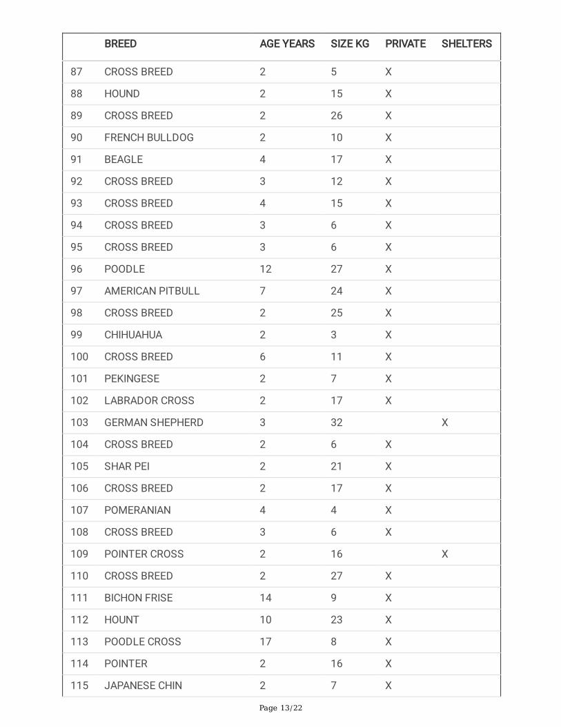
Table 5Characteristics of the population
BREED AGE YEARS SIZE KG PRIVATE SHELTERS
1 HOUND 5 10 X
2 HOUND 4 15 X
3 CROSS BREED 4 7 X
4 PINCHER CROSS 4 5 X
5 BISCON FRISE 11 5 X
6 ENGLISH SPRINGER SPANIEL 4 13 X
7 TERRIER 3 11 X
8 CROSS BREED 4 5 X
9 POINTER 4 15 X
10 ROTTWEILER 3 27 X
11 AMER.STAFFOR.TERRIER 4 20 X
12 POODLE 3 5 X
13 POMERANIAN 4 5 X
14 HOUND 4 10 X
15 POODLE 7 8 X
16 HUSKY 4 32 X
17 POODLE 7 4 X
18 LABRADOR 6 30 X
19 MINI DOBERMAN 4 8 X
20 CROSS BREED 4 7 X
21 CHIHUAHUA 3 3 X
22 LABRADOR 3 25 X
23 CROSS BREED 3 5 X
24 BICHON FRISE 3 8 X
25 BICHON FRISE 3 6 X
26 BICHON FRISE 3 8 X
27 CROSS BREED 4 12 X
28 POODLE 4 6 X

Page 11/22
BREED AGE YEARS SIZE KG PRIVATE SHELTERS
29 POODLE 3 5 X
30 Yorkshire TERRIER 3 5 X
31 CROSS BREED 3 9 X
32 POMERANIAN 3 3 X
33 BELGIAN MALINOIS 4 17 X
34 CAIRN TERRIER 3 12 X
35 MALTEZE 3 5 X
36 CANE CORSO 3 40 X
37 Yorkshire TERRIER 3 7 X
38 DOBERMAN 3 31 X
39 CROSS BREED 6 12 X
40 CROSS BREED 3 10 X
41 CROSS BREED 3 10 X
42 HOUND 4 18 X
43 LABRADOR 11 43 X
44 HOUND 11 34 X
45 CROSS BREED 4 8 X
46 LABRADOR 3 27 X
47 BICHON FRISE 3 7 X
48 HUSKY 4 21 X
49 VIZLA CROSS 3 10 X
50 CROSS BREED 3 5 X
51 CROSS BREED 8 10 X
52 GERMAN SHEPERD CROSS 3 10 X
53 LABRADOR 5 17 X
54 BOXER 3 27 X
55 HOUND 3 15 X
56 POMERANIAN 3 5 X
57 BEAGLE 7 19 X

Page 12/22
BREED AGE YEARS SIZE KG PRIVATE SHELTERS
58 JACK RUSSELL 5 5 X
59 LABRADOR 3 19 X
60 POODLE 12 11 X
61 SCHNAUZER 3 21 X
62 HUSKY 3 35 X
63 CROSS BREED 3 15 X
64 ROTTWEILER 2 30 X
65 SPRINGER SPANIEL 3 15 X
66 TERRIER 2 11 X
67 HUSKY 4 23 X
68 GERMAN POINTER 3 29 X
69 BEAGLE 3 15 X
70 CROSS BREED 6 5 X
71 CROSS BREED 4 5 X
72 DACHSHUND 15 10 X
73 BEAGLE 3 15 X
74 CROSS BREED 2 10 X
75 POODLE 5 7 X
76 BEAGLE 6 17 X
77 POINTER 3 18 X
78 CROSS BREED 4 18 X
79 HOUND 5 15 X
80 YORKSHIRE-TERRIER 10 4 X
81 CROSS BREED 6 15 X
82 CROSS BREED 4 12 X
83 YORKSHIRE-TERRIER 2 5 X
84 LABRADOR CROSS 6 50 X
85 CROSS BREED 2 9 X
86 CROSS BREED 2 5 X
Page 13/22
BREED AGE YEARS SIZE KG PRIVATE SHELTERS
87 CROSS BREED 2 5 X
88 HOUND 2 15 X
89 CROSS BREED 2 26 X
90 FRENCH BULLDOG 2 10 X
91 BEAGLE 4 17 X
92 CROSS BREED 3 12 X
93 CROSS BREED 4 15 X
94 CROSS BREED 3 6 X
95 CROSS BREED 3 6 X
96 POODLE 12 27 X
97 AMERICAN PITBULL 7 24 X
98 CROSS BREED 2 25 X
99 CHIHUAHUA 2 3 X
100 CROSS BREED 6 11 X
101 PEKINGESE 2 7 X
102 LABRADOR CROSS 2 17 X
103 GERMAN SHEPHERD 3 32 X
104 CROSS BREED 2 6 X
105 SHAR PEI 2 21 X
106 CROSS BREED 2 17 X
107 POMERANIAN 4 4 X
108 CROSS BREED 3 6 X
109 POINTER CROSS 2 16 X
110 CROSS BREED 2 27 X
111 BICHON FRISE 14 9 X
112 HOUNT 10 23 X
113 POODLE CROSS 17 8 X
114 POINTER 2 16 X
115 JAPANESE CHIN 2 7 X

Page 14/22
BREED AGE YEARS SIZE KG PRIVATE SHELTERS
116 LABRADOR CROSS 3 15 X
117 PUG 2 8 X
118 POODLE CROSS 1 6 X
119 CROSS BREED 2 5 X
120 POODLE 2 9 X
121 LABRADOR CROSS 9 33 X
122 CROSS BREED 2 7 X
123 CROSS BREED 11 10
124 GERMAN SHEPHERD 2 29 X
125 GERMAN SPITZ 3 10 X
126 HOUND 2 10 X
127 TERRIER 1 8 X
128 TERRIER 3 11 X
129 SAMOYED 8 36 X
130 PIT BULL 1 22 X
131 TERRIER 2 7 X
132 GERMAN SHEPERD CROSS 7 35 X
133 ROTTWEILER 1 35 X
134 CROSS BREED 1 12 X
135 POODLE CROSS 6 13 X
136 CROSS BREED 1 5 X
137 CROSS BREED 2 7 X
138 GERMAN SHEPHERD 6 42 X
139 CROSS BREED 1 19 X
140 SALUKI 2 22 X
141 GERMAN SPITZ 1 4 X
142 AMER.STAFFOR.TERRIER 1 22 X
143 POODLE 2 8 X
144 ROTTWEILER 1 14 X

Page 15/22
BREED AGE YEARS SIZE KG PRIVATE SHELTERS
145 CROSS BREED 11 12 X X
146 GERMAN SHEPERD CROSS 2 21 X
147 HOUND 2 12
148 ROTTWEILER 4 42 X
149 PUG 2 9 X
150 POINTER 2 20 X
151 CROSS BREED 0.8 6 X
152 HUSKY 1 25 X
153 CROSS BREED 1 9 X
154 HOUND 2 15 X
155 CROSS BREED 4 7 X
156 HOUND 1 15 X
157 GERMAN SHEPERD CROSS 2 21 X
158 CROSS BREED 0.8 19 X
159 WEIMARANER 1 17 X
160 VISZLA 1 18 X
161 HOUND 2 13 X
162 POINTER 3 20 X
163 PINCHER 2 7 X
164 POINTER 2 18 X
165 CHIHUAHUA 3 4 X
166 HOUND 2 18 X
167 HOUND 3 25 X
168 CROSS BREED 2 15 X
169 POINTER 1 30 X
170 STAFFORDSHIRE TERRIER 3 22 X
171 HOUND 7 20 X
172 HOUND 1 16 X
173 GERMAN SHEPERD 1 22 X

Page 16/22
BREED AGE YEARS SIZE KG PRIVATE SHELTERS
174 FRENCH BULLDOG 8 11 X
175 COCKER SPANIEL 2 15 X
176 DOBERMAN 3 38 X
177 ROTTWEILER 3 43 X
178 LABRADOR 1 16 X
179 SHARPEI 2 18 X
180 GERMAN SHEPHERD 6 37 X
181 CROSS BREED 5 16 X
182 PEKINGESE 3 7 X
183 HOUND 1 10 X
184 CROSS BREED 6 6 X
185 LABRADOR 2 20 X
186 STAFFORDSHIRE TERRIER 6 28 X
187 POODLE 2 9 X
188 LABRADOR 2 22 X
189 POODLE 2 11 X
190 LABRADOR 1 18 X
191 HOUND 1 15 X
192 PINCHER 2 10 X
193 PINCHER 1 8 X
194 CROSS BREED 2 7 X
195 CROSS BREED 2 9 X
196 LABRADOR 4 27 X
197 COCKER SPANIEL 10 13 X
198 POODLE 1 8 X
199 HOUND 1 12 X
200 POODLE 2 10 X
The surgical procedures were performed by a single veterinary surgeon (CY) in order to avoid interobservervariability. Data were recorded and included breed, bodyweight, age, total surgical time, lifestyle

Page 17/22
(private/shelter) and the encountered complications up to 10 days post surgery.
All protocols were standardized prior to commencing the study. These included: preparation for asepticsurgery, surgical procedure, recovery protocol, postoperative instructions and client communication.Orchiectomy time was measured from immediately prior the skin incision to apposition of the incision siteand dogs were categorized based on body weight into small (< 10kg), medium (10 to 20 kg) and large breed(> 20 kg). Associated surgical complications were documented including automutilation, incisionalcomplications (hemorrhage, bruising, swelling, erythema) and scrotal complications (swelling, bruising,hematoma). The treatments/measures employed to address the encountered complications were bustercollar application (BC) and ice packs (IP) in all complications and additional analgesia with non-steroidalanti-in�ammatory medication and antibiotics in the cases with combination of incisional and scrotalcomplications.
Perioperative managementAll dogs were anaesthetised by use of standard clinical protocols. Young healthy dogs were premedicatedwith 0·02 mg/kg medetomidine (Domitor P�zer) intravenously (iv) and 0·02 mg/kg butorphanol (DolorexMerck) iv. Induction to anesthesia was achieved with 1 to 3 mg/kg propofol (Propofol 1%, Fresenius pharma)slowly iv to effect and maintained with 2% inspired iso�urane (iso�o 100%, Abbott pharma) in oxygen withthe use of a Bain circuit. In patients over 10 years old, medetomidine was replaced by 0·2 mg/kg midazolam(Dormicum Roche) iv. Anaesthesia was monitored by clinical assessment and a multi parameter anaesthesiamonitor. All dogs received 0·2 mg/kg meloxicam (Metacam Boehringer) iv before anesthesia. Additionalanalgesia with butorphanol was administered after surgery in patients with evidence of pain (WSAVA, 2014).A small square area around the incision site was clipped with slow-powered clippers with 0,5mm blade. Carewas taken not to traumatize the sensitive scrotal skin during clipping. The skin was prepared with surgicalscrub (Dermanios Scrub CG, Anios lab) followed by 2% Chlorhexidine Digluconate solution (ChlorhexidineConcentrate 5 vol. % CD, Krusan). The patients were positioned in dorsal recumbency and draped with around fenestrated self-adhesive drape.
Surgical proceduresThe left testicle was grasped between the thumb and index of the surgeon and directed caudoventrally whilepulling the scrotal skin taut over the testicle. The scrotal skin cranial and ventral to the median raphe at theapex of the scrotum, was incised with a monopolar electrosurgery device (Eschmann TD830), in pure cuttingmode at 100-150W output (small and medium-large patients respectively), approximately one-third of thelength of the long axis of the testicle (0,5 − 2 cm) (Fig. 1A). The incision was continued through the spermaticfascia until the parietal tunic was seen. The testicle within the parietal layer of the vaginal process wasexteriorised, and the spermatic cord was exposed by re�ecting all visible connective tissue from the parietaltunic with a gauze sponge. A 10-mm instrument (LigaSure Atlas Sealer/Divider—LS10) connected to abipolar, feedback-controlled vessel sealing device (LigaSure Generator-Valleylab, Boulder, Colorado, USA) witha power setting of three bars was applied to the spermatic cord and the instrument was activated to achievehemostasis (Fig. 1B). The sealing and transection time was less than 5 seconds per cord, appreciably fasterthan suture ligation. After hemostasis was completed, the spermatic cord was transected with scissors and

Page 18/22
the stump was returned to the scrotum (Fig. 1C,D). The second testicle was removed using the identicaltechnique. After completing the procedure the skin was opposed by gentle approximation of the incisionedges (Fig. 1E).
Postoperative careAll dogs were hospitalised for 6–8 hours after surgery. During this period they were closely monitored forsigns of pain, haemorrhage and swelling of the surgical site. The assessment of the amount of pain wascarried out hourly and was based on the observation of the demeanor and posture of the animal in the kenneland during the interaction with the dog while examining the wound for signs of surgery associatedcompilations. Following discharge from the clinic, all owners were instructed to rest their dogs for 1 day.Short lead walks four to �ve times a day for toileting were permitted. Progressive return to exercise over oneweek was advised, if healing was uncomplicated. Owners were instructed not to dress or treat the wound. Abuster collar was provided to the owners but it was not applied to the animals. Owners were advised tomonitor the surgical wound twice daily and apply the buster collar and contact the members of the staff ifself-trauma, discomfort or swelling were evident at the surgical site.
Follow up phone calls to the owners were conducted on day 1 after discharge and on day 10. An additionalphone call was conducted on day 5 in the cases that any complication was reported. During thecommunication, a standard questionnaire format was employed and the owner’s comments weredocumented.
Statistical analysisData were recorded into Microsoft O�ce Excel 2016 and then imported into SPSS (version 22.0; SPSS Inc.,Chicago IL, USA) for statistical analysis. Descriptive statistics were performed for the continuous variables ofage and duration of surgery, as well as for the following categorical variables: breed, body weight (small,medium and large), lifestyle (private or shelter), indications for surgery (neutering, prostatic disease, testiculardisease and other) and complications (none, AMIC, AMICSC). Independent t-test was used to evaluate forassociations between continues variables and complications. To investigate any association forcomplications and categorical variables a univariable analysis conducted using Chi-square test. Amultivariable logistic regression was performed to test for possible risk factors associated withcomplications. The independent variables that yielded P-values of < 0.2 in a univariable analysis were thentested in a multivariable logistic regression analysis. A P-value ≤ 0.05 was considered statistically signi�cant.The P-values with odds ratio (OR) and 95% con�dence interval (CI) were reported.
AbbreviationsAM: automutilation; ANG: Analgesia; ATB: Antibiotics; BC: Buster collar; CI: con�dence interval; IC: incisionalcomplications; IP: Ice Packs; OR: odds ratio; SC: scrotal complications; SD : standard deviation
DeclarationsEthics approval and consent to participate

Page 19/22
All procedures were approved by the Ministry of Agriculture, Development and Environment, VeterinaryServices, (protocol number 1/2021) and performed according to the Cypriot legislation [The Dogs LAW, N. 184(I)/2002]. All owners gave informed written consent for their dogs inclusion in the study. The study wascarried out in compliance with the ARRIVE guidelines.
Consent for publication
Not applicable.
Availability of data and materials
All data generated or analysed during this study are included in this published article.
Competing interests
The authors declare that they have no competing interests.
Funding
Not applicable.
Authors' contributions
CY, CO and JM made substantial contributions to the conception of the study design, acquisition, analysisand interpretation of data; MP and TV contributed to the data analysis and critically revised importantintellectual content. All authors read and approved the �nal manuscript.
Acknowledgements
Not applicable.
References1. Pollari FL, Bonnett BN, Bamsey SC, Meek AH, Allen DG. Postoperative complications of elective surgeries
in dogs and cats determined by examining electronic and paper medical records. JAVMA 1996;208:1882-6.
2. Howe LM. Surgical methods of contraception and sterilization. Theriogenol. 2006;66:500-09.
3. Reichler IM. Gonadectomy in cats and dogs: a review of risks and bene�ts. Reprod Dom Anim.2009;44:29-35.
4. Adin CA. Complications of ovariohysterectomy and orchidectomy in companion animals. Vet Clin NorthAm-Small Anim Pract. 2011; 41:1023-39.
5. Hamilton K, Henderson E, Toscano M, Chanoit G. Comparison of postoperative complications in healthydogs undergoing open and closed orchidectomy. JSAP. 2014;55:521–6.
�. Crane SW. Orchiectomy of descended and retained testes in the dog and cat. In: Bojrab, MJ, edior. CurrentTechniques in Small Animal Surgery, 4th edn. Baltimore:Williams & Wilkins; 1998. p. 517-523.

Page 20/22
7. Goethem VB. Orchiectomy. In: Griffon D, Hamaide A, editors. Complications in Small Animal Surgery.John Wiley & Sons, Inc, 2016. p. 528-533.
�. Johnston DE, Archibald J. Male genital system. In: Archibald J, editor. Canine Surgery. 2nd ed. SantaBarbara, CA: American Veterinary Publications; 1974. p. 703-749.
9. Bushby PA. Surgical techniques for spay/neuter. In: Zawistowski ML, editor. Shelter Medicine forVeterinarians and Staff. 2nd ed., Wiley-Blackwell; 2013. p. 625-645.
10. Woodruff K, Bushby PA, Rigdon-Brestle K, Wills R. Scrotal castration versus prescrotal castration in dogs.Vet Med. 2015;110:131–5.
11. Boothe HW. Testes and epididymides. In: Slatter DH, editor. Textbook of Small Animal Surgery. 3rd edn.W. B. Saunders:Philadelphia; 2003. p. 1521-1531.
12. Hedlund CS. Surgery of the reproductive and genital systems. In: Fossum TW, editor. Small AnimalSurgery, 3 rd Ed., St.Louis:Mosby, Elsevier, 2007. p. 702-774.
13. Gracia-Calvo LA, Martín-Cuervo M, Jiménez J, Vieítez V, Durán ME, Argüelles D, Ezquerra LJ. Intra andpostoperative assessment of re-sterilised Ligasure Atlas for orchidectomies in horses: clinical study. VetRec. 2012;171:98.
14. Diamantis T, Gialikaris S, Kontos M, Gakiopoulou C, Felekouras E, Papalois A, et al. Comparison of safetyand e�cacy of ultrasonic and bipolar thermal energy: an experimental study. Surg Lapar End PercutanTechn. 2008; 18:384-390.
15. Kennedy JS, Stranahan PL, Taylor KD, Chandler JG. High-burst-strength, feedback-controlled bipolarvessel sealing. Surg Endosc. 1998;12:876–8.
1�. Landman J, Kerbl K, Rehman J, Andreoni C., Humphrey PA., Collyer W, et al. Evaluation of a vesselsealing system, bipolar electrosurgery, harmonic scalpel, titanium clips, endoscopic gastrointestinalanastomosis vascular staples and sutures for arterial and venous ligation in a porcine model. J Urol.2003;169:697–700.
17. Launois MT, Vandeweerd JMEF, Perrin RAR, Brogniez L, Gabriel A, Coomer R. Evaluation of a vessel-sealing device to assist eye enucleation and exenteration in the horse. Equine Vet Educ. 2009;21:596-601.
1�. Miller KP, Rekers WL, Lena GDT, Blanchette JM, Milovancev M. Evaluation of sutureless scrotal castrationfor pediatric and juvenile dogs. JAVMA. 2018;253:1589-93.
19. Faluvégi A, Bedi D, Banhidy J, Demeter A, Molnár J, , et al. Neutering of dogs and cats using LigaSuredevice. Magyar Állatorvosok Lapja. 2018;140:217-21.
20. Adin CA. Complications of ovariohysterectomy and orchiectomy in companion animals. Vet Clin NorthAm Small Anim Pract. 2011;41:1023–39.
21. 21. Scott JE, Swanson EA, Pearce EC, Cooley J, Wills RW. Healing of canine skin incisions made withmonopolarelectrosurgery versus scalpel blade. Vet Surg. 2017;46:520-9.
22. Mckenna RJ, Brenner M, Gelb AF, Mullin M, Singh N, Peters H. A randomized prospective trial of stapledlung reduction versus laser bullectomy for diffuse emphysema. J Thorac Cardiovasc Surg.1996;111:317–22.

Page 21/22
23. Belch A, Matiasovic M, Rasotto R, Demetriou J. Comparison of the use of LigaSure versus a standardtechnique for tonsillectomy in dogs. Vet Rec. 2016;180:196.
24. Collard F, Nadeau ME, Carmel EN. Laparoscopic splenectomy for treatment of splenic hemangiosarcomain a dog. Vet Surg. 2010;39:870-2.
25. Proença LM. Two-Portal Access Laparoscopic Ovariectomy Using Ligasure Atlas in Exotic CompanionMammals, Vet Clin North Am Exot Anim Pract. 2015;18:587-96.
2�. Öhlund M, Höglund O, Olsson U, Lagerstedt AS. Laparoscopic ovariectomy in dogs: a comparison of theLigaSure™ and the SonoSurg™ systems. JSAP. 2011;52:290-4.
27. Wouters EGH, Buishand FO, Kik M, Kirpensteijn J. Use of a bipolar vessel-sealing device in resection ofcanine insulinoma. JSAP. 2011;52:139-45.
2�. Gardeweg S, Bockstahler B, Duprè G. Effect of multiple use and sterilization on sealing performance ofbipolar vessel sealing devices. PLoS One. 2019;20;14(8):e0221488.
Figures

Page 22/22
Figure 1
Surgical procedure. A, Scrotal incision. B Exteriorization of the testicle, spermatic cord exposure and LigaSureactivation for hemostasis. C Transection of the spermatic cord with scissors distal to the sealed spermaticcord. D Gentle approximation of the incision edges after stump retraction. E Final aspect of the surgery