prosthetics and international
TRANSCRIPT

The Journal of the International Society for Prosthetics and Orthotics
Prosthetics and
International
December 1977, Vol. 1, No. 3

Facts to keep in mind when selecting a Modular System
ORTHOPÄDISCHE INDUSTRIE KG 3428DUOERSTADT W-GERMANY
To be c o m p l e t e l y a c c e p t a b l e , a M o d u l a r S y s t e m m u s t b e c a p a b l e o f a c c u r a t e a l i g n m e n t in b o t h f r o n t a l a n d s a g i t t a l p l a n e s . Th is is w h y ou r c o n n e c t i o n m o d u l e s p r o v i d e fo r p o s i t i v e p o s i t i o n i n g in t h e s e t w o p l anes . Further, a M o d u l a r S y s t e m s h o u l d p r o v i d e fo r q u i c k a n d e a s y i n t e r c h a n g e o f f o o t a n d k n e e u n i t s w i t h o u t l o s i n g t h e p r e v i o u s l y d e t e r m i n e d a l i g n m e n t . O u r s y s t e m of s e t s c r e w s p l a c e d a t 9 0 ° t o e a c h o t h e r m a k e s u c h i n t e r c h a n g e s s i m p l e y e t a c c u r a t e O n l y O T T O B O C K c o n n e c t i o n m o d u l e s w i t h o u r f e e t a n d k n e e s
provide the features described above. And, only the OTTO BOCK Modular System offers the variety of foot, knee and hio joints for true versatility in patient fitting.
Prosthetics and Orthotics International
Co-editors: JOHN HUGHES
NORMAN JACOBS
Production Editor: RONALD G . DONOVAN
Editorial Board: A N D R É BÄHLER
RONALD G . DONOVAN
JOHN HUGHES
NORMAN JACOBS
K N U D JANSEN
GEORGE MU R D O C H
GEORGE VERES
Prosthetics and Orthotics Internat ional is published three times yearly by the International Society for
Prosthetics and Orthotics (ISPO), P O Box 42, DK-2900, Hellerup, Denmark . Subscription rate is
$14 (U.S.) per annum, single numbers $5 (U.S.). The journal is provided free to Members of ISPO.
The subscription ra te for Associate Members is $7 (U.S.) per annum. Remittances should be made
payable to ISPO.
Editorial correspondence, advertisement bookings and enquiries should be directed to Prosthetics and
Orthot ics Internat ional , Nat ional Centre for Training and Educat ion in Prosthetics and Orthotics,
University of Strathclyde, 73 Rot tenrow, Glasgow G 4 ONG, Scotland (Tel:. 041-552 4049).
ISSN 0309-3646
Produced by the Nat ional Centre for Training and Education in Prosthetics and Orthotics, Glasgow.
Printed by David J. Clark Limited, Glasgow.

Since its introduction in 1970 this Modular System has widely proved its reliability and has been fully
tested both by mechanical and field use.
ow knee
device, out.
m -ntrol with stabilised
a very
j Design Council Award 1976 Please write for further information to:— CHAS. A. BLATCHFORD & SONS LTD.
Basingstoke, Hampshire RG22 4AH, England Duke of Edinburgh's Designer's Prize

The Journal of the International Society for Prosthetics and Orthotics
December 1977, Vol. 1, No. 3
Contents
Editorial 133
The World Congress—1977
Congress report 135 A. STAROS and S. FISHMAN
Second triennial assembly 136 J. HUGHES
T h e international organizations—United Nat ions 138,140 Ms. H . SIMLA
B . MORSE
T h e international organizations—Rehabil i tat ion International 144 N . A C T O N
The K n u d Jansen Lecture—Above-knee prosthetics 146 C. W. RADCLIFFE
Graphical analysis of forces acting upon a simplified model of the foot 161 G. VERES
The foot and footwear 173 P. K. SETHI
Amputa t ion surgery in the lower extremity—part I I 183 G. M U R D O C H
Calendar of events 193
iii

Prosthetics and Orthotics International, 1977,1
Executive board of ISPO
Elected Members:
G. Murdoch (President) A . Staros (President Elect) C. Beyer (Vice-President) E . Marquard t (Vice-President) B. Klasson H . Ogishima J. A. Pentland H . Schmidl J. Hughes (Hon. Secretary) E. Lyquist (Hon. Treasurer)
Immediate Past President:
K. Jansen
Standing Committee Chairmen:
D . S. McKenzie (Membership) S. F ishman (Education) A. B. Wilson (Evaluation) J. Kjølbye (Finance) G. Veres (Publications) B. M . Persson (Research) E. Peizer (Resources) E. Lyquist (Design and Layout) K. Jansen (Protocol) R. Baumgartner (Conference) W. H o r n (Standards)
Regional Consultants: T. Keokarn
B . Sankaran
V. E. Angliss
N . Kondrashin
H . Ginko P. Prim H . Hosni J. E . Traub S. Sawamura G . Holmgren F . A. O. Owosina
United Kingdom United States Nether lands Germany Sweden Japan Canada Italy
United Kingdom
D e n m a r k
Denmark
United Kingdom United States United States D e n m a r k Norway Sweden Uni ted States D e n m a r k D e n m a r k Switzerland Luxembourg
South East Asia India Australasia Eastern Europe Central Europe Southern Europe Middle East Pan America Pan Pacific Scandinavia Africa
Interbor Consultant:
A. Bähler Switzerland
Consumer Consultants:
C. D u n h a m H . C. Chadder ton
United Kingdom Canada
Secretary
Aase Larsson D e n m a r k

Prosthetics and Orthotics International, 1977,1,133-134
Editorial The President
We must all congratulate the United States Nat ional Member Society on their performance in hosting our Second World Assembly and Congress. Despite a difficult financial climate and the distances involved for most of our membership many m a d e obvious sacrifices to come and learn, some to contr ibute and all to renew friendships and exchange experiences. The scientific p rogramme was comprehensive and of high quality. The instructional courses were very well attended and much appreciated; they have clearly come to stay. Congratulat ions to Anthony Staros, Sidney Fishman, Howard Thranhard t and their ha rd working colleagues !
The dust has settled and we must now rouse ourselves from the inevitable let down which follows great events. We constitute a Society which exists no t only to meet every three years. We have clear objectives embodied in our consti tution and reflected in our Society's structure. Our b road purpose is to help the disabled of the world and the disabled will not be t o o impressed by their vision of a thousand-odd people cavorting and disporting themselves in the fleshpots of New York, however well they have been successful in informing each other t o the patient 's ult imate benefit. W h a t will engage their interest will be hard news such as the foundation of a new Technical Commit tee of the International Standards Organization which will ensure proper s tandards for the equipment their disability demands . In our efforts we must seek to drive towards an end product either in the form of publication of the useful information or towards methods of operat ion and collaboration which will ensure direct benefits for our patients. These efforts must reflect needs and priorities and in prosecuting them we should be sensitive t o the responsibilities of the Uni ted Nat ions and the Wor ld Heal th Organization and work hand in hand with them in their endeavours.
The contributions t o our meeting in New York by Bradford Morse and Ms. Sipila are significant pointers to the way we must go and the pitfalls we must avoid.
You, the members , have responsibilities not only to your Nat ional Member Societies but also t o the international field by the very fact of your membership. Nevertheless you will be more effective by operating through your Nat ional Member Society; fostering the objectives of our Society, stimulating your membership to hold scientific meetings and instructional courses, contributing your ideas, offering to help in international projects and persuading your colleagues to similar efforts. Y o u can, for example, help in a simple and practical way by persuading university, college and hospital libraries to subscribe to "Prosthetics and Orthotics Internat ional" . Descriptions of your activities, problems and aspirations reported to the Secretariat regularly can be of enormous benefit to the Executive Board in making decisions, promot ing activities and in guiding the Society. If you have an idea or problem which seems to be directly within the responsibility of a particular standing committee, write direct to the relevant chairman and send a copy to the Secretariat.
The Standing Committees are presently planning their p rogramme of work and we hope to make appropria te statements of projects in the next number of this Journal . Suggestions and critiques either before or after their plans are announced will be welcome. Two of the new Standing Committees will be meeting shortly. First the Protocol Committee will be discussing the operat ion of the Society a n d recommending to the Executive Board guidelines on which they can base their work . It will have a continuing responsibility and will encompass such matters as constitutional interpretation and reform. Y o u r observations, comments and suggestions will be given serious consideration by the Committee. The second of these Committees—the Conference Commit tee—has a responsibility to create a protocol for the conduct of all ISPO Conferences. The protocol they will recommend to the Executive Board will lay down requirements for the conduct of our triennial Congresses and offer guidance t o those responsible for regional and national conferences. As a standing committee the members will individually and collectively always be available for advice and help.
133

134 Editorial
O u r next general assembly and congress will be held in the Nether lands subject to satisfactory arrangements being made . Our Du tch friends will have a major task on their hands in preparing for what we believe will be bigger and better than our previous efforts. AH members should give them support by making as firm a commit tment as they can to come to Hol land in 1980. We should all now prepare by organizing our work studies and researches so that we can make useful contr ibutions to the Congress. W e will certainly expect many contr ibutions on clinical, technical and scientific subjects of importance but it would be extremely valuable if nat ional member societies were to m a k e epidemiological studies of the various disability categories in their respective countries and regions. Nat ional statistics are not always reliable and even small studies, if meticulously conducted, will test the validity of existing statistics and prove of enormous value to the international agencies in their planning. W e seek, in fact, an updating of the Les Diablerets report .
Finally I wish to congratulate John Hughes, N o r m a n Jacobs and R o n Donovan and their editorial g roup on their eminently successful efforts in producing our Journal . Make it your responsibility to help them in their endeavour by sending in contr ibutions.
George Murdoch .
The Pas t President
The second ISPO Wor ld Congress truly confirmed the viability of our multi-disciplinary community. U p o n retiring from the chair, it is with feelings of pleasure and grati tude that I a m reviewing the development of our p rogramme with regard to its global expansion and with regard to advances in education and research.
The activities have been demanding indeed and the problems we are facing present a challenge to our Society in years to come. Millions of physically disabled remain without even basic opportunit ies for medical and technical care and rehabilitation. Competent staff is in short supply or non-existent in large areas. This is also true in the old world where there remain many unsolved problems or situations in need of improvement.
Our Society has a unique potential armed, as it is, with the highest competence and dedication.
I wish our new President and our new Board a fruitful period with progress and with successful effort. Negligence, outdated tradit ions, customs, regulations and other hurdles must be crossed. The investments by national and private funds must be properly co-ordinated and adjusted to regional needs and social condit ions.
The after effects of the educational procedures which have been applied in the developing countries have not been properly assessed. Our Society is co-operating with the United Nat ions and Dan ida in planning a seminar to analyse this important problem. The Clearing House for assistance programmes , operated in collaboration with the United Nat ions Rehabil i tat ion Office, should be restored and strengthened. Co-operat ion with the Wor ld Heal th Organization is mandatory t o our efforts. Liaison with other organizations with allied objectives, e.g. S ICOT and World Orthopaedic Concern may also prove beneficial for our progress.
In spite of some frustration in the past, it is my belief that we find ourselves in an excellent posit ion for further movement . Our basic concept, the team approach, has proven its validity and with faithful support by the membership ISPO will in the future provide better services to more people.
K n u d Jansen

Prosthetics and Orthotics International, 1977,1,13S
Congress Report
A N T H O N Y STAROS Congress President
and
S I D N E Y F I S H M A N Programme Chairman
The Commit tee responsible for organizing the Second World Congress of the International Society for Prosthetics and Orthotics (ISPO) has been most gratified by the response of the membership . Many complimentary comments have been received and an analysis of the registration figures indicates that more than 1000 attendees from over 40 countries, representing all of the major populat ion areas in the world, convened in New Y o r k City last May. In fact we believe that the 1977 New Y o r k Congress marked the coming of age of the Internat ional Society for Prosthetics and Or thotics as an internationally recognized professional society.
Until the formation of ISPO, individuals professionally concerned with prosthetic and orthot ic provision for the disabled had n o broadly based, interdisciplinary, international forum available for comprehensive discussion a n d debate. Al though meetings of related groups (Orthopaedic Surgery, Rehabil i tat ion Medicine, Physical and Occupational Therapy, etc.) often devoted small por t ions of their p rogramme to prosthetic and orthot ic mat ters , full at tention to pertinent subject matter and issues was never possible in these meetings.
The remarkably b road scope and diversity of ongoing research, clinical, and educational activities was evidenced by the programming in our Congress of 175 acceptable papers and five symposia covering the broad spectrum of prosthetics and orthotics and rehabili tation engineering. In addit ion t o these submitted papers , the plenary sessions offered 30 invited lectures by leading international authorities who p rovided an up-to-the-minute overview of all aspects of our field.
As an innovation, an extensive p rogramme of 41 instructional courses, ranging in length from two hours to five days, devoted to various topics of interest was offered and was most favourably received. A n instructional course faculty of 70 prominent members provided the highest order of technical knowledge to more than 1650 individual course registrants, who were part icularly enthused by the detailed, explicit coverage of the many technical topics which were available.
The part icipants carried away many facts and ideas which could be of immediate benefit to their patients.
The editors of "Prosthetics and Orthotics In te rna t iona l" will devote future issues to the publication of selected papers from the Congress. In this way the most important contributions from the Congress will be m a d e par t of the professional literature of prosthetics and orthotics and thus available for detailed study on a permanent basis.
A n undertaking of the size and complexity of 1977 Congress could no t have been implemented without the devoted assistance of numerous talented colleagues throughout the world—their number being t o o great to list individually. However, we wish t o recognize and express our heartfelt thanks and appreciation to all of these associates—and especially to Messrs. Baumgartner, Bähler, and Murdoch w h o provided us with an excellent model in our First ISPO Congress in Montreux, Switzerland in 1974. A special thank you is offered to our Past President Knud Jansen, who gave us very helpful guidance as the countdown for May 1977 in New York was underway.
135

Prosthetics and Orthotics International, 1977,1,136-137
Second Triennial Assembly, 1977 J O H N H U G H E S
Honorary Secretary ISPO
The Second Triennial Assembly of the Internat ional Society for Prosthetics and Orthotics was held on May 30th, 1977, in New York , U.S.A., at the t ime of the Second Wor ld Congress. The Assembly was opened by the retiring President, D r . K n u d Jansen.
Nat ional Representatives were welcomed and introduced from Australia, Belgium, Canada , Denmark , Egypt, West Germany, Israel, India, Japan, Netherlands, Norway, Sweden, Switzerland, Uni ted Kingdom and United States of America.
Repor ts from the Standing Committees of the Society were presented and are summarised following this report .
D r . Jansen introduced the new Executive Board which had been elected by the International Commit tee at its meeting on 28th May. The Officers a re :
President : Professor George Murdoch , U . K .
Vice-Presidents : Mr. Anthony Staros, U.S.A. (President Elect)
Professor Ernst Marquard t , West Germany
Mr . Cor Beyer, Netherlands Fellows : Mr . Bo Klasson, Sweden
Dr . H . Ogishima, Japan Mr . J. A . Pentland, Canada Mr . H . Schmidl, Italy
Hon . Secretary: Mr . J. Hughes, U . K . H o n . Treasurer : Mr . E. Lyquist, D e n m a r k Professor Murdoch assumed the chair and
closed the Assembly with the announcement that the Third World Congress and Assembly would take place in the Netherlands in 1980.
Messages from the new and retiring Presidents form the Editorial in this issue of the Journal .
Standing Committees Membership
Dr. McKenzie reported on the work of the Membership Committee. It had been approved by the Executive Board that in future the committee would consist of a Chai rman, a Vice-Chai rman and two other Fellows.
The current membership situation was : Members and Associate Members 1169 Fellows 64 Sponsor Members 9 Affiliated Societies 5 Libraries 25 Countries represented 60
The total membership had increased by about 1 0 % since 1974.
H e congratulated those active Nat ional Member Societies which were showing steady growth. He reminded those Societies which had not already done so of the requirement to submit their formal Nat ional Consti tution.
H e referred to the activities of the Ad H o c Committee on Therapists under the chairmanship of Joan Edelstein. This group would continue its work in the coming triennium.
Education The main activities of this committee during
the previous year had been centred round the prepara t ion of the Congress.
Professor F ishman referred to the variability of the curricula of different schools and the resulting s tandards of education. It was p ro posed to hold a workshop to develop more detailed curricula which might lead to an internationally accepted s tandard and thence to international accreditation. It was hoped to at tract funds for this.
The committee remained willing to assist in organising courses in the developing countries. A course was planned sponsored by the Danish Overseas Aid Agency (Danida) on the evaluation of teaching methods in this field.
Evaluation A previous intention to combine this com
mittee with the Research Commit tee had been reversed by the Executive Board. Al though the committee had not been active there was now intergovernmental interest in establishing an evaluation p rogramme under the auspices of ISPO.
136

Second triennial assembly 137
Finance Dr. Kjølbye presented information on the
budget for 1977. A substantial par t of the current allocation was to the new journal . It was hoped that eventually through advertising and increased subscription however "Prosthet ics and Orthotics In ternat ional" would become self-supporting.
Publications Andre Bahler, as Inter im Chai rman, reported
that emphasis was now being placed on developing the new journal . Within the format of this an internal ISPO News Section would be developed. H e particularly invited the Executive Board, Chairmen of Standing Committees and Nat ional Member Societies to pass information about their activities on to the Publications Committee. Nat ional Committees were also invited to nominate correspondents and to stimulate members to write articles for the journal and to inform the Publications Committee of other material which was potentially suitable for publication. It was hoped that in the future Guest Editors would be invited to produce material for symposium issues of "Prosthet ics and Orthotics Internat ional" . The Commit tee intended to establish a roster of referees for the assessment of submitted papers . It was hoped that it would prove possible to translate summaries of all articles and internal news. Ways of achieving this were currently being investigated. Research
George Murdoch reported on behalf of the
Research Committee. This committee had not been as fruitful as hoped due to the resignation of the Chai rman, Colin McLaur in and illness of the Inter im Chairman, Fedde Germans , who had subsequently been appointed. However, several projects previously initiated had developed. The Design Criterion Sub-Committee 's work had resulted in the formation of a new technical committee of the ISO. The Standards meeting to be held in Philadelphia would also further its objectives.
The fine work on Nomencla ture and Classification in Congenital L imb Deficiency under the Chairmanship of the late Hector Kay had not advanced beyond the publication of the ISPO System in several journals . However, it was apparent that several centres a round the world were using the system routinely and there is every hope that the work can be revived under a new chairman and with the co-operat ion of W H O . It will be necessary to increase the membership to include representatives from, for example, France, Japan, Spain and Italy.
Because of the disbanding of the Commit tee on Prosthetic Research and Development, it had not been possible to continue development of the Information Retrieval System. All copies of the first and second issues of the catalogue had been sold, but work was continuing on the third edition.
A Workshop on the Deformed Foot , which had been sponsored by the Swedish Government was held in Stockholm in March and a report would be available.

Prosthetics and Orthotics International, 1977,1,138-139
The international organizations—United Nations Ms. H . SIPILA
Assistant Secretary-General for Social Development and Humanitarian Affairs
Opening Plenary Session of the Second World Congress of the International Society for Prosthetics and Orthotics, 28 May 1977, New York City.
It is a great pleasure for me to speak on behalf of the United Nat ions at this opening session of the Second Wor ld Congress of your organization. Your Congress takes place at a t ime when it appears that the Member Governments of the United Nat ions are giving increased at tention t o the problems that disabled persons are facing in the communities in which they live. The Governments are also showing signs of willingness to take action in order to solve those problems.
I base this assumption on the fact that in the last two sessions of the United Nat ions General Assembly, in 1975 and 1976, the representatives of Member Governments have adopted important resolutions concerning disabled persons. In 1975 the Assembly proclaimed the Declarat ion on the Rights of Disabled Persons and in 1976 it proclaimed the year 1981 as International Year for Disabled Persons.
W e fully recognize that these actions alone do not render the life of disabled persons any better than it has been. But I a m convinced that with the efforts which the United Nat ions , its specialized agencies concerned and a great number of non-governmental organizations are presently under taking and, it is hoped, will under take in order to implement those resolutions, at least some improvements are bound to take place. I a m referring, in particular, to conditions in developing countries, in many of which rehabilitat ion services, including the provision of or thot ic and prosthetic appliances, are still extremely limited and reach but a minor part of the disabled populat ion.
I believe it is widely recognized that for many physically disabled persons, the provision of an or thot ic or prosthetic appliance is one of the most important elements of the total rehabilitat ion process. F o r many of them, such appliances, provided they are well fitting and made to meet each one 's specific needs, may restore their mobility and enable them to look for a proper place in the community. I do not claim any
specific knowledge of this field but it is obvious even for a lay person h o w important a phase the regaining of mobility is. Accordingly, the work that is done to make this happen, that is the design, preparat ion, product ion and fitting of orthotic and prosthetic appliances as well as all the other related work, must be of equal importance.
The United Nat ions has been involved during the past 25 years or so with the development of orthotic/prosthetic services al though the limited resources that have been available to us for this purpose have kept that involvement at a much lower level than we in the Secretariat would have wished it to be. In this effort, we have enjoyed the technical support and co-operation of your organization as well as its predecessor, the International Committee on Prosthetics and Orthotics and I only wish that close co-operat ion between our organizations will be maintained in the future. As you will hear more of our specific activities in this field later during your Congress I will not go into details now.
Instead, I should like to use this opportuni ty to draw your attention to some of the issues of more general nature related to technical assistance activities. As you may know, the United Nat ions social and economic activities, including those in the field of orthotics and prosthetics, are mainly aimed at assisting the developing countries to meet the basic needs of their populat ions. Within this framework efforts are also being made to help Governments t o establish and improve the services rendered for disabled persons. It is well known that despite all the efforts that have been made by the international community , multilaterally and bilaterally, in the social and economic fields in general, the fact remains that the gap in standards of living between the industrial and the developing countries is continuously broadening. Obviously the same applies to the levels of service to disabled persons in these two groups of countries. It has become obvious that some
138
The international organizations—United Nations 139
of the methods used in the past in assisting developing countries may not have been the right ones and that what is found to be good and desirable in this and other industrialized countries may not be relevant to the conditions in many of the developing countries.
In recent years, the United Nat ions and the specialized agencies concerned (ILO, W H O and U N E S C O ) as well as some of the interested nongovernmental organizations have been reevaluating these past efforts of assisting developing countries in the field of rehabilitation. It has been recognized that the necessary resources simply are not available to help most of the developing countries establish the same kind of rehabilitation programmes that have been created during the last 30 years in industrialized countries and that require a considerable number of highly specialized personnel, sophisticated equipment and expensive institutions. It has been suggested that alternative ways should be sought for rehabilitating disabled persons in developing countries which those countries could afford and manage and which would be relevant to their needs and resources.
This applies also to the provision of assistance to orthotic/prosthetic services. Some of these services which have been established during the past 20 years with the help of foreign assistance obviously are functioning well and providing the services that are expected from them. Other efforts may have been less successful and, n o doubt , complete failures can also be found. It would be useful, I believe, to carry out a thorough evaluation of some of the past technical assistance projects in this field in order to determine whether the advice that has been rendered has been relevant to the needs and resources of the countries concerned, whether t he advice has been heeded and, if so, with what results. Maybe, this body or your organization should give specific at tention to this mat ter and help us, as well as other organizations that provide technical assistance in this field to design the most appropr ia te methods for this assistance —methods that would result in the establish
ment of services covering the maximum number of disabled persons that need those services at the lowest possible expense.
I have been speaking to you about the relationship of orthotic/prosthetic questions to the work of the United Nat ions Centre for Social Development and Humani ta r ian Affairs where rehabili tation mat ters , among other social issues, are a regular par t of our work. Y o u may also be interested in knowing about the United Nat ions activities concerning the application of advances of science and technology in the developing countries. A major Uni ted Nat ions conference on this subject will be organized in 1979 to explore how science and technology can contr ibute to the economic and social development of developing countries. It will seek, among other things, concrete decisions on ways of applying science and technology in establishing a new international economic order. I would assume that some of the principles to be discussed in this conference may be of part icular interest t o your organization and to your membership and that you might want to find out whether the question of transferring the benefits of technological advances in orthotics and prosthetics to developing countries could be related to the subjects to be dealt with within the framework of the United Nat ions activities in science a n d technology. I n the Centre for Social Development and Humani ta r ian Affairs, we deal with social aspects of rehabilitation as well as of orthotics and prosthetics. Technological and scientific questions are handled by the Office for Science and Technology of the United Nat ions Depar tment of Economic and Social Affairs.
Finally, I should like t o use this opportuni ty of expressing our heartfelt thanks t o your organization for the co-operat ion and technical advice extended to us in connection with several Uni ted Nat ions training courses and urge you to give special at tention to the goals and objectives of the International Year for Disabled Persons 1981 in your work programmes for the next few years.

Prosthetics and Orthotics International, 1977,1,140-144
The international organizations—United Nations B R A D F O R D M O R S E
Réhabilitation International Luncheon, 31 May, 1911
I a m very pleased to join with you today and to have an opportuni ty to discuss the role of Uni ted Nat ions organizations in the field of prosthetics and orthotics and, more generally, of the rehabili tation of disabled persons. As a former Deputy Adminis t ra tor of the United States Veterans Administrat ion I have had some direct working responsibility for the welfare of disabled persons. I know just a little of your marvellous work and I have seen too much of the desperate plight of handicapped people in the developing countries not to believe that we in the United Nat ions development system must intensify our efforts in all possible ways to help those hundreds of millions of people.
I n the widest and longer-term context, our efforts of course must be preventive—the very process of integrated and co-ordinated development is one that seeks to eradicate the causes of many forms of disability. Thus , for example, the United Nat ions Development Programme is working with the World Heal th Organization and the World Bank, and many bilateral donors , in seven African countries bo th to eradicate the tiny blackfly that produces onchocerciasis — river blindness—and to build u p an integrated development p rog ramme for the entire area as it becomes safe for human settlement. In the same way, U N D P and W H O have launched a major new at tack on the six great tropical diseases that kill, debilitate and disable millions of people. Our work in the drive to increase product ion of nutr i t ious food in the developing countries, including applied research in plant genetics to breed into grain crops for a higher nutri t ional value, will also serve t o cut back nutri t ional causes of disablement. A new concentrat ion on problems of providing safe water for every village in the world by 1990, one of the goals set by the recent United Nat ions Water Conference, will yet again progressively reduce causes of the effects which you who are specialists in rehabili tation must cope with.
But I a m fully aware that all these massive longer-term development efforts do not reach to the hundreds of millions who are already disabled, or day by day are being born with
handicaps, or are becoming afflicted as children and adults. Let me therefore shorten the perspective, and try to give you a brief overview of United Nat ions work more directly in rehabilitation. Under the Administrative Commit tee on Co-ordination, which is chaired by the United Nat ions Secretary General , I believe that we are moving towards a more concerted inter-agency effort among the agencies concerned with the disabled. These, as you know, are the United Nat ions Social Development Division; the World Heal th Organizat ion; the International Labour Office especially in respect of Vocational Rehabil i tat ion; U N E S C O which deals with special education for the handicapped ; U N I C E F which works in the maternal , infant, and childhood areas ; and the High Commission for Refugees which must care for handicapped refugees.
At a special meeting of these agencies in 1975, a " N e w Concep t" was agreed, incorporating a set of objectives, aims and basic principles. I believe these are important as the foundation for a strategy and, not least, for an approach t o the disabled in developing countries that can at least begin to overcome the terrible difficulty of scarce financial resources and the pa ramount importance of finding cost-beneficial arguments for development planners. Let me quote to you some of the key points in this New Concept : " (a) the main objective of the p rog ramme should be to reduce the problems arising from disability for the disabled person himself, for his immediate surroundings, and for the community, utilizing all possible preventive and curative measures, or measures that might wholly or partly reverse an already present disability, or delay its progress. The action taken should not only be directed towards the individual but also towards factors in his surroundings or in the society at large ;
(b) the p rogramme should be directed towards vulnerable groups in the developing world with a high risk of disability and persons whose disability could be expected to be preventable or with w h o m a rehabilitative intervention was likely to be successful;
140

The international organizations—United Nations 141
(c) the principal aim of these measures should be the integration of the disabled into community life ( through such activities as the restorat ion of the disabled person 's mental and physical abilities and capacities, mobility and orientation, self-care independence, necessary education and training, improved chances for gainful employment or other economic activity and elimination of physical barriers and prejudices and discriminatory atti tudes which may form an obstacle to that integration);
(d) the programme should contain provisions for effective prevention of disabilities and for communi ty involvement and participation (including the participation of disabled persons themselves in decisions affecting them) ;
—it should emphasize the need for avoiding expensive institutional facilities and instead p romote development of low-cost high-yield services;
—it should, furthermore, underline the need for effective co-ordination of efforts at all levels (international, regional, nat ional , local) ;
—integration of disability prevention and rehabilitation services into national development plans should take place through the national programming process which would define national priorities and necessary measures and means for implementat ion;
(e) mechanisms should be established by which interested governments could be informed and advised, requisite national will and local interest assessed and the possibility of implementing on a long-term basis a series of comprehensive disability prevention/rehabili tation p ro jects evaluated."
There are points within these objectives to which I will wish to return in a moment , because they closely relate to what my own responsibility, for the United Nat ions Development Programme, may involve in intensified efforts of collaboration with you. But first, let me give you a quick sketch of U N D P itself, and how we work.
U N D P is engaged in programming, coordination, financing, monitoring, delivery and evaluation of some 8,000 projects in technical development co-operation in 149 countries and territories across the world. Its annual volume of expenditure now exceeds one billion dollars, almost half from U N D P resources voluntarily contributed by governments and the balance
from the direct counterpart contr ibutions of developing country governments to support these projects.
The projects cover virtually every economic, social and technological activity one can conceive, and they vary in size and durat ion from the provision of perhaps one international expert for three or six months , to large-scale efforts involving teams of experts, equipment , and fellowships extending for several years and costing a million or more dollars for each such project. As of last year, U N D P was support ing over 500 projects in Heal th fields, and another 400 in social welfare.
The Programme is operated through some 25 technical agencies, and in each developing country the work is led and co-ordinated by our U N D P Resident Representative and staff—we have thus a network of some 100 field offices in the developing countries for this vital purpose.
Now it is crucial to unders tand how the use of U N D P resources is determined, because in this there is a most important factor relating t o the extent of our work for disabled persons. U N D P ' s financial resources are massively decentralized. They are largely divided by our Governing Council into five-year "Indicat ive Planning Figures" for each developing country. Each government, working with the Resident Representative and his or her team, identifies its priorities for use of these funds, and draws up its "count ry p r o g r a m m e " and the projects from which the p rogramme is synthesized. O n the United Nat ions side, the Resident Representative is responsible for the effective co-ordination of this work among the specialized agencies and for helping the government, to the extent it chooses, in its co-ordination of external assistance from other sources. The Resident Representative is responsible for ensuring that each UNDP-suppor t ed project has relevant and realistic objectives, adequate support ing infrastructure, an effective and rat ional t imetable of activities, and the capacity for steady growth and productivity after U N D P involvement has been completed. The Resident Representative has financial authori ty to approve, on the spot, any project up to a cost of $400,000.
At once, then, you will unders tand that the decision, for each developing country, as to what priority to accord rehabilitation and services for disabled people in the use of its

142 B. Morse
assigned share of U N D P funds, is a decision made by its government. In the vast majority of cases, this decision-making is carried out on a partnership basis with our Resident Representative, who can obviously suggest areas of investment for the Government to consider. But at the end of the day it is the Government ' s choice and it is n o secret to those working in rehabilitation that , to date, those choices accord very low priority to the disabled.
Last year, our recorded country-level expenditures directly in rehabilitation of the disabled with the ILO, for example—including ILO-executed projects dealing with prosthetics and orthotics—came to some $850,000 world-wide in some 15 projects. There were, of course, other projects through U N E S C O and other agencies, but at the country levels, the total direct applicat ion of U N D P funds for services to the disabled remains very small.
W e have, of course, moved ahead to a limited extent at the level above individual country activity. As you may know, and I a m very pleased about this, U N D P is supporting the opening two-year phase of the new International Insti tute for Rehabil i tat ion in Developing Countries, which is hosted by I ran at Teheran, and which will draw upon W H O ' s Regional Training Centre, also located at Teheran. The Insti tute is to provide assistance to developing countries in the establishment and development of disability prevention and rehabilitation services at low cost. U N D P ' s assistance amounts to $237,000 over 1977 and 1978; the Government of I ran 's contr ibution is estimated at 3.9 million Rials. This is undoubtedly one important step forward.
Another new initiative by the Development Programme should also yield a steadily widening impact at the country levels. As part of a major new effort to help Governments and our Resident Representatives to focus the planning of U N D P activity in co-ordinated ways, we are issuing what we call Technical Advisory Notes— reasonably short papers that pose key questions for planners in a given sector or sub-sector of development, but that particularly seek also to help planners to "see over the sectoral fence" as it were, and identify the elements in a given objective that will require multi-sectoral and well co-ordinated effort. I a m very glad to be able to tell you that U N D P , with the specialized agencies I have mentioned that work
in the field of rehabilitation, is now actively discussing the preparat ion of such Notes in the field of Rehabil i tat ion of the Disabled. The discussions are also involving the Council of World Organizations Interested in The Handi capped whose chairman, as you know, is N o r m a n Acton, Secretary General of Rehabilitat ion International .
It is in these critical areas of communicat ing the needs—and the potential contributions—of the disabled to our national and international planners, that I would like to make a few suggestions to you. Firstly, I a m deeply concerned lest we allow the impression to persist that facilities, services and opportunit ies for handicapped people are solely the concern of two or three sectors and ministries; and that development benefiting the handicapped always takes the form of separate projects, so labelled. The specialized projects, not least those in prosthetics, are of course absolutely vital. But we must surely find better ways to demonstrate to planners that for many handicapped people a range of quite simple environmental adjustments and of community service opportunit ies could be programmed into a far wider spectrum of projects, and without special high costs.
Let me turn next to the technology of rehabilitation. A n d here, I hope that none of you will take offence if I suggest an analogy with a problem I a m grappling with every day—that the unit-cost of foreign expertise which, in the United Nat ions system, has to date necessarily been based on the costs of an expert from the richer countries, is steadily pricing itself out of feasibility for developing countries. Has not the unit-cost of prosthetics, based on the technology of rehabilitation in the industrialized countries, followed a quite similar curve?
Yet at the very other end of the scale, wherever I have gone in the developing countries, I have seen on all sides examples of crude, self-designed physical aids in use by handicapped people, in city streets and village lanes, that have made me wonder again and again. A n d we know in UNDP—indeed we will be re-doubling our efforts from what we already know—that the resources of indigenous technology, suitable for conditions in developing countries, have scarcely begun to be tapped as yet for organized development work in many sectors. I claim absolutely no expertise in this area and I do know that the science of prosthetics is one of the most wonder-

The international organizations—United Nations 143
fui, intricate, and ingenious accomplishments. But has enough been done to search out intermediate technology in this so terribly important aspect of rehabil i ta t ion? Both in terms of the materials and of the degree of complexity of design—even the range of manual and per-ambulatory achievement—is it possible that the technology of rehabili tation can be m a d e more appropria te to the resources of developing countries ?
Atti tudes in the developing countries are changing about this entire subject. Authorit ies have become profoundly aware in more and more fields and sectors that the wholesale importat ion, and total reliance upon technology models from industrialized countries is dangerous. The training of technicians in the design and use of technology based on the industrialized countries ' capacity to pay for high-cost materials and manufacture is more and more seen as training that may train a technician almost out of usefulness in his or her own country. And so I pose the question in a deliberately dramatic form: what price the prosthetics workshop in a developing country that is dependent upon aluminium materials which the country does not manufacture and cannot afford to import—when in the streets outside there are moving miracles of ingenuity among the handicapped themselves, and in the country as a whole there may be indigenous raw materials which certainly could not replace aluminium but might indeed be better than nothing at all for more generations of disabled people ?
I hope that it would be possible to plan for human settlements, rural and urban, in which there could be small workshops, staffed largely by handicapped people themselves, producing at least a range of more simple prosthetic equipment based upon inventories of minimal needs that could be met, and drawing upon all the ingenuity which re-oriented technicians could seek out in their own country among its raw materials. I admit quite freely that this vision is based, no t on specialized knowledge of this miraculous technology, but on sheer confidence that it cannot be beyond the gifts of inspired men and women to achieve in this field in less costly ways what people in the developing countries have long ago achieved and are now resuming—technology that springs from their own circumstances, their own resources, their
own adversity-driven inspirations.
As you may know, the United Nat ions Development Programme has been given the responsibility of launching a special system for Technical Co-operat ion between Developing Countries. This is a p rogramme to try to accelerate the exchange of wisdom and expertise in development between the developing countries themselves because their circumstances and their massive accumulated experience may so often be inter-changeable. We are developing a da ta bank and information referral system for this technical co-operation, and I most certainly want to ensure that it includes any and all work and expertise in these rehabilitation technologies. Here again is surely an opportuni ty for international act ion within our existing resources.
Finally, there is a resource for development which, however paradoxical and obvious it is when once stated, we have only begun t o understand and to tap—the treasure of h u m a n will, of community-wide motivation, of organized self-help in non-governmental organizations in the developing countries. It is an enormous subject, immensely rich in its potential . Let me single out only one basic suggestion related to improving the quality of life for handicapped persons in human settlement work.
The developing countries are increasingly evolving new echelons and cadres of para- or auxiliary workers in many sectors. Year by year, the re-structuring of professional, technical and administrative human resources is identifying these roles as distinct needs in development that do not rely on imported models . W h a t are the implications for handicapped people in the developing countries ? In the low-cost training, by non-governmental rehabilitation agencies, of hundreds of selected handicapped persons as auxiliary teachers of handicapped children bo th in ordinary schools and in village and slum centres for necessarily sheltered chi ldren? In the training of handicapped para-medical aides to nurses and midwives, to reinforce their reach to handicapped people? A n d the same in vocational t raining?
In these areas, too , I hope that the United Nat ions Development Programme will be able to help, not necessarily with direct financial resources for the reasons I have mentioned, but through our special new policy of seeking far more dynamic collaboration with non-govern-

Prosthetics and Orthotics International, 1977,1,144-145
mental organizations in the developing countries and thus meshing together their resources with ours .
I hope that in this inevitably sketchy overview, I may have given those of you w h o spend your lives working for the disabled some measure of increased hope and some ideas as to how we can work together more closely. You have my pledge that I a m deeply aware how far
we have to go to give full meaning to our own United Nat ions Declarat ion on the Rights of the Disabled. If we work together; if specialists in rehabilitation can find new means to engage the unders tanding of development p lanners ; if there are as yet un-tapped aspects of the new science of appropr ia te technology that can advance the use of prosthetics in desperately poor countries, then surely we can move ahead.
The international organizations—Rehabilitation International N O R M A N A C T O N
Rehabilitation International Luncheon, 31st May, 1977
I have been asked to speak to you about Rehabili tation International , and especially its p rogramme activities directly related to prosthetics and orthotics. I a m going to take the liberty of expanding that subject a bit, not because I could not quickly fill the t ime at my disposal with what Rehabili tation International is doing, but for two other reasons.
I hope that most of you already know, at least in a general way, what Rehabil i tat ion International is and does. Y o u know that for financial and other reasons we have an extremely modest technical assistance p rogramme, and consequently do not give our major at tention to direct help of that kind. We do operate a major p rogramme for the exchange of information with the bases of activity in Heidelberg and in Mexico City for the Spanish language countries, as well as in Stockholm where ICTA, our International Commission on Technical Aids. Hous ing and Transportat ion, distributes material relevant to those subjects. W e do include in these distribution mechanisms information of direct relevance to those who are working in the fields of prosthetics and orthotics, and the subjects, of course, are always important ones in our World Congresses, Regional, and other meetings.
Perhaps one of the most important credentials of Rehabili tat ion International in this particular audience is the fact that it has been the mother a n d father of the International Society for Prosthetics and Orthotics. As you know, ISPO started as a Technical Committee within Rehabilitation International , and its eventual separation as an independent organization was
planned and carried out as a progressive step with the agreement of bo th parties, and in a spirit of complete friendship and co-operation. ISPO remains an international member of Rehabili tation International and the co-operation continues in bo th spirit and reality.
My second reason for broadening the subject assigned to me is that I would like to take the opportuni ty of having you gathered in one audience to suggest some avenues of larger co-operation that are opsn to all of us that are working in this field. In this connection, I would like immediately t o emphasize that we are in the presence of a pat tern of opportunit ies that should be a major challenge to all of us who are interested in expanding the impact of international effort and the depth of its consequences in the various countries a round the world.
We are entering the final years of the Decade of Rehabili tation, which was proclaimed for the 1970s by Rehabili tat ion International . The Decade will culminate in the 14th Wor ld Congress, to be held in Winnipeg, Canada , June 2 2 -27, 1980. The time between now and then will be occupied by various regional and other specialized conferences and seminars, all dealing with the major themes that have been chosen for the 14th World Congress.
These themes have been selected following a survey of individuals and organizations in all par ts of the world to learn what are the major preoccupations of people working in rehabilitat ion activities. They are as follows: the prevention of disability, the integration of disabled persons into the community, the participat ion of disabled persons in the rehabilitation
144

The international organizations—Rehabilitation International 145
process, and the improvement of professional care and services. We are developing a comprehensive p rogramme for the 14th Wor ld Congress which will include attention to each of these subjects as well as t o the larger principles which bind them together in the rehabilitation process.
A large number of individuals and organizations have been involved in the selection of these themes, and even larger numbers will be involved in the various preparatory conferences and other activities leading up to the 14th Wor ld Congress. The objective of all of this is to engage as many people and organizations as possible in thinking that will produce a "Char te r for the 1980s". That is, a statement of principles and objectives upon which there is wide international agreement, and which can guide the further development of our collective effort during the next decade. This would be accompanied by a set of more specific recommendat ions capable of being implemented at the national level, making allowances for the varying stages of development in different areas and regions of the world.
As you have heard, the United Nat ions General Assembly has proclaimed 1981 the International Year for Disabled Persons. W e are engaged with the United Nat ions Secretariat in a very close co-ordination of planning so that the preparatory work for the Rehabili tat ion International 1980 Congress will serve also t o prepare the way for the International Year in 1981. The "Char te r for the 80s" and the special recommendat ions which we hope will emerge from our prepara tory process and Congress can be the basis for regional and national action during the International Year in 1981. We are discussing with the U . N . the possibility that the draft texts of these documents would be prepared by experts chosen not only by Rehabili tation International , but by the United Nat ions and other co-operating organizations, such as ISPO.
Dur ing the first seven years of the Decade of Rehabil i tat ion we have achieved successes in some areas, and there remains much to be done in others. I regard our work in the pursuit of the first three objectives of the Decade as having lead to significant developments. These were to increase public awareness of the problem of disability and the fact that something can be done about it, to encourage governments to give a higher priority t o services for disability prevention and rehabili tation, and to provide or
stimulate the provision of information, guidance, and advice needed for the development of rehabil i tat ion services. In each of these areas, there has been significant progress during recent years and, while I certainly would not at tempt to claim credit for all of that for Rehabil i tat ion International and its Decade of Rehabili tation, we can show that a number of these significant developments are based on the knowledge, the contacts and the stimulation resulting from the Decade Programme.
The other two objectives of the Decade were, first, to encourage an increase in the volume of training opportunit ies for personnel needed in the rehabil i tat ion field, and to expand the concepts of training to deal more effectively with today 's reality; and, second, to develop simpler and less expensive methods of providing needed rehabilitation services. In my opinion, it is in these last two areas that high priority attention needs to be given, not only during the balance of the Decade of Rehabil i tat ion, but in our plans for the future. We have seen in many different ways the statistical and other da ta which makes it evident that traditional methods are not coming to grips with the volume of disability a round the world, and it was Rehabili tation Internat ional 's privilege to introduce in 1969 the estimates u p o n which much of our global thinking since that t ime has been based. Y o u recall the figures we announced at that t ime, 450 million disabled persons in the world, 300 million of them in need of services that were no t available to them. W e may now estimate that there has been an addit ion of approximately 3 million disabled persons to that total each year, and I think none of us would pretend that we have been able to develop additional services to deal with that increase, or with the 300 million deficit with which we started the Decade.
I am, however, jumping ahead of the story, since the "Char te r for the 80s" and the recommendat ions for activity in the future will emerge from the process I have described. I hope you will agree that if we all join in these co-ordinated events in the preparat ion for and in the actual activities of the 14th Wor ld Rehabili tat ion Congress and the International Year for Dis abled Persons, we will succeed in generating a new level of understanding of our problems and our needs, and new levels of support for the kinds of activity we are all seeking to develop.

THE KNUD JANSEN LECTURE
Above-knee prosthetics C. W. R A D C L I F F E
Department of Mechanical Engineering, University of California, Berkeley
Introduction In any discussion of the development of technology as applied to the rehabilitation of the above-the-knee amputee , we must first acknowledge the many contr ibut ions of European pioneers in this field.
In Europe the needs resulting from World War I resulted in greatly accelerated research and development in the field of prosthetics technology during the next three decades. This research included scientific studies of the biomechanics of human walking, alignment principles, and methods of fitting for above-knee prostheses. Notable examples were the work of Braune and Fischer on human gait, and the publications of D r . Schede and others on scientific principles of fitting and alignment. T h e list of Ge rman and Austr ian contr ibutors to prosthetic technology is long and distinguished. Their literature and professional meetings have always included a series of lively discussions between proponents of different construction systems and prosthetic component designs.
In 1946 a U.S. Army Commission under the direction of Col. L. T. Peterson made a study of European prosthetics technology. They were particularly impressed with the work of Haberman and Schneider in Germany. A major recommendat ion of this commission was for the introduction of the so-called "suct ion socket" for the above-knee amputee into American prosthetic practice. Responsibility for supervision of this p rogramme was delegated to the University of California at Berkeley and a series of schools and clinical trials were successful in introducing this new fitting technique into the United States during the 1946-1948 period.
In 1949 a second group led by D r . V . T . Inman and Professor H . D . Eberhar t visited Europe
for another critical comparison of European and American practice, particularly in the area of lower l imb prosthetic components and fitting and alignment principles. They were intrigued with the results obtained by Striede in Austr ia using the quadrilateral suction socket shape. They also described and catalogued a large number of physiological, linkage, and friction brake knee designs, alignment devices and other fitting aids. A m o n g others, the condylar designs of Striede, Lang, Röck, and Habe rman were studied and evaluated as well as the b rake type designs of the Kleinkathöfer and J ü p a knees and the four-bar linkage design of Lammers . In addit ion, they observed the alignment principles and adjustable "walking machine" prosthesis of Schneider and other alignment systems such as those used by H a b e r m a n in Frankfurt , and others.
U p o n the return of the Inman-Eberhar t group, a series of clinical trials of devices and principles observed in Europe were undertaken at Berkeley in an effort t o evaluate their potential for improvement of the comfort and function of the suction socket above-knee prosthesis as it was then being fitted in the United States. An extensive review of the Ge rman literature revealed a wealth of information but n o universally recognized and accepted set of biomechanical principles which could guide the prosthetist in selecting components and methods for fitting the above-knee prosthesis. A number of different socket shapes were in use in Europe as well as considerable variety in the devices, materials and methods of fitting and alignment.
As a result of evaluations subsequent to the 1949 European tr ip, the following items were selected for more extensive study at Berkeley. (1) The quadrilateral ischial bearing socket
shape as fitted by Striede in Austria. (2) The benefits of polycentric knee construc
t ions; in particular, the Striede, Röck, and Lang knees from Germany.
(3) The use of adjustable prostheses for walking

trials and dynamic alignment adjustments as used by Schneider in Germany.
(4) Biomechanical principles of fitting and alignment of the suction socket above-knee prosthesis.
In addit ion to the above, the Inman-Eberhar t report noted the generally p o o r swing phase control in bo th American a n d European prostheses and proposed an addit ional project. (5) The development of improved swing control
devices which would simulate quadriceps and hamstring muscle action about a normal knee and allow walking over an increased range of walking speeds.
Dur ing the period 1949-55, the research staff at the Biomechanics Labora to ry continued their evaluation of the Ge rman technology and developed what was considered a rat ional biomechanical basis for fitting the carved willow wood suction socket. A n alignment system using the " A K adjustable leg" and the " A K alignment duplication j ig" was developed and tested with the co-operat ion of prominent American pros-thetists, notably T rau tman in Minneapolis, H a d d o n in Denver, and Thranhard t in Atlanta.
This information was organized in the form of a syllabus for the first "pi lot school in above-knee prosthet ics" held in August 1955 for the purpose of training future instructors for the new prosthetics education programmes then being developed at New Y o r k University and the University of California at Los Angeles. This material later became the basis for the Manual of Above-Knee Prosthetics edited by M. H . Anderson and R. E. Sollars and published January 1, 1957.
One of the major benefits of the 1955 technology was the introduction of a uniform and scientific terminology which enhanced the ability of members of the prosthetic clinic teams to communicate with each other in solving the various problems associated with amputee rehabilitation. The new education programmes in above-knee prosthetics were one step toward the eventual certification and improved professional education of American prosthetists.
The period 1955-60 saw the introduction of thermosett ing laminating resins into lower l imb prosthetics technology: first as a substitute for rawhide in the reinforcement of wooden prostheses, later as the basic material for plastic sockets moulded over plaster models of the s tump. The remarkable success of the laminated
plastic socket used with the patellar-tendon-bearing below-knee prosthesis led to the use of similar techniques with above-knee sockets. The adjustable brims developed at Berkeley and the fitting stand developed by V A P C in New Y o r k City, bo th of which appeared in the early sixties, were designed to aid in the plaster wrap casting of the above-knee s tump. These new tools m a d e the " to ta l con tac t" A K suction socket possible and helped to solve the problems of distal s tump oedema often experienced with "open -end" carved willow wood sockets.
Studies of the kinematics of various European polycentric, physiological, and brake-type knee constructions indicated a general confusion a s to the function, amputee benefits, and prescript ion criteria for such devices.
It was clear that a comprehensive theory which could relate the functional characteristics of such devices t o their alignment and the needs of t he amputee was required. It was hoped that studies of this type would show the relationship between the physical capabilities of the amputee , t he functional characteristics of the device, and the manner in which the prosthesis was used in walking. It became clear that it was instructive to discuss above-knee prosthetics in a somewhat different manner than had been typical in t he past . It was typical in the pre-1950 era to discuss fitting and alignment of the above-knee p ros thesis as almost separate problems in three areas : (1) socket shape and fitting methods (2) alignment for stability and swing through (3) selection of components .
Eventually, a somewhat different and hopefully more logical approach evolved as given below. W e will first discuss the functional characteristics of certain classes of prosthet ic knee designs and relate their function to alignment principles. The use of a certain class of knee mechanism plus the need to balance the body weight above the prosthesis during the stance phase creates a varying pat tern of b io mechanical pressures between s tump and socket. These pressure pat terns can be anticipated and influence the basic socket design. The m o s t difficult pa r t of the entire process is the fitting of the socket t o the individual anatomical characteristics of the amputee . Here the skillful prosthetist will " s h a p e " the socket to achieve a comfortable accommodat ion of the required biomechanical forces and pressures. In the actual fitting, the casting of the s tump and shaping a n d

adjustment of the socket are often the first steps in the procedure. However, the successful fitting also must include careful planning of the complete procedure including the influence of components and alignment on the type of socket fitting to be achieved.
Classification of prosthetic knee mechanisms A large number of different knee mechanisms
have either been invented or developed as research studies, and many are available commercially for fitting t o the above-knee amputee . T h e potential user often has great difficulty in evaluating the claims of the developer as to the benefits to be derived from the special features of a particular device. When a particular mechan i sm is considered in terms of its basic functional elements it is often possible to catalogue the device as a member of a certain class. There will, of course, be differences in construction, weight, durabili ty, and even the degree to which a part icular device approaches the optimal funct ional behaviour expected of a device of that class. In this discussion we will classify knee mechanisms in terms of (1) their special features which are related to enhancing knee stability, i.e., safety against collapse of the knee during the stance phase, and (2) the type of mechanism employed to control the flexion of the knee dur ing the swing phase of walking.
Bench alignment systems The basic means employed in many above-
knee prostheses to achieve knee stability is to align the knee axis in a location such that the load carried by the weight-bearing prostheses always passes ahead of the knee axis and forces the prosthetic knee against a mechanical s top in the fully extended posit ion. This simple principle is employed in all above-knee prostheses regardless of other means used to achieve stability and will be referred to as alignment stability in the following discussion.
Alignment stability is often ensured by a process which has become known as bench alignment. Two somewhat different philosophies for bench alignment have been proposed. In Ge rmany the prosthesis is often assembled in a fixture using a p lumb line viewed from the lateral side which passes downward from the centre of the socket brim through a point on the bisector of the length of the foot as shown
in Figure 1. The trochanter is sometimes used as the upper reference point. T o achieve what may be described as a rolling action of the hip over the foot the heel of the foot is elevated slightly in the reference position. The bench alignment diagram displays the prosthesis in a posit ion corresponding to the point where the amputee would begin to roll over the ball of the foot in walking, i.e., in a position where the hip joint is close to the highest point in its trajectory. The clearance under the heel is often called a "safety factor". An increased clearance results in a more rapid transfer of weight to the ball of the foot and improves knee stability at heel contact.
The position of the knee axis relative to the vertical reference line is dictated by the combination of knee kinematics and friction brake action inherent in a particular mechanism and very definite rules have been established for different combinat ions of knee and foot designs.
In the United States a similar p lumb line reference system has been used which has the trochanter as the upper reference point and which locates the ankle joint directly under the trochanter as shown in Figure 2. Rules have been established for locating the knee joint on or behind this reference line which has been called the T K A line. In the American system the
Figs. 1, 2 and 3. Bench alignment systems. Fig. 1 (left) German, fig. 2 (centre) TKA reference, fig. 3
(right) MKA reference.

heel is shown in contact with the floor in the reference position. This system has evolved because of the need to have a convenient way to check bench alignment prior to walking trials with adjustable devices without the need for special holding or limb assembly fixtures as are common to some European systems.
The American system may result in a prosthesis which is somewhat longer than a similar prosthesis if the pelvis level is checked with the limb in the alignment reference position.
As the American prosthesis rotates forward over the ball of the foot toward the Ge rman reference position, it is clear that the heel would rise from the floor and the hip joint would be elevated slightly. This explains in part the controversy which often occurs between the American prosthetist who prefers to construct the prosthesis shortened a few millimetres to make it easier for the amputee to walk and the physician who insists on a level pelvis with the prosthesis in the alignment reference position.
Errors in both systems can result when the trochanter is used as the upper reference point. It requires that the alignment be checked with the amputee wearing the prosthesis and precise location of the point of contact of the head of the trochanter against the lateral brim of the socket is often difficult.
The reference line shown in Figure 3 is suggested as an alternative which is particularly well suited to the fitting of the quadrilateral socket shape. The upper reference point is established at the bisector of the interior medial wall of the socket. In the modified American system the new reference line becomes the M K A line and the knee joint is located on the medial aspect of the prosthesis rather than the lateral side as with the use of the T K A line.
When using the T K A line, the knee axis is located on the lateral side of the prosthesis and for a typical prosthesis the knee axis is located approximately 6 m m (1/4") posterior to the T K A line.
The knee axis is typically aligned in 5 degrees external rotat ion, as necessary to prevent lateral movement (whip) of the foot in the swing phase. The medial end of the knee axis is forward of the lateral end by approximately 6 mm. Therefore, when using the M K A reference line along the medial aspect of the prosthesis, the knee axis can be located directly on the M K A line for a typical active above-knee amputee with medium to long
s tump. The usefulness of the German air space under
the heel is also acknowledged and where greater knee security is desired, the use of increased heel rise in the prosthetic foot is encouraged.
A spacer under the heel 6 to 10 m m thick at the t ime of bench alignment will give the amputee greatly increased knee security at heel contact. The prosthesis will not have its length increased with this procedure since the distance to the ball of the foot remains the same.
Polycentric knee mechanisms Alignment stability can be enhanced through
the use of polycentric knee mechanisms. A polycentric knee mechanism is any device where the instantaneous centre of rotat ion of the knee changes its position as the knee flexion angle increases or decreases. Kinematically all polycentric devices, where stability is controlled by the location of the instantaneous centre of rotat ion (the instant centre), are of the same class. However, there is an infinite variety of kinematic arrangements possible, each with its own special functional characteristics. The following discussion is an at tempt to explain the behaviour of such devices assuming, in the beginning, that there is no brake-type friction acting simultaneously as part of the function of a particular device.
There are several ways that polycentric mot ion between the thigh piece and shank can be created in a knee mechanism.
Figure 4 displays schematically most of the polycentric prosthetic knee mechanisms that have been used to date. In each diagram the point I represents the instantaneous centre of rotat ion of the thigh piece or knee block with respect to the fixed shank with the knee in full extension. In many cases, it has been possible to locate the point I in exactly the same position when the knee is fully extended.
Case A represents the double-skid mechanism often used for so-called "physiological" knee mechanisms. In this case the shape of the upper condylar surface determines the manner in which the instant centre changes its location with knee flexion. Point I is located at the intersection of the two lines which are perpendicular to the surfaces in contact at the two skid points .
Case B is a simple variation where one skid point has been replaced by a pivoted slider.

Point I is again located by two lines, one of which passes through the slider pivot perpendicular to the upper condylar surface.
In case C the rear skid point has been replaced by a two-joint link and the centre line of the posterior link becomes one of the lines required to locate point I.
Cases D and E are four-bar linkages with the same instant centre I bu t obviously different flexion characteristics. The knee block and shank structure form the second pair of " b a r s " as shown by dot ted lines.
Case F is also a four-bar linkage. The upper pivot of the anterior two-joint link is at tached to the shank in this case.
Case G represents a design involving pure rolling rather than a skidding action. In devices of this type, the instant centre I is always at the point of rolling contact.
Case H is also somewhat different. The posterior two-joint link acts in the same manner as in the four-bar linkage. The anterior guidance is provided by the skidding of the surface of knee block along a flat surface at tached to the shank. Since the shank surface is flat and horizontal , the line which locates I is always perpendicular to the shank surface at the point of contact .
Brake knee mechanisms The second basic way of enhancing knee
stability is through the incorporat ion of devices which resist the tendency of the knee to flex or collapse under load by the creation of a braking action which either locks or resists a tendency of the knee joint t o flex during the stance phase. There have been many designs proposed over the years which provide for braking action of this type. Figure 5 illustrates a few types which have been produced commercially. In Figure 5A the braking effect is due to friction developed between mating cylindrical surfaces on the knee block and shank. The surfaces are separated by a spring which is compressed as load is t ransferred to the prosthesis and allows the friction surfaces t o contact. The magni tude of the friction is influenced by the surface materials, spring stiffness, and other geometrical parameters. It should be noted that friction can also be present in the mechanisms shown in Figures 4A, 4B, 4 C and 4 H and often is a major functional feature in such arrangements .
Figure 5B illustrates a friction clamp which is actuated by a posterior rota t ion of the knee block about point P . The resulting pressure on
Fig. 4. Polycentric knee mechanisms.
Fig. 5. Brake type knee mechanisms.

the c lamp creates friction on the knee bolt which is rigidly at tached to the shank.
Figure 5C shows a hydraulic cylinder with a passage for fluid t o flow between upper and lower chambers . The valve V can be closed by a variety of mechanisms not shown which prevents the knee from flexing under load. A controlled ra te of flexion under load is possible if a small amoun t of fluid is allowed to flow through the closed valve.
Figure 5 D shows a friction brake of the differential band brake type. Load on the prosthesis causes the pivoted a rm to ro ta te upward against the compression spring. This relative mot ion increases the tension in the band wrapped a round a cylinder at tached to the shank. The sensitivity of the device in generating a friction brake moment is determined by the setting of the spring.
Voluntary control of knee stability The brake knee mechanisms shown in Figure
5 provide what might be called automat ic control of knee stability. When operating properly, the brake friction is created automatically each time the prosthesis assumes its weight bearing function.
The brake action is generally beneficial in level walking, particularly on rough surfaces. In some cases there may be difficulty in flexing the knee at the end of stance phase when walking on slopes and stairs.
Biomechanics of polycentric knee mechanisms* A linkage knee offers improved function for
the amputee because it is polycentric. As the knee flexes, the apparent or instantaneous centre of rota t ion between the thigh and shank changes in a manner which improves the stability characteristics of the knee and also allows an increased range of knee flexion. Swing control devices can be added to the basic mechanism.
The characteristics of any prosthetic knee are best described in terms of the relationship between four factors: (1) the instant centre of thigh-shank rotat ion, (2) the load line, (3) the brake moment or torque generated by the prosthetic knee, and (4) the hip moment which can be supplied voluntarily by the amputee .
Figure 6 illustrates a simple but important principle of mechanics. The combinat ion of a force P plus moment M as shown in Figure 6a
is equivalent t o the single force Q offset a distance d as shown in Figure 6b. W e see that Q is equal to P and that the distance d is calculated by noting that Qd=M. Hence
Using this principle we see that the posi t ion of the load line at heel contact or at toe off may be related to the load P carried through the h ip joint and the hip moment M H exerted by the amputee, either in flexion or extension. A n extension moment moves the load line ahead of the hip, a flexion moment moves it behind. Figure 7 shows a hypothetical situation where in Figure 7a the amputee is exerting a hip extension moment to maintain knee stability at heel contact. Figure 7c shows the situation where the amputee is exerting a hip flexion moment in an at tempt to flex the prosthetic knee while the prosthesis is continuing to support the body weight. These two diagrams are combined in Figure 7b and drawn relative to a fixed reference position of the l imb.
In Figure 7a we note that the prosthetic knee will be stable a t the critical instant of heel contact whenever the knee centre is aligned behind the line PQ. In Figure 7c it is apparent that the knee can be flexed voluntarily under load if the knee centre is located ahead of the line P1Q 1 . In Figure 7b the two zones E and F when superimposed define a c o m m o n region S, the zone of voluntary stability. Fo r the hypothetical amputee of Figure 7 the zone S would
*Excerpted from "Polycentric Linkages as Prosthetic Knee Mechanisms for the Through-Knee Amputee", ISPO, Montreux, October 1974.
Fig. 6. Single force Q equivalent to combined force P and moment M.

allow considerable variation in the alignment of the prosthetic knee joint while maintaining stability at heel contact and ease of knee flexion during push-off.
Figure 8 illustrates a more typical situation where the amputee either has reduced hip moment capabilities or prefers to use his hip musculature at less than maximum strength under ordinary conditions. In this case the zone of voluntary stability is dramatically reduced. Under these conditions the prosthetist fitting a single axis prosthetic knee joint must align the knee joint with its centre behind line P Q in order to ensure knee stability at heel contact. Such a location is also well behind line P 1 Q 1 and outside of the desirable region F . The result is a stable knee at heel contact but a knee which is difficult to flex under load at push-off. Voluntary knee flexion is important in achieving an aesthetic and energy saving gait.
As Figures 7b and 8 indicate, the ability of the amputee to control knee stability is influenced by the height of the instant centre of knee ro ta t ion above the floor. A high knee centre provides improved leverage for voluntary control of knee stability. Single axis knees provide little or no opportunity to make practical use of this fact because any significant change in the
vertical posit ion of the knee joint is cosmetically unacceptable. The polycentric knee can b e designed with the initial instantaneous centre of ro ta t ion located above the usual knee joint and well within the zone of voluntary stability, S. The prosthetist then has the opt ion of emphasizing either stability at heel contact or ease of knee flexion by appropr ia te alignment of the knee mechanism within the prosthesis.
The interrelation of all factors, including the effect of a friction brake moment M K developed by the knee joint , is given by the knee stability equation
where, as shown in Figure 9, M H = t h e muscle moment about the hip joint
on the amputated side required to maintain the weight-bearing knee in a stable flexed position.
M K = t h e knee moment created by mechanical or hydraulic friction in a " b r a k e " type design ( M K = O in the U C - B L four-bar knee).
L = t h e total length of the prosthesis from the hip to the bo t tom of the heel.
Fig. 7. The zone of voluntary stability, S.

P = t h e load carried along the long axis of the leg.
h = t h e vertical height of the instantaneous centre of knee rota t ion measured from the bo t tom of the heel,
d = t h e distance forward from the hip/heel line to the instantaneous centre of knee rotat ion.
Knee stability is influenced by three independently variable parameters : M K d, and h in equation (2). For a given kinematic arrangement it is obvious that the required M H can be reduced to zero if the mechanism is capable of developing a brake moment :
This principle has been exploited successfully in a wide variety of friction-stabilized knee brake mechanisms. It should be recognized that the friction which resists flexion to provide knee stability at the beginning of stance phase will interfere with knee flexion near the end of the
stance phase. As the amputee at tempts to flex the knee, the negative sign in equat ion (2) becomes positive because the friction moment is acting in the opposite direction, and the hip moment required to initiate knee flexion is increased as compared with a non-brake mechanism.
The parameter d in equat ion (2) governs the so-called alignment stability.
It is possible to reduce the variable d to zero by aligning the prosthesis in such a manner that the knee centre lies on the hip/heel line. With such an alignment, a muscle moment about the hip would not be required to maintain knee stability at heel contact. Unfortunately, however, such an alignment is not satisfactory because it provides too much stability later in the stance phase when weight is borne on the forefoot, thereby interfering with desirable flexing of the knee just prior to toe off.
The third parameter h in equation (2), in combinat ion with dimension d, provides what may be described as kinematic stability, as discussed below.
The kinematic characteristics of a polycentric linkage are determined by the length and arrangement of its links. A n infinite variety of arrangements is possible.
One measure of the functional characteristics attained with a particular linkage is a plot of the successive locations of the relative centre of rotat ion of the socket with respect to the shank as a function of the knee angle, as shown in Figure 10 for the University of California four-
Fig. 8. Typical stability diagram for an above-knee amputee.
Fig. 9. Dimensions used in the knee stability equation.

bar linkage above-knee prosthesis. A larger value for h and/or a smaller value for d at a part icular angle of knee flexion will result in a smaller h ip moment required to maintain knee security. However, during knee flexion a large h also results in a cosmetically undesirable forward translation of the knee block in the region of the anatomic knee centre. The knee centre must eventually move downward with knee flexion or the knee will not bend in the proper place during sitting. It is desirable to mainta in a large value
of h and thereby keep the knee centre high over the first 10 degrees of knee flexion in order to help the amputee recover from a stumble or other situation when knee stability is accidentally disturbed. The linkage shown in Figure 10 has been selected as a compromise which tends to maximize the desired functional benefits of the linkage while minimizing the cosmetic p roblems due to translation of the knee block. The kinematics of the U C - B L four-bar linkage has been found to be useful to a wide variety of young male amputees, including one bilateral above-knee amputee, over periods of use ranging up to 25 years.
Figure 11 indicates the manner in which the rotat ion centre (plotted on the moving socket) changes position during a full walking cycle. No te that it is possible to have a relatively large dimension d in full extension, which is beneficial to the amputee near the end of the stance phase. The elevated position of the centre of knee rotat ion compensates for the large value of d by allowing the amputee t o make better use of available muscle moments at the hip, and therefore provides improved voluntary control of knee stability. It is interesting to note that voluntary control is equally important in both flexion and extension, particularly to the younger, more active amputee .
Swing phase control A swing control is a device which can be
incorporated into the design of a prosthetic knee mechanism such that it simulates the action of the quadriceps and hamstring musculature which act about the normal knee joint during the swing phase of walking. Such a device has three functions : (1) to limit the maximum knee flexion angle and cause the shank-foot to swing forward smoothly in a manner similar to quadriceps action about a normal knee, (2) allow the knee to extend smoothly into full extension without impact, (3) provide automat ic changes in the level of the resistance pat terns to allow walking over an extended range of walking speeds.
One means of establishing design criteria for swing control mechanisms is to calculate the knee moment pat tern which must be generated by the device in order to cause the shank-foot to swing through space with a mot ion pat tern which approximates that of an average normal person. Figure 12 illustrates qualitatively the
Fig. 10. Linkage arrangement and path of the instant centre for the UC-BL four-bar linkage polycentric
knee.

results of such calculations. It is seen that the device should provide a resistance pat tern with four phases.
In normal walking the knee begins t o flex before the foot loses contact with the floor. The ra te of flexion is controlled by the quadriceps group acting over the anterior aspect of the knee joint . Such action at present is difficult to duplicate in prosthetic knee mechanisms although the gliding action of certain polycentric knee designs is an approximation.
After the foot leaves the ground the quadriceps continue to act to limit the knee flexion to a max imum of approximately 65 degrees. As extension of the knee begins, there is a second burst of quadriceps activity associated with forward acceleration of the shank-foot. Dur ing the forward swing the quadriceps action decreases abruptly a n d the shank-foot swings as a pendulum. Almost immediately the hamstring muscles begin to act behind the knee to decelerate the ra te of extension and allow the foot to move smoothly into contact with the floor.
This sequence is difficult to simulate accurately with the typical mechanical friction swing control mechanism. Figure 13 illustrates the resistance characteristic of a constant friction device with elastic extension bias. When properly adjusted, the amputee is able to walk reasonably well at the speed for which adjusted but the device lacks automat ic adjustment for changes in walking speed.
Hydraulic devices of the type shown in Figure 14 offer much better possibilities for meeting the design objectives shown in Figure 12. A typical design incorporates multiple holes in the cylinder wall which are progressively covered by the piston to reduce the available flow area and create a highly non-linear resistance characteristic. Hydraul ic devices can match the desired curves very closely. The peak moments of the characteristic curve are increased automatically with increasing walking speeds with a resistance level approximately proport ional to the square of the walking speed.
Another method of achieving a variable moment pat tern is with a pneumatic device of the type shown in Figure 15. The pneumat ic devices rely upon a combinat ion of air compression and controlled leak rate to achieve an approximation to the desired moment characteristic. As the walking speed is increased, the t ime available for air to leak from one side of the piston to another is decreased hence higher peak pressures are maintained at fast walking speeds.
Biomechanics of socket fitting The following discussion assumes a socket in
which the major support of the body weight is through the ischial tuberosity with additional contributions from the pressures acting upward against the gluteal musculature and provided by the flare of the anterior socket brim. Any pressure against the distal s tump tissues is
Fig. 11. Change in knee centre during walking.

assumed to be of low magni tude and of primary benefit in preventing the development of oedema in the distal s tump.
As an example, consider the pressure pat terns shown in Figure 16 acting between s tump and socket as viewed from the side. The pressure
Fig. 12. Prosthetic knee moment vs. knee angle during swing phase.
Fig. 13. Mechanical swing control with linear extension bias.

patterns shown are the result of the active use of the hip musculature to control stability at heel contact and initiate flexion at the end of the stance phase. Examinat ion of such pat terns reveals the need to pay particular at tention to the proximal anterior br im area. The socket must provide sufficient anterior support t o prevent the ischium from sliding forward and downward into the socket with inevitable painful contact of the pubic ramus on the medial
wall of the socket. At the same t ime, the fitting must anticipate the movement of the femur s tump within the soft tissue as the femur first presses posteriorly to maintain knee stability then moves anteriorly to initiate knee flexion in the swing phase. The ischial seat should be sloped slightly inward and rounded to avoid skin irritation, but must provide a definite contribution to support of the body weight. The gluteal area of the posterior br im should fit snugly
Fig. 14. Hydraulic swing control with linear extension bias.
Fig. 15. Pneumatic swing control.

against the musculature to distribute the pressure on the socket br im and relieve the ischial pressure particularly in those cases where the ischium may be sensitive to excessive pressure.
Figure 17 illustrates the pressure pat terns on the s tump as viewed from the rear. In this case the need for lateral contact between socket and s tump is clearly indicated. The lateral pressure pat tern is associated with the balancing of the body weight above the level of the socket brim. The support point in the region of the ischium serves as a fulcrum for the body weight acting downward on the pelvis. The gluteus medius muscle acts to stabilize the hip joint which results in abduct ion of the s tump against the lateral socket wall. This type of pressure pat tern
always accompanies a nar row base gait with erect torso. The need for lateral pressure on the s tump is diminished if the amputee walks with a wide walking base or leans the torso over the support point during the stance phase.
The quadrilateral socket shape The socket shapes shown in Figure 18 are
based u p o n the requirements as discussed in the previous section. The shape is not a duplicate of the free s tump since soft tissue must unavoidably be compressed in areas where higher pressures must be tolerated on soft tissue, particularly in the Scarpa's triangle area along the anterior aspect of the s tump and in the
Fig. 16. Anterior-posterior stump-socket pressures during the stance phase of walking.
gluteal region of the posterior brim. Regions of firm musculature such as along the rectus femoris muscle are channelled to avoid excessive pressure as required. Tendon insertions on the pelvis such as the hamstr ing tendons or adductor longus must be accommodated in appropr ia te channels in the posterior-medial and anterior-medial corners of the socket. The width of the medial wall of the socket is defined by the anatomical dimension measured between ischium and adductor longus. If this dimension is incorporated into the socket accurately, the pubic r amus will always be supported well above the medial b r im of the socket. If the medial dimension is too wide, the ischium will slide forward during weight bearing with inevitable painful pressure against the pubis .
The contours of the socket must always be considered in three dimensions. One of the most difficult problems in the fitting process is t o achieve the proper degree of tightness of fit along the length of the s tump. With open-end sockets carved from wood the socket circumference dimensions were often less than the free s tump circumference with the result that the soft
tissues of the s tump were pulled deeply into the socket as the socket was donned. With the modern total-contact suction socket, this reduction in socket circumference is often less and the socket dimensions more nearly approximate the free s tump measurements.
The pressure pat tern created between the socket and the tissues of the s tump and pelvis is a function of the forces and moments being transmitted at any instant during the walking cycle. The pressure pat tern varies dramatically over the walking cycle and it is remarkable that a socket of fixed shape can transmit these pressures comfortably for long periods of use.
Conclusion Much progress has been m a d e over the past
30 years in the refinement of principles, devices, fitting and alignment procedures and materials used for above-knee prostheses. Prosthetists all over the world have been trained in the applicat ion of modern methods and components and in many cases have successfully adapted local materials in the construction of prostheses.
There is considerable current interest in the design of modular components with international s tandards for dimensions which will allow all nations to benefit from advances in component design and manufacture.
The major problems to be solved are generally associated with the increasing age of the amputee populat ion. Lighter weight prostheses which can be produced quickly and at minimum cost are required. Problems associated with cosmesis remain to be solved. Improved patient management procedures to reduce the time between amputa t ion and fitting with a permanent prosthesis must be developed if the elderly amputee is t o be rehabilitated. Amputee training procedures which have proved successful with young vigorous amputees must be re-examined to account for lower levels of physical capability. Finally, we must encourage the dissemination of knowledge on an international basis so that all persons concerned with the welfare of above-knee amputees can benefit from the success of our more progressive institutions.
Fig. 17. Use of hip abductor musculature for lateral stabilization of the pelvis.

Fig. 18. The quadrilateral suction socket shape.

Graphic analysis of forces acting upon a simplified model of the foot
G. V E R E S
National College of Prosthetics, Sophies Mindes Orthopaedic Hospital, Oslo
Abstract Application of a graphical technique to analyse internal forces on a simplified model of the foot in various external loading patterns. The method is applied when the external load is acting purely upon the forefoot, the hindfoot and on bo th locations. The pes planus situation and the effect of the " r o c k e r " and inlay sole are studied.
Introduction It is often of interest for people taking care of
patients with foot diseases to know how the load is distributed on the internal structure of a normal and a pathological foot; how variat ion of the external load changes the internal load distribution pat tern and how a pathological structural change can be followed by a deviation from the normal loading pattern.
The answer to this problem can aid the or thopaedic surgeon in deciding what can be achieved by changing the structure of a foot through surgery. The foot orthotist can make use of the answer by constructing the or thopaedic footwear in such a way that the external load will be satisfactorily accommodated by the internal load distribution. (By external loads is meant the reaction forces from the floor acting upon the foot, while internal loads mean any force acting inside the body.)
The foot with its active and passive structures (muscles, bones, aponeuroses and ligaments) represents a complex three dimensional system. In addit ion it is not only purely a mechanical system, as an engineer would prefer, but a biological one controlled by the central nervous system. Even emotional conditions make a difference in how the foot acts in certain situations.
A method that is easy to use in daily practice is presented which analyses the relationship between the internal and external loads. This is extended to forecast how variations of the external load and structure of the foot, whether deliberate or not , influence its internal loading.
Fo r the purpose of the analyses the foot is considered to be a segmented solid coplanar body, placed in the sagittal plane of the foot and articulating with the crus in the talocrural joint . We will further simplify the structure of the foot by substituting the whole plantar passive apparatus with an idealized plantar ligament, taking into consideration just a few of the main muscles. All analyses are limited to static equilibr ium situations in the above mentioned sagittal plane. All forces are given in Newtons.
The externally loaded forefoot Figure 1 shows the forefoot bearing the total
bodyweight assumed to be about 800N. This corresponds to the situation which is found when standing on one foot, for example on a stair.
Fo r simplicity, let us start with a foot consisting only of one segment which articulates with the crus through the talocrural joint. The foot as a free body is acted upon by three forces; the ground reaction force R, the load T C on the trochlea tali through the axis of the talocrural (tc) joint and the force TS in the muscle triceps surae.
The reaction force R can be measured and its magnitude and direction are known. However only the direction of TS is known and the only information we have about T C is that it must pass through the axis of the talocrural joint (tc).
We start by drawing the force R on the force diagram and continue downwards with the action line of TS as is shown in Figure 1a. The point 0 is chosen quite freely and is connected to the head of R by means of line 1 and

Fig. 1. Forefoot bearing the total body weight. (Foot considered as a single body.)

t o the tail of R by means of line 2. O n the schematic architectural d iagram of the foot we start from the axis of the talocrural joint (tc) and construct line 1 parallel to the above mentioned line until it crosses the action line of R. F r o m
this point there must be drawn a construction line parallel to the line 2 on the force diagram. The intersection point on the action line of TS must be connected to the starting point tc which results in construction line 3. A parallel t o this
Fig. 2. Forefoot bearing the total body weight. (Foot considered as a two-member body.)

Fig. 3. Forefoot bearing the total body weight by using a shoe with rocker.

from point 0 on the force diagram will cut out the correct length of the force TS . By closing the force diagram we can obtain the magni tude a n d direction of the unknown force T C and the unknown magni tude of the force TS.
Figure 2 shows the same situation as before, but we now take into consideration the existence of the talonavicular joint . In this way the " foo t " consists of a solid hindpar t and forepart which are linked together on the plantar side by means of the idealized plantar passive structure, ligament LP . The forefoot is acted upon by three coplanar forces. The equilibrium in this case demands that these must have a c o m m o n intersection point . This immediately gives the direction of the load on the talonavicular joint which must pass through the axis of the joint (tn).
The force diagram of the forefoot as a free body in equilibrium can be constructed as shown in figure 2a.
Similarly the force diagram of the hindfoot is shown in figure 2b. The directions of forces T N and L P are changed in relation to figure 2a, because these are now taken into consideration
as forces acting on the hindfoot. The directions of the forces acting on the
hindfoot are shown in the figure of the schematic foot architecture.
It is interesting to observe that the load on the talocrural joint is about three times greater than the total reaction force.
The foot orthotist can reduce the large internal loads on the foot by using a rocker on the sole of the footwear. Figure 3 shows a rocker which moves the reaction force R posteriorly by a distance of about 15 per cent of the length of the footwear compared with the R shown in figure 2. At first we handle the foot as one solid single free body acted upon by the forces R , T C a n d TS. By analogy with the graphical me thod applied in figure 1 we can get the load T C and TS (fig. 3b). Figure 3a shows the force diagram of the forefoot and figure 3c the same for the hindfoot. The rocker reduces the load on the talocrural joint by about 20 per cent and on the idealized plantar ligament by abou t 40 per cent.
It is informative to take a look a t the foot when it is loaded externally, bu t with a modera te
Fig. 4. Flexed forefoot bearing the total body weight.

flexion as shown in figure 4. The graphical analysis gives the force diagram for the forefoot (fig. 4a) and for the hindfoot (fig. 4b). N o t e that the flexion in this configuration decreases the internal forces, for example the load on the talocrural joint falls by about 20 per cent.
Figure 5 indicates how the internal load dist r ibut ion will change in the case of pes planus. The pes planus situation is approximated by moving the axis of talocrural and talonavicular joints distally ( t c 1 and t n 1 ) compared with the normal foot (tc and tn) . Figure 5a shows the force diagram of the forefoot. In this approximat ion we maintain the form of the foot and therefore the loads T C and TS are the same as in the case of the normal foot. (On the schematic architecture of the foot the forces are shown as they act upon the forefoot).
The results of the four cases we have discussed can be most conveniently compared by looking a t table I.
The externally loaded hindfoot It is also important to know how the load on
the hindfoot affects the internal load distribution. Let us examine the case when standing on a stair with the heel as shown in figure 6. W e assume that the foot as a solid free body is acted
Table I. Comparison of internal loading in different circumstances as a result of externally loading the forefoot (all values are given in Newtons).
Fig. 5. Forefoot bearing the total body weight in the case of pes planus.
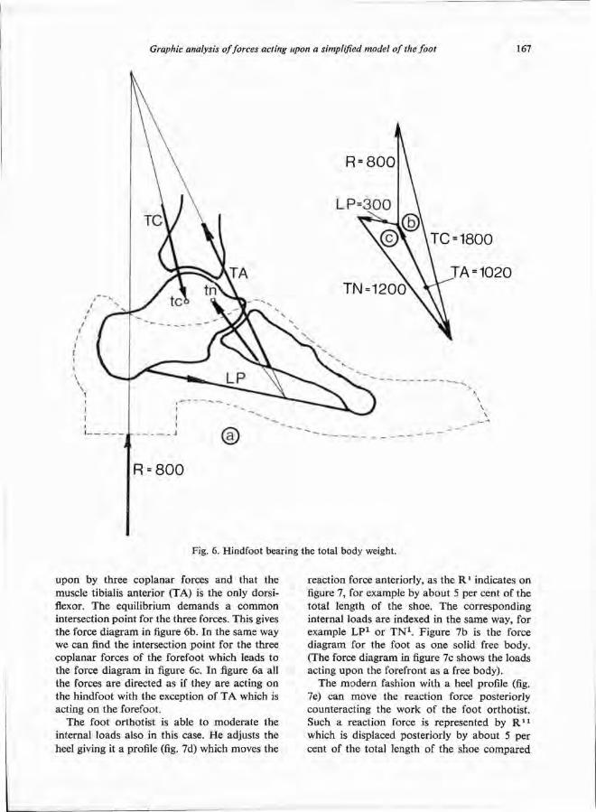
upon by three coplanar forces and that the muscle tibialis anterior (TA) is the only dorsi-flexor. The equilibrium demands a common intersection point for the three forces. This gives the force d iagram in figure 6b. In the same way we can find the intersection point for the three coplanar forces of the forefoot which leads t o the force diagram in figure 6c. In figure 6a all the forces are directed as if they are acting on the hindfoot with the exception of T A which is acting on the forefoot.
The foot orthotist is able to modera te the internal loads also in this case. He adjusts the heel giving it a profile (fig. 7d) which moves the
reaction force anteriorly, as the R 1 indicates on figure 7, for example by about 5 per cent of the total length of the shoe. The corresponding internal loads are indexed in the same way, for example L P 1 or T N 1 . Figure 7b is the force diagram for the foot as one solid free body. (The force diagram in figure 7c shows the loads acting upon the forefront as a free body) .
The modern fashion with a heel profile (fig. 7e) can move the reaction force posteriorly counteracting the work of the foot orthotist . Such a reaction force is represented by R 1 1
which is displaced posteriorly by about 5 per cent of the total length of the shoe compared
Fig. 6. Hindfoot bearing the total body weight.

with the normal situation (R). The corresponding internal forces are indexed in the same way, for example L P 1 1 or T N 1 1 .
The internal load distributions for externally loaded hindfoot situations are shown in table I I .
Fig. 7. Influence of the heel profile variation on the internal force distribution when the hindfoot is bearing the total body weight.

The simultaneously externally loaded hindfoot and forefoot
The graphical construction methods applied previously can be useful in analysing the internal load distribution in a situation when the reaction force is acting simultaneously on the forefoot and the hindfoot. Such a case is shown in figure 8 with symmetrically divided equal partial loads (each 400N).
In the first step we determine the loads acting on the foot as a one-piece solid body and we consider the total reaction force R = R 1 + R 2 and construct the force diagram in figure 8a. In this
Table II. Comparison of internal loadings using different heel profiles when externally loading the hindfoot (all values given in Newtons).
Fig. 8. Symmetrically loaded forefoot and hindfoot.

way we can determine the forces T C and TS . I n the next step we can find the loads acting on the forefoot as is done in figure 8b. Figure 8c shows the force diagram of the hindfoot. (The forces on the schematic architecture of the foot are directed as they are acting on the hindfoot).
H o w a pes planus will behave in this combined externally loaded case is shown in figure 9. The pes p lanus situation is indicated in the same way as in figure 4. The graphical construction method is similar t o the one that is used before. It is wor th noticing that the load in the idealized
ligament increases in this case by one fourth compared with the normal situation.
The foot orthotist helps the patient by applying an inlay sole and moving the hind part ial reaction anteriorly as shown in figure 10, the unchanged total reaction force R will be divided into part ial reaction forces in inverse ra t io t o their distance from R. The construct ion of force diagrams are similar t o the ones we have used before. A significant reduct ion in the magni tude of the internal load is obtained.
The results of the investigations on the foot
Fig. 9. Influence of pes planus configuration on the internal forces of the symmetrically loaded forefoot and hindfoot.

acted upon by the reaction forces acting simultaneously on the forefoot and hindfoot are shown in table III .
The destabilizing trend of the pes planus The figure 11 shows the foot structure in a n
even more simplified form than before. T h e reason for this is to show as clearly as possible the important feature of the pathomechanics of the pes planus. The forefoot and hindfoot are substituted by solid bars articulating at one idealized ankle joint and held together by the idealized plantar ligament. We assume that only the passive structures are engaged to maintain equilibrium with the external load.
Figure 11e shows the model of the normal foot. The force diagram in figure 11a belongs to the hindfoot and in figure 11b to the forefoot of such a model .
If the plantar passive structure in any way is
Fig. 10. Influence of pes planus inlay on the internal force.
Table III. Comparison of internal loading in different circumstances as a result of simultaneously loading the forefoot and the hindfoot (all values given in Newtons).

too weak to resist the load acting on it, the structure will give way and be lengthened. This lengthened passive plantar structure causes a pes p lanus architecture where the lengths of the solid forefoot and hindfoot are constant, but the height of the ankle joint will fall as shown in figure 11f. The forces acting on the forefoot in this case are shown in figure 11d and on the hindfoot in figure 11c. The load on the plantar passive structure in this case increases by about 60 per cent compared with the normal foot. This means that if the plantar passive structure
is unable to resist a normal load it will cause an architectural change which results in an even greater load on the same structure. This is a runaway trend which should be stopped.
In closing it must be remembered that all these analyses are carried out by reducing to the utmost the complex situation to get an easy method which gives the relative magni tude and directions of these loads depending on various situations.
Fig. 11. Diagram showing the destabilizing effect of pes planus.
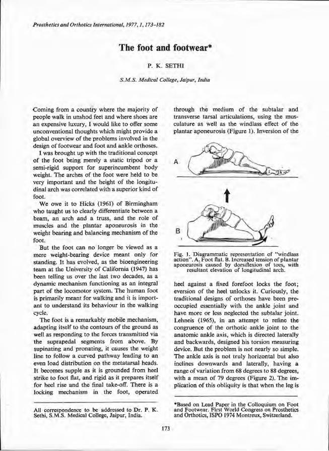
The foot and footwear*
P. K. SETHI
S.M.S. Medical College, Jaipur, India
*Based on Lead Paper in the Colloquium on Foot and Footwear. First World Congress on Prosthetics and Orthotics, ISPO 1974 Montreux, Switzerland.
Coming from a country where the majority of people walk in unshod feet and where shoes are a n expensive luxury, I would like t o offer some unconventional thoughts which might provide a global overview of the problems involved in the design of footwear and foot and ankle orthoses.
I was brought up with the traditional concept of the foot being merely a static t r ipod or a semi-rigid support for superincumbent body weight. The arches of the foot were held to be very important and the height of the longitudinal arch was correlated with a superior kind of foot.
W e owe it to Hicks (1961) of Birmingham w h o taught us to clearly differentiate between a beam, an arch and a truss, and the role of muscles and the plantar aponeurosis in the weight bearing and balancing mechanism of the foot.
But the foot can no longer be viewed as a mere weight-bearing device meant only for standing. It has evolved, as the bioengineering team at the University of California (1947) has been telling us over the last two decades, as a dynamic mechanism functioning as an integral pa r t of the locomotor system. The h u m a n foot is primarily meant for walking and it is importan t to unders tand its behaviour in the walking cycle.
The foot is a remarkably mobile mechanism, adapt ing itself to the contours of the ground as well as responding to the forces transmitted via the suprapedal segments from above. By supinating and pronat ing, it causes the weight line to follow a curved pathway leading to an even load distribution on the metatarsal heads. It becomes supple as it is grounded from heel strike to foot flat, and rigid as it prepares itself for heel rise and the final take-off. There is a locking mechanism in the foot, operated
through the medium of the subtalar and transverse tarsal articulations, using the musculature as well as the windlass effect of the plantar aponeurosis (Figure 1). Inversion of the
heel against a fixed forefoot locks the foot ; eversion of the heel unlocks it. Curiously, the traditional designs of orthoses have been preoccupied essentially with the ankle joint and have more or less neglected the subtalar joint . Lehneis (1965), in an a t tempt to refine the congruence of the orthotic ankle joint to the anatomic ankle axis, which is directed laterally and backwards, designed his torsion measuring device. But the problem is not nearly so simple. The ankle axis is not truly horizontal but also inclines downwards and laterally, having a range of variation from 68 degrees to 88 degrees, with a mean of 79 degrees (Figure 2). The implication of this obliquity is that when the leg is
Fig. 1. Diagrammatic representation of "windlass action". A. Foot flat. B. Increased tension of plantar aponeurosis caused by dorsiflexion of toes, with
resultant elevation of longitudinal arch.
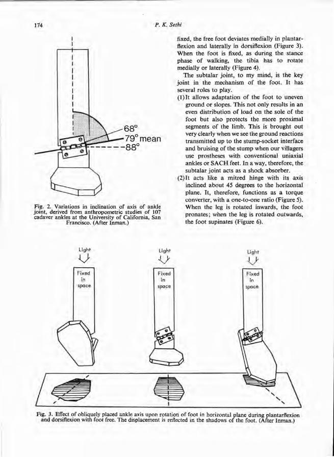
fixed, the free foot deviates medially in plantar-flexion and laterally in dorsiflexion (Figure 3). When the foot is fixed, as during the stance phase of walking, the tibia has to rotate medially or laterally (Figure 4).
The subtalar joint , to my mind, is the key joint in the mechanism of the foot. It has several roles to play. (1) It allows adapta t ion of the foot to uneven
ground o r slopes. This no t only results in an even distribution of load on the sole of the foot but also protects the more proximal segments of the limb. This is brought out very clearly when we see the ground reactions transmitted up to the stump-socket interface and bruising of the s tump when our villagers use prostheses with conventional uniaxial ankles or S A C H feet. In a way, therefore, the subtalar joint acts as a shock absorber.
(2) It acts like a mitred hinge with its axis inclined about 45 degrees to the horizontal plane. It, therefore, functions as a torque converter, with a one-to-one rat io (Figure 5). When the leg is rotated inwards, the foot pronates ; when the leg is rotated outwards , the foot supinates (Figure 6).
Fig. 2. Variations in inclination of axis of ankle joint, derived from anthropometric studies of 107 cadaver ankles at the University of California, San
Francisco. (After Inman.)
Fig. 3. Effect of obliquely placed ankle axis upon rotation of foot in horizontal plane during plantarflexion and dorsiflexion with foot free. The displacement is reflected in the shadows of the foot. (After Inman.)

(3)If there were no subtalar joint , the obliquity of the metatarsophalangeal break would cause the leg to tilt out during heel rise and
toe-off. Here , the subtalar joint acts as a correcting device, allowing the leg to remain vertical (Figure 7).
Fig. 4. Foot fixed to floor. Plantarflexion and dorsiflexion of the ankle produce horizontal rotation of the leg because of the obliquity of the ankle axis.
(After Inman.)
Fig. 5A. Action of mitred hinge. If the axis of the hinge is at 45 degrees, a simple torque converter is created. Rotation of the vertical member causes equal rotation of the horizontal member, with a
one-to-one relationship.
Fig. 5B. To prevent the entire horizontal segment from rotating, the horizontal member has been divided into a short proximal and a long distal
segment with a pivot in between. (After Inman.)
Fig. 6. Distal portion of horizontal member replaced by two structures A and B. Mechanical analogue of principal components of foot C and D. Mechanical components inserted into foot and leg. External rotation of the leg causes inversion of the heel, elevation of the medial side of the foot and depression of the lateral side. Internal rotation of the leg produces the opposite effect on the foot. (After
Inman.)

The close relationship between subtalar movement and horizontal ro ta t ion of the tibia during gait cycle has been demonstrated in several studies (Figure 8). F r o m heel strike t o foot flat, the foot pronates and the tibia rotates medially. Dur ing mid-stance, the foot supinates and the tibia reverses its rotat ion. At heel rise and toe-off, the heel is in inversion, the tibia is ro ta ted out. This tibial ro ta t ion has not received the attention it deserves in the design of foot orthoses and prostheses. I would like to emphasize that most quantitative da ta on subtalar movement and tibial ro ta t ion has been collected on level walkway studies. It is inevitable that the dimensions of these movements would be considerably greater when walking on irregular ground.
Any footwear, shoe modification, or a prosthetic foot should respect this system of linkage between the various articulations of the h u m a n foot.
In unshod communities the foot muscles get freedom for exercise and the joints remain supple. This is why functional disorders of the foot are so rarely seen in such people.
However, footwear becomes necessary t o protect the skin of the sole against rough ground or ha rd pavements and to keep the foot wa rm in cold climates. Over the ages, various communit ies have used locally available raw materials to design footwear which suit their own needs, and it is unnecessary and unwise to dictate to them that only a part icular design of footwear is correct. In fact I sometimes like to look upon closed shoes as braces, a necessary evil, which take u p the work of muscles, causing them to a t rophy from disuse and make
the joints stiff. I feel it is wise, at least for some time during the day, to take the shoes off and allow the foot to function unhampered. Use of an open sandal without a heel s trap, where the toes have to grip t o prevent their slipping off, is an excellent exercise for the intrinsic muscles. Fo r those who may argue that this cannot be applicable for the cold climates, I suggest the example of a Japanese Geta and Tobi , a very sensible combinat ion t o provide warmth as well as freedom of foot action (Figure 9).
In short, for normal feet, the use of a shoe is optional . But when a foot is abnormal a shoe becomes compulsory, to realign the foot, to redistribute pressure especially in an anaesthetic sole, to accommodate deformities, to equalize leg lengths, or to act as a foundation over which an orthosis has to be erected.
I feel a little puzzled about the long and repetitive list of shoe modifications which are illustrated in most texts on foot ailments. Whenever many alternatives are available for a particular situation it usually means that none is really effective. Also, I have never been sure that when I prescribe a wedged heel or an arch support , I a m really being rat ional . I a m particularly sceptical about arch supports which are based on a static concept of the foot. The advertising media exploit such devices, and I d o feel that some long term controlled clinical trials should be held to evaluate these devices. All useless and occasionally harmful modifications should be pruned. I must confess, however, that there is a great temptat ion to use them as placebos.
There is a need for controlling an abnormal inclination of the heel such as in valgus feet. Plastic heel seats were described by Helfet (1956) and later modified by Rose. Fo r some reason, I have not seen them being used. I wonder why? The U C B L foot support (Henderson and Campbell , 1969) developed at the Biomechanics Laboratory , University of California appeals t o me. Its fabrication, however, is not such as to be used except by skilled orthotists and it requires raw materials not available to us in the underdeveloped countries. I do feel that there has been no clear understanding of this problem and its solution so far.
F r o m my basic theme of emphasizing the mobility of the foot, I now move in the opposite direction, that is, the need in some situations, for rigid footwear. Fur ther work on the design of
Fig. 7. In spite of the obliquity of the metatarsophalangeal break, raising of the heel allows the leg to remain vertical. This is another function of the
subtalar joint. (After Inman.)

rigid footwear has become necessary. This is so for several reasons. An absolute indication for such rigid footwear is the anaesthetic foot with
healed ulcers or with toe deformities. Once the toes have lost their bite during push off, the metatarsal heads are exposed to t remendous
Fig. 8a. Subtalar rotation and horizontal rotation of tibia in gait cycle. Heel-strike to foot-flat—the heel is elevated and the tibia rotates medially.

pressure. There is also the shearing effect due t o the roll of the metatarsal heads on the soft tissues underneath them. A microcellular rubber insole of 15 shore hardness, as described by Paul Brand (Ward 1964) can only prevent ulcers in an unscarred balanced foot. This resilient insole, over a wooden clog with a rockered sole, abolishes peak pressures on the sole during the walking cycle. I think there is still considerable scope for research in the problem of redistribution of load on the
anaesthetic skin. A resilient insole is only one approach. There can be other approaches.
Such rigid wooden clogs have also been used in a completely different context by Hucks tep (1974) in Uganda for polio patients. Huckstep was concerned about the cost of leather shoes, the long waiting lists piling up in his workshop , and the lack of trained orthotists. Therefore, he tried to simplify the fabrication of calipers by using wooden soles and a rocker bar as a substitute for or thodox shoes. This is a realistic
Fig. 8b. During mid-stance—the foot supinates and the tibia reverses its rotation.

approach in underdeveloped countries. Rigid footwear with a rocker sole has been
advocated in a more sophisticated setting by John Glancy (1971) where he has used it to p rogramme the heel rise by the body tripping
over a rocker bar . A preset toe-off metal shoe plate can convert the entire length of the shoe into an efficient "resistance a r m " which not only prolongs the life of a shoe, but allows a "boos te r " spring to act efficiently.
Fig. 8c. At heel rise and toe-off, the heel is in inversion and the tibia rotates out.
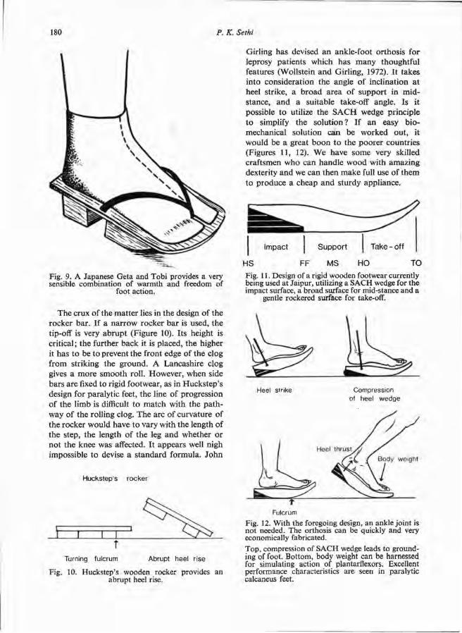
The crux of the matter lies in the design of the rocker bar . If a na r row rocker bar is used, the tip-off is very abrupt (Figure 10). Its height is critical; the further back it is placed, the higher it has to be to prevent the front edge of the clog from striking the ground. A Lancashire clog gives a more smooth roll. However, when side bars are fixed to rigid footwear, as in Huckstep 's design for paralytic feet, the line of progression of the limb is difficult to match with the pa thway of the rolling clog. The arc of curvature of the rocker would have to vary with the length of the step, the length of the leg and whether or not the knee was affected. I t appears well nigh impossible to devise a s tandard formula. John
Girling has devised an ankle-foot orthosis for leprosy patients which has many thoughtful features (Wollstein and Girling, 1972). It takes into consideration the angle of inclination at heel strike, a b road area of support in mid-stance, and a suitable take-off angle. Is it possible to utilize the S A C H wedge principle to simplify the solut ion? If an easy b io-mechanical solution can be worked out , it would be a great boon to the poorer countries (Figures 11, 12). W e have some very skilled craftsmen who can handle wood with amazing dexterity and we can then make full use of them to produce a cheap and sturdy appliance.
Fig. 9. A Japanese Geta and Tobi provides a very sensible combination of warmth and freedom of
foot action.
Fig. 10. Huckstep's wooden rocker provides an abrupt heel rise.
Fig. 11. Design of a rigid wooden footwear currently being used at Jaipur, utilizing a SACH wedge for the impact surface, a broad surface for mid-stance and a
gentle rockered surface for take-off.
Fig. 12. With the foregoing design, an ankle joint is not needed. The orthosis can be quickly and very economically fabricated. Top, compression of SACH wedge leads to grounding of foot. Bottom, body weight can be harnessed for simulating action of plantarflexors. Excellent performance characteristics are seen in paralytic calcaneus feet.

Finally a word about foot and ankle orthoses. There has been an unfortunate "or thot ic lag". In spite of rapid progress in the field of prosthetics, the design of orthoses has witnessed, till lately, no basic change over the last half century. Only recently, a break-through has appeared. There is a greater realization that any mechanical analogue should match the bio
mechanical system. I have already referred to the value of subtalar movement in the walking cycle and I would like to have a device which preserves it, especially when most of my patients have to walk on a rugged terrain. The U C B L dual-axis ankle was, in my opinion, a step in the right direction. But the recent work by Lehneis (1972) at New York University, such as the use of a plastic spiral ankle-foot orthosis, is much more appealing because these orthoses rely on the natural anatomic axes rather than some arbitrarily imposed external joints of the traditional brace systems. While plastics seem to have a great future, they are still very expensive and being rigid, require a great deal of expertise in the fabrication of such orthoses to avoid the creation of pressure points. Also, I am not quite sure whether these can withstand the merciless beating in our rugged terrain. It is in this context that I want you to consider the use of a cord-cum-rubber bracing, which is more akin to the properties of skeletal muscle
Fig. 13. Jaipur Foot—The external covering is vulcanized rubber with rayon cord reinforcement.
Fig. 14. Jaipur version of Syme's prosthesis. The cord-cum-rubber provides a combination of re
silience and stability.
Fig. 15. Schematic design of a foot orthosis made of vulcanized rubber and cord reinforcement. Movements are permitted at natural joint axes. Reasonable stability is conferred on a paralytic foot. The
orthosis is worn like a gym shoe.

and is not subject to the failure encountered in all mechanical spring systems. As a "spin-off" effect of our work on the Jaipur F o o t (Figure 13) and its modification for Syme's amputa t ion (Figure 14), it has recently occurred to us that we could extend the same principle for orthotic purposes. If a Syme's amputee can feel secure
in this resilient container, I see no reason why a flail ankle and foot cannot be stabilized by this device. I a m sure it can be used to p rogramme mot ion in any direction. W e intend to pursue this idea and have a feeling that , at least for problems of the foot and ankle, it may answer many of our needs (Figure 15).
REFERENCES
GLANCY, J. (1971). Lower extremity orthotic components—applications and indications. Instructional Course Lectures, American Academy of Orthopaedic Surgeons, Vol. XX, 130-141. The C. V. Mosby Co., St. Louis.
HELFET, A. J. (1956). A new way of treating flat feet in children. Lancet, 1, 262-264.
HENDERSON, W. H . and CAMPBELL, J. W. (1969). UC-BL shoe insert—casting and fabrication. Bull. Pros. Res., 1 0 - 1 1 , 215-235.
HICKS, J. (1961). The three weight-bearing mechanisms of the foot. Biomechanical Studies of the Musculoskeletal System. Ed. Evans, F. G . , 161-191. Charles C. Thomas, Springfield, Illinois.
HUCKSTEP, R. L. (1974). Orthopaedic problems in East Africa. Annals Royal College of Surgeons of Edinburgh, 2 , 214.
INMAN, V. T. and MANN , R. A. (1973). Biomechanics of the foot and ankle. DuVries' Surgery of the Foot. Ed. Inman, V. T., 3-22. The C. V. Mosby Co., St. Louis.
LEHNEIS, H. R. (1965). Orthotic measurement board for tibial torsion and toe-out. Orth, and Pros. Appl. J., 1 9 , 209-214.
LEHNEIS, H. R. (1972). The plastic spiral BKO. Final Report, Bioengineering Design and Development of Lower Extremity Orthotic Devices. Institute of Rehabilitation Medicine, New York University Medical Centre.
SETHI, P. K. (1974). Lower limb prosthesis for amputees in rural areas. Surgical Rehabilitation in Leprosy. Ed. McDowell, F. and Enna, C. D., 396-402. The Williams and Wilkins Co., Baltimore.
UNIVERSITY OF CALIFORNIA (1947). Fundamental studies of human locomotion and other information relating to design of artificial limbs. Report to National Research Council, Committee on Artificial Limbs.
WARD , D. J. (1962). Footwear in leprosy. Leprosy Review, 33, 97-105.
WOLLSTEIN, L. and GIRLING, J. (1972). Orthopaedic appliances for the leprosy patient—the fixed ankle brace walker. Leprosy Review, 4 2 , 162-164.

Amputation surgery in the lower extremity—part II G. M U R D O C H
Department of Orthopaedic Surgery and Traumatology, University of Dundee
Introduction Part I of this contribution was published in the last issue of "Prosthetics and Orthotics Interna t iona l" (Vol. 1, N o . 2). This par t presents the application of the techniques and considerations previously discussed in a variety of special circumstances.
Vascular disease Vascular disease has played an increasingly
important role in the field of amputa t ion . Atherosclerosis is widespread in Nor the rn Europe , N o r t h America and elsewhere in the world and several publications (Hansson, 1965 and Robinson, 1976) demonstrate that over 80 per cent of pr imary amputa t ions are now performed on patients with vascular disease and this despite a number of significant advances in vascular surgery. Our experience in the Tayside area of Scotland with a very stable populat ion, indicates a figure of 86 per cent (Murdoch et al, 1977).
Typically, these pat ients are elderly and subject t o other handicaps which are often multiple (Troup, et al, 1973). Mazet et al(1959) and Olejniczak (1967) indicate that as recently as twenty years ago some 90 per cent of these patients had above-knee amputa t ions performed.
One event focused at tention on the lot of the amputee—the advent of immediate postsurgical fitting of prostheses. Following the first report of Berlemont (1961) experience in a variety of places, notably that of Burgess and R o m a n o (1968) 1 and Sarmiento et al (1970), has underlined that a high propor t ion of these patients can be rehabilitated following successful fitting with a prosthesis. While the emphasis on immediate post-surgical fitting itself has now
diminished a number of centres a round the world have achieved similar success. Our own studies have demonstrated that close to 90 per cent can be returned, independent in some degree, to a home environment. We also know that this rehabilitation need take no more than forty or fifty days. However, to achieve this kind of result an integrated p rogramme of rehabili tation with a properly co-ordinated management of concurrent handicaps is required. Moreover, the knee joint should be saved when possible and to achieve this, the accuracy of level selection and the success of surgery are critical. Should level assessment be in error or the surgery fail for any reason the rehabili tation time is doubled.
Once presented for possible amputa t ion the dysvascular patient must be aggressively investigated as every day lost causes deterioration with increasing depression, muscle wasting, the development of flexion contractures and a variety of systemic changes due to inadequate fluid intake. The assessment must be of the patient as a whole and of the vascular tree in particular to decide whether vascular surgery can offer a solution. This usually involves arteriography which, however, is not in itself of particular value in determining any proposed level of amputa t ion (Murdoch, 1967; Burgess et al, 1971; Burgess and R o m a n o , 1971). The clinical appearances of the l imb remain impor tan t providing information about peripheral pulses, frank gangrene or partial ischaemia and particularly the state of the skin. The surgeon need not be put off the lower levels of amputa t ion by hairless, thin, inelastic skin but he will certainly have doubts where there are frank ischaemic ulcers or a severe temperature d rop is noted.
Fo r a number of years a variety of ancillary methods of investigation have been available and all provide information of value al though, even at this date, one cannot make categorical decisions on the basis of any single technique.

These methods include plethysmography, skin b lood flow and skin blood pressure estimations (Browse, 1973; Lassen and Holstein, 1974), the use of the Doppler ul t rasound in providing information about the total blood flow (Barnes et al, 1976) and thermography (Murdoch, 1975). Significantly useful results have been claimed for each of these methods . However it is necessary that work continues so that not only the crude level of amputa t ion , for example, above-knee or below-knee, is selected but also in the case of possible below-knee amputa t ion, the precise state of each of the tissues involved can be determined permitt ing the surgeon to fashion skin flaps appropria te to the individual case, according to the state of the pathology and, if need be, to excise muscle groups which are likely not t o survive the assault of the operat ion. Many of the successes reported in this field have been dependent on the ability to achieve amputa t ion at below-knee level and there is the suggestion that the employment of the posterior flap operat ion has also contr ibuted (Burgess and R o m a n o , 1968 2 ; Burgess et al, 1969; Murdoch , 1970). When it is considered that below-knee amputa t ion is not feasible, and perhaps the final decision should be made only at operat ion, through-knee amputa t ion, supracondylar amputa t ion such as the Gritti-Stokes and above-knee amputat ions are all available. Several publications testify to good results from the through-knee amputa t ion (Howard et al, 1969; Newcombe and Marcuson, 1972) and similarly good results with supracondylar amputa t ion (Weale, 1969; Mar t in et al, 1967; Middleton and Webster, 1962). However, these publications do not include the composite view of the pat ient 's life after amputa t ion nor, in some instances, indicate on what basis these other levels of amputa t ion have been selected. Whatever technique is employed one must pay respect to the importance of at tachment of the muscles to the bone end, sculpturing of the bone end where the bone is divided, high division of the nerve, adequate haemostasis and proper abutment of the skin edges without tension.
In summary, the level of amputa t ion selected should be based on as much information as possible, bearing in mind the value of the knee joint in the rehabilitation of the patient and in the quality of his life thereafter. The surgeon should have some knowledge of the prostheses available and confidence in his prosthetists to
fit the patient competently. The surgery and prosthetic fitting should take place in an environment which ensures the proper t reatment of concurrent handicaps, stimulates motivat ion and assures the patient an adequate follow-up in the fullest terms.
Diabetes Depending on the popula t ion at risk, some
25-50 per cent presenting for amputa t ion for vascular disease suffer from diabetes. The latter are often described as suffering from diabetic gangrene. There is no such single clinical entity. Meggitt (1973) emphasizes this in pointing out that the pathology can be the result of small vessel disease, major vessel occlusion as in atherosclerosis and in the absence of diabetes, neuropathy or infection t o which diabetic tissue is more prone . Some or all of these factors may have an influence in a single case and thus each case must be assessed individually. Where there is no major vessel occlusion Meggitt has shown that some diabetic feet can be saved by careful surgery, excising all dead tissue, opening all infected areas and by careful pressure dressings (Figure 1). In some cases local toe amputat ions ,
Fig. 1. Ischaemic and infected foot in diabetes. Wide exploration of tissues in sole of foot with excision of all dead and infected tissues. Sound healing achieved
without amputation.

skin grafting and partial foot amputa t ion such as the transmetatarsal procedure are appropriate. It has been shown by Meggitt and later by Wagner (1977) dealing with the same group of patients that Syme's amputa t ion in the diabetic dysvascular patient is a useful p ro cedure which can be performed with confidence provided level assessment has been carefully judged. Experience of the Syme's amputa t ion in atherosclerosis without diabetes has been very variable and is no t advocated. Where even a Syme's amputa t ion is no t deemed feasible the preponderance of major vessel occlusion in vessels below the knee rather than above the knee will normally permit a below-knee amputa t ion .
Trauma Amputa t ion in the severe t r auma resulting
from road traffic accidents, war injuries and the like, can provide many serious but interesting problems. The long established principles will hold and the principal one to conserve all viable tissue should, in the initial operat ion, be mandatory . In a few cases in those regions where replantat ion teams have been organized and micro-surgical competence is established the completely severed l imb with little local tissue damage should be presented with the patient as soon as possible in case replantat ion is feasible. (Chung-Wei, Che'en, 1962; Malt and McKhann , 1964). A considerable experience has been gained since these classic contr ibut ions al though n o conclusions can be reached (Editorial B.M.J. , 1975; China Med. J., 1975).
The judgement as t o whether a tissue is viable or not may be difficult but one should err on the side of conservation. W o u n d s should be closed when feasible. If there is much doubtful tissue destruction then open flaps should be employed. The contr ibutions of Welch et al (1969), Mayfield et al (1972) and Brown (1970) suggest that skin traction has still an important par t to play in this instance. By this approach and with the aid of plastic surgery joints can be saved and s tump length maintained.
The problems that remain are clouded by a philosophical one, namely that given a well motivated patient a n d a skilled prosthetist even the most badly scarred stumps can be fitted well enough. However, the patients normally concerned have a life expectancy of 30, 40 or 50 years and accordingly the question of re
construction of the s tump when indicated must be formally presented to the patient at an early date. S tump reconstruction is often necessary t o ensure that the s tump will give the best service to the patient over the years and not interfere with his employment and social aspirations. The further possible procedures that may be discussed include excision of the fibula to increase load bearing area in very short s tumps (Spira a n d Steinbach 1973), osteomyoplasty, scar excisions and mobilizations. Even if s tump reconstruction is inconvenient at the time of consultat ion the patient must be informed of the long term implications of deficiencies in the s tump so that he is in a posit ion t o make decisions regarding his future. The plastic surgeon in some instances should be consulted at this t ime to determine what role he may adopt .
Tumours Osteosarcoma appears generally in the ages
between 10 and 20 and continues to present horrifying decisions for the patients, parents and surgeons and, until recently, a five year survival rate of about 20 per cent was the c o m m o n experience. Dahl in and Coventry (1967) confirmed the typical occurrence a round the knee and the place of amputa t ion in t reatment . Radiotherapy has not significantly improved the pat ient 's chances of survival a l though its application as recommended by Cade (1955), has spared many patients needless amputa t ions by reserving the procedure for those most likely to survive. A series of publications since Jaffe's (1972) report on the use of high-dose methotrexate with ci t rovorum factor have awakened interest in chemotherapy because of the apparent improvement in survival rates. Marcove et al (1970) had shown the close relationship between the recognition of pulmonary metastases and death of the patient and so chemotherapy is now employed for treatment of the condit ion pr ior t o the known existence of any pulmonary metastases and instituted at the time of amputa t ion . Since then publications abound relating experience with a variety of drugs individually and in combinat ion in differing doses (Sutow et al, 1974). They highlight in part icular methotrexate in massive doses or adriamycin or combinat ions of bo th (Cortes et al, 1974). Jaffe and Wat ts (1976) in an editorial comment review the situation and point up some of the pathological and philo-

sophical problems. Much of the emphasis on chemotherapy is based on the view that the time interval between diagnosis of osteosarcoma and the appearance of lung metastases on radiological examination does not represent spread of the disease but ra ther the unveiling of disease which is already present. Jaffe points out further that while reports confirm that chemotherapy has substantially improved the prognosis in patients with osteosarcoma he wonders whether the results will s tand up to the passage of time. Prior to chemotherapy if a patient survived two years he could be considered "cured" . It is not possible to say this about the new treatments yet. These drug regimes also carry a high degree of toxicity. There is, therefore, a clear need for collaboration between centres as in the United Kingdom Medical Research Council trial to determine the efficacy of these drugs, their op t imum dosage and the best ways of reducing toxic effects.
F o r the surgeon while chemotherapy slows down the healing of amputat ion wounds, it also permits amputa t ion through the affected bone provided it is at least 70-100 m m above the most proximal area of bone reaction seen on bone scan. Amputa t ion remains the main weapon of t reatment but the way may now be open for resection of tumour rather than amputat ion in some cases. I wholly agree with Jaffe and Wat t s ' plea for referral of patients to centres staffed by experts who can provide the best overall results.
Soft tissue sarcomas in the extremities A report by Simon and Enneking (1976)
regarding the management of soft tissue sarcomas of the extremities outline their approach and decision leading to radical local resection or amputa t ion . They place particular emphasis on the value of angiography and bone-scanning. Any surgeon contemplating radical local resection should read this paper with care.
Chondrosarcoma Chondrosarcoma is not normally amenable
to radiation and is difficult to categorically exclude in any individual case of chondroma, especially when the tumour is large and in the pelvis. Where X-ray examination, bone scans and the like have been performed, it is often possible to m a k e a categorical diagnosis of chondrosarcoma and proceed to amputa t ion as survival rates are higher than in osteosarcoma.
The pelvic tumours usually require a hind-quarter amputat ion. Any argument for amputa tion in other cases will depend on a full histological examination of the tumour removed.
Other tumours for a variety of reasons may bring about the need for amputa t ion but then usually because of failed local excision or p ro gression of the tumour to the point where there is gross bone destruction not amenable to other forms of treatment.
Infection Occasionally emergency amputa t ion is re
quired in the case of gas gangrene. Otherwise the question of amputa t ion in infection is usually confined to cases of longstanding infection associated often with ununited fractures and partial ischaemia of the tissues. In these cases the infection is walled off and amputa t ion can be carried out through the affected bone.
Congenital limb deficiency Congenital limb deficiency is comparatively
rare in the experience of any one surgeon and, in my view, the management of these cases is perhaps best under taken by special Centres which by reason of their competence have accumulated a large experience. Even in these circumstances concern about a basic understanding of the nature of the defects and the difficulties in comparing methods of management has been expressed by a number of au thors at tempting to name and classify the defects. The International Society for Prosthetics a n d Orthotics took a lead in this regard, following the interest displayed by the Committee on Prosthetic Research and Development at the Nat ional Academy of Sciences in the United States, and created a Sub-committee to look at this problem. This group, through a series of workshops, has reached a measure of agreement and the results of their work has been published and tested under field conditions (Kay, 1974). It is intended that further work and trials will be conducted to revise the system as referred t o in Part I. There is fairly general agreement that congenital absence of the fibula is best treated by ablation of the foot. O n the other hand , in for example, proximal focal femoral deficiency, controversy continues and the debate on surgical management must include procedures such as arthrodesis of t he

knee, Syme's amputa t ion, osteotomies and the more exotic procedures such as the Van Ness operat ion. Other deformities provoke similar controversy. In my view the clinic team, before proceeding to any operation, should have a clear picture of the likely natural history of the deficiency itself and of the patient in different situations. Parents should be fully informed and any decisions taken should be on the basis of a full understanding of the situation. Limb discrepancy, deformity and paralysis
There remains a broad group of disease and disability categories which result in various combinat ions of deformity, paralysis and l imb discrepancy. This group can provide the most difficult problems as amputa t ion, if employed, is not a life saving procedure, nor indeed is it necessary to p romote locomotion. A careful analysis of each of the elements of the problem must be under taken and the team must have an intimate knowledge of the patient, her family and her aspirations if a correct decision is to be made .
Typical pathologies involved include septic arthritis of infancy, poliomyelitis, tuberculosis with premature closure of the epiphysis, the late effects of t r auma and spina bifida. The result may include gross shortening, joint instability, arthritis, degrees of paralysis, deformity, neuropathy and ischaemia.
Westin (1967) in outlining the criteria for leg lengthening points out that the integrity of bo th hip and knee joints must be carefully evaluated and that in cases of extreme shortening amputat ion may be the t reatment of choice. In his series of twenty-six patients, lengthening from 15 to 60 m m was achieved with residual discrepancies ranging from 15 to 110 mm. Over half the patients had major complications for example dislocation of the hip, fractures, osteomyelitis. This experience suggests that in discrepancies over 130 or 140 m m amputa t ion should be considered.
Each case must be considered on its merits but knee disarticulation and Syme's amputa t ion can be deployed with benefit as the usual cosmetic disadvantages do not apply as, by virtue of the shortening, the bulbous end can be contained within the prosthetic thigh or shank.
Joint instability may result from a variety of causes, for example paresis, architectural deficiencies o r the development of arthritis. Each joint must be assessed pr ior to amputa t ion
as t o h o w it will be affected by amputa t ion and prosthetic fitting. It may be improved by some other surgical procedure, be eliminated by the amputa t ion itself, or be so gross that after amputa t ion it will still require external support with perhaps only marginal benefits. This applies especially to proposed amputa t ions below an unstable knee.
If a joint is arthritic it is amenable to s tandard treatments such as joint replacement, it may be removed by the amputa t ion or the operat ion can be carried out through the affected jo int .
Deformity of the short l imb may be compounded of many factors such as reduction or enlargement of the par t , gross wasting and angulation of the major bones or at major joints. The deformity may be eliminated by the amputa t ion or by a separate procedure, for example osteotomy above the level of amputation.
The decision cannot rest solely on functional considerations even in the male. The t rap for the operating surgeon is to replace observed ugliness in static terms with an equally unpleasant and irremediable dynamic ugliness. Amputa t ion in these cases may be a valid procedure but often it is only one of a p r o gramme of operations designed to ensure an improvement in both appearance and locomoto r capacity. Surgeons not faced with prosthetic problems in their normal working lives are strongly recommended to consult surgeons in this field before undertaking the irrevocable step of amputa t ion .
Special situations Growth period
The surgeon must be especially cautious in undertaking amputa t ion during the growth period. This caution has already been underlined with regard to congenital limb deficiencies but for the most part we are considering the question of t r auma in children. Every effort should be m a d e to retain joints and if a jo in t cannot be retained, the epiphysis may be saved. This should be at tempted even when there is major skin loss. Quite extensive areas can be covered by split skin grafts and later, if need be , by full thickness skin. Split skin grafts are able to survive direct and shear forces during the growth period better than they do in the fully ma tu re adult (Figure 2). Even when the epiphysis cannot be retained stumps should be as long

as possible. If overgrowth of the bone occurs in relation to the soft tissues then tr imming of the bone can be performed at later procedures. More proximal amputat ions should not be performed to avoid these further t r imming procedures nor should operat ions such as epiphyseal arrest be considered. The p ro cedures, therefore, which give the best long term results are clearly through-knee amputations and the Syme's procedure if they are justified on other grounds. I n amputa t ions through the shaft of a bone the general principles of tissue management should be maintained. At below-knee level it is my view that an osteomyoplasty should be performed if possible. Later developments such as a valgus deformity can be corrected by an addit ional surgical procedure as required.
Stump revisions Revisions of initial amputa t ion surgery are
indicated from time to time. They should only
be performed with clear objectives in mind and any procedure should be discussed with the prosthetist as he may be able to offer solutions which avoid surgery. The revision proposed may be required to eliminate adherent scars, bony spikes, neuromata etc. The surgeon in performing these procedures should be sensitive to the requirements of the " idea l" s tump. Fo r example, in my view it is rarely sufficient to excise a neuroma which is seen to be the cause of disabling symptoms. It is usually also necessary to reconstruct the end of the s tump ensuring secure muscle fixation, a well sculptured bone end, and a newly cut end of the nerve so placed in healthy muscle that it will not be involved again in the development of terminal scar tissue. In longstanding stumps the surgeon should look carefully for evidence of local terminal ischaemia. This can be demonstrated by a significant d r o p in temperature a n d sensory changes. Before proceeding to s tump surgery it is often worthwhile to consult the vascular
Fig. 2. Below-knee amputation in a boy of 12. Extensive skin loss. Survival of split skin grafts with prosthetic sore some years later. Refitting of PTB socket permitted sound healing.

surgeon with a view to possible lumbar sympathectomy. Close co-operat ion with the prosthetist is essential if the resultant scars are to be properly placed in relation to the load bearing areas.
In vascular cases it is all t oo common a n experience for surgery to fail at below-knee level as the precise level of tissue viability is often difficult to determine. Before proceeding to a knee disarticulation or above-knee amputat ion the surgeon should carefully consider whether local excision of ischaemic and infected tissues can be done with a reasonable hope of pr imary wound healing and the retention of a useful below-knee s tump. The au thor recommends a wedge resection certainly in those cases who have had a posterior flap amputa t ion performed (Figure 3). In this procedure an undissected wedge of tissue is excised. After outlining the incisions to encompass the tissue, sharp dissection proceeds through the retained gastrocnemius muscle posteriorly to above the level of the affected tissue and the wedge is then removed en bloc by taking a Gigli saw upwards through soft tissue and bone alike. After minor bone sculpture at the anterior distal end of the
tibia wound closure can be affected in a neat and precise way.
Amputation in the anaesthetic limb Amputa t ion in this situation is usually con
sidered in cases of high sciatic nerve damage, leprosy, spina bifida etc., and becomes necessary because of intractable infected t rophic ulcers which have usually invaded bone or joint . The t rap for the amputa t ing surgeon here is that he may proceed to amputa t ion a t too high a level in an effort to remove anaesthetic skin. H e should remember that the skilled prosthetist is able to transfer force actions via the socket in a much better way than is normally effected by footwear for example. The surgeon should consult the prosthetist in selecting the level of amputa t ion in the confidence that the prosthetist will be able to design a socket to transfer the forces of weight bearing without irremediable skin damage. Even in anaesthetic end bearing s tumps successful prosthetic fittings have been achieved (Scrinivasan, 1973).
Flexion deformities Flexion deformities are the bane of the
surgeon and prosthetist 's life. The best treatment is prevention and the clinic team's efforts in this direction must be initiated where possible from before amputa t ion and throughout the period of rehabilitation. It is tempting t o give angles of flexion deformity which can be accommodated by the prosthetic design but individual cases are best discussed with the prosthetist . These deformities usually occur as a result of contracting scar tissue or because of sustained flexion prior t o amputa t ion . In the latter instance it is usually an elderly patient with vascular disease who has had a long period of enforced bed rest accompanied by severe pain. The t rap for the surgeon here is that he may be tempted to operate at a higher level because of a n apparent gross flexion deformity. H e should leave his final decision until the patient is completely anaesthetized. In the post-operative period splintage of the proximal joint may avoid the development of a chronic flexion deformity. Needless to say the role of the physical therapist is of pa ramoun t importance. Harmful s tump postures in the rehabilitation period are well known and instruction in their avoidance should be par t of the clinic team's p rogramme of management .
Fig. 3. Failed posterior flap amputation. Wedge resection with en bloc removal of infected and
ischaemic tissues. Primary wound healing.

Where physiotherapy and prosthetic alignment measures have failed to correct the flexion deformity surgery may be considered. At the hip this would certainly include elevation of the origins of the s trap muscles and tensor fascia lata from the anterior superior iliac spine and adjacent iliac crest and division of the ilio-psoas tendon. It may also require elevation or division of the origins of rectus femoris. At the knee it may require division or elongation of the hamstrings and perhaps even division of the posterior capsule of the knee joint.
Recent advances
Skeletal attachment
W o r k on skeletal a t tachment of the prosthesis continues. Acceptable clinical solutions have been achieved in individual cases and the main problem relates to the percutaneous interface with the search for biocompatible materials and shapes. Other considerations will assume greater importance when the philosophy is applied to the lower extremity.
Biofeedback training for amputees
This approach appears to have benefits in training the amputee to walk in an op t imum manner with his prosthesis and are clearly of great value in the primary, elderly amputee . These techniques are usually based on making the patient aware of the prosthetic knee position, t runk att i tude, step length and weight t ransference by audible or tactile signals.
Stump environment
Since the pioneering work of the Seattle group many Centres employ a rigid cast as the initial dressing following amputa t ion with benefits in terms of tissue stabilization, non-inrteference with the wound, freedom to move and the ability to exercise the muscle (Burgess and R o m a n o , 1968 1 ; Mooney et al, 1971). Reference has already been made to controlled environment treatment and its benefits. One important aspect of this innovation is that it provides a " l abora to ry" for the study of wound healing in a controlled environment.
Fig. 4. Gross chronic indurated lymphatic oedema of leg and foot. Result following cyclic pressure in controlled environment treatment device and plastic surgery. No amputation.

Conclusion The experience of the last twenty years
demonstrates that the amputat ing surgeons need n o longer be regarded as they were by Guy Pat in in the 17th century
" M e r e booted lackeys—a race of extravagant coxcombs who wear moustaches and flourish razors . "
The surgery is now more considerate, more conservative and more in tune with the pat ient 's needs. Indeed the surgeon involved in amputation surgery is perhaps more sensitive to solutions other than amputa t ion because of his awareness of the life of the amputee . Figure 4 shows a grossly ugle leg due to chronic indurated lymphatic oedema of doubtful origin. The patient was presented for amputa t ion . Employing Controlled Environment t reatment the oedema was gradually dissipated permitt ing
plastic surgery and a very satisfactory result without amputat ion.
It has been demonstrated that this field of scientific endeavour covers the whole range of disciplines in medicine and the physical sciences. It is now clear that the rehabilitation of the amputee does not depend solely on the operating theatre and the prosthetist 's shop but that a team approach is necessary if the amputee is to be returned to his h o m e and work, integrated with his prosthesis and the community . There are clear implications for funding authorities in terms of the organization of systems to ensure that all the professional skills are brought together in a collaborative way for the benefit of the patient and underlying all the problems is the need for proper education of all professionals bo th at undergraduate and postgraduate level.
REFERENCES
BARNES, R . W., SHANIK, G. D. and SLAYMAKER, E. E. (1976). Proceedings : an index of healing in below-knee amputation : leg blood pressure by Doppler ultrasound. J. Cardiovasc. Surg. (Torino) 17(1): 101-2: also Surgery 79(1): 13-20, Jan. 1976.
BERLEMONT, M. (1961). Notre experience de l'appareillage précoce des amputés de membre inférieur aux establ'sements. Helio-Marins de Berck. Annals de Medicine Physique, iv, 4.
BROWN, P. W. (1970). Rehabilitation of bilateral lower-extremitv amputees. J. Bone and Joint Surg., 5 2 A , 687-699.
BROWSE, N . L. (1973). Choice of level of amputation in ischaemic arterial disease. Scand. J. Clin. Lab. Invest., 3 1 , Suppl. 128, 249-252.
BURGESS, E. M. and ROMANO R . L. (1968)1. The management of lower extremity amputees using immediate post-surgical prostheses. Clin. Orthop. and Rel. Res., 5 7 , 137-146.
BURGESS, E. M. and ROMANO, R . L. (1968)2. The stabilization of muscles in lower extremity amputations J. Bone and Joint Surg., 50A, 1486-1487.
BURGESS, E. M., ROMANO, R . L. and ZETTL, J. H. (1969). The management of lower extremity amputations. TR10-6, August, Washington: U . S . Government Printing Office.
BURGESS, E. M. and ROMANO R . L. (1971). Level selection in lower extremity amputations. Clin. Orthop. 7 4 , 177-184.
BURGESS, E. M., ROMANO, R . L., ZETTLE, J. H. and SCHRoCK, R. B. (1971). Amputations of the leg for peripheral vascular insufficiency. J. Bone and Joint Surg., 5 3 A , 874-889.
CADE, S. (1955). Osteogenic sarcoma. A study based on 133 patients. J. Royal Coll. Surg. Edin., 1, 79-111.
CHUNG-WEI, CH'EN (1962). Salvage of the forearm following complete traumatic amputation. China Med. J., 82, 632.
CORTES, E . P., HOLLAND, J. F . , WANG , J. J., SINKS, L. F . , BLOM, J., SENN, H . , BANK, A. and GLIDE-WELLS, O. (1974). Amputation and adriamycin in primary osteosarcoma. New England J. Med., 291, 998-1000.
DAHLIN, D . C. and COVENTRY, M. B . (1967). Osteogenic sarcoma. J. Bone and Joint Surg., 49A, 102-110.
EDITORIAL (1975). Replantation of severed limbs. Brit. Med. J., 3 (5986), 722. Replantation of severed limbs. Analysis of 40 cases. China Med. J. 1 (4), 265-74, July.
HANSSON, J. (1965). Leg amputees, Pros. Int., 2,4-5.
HOWARD, R. R. S,, CHAMBERLAIN, J. and MAC-PHERSON, A. I. S. (1969). Through-knee amputations in peripheral vascular disease. Lancet, 2, 240.
JAFFE, N . (1972). Recent advances in the chemotherapy of metastatic osteogenic sarcoma. Cancer 3 0 , 1627-1631.
JAFFE, N . , FREI, E. , TRAGGIS, D . and BISHOP, Y . (1974). Adjuvant methotrexate and citrovorum-factor treatment of osteogenic sarcoma. New England J. Med., 291, 994-997.
JAFFE, N . and WATTS, H . G . (1976). Multidrug chemotherapy in primary treatment of osteosarcoma. J. Bone and Joint Surg., 5 8 A , 6 3 4 - 6 3 5 .

KAY, H . W . (1974). A proposed international terminology for the classification of limb deficiencies. Orth, and Pros., 2 8 : 2 , 33-48.
LASSEN, N . A. and HOLSTEIN, P. (1974). Use of radioisotopes in assessment of distal blood pressure in arterial insufficiency. Surg. Clin. N. Amer., 5 4 , 39-55.
MALT. R . A. and MCKHANN , C. F . , (1964). Replantation of severed arms. J. Amer. Med. Ass., 1 8 9 : 1 0 , 716-722.
MARCOVE, R . C., MIKE, V., HUTTER, R . V. P., Huvos, A. G., SHOJI, H . , MILLER, T. R . and KOSSLOFF, R . (1970). Chondrosarcoma of the pelvis and upper end of the femur. J. Bone and Joint Surg., 5 4 A , 561-572.
MARTIN, P., REN WICK, S. and MAELOR-THOMAS, E. (1967). Gritti-Stokes amputation in atherosclerosis—a review pf 237 cases. Brit. Med. J., 3 , 837.
MAYFIELD, G. W . , BURKHALTER, W . E. and BALLARD, A. (1972). Ambulant treatment of lower extremity amputees with open stumps. J. Bone and Joint Surg., 5 4 A , 7, 1571.
MAZET, R., SCHILLER, F . J., D U N N , O . J. and NEUFELD, A. J. (1959). Influence of prosthetic healing on the health of geriatric amputees. Report of Project, 431. Washington, D. C.: Office of Vocational Rehabilitation.
MEGGITT, B . F . (1973). Orthopaedic management of foot breakdown in the diabetic patient. Proceedings and Reports of Councils and Associations. J. Bone and Joint Surg., 5 5 B , 882.
MIDDLETON, M . D. and WEBSTER, C. V. (1962). Clinical review of the Gritti-Stokes amputation. Brit. Med. J., 2, 574.
MOONEY, V., HARVEY, J. P., MCBRIDE , E. and SNELSON, R. (1971). Comparison of post-operative stump management: Plaster vs. soft dressings. J. Bone and Joint Surg., 5 3 A , 241-249.
MURDOCH, G. (1967). Levels of amputation and limiting factors. Annals Royal Coll. Surg. England, 4 0 , 204-216.
MURDOCH, G. (1970). The surgery of the below-knee amputation. Prosthetic and Orthotic Practice. Ed. Murdoch, G., 45-60. Edward Arnold, London.
MURDOCH, G. (1975). Research and development within surgical amputee management. Acta ortho. Scand., 46, 526-547.
MURDOCH, G., WOOD, M. A., TROUP, I. M . , DONEGAN, N. and JOY, E . (1977). Nursing barriers to the effective rehabilitation of the lower limb amputee. Presented at Second World Congress, I.S.P.O., New York, May.
NEWCOMBE, J. F. and MARCUSON, R. W. (1972). Through-knee amputation. Brit. J. Surg., 59, 260.
OLEJNICZAK, S. (1967). Summary of leg amputations and prosthetic replacement at Wayne County General Hospital during 1961-1965. Michigan Medical News, 66, 723-726.
ROBINSON, K . P. (1976). Long posterior flap amputation in geriatric patients with ischaemic disease. Annals Royal Coll. Surg. England, 58, 440-451.
SARMIENTO, A., MAY, B . J., SINCLAIR, W. F. , MCCOLLOUGH, N. C. III and WILLIAMS, E . M . (1970). The impact of immediate post-surgical fitting. Clin. Orthop., 68, 22-31.
SCRINIVASAN, H. (1973). Syme's amputation in insensitive feet. A review of twenty cases. J. Bone and Joint Surg., 55A, 558-562.
SIMON, M. A. and ENNEKING, W. F. (1976). The management of soft-tissue sarcomas of the extremities. J. Bone and Joint Surg., 58A, 317-327.
SPIRA, E . and STEINBACH, T., (1973). Fibulectomy and resection of the peroneal nerve for short tibia stumps. Acta, ortho. Scand., 44, 589-596.
SUTOW, W. W,, SULLIVAN, M. P. and FERNBACH, J. D. (1974). Adjuvant chemotherapy in primary treatment of osteogenic sarcoma. Proceedings of the American Association for Cancer Research, 51, 20.
TROU , I. M . , MURDOCH, G. and CONDIE, D . N. (1974). A statistical survey of amputee care within a specialized prosthetic unit. Presented at First World Congress, I.S.P.O., Montreux, October.
WAGNER, W. F., (1977). Amputation of the foot and ankle. Current status. Clin. Orthop., 122, 62-69.
WEALE, F . E . (1969). The supra-condylar amputation with patellectomy. Brit. J. Surg., 56, 589.
WELCH, R. B . , MeTZ , C. W., BECKER, Q. H. and RANKIN, E. A. (1969). The management of the battle incurred lower extremity amputee. J. Boen and Joint Surg., 51A, 1041.
WESTiN, W. G., (1967). Femoral lengthening using a periosteal sleeve. J. Bone and Joint Surg., 49A, 836-854.

Prosthetics and Orthotics International, 1977,1,193-199
Calendar of Events
P R E L I M I N A R Y N O T I C E
A COURSE ON ABOVE-KNEE PROSTHETICS
DENMARK
5th to 11th NOVEMBER, 1978
A course covering all aspects of Above-Knee Prosthetics will be held in Rungsted, Denmark , from 5th to 11th November , 1978. Consideration will be given to medical and surgical factors; locomot ion; biomechanics; socket shape; function; fabrication; components and their function; al ignment; gait training. New practices in the fitting and design of the A K prosthesis will be highlighted and the different systems currently available will be discussed.
The Registration Fee will be U.S . $190. Accommodat ion will be available at the Course Centre at a cost for full boa rd of $240 (including the Course Dinner) .
Attendance will be strictly limited to 90 participants.
A n indication of your interest in at tending should be sent to the Secretariat in Copenhagen. The next issue of Prosthetics and Orthotics International will contain an application form.
January 1978 Second International Conference on Legislation Concerning the Disabled, Manila , Philippines. Informat ion: Rehabili tat ion International , 122 East 23rd St., New York, N . Y . 10010.
2-13 January, 1978 746 Course in Upper Limb Prosthetics for Prosthetists. Informat ion: New York University, Post -Graduate Medical School, Prosthetics and Orthotics, 317 East 34th Street, New York , N . Y . 10016.
7-8 January, 1978 Instructional Course, British Orthopaedic Association, City University, London . Informat ion: Miss M . Bennett, Honora ry Secretary, B.O.A., Royal College of Surgeons, Lincolns Inn Fields, London W C 2 A 3PN.
9-20 January, 1978 N C 203 Course in Knee-Ankle-Foot and Hip-Knee-Ankle-Foot Orthotics for Orthotists. Informat ion: J. Hughes , Director, Nat ional Centre for Training and Educat ion in Prosthetics and Orthotics, University of Strathclyde, 73 Rot tenrow, Glasgow G 4 0 N G .
16-27 January, 1978 756 Course in Spinal Orthotics for Orthotists. Informat ion: New York University, Pos t -Graduate Medical School, Prosthetics and Orthotics, 317 East 34th Street, New York , N . Y . 10016.
193

194 Calendar of events
23-27 January, 1978 622-C, 623-C Courses in Lower Limb Prosthetics for Physicians, Surgeons and Therapists. Informat ion: Charles M. Fryer, Director, Prosthetic-Orthotic, Center, Northwestern University Medical School, 345 East Superior Street, R o o m 1723, Chicago, Illinois 60611.
23 January-3 February, 1978 N C 205 Course in Above-Knee Prosthetics for Prosthetists. Informat ion: J. Hughes , Director, Nat ional Centre for Training and Educat ion in Prosthetics and Orthotics, University of Strathclyde, 73 Rot tenrow, Glasgow G 4 0 N G .
26-27 January, 1978 681-C Course in Immediate Postsurgical Fitt ing for Prosthetists. Informat ion: Charles M. Fryer, Director, Prosthetic-Orthotic Center, Northwestern University Medical School, 345 East Superior Street, R o o m 1723, Chicago, Illinois 60611.
30 January-3 February, 1978 744A Course in Upper Limb Prosthetics and Orthotics for Physicians and Surgeons. Informat ion: New York University, Post -Graduate Medical School, Prosthetics and Orthotics, 317 East 34th Street, New York, N . Y . 10016.
6-17 February, 1978 742C Course in Lower Limb Prosthetics for Therapists. Informat ion: New York University, Post -Graduate Medical School, Prosthetics and Orthotics, 317 East 34th Street, New York, N . Y . 10016.
13-17 February, 1978 N C 206 Course in Upper Limb Orthotics for Orthotists. Informat ion: J. Hughes, Director, Nat ional Centre for Training and Educat ion in Prosthetics and Orthotics, University of Strathclyde, 73 Rot tenrow, Glasgow G 4 0 N G .
13-17 February, 1978 6 2 2 - D , 623-D Courses in Lower Limb Prosthetics for Physicians, Surgeons and Therapists. Informat ion: Charles M. Fryer, Director, Prosthetic-Orthotic Center, Northwestern University Medical School, 345 East Superior Street, R o o m 1723, Chicago, Illinois 60611.
14-15 February, 1978 International Society for Prosthetics and Orthotics. International Symposium ' P R O T E C H . ' Mart inihai Centrum, Groningen, The Netherlands. Informat ion: Mart inihal Centrum, Afdeling Congreszaken, Postbus 8010 Groningen (V.K. 9700), The Netherlands.
16-17 February, 1978 6 8 1 - D Course in Immediate Postsurgical Fitt ing for Prosthetists. Informat ion: Charles M. Fryer, Director, Prosthetic-Orthotic Center, Northwestern University Medical School, 345 East Superior Street, R o o m 1723, Chicago, Illinois 60611.
20-24 February, 1978 N C 101B Course in Lower Limb Prosthetics for Physicians and Surgeons. Informat ion: J. Hughes , Director, Nat ional Centre for Training and Educat ion in Prosthetics and Orthotics, University of Strathclyde, 73 Rot tenrow, Glasgow G 4 0 N G .
27 February-3 March, 1978
7 4 5 A Course in Upper Limb Prosthetics for Therapists. Informat ion: New York University, Post -Graduate Medical School, Prosthetics and Orthotics, 317 East 34th Street, New York, N . Y . 10016.

Calendar of events 195
6-10 March, 1978 N C 102B Course in Lower Limb Orthotics for Physicians and Surgeons. Informat ion: J. Hughes, Director, Nat ional Centre for Training and Educat ion in Prosthetics and Orthotics, University of Strathclyde, 73 Rot tenrow, Glasgow G 4 0 N G .
6-10 March, 1978 652-B, 653-B Courses in Upper Limb Prosthetics and Orthotics for Physicians, Surgeons and Therapists . Informat ion: Charles M. Fryer, Director, Prosthetic-Orthotic Center, Northwestern University Medical School, 345 East Superior Street, R o o m 1723, Chicago, Illinois 60611.
6-11 March, 1978 741C Course in Lower Limb Prosthetics for Physicians and Surgeons. Informat ion: New York University, Pos t -Graduate Medical School, Prosthetics and Orthotics, 317 East 34th Street, New York , N .Y. 10016.
13-17 March, 1978 7th Congress of the World Federat ion of Occupation Therapists, Tel Aviv, Israel. Informat ion: (President of the Organizing Committee) Mrs . M . Clyman, 65 Aloof David Street, Rama t -Gan , Israel.
13-17 March, 1978 75IB Course in Lower Limb Orthotics for Physicians and Surgeons. Informat ion: New Y o r k University, Pos t -Graduate Medical School, Prosthetics and Orthotics, 317 East 34th Street, New York , N . Y . 10016.
13-17 March, 1978 752B Course in Lower Limb Orthotics for Therapists. Informat ion: New York University, Post -Graduate Medical School, Prosthetics and Orthotics, 317 East 34th Street, New York , N . Y . 10016.
13-17 March, 1978 722-A, 723-A Courses in Lower Limb and Spinal Orthotics for Physicians, Surgeons and Therapists . Informat ion: Charles M . Fryer, Director, Prosthetic-Orthotic Center, Northwestern University Medical School, 345 East Superior Street, R o o m 1723, Chicago, Illinois 60611.
13-23 March, 1978 N C 207 Course in Spinal Orthotics for Orthotists. Informat ion: J. Hughes , Director, Nat ional Centre for Training and Educat ion in Prosthetics and Orthotics, University of Strathclyde, 73 Rot tenrow, Glasgow G 4 0 N G .
20-24 March, 1978 757A Course in Upper Limb Orthotics for Therapists. Informat ion: New Y o r k University, Pos t -Graduate Medical School, Prosthetics and Orthotics, 317 East 34th Street, New York , N . Y . 10016.
20-24 March, 1978 622-E, 623-E Courses in Lower Limb Prosthetics for Physicians, Surgeons and Therapists. Informat ion: Charles M. Fryer, Director, Prosthetic-Orthotic Center, Northwestern University Medical School, 345 East Superior Street, R o o m 1723, Chicago, Illinois 60611.

196 Calendar of events
23-24 March, 1978
681-E Course in Immediate Postsurgical Fitt ing for Prosthetists. Informat ion: Charles M,- Fryer, Director, Prosthetic-Orthotic Center, Northwestern University Medical School, 345 East Superior Street, R o o m 1723, Chicago, Illinois 60611.
27-31 March, 1978
7561 Course in Advanced Spinal Orthotics for Orthotists. Informat ion: New Y o r k University, Post -Graduate Medical School, Prosthetics and Orthotics, 317 East 34th Street, New York, N . Y . 10016.
3-7 April, 1978
622 -F , 623 -F Course in Lower Limb Prosthetics for Physicians, Surgeons and Therapists. Informat ion: Charles M. Fryer, Director, Prosthetic-Orthotic Center, Northwestern University Medical School, 345 East Superior Street, R o o m 1723, Chicago, Illinois 60611.
5 April, 1978
Internat ional Society for Prosthetics and Orthotics, United Kingdom, 6th Scientific Meeting. T o be held in conjunction with British Orthopaedic Association Spring Meeting, University of Surrey, Guildford. Informat ion : John Williams, Secretary, U . K . Nat ional Member Society, ISPO, Trustees Office, Queen Mary ' s Hospital , Roehampton , London SW15 5PN.
6 - 8 April, 1978 Spring Meeting, British Orthopaedic Association, University of Surrey, Guildford. Informat ion: Miss M. Bennett, Honora ry Secretary, B.O.A., Royal College of Surgeons, Lincolns Inn Fields, London W C 2 A 3PN.
6-7 April, 1978 6 8 1 - F Course in Immediate Postsurgical Fitt ing for Prosthetists. Informat ion: Charles M. Fryer, Director, Prosthetic-Orthotic Center, Northwestern University Medical School, 345 East Superior Street, R o o m 1723, Chicago, Illinois 60611.
10-14 April, 1978 744B Course in Upper Limb Prosthetics and Orthotics for Physicians and Surgeons. Informat ion: New York University, Post -Graduate Medical School, Prosthetics and Orthotics, 317 East 34th Street, New York , N . Y .
10-14 April, 1978 652-C, 653-C Courses in Upper Limb Prosthetics and Orthotics for Physicians, Surgeons and Therapists. Informat ion: Charles M. Fryer, Director, Prosthetic-Orthotic Center, Northwestern University Medical School, 345 East Superior Street, R o o m 1723, Chicago, Illinois 60611.
17-28 April, 1978
742D Course in Lower Limb Prosthetics for Therapists. Informat ion: New York University, Post -Graduate Medical School, Prosthetics and Orthotics, 317 East 34th Street, New York, N . Y . 10016.

Calendar of events 197
1-5 May, 1978 751C Course in Lower Limb Orthotics for Physicians and Surgeons. Informat ion: New York University, Pos t -Graduate Medical School, Prosthetics and Orthotics, 317 East 34th Street, New York, N . Y . 10016. 752C Course in Lower Limb Orthotics for Therapists. Information: New York University, Post -Graduate Medical School, Prosthetics and Orthotics, 317 East 34th Street, New York, N . Y . 10016.
1-5 May, 1978 772-B, 723-B Courses in Lower Limb and Spinal Orthotics for Physicians, Surgeons and Therapists. Informat ion: Charles M. Fryer, Director, Prosthetic-Orthotic Center, Northwestern University Medical School, 345 East Superior Street, R o o m 1723, Chicago, Illinois 60611.
8-12 May , 1978 622-G, 623-G Courses in Lower Limb Prosthetics for Physicians, Surgeons and Therapists. Informat ion: Charles M. Fryer, Director, Prosthetic-Orthotic Center, Northwestern University Medical School, 345 East Superior Street, R o o m 1723, Chicago, Illinois 60611.
8-13 May , 1978 741D Course in Lower Limb Prosthetics for Physicians and Surgeons. Informat ion: New York University, Pos t -Graduate Medical School, Prosthetics and Orthotics 317 East 34th Street, New York, N . Y . 10016.
11-12 May , 1978 681-G Course in Immediate Postsurgical Fitt ing for Prosthetists. Informat ion: Charles M. Fryer, Director, Prosthetic-Orthotic Center, Northwestern University Medical School, 345 East Superior Street, R o o m 1723, Chicago, Illinois 60611.
15-18 May , 1978 631-B, 632-B, 633-B Courses in Management of the Juvenile Amputee for Physicians, Surgeons, Therapists and Prosthetists. Informat ion: Charles M. Fryer, Director, Prosthetic-Orthotic Center, Northwestern University Medical School, 345 East Superior Street, R o o m 1723, Chicago, Illinois 60611.
15-26 May , 1978 758 Course in Upper Limb Orthotics for Orthotists. Informat ion: New York University, Pos t -Graduate Medical School, Prosthetics and Orthotics, 317 East 34th Street, New York, N . Y . 10016.
22-26 May , 1978 622-H, 623-H Courses in Lower Limb Prosthetics for Physicians, Surgeons and Therapists. Informat ion: Charles M. Fryer, Director, Prosthetic-Orthotic Center, Northwestern University Medical School, 345 East Superior Street, R o o m 1723, Chicago, Illinois 60611.
25-26 May , 1978 681-H Course in Immediate Postsurgical Fitt ing for Prosthetists. Informat ion: Charles M. Fryer, Director, Prosthetic-Orthotic Center, Northwestern University Medical School, 345 East Superior Street, R o o m 1723, Chicago, Illinois 60611.

198 Calendar of events
June, 1978 8th International Congress of the World Federat ion for Physical Therapy, Tel Aviv, Israel. Informat ion: Secretary General , Miss M. M . McKay, W.C.P.T. , Brigray House , 20/22 Mor t imer Street, London W 1 P 1AA.
5-9 June, 1978 750B Course in Prosthetics and Orthotics for Rehabil i tat ion Counselors. Informat ion: New York University, Pos t -Graduate Medical School, Prosthetics and Orthotics, 317 East 34th Street, New York, N . Y . 10016.
5-9 June, 1978 622-1, 623-1 Courses in Lower Limb Prosthetics for Physicians, Surgeons and Therapists. Informat ion: Charles M. Fryer, Director, Prosthetic-Orthotic Center, Nor thwestern University Medical School, 345 East Superior Street, R o o m 1723, Chicago, Illinois 60611.
5-23 June, 1978 753 Course in Lower Limb Orthotics for Orthotists. Informat ion: New York University, Pos t -Graduate Medical School, Prosthetics and Orthotics, 317 East 34th Street, New York, N . Y . 10016.
8-9 June, 1978 681-1 Course in Immediate Postsurgical Fitt ing for Prosthetists. Informat ion: Charles M. Fryer, Director, Prosthetic-Orthotic Center, Northwestern University Medical School, 345 East Superior Street, R o o m 1723, Chicago, Illinois 60611.
12-16 June, 1978 745B Course in Uppe r Limb Prosthetics for Therapists. Informat ion: New York University, Post -Graduate Medical School, Prosthetics and Orthotics, 317 East 34th Street, New York, N . Y . 10016.
19-21 June, 1978 755 Course in Spinal Orthotics for Physicians and Surgeons. Informat ion: New Y o r k University, Post -Graduate Medical School, Prosthetics and Orthotics, 317 East 34th Street, New York, N . Y . 10016.
26-30 June, 1978 757B Course in Uppe r Limb Orthotics for Therapists. Informat ion: New Y o r k University, Pos t -Graduate Medical School, Prosthetics and Orthotics, 317 East 34th Street, New York , N . Y . 10016.
26-30 June, 1978 751-A Course in Fit t ing and Fabricat ion of the C.T.L.S.O. (Milwaukee-type Orthosis) for Orthot is ts Informat ion: Charles M . Fryer, Director, Prosthetics-Orthotic Center, Northwestern University Medical School, 345 East Superior Street, R o o m 1723, Chicago, Illinois 60611.
26-30 June, 1978 801-A Course in Pedorthic Management of the Foot . Informat ion: Charles M . Fryer, Director, Prosthetic-Orthotic Center, Northwestern University Medical School, 345 East Superior Street, R o o m 1723, Chicago, Illinois 60611.

Calendar of events 199
2-7 July, 1978 3rd Wor ld Congress of the International Rehabili tat ion Medicine Association, Basle, Switzerland. Informat ion: D r . W. M . Zinn, Thermes, C H 7310 Bad Ragaz, Switzerland.
2-8 July, 1978 7th Pan-American Congress on Rheumat ic Diseases, Bogota, Columbia. Informat ion: Secretariat, Associacion Colombiana de Reumatologia, Apar t ado Aeteo 90331, Bogota, D . E., Columbie.
5-21 July, 1978 743 Course in Above-Knee Prosthetics for Prosthetists. Informat ion: New York University, Pos t -Graduate Medical School, Prosthetics and Orthotics, 317 East 34th Street, New York , N . Y . 10016.
8-11 August, 1978 Seminar on Funct ional Rehabili tation—Devices, Costs and Effectiveness, University of Strathclyde, Glasgow, Scotland. Informat ion: Professor R. M. Kenedi , University of Strathclyde, Bioengineering Unit , Wolfson Centre, 106 Rot tenrow, Glasgow G 4 0 N W , Scotland.
13-25 August, 1978 British Council Course 836 on Biomechanics of the H u m a n Body with Reference to Orthopaedic Implants , Prostheses and Orthoses, University of Strathclyde, Glasgow, Scotland. Informat ion: The Courses Depar tment , The British Council, 65 Davies Street, London W 1 Y 2AA, o r your local British Council Office.
23-28 August, 1978 9th Congress of the International Association of Educators for Handicapped Youth , Montreal , Canada . Informat ion: (President of the Organizing Committee) M. Marcel Saint-Jacques, C.E.C.M., 3737 Est, Sherbrooke, Montreal , P.Q. , Canada .
28 August-1 September, 1978 The Sixth International Symposium on External Control of H u m a n Extremities, Dubrovnik , Yugoslavia. Announcement and call for papers . Authors willing to submit papers are requested to send abstracts no t later than February 1, 1978. Informat ion: G o r d a n a Aleksic, Yugoslav Committee for E T A N , P.O. Box 356, 11001 Belgrade, Yugoslavia.
20-22 September, 1978
A u t u m n Meeting, British Orthopaedic Association, London (combined with Spanish Orthopaedic Association). Informat ion: Miss M . Bennett , Honora ry Secretary, B.O.A., Royal College of Surgeons, Lincolns Inn Fields, London W C 2 A 3PN.

A N N O U N C E M E N T
A B S T R A C T S O F T H E P A P E R S S U B M I T T E D T O T H E 1 9 7 7 I S P O / A O P A W O R L D C O N G R E S S A N D T H E N A M E S A N D A D D R E S S E S O F A L L C O N G R E S S C O N T R I B U T O R S A R E N O W AVAILABLE. (These were contained in the Programme of the Congress; therefore those persons who at tended the Congress should already have a set.)
T o order Abstracts and Address List—Fill out below and send with $7.00 to cover handling and mailing costs t o :
1977 ISPO Wor ld Congress, A B S T R A C T S , c/o USISPO, 1444 N . Street, N . W . , Washington, D .C . 20005
Make Cheques Payable to 1977 World Congress. Please Print :
Name
Address
State Country
200

Internationally Recognized
Compar ison Burn Scar BEFORF
omparisoπ Burn Scar A N T I / B U R N S C A I ^ G A R M E N T
Compar ison Burn Scar AFTER r
Now Made In Ireland
/ M Full
4^F Length
Sleeve
JOBST® Garments for Severe Vascular Disorders and Control of Hypertrophic Scarring and Contractures S e v e r e v a s c u l a r d i s o r d e r s , a s w e l l a s t h e c o n t r o l o f
h y p e r t r o p h i c s c a r r i n g a n d c o n t r a c t u r e s m a y b e
c o n t r o l l e d b y u s i n g c l i n i c a l l y p r o v e n J O B S T V E N O U S
P R E S S U R E G R A D I E N T ® S u p p o r t s a n d J O B S T A N T I /
B U R N S C A R ™ G a r m e n t s . P l e a s e s e n d f o r c o m p l e t e
i n f o r m a t i o n a n d s p e c i a l m e a s u r i n g t a p e s t o o u r n e w
m a n u f a c t u r i n g f a c i l i t y i n I r e l a n d . A l l p r o d u c t s a r e
m a n u f a c t u r e d u n d e r d i r e c t A m e r i c a n e n g i n e e r i n g
s u p e r v i s i o n .
© 1977 Jobst 1
Reg. U.S. Pat. O f f .
JOBST JOBST Ireland, Ltd. Thurles Industr ia l Estate Thurles, County Tipperary Republ ic of I reland
L .
I w o u l d l ike more i n f o r m a t i o n and measur ing tapes for Jobst Garments.
Name Hospi ta l . Address
" J


KNIT-RITE PROSTHETIC
SOCKS Dependable, Quality
Since 1923 No Corners Cut For
Production Economies Highest quality yarns
Carefully knitted & hand finished
SOFT — DURABLE — COMFORTABLE
(L
I ^ I V I ' I - W I ' I I C , I N C .
2 0 2 0 G R A N D A V E N U E • P. O . B O X 2 0 8 • K A N S A S C I T Y , M I S S O U R I 6 4 1 4 1
P H O N E : 8 1 6 - 2 2 1 - 0 2 0 6
vii

Your B u y i n g
Orthoses Materials
O R T H O L E N - ; ^ jm Cervical Brace* ORTHOLEI\
Drop Foot
Splint
Three Point Corset REPONA-Cervical Brace Drop Foot Splint
S A D U R T e p e f o m ® T e p d e n ®
PVA β P V O <5%> W I L H J U L T E U F E L D7000 Stuttgart 1

INTEGRATED PROSTHETIC ORTHOPAEDIC SERVICES
Integrates conventional alignment principles into the prosthesis—no new alignment techniques involved. The Adapter Chain allows these advantages: Hip joint installation based on the principles of the Canadian hip joint—but fully alignable. 30° alignment range in all links in all planes below the hip, above and below the knee and above the ankle. V.A.—recommended alignment capability at ankle, including plantar and dorsiflexion range and toe-out adjustment. Static and dynamic alignment of the same system—no transfer and duplication.
i p o s ipos kg Postfach 1845 D-3140 Lüneburg Germany
The MODULAR System With The IPOS Adapter Chain

ET an outstanding tool in biomechanics: T U E MCIAf Q u a r t z multicomponent I MC N C W measuring platform for
sports
N E W ! Quartz Multicomponent Measuring Platform for Biomechanics -this unique instrument determines together with a special electronic unit the following 6 measurands: • the 3 components F x, F y and F z
of any force acting on it • the coordinates a„ and a y of the instantaneous point of force application • the free moment M'z about an axis normal to the platform
orthopedics neurology
This system is an indispensable tool in various fields such as • sports: reaction forces in broad and high jump, shot put, running etc. • orthopedics: gait analysis, evaluating prosthesis, monitoring rehabilitation etc. • physiology of work: stress on workers, human engineering etc. • neurology: "Romberg" test, psychomotor phenomena. • posturography: more than a simple statokinesimeter, as the horizontal forces (in 2 components!) exerted in maintaining equilibrium are measured, too. Over 110 systems are successfully used all over the world! Special version with glass top plate available on request.
The outstanding features are: • amazingly simple operation • factory calibrated • extremely wide measuring range • very low cross talk (typically <1%) • linear and free of hysteresis • natural frequency over 1 kHz • interchangeable top plates • simplified mounting. We offer a complete line of piezoelectric transducers for pressure, force and torque, multicomponent force, acceleration and charge amplifiers, also in miniature versions, and other electronic equipment.
Piezo-Messtechnik <ISTLER Kistler Instrumente AG CH-8408 Winterthur/Switzerland Phone: (052) 25 2821 Telex: 76458
X

JUééiauen Specials Illustrated literature available on all items shown.
TORONTO PERTHES
TRILATERAL PERTHES
'A" FRAME
PH9 " F O R H I P D Y S P U S I A = P A V L I K H A R N E S S • • • V O N R O S E N S P L I N T
ill ' O R T H O P E D I C ESTABLISHED 1935
P.O. BOX 1678 CHATTANOOGA, TENN. 37401
Sclerotherapy, Operation or both. Whichever method you choose, medical elastic compression stockings and panties
SIGVARIS (
FINEIA® wi l l aid and ensure successful treatment.
S IGVARIS®+ FINELA®have the physio logi cally correct compression values and are being satisfactorily worn by millions of patients.
Further indicat ions: Prophylaxis of thrombosis, venous insufficiency of various genesis, after ulcus cruris, after leg fractures.
Ctj)— registered trade mark of Gaπzoni & Cie AG. St Gallen/Switzerland
Manufacturer: GANZONI & CIE AG. 9014 St Galleπ/Switzerland Licensee: GANZONI 8. CIE SA, 68304 St-Louis/Fraπce Distributors tor USA, Canada and Great Britain: CAMP
Xİ

J O I N T S A N D B R A C E S
L e g a n d h i p j o i n t s , b r a c e s a n d
s h o e s t i r r u p s
M A T E R I A L
F a b r i c a t i n g s u p p l i e s , t o o l s
L E A T H E R A N D F A B R I C S
S P A R E P A R T S
F e e t , k n e e s , s o c k e t b l o c k s , h o o k s ,
h a n d s , w r i s t a n d e l b o w u n i t s
T O O L S A N D M A C H I N E S
' M I R A '
A b d u c t i o n p i l l o w s p l i n t s
E. LINK & CO P . O . B o x 3 9 4
D-7200 TUTTLINGEN T E L E P H O N E ( 0 7 4 6 1 ) 5 0 1 8
MASCH IN EI\I-SCH MID A long and successful
tradition in machinery for
Orthopaedic Supplies
• R O B U S T
I • S T E A D I L Y I M P R O V E D
• D U S T E X T R A C T I N G
Machinery and tools
made in Germany
by specialists for specialists
For detailed information please write to:
M a s c h i n e n - S c h m i d , M o r e n a s t r 3 , D 8 0 0 0 M u n i c h G e r m a n y T e l e p h o n e 0 8 9 / 8 3 2 5 1 0
[SCHMİDİ
xü

N e w f r o m P r o t e k :
O r i g i n a l M . E . M ü l l e r t o t a l h i p p r o s t h e s e s I I I Mir lllk\\**S*' MM / / /A4\≤f W l!= m\v&?
w i t h WOLOX A l j O ^ c e r a m i c h e a d s B i o l o x ® c e r a m i c i s c h e m i c a l l y
i d e n t i c a l w i t h t h e j e w e l s a p p h i r e
F o r a n o u t s t a n d i n g f r i c t i o n
a n d w e a r b e h a v i o u r :
H i g h d e n s i t y R C H 1 0 0 0
P O L Y E T H Y L E N E c u p
c o m b i n e d w i t h a
B I O L O X β A I 2 0 3 C E R A M I C h e a d
B i o l o x ® c e r a m i c c o m p a r e d t o m e t a l h a s
a b e t t e r b i o s t a b i ! i t y a b e t t e r b i o c o m p a t i b i l i t y a h i g h e r c o r r o s i o n r e s i s t a n c e a b e t t e r w e t t a b i l i t y b y f l u i d s a h i g h e r h a r d n e s s a h i g h e r s c r a t c h r e s i s t a n c e
P.O. Box 2016 3001 Berne Switzerland
' Registered trade mark of Sulzer Bros. Ltd., Winterthur "Regis tered trade mark of Feldmühle AG, Plochingen

xiv

MANUFACTURERS AND SUPPLIERS OF PROSTHETICS ORTHOTICS AND MEDICAL EQUIPMENT
H u g h S t e e p e r ( R o e h a m p f o n ) L i m i t e d
Registered Office: Registered No. 475291 ENGLAND 237-239 Roehampton Lane, London SW15 4LB Te lephone: 01-788 8165-9 Telex No. 261787 Te legraphic Address: Steeper L o n d o n .
List of advertisers Charles A. Blatchford & Sons Ltd. ii
O t to Bock Orthopädische Industr ie K G I F C
Fillauer Orthopedic xi
Ganzoni & Cie A G xi
IPOS K G ix
Jobst (Ireland) Ltd. V
Kingsley Manufacturing C o . vi
Kistler Instruments A G X
Knit-Rite Inc. vii
E. Link & Co . xii
Protek A G Bern xiii
Maschinen-Schmid xii
H u g h Steeper (Roehampton) Ltd. XV
Wilh. Jul . Teufel viii
Uni ted States Manufacturing Co . O B C
Yaesu Rehabil i tat ion Co . Ltd. I B C
Julius Zorn G M B H XİY
XV

Prosthetics and Orthotics International, 1977
Information for Contributors Contributions should be sent to Prosthetics and Orthotics International , Nat ional Centre for Training and Educat ion in Prosthetics and Orthotics, University of Strathclyde, 73 Rot tenrow,Glasgow G 4 0 N G . In the meant ime considerable difficulty and delay is entailed in processing contr ibutions in languages other than English. Authors are asked to provide three copies of text, tables and figures. Papers are accepted on the understanding tha t they may be subject t o editorial revision a n d that n o substantial pa r t has been, or will be published elsewhere. Subsequent permission to reproduce articles must be obtained from the publishers. Manuscripts should be typewritten in double line spacing on one side of paper only with margins of 25 mm. Papers must commence with an abstract not exceeding 250 words . O n a separate sheet must b e :
(1) Title and short title. The short title should appear at the head of each page and should not exceed forty-five characters including spaces.
(2) Authors ' names, initials and title. The present address of any author if different from the place where the work was done, may be shown as a footnote.
(3) D e p a r t m e n t s ) in which the work was done. (4) The name and full postal address of the author to w h o m correspondence and requests for
reprints should be directed. This will appear as a footnote.
Illustrations All illustrative material should be lightly marked on the back in pencil with the figure number in arabic numerals, title of paper, au thors ' name and a clear indication of the top of the figure. The approximate location in the text should be marked. Figure captions should be typed on a separate sheet. Tables should be used only when necessary t o clarify impor tant points . Each table should be typed on a separate sheet and numbered consecutively in arabic numerals .
References References in the text should follow the author /date system for example: Peizer (1971). If there are more than two authors—Solomonidis et al. (1974). References at the end of articles should be listed on a separate sheet in alphabetical order of (first) author ' s name, as follows: Marx, H . W. (1974). Lower limb orthotic designs for the spastic hémiplégie patient. Orthotics and Prosthetics, 28(2), 14-20. Journal titles must be given in full.
References to articles in books should include author , year of publication, article title, book title, edition, editor (if different from author) , first and last pages, publisher and place of publication. F o r example, Hughes , J. (19"'5). Recent developments in prosthetics and orthotics. Recent Advances in Orthopaedics (2) Ed. McKibbin , B, , 196-216, Churchill Livingstone, Edinburgh.
Reprints Ten reprints will be supplied free of charge to the first named author . Addit ional reprints will be available at cost if they are ordered when the proofs are returned.
XV i

L E A D I N G R E H A B I L I T A T I O N T R A I N I N G E Q U I P M E N T M A N U F A C T U R E R O F J A P A N
Y A E S U Automatic recording of weight bearing balance Quantitative evaluation of weight bearing in lower extremity and loading in upper extremity Adjustable to match the need of each patient for evaluation and training
W E I G H T B A L A N C E A N A L Y Z E R
R G - 8 0 0
This new electronic balance analyzer makes it possible to give difference in weight bearing in training function of lower extremities, which would be ideal for prosthetic training.
Y a e s u R e h a b i l i t a t i o n C o m p a n y L t d
Yαesu Headquarter Building
3-6-13 Kayabacho, Nihonbashi, Chuo-ku,
Tokyo, Japan
tel:(03)666-7911 tlx:2524526 YAEHON J
In upper extremity this balance analyzer gives differential value of loading. Recording outlet is attached and this can be easily connected to various types of auto-recording system.

Hinged Ankle Joint Fracture Cast Brace
Part No. 1S 301
Sold on prescription only.
İ6rVie U N I T E D S T A T E S M A N U F A C T U R I N G C O . P O B o x 1 1 0 , 6 2 3 S o u t h C e n t r a l A v e n u e G l e n d a l e , Ca l i fo rn i a 9 1 2 0 9 U S A P h o n e { 2 1 3 ) 2 4 5 6 8 5 5 C a b l e L I M B R A C E TWX No 910-4972279
The Hinged Ankle Joint Fracture Cast Brace is a new addition to United States
Manufacturing Company's family of fracture bracing components. Designed
as a single axis joint orthosis, rather than a
polycentric one, the hinged ankle joint brace
allows normal tibial torsion which reduces
fracture site strain. Strain applied to the brace itself is also reduced, eliminating the breakage
problem encountered by polycentric type orthoses. Free plantar dorsiflexion motion is achieved by
positioning the axis of the orthosis directly over the anatomical axis of the ankle.
Strong, lightweight and ribbed for extra rigidity and strength, the hinged ankle joint brace is supplied with a Velcro strap which facilitates easy donning
of the patient's shoe. The brace is available in 3 sizes: small, medium and large — left or right, and may be used with plaster bandages Hexcelite Orthoplast, Zorac,
or any other equivalent substitute.