prosurvival ambra1 turns into a proapoptotic bh3-like
TRANSCRIPT

BASIC RESEARCH PAPER
Prosurvival AMBRA1 turns into a proapoptotic BH3-like protein during mitochondrialapoptosis
Flavie Strappazzona, Anthea Di Ritaa,b, Valentina Cianfanellic, Melania D’Oraziob, Francesca Nazioa,b, Gian Maria Fimiad,e,and Francesco Cecconia,b,c
aIRCCS Fondazione Santa Lucia, Rome, Italy; bDepartment of Biology, University of Rome Tor Vergata, Rome, Italy; cUnit of Cell Stress and Survival,Danish Cancer Society Research Center, Copenhagen, Denmark; dDepartment of Biological and Environmental Sciences and Technologies (DiSTeBA),University of Salento, Lecce, Italy; eNational Institute for Infectious Diseases ‘L. Spallanzani’ IRCCS, Rome, Italy
ARTICLE HISTORYReceived 24 September 2015Revised 2 March 2016Accepted 7 March 2016
ABSTRACTAutophagy and apoptosis are 2 stress-response mechanisms that are closely interconnected. However, themolecular interplays between these 2 pathways remain to be clarified. Here we report that the crucialproautophagic factor AMBRA1 can act as a positive mediator of mitochondrial apoptosis. Indeed, we showthat, in a proapoptotic positive feedback loop, the C-terminal part of AMBRA1, generated by CASP/CASPASE cleavage upon apoptosis induction, inhibits the antiapoptotic factor BCL2 by a direct bindingthrough its BH3-like domain. The mitochondrial AMBRA1-BCL2 complex is thus at the crossroad betweenautophagy and cell death and may represent a novel target in development of therapeutic approaches inclinical diseases.
KEYWORDSAMBRA1; apoptosis;autophagy; BCL2; BH3domain
Introduction
Autophagy is an important eukaryotic process involved in thelysosomal degradation of cytosolic components, both underphysiological and pathological conditions. During autophagy,the autophagosomes—specific double-membrane vesicles—engulf a number of different cargoes and then fuse with lyso-somes for subsequent recycling of their content. Several keyproteins are involved in autophagosome formation, such asBECN1/Beclin 1 and its positive regulator AMBRA1; AMBRA1and BECN1 are present in the class III phosphatidylinositol 3-kinase (PtdIns3K) complex,1,2 and are involved in autophago-some nucleation, an early step of autophagy induction. Morerecently, it has been demonstrated that AMBRA1 and BECN1can also regulate autophagy at different steps of the process, bybinding other key interactors.3-6 Interestingly, a pool ofAMBRA1 is localized at the mitochondria, where its proauto-phagic activity is inhibited by mitochondrial resident BCL2.7
Moreover, AMBRA1 regulates mitophagy, a form of autophagythat ensures the specific removal of damaged mitochondria,both dependently or independently of the E3-ubiquitin LigasePARK2/PARKIN.8
The fact that AMBRA1 interacts with the antiapoptotic factorBCL2 places it at the crossroad between autophagy and cell death.In fact, BCL2, a well-known antiapoptotic factor whose overex-pression protects against a wide range of apoptotic inducers, canregulate BECN1-induced autophagy at the endoplasmic reticu-lum by a direct binding to the BECN1 BH3 domain and with the
contribution of CISD2/NAF-1 (CDGSH iron sulfur domain2).9,10 BCL2 can also regulate BECN1-induced autophagy bysequestering AMBRA1 (the activator of BECN1) at the mito-chondria.6 Conversely, it has been demonstrated that a numberof regulators of autophagy, such as BECN1 and PI3K3C3/Vps34,11,12,13,14,15 ATG4A/ATG4, ATG5,16,17,18,19 or AMBRA1,20
are subjected to proteolytic cleavage during cell death so as toabrogate their proautophagic function. Another point of conver-gence between autophagy and apoptosis is the binding betweenATG12 and BCL2 family members that takes place in order toinduce mitochondrial apoptosis.21
Besides AMBRA1 binding to BCL2 in the mitochondrialfraction, we have previously demonstrated that this event isreduced following apoptosis induction,7 this indicating thatAMBRA1 could be involved in cell death regulation.
In this work, we explore the relationship betweenAMBRA1 and the death machinery by investigating whetherAMBRA1 could disrupt BCL2’s ability to protect against apo-ptosis. In particular, because AMBRA1 is cleaved by CASPduring apoptosis, we decided to study the effect of the C-ter-minal fragment of AMBRA1, which is released by this CASPcleavage, on BCL2. We found that this fragment, through aBH3-like domain, is able to inhibit the antiapoptotic functionof BCL2 so as to enhance apoptosis. To summarize, we pro-pose a model in which AMBRA1 is involved in both autoph-agy and apoptosis, depending upon the presence of its full-length or cleaved form.
CONTACT Flavie Strappazzon [email protected]; Francesco Cecconi [email protected], [email protected] data for this article can be accessed on the publisher’s website.
© 2016 Flavie Strappazzon, Anthea Di Rita, Valentina Cianfanelli, Melania D’Orazio, Francesca Nazio, Gian Maria Fimia, and Francesco Cecconi. Published with license by Taylor & Francis.This is an Open Access article distributed under the terms of the Creative Commons Attribution-Non-Commercial License (http://creativecommons.org/licenses/by-nc/3.0/), which permitsunrestricted non-commercial use, distribution, and reproduction in any medium, provided the original work is properly cited. The moral rights of the named author(s) have been asserted.
AUTOPHAGY2016, VOL. 12, NO. 6, 963–975http://dx.doi.org/10.1080/15548627.2016.1164359
Results
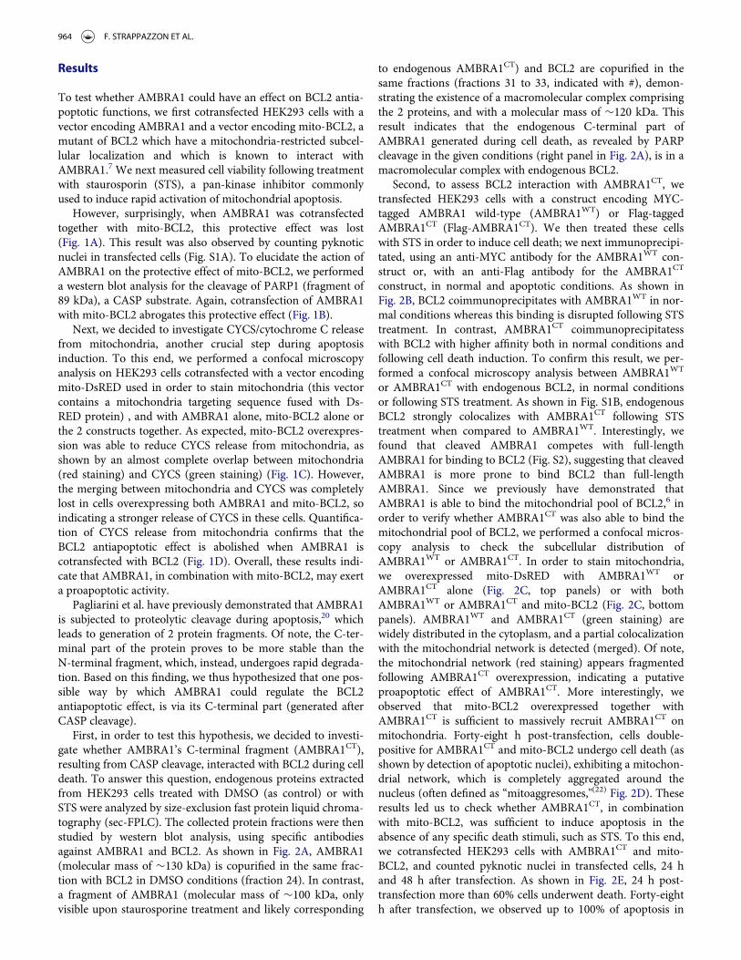
To test whether AMBRA1 could have an effect on BCL2 antia-poptotic functions, we first cotransfected HEK293 cells with avector encoding AMBRA1 and a vector encoding mito-BCL2, amutant of BCL2 which have a mitochondria-restricted subcel-lular localization and which is known to interact withAMBRA1.7 We next measured cell viability following treatmentwith staurosporin (STS), a pan-kinase inhibitor commonlyused to induce rapid activation of mitochondrial apoptosis.
However, surprisingly, when AMBRA1 was cotransfectedtogether with mito-BCL2, this protective effect was lost(Fig. 1A). This result was also observed by counting pyknoticnuclei in transfected cells (Fig. S1A). To elucidate the action ofAMBRA1 on the protective effect of mito-BCL2, we performeda western blot analysis for the cleavage of PARP1 (fragment of89 kDa), a CASP substrate. Again, cotransfection of AMBRA1with mito-BCL2 abrogates this protective effect (Fig. 1B).
Next, we decided to investigate CYCS/cytochrome C releasefrom mitochondria, another crucial step during apoptosisinduction. To this end, we performed a confocal microscopyanalysis on HEK293 cells cotransfected with a vector encodingmito-DsRED used in order to stain mitochondria (this vectorcontains a mitochondria targeting sequence fused with Ds-RED protein) , and with AMBRA1 alone, mito-BCL2 alone orthe 2 constructs together. As expected, mito-BCL2 overexpres-sion was able to reduce CYCS release from mitochondria, asshown by an almost complete overlap between mitochondria(red staining) and CYCS (green staining) (Fig. 1C). However,the merging between mitochondria and CYCS was completelylost in cells overexpressing both AMBRA1 and mito-BCL2, soindicating a stronger release of CYCS in these cells. Quantifica-tion of CYCS release from mitochondria confirms that theBCL2 antiapoptotic effect is abolished when AMBRA1 iscotransfected with BCL2 (Fig. 1D). Overall, these results indi-cate that AMBRA1, in combination with mito-BCL2, may exerta proapoptotic activity.
Pagliarini et al. have previously demonstrated that AMBRA1is subjected to proteolytic cleavage during apoptosis,20 whichleads to generation of 2 protein fragments. Of note, the C-ter-minal part of the protein proves to be more stable than theN-terminal fragment, which, instead, undergoes rapid degrada-tion. Based on this finding, we thus hypothesized that one pos-sible way by which AMBRA1 could regulate the BCL2antiapoptotic effect, is via its C-terminal part (generated afterCASP cleavage).
First, in order to test this hypothesis, we decided to investi-gate whether AMBRA1’s C-terminal fragment (AMBRA1CT),resulting from CASP cleavage, interacted with BCL2 during celldeath. To answer this question, endogenous proteins extractedfrom HEK293 cells treated with DMSO (as control) or withSTS were analyzed by size-exclusion fast protein liquid chroma-tography (sec-FPLC). The collected protein fractions were thenstudied by western blot analysis, using specific antibodiesagainst AMBRA1 and BCL2. As shown in Fig. 2A, AMBRA1(molecular mass of »130 kDa) is copurified in the same frac-tion with BCL2 in DMSO conditions (fraction 24). In contrast,a fragment of AMBRA1 (molecular mass of »100 kDa, onlyvisible upon staurosporine treatment and likely corresponding
to endogenous AMBRA1CT) and BCL2 are copurified in thesame fractions (fractions 31 to 33, indicated with #), demon-strating the existence of a macromolecular complex comprisingthe 2 proteins, and with a molecular mass of »120 kDa. Thisresult indicates that the endogenous C-terminal part ofAMBRA1 generated during cell death, as revealed by PARPcleavage in the given conditions (right panel in Fig. 2A), is in amacromolecular complex with endogenous BCL2.
Second, to assess BCL2 interaction with AMBRA1CT, wetransfected HEK293 cells with a construct encoding MYC-tagged AMBRA1 wild-type (AMBRA1WT) or Flag-taggedAMBRA1CT (Flag-AMBRA1CT). We then treated these cellswith STS in order to induce cell death; we next immunoprecipi-tated, using an anti-MYC antibody for the AMBRA1WT con-struct or, with an anti-Flag antibody for the AMBRA1CT
construct, in normal and apoptotic conditions. As shown inFig. 2B, BCL2 coimmunoprecipitates with AMBRA1WT in nor-mal conditions whereas this binding is disrupted following STStreatment. In contrast, AMBRA1CT coimmunoprecipitatesswith BCL2 with higher affinity both in normal conditions andfollowing cell death induction. To confirm this result, we per-formed a confocal microscopy analysis between AMBRA1WT
or AMBRA1CT with endogenous BCL2, in normal conditionsor following STS treatment. As shown in Fig. S1B, endogenousBCL2 strongly colocalizes with AMBRA1CT following STStreatment when compared to AMBRA1WT. Interestingly, wefound that cleaved AMBRA1 competes with full-lengthAMBRA1 for binding to BCL2 (Fig. S2), suggesting that cleavedAMBRA1 is more prone to bind BCL2 than full-lengthAMBRA1. Since we previously have demonstrated thatAMBRA1 is able to bind the mitochondrial pool of BCL2,6 inorder to verify whether AMBRA1CT was also able to bind themitochondrial pool of BCL2, we performed a confocal micros-copy analysis to check the subcellular distribution ofAMBRA1WT or AMBRA1CT. In order to stain mitochondria,we overexpressed mito-DsRED with AMBRA1WT orAMBRA1CT alone (Fig. 2C, top panels) or with bothAMBRA1WT or AMBRA1CT and mito-BCL2 (Fig. 2C, bottompanels). AMBRA1WT and AMBRA1CT (green staining) arewidely distributed in the cytoplasm, and a partial colocalizationwith the mitochondrial network is detected (merged). Of note,the mitochondrial network (red staining) appears fragmentedfollowing AMBRA1CT overexpression, indicating a putativeproapoptotic effect of AMBRA1CT. More interestingly, weobserved that mito-BCL2 overexpressed together withAMBRA1CT is sufficient to massively recruit AMBRA1CT onmitochondria. Forty-eight h post-transfection, cells double-positive for AMBRA1CT and mito-BCL2 undergo cell death (asshown by detection of apoptotic nuclei), exhibiting a mitochon-drial network, which is completely aggregated around thenucleus (often defined as “mitoaggresomes,"(22) Fig. 2D). Theseresults led us to check whether AMBRA1CT, in combinationwith mito-BCL2, was sufficient to induce apoptosis in theabsence of any specific death stimuli, such as STS. To this end,we cotransfected HEK293 cells with AMBRA1CT and mito-BCL2, and counted pyknotic nuclei in transfected cells, 24 hand 48 h after transfection. As shown in Fig. 2E, 24 h post-transfection more than 60% cells underwent death. Forty-eighth after transfection, we observed up to 100% of apoptosis in
964 F. STRAPPAZZON ET AL.
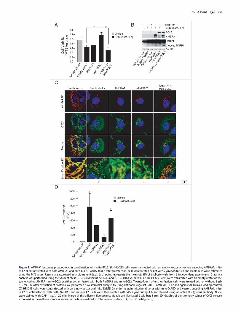
Figure 1. AMBRA1 becomes proapoptotic in combination with mito-BCL2. (A) HEK293 cells were transfected with an empty vector or vectors encoding AMBRA1, mito-BCL2 or cotransfected with both AMBRA1 and mito-BCL2. Twenty-four h after transfection, cells were treated or not with 2 mM STS for 3 h and viable cells were estimatedusing the MTS assay. Results are expressed as arbitrary unit (a.u). Each point represents the mean (§ SD) of triplicate wells from 3 independent experiments. Statisticalanalysis was performed using the Student t test (�P< 0.05) versus pcDNA3 and (#, P< 0.05) vs. mito-BCL2. (B) HEK293 cells were transfected with an empty vector or vec-tors encoding AMBRA1, mito-BCL2 or either cotransfected with both AMBRA1 and mito-BCL2. Twenty-four h after transfection, cells were treated with or without 2 mMSTS for 3 h. After extraction of proteins, we performed a western blot analysis by using antibodies against PARP1, AMBRA1, BCL2 and against ACTB (as a loading control).(C) HEK293 cells were cotransfected with an empty vector and mito-DsRED (in order to stain mitochondria) or with mito-DsRED and vectors encoding AMBRA1, mito-BCL2 or cotransfected with both AMBRA1 and mito-BCL2. Cells were then treated with STS 2 mM during 4 h and stained using an anti-CYCS (green) antibody. Nucleiwere stained with DAPI 1mg/ml 20 min. Merge of the different fluorescence signals are illustrated. Scale bar: 8 mm. (D) Graphic of densitometry values of CYCS release,expressed as mean fluorescence of individual cells, normalized to total cellular surface (F:A, n D 30 cells/groups).
AUTOPHAGY 965
Figure 2. For figure legend, see next page.
966 F. STRAPPAZZON ET AL.

cells overexpressing both AMBRA1CT and mito-BCL2. Theseresults demonstrate that AMBRA1CT, in combination withmito-BCL2, induces apoptosis.
At this point, to demonstrate that the C-terminal fragmentof AMBRA1 could become proapoptotic following cell deathsignals, we decided to use a mutant of AMBRA1 resistant toCASP cleavage (AMBRA1D482A). In fact, the AMBRA1D482A
mutant, carrying a single-point mutation (D482 ! A) was ini-tially mutated in order to disrupt the predicted putative caspasecleavage site.20 We overexpressed either a vector encodingAMBRA1D482A or AMBRA1CT in HEK293 cells, and treatedthem with STS to induce apoptosis. We next checked, by confo-cal microscope, CYCS release from mitochondria. As shown inFig. 3A (top panels), AMBRA1D482A did not induce CYCSrelease in normal condition. Also, following STS treatment, weobserved that cells overexpressing AMBRA1D482A were healthy,with a still significant overlap of CYCS staining with mitochon-dria, indicating an antiapoptotic activity of this mutant. By con-trast, overexpression of AMBRA1CT is sufficient to induce, innormal conditions, CYCS release from mitochondria (Fig. 3B,top panels), and this release is also visible in cells treated withSTS (Fig. 3B, bottom panels). The graphic of Fig. 3C illustratesquantification of CYCS release. Last, by performing a westernblot analysis, we observed that BAX activation, following STStreatment, is reduced in cells overexpressing AMBRA1D482A
compared with cells overexpressing AMBRA1WT orAMBRA1CT (Fig. 3D). Altogether, these results indicate that,during apoptosis, AMBRA1 cleavage by CASP is fundamentalto support BAX activation.
This being the case, in contrast with cells overexpressingAMBRA1CT, cells overexpressing AMBRA1D482A are expectedto be protected from cell death. We thus decided to monitorcell death by counting pyknotic nuclei in transfected cells. Asshown in Fig. 3E, cells positive for AMBRA1D482A undergo celldeath to a lesser extent than to AMBRA1CT-transfected cells. Infact, it has been demonstrated in another cell system thatAMBRA1D482A delays cell death by maintaining its proauto-phagic activity.20 Our observation confirms that AMBRA1CT
favors apoptosis, whereas its mutant resistant to CASP cleavagecan partially prolong cell survival.
Next, we decided to investigate the molecular mechanism ofAMBRA1CT activity on BCL2. BCL2 is a member of the BCL2protein family; this family shares a consensus sequence calledBH3 domain; often, proapoptotic members of this family regu-late, through direct binding via their BH3 domain, other BH3-containing proteins. We thus hypothesized that AMBRA1CT
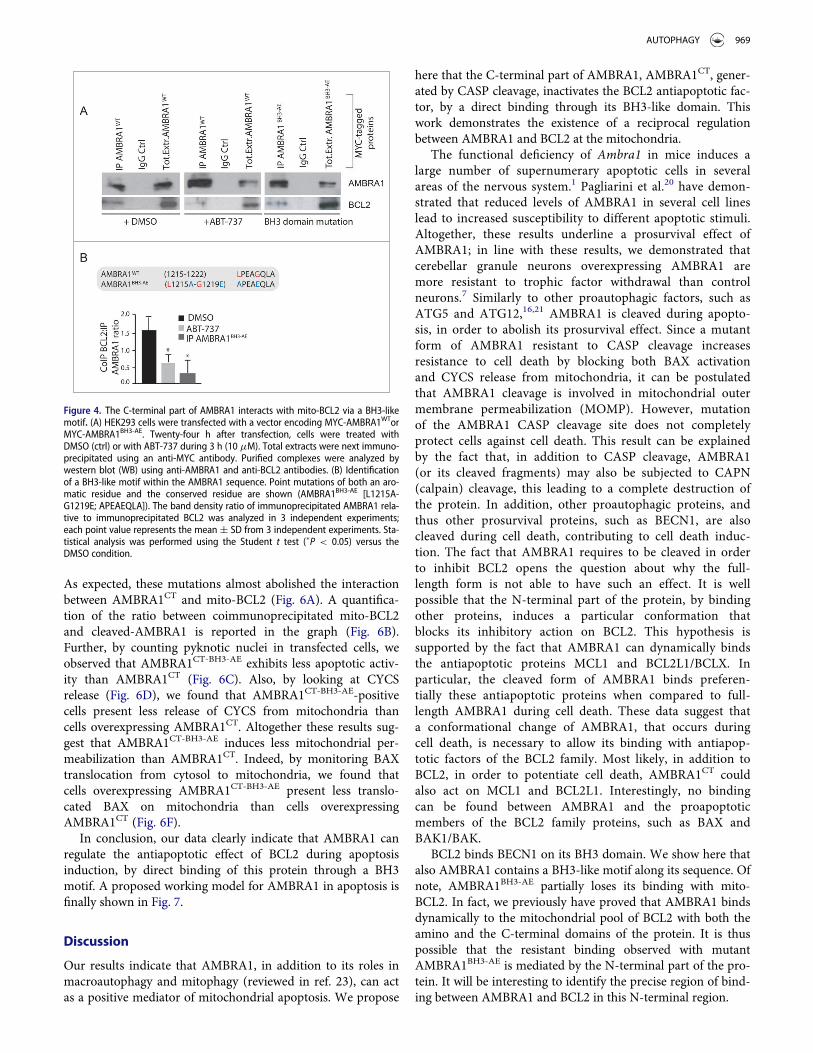
could act on BCL2 as a BH3 protein. First, we used a well-known BH3-mimetic, ABT-737, to test whether the interactionbetween AMBRA1 and mito-BCL2 could occur through a BH3domain. We thus treated HEK293 cells overexpressing MYC-AMBRA1WT with 10 mM ABT-737 for 3 h. We next immuno-precipitated AMBRA1 with an anti-MYC antibody. As shownin Fig. 4A, mito-BCL2 is associated with AMBRA1 in normalconditions, this binding being reduced by ABT-737. We nextexamined in silico the AMBRA1 sequence and found thatAMBRA1 possesses, indeed, a putative BH3 motif in its C-ter-minal region (1215LPEAGQLA1222, see Fig. 4B). Therefore, inprinciple, AMBRA1 could bind BCL2 through this motif. Asexpected, 2 point mutations of this putative BH3 domain(AMBRA1BH3-AE, [L1215A and G1219E]) almost abolished theinteraction between MYC-AMBRA1WT and mito-BCL2(Fig. 4A). These results indicate that AMBRA1 can interactwith mito-BCL2 through a previously unrevealed BH3 domain.A quantification of the ratio between mito-BCL2 coimmuno-preciptated on the quantity of precipitated AMBRA1 isreported in the graph (Fig. 4B).
Finally, we investigated the effect of AMBRA1BH3-AE on apo-ptosis induction. To this end, we performed a confocal micros-copy analysis and checked for CYCS release frommitochondria, upon STS treatment, in cells overexpressingAMBRA1WT or AMBRA1BH3-AE. CYCS release was reduced incells overexpressing AMBRA1BH3-AE following STS treatment(Fig. 5B and C). Again, we quantified apoptosis upon STS treat-ment in cells overexpressing this novel mutant construct. Asshown in Fig. 5D, AMBRA1BH3-AE can delay apoptosis similarlyto AMBRA1D482A, confirming that the AMBRA1CT acts onBCL2, through its BH3 domain, during apoptosis. Last, by per-forming a western blot analysis using an antibody against acti-vated BAX, we observed that cells overexpressingAMBRA1BH3-AE exhibited a lower basal activation of BAXwhen compared with AMBRA1CT (Fig. 5E). Also, the capabilityof AMBRA1 fragments to bind BH3-domain-containing pro-teins is further proved by their dynamic interactions with otherantiapoptotic members of the BH3 family, as shown in Fig. S4.
At this point, our data suggest that AMBRA1 can regulatethe antiapoptotic effect of BCL2 during apoptosis induction bya direct binding of this protein through a BH3 motif. Tostrengthen these results, we decided to render AMBRA1 pro-tein both autophagy deficient and BCL2-binding deficient. Tothis end, we performed 2 point mutations of the BH3 domainon the cleaved form of AMBRA1, which is deficient for auto-phagy activity (Pagliarini et al., and Fig. S5; AMBRA1CT-BH3-AE).
Figure 2. (See previous page). The C-terminal part of AMBRA1, resulting from CASP cleavage, interacts with BCL2 and increases cell death following STS treatment.(A) 2 mg of HEK293 cell lysate, obtained from DMSO-treated cells (control cells) or staurosporine-treated cells, were injected onto a superose 6 HR 10/30 FPLC gel filtrationcolumn. Proteins were collected in 500 ml fractions. Equal amounts of each fraction have been analyzed by western blot using antibodies against AMBRA1 and BCL2. Tocontrol that the STS treatment was efficient, we analyzed PARP cleavage by using an antibody against PARP. (B) HEK293 cells were transfected with a vector encodingMYC-AMBRA1WT or Flag-AMBRA1CT. Twenty-four h after transfection, cells were treated or not with STS (2 mM, 2 h). Protein extracts were immunoprecipitated using ananti-MYC or anti-Flag antibodies. Purified complexes and corresponding total extracts were analyzed by western blot (WB) using anti-AMBRA1 and anti-BCL2 antibodies.(C) HEK293 cells were cotransfected with a vector encoding mito-DsRED (to stain mitochondria) and a vector encoding MYC-AMBRA1WT or AMBRA1CT (lane 1) or in addi-tion with a vector encoding mito-BCL2 (lane 2). Twenty-four h after transfection, cells were then stained using antibody anti-AMBRA1 (AMBRA1WT and AMBRA1CT, green).Nuclei were stained with DAPI 1 mg/ml 20 min. The merging of the different fluorescence signals is illustrated. Scale bar: 8 mm. (D) HEK293 cells were cotransfected witha vector encoding AMBRA1CT and a vector encoding mito-BCL2. Forty-eight h after transfection, cells were fixed and stained using anti-AMBRA1 antibody (AMBRA1CT,red). Nuclei were stained with DAPI 1mg/ml 20 min. The merging of the different fluorescence signals is illustrated. Scale bar: 8 mm. (E) HEK293 cells were cotransfectedwith a vector encoding Flag-AMBRA1CT and a vector encoding mito-BCL2. Twenty-four or 48 h following transfection, cells were fixed and assessed by immunolabellingusing anti-Flag antibody and cells with condensed or fragmented nuclei were scored as pyknotic. Results are expressed as the percentage of transfected cells displayingpyknotic nuclei. For each condition, transfected cells were counted in 10 random fields from 3 independent experiments. Values are given as a percentage (§ SD) of celldeath.
AUTOPHAGY 967

Figure 3. The C-terminal part of AMBRA1 functions upstream of MOMP. (A) HEK293 cells were cotransfected with a vector encoding AMBRA1D482A and mito-DsRED (inorder to stain mitochondria) or (B) with mito-DsRED and vectors encoding AMBRA1CT. Twenty-four h after transfection, cells were treated with or without STS (2 mM) for4 h. Cells were fixed and then stained using antibodies against CYCS (green) and against AMBRA1 (blue staining) that recognize only the overexpressed form of the pro-tein. Nuclei were stained with DAPI 1mg/ml 20 min. The merging of the different fluorescence signals is illustrated. Scale bar: 8 mm. (C) Graphic of densitometric valuesof CYCS release, expressed as mean fluorescence of individual cells, normalized to total cellular surface (F:A, nD30 cells/groups). (D) HEK293 cells were transfected withvectors encoding AMBRA1WT, AMBRA1D482A or AMBRA1CT. Twenty-four h after transfection, cells were treated with 2 mM STS for 2 h. After extraction of proteins, we per-formed a western blot analysis using antibodies against AMBRA1, activated BAX, and total BAX (as a loading control). The band density ratio of activated BAX/total BAXwas analyzed in 3 independent experiments; each point value represents the mean § SD from 3 independent experiments. Statistical analysis was performed using theStudent t test (�, P < 0.05). (E) HEK293 cells were transfected with an empty vector or with a vector encoding AMBRA1D482A or AMBRA1CT. Twenty-four h following trans-fection, cells were treated with 2 mM STS for 2 h. Cells were next fixed and examined by immunolabelling using anti-AMBRA1 antibody and cells with condensed or frag-mented nuclei were scored as pyknotic. Results are expressed as the percentage of transfected cells displaying pyknotic nuclei. For each condition, transfected cells werecounted in 10 random fields from 3 independent experiments. Values are given as a percentage (§ SD) of cell death.
968 F. STRAPPAZZON ET AL.
As expected, these mutations almost abolished the interactionbetween AMBRA1CT and mito-BCL2 (Fig. 6A). A quantifica-tion of the ratio between coimmunoprecipitated mito-BCL2and cleaved-AMBRA1 is reported in the graph (Fig. 6B).Further, by counting pyknotic nuclei in transfected cells, weobserved that AMBRA1CT-BH3-AE exhibits less apoptotic activ-ity than AMBRA1CT (Fig. 6C). Also, by looking at CYCSrelease (Fig. 6D), we found that AMBRA1CT-BH3-AE-positivecells present less release of CYCS from mitochondria thancells overexpressing AMBRA1CT. Altogether these results sug-gest that AMBRA1CT-BH3-AE induces less mitochondrial per-meabilization than AMBRA1CT. Indeed, by monitoring BAXtranslocation from cytosol to mitochondria, we found thatcells overexpressing AMBRA1CT-BH3-AE present less translo-cated BAX on mitochondria than cells overexpressingAMBRA1CT (Fig. 6F).
In conclusion, our data clearly indicate that AMBRA1 canregulate the antiapoptotic effect of BCL2 during apoptosisinduction, by direct binding of this protein through a BH3motif. A proposed working model for AMBRA1 in apoptosis isfinally shown in Fig. 7.
Discussion
Our results indicate that AMBRA1, in addition to its roles inmacroautophagy and mitophagy (reviewed in ref. 23), can actas a positive mediator of mitochondrial apoptosis. We propose
here that the C-terminal part of AMBRA1, AMBRA1CT, gener-ated by CASP cleavage, inactivates the BCL2 antiapoptotic fac-tor, by a direct binding through its BH3-like domain. Thiswork demonstrates the existence of a reciprocal regulationbetween AMBRA1 and BCL2 at the mitochondria.
The functional deficiency of Ambra1 in mice induces alarge number of supernumerary apoptotic cells in severalareas of the nervous system.1 Pagliarini et al.20 have demon-strated that reduced levels of AMBRA1 in several cell lineslead to increased susceptibility to different apoptotic stimuli.Altogether, these results underline a prosurvival effect ofAMBRA1; in line with these results, we demonstrated thatcerebellar granule neurons overexpressing AMBRA1 aremore resistant to trophic factor withdrawal than controlneurons.7 Similarly to other proautophagic factors, such asATG5 and ATG12,16,21 AMBRA1 is cleaved during apopto-sis, in order to abolish its prosurvival effect. Since a mutantform of AMBRA1 resistant to CASP cleavage increasesresistance to cell death by blocking both BAX activationand CYCS release from mitochondria, it can be postulatedthat AMBRA1 cleavage is involved in mitochondrial outermembrane permeabilization (MOMP). However, mutationof the AMBRA1 CASP cleavage site does not completelyprotect cells against cell death. This result can be explainedby the fact that, in addition to CASP cleavage, AMBRA1(or its cleaved fragments) may also be subjected to CAPN(calpain) cleavage, this leading to a complete destruction ofthe protein. In addition, other proautophagic proteins, andthus other prosurvival proteins, such as BECN1, are alsocleaved during cell death, contributing to cell death induc-tion. The fact that AMBRA1 requires to be cleaved in orderto inhibit BCL2 opens the question about why the full-length form is not able to have such an effect. It is wellpossible that the N-terminal part of the protein, by bindingother proteins, induces a particular conformation thatblocks its inhibitory action on BCL2. This hypothesis issupported by the fact that AMBRA1 can dynamically bindsthe antiapoptotic proteins MCL1 and BCL2L1/BCLX. Inparticular, the cleaved form of AMBRA1 binds preferen-tially these antiapoptotic proteins when compared to full-length AMBRA1 during cell death. These data suggest thata conformational change of AMBRA1, that occurs duringcell death, is necessary to allow its binding with antiapop-totic factors of the BCL2 family. Most likely, in addition toBCL2, in order to potentiate cell death, AMBRA1CT couldalso act on MCL1 and BCL2L1. Interestingly, no bindingcan be found between AMBRA1 and the proapoptoticmembers of the BCL2 family proteins, such as BAX andBAK1/BAK.
BCL2 binds BECN1 on its BH3 domain. We show here thatalso AMBRA1 contains a BH3-like motif along its sequence. Ofnote, AMBRA1BH3-AE partially loses its binding with mito-BCL2. In fact, we previously have proved that AMBRA1 bindsdynamically to the mitochondrial pool of BCL2 with both theamino and the C-terminal domains of the protein. It is thuspossible that the resistant binding observed with mutantAMBRA1BH3-AE is mediated by the N-terminal part of the pro-tein. It will be interesting to identify the precise region of bind-ing between AMBRA1 and BCL2 in this N-terminal region.
Figure 4. The C-terminal part of AMBRA1 interacts with mito-BCL2 via a BH3-likemotif. (A) HEK293 cells were transfected with a vector encoding MYC-AMBRA1WTorMYC-AMBRA1BH3-AE. Twenty-four h after transfection, cells were treated withDMSO (ctrl) or with ABT-737 during 3 h (10 mM). Total extracts were next immuno-precipitated using an anti-MYC antibody. Purified complexes were analyzed bywestern blot (WB) using anti-AMBRA1 and anti-BCL2 antibodies. (B) Identificationof a BH3-like motif within the AMBRA1 sequence. Point mutations of both an aro-matic residue and the conserved residue are shown (AMBRA1BH3-AE [L1215A-G1219E; APEAEQLA]). The band density ratio of immunoprecipitated AMBRA1 rela-tive to immunoprecipitated BCL2 was analyzed in 3 independent experiments;each point value represents the mean § SD from 3 independent experiments. Sta-tistical analysis was performed using the Student t test (�P < 0.05) versus theDMSO condition.
AUTOPHAGY 969

Human cancer grows by evading cell death. In fact, somecancers, and in particular breast cancers, express high levelsof BCL2. Proteins from the BCL2 family are involved inMOMP, a phenomenon mediating CYCS release from mito-chondria (essential in cell death induction) and, for this
reason, MOMP is a good target in cancer therapy. Indeed,BH3 mimetics are already used in chemotherapy and haveprovided beneficial insights into the regulation of theBCL2-BECN1 complex as well as in identifying additionalpathways involved in autophagic cell death. ABT-737 and
Figure 5. The mutant of AMBRA1 that cannot bind BCL2 loses its proapoptotic effect during cell death. (A) HEK293 cells were cotransfected with a vector encodingAMBRA1WT or (B) AMBRA1BH3-AE and mito-DsRED (in order to stain mitochondria). Twenty-four h after transfection, cells were treated with or without STS (2 mM) for 4 h.Cells were fixed and then stained using the anti-CYCS (green) antibody. Nuclei were stained with DAPI 1mg/ml 20 min. The merging of the different fluorescence signalsis illustrated. Scale bar: 8 mm. (C) HEK293 cells were transfected with an empty vector or with a vector encoding AMBRA1D482A, AMBRA1CT (see Fig. 3D) or with a vectorencoding AMBRA1BH3-AE. Twenty-four h following transfection, cells were treated with 2 mM STS during 3 h. Cells were next fixed and examined by immunolabellingusing anti-AMBRA1 antibody that recognizes only the overexpressed form of the protein and cells with condensed or fragmented nuclei were scored as pyknotic. Resultsare expressed as the percentage of transfected cells displaying pyknotic nuclei. For each condition, transfected cells were counted in 10 random fields from 3 independentexperiments. Values are given as a percentage (§ SD) of cell death. (D) Graphic of densitometric values of CYCS release, expressed as mean fluorescence of individualcells, normalized to total cellular surface (F:A, nD30 cells/groups). (E) HEK293 cells transfected with a vector encoding AMBRA1CT or AMBRA1BH3-AE were analyzed by west-ern blot using antibodies against activated BAX and total BAX. The band density ratio of activated BAX/total BAX was analyzed in 3 independent experiments; each pointvalue represents the mean § SD from 3 independent experiments. Statistical analysis was performed using the Student t test (�P < 0.05).
970 F. STRAPPAZZON ET AL.

Figure 6. For figure legend, see next page.
AUTOPHAGY 971

HA14-1 also stimulate other proautophagic pathways andhence activate the nutrient sensors SIRT1 (sirtuin 1) andAMPK, inhibit MTOR, deplete cytoplasmic TP53/p53, andtrigger the CHUCK/IKKa and IKBKB/IKKb kinases.24 Herewe propose a novel mechanism of action of ABT-737,which disrupts the interaction between AMBRA1 and mito-BCL2, thus contributing to autophagy induction. Conse-quently, exploiting the AMBRA1-BCL2 interaction could beused to develop novel anticancer therapies.
BCL2 is upregulated in human breast cancers and medi-ates the resistance of these cancers to chemotherapeuticstrategies,25,26 while, by contrast, AMBRA1 is an haploinsuf-ficient tumor suppressor gene;27 given both these factors,modulating the reciprocal AMBRA1 and BCL2 levels incancer cells by targeting their interaction could contributeto early diagnosis and to predicting prognosis for breastcancer.
Further studies are needed to discover binding partnersthat can alter the reciprocal affinity of BCL2 and AMBRA1at the mitochondria. It should also be interesting to investi-gate whether post-translational modifications on AMBRA1(such as its phosphorylation by kinases) regulate this
binding. Developing combination therapies between BH3mimetics and kinase activators could provide a powerful“double effect” on the AMBRA1-BCL2 complex. In conclu-sion, identification of the mechanisms that maintain or dis-rupt this complex may allow us to develop additional drugsthat can target it, thus ensuring an improved therapeuticoutcome.
Materials and methods
Antibodies
Mouse monoclonal anti-ACTB/b actin (Sigma-Aldrich,A2228), monoclonal anti-BCL2 (Santa Cruz Biotechnology, sc-7382), polyclonal anti-AMBRA1 (Novus, 26190002), monoclo-nal anti-AMBRA1 (Santa Cruz Biotechnology, sc-398204),mouse monoclonal anti-CYCS (Assay designs-Enzo Life Scien-ces, 6H2-B4), polyclonal anti-LC3 (Cell Signaling Technology,2775), monoclonal anti-SQSTM1/p62 (Santa Cruz Biotechnol-ogy, sc-28369), polyclonal anti-activated BAX (6A7; Abcam5714), polyclonal anti-BAX (Santa Cruz Biotechnology, sc-493,N-20), polyclonal anti-PARP1 (Cell Signaling Technology,9542), goat anti-BAK1 (G-23; Santa Cruz Biotechnology, sc-832), mouse monoclonal anti-MYC (9E10; Santa Cruz Biotech-nology, sc-4), mouse monoclonal anti-Flag (Sigma-Aldrich,F3165), rabbit polyclonal anti-SOD2/MnSOD (Enzo Life Scien-ces, 110F) and mouse monoclonal anti-TUBB/b tubulin(Sigma-Aldrich, T9026).
Cloning and plasmids
Construct coding for AMBRA1WT, AMBRA1D482A andAMBRA1CT (483-1300 amino acids) was cloned in pLPCX vec-tor (Clontech, 631511).18
The construct coding for “mito-BCL2,” a mitochondrial-tar-geted BCL2 in which the C�terminal hydrophobic sequence ofBCL2 is exchanged for an equivalent sequence from modifiedActA (Listeria monocytogenes actin assembly-inducing protein)which binds specifically to the cytoplasmic face of mitochon-drial outer membranes, was kindly provided by Beth Levine9
and was cloned in prcCMV vector.The mito-DsRED construct encodes for human Cox8A
mitochondria signal which is fused with wild-type-DsRED inpcDNA3 vector (Invitrogen).
We summarized in the table all constructs used in thepresent work:
Figure 6. (See previous page). The mutant form of cleaved AMBRA1 that cannot bind BCL2 loses its proapoptotic effect during cell death. (A) HEK293 cells were trans-fected with a vector encoding Flag-AMBRA1CT or Flag-AMBRA1CT-BH3-AE. Twenty-four h after transfection, total extracts were next immunoprecipitated using an anti-Flagantibody. Purified complexes were analyzed by western blot (WB) using anti-AMBRA1 and anti-BCL2 antibodies. (B) The band density ratio of immunoprecipitatedAMBRA1 relative to immunoprecipitated BCL2 was analyzed in 3 independent experiments; each point value represents the mean§ SD from 3 independent experiments.Statistical analysis was performed using the Student t test (�, P< 0.05) vs. CT. (C) HEK293 cells were transfected with a vector encoding Flag-AMBRA1CT or Flag-AMBRA1CT-BH3-AE. Twenty-four h following transfection, cells were treated with 2 mM STS during 2 h. Cells were next fixed and examined by immunolabelling using anti-AMBRA1antibody that recognizes only the overexpressed form of the protein and cells with condensed or fragmented nuclei were scored as pyknotic. Results are expressed asthe percentage of transfected cells displaying pyknotic nuclei. For each condition, transfected cells were counted in 10 random fields from 3 independent experiments.Values are given as a percentage (§ SD) of cell death. (D) HEK293 cells were cotransfected with a vector encoding Flag-AMBRA1CT or Flag-AMBRA1CT-BH3-AE and mito-DsRED (in order to stain mitochondria). Cells were then treated with STS 2 mM during 4 h and stained using an anti-CYCS antibody (green). Nuclei were stained withDAPI 1mg/ml during 20 min. The merging of the different fluorescence signals is illustrated. Scale bar: 8 mm. (E) Graphic of densitometric values of CYCS release,expressed as mean fluorescence of individual cells, normalized to total cellular surface (F:A, nD30 cells/groups). (F) HEK293 cells were transfected with a vector encodingFlag-AMBRA1CT or Flag-AMBRA1CT-BH3-AE. Twenty-four h after transfection, cytosolic and mitochondrial fractions were analyzed by western blot (WB) using anti-BAX andanti-SOD2 antibodies. The band density ratio of translocated BAX on mitochondria was analyzed in 3 independent experiments; each point value represents the mean §SD from 3 independent experiments. Statistical analysis was performed using the Student t test (��, P < 0.01) versus CT.
Figure 7. The proposed working model for AMBRA1 in mitochondrial apoptosis.Upon induction of apoptosis, AMBRA1 is subjected to CASP cleavage. In the initialphase of apoptosis, the C-terminal fragment of AMBRA1 is stable and interactsdirectly with BCL2 through its BH3-like domain, thus likely increasing cell death(cell death amplification loop).
972 F. STRAPPAZZON ET AL.

Point mutations were generated using the QuickChange site-directed mutagenesis kit (Stratagene, 200519) and all plasmidconstructs made in this study were verified by DNA sequencing(Eurofins). The oligonucleotides used for mutagenesis, PCR,and DNA sequencing were purchased from Invitrogen.
Cell cultures
The human embryonic kidney HEK293 cells and HeLa cellswere cultured in Dulbecco’s modified Eagle’s medium (Lonza,BE12-604F) supplemented with 10% FBS (Gibco; ThermoFisher Scientific, 10270-106) , and 1% penicillin-streptomycinsolution (Lonza, 17-602 E) at 37�C under 5% CO2.
Cell culture transfection
Transient transfections of expression plasmids into HEK293cells were performed using TurboFect according to the suppli-er’s instructions (Thermo Fisher Scientific, R0532).
Determination of cell viability
Cell survival was estimated by using the MTS assay or bycounting the number of condensed or fragmented nuclei asobserved using DAPI (Sigma Aldrich, D9542) staining. The tet-razolium salt MTS (3-[4,5-dimethyl-2-thiazolyl]-2-5diphenyl-2H tratrazolium bromide; Sigma Aldrich, M2128) was added tocell cultures (1 mg/ml) and incubated for 30 min a 37�C. Cellswere then lysed in DMSO (Sigma Aldrich, 472301). Formazanproduction by living cells was assessed by measuring absor-bance at 540 nm using a Biotek Elx-800 microplate reader(Mandel Scientific Inc.) For cell counting, cultures were fixedin 4% paraformaldehyde (Merck Millipore, 104005) in PBS(UCS Diagnostic, PBS1199) for 10 min at 4�C and stained withDAPI in PBS for 20 min at room temperature. Cells werewashed 3 times with PBS, then mounted and observed under aZEISS confocal laser microscope (CLSM700; Jena, Germany).For each condition, random images were captured and ana-lyzed using Zen Light edition software. Cell viability was thenscored on the basis of nuclear morphology: cells containingcondensed or fragmented nuclei were counted as dying or deadcells.
Immunocytochemistry
Cells were washed in PBS and fixed with 4% paraformaldehydein PBS for 15 min. After permeabilization with 0.4% Triton X-100 (Sigma Aldrich, X100) in PBS for 5 min, cells were blockedin 3% normal goat serum (Sigma-Aldrich, G9023) in PBS andincubated overnight at 4 degrees with primary antibodies. Weused the antibodies directed against AMBRA1 and CYCS. Cellswere then washed in blocking buffer and incubated for 1 h withlabeled anti-mouse (Alexa Fluor 488; Thermo Fisher Scientific,A11017) or anti-rabbit (CyTM3; Jackson ImmunoResearch,115-165-166) secondary antibodies. Nuclei were stained with1 mg/ml DAPI and examined under a Zeiss LSM 700 100x oil-immersion objective (CLSM700; Jena, Germany). We used“ZEN 2009 Light edition” software for image analysis. Allacquisitions were performed in non-saturated single z-confocalplanes.
Western blot analysis
Cell extracts were centrifuged at 13,000 g for 10 min at 4�C.Protein concentrations were determined with the Bio-Rad Pro-tein Assay Kit (Bio-Rad, 5000001). Cell extracts or immunopre-cipitates were separated by SDS-PAGE and transferred ontonylon membranes (Immobilon P; Merck-Millipore,IPFL10100). Membranes were incubated with primary antibod-ies followed by horseradish peroxidase-conjugate secondaryantibody (Bio-Rad, 1706515 and 1721011) and visualized withECL (Merck-Millipore WBKLS0500).
Immunoprecipitation
Cells were lysed in RIPA buffer plus protease inhibitor cocktail(Sigma Aldrich, P8340). Equal amounts of protein (500 mg)were incubated with 2 ml of monoclonal anti-MYC antibodyfor 4 h followed by 60 min incubation with 30 ml of protein A
Constructs Type of mutation Feature
Flag-tagged-AMBRA1D482A
Single-point mutation (D482! A) on the predictedcaspase cleavage site ofAMBRA1
This mutant of AMBRA1 isuncleavable bycaspases followingapoptosis inductionand maintains itsproautophagic activity
MYC-tagged-AMBRA1WT
No mutation This construct encodes forthe wild-type form ofAMBRA1
MYC-tagged-AMBRA1BH3-AE
Two-point mutations in theBH3 domain of AMBRA1L1215! A and G1219! E
This mutant of AMBRA1partially loses itsbinding with mito-BCL2protein
Flag-tagged-AMBRA1CT
Terminal fragment of AMBRA1resulting from caspasecleavage (483-1300 aminoacids)
This construct encodes forthe C-terminal part ofAMBRA1 resultingfronm caspasecleavage. This fragmentbinds mito-BCL2
Flag-tagged-AMBRA1CT-BH3-AE
Two-point mutations in theBH3 domain of the C-terminal fragment ofAMBRA1L1215! A andG1219! E resulting fromcaspase cleavage (483-1300amino acids)
This construct encodes a C-terminal fragment ofAMBRA1 which partiallyloses its binding withmito-BCL2
Mito-BCL2 The mitochondrial-targetedBCL2 presents the C-terminal hydrophobicsequence of BCL2exchanged for anequivalent sequence frommodified ActA (Listeriamonocytogenes Actinassembly-inducing protein),which binds specifically tothe cytoplasmic face ofmitochondrial outermembranes. In this caseBCL2 is fused with ActAsequence.
This construct encodes aBCL2 protein that it isspecifically targeted tothe mitochondriawhere it acts as anantiapoptotic factor.
Mito-DsRED Human Cox8A mitochondriasignal is fused with wild-type-DsRED protein inpcDNA3 vector.
This construct encodes fora red protein that istargeted to themitochondria. It is usedin microscopy analysisin order to stain themitochondria network.
AUTOPHAGY 973

agarose beads (Roche, 11719408001). The beads were collectedby centrifugation and washed 4 times with the RIPA buffer.Proteins bound to the beads were eluted with 30 ml of SDS-polyacrylamide gel electrophoresis sample buffer and heated to95�C for 10 min.
Mitochondria/cytosol purification
Mitochondria and cytosol purification occurs by standard dif-ferential centrifugations. HEK293 cells were suspended inhomogenization buffer (0.25 M sucrose, 10 mM HEPES, pH 7,1 mM EDTA [Sigma-Aldrich, S0389, H4034, and ED-100,respectively]) with protease and phosphatase inhibitors. Mito-chondria suspensions were obtained homogenizing the solutionwith 40�–60 pulses (one pulse corresponds to approximatelyone sec), in vertical movements. After homogenization, the sus-pension was centrifuged at 11 000 g for 15 min at 4�C. Themitochondrial pellet fraction was suspended in isolation buffer(0.2 M sucrose, 10 mM Tris-MOPS [Sigma, M1254], pH 7.4,0.1 mM EGTA-Tris [Sigma Aldrich, E4378]). The supernatantfraction corresponded to soluble cytosolic proteins.
Size-exclusion fast protein liquid chromatography(sec-FPLC)
Two milligrams of HEK293 cell lysate, obtained from DMSO-treated cells or staurosporine-treated cells, were injected onto aSuperose 6 HR 10/30 fast protein liquid chromatography(FPLC) gel filtration column (Amersham Pharmacia Bioscien-ces, 17-0537-01) connected to a Pharmacia LKB LCC 501 PlusFPLC System (Amersham Pharmacia LKB Biotechnologies,Piscataway, N.J.). Lysate preparation and column equilibrationhave been previously described.5 CAPN (calpain II inhibitor,50 mM; Sigma-Aldrich, A6060) and CASP (Q-VD-OPH, 100mM; Santa Cruz Biotechnology, sc-222230) inhibitors wereadded in the lysis buffer in order to prevent AMBRA1 cleavageduring the fraction separation. Proteins were collected in500 ml fractions, precipitated with 10% trichloroacetic acid,and resolved in SDS-PAGE for western blot analyses. Gel filtra-tion column was calibrated as previously described.25 Equalamounts of each fraction were analyzed by western blot.
Quantification of CYCS release
Quantification of CYCS release from mitochondria wasperformed on transfected cells by densitometric analyses, aspreviously described.28 After background subtraction, CYCScell-associated signals were quantified by manually outliningindividual cells and measuring cell-associated fluorescenceintensity with the ImageJ software (http://rsb.info.nih.gov/ij/).The F:A ratio defines mean fluorescence of individual cells (F)normalized to total cellular surface (A).
Statistical analyses
Statistical analyses were performed using the Student 2-tailedttest. Data were shown as means § SD of 3 independent experi-ments. Values of p < 0,05 were considered significant.
Abbreviations
AMBRA1 autophagy/Beclin 1 regulator 1AMBRA1BH3-AE autophagy/Beclin 1 regulator 1, mutated
in BH3 motifAMBRA1CT C-terminal part of AMBRA1AMBRA1CT-BH3-AE C-terminal part of AMBRA1, mutated in
BH3 motifBAX BCL2-associated X proteinBCL2 B-cell CLL/lymphoma 2BECN1 Beclin 1, autophagy relatedBH3 BCL2 homology domain 3CASP caspaseCYCS cytochrome c, somaticFPLC fast protein liquid chromatographyHEK293 human embryonic kidneyMOMP mitochondrial outer membrane
permeabilizationMito-BCL2 mitochondrial targeted BCL2PARK2 parkin RBR E3 ubiquitin protein ligaseSTS staurosporin
Disclosure of potential conflicts of interest
The authors declare no conflict of interest.
Acknowledgments
We thank M. Acu~na Villa andM. Bennett for secretarial and proofreadingwork, and V. Nanni and B. Biferali for research assistance. We are indebtedto B. Levine (Dallas, TX), for kindly providing us with the mito-BCL2 con-struct and M. Priault (Bordeaux, France) for kindly providing us with theMCL1 and BCL2L1 constructs.
Funding
This work was supported in part by grants from the Telethon Foundation(GGP14202), AIRC (IG2013 to FC, IG2012-13529 and IG 2015-17404 toGMF), FISM (2013), the Italian Ministry of University and Research (FIRBAccordi di Programma 2011), the Italian Ministry of Health (RicercaFinalizzata and Ricerca Corrente to FC and GMF, Progetto Giovani Ricer-catori GR2011-2012 to FS). VC is supported by the Lundbeck Foundation(R165-2013-15982). This work has been also supported by a KBVU grantfrom the Danish Cancer Society (R72-A4408). We are also grateful to theBjarne Saxhof Foundation, the Lundbeck Foundation (R167-2013-16100),and the NovoNordisk Foundation (7559). Further, FC lab in Copenhagenis part of the Center of Excellence in Autophagy, Recycling and Disease(CARD), funded by the Danish National Research Foundation.
References
[1] Fimia GM, Stoykova A, Romagnoli A, Giunta L, Nardacci R, Coraz-zari M, Fuoco C, Ucar A, Schwartz P, Gruss P, et al. Ambra1 regu-lates autophagy and development of the nervous system. Nature2007; 447: 1121-1125; PMID:17589504
[2] Di Bartolomeo S, Corazzari M, Nazio F, Oliverio S, Lisi G, AntonioliM, Paglierini V, Mattoni S, Fuoco C, Giunta L, et al. The dynamicinteraction of AMBRA1 with the dynein motor complex regulatesmammalian autophagy. J Cell Biol. 2010; 191:155-68;PMID:20921139; http://dx.doi.org/10.1083/jcb.201002100
[3] He C, Levine B. The Beclin 1 interactome. Curr Opin Cell Biol. 2010;2:140-9; http://dx.doi.org/10.1016/j.ceb.2010.01.001
[4] Nazio F, Strappazzon F, Antonioli M, Bielli P, Cianfanelli V, BordiM, Gretzmeier C, Dengjel J, Piacentini M, Fimia GM, et al. mTORinhibits autophagy by controlling ULK1 ubiquitination, self-associa-tion and function via AMBRA1 and TRAF6. Nat Cell Biol. 2013;4:406-16; http://dx.doi.org/10.1038/ncb2708
974 F. STRAPPAZZON ET AL.

[5] Cianfanelli V, Fuoco C, Lorente M, Salazar M, Quondamatteo F,Gherardini PF, De Zio D, Nazio F, Antonioli M, D’Orazio M, et al.AMBRA1 links autophagy to cell proliferation and tumorigenesis bypromoting c-Myc dephosphorylation and degradation. Nat Cell Biol.2015; 17:20-30; PMID:25438055; http://dx.doi.org/10.1038/ncb3072
[6] Antonioli M, Albiero F, Nazio F, Vescovo T, Perdomo AB, CorazzariM, Marsella C, Piselli P, Greztmeier P, Denjiel J, et al. AMBRA1interplay with cullin E3 ubiquitin ligases regulates autophagy dynam-ics. Dev Cell. 2014; 6:734-46; http://dx.doi.org/10.1016/j.devcel.2014.11.013
[7] Strappazzon F, Vietri-Rudan M, Campello S, Nazio F, Floren-zano F, Fimia GM, Piacentini M, Levine B, Cecconi F. Mito-chondrial BCL-2 inhibits AMBRA1-induced autophagy. Embo j.2011; 30:1195-208; PMID:21358617; http://dx.doi.org/10.1038/emboj.2011.49
[8] Strappazzon F, Nazio F, Corrado M, Cianfanelli V, Romagnoli A,Fimia GM, Campello S, Nardacci R, Piacentini M, Campanella M.et al. AMBRA1 is able to induce mitophagy via LC3 binding, regard-less of PARKIN and p62/SQSTM1. Cell Death Differ. 2015; 3:419-32; http://dx.doi.org/10.1038/cdd.2014.139
[9] Pattingre S, Tassa A, Qu X, Garuti R, Liang XH, Mizushima N,Packer M, Schneider MD, Levine B. Bcl-2 antiapoptotic proteinsinhibit Beclin 1-dependent autophagy. Cell 2005; 122:927-39;PMID:16179260; http://dx.doi.org/10.1016/j.cell.2005.07.002
[10] Chang NC, Nguyen M, Germain M, Shore GC. Antagonism of Beclin1-dependent autophagy by BCL-2 at the endoplasmic reticulumrequires NAF-1. Embo J 2010; 29:606-18; PMID:20010695; http://dx.doi.org/10.1038/emboj.2009.369
[11] Cho DH, Jo YK, Hwang JJ, Lee YM, Roh SA, Kim JC. Caspase-medi-ated cleavage of ATG6/Beclin-1 links apoptosis to autophagy inHeLa cells. Cancer Lett. 2009; 274:95-100; PMID:18842334; http://dx.doi.org/10.1016/j.canlet.2008.09.004
[12] Luo S, Rubinsztein DC. Apoptosis blocks Beclin 1-dependent auto-phagosome synthesis: an effect rescued by Bcl-xL. Cell Death Differ2010; 17:268-277; PMID:19713971; http://dx.doi.org/10.1038/cdd.2009.121
[13] Zhu Y, Zhao L, Liu L, Gao P, Tian W, Wang X, Jin H, Xu H, Chen Q.Beclin 1 cleavage by caspase-3 inactivates autophagy and promotesapoptosis. Protein Cell 2010; 1:468-477; PMID:21203962; http://dx.doi.org/10.1007/s13238-010-0048-4
[14] Djavaheri-Mergny M, Maiuri MC, Kroemer G. Cross talk betweenapoptosis and autophagy by caspase-mediated cleavage of Beclin 1.Oncogene 2010; 12:1717-9; http://dx.doi.org/10.1038/onc.2009.519
[15] Maiuri MC, Criollo A, Kroemer G. Crosstalk between apoptosis andautophagy within the Beclin 1 interactome. Embo J 2010; 29:515-6;PMID:20125189; http://dx.doi.org/10.1038/emboj.2009.377
[16] Pyo JO, Jang MH, Kwon YK, Lee HJ, Jun JI, Woo HN, Cho DH, ChoiB, Lee H, Kim JH, et al. Essential roles of Atg5 and FADD in auto-phagic cell death: dissection of autophagic cell death into vacuole
formation and cell death. J. Biol. Chem. 2005; 280:20722-9;PMID:15778222; http://dx.doi.org/10.1074/jbc.M413934200
[17] Yousefi S, Perozzo R, Schmid I, Ziemiecki A, Schaffner T, ScapozzaL, Brunner T, Simon HU. Calpain-mediated cleavage of Atg5switches autophagy to apoptosis. Nat. Cell Biol. 2006; 8:1124-32;PMID:16998475; http://dx.doi.org/10.1038/ncb1482
[18] Zalckvar E, Yosef N, Reef S, Ber Y, Rubinstein AD, Mor I, Sharan R,Ruppin E, Kimchi A. A systems level strategy for analyzing the celldeath network: implication in exploring the apoptosis/autophagyconnection. Cell Death Differ. 2010; 17:1244-53; PMID:20150916;http://dx.doi.org/10.1038/cdd.2010.7
[19] Maskey D, Yousefi S, Schmid I, Zlobec I, Perren A, Friis R, SimonHU. ATG5 is induced by DNA-damaging agents and promotesmitotic catastrophe independent of autophagy. Nat Commun. 2013;4:2130; PMID:23945651; http://dx.doi.org/10.1038/ncomms3130
[20] Pagliarini V, Wirawan E, Romagnoli A, Ciccosanti F, Lisi G, LippensS, Cecconi F, Fimia GM, Vandenabeele P, Corazzari M, et al. Proteol-ysis of Ambra1 during apoptosis has a role in the inhibition of theautophagic pro-survival response. Cell Death Differ. 2012; 9:1495-504; http://dx.doi.org/10.1038/cdd.2012.27
[21] Rubinstein AD, Eisenstein M, Ber Y, Bialik S, Kimchi A. The autoph-agy protein Atg12 associates with antiapoptotic Bcl-2 family mem-bers to promote mitochondrial apoptosis. Mol Cell. 2011; 5:698-709;http://dx.doi.org/10.1016/j.molcel.2011.10.014
[22] Lee JY, Nagano Y, Taylor JP, Lim KL, Yao TP. Disease-causing muta-tions in parkin impair mitochondrial ubiquitination, aggregation,and HDAC6-dependent mitophagy. J Cell Biol. 2010; 4:671-9; http://dx.doi.org/10.1083/jcb.201001039
[23] Cianfanelli V, De Zio D, Di Bartolomeo S, Nazio F, Strappazzon F,Cecconi F. Ambra1 at a glance. Cell Science at a glance 2015;11:2003-8; http://dx.doi.org/10.1242/jcs.168153
[24] Malik SA, Shen S, Mari~no G, BenYoun�es A, Maiuri MC, Kroemer G.BH3 mimetics reveal the network properties of autophagy-regulatorysignaling cascades. Autophagy. 2011; 8:914-6; http://dx.doi.org/10.4161/auto.7.8.15785
[25] Huang Z. Bcl-2 family proteins as targets for anticancer drug design.Oncogene 19:6627-31; PMID:11426648; http://dx.doi.org/10.1038/sj.onc.1204087
[26] Karnak D, Xu L. Chemosensitization of prostate cancer by modulat-ing Bcl-2 family proteins. Curr Drug Targets 2010; 11:699-707;PMID:20298153; http://dx.doi.org/10.2174/138945010791170888
[27] Cianfanelli V, D’Orazio M, Cecconi F. AMBRA1 and BECLIN 1interplay in the crosstalk between autophagy and cell proliferation.Cell cycle 2015; 14:959-63; PMID:25803737; http://dx.doi.org/10.1080/15384101.2015.1021526
[28] Oddi S, Latini L, Viscomi MT, Bisicchia E, Molinari M, MaccarroneM. Distinct regulation of nNOS and iNOS by CB2 receptor in remotedelayed neurodegeneration. J Mol Med (Berl) 2012; 90:371-87;PMID:22198001; http://dx.doi.org/10.1007/s00109-011-0846-z
AUTOPHAGY 975