protective effects of mentha piperita l. leaf …...research open access protective effects of...
TRANSCRIPT

RESEARCH Open Access
Protective effects of Mentha piperita L. leafessential oil against CCl4 induced hepaticoxidative damage and renal failure in ratsKhaled Bellassoued1*, Anis Ben Hsouna2, Khaled Athmouni3, Jos van Pelt4, Fatma Makni Ayadi5, Tarek Rebai6
and Abdelfattah Elfeki1
Abstract
Background: Mentha piperita L. is a flowering plant belonging to the Lamiaceae family. Mentha plants constituteone of the main valuable sources of essential oil used in foods and for medicinal purposes.
Methods: The present study aimed to investigate the composition and in vitro antioxidant activity of Mentha piperitaleaf essential oil (MpEO). A single dose of CCl4 was used to induce oxidative stress in rats, which was demonstrated bya significant rise of serum enzyme markers. MpEO was administrated for 7 consecutive days (5, 15, 40 mg/kg body weight)to Wistar rats prior to CCl4 treatment and the effects on serum alanine aminotransferase (ALT), aspartate aminotransferase(AST), alkaline phosphatase (ALP), lactate dehydrogenase (LDH), and γ -glutamyl transpeptidase (γ-GT) levels, as well as theliver and kidney superoxide dismutase (SOD), catalase (CAT) and glutathione peroxidase (GPx) activity and thiobarbituricacid reactive substances (TBARS) levels were evaluated. In addition, histopathological examinations of livers and kidneyswas performed.
Results: The in vitro antioxidant activity of MpEO was lower than that of silymarin. Pretreatment of animals with MpEO ata dose of 5 mg/kg did not have a significant effect on ALT, AST, ALP, LDH, γGT, urea or creatinine levels in CCl4-inducedstress. Whereas pretreatment with MpEO at doses of 15 and 40 mg/kg prior to CCl4, significantly reduced stress parameters(ALT, AST, ALP, LDH, γGT, urea and creatinine) compared to the CCl4-only group. Moreover, a significant reduction in hepaticand kidney lipid peroxidation (TBARS) and an increase in antioxidant enzymes SOD, CAT and GPx was also observed aftertreatment with MpEO (40 mg/kg) compared to CCl4-treated rats. Furthermore, pretreatment with MpEO at 40 mg/kg canalso markedly ameliorate the histopathological hepatic and kidney lesions induced by administration of CCl4.
Conclusions: We could demonstrate with this study that MpEO protects liver and kidney from CCl4-induced oxidativestress and thus substantiate the beneficial effects attributed traditionally to this plant.
Keywords: M. piperita, Essential oil, Liver, Kidney, Oxidative stress, Histopathological
BackgroundReactive oxygen species (ROSs) are various forms of acti-vated oxygen. A disproportion of the reactive oxygen speciesand the absence of their scavenge systems in cells lead tooxidative stress and increases the risk of several humanchronic diseases [1]. ROS contributes to the development ofvarious diseases such as diabetes, atherosclerosis, cancer,neurodegenerative diseases, liver cirrhosis and the aging
process [2]. The liver plays a central role in the maintenanceof systemic lipid homeostasis and is especially susceptible toROS damage. CCl4 is now of greatest concern as an envir-onmental contaminant [3]. It was reported that CCl4 wasone of the most commonly used toxins in the experimentalstudy of liver diseases [4]. Abraham et al. [5] showed thatthe nephrotoxic effects of CCl4 were also associated withfree radical production.To prevent the damage caused by ROS, living organisms
have developed an antioxidant defense system that includesthe presence of non-enzymatic antioxidants and enzymessuch as catalase (CAT), superoxide dismutase (SOD) and
* Correspondence: [email protected] of Life Sciences, Animal Ecophysiology Laboratory, Faculty ofsciences, University of Sfax Tunisia, Road of Soukra Km 3.5, BP 1171, PC 3000Sfax, TunisiaFull list of author information is available at the end of the article
© The Author(s). 2018 Open Access This article is distributed under the terms of the Creative Commons Attribution 4.0International License (http://creativecommons.org/licenses/by/4.0/), which permits unrestricted use, distribution, andreproduction in any medium, provided you give appropriate credit to the original author(s) and the source, provide a link tothe Creative Commons license, and indicate if changes were made. The Creative Commons Public Domain Dedication waiver(http://creativecommons.org/publicdomain/zero/1.0/) applies to the data made available in this article, unless otherwise stated.
Bellassoued et al. Lipids in Health and Disease (2018) 17:9 DOI 10.1186/s12944-017-0645-9

glutathione peroxidase (GPx) [6]. It has been anticipatedthat in addition to these natural antioxidants, other syn-thetic or natural ROS scavengers may reduce the incidenceof free radical-mediated diseases. The use of antioxidants inthe prevention and cure of various diseases is intensifying,and there is considerable interest in the study of the anti-oxidant activities of molecules such as plant polyphenolicand carotenoid components [6, 7]. Antioxidants appear toact against disease processes by increasing the levels ofendogenous antioxidant enzymes and decreasing lipidperoxidation [8].A number of studies showed that various herbal ex-
tracts could protect liver and kidney against CCl4-in-duced oxidative stress by inhibiting lipid peroxidationand enhancing antioxidant enzyme activity [9]. Sily-marin, a flavonolignan mixture of milk thistle (Silybummarianum), is one such important herbal hepatoprotec-tive drug. Silymarin exhibits hepatoprotective effects byaltering cytoplasmic membrane architecture and, in turn,preventing the penetration of hepatotoxic substances,such as carbon tetrachloride (CCl4), thioacetamide andD-galactosamine [10].The well-known and widely used peppermint (Mentha
piperita L.) (Lamiaceae) is a cultivated natural hybrid ofMentha aquatica L. (water mint) and Mentha spicata L.(spearmint). Although a native genus of the Mediterraneanregion, it is cultivated all over the world for its use in flavor,fragrance, medicinal, and pharmaceutical applications.Peppermint oil is one of the most widely produced andconsumed essential oils [11, 12]. Besides its uses in food,herbal tea preparations, and confectioneries, the medicinaluses of mint, which date back to ancient times, include car-minative, anti-inflammatory, antispasmodic, antiemetic,diaphoretic, analgesic, stimulant, emmenagogue, and antic-atharrhal application. It is also used against nausea, bron-chitis, flatulence, anorexia, ulcerative colitis, and livercomplaints. Mint essential oils are generally used externallyfor antipruritic, astringent, rubefacient, antiseptic, and anti-microbial purposes, and for treating neuralgia, myalgia,headaches, and migraines [13, 14].From the experimental and clinical studies performed
on Mentha piperita leaf essential oil (MpEO), it seemsthat most of its pharmacological actions are due to itsantioxidant activity which is mainly due to its ability toscavenge free radicals and/or inhibit lipid peroxidation[15, 16]. Antioxidants are substances that delay or pre-vent the oxidation of inter- or intra-cellular oxidizablesubstrates from oxidative stress. In this study, we reportthe chemical composition and antioxidant effects ofMpEO in several in vitro systems (DPPH and super-oxide scavenging activities). Besides, we are interestedin determining the possible protective effects ofMpEO against oxidative damage of the liver and kidneyfollowing an intraperitoneal administration of CCl4, by
assessing the oxidative stress profile and some serum bio-chemical parameters.
MethodsPlant materialFresh leaves of M. piperita L. samples were harvested fromthe local market at Sfax (Tunisia) (N: 34.4426°, E: 10.4537°)during the vegetative stage in June 2013. The samples wereidentified and authenticated by a senior botanist, Pr. FerjaniBen Abdallah, at the Faculty of Science of Sfax, University ofSfax (Tunisia). From 50 individual M. piperita L. plantseach, a total of 80–100 leaves (≈ 12 cm2 in size) were ran-domly collected from the base to the apex. The fresh leaveswere mixed and immediately dried in the shade away fromlight at room temperature. After drying, the samples weregrounded to a fine powder that was used for the extractionof essential oil.
Essential oil preparationMpEO was extracted by the steam distillation method. Amass of 3 kg of dry plant material was hydrodistillatedfor 2 h in a Clevenger-type apparatus. The recovered(0.47%) essential oil was dried with anhydrous Na2SO4,and stored at 4 °C.
Mentha piperita essential oil compositionMpEO compositional analysis of the volatile constituentswas performed on a Hewlett-Packard gas chromatographGC: 5890 series II. The fused HP-Innowax capillarycolumn (polyethylene glycol, 30 m, 0.25 μm, ID, 0.25 mmfilm thickness) was directly connected to the mass spec-trometer. Nitrogen was used as a carrier gas at a flow rateof 1.2 ml/min. Oven temperature was initially set at 50 °C(1 min) and gradually raised to 250 °C (5 min) at 7 °C/min. The temperatures of the injection port and detectorwere maintained at 250 and 280 °C, respectively. The massspectrometer was operated (full scan-mode) in the EI-mode at 70 eV.
Component identificationThe essential oil components were identified based ontheir mass spectra and computer matching with the dataavailable in the Wiley 275 library (Wiley, New York).
In vitro antioxidant activities testThe antioxidant activity of the MpEO was determined bytwo methods and compared with the activity of silymarin,a standardized extract of the milk thistle seeds that con-taines a mixture of flavonolignans. Silymarin has a numberof potential mechanisms including chemoprotectiveeffects from environmental toxins and anti-inflammatoryactivity and is used as a drug.
Bellassoued et al. Lipids in Health and Disease (2018) 17:9 Page 2 of 14

Measurement of free radical-scavenging action2,2-Diphenyl picrylhydrazyl (DPPH) free radicals scaven-ging activity was assessed according to Blois [17], with aslight modification. Different concentrations of the MpEOand silymarin (5–100 μg/ml) were mixed with 1 ml of0.1 mM DPPH in ethanol solution and 450 μL of 50 mMTris-HCl buffer (pH 7.4) was added. The solution wasincubated at 37 °C for 30 min and the reduction of DPPHfree radicals was measured by reading the absorbanceat ʎ = 517 nm. Silymarin was used as reference stand-ard. The activity is given as % DPPH scavenging andcalculated according to the following equation:
%DPPH scavenging ¼ control OD� sample ODð Þ=control OD½ � � 100
The antioxidant activity of MpEO is expressed as IC50,defined as the concentration of MpEO required to cause a50% decrease in initial DPPH concentration. Each samplewas analyzed six times.
Scavenging of superoxide anionThe influence of MpEO on the generation of superoxideanion was measured according to the method describedby Yen & Chen, 1995 [18]. Superoxide anion was gener-ated in a non-enzymatic system and determined by spec-trophotometric measurement for the reduction ofnitroblue tetrazolium. The reaction mixture, which con-tained 100 μL of essential oil in ethanol, 800 μL of 1 Mphosphate buffer (pH 7.4), 400 μL of distilled water,100 μL of 0.1 M Na4EDTA, 100 μL of 1.5 mM NBT and50 μL of 0.12 mM riboflavin was incubated at ambienttemperature for 5 min, and the color was read at ʎ = 560 nmagainst blank samples.
%superoxide anion scavenging ¼ blank OD� sample ODð Þ=OD blank½ � � 100
Where blank OD is the absorbance of the control re-action and sample OD is the absorbance in the presenceof MpEO. The IC50 was calculated from the plot of theinhibition percentage against the essential oil concentra-tion. Each sample was analyzed six times.
In vivo antioxidant propertiesAnimalMale Wistar rats, weighing about 200–220 g, were pur-chased from the Central Pharmacy of Tunisia (SIPHAT,Tunisia). They were housed at 22 ± 3 °C with light/darkperiods of 12 h and a minimum relative humidity of40%. The animals had free access to commercial pelletdiet (SICO, Sfax, Tunisia) and water ad libitum. Thegeneral guidelines for the use and care of living animalsin scientific investigations were followed [19]. The hand-ling of the animals was approved by the Tunisian EthicalCommittee for the Care and Use of laboratory animals.
Experimental designAfter acclimatizing to the laboratory conditions for 1week, 70 rats were divided into 7 groups of 10 animalsand treated for 7 days as follow [20]:The rats of group 1 served as normal control and
received saline orally daily for 7 days and were injectedwith 1 ml/kg BW of just olive oil (the solvent of CCl4) onthe 7 day. The rats of group 2 served as CCl4-hepato andrenotoxicity control and were received saline orally dailyfor 7 days and were injected with 1 ml/kg BW of CCl4 andolive oil mixture on the 7 day (a single intraperitonealinjection). The CCl4 dose was selected according to thereference dose for chronic oral exposure (RFD) as recom-mended for CCl4 (CASRN 56–23-5) [21].The rats of group 3 were pretreated orally seven times
with a dose of 50 mg/kg BW of reference drug silymarinwith an interval of 24 h [22].The rats of groups 4, 5, 6 and 7 were pretreated orally
seven times with doses of 5, 15 and 40 mg MpEO /kgBW, respectively with an interval of 24 h [23].After pretreatment with either silymarin or MpEO for
7 days, the rats of groups 3, 4, 5 and 6 received a singleintraperitoneal injection of CCl4 (1 ml/kg BW) on the7 day.Rats were killed 24 h after vehicle or CCl4 single injection.
The animals in the different groups were killed by cervicaldecapitation to avoid stress conditions.
Sample collectionSerum was prepared by centrifugation (1500×g, 15 min, 4 °C; Beckman-Coulter, Marseille, France) and stored at −80 °C for further biochemical assays. The liver and kidneytissues were immediately removed and dissected over ice-cold glass slides and a part was homogenized (10% w/v)with an Ultra Turrax homogenizer in ice-cold, 1.15% KCl-0.01 M sodium, potassium phosphate buffer. Homogenateswere centrifuged at 10000×g for 20 min at 4 °C. The result-ing supernatants were used for immediate lipid peroxida-tion and protein oxidation determination. Homogenatealiquots were stored at −80 °C for further biochemicalassays. Other parts of these livers and kidney tissues werefixed in 10% formaldehyde solution and processed forparaffin sectioning and histological studies.
Biochemical assaysBiochemical markers in plasmaPlasma levels of aspartate aminotransferase (AST), alanineaminotransferase (ALT), alkaline phosphatase (ALP), γ-glutamyl transpeptidase (γ-GT), cholesterol (TC), triglyc-erides (TG), low-density lipoprotein (LDL), high-densitylipoprotein (HDL), creatinine and urea rates were measuredin plasma samples by standardized enzymatic proceduresusing commercial kits from (Biolabo, Maizy, France) on an
Bellassoued et al. Lipids in Health and Disease (2018) 17:9 Page 3 of 14

automatic biochemistry analyzer (Vitalab Flexor E, Dia-mond Diagnostics, Holliston, MA).
Protein quantificationProtein content in liver and kidney tissues were determinedaccording to the method of Lowry et al. [24] using bovineserum albumin as a standard.
Lipid peroxidationMalondialdehyde concentrations (marker for lipid peroxida-tion) in liver and kidney tissues were determined spectro-photometrically according to Draper & Hadley [25]. Briefly,an aliquot of liver and kidney extracts supernatant wasmixed with 1 ml of 5% trichloroacetic acid and centrifugedat 2500×g for 10 min. One ml of thiobarbituric acid reagent(0.67%) was added to 500 μl of supernatant and heated at90 °C for 15 min. The mixture was cooled and the absorb-ance measured at 532 nm using a spectrophotometer(Jenway UV-6305, Essex, England). The malondialdehydevalues were calculated using 1,1,3,3-tetraethoxypropane asstandard and expressed as nmol of malondialdehyde/mgof protein.
Determination antioxidant enzyme activities in liver andkidney tissueCatalase (CAT) activity was measured according to Aebi[26]. A total of 20 μL tissue homogenate (about 1.5 mg pro-teins) was added to 1 ml phosphate buffer (0.1 M, pH 7)containing 100 mM H2O2. Rate of H2O2 decompositionwas followed by measuring the decrease in absorbance at240 nm for 1 min. The enzyme activity was calculated usingan extinction coefficient of 0.043 mM−1 cm−1 andexpressed in international units (I.U.), i.e. in μmol H2O2
destroyed/min/ mg protein, at 25 °C.Superoxide dismutase (SOD) activity was estimated
according to Beyer and Fridovich [27]. The reaction mix-ture contained 50 mM of tissue homogenates in potas-sium phosphate buffer (pH 7.8), 0.1 mM EDTA, 13 mML-methionine, 2 mM riboflavin and 75 mM nitro bluetetrazolium (NBT). The developed blue color in the reac-tion was measured at 560 nm. Units of SOD activity wereexpressed as the amount of enzyme required to inhibit thereduction of NBT by 50% and the activity was expressedas units/mg of protein, at 25 °C. Glutathione peroxidase(GPx) activity was measured by the procedure of Floheand Gunzler [28]. One milliliter of reaction mixture con-taining 0.3 ml of phosphate buffer (0.1 M, pH 7.4), 0.2 mlof 2 mM glutathione (GSH), 0.1 ml of sodium azide(10 mM), 0.1 ml of H2O2 (1 mM) and 0.3 ml of liver andkidney supernatant were prepared. After incubation at37 °C for 15 min, the reaction was terminated by adding0.5 ml 5% TCA. Tubes were centrifuged at 1500×g for10 min and the supernatant was collected. To 0.1 ml ofthis reaction supernatant, 0.2 ml of (0.1 M pH 7.4) and
Table 1 Chemical composition (%) of leaves essential oil fromTunisian M.piperita as identified by GC/MS analysis
Peak Compounds Retention Time (min) Percentage (%)
1 α-pinene 4.42 1.80
2 β-pinene 6.07 0.14
3 Sabinene 6.42 0.25
4 Myrcene 7.81 1.30
5 Limonene 8.30 8.0
6 1,8-cineole 8.48 2.80
7 3-octanone 10.13 0.45
8 3-octanol 10.41 0.53
9 Limoneneoxide
13.44 0.59
10 α-terpineol 18.62 0.37
11 Linalool 15.73 2.64
12 iso-menthone 15.14 33.00
13 Menthylacetate
16.35 0.68
14 Iso-pulegol 16.71 2.40
15 Isomenthol 17.29 0.28
16 Neo-iso-menthol
17.90 0.45
17 Menthol 18.25 33.59
18 Pulegone 18.39 1.6
19 Neryl acetate 18.45 0.8
20 Piperitone 19.09 3.2
21 Myrtenol 19.56 0.55
22 Carveol 19.99 0.31
23 Caryophyllene 17.33 1.95
24 Caryophylleneoxide
20.10 0.11
25 Germacrene D 20.80 0.11
26 Δ –Cadinene 21.92 0.27
Monoterpene hydrocarbons (%) 16.23
Oxygenated monoterpenes (%) 79.5
Sesquiterpene hydrocarbons (%) 2.44
Total (%) 98.17
Table 2 MpEO effects and positive controls on the in vitro freeradical(DPPH and superoxide)
MpEO Silymarin
50% scavenging concentration (μg/ml)on DPPH radical
61.28 ± 0.02* 21.25 ± 0.13
50% scavenging concentration (μg/ml)on superoxide anion
356.45 ± 0.35* 39.04 ± 1.02
Values are represented as mean ± SEM of six different experiments*p < 0.05 versus silymarin
Bellassoued et al. Lipids in Health and Disease (2018) 17:9 Page 4 of 14
0.7 mL of 5,5 dithiobis-(2-Nitrobenzoic acid) (DTNB,0.4 mg/ml) were added. After mixing, absorbance was re-corded at 420 nm and the enzyme activity was calculatedas μmol of GSH oxidized/min/mg protein.
Histopathological studiesAt the time of sacrifice, the liver and kidney tissues wereremoved and fixed in 10% formaldehyde solution andwashed. The tissues were dehydrated in increasing gradientof ethanol, finally cleared in toluene and embedded in mol-ten paraffin wax. Sections were cut at 4–5 μm thickness andstained with hematoxylin and eosin (H&E). The slides werephotographed with an Olympus UTU1X-2 camera con-nected to an Olympus CX41 microscope (Tokyo, Japan).The histological damage in liver was quantified by
measuring the index of tissue large numbers of inflam-matory cells such as lymphocytes together with hepaticsinusoidal inflammation, hepatocyte necrosis and devas-tating liver architecture. Moreover, the histological dam-age in kidney was quantified by measuring the index oftissue the glomerular and tubular necrosis. To evaluatethe severity of lesions, the degree of liver and kidneydamage was graded according to a zero to four-point
scoring system [29], where 0 indicates no damage, I indi-cates slight damage (1–25%), II indicates discrete dam-age (26–50%), III indicates moderate damage (51–75%)and IV indicates severe damage (76–100%).The tabula-tion of data and the statistical analysis were made inaccordance with the number of animals with establishedscores. All the parameters were quantified by a singleobserver who was not aware of the treatment groups.
Statistical analysisAll values are expressed as mean ± SE for continues vari-ables or as median with inter quartile range [25%, 75%]where appropriate. The results were analyzed by One-Way Analysis of Variance (ANOVA) followed by Tukeytest for multiple comparisons using SPSS for Windows(version. 12) or ANOVA-on-ranks with Dunn’s correction.Differences were considered significant at p < 0.05.
ResultsChemical constitution of Mentha piperita L. leaf essential oilChemical composition of MpEO was determined by GC/MS analysis. The compounds, their percentages as well as
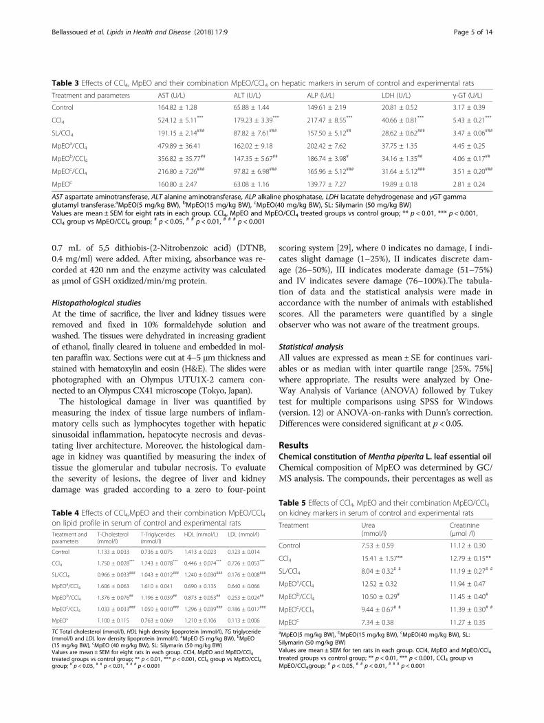
Table 3 Effects of CCl4, MpEO and their combination MpEO/CCl4 on hepatic markers in serum of control and experimental rats
Treatment and parameters AST (U/L) ALT (U/L) ALP (U/L) LDH (U/L) γ-GT (U/L)
Control 164.82 ± 1.28 65.88 ± 1.44 149.61 ± 2.19 20.81 ± 0.52 3.17 ± 0.39
CCl4 524.12 ± 5.11*** 179.23 ± 3.39*** 217.47 ± 8.55*** 40.66 ± 0.81*** 5.43 ± 0.21***
SL/CCl4 191.15 ± 2.14### 87.82 ± 7.61### 157.50 ± 5.12## 28.62 ± 0.62### 3.47 ± 0.06###
MpEOa/CCl4 479.89 ± 36.41 162.02 ± 9.18 202.42 ± 7.62 37.75 ± 1.35 4.45 ± 0.25
MpEOb/CCl4 356.82 ± 35.77## 147.35 ± 5.67## 186.74 ± 3.98# 34.16 ± 1.35## 4.06 ± 0.17##
MpEOc/CCl4 216.80 ± 7.26### 97.82 ± 6.98### 165.96 ± 5.12### 31.64 ± 5.12### 3.51 ± 0.20###
MpEOc 160.80 ± 2.47 63.08 ± 1.16 139.77 ± 7.27 19.89 ± 0.18 2.81 ± 0.24
AST aspartate aminotransferase, ALT alanine aminotransferase, ALP alkaline phosphatase, LDH lacatate dehydrogenase and γGT gammaglutamyl transferase.aMpEO(5 mg/kg BW), bMpEO(15 mg/kg BW), cMpEO(40 mg/kg BW), SL: Silymarin (50 mg/kg BW)Values are mean ± SEM for eight rats in each group. CCl4, MpEO and MpEO/CCl4 treated groups vs control group; ** p < 0.01, *** p < 0.001,CCl4 group vs MpEO/CCl4 group;
# p < 0.05, # # p < 0.01, # # # p < 0.001
Table 4 Effects of CCl4,MpEO and their combination MpEO/CCl4on lipid profile in serum of control and experimental ratsTreatment andparameters
T-Cholesterol(mmol/l)
T-Triglycerides(mmol/l)
HDL (mmol/L) LDL (mmol/l)
Control 1.133 ± 0.033 0.736 ± 0.075 1.413 ± 0.023 0.123 ± 0.014
CCl4 1.750 ± 0.028*** 1.743 ± 0.078*** 0.446 ± 0.074*** 0.726 ± 0.053***
SL/CCl4 0.966 ± 0.033### 1.043 ± 0.012### 1.240 ± 0.030### 0.176 ± 0.008###
MpEOa/CCl4 1.606 ± 0.063 1.610 ± 0.041 0.690 ± 0.135 0.640 ± 0.066
MpEOb/CCl4 1.376 ± 0.076## 1.196 ± 0.039## 0.873 ± 0.053## 0.253 ± 0.024##
MpEOc/CCl4 1.033 ± 0.033### 1.050 ± 0.010### 1.296 ± 0.039### 0.186 ± 0.017###
MpEOc 1.100 ± 0.115 0.763 ± 0.069 1.210 ± 0.106 0.113 ± 0.006
TC Total cholesterol (mmol/l), HDL high density lipoprotein (mmol/l), TG triglyceride(mmol/l) and LDL low density lipoprotein (mmol/l). aMpEO (5 mg/kg BW), bMpEO(15 mg/kg BW), cMpEO (40 mg/kg BW), SL: Silymarin (50 mg/kg BW)Values are mean ± SEM for eight rats in each group. CCl4, MpEO and MpEO/CCl4treated groups vs control group; ** p < 0.01, *** p < 0.001, CCl4 group vs MpEO/CCl4group; # p < 0.05, # # p < 0.01, # # # p < 0.001
Table 5 Effects of CCl4, MpEO and their combination MpEO/CCl4on kidney markers in serum of control and experimental rats
Treatment Urea(mmol/l)
Creatinine(μmol /l)
Control 7.53 ± 0.59 11.12 ± 0.30
CCl4 15.41 ± 1.57** 12.79 ± 0.15**
SL/CCl4 8.04 ± 0.32# # 11.19 ± 0.27# #
MpEOa/CCl4 12.52 ± 0.32 11.94 ± 0.47
MpEOb/CCl4 10.50 ± 0.29# 11.45 ± 0.40#
MpEOc/CCl4 9.44 ± 0.67# # 11.39 ± 0.30# #
MpEOc 7.34 ± 0.38 11.27 ± 0.35aMpEO(5 mg/kg BW), bMpEO(15 mg/kg BW), cMpEO(40 mg/kg BW), SL:Silymarin (50 mg/kg BW)Values are mean ± SEM for ten rats in each group. CCl4, MpEO and MpEO/CCl4treated groups vs control group; ** p < 0.01, *** p < 0.001, CCl4 group vsMpEO/CCl4group;
# p < 0.05, # # p < 0.01, # # # p < 0.001
Bellassoued et al. Lipids in Health and Disease (2018) 17:9 Page 5 of 14

a
b
Fig. 1 a Effects of CCl4, MpEO and their combinations MpEO/CCl4 on hepatic TBARS of control (Con) and experimental rats. Con, control group;mod, CCl4-model group; SL/CCl4, silymarin 50 mg/kg + CCl4; MpEO/CCl4 5 mg/kg + CCl4 group; MpEO/CCl4 15 mg/kg + CCl4 group; MpEO/CCl440 mg/kg + CCl4 group; MpEO 40 mg/kg group. Values are mean ± SEM for ten rats in each group. CCl4, MpEO, MpEO/CCl4 treated groups vscontrol group; *p < 0.05, **p < 0.01, *** p < 0.001, CCl4 group vs (MpEO/CCl4) group; #p < 0.05, ##p < 0.01, ###p < 0.001. b. Effects of CCl4, MpEOand their combinations (MpEO/CCl4) on kidney TBARS of control (Con) and experimental rats. Con, control group; mod, CCl4-model group; SL/CCl4, silymarin 50 mg/kg + CCl4; MpEO/CCl4 5 mg/kg + CCl4 group; MpEO/CCl4 15 mg/kg + CCl4 group; MpEO/CCl4 40 mg/kg + CCl4 group; MpEO40 mg/kg. Values are mean ± SEM for ten rats in each group. CCl4, MpEO, MpEO/CCl4 treated groups vs control group; *p< 0.05, **p< 0.01, *** p< 0.001,CCl4 group vs (MpEO/CCl4) group; #p< 0.05, ##p< 0.01, ###p< 0.001
Bellassoued et al. Lipids in Health and Disease (2018) 17:9 Page 6 of 14

their retention indices are listed in Table 1. MpEO is amixture with 26 compounds representing 98.17% of thetotal oil composition. The most abundant chemicals cat-egories for MpEO are oxygenated monoterpenes (79.50%),followed by monoterpene hydrocarbons (16.23%) and ses-quiterpene hydrocarbons (2.44%). The major componentsof MpEO are menthol (33.59%) and iso-menthone(33.00%). In lower amounts we found a variety of com-pounds including limonene (8.00%), piperitone (3.20%),1,8-cineole (2.80%), linalool (2.64%), iso-pulegol (2.40%),caryophyllene (1.95%) and pulegone (1.60%).
Essential oil antioxidant activityThe antioxidant activity of MpEO was compared to thatof silymarin, a well-known antioxidant, using two differentassays, namely DPPH and superoxide oxygen radicals in-hibition, the results are reported in Table 2. DPPH showedfor MpEO an IC50 value around 3 times higher than theone recorded for silymarin indicating that antioxidantactivity of MpEO was lower than that of silymarin.
Serum biochemical parametersThe results of biochemical indicators of liver and kidneyfunction are summarized in Tables 3, 4 and 5. The admin-istration of CCl4 caused severe hepato and reno-toxicity inthe treated rats, as evidenced by the significant elevationsof serum ALT, AST, ALP, LDH, γGT, total cholesterol, tri-glycerides, LDL urea and creatinine levels, while HDLlevel was decreased compared to control animals.Pretreatment with the MpEO at doses of 15 or 40 mg/kg
significantly reduced levels of ALT, AST, ALP, LDH, γGT,total cholesterol, triglycerides, LDL urea and creatinine andincreased the level of HDL compared to the CCl4 group. Itis worth noting that the treatment with 5 mg/kg MpEO didnot induce any significant changes in the biochemicalparameters (ALT, AST, ALP, LDH, γGT, total cholesterol,triglycerides, LDL, urea, creatinine or HDL) when com-pared to the CCl4 group. Treatment of rats with onlyMpEO (40 mg/kg BW) did not result in significant alter-ations in biochemical parameters compared to control rats.Pretreatment with silymarin (50 mg/kg), used as posi-
tive control, significantly decreased the elevated levels ofALT, AST, ALP, LDH, γGT, total cholesterol, triglycer-ides, LDL urea and creatinine and increased of HDLlevel as compared to CCl4 group. Its effect was compar-able in reducing of liver and kidney damage induced byCCl4 with that observed for the highest dose of MpEO(40 mg/kg).
Effects on lipid peroxidationTBARS level is widely used as a marker for free radical me-diated lipid peroxidation injury. We determined TBARSlevels in the liver and kidney tissues of the investigated
animals and our results are shown in Fig. 1a, b. The levels ofTBARS were significantly increased in both liver and kidneytissues of CCl4-treated animals when compared to controluntreated rats.Pre-treatment with the MpEO at doses of 15 and
40 mg/kg BW significantly reduced levels of TBARS inliver and kidney tissues as compared to CCl4 group. Therewas a dose effect; treatment with MpEO at 5 mg/kg BWdid not induce any significant decrease in the levels ofTBARS in liver and kidney as compared to CCl4 group.When rats were treated with only MpEO (40 mg/kg BW),no significant differences in the TBARS values wasobserved compared to control rat. Pretreatment with sily-marin (50 mg/kg) significantly decreased the elevatedlevels of TBARS in both liver and kidney compared toCCl4 control. Moreover, the effect of silymarin (50 mg/kg)in attenuation of TBARS levels in liver and kidney wascomparable with highest dose of MpEO (40 mg/kg).
Effects on antioxidant enzymesResults presented in Tables 6 and 7 showed a significantdecrease in the levels of CAT, SOD, GPx in liver and
Table 6 Effects of CCl4, MpEO and their combination MpEO/CCl4 on the activities of enzymatic antioxidants in liver ofcontrol and experimental ratsTreatment SOD
(Units/mg protein)CAT(μmol H2O2/min/mgprotein)
GPx(μmol GSH/min/mgprotein)
Control 16.04 ± 0.11 14.03 ± 0.29 7.66 ± 0.51
CCl4 13.60 ± 0.50*** 11.13 ± 0.37*** 5.25 ± 0.25**
SL/CCl4 15.80 ± 0.50## 13.48 ± 0.25### 7.41 ± 0.16###
MpEOa/CCl4 13.92 ± 0.40 11.34 ± 0.41 5.41 ± 0.31
MpEOb/CCl4 14.98 ± 0.19# 12.32 ± 0.32# 6.04 ± 0.17#
MpEOc/CCl4 15.65 ± 0.44## 13.20 ± 0.19### 7.26 ± 0.22###
MpEOc 15.83 ± 0.31 13.85 ± 0.33 7.45 ± 0.33
aMpEO(5 mg/kg BW), bMpEO (15 mg/kg BW), cMpEO(40 mg/kg BW), SL: Silymarin(50 mg/kg BW)Values are mean ± SEM for ten rats in each group. CCl4, MpEO and MpEO/CCl4treated groups vs control group; ** p < 0.01, *** p < 0.001, CCl4 group vs MpEO/CCl4group; # p < 0.05, # # p < 0.01, # # # p < 0.001
Table 7 Effects of CCl4, MpEO and their combination MpEO/CCl4 on the activities of enzymatic antioxidants in kidney ofcontrol and experimental ratsTreatment SOD
(Units/mg protein)CAT(μmol H2O2/min/mgprotein)
GPx(μmol GSH/min/mgprotein)
Control 15.13 ± 0.22 12.90 ± 0.15 5.68 ± 0.27
CCl4 12.12 ± 0.50*** 10.05 ± 0.34*** 4.06 ± 0.28**
SL/CCl4 14.52 ± 0.35## 12.23 ± 0.24### 5.56 ± 0.14##
MpEOa/CCl4 12.92 ± 0.20 10.80 ± 0.22 4.46 ± 0.33
MpEOb/CCl4 13.69 ± 0.10# 11.02 ± 0.22# 5.10 ± 0.28#
MpEOc/CCl4 14.20 ± 0.35## 12.04 ± 0.19### 5.32 ± 0.06##
MpEOc 14.91 ± 0.21 12.62 ± 0.33 5.59 ± 0.27
aMpEO(5 mg/kg BW), bMpEO(15 mg/kg BW), cMpEO(40 mg/kg BW), SL: Silymarin(50 mg/kg BW)Values are mean ± SEM for ten rats in each group. CCl4, MpEO and MpEO/CCl4treated groups vs control group; ** p < 0.01, *** p < 0.001, CCl4 group vs MpEO/CCl4group; # p < 0.05, # # p < 0.01, # # # p < 0.001
Bellassoued et al. Lipids in Health and Disease (2018) 17:9 Page 7 of 14
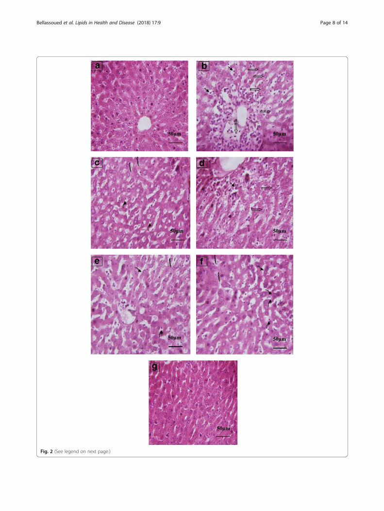
Fig. 2 (See legend on next page.)
Bellassoued et al. Lipids in Health and Disease (2018) 17:9 Page 8 of 14

kidney in CCl4-treated group when compared to controlgroup. The decrease in hepatic and kidney CAT, SODand GPx levels induced by CCl4 injection were signifi-cantly restored (elevated) in the MpEO and silymaringroups, and this effect was more pronounced with theincrease of essential oil concentration. Pretreatment withMpEO at doses of 15 and 40 mg/kg significantly in-creased levels of hepatic and kidney CAT, SOD and GPxas compared to CCl4 group. It is worth noting that thetreatment with MpEO at a dose 5 mg/kg did not induceany significant increase in the levels of hepatic and kid-ney CAT, SOD and GPx as compared to CCl4 group. Nosignificant differences in the values were observed in ratstreated with MpEO only (40 mg/kg) compared to con-trol rat values. Moreover, the effect of silymarin (50 mg/kg) was comparable in attenuation of levels of hepaticand kidney CAT, SOD and GPx with highest dose ofMpEO (40 mg/kg).
Histopathological findingsThe normal liver architecture was observed in liver histologyof control group (Fig. 2a). Large numbers of inflammatorycells such as lymphocytes together with hepatic sinusoidalinflammation, hepatocyte necrosis and devastating liverarchitecture were observed in the CCl4 group (Fig. 2b). How-ever, pretreatment with MpEO (40 mg/kg, Fig. 2f) can re-markably ameliorate the histopathological hepatic lesionsinduced by administration of CCl4. MpEO 15 mg/kg showedvery few inflammatory cells along with prominent nucleolus(Fig. 2e). The highest dose of MpEO (40 mg/kg) and sily-marin (50 mg/kg) significantly attenuated the damaged liverdepicting marked focal regenerative changes which are illus-trated by presence of actively dividing cells with a prominentnucleolus (Fig. 2f). In addition, silymarin at the dose of50 mg/kg has shown to produce hepatoprotection evidencedby area of regeneration and dark nucleus (Fig. 2c). The histo-logical pattern was almost normal in rats treated with MpEOoil alone. By analyzing the histopathological scoring attrib-uted to the liver tissues it is possible to note that the highestdose of MpEO (40 mg/kg) or silymarin (50 mg/kg) pretreat-ment just conferred good protection on CCl4-induced liverdamage (Table 8).Kidney sections of normal histological appearance (Fig. 3a)
and the CCl4 control group showed some nephrotoxiclesions, as evidenced by the glomerular and tubular necrosis(Fig. 3b). However, pretreatment with MpEO (40 mg/kg,Fig. 3f) can remarkably ameliorate the histopathological
kidney lesions induced by administration of CCl4 (Fig. 3f).In addition, silymarin at the dose of 50 mg/kg has shown toproduce renoprotection evidenced by amelioration thehistopathological kidney lesions induced by injection ofCCl4 (Fig. 3c). The histological pattern in kidney was almostnormal in rats treated with MpEO alone. By analyzing thehistopathological scoring attributed to the kidney tissues it ispossible to note that the highest dose of MpEO (40 mg/kg)or silymarin (50 mg/kg) pretreatment just conferred goodprotection on CCl4-induced kidney damage (Table 9).
DiscussionChemical composition of the essential oil obtained fromMpEO was determined by GC-MS analysis. The com-pounds, their percentages as well as the retention indicesare listed in Table 1. The essential oil is a complex mixturewith 26 compounds representing 98.17% of the total oilcomposition. The major component of the essential oil ismenthol (33.59%) followed by iso-menthone (33.00%). Inlower amounts we found a variety of compounds includ-ing limonene (8.00%), piperitone (3.20%), 1,8-cineole(2.80%), linalool (2.64%), iso-pulegol (2.40%), caryophyl-lene (1.95%) and pulegone (1.60%). The obtained resultsare in accordance with previous studies of M. piperita oilsfrom Turkey, Spain (Barcelona), Norway and Poland thatalso had menthone and menthol as their most importantcomponents [30–32]. On the other hand, the compositionof the essential oil from Iran is totally different, with α-terpinene (19.70%), isomenthone (10.30%), trans-carveol(14.50%), pipertitinone oxide (19.30%) and β-caryophyllene(7.60%) as the major compounds, and also the oil from theGirona region (Spain) is different, where limonene and 1.8-
Table 8 Grades of inflammatory cells and cellular necrosis inrat liverPathologic grading of inflammatory cells and cellular necrosis
Groups Zero I II III IV N P value vs Control P value vs CCl4
Control 10 0 0 0 0 10 –
CCl4*** 0 1 3 3 3 10 P < 0.001 –
SL/CCl4### 8 2 0 0 0 10 ns P < 0.001
MpEOa/CCl4 0 2 6 1 1 10 P < 0.05 ns
MpEOb/CCl4# 6 4 0 0 0 10 ns P < 0.05
MpEOc/CCl4### 7 3 0 0 0 10 ns P < 0.001
MpEOc 9 1 0 0 0 10 ns P < 0.001
aMpEO (5 mg/kg BW), bMpEO (15 mg/kg BW), cMpEO (40 mg/kg BW), SL: Silymarin(50 mg/kg BW)
(See figure on previous page.)Fig. 2 Effect of MpEO on CCl4-induced liver damage. a Control group; (b) CCl4-model group showing marked inflammatory cells, necrosis andreduced lesions of necrosis; (c) Silymarin 50 mg/kg + CCl4 group; (d) MpEO 5 mg/kg + CCl4 group (e); MpEO 15 mg/kg + CCl4 group; (f) MpEO40 mg/kg + CCl4 group; (g) MpEO 40 mg/kg group. Hematoxylin/eosin staining; magnification ×400. : Marked inflammatory cells;
: Necrosis cells; : Reduced lesions of necrosis; : Regeneration area; : Prominent nucleolus; : Mild inflammatory cells
Bellassoued et al. Lipids in Health and Disease (2018) 17:9 Page 9 of 14

Fig. 3 (See legend on next page.)
Bellassoued et al. Lipids in Health and Disease (2018) 17:9 Page 10 of 14

cineole, eucalyptol are the main compounds (33.37% and30.75%, respectively) [33, 34].These studies showed variable chemotypes of M.
piperita L. extracts with various major oil components.Differences in chemical composition observed for essen-tial oils is likely related to abiotic factors such as soiltype and climate specific regions of provenance samplesand geographical factors [35]. Furthermore, menthol andiso-menthone, found at relatively high concentrations inthe MpEO used in the present study, have been reportedto exhibit anti-inflammatory activity [14], making MpEOuse a promising candidate against oxidative damage ofthe liver and kidney following an intraperitoneal admin-istration of CCl4. To be noted: working with naturalextracts, the antioxidant activity is considered to be pri-mary related to the major active compounds in theessential oil such as menthol and its derivatives [15].However the antioxidant activity could also come from aminor compound interacting in a synergistic or antagon-istic way, to create an effective system against free radi-cals [36, 37], this has to be realized when evaluatingdifferent MpEO preparations.Liver injury after CCl4 exposure is characterized by the
elevated levels of serum hepatic marker enzymes indicat-ing the cellular leakage and loss of functional integrity ofhepatic membrane architecture. High levels of ALT, AST,ALP, LDH and γGT activities are sensitive indicators ofliver cell injury and are most helpful in recognizing hep-atic diseases [38]. CCl4-treated rats show increased activ-ities of these enzymes, reflecting damage to the liver cellsor changes in the cell membrane permeability leading to
leakage of enzymes from cells to the circulation [39]. Inthe present study increased levels of serum hepaticmarkers suggested that an extensive liver injury was occa-sioned by CCl4 due to increased lipid peroxidation whichhad the ability to cause membrane damage. It is now gen-erally accepted that CCl4 hepatotoxicity is the result ofreductive dehalogenation, which is catalyzed by its specificisoenzyme of cytochrome P450 2E1, and which forms thehighly reactive free radical. Hence, the suppression ofP450 2E1 could result in reduced levels of reactive metab-olites, and thus decreased tissue damage [40].The liver plays a fundamental role in the metabolism of
lipids. Injection of CCl4 caused a significant increase inthe triglyceride, total cholesterol, and LDL levels anddecrease in HDL level. Increase in the cholesterol levelsmight be due to the increased esterification of fatty acids,inhibition of fatty acid β-oxidation, and decreased excre-tion of cellular lipids [41]. CCl4 stimulates the transfer ofacetate into liver cells (probably by increasing access to acet-ate) and leads to an increase in cholesterol synthesis. It alsoincreases the synthesis of fatty acids and triglyceride fromacetate and enhances lipid esterification [42]. The accumula-tion of triglyceride in liver might occur due to the inhibitionof lysosomal lipase activity and VLDL secretion [43].The administration of CCl4 induced also renal toxicity
evidenced by an elevation of serum creatinine and urea[44, 45]. These pathological changes can also be attributedto damages touching the structural integrity of nephrons[46], which is consistent with reports confirming that thelevel of serum creatinine increases only if at least half ofthe kidney nephrons are already damaged [47].Treatment of rats with MpEO prior to CCl4 exposure
resulted in a dramatically protective effect against acutehepato and renotoxicity and oxidative stress, which wasfurther also confirmed by the hepatic histopathologicalexaminations. The stimulation of hepatic regenerationmakes the liver more resistant to damage by the toxin[48]. Treatment with silymarin (50 mg/kg) or MpEO(40 mg/kg) significantly decreased the elevated levels ofALT, AST, ALP, LDH, γGT, total cholesterol, triglycerides,LDL urea and creatinine and increased of HDL level ascompared to CCl4 group. Pharmacological studies haveshown that essential oil derived from various plant mate-rials possesses anti-inflammatory activities [49, 50] Know-ing that sesquiterpenes have excellent anti-inflammatoryactivities [51], the anti-inflammatory activity of M. piperitaL. leaf essential oil could be partly explained by the pres-ence of sesquiterpenes, such as spathulenol, cadinene,
Table 9 Grades of glomerular and epithelial cells of theproximal tubules necrosis in rat kidney
Pathologic grading of glomerular and epithelial cells of the proximaltubules necrosis
Groups Zero I II III IV N P value vsControl
P value vsCCl4
Control 10 0 0 0 0 10 –
CCl4*** 0 2 2 3 3 10 P < 0.001 –
SL/CCl4### 8 2 0 0 0 10 ns P < 0.001
MpEOa/CCl4 0 2 5 3 0 10 P < 0.05 ns
MpEOb/CCl4# 6 4 0 0 0 10 ns P < 0.05
MpEOc/CCl4### 7 3 0 0 0 10 ns P < 0.001
MpEOc 9 1 0 0 0 10 ns P < 0.001aMpEO (5 mg/kg BW), bMpEO (15 mg/kg BW), cMpEO (40 mg/kg BW), SL:Silymarin (50 mg/kg BW)
(See figure on previous page.)Fig. 3 Effect of MpEO on CCl4-induced kidney damage. a Control group; (b) CCl4-model group showing some nephrotoxic lesions, as evidencedby the glomerular and tubular necrosis; (c) Silymarin 50 mg/kg + CCl4 group; (d) MpEO 5 mg/kg + CCl4 group (e); MpEO 15 mg/kg + CCl4 group;(f) MpEO 40 mg/kg + CCl4 group; (g) MpEO 40 mg/kg group. Hematoxylin/eosin staining; magnification ×400. : glomerular necrosis;
: necrosis in epithelial cells of the proximal tubules
Bellassoued et al. Lipids in Health and Disease (2018) 17:9 Page 11 of 14

caryophyllene and caryophyllene oxide. The ethanolic ex-tract of parsley leaves also showed significant anti-inflammatory [52] and antioxidant activities [53, 54] whichmay contribute to its hepatoprotective action. Furthermore,menthol and iso-menthone, found at relatively high con-centrations in the MpEO used in the present study, havebeen reported to exhibit anti-inflammatory activity [14].In the present study, the two fold increase in the TBARS
levels and reduce activity of SOD, CAT and GPx observedin liver and kidney homogenate of CCl4-intoxicated rats.Silymarin significantly reversed CCl4-induced TBARSlevels elevation but values obtained with MpEO at thehighest dose was comparable in attenuation of TBARSlevels in liver and kidney. Silymarin reversed CCl4-inducedSOD, CATand GPx activities decrease but values obtainedwith MpEO at the highest dose (40 mg/kg) was compar-able in attenuation of SOD, CAT and GPx activities inliver and kidney.These results suggested that MpEO could exert its anti-
oxidant and/or radical scavenging activities thus prevent-ing the formation of the carbon free radicals originatedfrom CCl4 metabolism as well as ROS and peroxidationproducts. This hypothesis is supported by the recent find-ings on the in vitro antiradical and antioxidative activitiesof MpEO [16]. Previous studies showed that menthol andits derivatives were the major compounds responsible forantioxidant activity of MpEO [15].In the present study, the rats of group 2 served as CCl4-
hepato and renotoxicity control and rats of groups 3, 4, 5and 6 were injected with 1 ml/kg BW of CCl4 and olive oilmixture on day seven (a single intraperitoneal injection). Ithas already been shown that a single dose of CCl4 initiateslipid peroxidation [55–58] that results in the disruption ofcellular and organelle membrane integrity and subsequentleakage of cellular contents into the blood [59, 60]. CCl4 isfurther well known to induce fibrosis of the hepatic tissuethat may further progress to cirrhosis if the stimuli persists[61–65]. Thus, a single CCl4 injection in mice can be usedas an attractive and highly reproducible model of liverregeneration after toxic injury. The first appearance ofhistological fibrosis and scarring fibers is usually observedafter repeated CCl4 treatment for 2 to 3 weeks, dependingon the dosage and mouse strains used [66]. In the presentwork, the hepatic histoarchitecture of the CCl4-treated ratsresulted large numbers of inflammatory cells such as lym-phocytes along with hepatic sinusoidal inflammation, hep-atocyte necrosis and devastating liver architecture Thehighest dose of MpEO (40 mg/kg) or silymarin (50 mg/kg)significantly attenuated the damaged liver depicting markedfocal regenerative changes which are illustrated by presenceof actively dividing cells with a prominent nucleolus. Theadministration of MpEO reducing the histological alter-ations in liver provoked by CCl4 was quite noticeable. Infact, the histological changes seen in the kidney of rats
treated with CCl4 were characterized by some nephrotoxiclesions, as evidenced by the glomerular and tubular necro-sis. Our results confirmed previous findings of Ozturk et al.[67] who had found degenerative changes in kidney of ratsexposed to CCl4. The results suggest that MpEO treatmentprior to CCl4 intoxication could prevent the CCl4-inducedalterations in kidney tissues of treated animals.
ConclusionsThe contents of MpEO not only protect the integrity ofplasma membrane but, at the same time, increased theregenerative and reparative capacity of the liver and kid-ney. These results suggest that the compound present inMpEO has hepatorenal protective effects against CCl4induced oxidative stress in rats. Further investigationsare essential to elucidate the precise mechanism of ac-tive agents of MpEO protection against CCl4-inducedhepatotoxicity and nephrotoxicity and it has to be testedagainst other biological parameters.
AbbreviationsBHT: Butylated hydroxytoluene; CAT: Catalase; DPPH: 1-Diphenyl-2-picrylhydrazyl; DTNB: 5,5′-Dithio-bis(2-nitrobenzoic acid);EDTA: Ethylenediaminetetraacetic acid; GPX: Glutathione peroxidase;GSH: Glutathione; M. piperita: Mentha piperita; MpEO: Mentha piperitaessential oil; NBT: Nitroblue Tetrazolium; ROS: Reactive oxygen species;SD: Standard deviation; SOD: Superoxide dismutase; TBA: Thiobarbituric Acid;TBARS: Thiobarbituric acid reactive substances; TBS: Tris Buffered Saline;TCA: Trichloroacetic Acid; TRIS: Trishydroxymethyl aminomethane
AcknowledgementsNot applicable.
FundingNot applicable.
Availability of data and materialsAll data generated or analyzed during this study are included within the article.
Authors’ contributionsKB, ABH, KA, JvP, FMA, TR and AE designed and wrote the paper. All authorshave read and approved the final manuscript.
Ethics approval and consent to participateNot applicable.
Consent for publicationNot applicable.
Competing interestsThe authors declare that they have no competing interests.
Publisher’s NoteSpringer Nature remains neutral with regard to jurisdictional claims inpublished maps and institutional affiliations.
Author details1Department of Life Sciences, Animal Ecophysiology Laboratory, Faculty ofsciences, University of Sfax Tunisia, Road of Soukra Km 3.5, BP 1171, PC 3000Sfax, Tunisia. 2Biotechnology and Plant Improvement Laboratory, Centre ofBiotechnology of Sfax, PO Box 1177, Road Sidi Mansour km 6, 3018 Sfax,Tunisia. 3Department of life sciences, Laboratory of Biodiversity and AquaticEcosystems, Faculty of sciences, University of Sfax Tunisia, Road of Soukra Km3.5, BP 1171, PC 3000 Sfax, Tunisia. 4Laboratory of Clinical DigestiveOncology, Department of Oncology, KU, Leuven, Belgium. 5Biochemistry
Bellassoued et al. Lipids in Health and Disease (2018) 17:9 Page 12 of 14

Laboratory, CHU Habib Bourguiba of Sfax, Faculty of Medicine, University ofSfax Tunisia, Road Menzel Chaker km 0.5, CP 3029 Sfax Sfax, Tunisia.6Laboratory of Histology and Embryology, Faculty of Medicine of Sfax,University of Sfax Tunisia, Road Menzel Chaker km 0.5, CP 3029 Sfax, Tunisia.
Received: 29 September 2017 Accepted: 17 December 2017
References1. Sies H, Stahl W, Sevanian A. Nutritional, dietary and postprandial oxidative
stress. J Nutr. 2005;135:969–72.2. Basaga HS. Biochemical aspects of free radicals. Biochem Cell Biol. 1990;68:989–98.3. ATSDR "Agency for Toxic Substance and Diseases Registry". Toxicological
profile for carbon tetrachloride, U.S. Department of Health and HumanServices, Public Health Service, TP-39/02. Atlanta, CA, 1994.
4. Wang CY, Ma FL, Liu JT, Tian JW, Fu FH. Protective effect of salvianic acid aon acute liver injury induced by carbon tetrachloride in rats. Biol Pharm Bull.2007;30:44–7.
5. Abraham P, Wilfred G, Cathrine SP. Oxidative damage to the lipids andproteins of the lungs, testis and kidney of rats during carbon tetrachlorideintoxication. Clin Chim Acta. 1999;289:177–90.
6. Valko M, Leibfritz D, Moncol J, Cronin MT, Mazur M, Telser J. Free radicalsand antioxidants in normal physiological functions and human disease. Int JBiochem Cell Biol. 2007;39:44–84.
7. Fang YZ, Yang S, Wu G. Free radicals antioxidants and nutrition. Nutrition.2002;18:872–9.
8. Bansal AK, Bansal M, Soni G, Bhatnagar D. N-nitrosodiethylamine inducedoxidative stress in rat liver. Chem Biol Interact. 2005;156:101–11.
9. Shahjahan M, Sabitha KE, Jainu M, Shyamala Devi CS. Effect of Solanumtrilobatum against carbon tetrachloride induced hepatic damage in albinorats. Indian J Med Res. 2004;120:194–8.
10. Li CC, Hsiang CY, Wu CL, Ho TY. Identification of novel mechanisms ofsilymarin on the carbontetrachloride-induced liver fibrosis in mice bynuclear factor-KB bioluminescent imaging-guided transcriptomic analysis.Food Chem Toxicol. 2012;50:1568–75.
11. Heywood VH. Flowering plants of the world. Oxford, U.K: Oxford UniversityPress; 1979.
12. Brown D. Encyclopaedia of herbs and their uses. London, U.K: DorlingKindersley; 1995.
13. Foster S. Peppermint, Mentha piperita, In Botanical Series, AmericanBotanical Council: Austin, Texas, no 306, 1990.
14. Cowan MM. Plant products as antimicrobial agents. Clin Microbiol Rev.1999;12:564–82.
15. Lee CP, Shih PH, Hsu CL, Yen GC. Hepatoprotection of tea seed oil (Camelliaoleifera Abel.) against CCl4-induced oxidative damage in rats. Food ChemToxicol. 2007;45:888–95.
16. Sun Z, Wang H, Wang J, Zhou L, Yang P. Chemical Composition and Anti-Inflammatory, Cytotoxic and Antioxidant Activities of Essential Oil fromLeaves of Mentha piperita Grown in China, Plos One. https://doi.org/10.1371/journal.pone.0114767, (2014) 1–15.
17. Blois MS. Antioxidant determinations by the use of stable free radical.Nature. 1958;26:199–200.
18. Yen GC, Chen HY. Antioxidant activity of various tea extracts in relation totheir antimutagenicity. J Agr Food Chem. 1995;43:27–32.
19. Council of European Communities. Council instructions about theprotection of living animals used in scientific investigations. Off J EuroCommun. 2010; 276:1-33.
20. Mihailović V, Mihailović M, Uskoković A, Arambašić J, Mišić D, Stanković V,Katanić J, Mladenović M, Solujić S, Matić S. Hepatoprotective effects ofGentiana asclepiadea L. extracts against carbon tetrachloride induced liverinjury in rats. Food Chem Toxicol. 2013;52:83–90.
21. Bruckner JV, MacKenzie WF, Muralidhara S, Luthra R, Kyle M, Acosta D. Oraltoxicity of carbon tetrachloride: acute, subacute and subchronic studies inrates. Fundam Appl Toxicol. 1986;6:16–34.
22. Sahreen S, Khan MR, Khan RA. Hepatoprotective effects of methanol extractof Carissa opaca leaves on CCl4-induced damage in rat. Complement AlternMed. 2011;1:1–9.
23. Spindler P, Madsen C. Subchronic toxicity study of peppermint oil in rats.Toxicol letter. 1992;62:215–20.
24. Lowry OH, Rosebrough NJ, Farr AL, Randall RJ. Protein measurement withFolin phenol reagent. J Biol Chem. 1951;193:265–75.
25. Draper HH, Hadley M. Malondialdehyde determination as index of lipidperoxidation. Methods Enzymol. 1990;2:421–31.
26. Aebi H. Catalase in vitro. Methods Enzymol. 1984;2:121–6.27. Beyer JWF, Fridovich I. Assaying for superoxide dismutase activity: some large
consequences of minor changes in conditions. Anal Biochem. 1987;113:559–66.28. Flohé L, Günzler WA. Assays for glutathione peroxidase. Methods Enzymol.
1984;105:114-21.29. Scheuer PJ. Classification of chronic viral hepatitis: a need for reassessment.
J Hepatol. 1991;13:372–4.30. Kim NS, Lee DS. Comparison of different extraction methods for the analysis
of fragrances from Lavandula species by gas chromatography-massspectrometry. J Chromatogr. 2002;22:31–47.
31. Rohloff J, Dragland S, Mordal R, Iversen TH. Effect of harvest time anddrying method on biomass production, essential oil yield, and quality ofpeppermint (Mentha×piperita L.). J Agric Food Chem. 2005;53:4143–8.
32. Skalicka-Wozniak K, Walasek M. Preparative separation of menthol and pulegonefrom peppermint oil (Mentha piperita L.) by high-performance counter-currentchromatography. Phytochem Lett. 2014;728: https://doi.org/10.1016/j.phytol.06.007.
33. Yadegarinia D, Gachkar L, Rezaei MB, Taghizadeh M, Alipoor-Astaneh S,Rasooli I. Biochemical activities of Iranian Mentha piperita L. and Myrtuscommunis L. essential oils. Phytochemistry. 2006;60:1249–55.
34. Ruiz Del Castillo ML, Blanch GP, Herraiz M. Natural variability of theenantiomeric composition of bioactive chiral terpenes in Mentha piperita.J Chromatogr A. 2004;1054:87–93.
35. Orav A, Raal A, Arak E. Comparative chemical composition of the essentialoil of Mentha piperita L. from various geographical sources. Proc EstonianAcad Sci Chem. 2004;53:174–81.
36. Lu Y, Foo LY. Antioxidant activities of polyphenols from sage (Salviaofficinalis). Food Chem. 2001;75:197–202.
37. Singh G, Marimuthu P, De Heluani CS, Catalan CAN. Antioxidant andbiocidal activities of Carum nigrum (seed) essential oil, oleoresin, and theirselected components. J Agric Food Chem. 2006;54:174–81.
38. Pradeep K, Victor Raj Mohan C, Gobianand K, Karthikeyan S. Protective effectof Cassia fistula Linn. On diethylnitrosamine induced hepatocellular damageand oxidative stress in ethanol pretreated rats. Biol Res. 2010;43:113–25.
39. Botsoglou NA, Taitzoglou IA, Botsoglou E, Lavrentiadou SN, Kokoli AN,Roubies N. Effect of long-term dietary administration of oregano on thealleviation of carbon tetrachloride-induced oxidative stress in rats. JAgricFood Chem. 2008;56:6287–93.
40. Zangar RC, Benson JM, Burnett VL, Springer DL. Cytochrome P4502E1 is theprimary enzyme responsible for low-dose carbon tetrachloride metabolismin human liver microsomes. Chem Biol Interact. 2000;125:233–43.
41. Fernandez ML, West KL. Mechanisms by which dietary fatty acids modulateplasma lipids. J Nutr. 2005;135:2075–8.
42. Weber LW, Boll M, Stampfl A. Hepatotoxicity and mechanism of action ofhaloalkanes: carbon tetrachloride as a toxicological model. Crit Rev Toxicol.2003; 33: https://doi.org/10.1080/713611034.
43. Marimuthu S, Adluri RS, Rajagopalan R, Menon VP. Protective role of ferulicacid on carbon tetrachloride-induced hyperlipidemia and histologicalalterations in experimental rats. J Basic Clin Physiol Pharmacol. 2013;24:59–66. https://doi.org/10.1515/jbcpp-2012-0053
44. Abdel Moneim AE, El-Deib KM. The possible protective effects of Physalisperuviana on carbon tetrachloride-induced nephrotoxicity in male albinorats. Life Sci J. 2012;9:1038–52.
45. Al-Yahya M, Mothana R, Al-Said M, Al-Dosari M, Al-Musayeib N, Al-Sohaibani M,Parvez MK, Rafatullah S. Attenuation of CCl4-Induced Oxidative Stress andHepatonephrotoxicity by Saudi Sidr Honey in Rats, Evidence-Based Complementaryand Alternative Medicine. 2013; https://doi.org/10.1155/2013/569037.
46. Khan RA, Khan MR, Sahreen S, Bokhari J. Prevention of CCl4-inducednephrotoxicity with Sonchus Asper in rat. Food Chem Toxicol. 2010;48:2469–76.
47. Bhattacharya H, Lun L, Gomez R. Biochemical Effects to Toxicity of CCl4 onRosy Barbs (Puntius conchonius). Our Nat. 2005;3:20-25.
48. Sadasivan S, Latha PG, Sasikumar JM, Rajashekaran S, Shymal S, Shine VJ.Hepatoprotective studies on Hedyotis Corymbosa (L.) lam. J Ethnopharmcol.2006;106:25–249.
49. Martins FT, Doriguetto AC, De Souza TC, De Souza KR, Moreira ME, Barbosa LC.Composition, and Antiinflammatory and antioxidant activities of the volatile oilfrom the fruit peel of Garcinia brasiliensis. Chem Biodivers. 2008;5:251–8.
50. Kim JY, Oh TH, Kim BJ, Kim SS, Lee NH, Hyun CG. Chemical compositionand anti-inflammatory effects of essential oil from Farfugium japonicumflower. J Oleo Sci. 2008;5:623–8.
Bellassoued et al. Lipids in Health and Disease (2018) 17:9 Page 13 of 14
51. Liu X, Sun Z, Zhang M, Meng X, Xia X, Yua NW, Xue F, Liu C. Antioxidantand antihyperlipidemic activities of polysaccharides from sea cucumberApostichopus japonicas. Carbohydr Polym. 2012;90:1664–70.
52. Al-Howiriny T, Al-Sohaibani M, El-Tahir K, Rafatullah S. Prevention ofexperimentally-induced gastric ulcers in rats by an ethanolic extract of"parsley" Petroselinum crispum. Am J Chin Med. 2003;31:699–711.
53. Wong PYY, Kitts DD. Studies on the dual antioxidant and antibacterialproperties of parsley (Petroselinum crispum) and cilantro (Coriandrumsativum) extracts. Food Chem. 2006;97:505–15.
54. Zhang H, Chen F, Wang X, Yao HY. Evaluation of antioxidant activity ofparsley (Petroselinum crispum) essential oil and identification of itsantioxidant constituents. Food Res Int. 2006;39:833–9.
55. Hafeman DG, Hoekstra WG. Protection against carbon tetrachloride-inducedlipid peroxidation in the rat by dietary vitamin E, selenium, and methionineas measured by ethane evolution. J Nutr. 1977;107:656–65.
56. Lee PY, Mccay PB, Hornbrook KR. Evidence for carbon tetrachloride-inducedlipid peroxidation in mouse liver. Biochem Pharmacol. 1982;31:405–9.
57. Burk RF, Lane JM, Patel K. Relationship of oxygen and glutathione inprotection against carbon tetrachloride-induced hepatic microsomal lipidperoxidation and covalent binding in the rat. Rationale for the use ofhyperbaric oxygen to treat carbon tetrachloride ingestion. J Clin Invest.1984;74:1996–2001.
58. Recknagel RO, Glende EA, Dolak JA, Waller RL. Mechanisms of carbontetrachloride toxicity. Pharmacol Ther. 1989;43:139–54.
59. Recknagel RO. A new direction in the study of carbon tetrachloridehepatotoxicity. Life Sci. 1983;33:401–8.
60. Brattin WJ, Glende EA, Recknagel RO. Pathological mechanisms in carbontetrachloride hepatotoxicity. J Free Radic Biol Med. 1985;1:27–38.
61. Post J, Earle DP, Patek AJ, Victor J. Effects of yeast and food intake onexperimental carbon tetrachloride cirrhosis of the liver in the rat. Am JPathol. 1942;18:661–73.
62. Constandinou C, Henderson N, Iredale JP. Modeling liver fibrosis in rodents.Methods Mol Med. 2005;117:237–50.
63. Smyth R, Munday MR, York MJ, Clarke CJ, Dare T, Turton JA. Comprehensivecharacterization of serum clinical chemistry parameters and theidentification of urinary superoxide dismutase in a carbon tetrachloride-induced model of hepatic fibrosis in the female Hanover Wistar rat. Int JExp Pathol. 2007;88:361–76.
64. Smyth R, Lane CS, Ashiq R, Turton JA, Clarke CJ, Dare TO, York MJ, GriffithsW, Munday MR. Proteomic investigation of urinary markers of carbon-tetrachloride-induced hepatic fibrosis in the Hanover Wistar rat. Cell BiolToxicol. 2009;25:499–512.
65. Fujii T, Fuchs BC, Yamada S, Lauwers GY, Kulu Y, Goodwin JM, Lanuti M,TANABE KK. Mouse model of carbon tetrachloride induced liver fibrosis:Histopathological changes and expression of CD133 and epidermal growthfactor. BMC Gastroenterol. 2010;10:79.
66. Liedtke C, Luedde T, Sauerbruch T, Scholten D, Streetz K, Tacke F, Tolba R,Trautwein C, Trebicka J, Weiskirchen R. Experimental liver fibrosis research:update on animal models, legal issues and translational aspects.Fibrogenesis Tissue Repair. 2013;6:19.
67. Ozturk F, Ucar M, Ozturk IC, Vardi N, Batcioglu K. Iinduced nephrotoxicity andprotective effect of betaine in Sprague-Dawley rats. Urology. 2003;62:353–6.
• We accept pre-submission inquiries
• Our selector tool helps you to find the most relevant journal
• We provide round the clock customer support
• Convenient online submission
• Thorough peer review
• Inclusion in PubMed and all major indexing services
• Maximum visibility for your research
Submit your manuscript atwww.biomedcentral.com/submit
Submit your next manuscript to BioMed Central and we will help you at every step:
Bellassoued et al. Lipids in Health and Disease (2018) 17:9 Page 14 of 14