protein localization in disease and therapyjcs.biologists.org/content/joces/124/20/3381.full.pdf ·...
TRANSCRIPT

Commentary 3381
IntroductionAll eukaryotic cells are surrounded by plasma membranes andcontain elaborate organelles and a complex endomembrane system.These organelles provide distinct compartments for differentmetabolic activities. Protein translation is confined to only one ofthese compartments, the cytosol. The translocation of proteins is afundamental requirement for proteins to be able to exert theirfunctions in different organelles. Indeed, approximately half of theproteins generated by a cell have to be transported into or acrossat least one cellular membrane to reach their functional destination(Chacinska et al., 2009).
The regulation of protein trafficking relies on information thatis encoded within the protein sequence and occurs by two majormechanisms, namely co-translational and post-translationaltranslocation (Rapoport, 2007; Schnell and Hebert, 2003; Wicknerand Schekman, 2005). Proteins targeted to the mitochondria,peroxisomes or the nucleus are post-transcriptionally translocated(Fig. 1) (Schmidt et al., 2010; Suntharalingam and Wente, 2003;Terry et al., 2007; Wickner and Schekman, 2005; Wolf et al.,2010). Even though protein trafficking into each of these organellesinvolves distinct mechanisms, there are some common features,such as the presence of signal sequences, which are recognized byspecific receptors, and the import through membrane-spanningpores. Whereas the transport systems for the post-translationaltranslocation of proteins into the mitochondria rely on the activityof chaperones to provide unfolded polypeptide chains astranslocation substrates (Rapoport, 2007; Schmidt et al., 2010), thenuclear envelope is perforated by nuclear pores that allow the passage of folded proteins either by simple diffusion or mediatedby soluble transport receptors (Strambio-De-Castillia et al., 2010;Suntharalingam and Wente, 2003; Terry et al., 2007). By contrast,many other proteins, including those destined for the secretorypathway and integral membrane proteins, are transported into theendoplasmic reticulum (ER) during synthesis, a process known as
co-translational translocation (Fig. 1) (Wickner and Schekman,2005).
Subcellular localization is essential to protein function and hasbeen suggested as a means to achieve functional diversity and, at thesame time, economize on protein design and synthesis (Butler andOverall, 2009). Subcellular localization determines the access ofproteins to interacting partners and the post-translational modificationmachinery and enables the integration of proteins into functionalbiological networks. Aberrantly localized proteins have been linkedto human diseases as diverse as Alzheimer’s disease, kidney stonesand cancer. In this Commentary, we will, therefore, summarize ourcurrent knowledge on the mechanisms that regulate subcellularprotein localization and those that have been implicated in thepathogenesis of human disease, and we will discuss emergingtherapeutic strategies that target protein localization.
Subcellular mislocalization of proteins inhuman diseasesProtein translocation accomplishes the movement of material andinformation within the eukaryotic cell and is essential for thenormal functioning of the cell. The protein transport machinery ofcells ensures that the right amount of protein is present at the righttime and place (Fig. 1). Aberrant protein localization that is causedby mutation, altered expression of cargo proteins or transportreceptors or by deregulation of components of the traffickingmachinery is a prominent feature of many human diseases.Deregulation of protein trafficking can lead to mislocalization ofproteins and hence their inactivation (i.e. loss of function),misregulation or a harmful activity at the wrong place (i.e. toxicgain of function). Abnormalities in the subcellular localization ofproteins that are important for the signaling, metabolic or structuralproperties of the cell can cause disorders that involve biogenesis,protein aggregation, cell metabolism or signaling. Table 1 listssome proteins whose localization to the wrong subcellular
SummaryThe eukaryotic cell is organized into membrane-covered compartments that are characterized by specific sets of proteins andbiochemically distinct cellular processes. The appropriate subcellular localization of proteins is crucial because it provides thephysiological context for their function. In this Commentary, we give a brief overview of the different mechanisms that are involvedin protein trafficking and describe how aberrant localization of proteins contributes to the pathogenesis of many human diseases, suchas metabolic, cardiovascular and neurodegenerative diseases, as well as cancer. Accordingly, modifying the disease-related subcellularmislocalization of proteins might be an attractive means of therapeutic intervention. In particular, cellular processes that link proteinfolding and cell signaling, as well as nuclear import and export, to the subcellular localization of proteins have been proposed as targetsfor therapeutic intervention. We discuss the concepts involved in the therapeutic restoration of disrupted physiological proteinlocalization and therapeutic mislocalization as a strategy to inactivate disease-causing proteins.
Key words: Human disease, Nucleo-cytoplasmic transport, Protein trafficking, Subcellular protein mislocalization, Theraputic mistargeting,Theraputic rescue
Journal of Cell Science 124, 3381–3392 © 2011. Published by The Company of Biologists Ltddoi:10.1242/jcs.089110
Protein localization in disease and therapyMien-Chie Hung1 and Wolfgang Link2,*1Department of Molecular and Cellular Oncology, The University of Texas M. D. Anderson Cancer Center, Houston, TX 77030, USA2Experimental Therapeutics Program, Centro Nacional de Investigaciones Oncologicas (CNIO), Melchor Fernandez Almagro 3, 28029 Madrid,Spain*Author for correspondence ([email protected])
Jour
nal o
f Cel
l Sci
ence

compartment has been associated with human diseases, and in thefollowing sections we will discuss some of the mechanisms thatcan lead to such changes in protein localization.
Mislocalization through alterations of theprotein trafficking machineryDysregulation of the protein trafficking machinery can havedramatic effects on general protein transport processes, modifyingcell morphology and physiology. Along these lines, changes in thenuclear pore complex (NPC) have been linked to several geneticdisorders (Chahine and Pierce, 2009). For example, in patientswith familial atrial fibrillation, the homozygous mutation R391Hin the nucleoporin NUP155 has been shown to reduce nuclearenvelope permeability and affect the export of Hsp70 mRNA andimport of HSP70 protein (Zhang et al., 2008). That study was thefirst to link a nucleoporin defect to cardiovascular disease.Mutations in other components of the NPC, such as the nucleoporinp62 protein and ALADIN (alacrima achalasia adrenal insufficiencyneurologic disorder, officially known as AAAS) are thought to
cause the neurodegenerative diseases infantile bilateral striatalnecrosis and triple A syndrome, respectively (Basel-Vanagaite etal., 2006; Kiriyama et al., 2008). Mutant ALADIN prevents nuclearentry of the DNA repair proteins aprataxin and DNA ligase I and,therefore, results in increased DNA damage and subsequent celldeath caused by oxidative stress (Kiriyama et al., 2008).
In a similar fashion, protein import into other organelles can beaffected by mutations in the trafficking machinery. For instance,mutations in the peroxin gene PEX7, which encodes a peroxisomalimport receptor that is responsible for the transport of severalessential peroxisomal enzymes, have been found to cause theperoxisome biogenesis disorder rhizomelic chondrodysplasiapunctata type 1 (RCDP1) (Braverman et al., 1997).
Mislocalization through altered proteintargeting signalsTargeting signals tend to be conserved between proteins and thussensitive to alterations (Laurila and Vihinen, 2009). Considerableeffort has been devoted to developing reliable methods to predictthe effect of mutations on the subcellular localization of disease-related proteins (Emanuelsson et al., 2007; Laurila and Vihinen,2009; Nair and Rost, 2008). A substantial amount of experimentaldata has been collected on mislocalization of disease-causingnuclear proteins. For example, loss of the nuclear localizationsignal (NLS) in the sex-determining region Y protein (SRY) hasbeen shown to be associated with XY sex reversal in Swyersyndrome. Similarly, missense mutations within two NLSs thatreduce the nuclear localization of SRY have been characterized inpatients with this syndrome (McLane and Corbett, 2009).
A similar alteration has been reported for the non-canonicalNLS of the cell-type specific transcriptional activator short staturehomeobox (SHOX) gene in patients with Léri–Weilldyschondrosteosis and Langer mesomelic dysplasia, two raregenetic disorders that result in dwarfism. The missense mutationR173C within the SHOX NLS abolishes nuclear localization andconsequently the downstream transcriptional activation by SHOX(Sabherwal et al., 2004). Interestingly, alterations within one copyof the NLS lead to phenotypes identical to SHOXhaploinsufficiency in Léri–Weill dyschondrosteosis, as only halfthe amount of protein can be imported into the nucleus. If bothNLS copies are mutated, all of the SHOX protein remains in thecytoplasm, which leads to the clinically severe, homozygous formof Langer mesomelic dysplasia (Sabherwal et al., 2004).
Additional mutations in several NLSs of disease-relevant proteinshave been identified, including trichorhinophalangeal syndrome I(TRPS1) (Kaiser et al., 2004), aristaless related homeobox (ARX)(Shoubridge et al., 2010) and forkhead box P2 (FOXP2) (Mizutaniet al., 2007), which are involved in tricho-rhino-phalangealsyndrome, X-linked lissencephaly with ambiguous genitalia and aspeech–language disorder, respectively. In contrast with thepreviously described examples, mutations that disrupt the zincfinger domain of the transcription factor autoimmune regulator(AIRE), which are present in patients with autoimmunepolyendocrinopathy-candidiasis-ectodermal dystrophy, have beenreported to cause cytoplasmic retention despite an intact nucleartargeting signal (Bjorses et al., 2000). Mutations within thenucleolar localization signal (NoS) of the ribosomal protein S19(RPS19) have also been reported to be involved in diseasedevelopment: in patients with Diamond–Blackfan anemia thesemutations prevent the correct targeting of RPS19 to the nucleoli(Da Costa et al., 2003a).
3382 Journal of Cell Science 124 (20)
Protein
Nucleus
Nucleolus
Peroxisome
Mitochondrion
Signalsequence
Endoplasmicreticulum
RNA
Ribosome
DNA
Golgi complex
Lysosome
1
8
6
5 4
3
2
7
9
Key
Fig. 1. Schematic overview of intracellular protein trafficking. The majorcomponents of the eukaryotic cell are the cytosol, the nucleus, the nucleolus,the endoplasmic reticulum (ER), the Golgi complex, mitochondria and theperoxisome. Whereas gene transcription takes place within the nucleus (1),protein synthesis is confined to the cytosol and takes place either on free RNAribosomes (2) or on ribosomes associated with the ER (3). Most proteinsdestined to be secreted from the cell (4), or to reside in the plasma membrane,the lysosomes (5), the Golgi apparatus or the ER, follow the secretory pathwayand enter the ER before the end of translation. Proteins targeted to themitochondria (6), peroxisome (7) and nucleus (8) are translocated after theirsynthesis is complete. Subnuclear localization signals include nucleolarretention signals (9), nuclear-matrix-targeting signals and signals that targetproteins to splicing speckles (Mekhail et al., 2007).
Jour
nal o
f Cel
l Sci
ence

Genetic alterations that affect protein targeting signals have alsobeen associated with several metabolic disorders that arecharacterized by defects in specific organelle functions, such aslysosomal and peroxisomal functions. Mislocalization of theperoxisomal enzyme alanine–glyoxylate aminotransferase (AGT)to the mitochondria in patients with the hereditary kidney-stonedisease primary hyperoxaluria type 1 (PH1) has been shown to becaused by the synergistic interaction between the common P11Lpolymorphism and a disease-specific G170R mutation (Djordjevicet al., 2010). Whereas the polymorphism generates a crypticmitochondrial targeting sequence (Purdue et al., 1991), thepolymorphism and mutation together inhibit AGT dimerizationand allow unfolding of the protein, which is a requirement forsubsequent mitochondrial import (Danpure, 2006).
In contrast with genetic alterations, inactivation of proteintargeting signals by post-transcriptional modifications and protein–protein interactions represents an important regulatory mechanismof reversible protein translocation. A striking example is thephosphorylation-dependent unmasking of the NLS that is presentin nuclear factor B (NF-B), a transcription factor known to becrucial for inflammatory processes and the development andprogression of several malignancies (Karin and Greten, 2005; Luoet al., 2005). In unstimulated cells the NF-B NLS is masked bythe inhibitor protein IB. Most activating agents employ a commonpathway that involves the IB kinase (IKK)-mediatedphosphorylation of IB, causing the ubiquitylation and subsequentdegradation of IB proteins, allowing the nuclear translocation ofNF-B through unmasking of the NLS (Hayden and Ghosh, 2008).Nuclear localization of NF-B is associated with tumorigenesisand various inflammatory diseases and hence altering its cellularlocalization has been considered as an attractive therapeutic target.
Mislocalization through changes in proteininteraction or modificationIn addition to altering the localization signal of a protein, disease-related mutations can result in mislocalization of a protein through
protein sequestration. Mutations within the LMNA (lamin A/C)gene, which cause laminopathies, have recently been reported toresult in mislocalization of the DNA-binding transcriptionalrepressor zinc finger protein 239 (ZNF239, also known as MOK2).The pathogenic lamin A/C mutant protein sequesters ZNF239 intonuclear aggregates, and this process is thought to subsequentlyderegulate ZNF239 target genes (Dreuillet et al., 2008).
Hereditary genetic changes that alter the post-translationalmodifications that are relevant for correct protein localizations arealso associated with human diseases. In a recent study, Cordedduet al. reported a genetic change in patients with Noonan-likesyndrome that introduces an N-myristoylation site in SHOC2, aleucine-rich repeat-containing protein. This acquired fatty acidmodification results in aberrant SHOC2 targeting to the plasmamembrane and impaired translocation to the nucleus upon growthfactor stimulation (Cordeddu et al., 2009).
Mislocalization of misfolded proteinsAberrant folding of proteins with important cellular roles can causediseases in which loss of the protein function rather than itsmislocalization drives the pathology (Hung et al., 1997; Payne et al., 1998; Skach, 2000; Tanaka et al., 2003). Misfolding can alsoresult in proteins that retain intrinsic function yet become misroutedand, as a consequence of their mislocalization, cease to functionnormally (Conn et al., 2007). Accordingly, aberrant localization ofmisfolded proteins can have deleterious gain-of-function ordominant-negative effects in many diseases. Several hereditarygenetic disorders have been linked to altered trafficking ofmisfolded G-protein-coupled receptors (GPCRs); examples of suchdisorders include retinitis pigmentosa (Conn et al., 2007; Mendeset al., 2005) and nephrogenic diabetes insipidus (Robben et al.,2006). In some disease states, the defect in cell surface membraneexpression of GPCRs is due to a dominant-negative effect of themisfolded receptor on its wild-type counterpart owing to theirassociation in the ER and misrouting of the resulting complex(Brothers et al., 2004; Conn et al., 2007; Gehret et al., 2006; Karpa
3383Disease-relevant protein mislocalization
Table 1. Mislocalized proteins that have been associated with human diseases
Protein Disease Mechanism Mislocalization Reference
SRY Swyer syndrome Mutation of NLS Loss of nuclear localization (McLane and Corbett, 2009)SHOX Léri–Weill dyschondrosteosis Mutation of NLS Cytoplasmic retention (Sabherwal et al., 2004)TRPS1 TRPS Mutation of NLS Loss of nuclear localization (Kaiser et al., 2004)ARX XLAG Mutation of NLS Loss of nuclear localization (Shoubridge et al., 2010)FOXP2 Speech–language disorder Mutation of NLS Loss of nuclear localization (Mizutani et al., 2007)AIRE APECED Mutation of ZFD Cytoplasmic retention (Bjorses et al., 2000)RPS19 Diamond–Blackfan anemia Mutation of NoS Loss of nucleolar localization (Da Costa et al., 2003b)AGT Primary hyperoxaluria type 1 Polymorphism and/or mutation Mitochondrial mislocalization (Djordjevic et al., 2010)hsMOK2 Laminopathy Mutation of lamin A⁄C Formation of nuclear aggregates (Dreuillet et al., 2008)SHOC2 Noonan-like syndrome Acquired N-myristoylation Mislocalization to the plasma (Cordeddu et al., 2009) membraneRhodopsin Retinitis pigmentosa Mutations ER retention (Mendes et al., 2005)AVPR2 Nephrogenic diabetes insipidus Mutations ER retention (Robben et al., 2006)ATP7B Wilson disease H1069Q mutation ER retention (Payne et al., 1998)ABCA1 Tangier disease Mutations Loss of plasma membrane localization (Tanaka et al., 2003)Tau Neurodegenerative diseases Hyperphosphorylation Mislocalization to dendritic spines (Hoover et al., 2010)TARDBP ALS and FTLD Unknown Cytoplasmic mislocalization (Winton et al., 2008)FUS FTLD Mutations Cytoplasmic mislocalization (Vance et al., 2009)FOXO Various types of cancer Post-translational modifications Cytoplasmic mislocalization (Dansen and Burgering, 2008)p53 Various types of cancer Mutations, post-translational Cytoplasm (Fabbro and Henderson, 2003) modifications
APECED, autoimmune polyendocrinopathy–candidiasis–ectodermal dystrophy; ALS, amyotrophic lateral sclerosis; FTLD, frontotemporal lobar degeneration;TRPS, trichorhinophalangeal syndrome; XLAG, X-linked lissencephaly with absent corpus callosum and ambiguous genitalia.
Jour
nal o
f Cel
l Sci
ence
et al., 2000). Mislocalized misfolded proteins that exert toxic gain-of-function or dominant-negative effects are increasinglyrecognized as a cardinal feature of many neurodegenerative diseases(Box 1).
Mislocalization of signaling proteinsSignal transduction involves the transmission of a signal in timeand space. Because proteins cannot diffuse as quickly as small-molecule second messengers, the subcellular localization ofsignaling proteins in proximity to their downstream targets is a keyelement of many signal transduction circuits (Scott and Pawson,2009). Translocation of signaling proteins provides an efficientmeans to carry the signal over a substantial distance or betweencellular compartments. Deregulation of the spatiotemporal signalingdynamics has been shown to be involved in tumorigenesis, tumor
growth and metastasis (Kau et al., 2004; Wang and Hung, 2005).Several important tumor suppressors require the ability to localizeto the nucleus to perform their function, and their cytoplasmiclocalization can serve as an inactivation mechanism that gives riseto uncontrolled cell proliferation and the onset of disease (Fabbroand Henderson, 2003; Salmena and Pandolfi, 2007; Turner andSullivan, 2008; Yashiroda and Yoshida, 2003). Consequently,mislocalization of nuclear proteins to the cytoplasm has beenproposed as a generalized mechanism for the inactivation of tumorsuppressors (Kau et al., 2004; Salmena and Pandolfi, 2007).
The transcription factor forkhead box O3a (FOXO3a), a memberof the forkhead family of proteins, provides an example of a tumorsuppressor whose function is altered by mislocalization. FOXO3aregulates the expression of, for example, the apoptotic proteinsFasL (Brunet et al., 1999) and Bim (Gilley et al., 2003), and thecell cycle inhibitor p27 (Dijkers et al., 2000). Cytoplasmiclocalization of FOXO3a has been shown to correlate with poorsurvival in breast cancer (Hu et al., 2004), whereas nuclearlocalization correlates with an increased sensitivity to radiation(Chen et al., 2008). Three oncokinases have been shown to regulateFOXO3a localization: AKT (Brunet et al., 1999), IKK (Hu et al.,2004) and extracellular signal-regulated kinase 1/2 (ERK1/2) (Yanget al., 2008). Whereas all three kinases phosphorylate FOXO3a ondifferent residues (AKT on T32, S253 and S315; IKK on S644;and ERK1/2 on S294, S344 and S425), the outcome is the same,namely export of FOXO3 from the nucleus and subsequentdegradation.
The roles of the cell cycle inhibitors p21 and p27 are alsoregulated by their cellular localization. p21 and p27 are traditionallyconsidered to be tumor suppressors that act in the nucleus andbecome oncogenic when localized in the cytoplasm. p21 wasoriginally shown to inhibit tumorigenesis and was suggested as agood candidate for gene therapy (Katayose et al., 1995; Yang et al.,1995). However, it subsequently became apparent that p21 is alsoassociated with anti-apoptotic functions when it is localized in thecytosol. It was shown that the binding of p21 to mitogen-activatedprotein kinase (MAPK) kinase kinase 5 (MAP3K5, also known asASK1) in the cytoplasm inhibits the MAPK cascade (Asada et al.,1999; Huang et al., 2003). In addition, phosphorylation of p21 onT145 by AKT induces nuclear export of p21, which enhances cellgrowth (Li et al., 2002; Rossig et al., 2001; Zhou et al., 2001).Furthermore, cytoplasmic p21 was found to have an oncogenicrole in mammary tumorigenesis and metastasis in vivo (Cheng et al., 2010). The physiological relevance of p21 in the cytoplasmis emphasized further by the clinical observation that cytoplasmiclocalization of p21 is a poor prognostic marker for breast cancer(Xia et al., 2004), is involved in mediating resistance to anticancerdrugs (Koster et al., 2010; Ruan et al., 1999; Zhang et al., 1995)and has been shown to result in a poor response to tamoxifen(Pérez-Tenorio et al., 2006).
Similar to p21, the cell cycle inhibitor p27 has been found tohave oncogenic roles when localized in the cytoplasm. AKTphosphorylates p27 on T157, which blocks the nuclear import ofp27 (Liang et al., 2002; Shin et al., 2002; Viglietto et al., 2002).Phosphorylation of p27 on S10 has also been shown to inducecytoplasmic localization of p27 without leading to its degradation(Rodier et al., 2001). As is the case for p21, the cytoplasmiclocalization of p27 has been found to be a poor prognostic factorin cancers, including breast (Liang et al., 2002), hepatocellular(Nan et al., 2004), colon (Ogino et al., 2009) and ovarian cancer(Rosen et al., 2005), and Barrett’s carcinoma (Singh et al., 1998).
3384 Journal of Cell Science 124 (20)
Box 1. Aberrant protein localization inneurodegenerative diseasesDespite a wealth of experimental data on neurodegenerativediseases, no consensus has yet emerged on the nature of theneuropathogenic species and how they promote degeneration ofneurons in, for example, Alzheimer’s, Parkinson’s, Huntington’sand prion diseases, amyotrophic lateral sclerosis (ALS) orfrontotemporal lobar degeneration (FTLD). Considerableevidence has been accumulated in recent years to indicate that the earlier stages of the protein mislocalization process aremore directly tied to pathogenesis than the filamentous protein aggregates themselves. The formation of large proteinaggregates and inclusion bodies might even represent abeneficial defense mechanism that operates to eliminateirreversibly aggregated proteins. Accordingly, it has been shownin mouse models that neurofibrillary tangles composed ofaggregates of a hyperphosphorylated form of the microtubule-associated protein tau (MAPT) exert negligible neurotoxicitycompared with that of soluble tau (Oddo et al., 2006; Santacruzet al., 2005). Mislocalization of tau to dendritic spines hasrecently been reported to mediate a synaptic dysfunction that isassociated with impaired brain function at the preclinical diseasestages that immediately precede neurodegeneration. Theaccumulation of hyperphosphorylated tau within intact dendriticspines impairs glutamate receptor trafficking or synapticanchoring (Hoover et al., 2010). In patients with ALS and FTLD,affected neurons exhibit a striking redistribution of TAR DNA-binding protein (TARDBP, also known as TDP43) or the ALS-associated protein fused in sarcoma (FUS) from the nucleus tothe cytoplasm (Neumann et al., 2006). Interestingly, a recentstudy showed that TDP43-mediated neurotoxicity was associatedwith an increased cytoplasmic localization of this protein,whereas TDP43 inclusion bodies were not necessary for thetoxicity and did not affect the risk of cell death (Barmada et al.,2010; Kwiatkowski et al., 2009; Lagier-Tourenne and Cleveland,2009; Vance et al., 2009).
Aggregation of amyloidogenic proteins can result in thesequestration and, hence, aberrant localization of numerousproteins, including importin alpha and phosphorylated SMAD3,which suggests that there is a functional impairment of nucleartrafficking in Alzheimer’s disease (Chalmers and Love, 2007; Zhuet al., 2002). It has been hypothesized that an altered localizationof transcription factors such as NF-B, activating transcriptionfactor 2 (ATF2), cAMP response element-binding (CREB), p53,E2F transcription factor and NF-E2-related factor 2 (NRF2) mightcontribute to cell death commitment in several neurodegenerativediseases (Chu et al., 2007).
Jour
nal o
f Cel
l Sci
ence

In addition to tumor suppressors, it has gradually become clearthat receptor tyrosine kinases (RTKs), traditionally regarded ascell-membrane-bound proteins, are also able to translocate to andfunction in the nucleus (Carpenter and Liao, 2009; Gomes et al.,2008; Reilly and Maher, 2001; Sehat et al., 2010; Stachowiak et al., 1997; Wang and Hung, 2009). Recently, a new traffickingmechanism for EGFR from cell surface to the nucleus has beenreported. EGFR trafficking involves endocytosis, followed byretrograde trafficking from the Golgi to the ER by COPI-mediatedvesicular transport. The receptor is then translocated through theER to the nucleus, where SEC61B is able to release it from the inner nuclear membrane (Wang et al., 2010a; Wang et al.,2010b). It is worthwhile mentioning that the EGFR remainsmembrane-bound throughout the entire trafficking pathway fromthe cell surface to the nucleus. Nuclear localization of EGFR iscorrelated with many tumor types (Hadzisejdic et al., 2010; Hoshinoet al., 2007; Lo et al., 2005a; Psyrri et al., 2008; Xia et al., 2009)and has been shown to be involved in mediating resistance to theanticancer agents cetuximab (Li et al., 2009) and cisplatin (Hsu etal., 2009). Consistently, nuclear EGFR has been associated withcell proliferation, DNA repair, drug resistance and nuclear EGFRand ERBB2 have been shown to bind to promoters and activatetranscription (Hanada et al., 2006; Hung et al., 2008; Lin et al.,2001; Lo et al., 2005b; Wang et al., 2004).
Therapeutic manipulation of proteinlocalization in human diseasesBecause the deregulation of intracellular protein transport iscrucially involved in the pathophysiology of a broad range ofmedical conditions and diseases, it offers molecular targets at manydifferent levels for the attempt to normalize or to interfere withprotein localization with a therapeutic strategy. In particular, cellularprocesses that link protein folding, cell signaling, and nuclearimport and export to subcellular localization of proteins have beenproposed as targets for therapeutic intervention, and in some casesagents have been developed to successfully influence subcellularprotein distribution in disease states (Table 2).
Stabilizing correct protein foldingMany disease-causing mutations disrupt the three-dimensionalconformation but not the active domain of the affected polypeptide,which results in an unstable rather than an inactive protein. Inrecent years, it has been demonstrated that the subcellularlocalization and function of some of these unfolded proteins issalvageable through the binding of small molecule compounds thatstimulate correct folding of these proteins or that stabilize theirnative-like conformation. In analogy to protein chaperones, thesesmall molecule agents have been named pharmacologicalchaperones and have been shown to reverse the intracellularretention of several different misfolded proteins (Morello et al.,2000; Ringe and Petsko, 2009). For instance, there is considerableexperimental evidence that the responsiveness of patients withmild phenylketonuria to an established therapy usingtetrahydrobiopterin is owing to its function as a pharmacologicalstabilizer of the mutated enzyme phenylalanine hydroxylase (Peyet al., 2004).
A number of other pharmacological chaperones are in late-stageclinical trials, including drugs for transthyretin-based amyloidosis,the imino sugar isofagomine for Gaucher disease and 1-deoxygalactonojirimycin for Anderson–Fabry disease (Ringe andPetsko, 2009). GPCRs comprise the largest family of drug targetsfor such pharmacological chaperons, so this approach has enormouspotential to promote the functional rescue of those proteins involvedin human diseases (Conn et al., 2007).
Targeting signaling pathways that regulate subcellularlocalizationThe subcellular distribution of many disease-relevant proteins canbe influenced by binding to other biomolecules and by post-translational modification, including phosphorylation, acetylation,ubiquitylation, farnesylation and proteolytic processing (Fig. 2)(Butler and Overall, 2009). Accordingly, manipulation of theupstream regulatory processes has the potential to restore thecorrect location and function of aberrantly localized proteins. Inaddition, from a chemical and pharmacological perspective, the
3385Disease-relevant protein mislocalization
Table 2. Examples of agents that can interfere with protein trafficking
Agent Direct target Localization effect Potential application Reference
IN3 Gonadotropin-releasing Cell surface expression of GNRHR Reproductive disorders (Finch et al., 2010) hormone receptor (GNRHR)Rapamycin MTOR Restoration of nuclear TARDBP Various neurodegenerative diseases (Caccamo et al., 2009)CF35Es Mutant rhodopsin Proper rhodopsin trafficking Retinitis pigmentosa (Ohgane et al., 2010)SMIP001/004 Unknown Nuclear p27KIP localization Prostate cancer (Rico-Bautista et al., 2010)ETP-45648 PI3K Nuclear FOXO localization Various types of cancer (Link et al., 2009)CHS828 IKK Cytoplasmic NF-B Various types of cancer (Olsen et al., 2004)PITs Pleckstrin homology domain Loss of AKT at the plasma membrane Various types of cancer (Miao et al., 2010)Palmostatin B APT1 Loss of precise RAS localization Lung cancer (Dekker et al., 2010)Tipifarnib Farnesylated proteins Mistargeting of farnesylated proteins Hematologic malignancies (Martinelli et al., 2008)Poloxin PLK1 PLK1 mislocalization Various types of cancer (Reindl et al., 2008)Resveratrol SIRT1 Nuclear FOXO1 Various types of cancer (Frescas et al., 2005)GSIs NOTCH1 Loss of ICN1 T cell acute lymphoblastic leukemia (Real et al., 2009)Elliticine Unknown Increased nuclear p53 localization Various types of cancer (Xu et al., 2008)INCAs Calcineurin and NFAT Cytoplasmic NFAT Inflammatory and autoimmune (Roehrl et al., 2004) diseasesWGA N-Acetyl-D-Glucosamine Unspecific nuclear exclusion ND (Gasiorowski and Dean, (GlcNac) 2003)bimax1/2 Importin-a Resistance to nuclear cargo release Viral infection, Atherosclerosis (Kosugi et al., 2008)LMB analogues CRM1 Unspecific nuclear trapping Various types of cancer (Mutka et al., 2009)
ND, not determined.
Jour
nal o
f Cel
l Sci
ence

action of many proteins that are involved in these signalingpathways can be modulated by small molecules.
The role of protein kinase inhibitors in intracellular proteintrafficking is probably the most studied modification of such aprocess, and these inhibitors have contributed to many of theconcepts of spatiotemporal regulation of protein function. Thephosphoinositide 3-kinase (PI3K)–AKT signaling networkrepresents a paradigm for the development of protein kinaseinhibitors that influence subcellular translocation of disease-relevantdownstream targets (Carnero et al., 2008; Hennessy et al., 2005).PI3K stimulates the production of phosphatidylinositol (3,4,5)-triphosphate [PtdIns(3,4,5)P3], which results in the translocation ofthe downstream kinases PDK1 and AKT from the cytoplasm to theinner surface of the plasma membrane. Activated AKTphosphorylates a broad range of substrate proteins and in turnregulates the subcellular trafficking of downstream targets, such asmembers of the FOXO family of transcription factors (Zanella et al., 2010a), the cell cycle inhibitor p27 (Liang et al., 2002), thetuberous sclerosis protein 2 (tuberin) (Cai et al., 2006), the E3 ubiquitin ligase MDM2 (Mayo and Donner, 2001) and theglucose transporters GLUT1 and GLUT4. Numerous studies usingthe PI3K inhibitors LY294002 and wortmannin indicate that thesetranslocation events are sensitive to upstream pathway inhibition(Anderson et al., 1998; Brunet et al., 1999; Cai et al., 2006; Kim et al., 2009; Wolf et al., 2010). Several small chemicalcompounds that specifically inhibit different lipid and proteinkinases within the PI3K–AKT pathway, including PI3K, AKT andPDK, have been shown to influence the subcellular localization ofdownstream targets. For example, a nuclear accumulation of FOXOtranscription factors has been demonstrated upon treatment withthe selective PI3K inhibitors ETP-45658, PI-301 and PIK-75, thePI3K inhibitor D000, the AKT inhibitors AI, AI-VIII and AI-X,as well as the protein tyrosine kinase inhibitor genistein (Zanellaet al., 2008).
In contrast to therapeutic strategies that attempt to restore normalprotein folding and localization, other therapies aim to deliberatelycause protein mislocalization, and thereby inhibit protein function.Accordingly, therapeutic mislocalization of oncoproteins hasemerged as a promising treatment strategy for cancer diseases. Forexample, perifosine, a lipid-based compound that targets thepleckstrin homology domain of AKT, and therefore prevents itstranslocation to the plasma membrane (Kondapaka et al., 2003),represents an example for this approach and is currently beingtested in Phase II and III clinical trials (Penel et al., 2010). Inaddition, recent studies report the development of allosteric non-lipid-based small molecules that target the interaction ofphosphatidylinositol 3-phosphate (PtdIns3P) and the pleckstrinhomology domains of different proteins at the plasma membrane(Kim et al., 2010; Miao et al., 2010). Similarly, the Polo-likekinase 1 (PLK1) can be inhibited by small molecules that interferewith its intracellular localization by inhibiting the function of thePolo-box domain, which is responsible for the interaction with the intracellular anchoring sites (Reindl et al., 2008). A recentstudy reported that the acyl protein thioesterase 1 (APT1) inhibitorpalmostatin B, which perturbs depalmitoylation, can interfere withoncogenic Ras signaling, thereby causing loss of precise steady-state localization of palmitoylated Ras (Dekker et al., 2010).Conversely, the antitumor activity of farnesyltransferase inhibitors(FTIs) against non-Ras-dependent cancers suggests that these signaltransduction inhibitors can exert their effects by mistargeting otherfarnesylated proteins (Sousa et al., 2008).
3386 Journal of Cell Science 124 (20)
AC PP P
MUb
Compartment X Compartment Y
Phosphorylation
Farnesylation
Proteolytic processing
Monoubiquitylation
Acetylation
Deacetylation
Mutation
Conformationalchange
Dephosphorylation
Examples
AGT
FOXOs
Ras
Notch
Changes in oligomeric stateSTATs
NF-κB
GR
PTEN
Ub
AC
M
PP P
A
B
Nucleus
Peroxisome
Mitochondrion
Endoplasmicreticulum
Golgi complex
Key
Ub
P
AC Acetyl group
Ubiquitin
Phosphate group
M Mutation
Fig. 2. Examples of mechanisms that regulate protein translocation.(A)The subcellular distribution of many disease-relevant proteins has beenshown to be influenced by genetic alterations, binding to other biomoleculesand by post-translational modifications including mutation, phosphorylation,acetylation, ubiquitylation, farnesylation and proteolytic processing (Butlerand Overall, 2009). (B)Changes in cellular localization as a result of themechanisms described for the specific example proteins shown in A. GR,glucocorticoid receptor.
Jour
nal o
f Cel
l Sci
ence
There is a wide variety of non-kinase components of signalingpathways, including proteases, histone deacetylases, phosphatases,ubiquitin ligases, farnesyltransferases and protein chaperones,whose manipulation has been shown to influence the subcellularlocalization of disease-relevant proteins. Inhibitors of the protease-secretase, initially developed for the treatment of Alzheimer’sdisease, have been shown to prevent the proteolytic generation ofthe intracellular domain of Notch molecules, and hence theirsubsequent nuclear translocation and the upregulation of Notchtarget genes (Real et al., 2009). Resveratrol, a small-moleculeactivator of NAD-dependent deacetylases (the sirtuins) overridesthe phosphorylation-dependent nuclear exclusion of forkhead box O1 (FOXO1) that is caused by growth factors and results innuclear translocation of FOXO1 in hepatocytes. Selectivemodulation of the FOXO–sirtuin interactions represents a promisingtherapeutic modality for metabolic disorders (Frescas et al., 2005).The immunodepressive drugs cyclosporin A and FK506 inhibit the phosphatase activity of calcineurin and, in turn, thedephosphorylation-mediated unmasking of the NLS in the nuclearfactor of activated T cells (NFAT) transcription factor, preventingits nuclear import. More specific inhibitors of NFAT–calcineurinassociation (e.g. INCA) have been developed and shown to inhibitnuclear NFAT localization (Roehrl et al., 2004).
Nuclear import as a therapeutic targetTherapeutic targeting of the import of nuclear proteins providesanother strategy for the indirect manipulation of protein localization.However, this has not yet been extensively explored (Davis et al.,2007). In fact, there is currently no small-molecule compoundavailable to interfere with the nuclear import of proteins, butinsight into the process of nuclear protein import has providedpromising anticancer, antiviral and anti-inflammatory strategies(Faustino et al., 2007). For example, replication of humanimmunodeficiency virus 1 (HIV1) requires the nuclear translocationof the HIV1 pre-integration complex (PIC). The HIV1 proteinintegrase has been proposed as the karyophilic agent that recruitsthe cellular nuclear import machinery to transport HIV1 cDNAthrough an intact nuclear envelope. These observations thushighlight important potential therapeutic targets for impeding theprogression of HIV and AIDS (Chahine and Pierce, 2009).
Several general regulators of the nuclear import process,including the NPC, the transport receptors and the import partnershave also been proposed as targets for therapeutic intervention(Chahine and Pierce, 2009; Davis et al., 2007). Monoclonalantibodies directed against the FG-repeats of nucleoporins, whichare thought to mediate the sequential binding of nuclear importreceptors during the translocation, have been used successfully inrat liver nuclear envelopes to prevent cargo association with theNPC and block the translocation of proteins (Gasiorowski andDean, 2003; Snow et al., 1987). Nuclear import can also be blockedby the plant lectin wheat germ agglutinin (WGA), but themechanism underlying this phenomenon is still elusive (Chahineand Pierce, 2009).
Interfering with transport receptors represents an alternativeapproach to modulate the nuclear import of proteins. This concepthas been proved valid with the development of peptide nuclearimport inhibitors on the basis of activity profiling of systematicallymutated NLS peptide templates. Using this method, the peptidesbimax1 and bimax2, which specifically inhibit the classical nuclearimport pathway, mediated by importin alpha, were generated(Kosugi et al., 2008). However, the therapeutic use of the currently
available agents for the inhibition of the nuclear import is limited,as they block the transport of all nuclear proteins through the porein a non-specific manner.
Targeting nuclear exportLike the inhibition of the nuclear import through a variety ofdrugs, targeting the nuclear export machinery is a non-selectivestrategy to trap proteins in a specific cellular compartment. Recentevidence indicates that nuclear export inhibitors might be ofsubstantial therapeutic use (Mutka et al., 2009). For instance,blocking the nuclear export dependent on CRM1 (chromosomeregion maintenance 1; also referred to as exportin1 or Xpo1) torestore the functions of nuclear tumor suppressors has beenconsidered as an attractive therapeutic approach for the treatmentof cancer (Turner and Sullivan, 2008). The first specific CRM1inhibitor, leptomycin B (LMB), was originally identified as anantifungal agent isolated from Streptomyces bacteria (Hamamotoet al., 1983) and, in the late 1990s, was shown to be an inhibitorof the nucleo-cytoplasmic translocation of HIV mRNA, which ismediated by the RNA-binding protein HIV1 Regulator of virion(Rev) (Wolff et al., 1997). LMB covalently binds to a singlecysteine residue and prevents binding of the nuclear export signal(NES) to CRM1 (Kudo et al., 1999). Unfortunately, LMB wasfound to exhibit severe toxicities in a Phase I clinical trial, whichmeans that it is currently not suitable for therapeutic use (Newlandset al., 1996). In addition to LMB, ratjadones, a group ofmyxobacterial cytotoxins that are chemically unrelated to LMB,have been shown to block CRM1-dependent nuclear export by anidentical mechanism (Kalesse et al., 2001; Meissner et al., 2004).
The recent advent of high-content screening technology (Box 2)has provided a unique tool to conduct large-scale experiments,with the aim of identifying nuclear export inhibitors with image-based monitoring of NES-containing reporter proteins (Zanella et al., 2010b; Zanella et al., 2008; Zanella et al., 2009). Severalchemical series that contain compounds that block the nuclearexport by known and new pharmacological mechanisms wereidentified by this method (Kau et al., 2003; Link et al., 2009). Amedicinal chemistry approach based on modifying LMB resultedin several semi-synthetic LMB derivatives that maintain the highpotency of LMB, but are up to 16-fold more well-tolerated thanLMB in vivo and show substantial efficacy in multiple mousexenograft models (Mutka et al., 2009). These data provide proofof concept that nuclear export can be inhibited with manageabletoxicities in vivo and should further fuel efforts to conduct massiveimage-based screens aimed at the identification of reversible less-toxic inhibitors of the general nuclear export machinery.
ConclusionsProteins exert their biological functions within the spatiotemporalcontext of an intact cell. To be at the right place at the right timeis of paramount importance for a protein to gain access toappropriate molecular interaction partners. As a consequence,aberrant protein localization is a prominent feature of a broadrange of medical conditions and diseases. Protein mislocalizationcould be caused by alterations such as mutations within signalsequences, changes in post-translational modifications or expressionlevel of the cargo protein itself or by deregulation of the proteintrafficking machinery. Deleterious gain-of-function or dominant-negative effects caused by aberrant subcellular localization ofmisfolded proteins have been implicated in the pathophysiology of several neurodegenerative diseases. Deregulation of the
3387Disease-relevant protein mislocalization
Jour
nal o
f Cel
l Sci
ence
spatiotemporal dynamics of signaling proteins has been shown topromote tumorigenesis and metastasis. Similarly, aberrantlocalization of several essential enzymes results in differentmetabolic diseases.
The substantial progress in our understanding of the mechanismsinvolved in the pathogenesis of many of these diseases, togetherwith major technological advances that allow the image-basedidentification of small-molecule compounds or molecular targets,have opened new horizons for therapeutic intervention. Consistentwith this development, targeting protein localization has beenconceptualized as a promising therapeutic strategy for the treatmentof several human diseases. However, the spectrum of agents thatspecifically target protein localization in clinical use or preclinicaldevelopment is still limited. From a conceptual point of view, these
agents can be divided into two main classes: relocators that restorephysiological protein localization and function, and mislocatorsthat aim to deliberately cause protein mislocalization and therebyinhibit the function of disease-causing proteins. Non-selectivestrategies to trap proteins in specific cellular compartments,including the inhibition of nuclear protein import and export, havebeen developed and hold promise for developing effective futuretherapies against viral infections and cancer. In some casespharmacological chaperones have been developed to successfullystabilize polypeptide folding and thereby restore the correctlocalization and function of the protein. Small-molecule andbiological inhibitors of signaling proteins represent the fastestgrowing segment of agents that influence protein localization. Thedevelopment of high-quality antibodies and organelle-specific
3388 Journal of Cell Science 124 (20)
Box 2. High-content screening to monitor protein localization
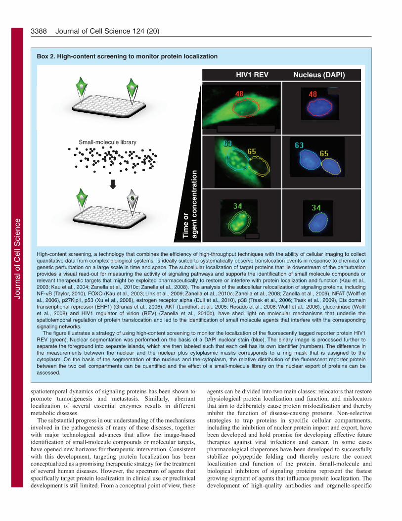
High-content screening, a technology that combines the efficiency of high-throughput techniques with the ability of cellular imaging to collectquantitative data from complex biological systems, is ideally suited to systematically observe translocation events in response to chemical orgenetic perturbation on a large scale in time and space. The subcellular localization of target proteins that lie downstream of the perturbationprovides a visual read-out for measuring the activity of signaling pathways and supports the identification of small molecule compounds orrelevant therapeutic targets that might be exploited pharmaceutically to restore or interfere with protein localization and function (Kau et al.,2003; Kau et al., 2004; Zanella et al., 2010c; Zanella et al., 2008). The analysis of the subcellular relocalization of signaling proteins, includingNF-B (Taylor, 2010), FOXO (Kau et al., 2003; Link et al., 2009; Zanella et al., 2010c; Zanella et al., 2008; Zanella et al., 2009), NFAT (Wolff etal., 2006), p27Kip1, p53 (Xu et al., 2008), estrogen receptor alpha (Dull et al., 2010), p38 (Trask et al., 2006; Trask et al., 2009), Ets domaintranscriptional repressor (ERF1) (Granas et al., 2006), AKT (Lundholt et al., 2005; Rosado et al., 2008; Wolff et al., 2006), glucokinase (Wolffet al., 2008) and HIV1 regulator of virion (REV) (Zanella et al., 2010b), have shed light on molecular mechanisms that underlie thespatiotemporal regulation of protein translocation and led to the identification of small molecule agents that interfere with the correspondingsignaling networks.
The figure illustrates a strategy of using high-content screening to monitor the localization of the fluorescently tagged reporter protein HIV1REV (green). Nuclear segmentation was performed on the basis of a DAPI nuclear stain (blue). The binary image is processed further toseparate the foreground into separate islands, which are then labeled such that each cell has its own identifier (numbers). The difference inthe measurements between the nuclear and the nuclear plus cytoplasmic masks corresponds to a ring mask that is assigned to thecytoplasm. On the basis of the segmentation of the nucleus and the cytoplasm, the relative distribution of the fluorescent reporter proteinbetween the two cell compartments can be quantified and the effect of a small-molecule library on the nuclear export of proteins can beassessed.
Tim
e o
rag
ent
con
cen
trat
ion
HIV1 REV
Small-molecule library
Ti
Nucleus (DAPI)
Jour
nal o
f Cel
l Sci
ence
fluorescent dyes will facilitate the characterization of pathologicalprotein localization and could be used as predictive and diagnosticbiomarkers for many human diseases. As cancer is increasinglyperceived as a disease of pathway lesions associated with thereversible mislocalization of essential signaling proteins, thisstrategy is expected to have its most immediate impact on thedevelopment of new anticancer therapies.
AcknowledgementsWe thank J. F. Martinez for helpful discussions and critical reading ofthis paper.
FundingThe work of our laboratory was supported by a grant from the SpanishMEC [project BIO2006-02432].
ReferencesAnderson, K. E., Coadwell, J., Stephens, L. R. and Hawkins, P. T. (1998). Translocation
of PDK-1 to the plasma membrane is important in allowing PDK-1 to activate proteinkinase B. Curr. Biol. 8, 684-691.
Asada, M., Yamada, T., Ichijo, H., Delia, D., Miyazono, K., Fukumuro, K. andMizutani, S. (1999). Apoptosis inhibitory activity of cytoplasmic p21(Cip1/WAF1) inmonocytic differentiation. EMBO J. 18, 1223-1234.
Barmada, S. J., Skibinski, G., Korb, E., Rao, E. J., Wu, J. Y. and Finkbeiner, S.(2010). Cytoplasmic mislocalization of TDP-43 is toxic to neurons and enhanced by amutation associated with familial amyotrophic lateral sclerosis. J. Neurosci. 30, 639-649.
Basel-Vanagaite, L., Muncher, L., Straussberg, R., Pasmanik-Chor, M., Yahav, M.,Rainshtein, L., Walsh, C. A., Magal, N., Taub, E., Drasinover, V. et al. (2006).Mutated nup62 causes autosomal recessive infantile bilateral striatal necrosis. Ann.Neurol. 60, 214-222.
Bjorses, P., Halonen, M., Palvimo, J. J., Kolmer, M., Aaltonen, J., Ellonen, P.,Perheentupa, J., Ulmanen, I. and Peltonen, L. (2000). Mutations in the AIRE gene:effects on subcellular location and transactivation function of the autoimmunepolyendocrinopathy-candidiasis-ectodermal dystrophy protein. Am. J. Hum. Genet. 66,378-392.
Braverman, N., Steel, G., Obie, C., Moser, A., Moser, H., Gould, S. J. and Valle, D.(1997). Human PEX7 encodes the peroxisomal PTS2 receptor and is responsible forrhizomelic chondrodysplasia punctata. Nat. Genet. 15, 369-376.
Brothers, S. P., Cornea, A., Janovick, J. A. and Conn, P. M. (2004). Human loss-of-function gonadotropin-releasing hormone receptor mutants retain wild-type receptors inthe endoplasmic reticulum: molecular basis of the dominant-negative effect. Mol.Endocrinol. 18, 1787-1797.
Brunet, A., Bonni, A., Zigmond, M. J., Lin, M. Z., Juo, P., Hu, L. S., Anderson, M.J., Arden, K. C., Blenis, J. and Greenberg, M. E. (1999). Akt promotes cell survivalby phosphorylating and inhibiting a Forkhead transcription factor. Cell 96, 857-868.
Butler, G. S. and Overall, C. M. (2009). Proteomic identification of multitasking proteinsin unexpected locations complicates drug targeting. Nat. Rev. Drug Discov. 8, 935-948.
Caccamo, A., Majumder, S., Deng, J. J., Bai, Y., Thornton, F. B. and Oddo, S. (2009).Rapamycin rescues TDP-43 mislocalization and the associated low molecular massneurofilament instability. J. Biol. Chem. 284, 27416-27424.
Cai, S. L., Tee, A. R., Short, J. D., Bergeron, J. M., Kim, J., Shen, J., Guo, R.,Johnson, C. L., Kiguchi, K. and Walker, C. L. (2006). Activity of TSC2 is inhibitedby AKT-mediated phosphorylation and membrane partitioning. J. Cell Biol. 173, 279-289.
Carnero, A., Blanco-Aparicio, C., Renner, O., Link, W. and Leal, J. F. (2008). ThePTEN/PI3K/AKT signalling pathway in cancer, therapeutic implications. Curr. CancerDrug Targets 8, 187-198.
Carpenter, G. and Liao, H. J. (2009). Trafficking of receptor tyrosine kinases to thenucleus. Exp. Cell Res. 315, 1556-1566.
Chacinska, A., Koehler, C. M., Milenkovic, D., Lithgow, T. and Pfanner, N. (2009).Importing mitochondrial proteins: machineries and mechanisms. Cell 138, 628-644.
Chahine, M. N. and Pierce, G. N. (2009). Therapeutic targeting of nuclear protein importin pathological cell conditions. Pharmacol. Rev. 61, 358-372.
Chalmers, K. A. and Love, S. (2007). Neurofibrillary tangles may interfere with Smad2/3 signaling in neurons. J. Neuropathol. Exp. Neurol. 66, 158-167.
Chen, M. F., Fang, F. M., Lu, C. H., Lu, M. S., Chen, W. C., Lee, K. D. and Lin, P.Y. (2008). Significance of nuclear accumulation of Foxo3a in esophageal squamous cellcarcinoma. Int. J. Radiat. Oncol. Biol. Phys. 71, 1220-1229.
Cheng, X., Xia, W., Yang, J.-Y., Hsu, J. L., Chou, C.-K., Sun, H.-L., Wyszomierski, S.L., Mills, G. B., Muller, W. J., Yu, D. et al. (2010). Activation of p21(CIP1/WAF1) inmammary epithelium accelerates mammary tumorigenesis and promotes lung metastasis.Biochem. Biophys. Res. Commun. 403, 103-107.
Chu, C. T., Plowey, E. D., Wang, Y., Patel, V. and Jordan-Sciutto, K. L. (2007).Location, location, location: altered transcription factor trafficking in neurodegeneration.J. Neuropathol. Exp. Neurol. 66, 873-883.
Conn, P. M., Ulloa-Aguirre, A., Ito, J. and Janovick, J. A. (2007). G protein-coupledreceptor trafficking in health and disease: lessons learned to prepare for therapeuticmutant rescue in vivo. Pharmacol. Rev. 59, 225-250.
Cordeddu, V., Di Schiavi, E., Pennacchio, L. A., Ma’ayan, A., Sarkozy, A., Fodale, V.,Cecchetti, S., Cardinale, A., Martin, J., Schackwitz, W. et al. (2009). Mutation ofSHOC2 promotes aberrant protein N-myristoylation and causes Noonan-like syndromewith loose anagen hair. Nat. Genet. 41, 1022-1026.
Da Costa, L., Narla, G., Willig, T. N., Peters, L. L., Parra, M., Fixler, J., Tchernia, G.and Mohandas, N. (2003a). Ribosomal protein S19 expression during erythroiddifferentiation. Blood 101, 318-324.
Da Costa, L., Tchernia, G., Gascard, P., Lo, A., Meerpohl, J., Niemeyer, C., Chasis,J. A., Fixler, J. and Mohandas, N. (2003b). Nucleolar localization of RPS19 proteinin normal cells and mislocalization due to mutations in the nucleolar localization signalsin 2 Diamond-Blackfan anemia patients: potential insights into pathophysiology. Blood101, 5039-5045.
Danpure, C. J. (2006). Primary hyperoxaluria type 1, AGT mistargeting highlights thefundamental differences between the peroxisomal and mitochondrial protein importpathways. Biochim. Biophys. Acta 1763, 1776-1784.
Dansen, T. B. and Burgering, B. M. (2008). Unravelling the tumor-suppressive functionsof FOXO proteins. Trends Cell Biol. 18, 421-429.
Davis, J. R., Kakar, M. and Lim, C. S. (2007). Controlling protein compartmentalizationto overcome disease. Pharm. Res. 24, 17-27.
Dekker, F. J., Rocks, O., Vartak, N., Menninger, S., Hedberg, C., Balamurugan, R.,Wetzel, S., Renner, S., Gerauer, M., Scholermann, B. et al. (2010). Small-moleculeinhibition of APT1 affects Ras localization and signaling. Nat. Chem. Biol. 6, 449-456.
Dijkers, P. F., Medema, R. H., Pals, C., Banerji, L., Thomas, N. S., Lam, E. W.,Burgering, B. M., Raaijmakers, J. A., Lammers, J. W., Koenderman, L. et al.(2000). Forkhead transcription factor FKHR-L1 modulates cytokine-dependenttranscriptional regulation of p27(KIP1). Mol. Cell. Biol. 20, 9138-9148.
Djordjevic, S., Zhang, X., Bartlam, M., Ye, S., Rao, Z. and Danpure, C. J. (2010).Structural implications of a G170R mutation of alanine:glyoxylate aminotransferasethat is associated with peroxisome-to-mitochondrion mistargeting. Acta Crystallogr.Sect. F Struct. Biol. Cryst. Commun. 66, 233-236.
Dreuillet, C., Harper, M., Tillit, J., Kress, M. and Ernoult-Lange, M. (2008).Mislocalization of human transcription factor MOK2 in the presence of pathogenicmutations of lamin A/C. Biol. Cell 100, 51-61.
Dull, A., Goncharova, E., Hager, G. and McMahon, J. B. (2010). Development of animage analysis screen for estrogen receptor alpha (ERalpha) ligands throughmeasurement of nuclear translocation dynamics. J. Steroid Biochem. Mol. Biol. 122,341-351.
Emanuelsson, O., Brunak, S., von Heijne, G. and Nielsen, H. (2007). Locating proteinsin the cell using TargetP, SignalP and related tools. Nat. Protoc. 2, 953-971.
Fabbro, M. and Henderson, B. R. (2003). Regulation of tumor suppressors by nuclear-cytoplasmic shuttling. Exp. Cell Res. 282, 59-69.
Faustino, R. S., Nelson, T. J., Terzic, A. and Perez-Terzic, C. (2007). Nuclear transport:target for therapy. Clin. Pharmacol. Ther. 81, 880-886.
Finch, A. R., Caunt, C. J., Armstrong, S. P. and McArdle, C. A. (2010). Plasmamembrane expression of gonadotropin-releasing hormone receptors: regulation bypeptide and nonpeptide antagonists. Mol. Endocrinol. 24, 423-435.
Frescas, D., Valenti, L. and Accili, D. (2005). Nuclear trapping of the forkheadtranscription factor FoxO1 via Sirt-dependent deacetylation promotes expression ofglucogenetic genes. J. Biol. Chem. 280, 20589-20595.
Gasiorowski, J. Z. and Dean, D. A. (2003). Mechanisms of nuclear transport andinterventions. Adv. Drug Deliv. Rev. 55, 703-716.
Gehret, A. U., Bajaj, A., Naider, F. and Dumont, M. E. (2006). Oligomerization of theyeast alpha-factor receptor: implications for dominant negative effects of mutantreceptors. J. Biol. Chem. 281, 20698-20714.
Gilley, J., Coffer, P. J. and Ham, J. (2003). FOXO transcription factors directly activatebim gene expression and promote apoptosis in sympathetic neurons. J. Cell Biol. 162,613-622.
Gomes, D. A., Rodrigues, M. A., Leite, M. F., Gomez, M. V., Varnai, P., Balla, T.,Bennett, A. M. and Nathanson, M. H. (2008). c-Met must translocate to the nucleusto initiate calcium signals. J. Biol. Chem. 283, 4344-4351.
Granas, C., Lundholt, B. K., Loechel, F., Pedersen, H. C., Bjorn, S. P., Linde, V.,Krogh-Jensen, C., Nielsen, E. M., Praestegaard, M. and Nielsen, S. J. (2006).Identification of RAS-mitogen-activated protein kinase signaling pathway modulatorsin an ERF1 redistribution screen. J. Biomol. Screen. 11, 423-434.
Hadzisejdic, I., Mustac, E., Jonjic, N., Petkovic, M. and Grahovac, B. (2010). NuclearEGFR in ductal invasive breast cancer: correlation with cyclin-D1 and prognosis. Mod.Pathol. 23, 392-403.
Hamamoto, T., Seto, H. and Beppu, T. (1983). Leptomycins A and B, new antifungalantibiotics. II. Structure elucidation. J. Antibiot. 36, 646-650.
Hanada, N., Lo, H. W., Day, C. P., Pan, Y., Nakajima, Y. and Hung, M. C. (2006). Co-regulation of B-Myb expression by E2F1 and EGF receptor. Mol. Carcinog. 45, 10-17.
Hayden, M. S. and Ghosh, S. (2008). Shared principles in NF-kappaB signaling. Cell132, 344-362.
Hennessy, B. T., Smith, D. L., Ram, P. T., Lu, Y. and Mills, G. B. (2005). Exploitingthe PI3K/AKT pathway for cancer drug discovery. Nat. Rev. Drug Discov. 4, 988-1004.
Hoover, B. R., Reed, M. N., Su, J., Penrod, R. D., Kotilinek, L. A., Grant, M. K.,Pitstick, R., Carlson, G. A., Lanier, L. M., Yuan, L. L. et al. (2010). Taumislocalization to dendritic spines mediates synaptic dysfunction independently ofneurodegeneration. Neuron 68, 1067-1081.
Hoshino, M., Fukui, H., Ono, Y., Sekikawa, A., Ichikawa, K., Tomita, S., Imai, Y.,Imura, J., Hiraishi, H. and Fujimori, T. (2007). Nuclear expression of phosphorylatedEGFR is associated with poor prognosis of patients with esophageal squamous cellcarcinoma. Pathobiology 74, 15-21.
3389Disease-relevant protein mislocalization
Jour
nal o
f Cel
l Sci
ence
Hsu, S., Miller, S., Wang, Y. and Hung, M. (2009). Nuclear EGFR is required forcisplatin resistance and DNA repair. Am. J. Transl. Res. 1, 249-258.
Hu, M. C., Lee, D. F., Xia, W., Golfman, L. S., Ou-Yang, F., Yang, J. Y., Zou, Y., Bao,S., Hanada, N., Saso, H. et al. (2004). IkappaB kinase promotes tumorigenesis throughinhibition of forkhead FOXO3a. Cell 117, 225-237.
Huang, S., Shu, L., Dilling, M. B., Easton, J., Harwood, F. C., Ichijo, H. and Houghton,P. J. (2003). Sustained activation of the JNK cascade and rapamycin-induced apoptosisare suppressed by p53/p21(Cip1). Mol. Cell 11, 1491-1501.
Hung, I. H., Suzuki, M., Yamaguchi, Y., Yuan, D. S., Klausner, R. D. and Gitlin, J. D.(1997). Biochemical characterization of the Wilson disease protein and functionalexpression in the yeast Saccharomyces cerevisiae. J. Biol. Chem. 272, 21461-21466.
Hung, L. Y., Tseng, J. T., Lee, Y. C., Xia, W., Wang, Y. N., Wu, M. L., Chuang, Y. H.,Lai, C. H. and Chang, W. C. (2008). Nuclear epidermal growth factor receptor(EGFR) interacts with signal transducer and activator of transcription 5 (STAT5) inactivating Aurora-A gene expression. Nucleic Acids Res. 36, 4337-4351.
Kaiser, F. J., Brega, P., Raff, M. L., Byers, P. H., Gallati, S., Kay, T. T., de Almeida,S., Horsthemke, B. and Ludecke, H. J. (2004). Novel missense mutations in theTRPS1 transcription factor define the nuclear localization signal. Eur. J. Hum. Genet.12, 121-126.
Kalesse, M., Christmann, M., Bhatt, U., Quitschalle, M., Claus, E., Saeed, A., Burzlaff,A., Kasper, C., Haustedt, L. O., Hofer, E. et al. (2001). The chemistry and biologyof ratjadone. Chembiochem 2, 709-714.
Karin, M. and Greten, F. R. (2005). NF-kappaB: linking inflammation and immunity tocancer development and progression. Nat. Rev. Immunol. 5, 749-759.
Karpa, K. D., Lin, R., Kabbani, N. and Levenson, R. (2000). The dopamine D3 receptorinteracts with itself and the truncated D3 splice variant d3nf: D3-D3nf interactioncauses mislocalization of D3 receptors. Mol. Pharmacol. 58, 677-683.
Katayose, D., Wersto, R., Cowan, K. H. and Seth, P. (1995). Effects of a recombinantadenovirus expressing WAF1/Cip1 on cell growth, cell cycle, and apoptosis. CellGrowth Differ. 6, 1207-1212.
Kau, T. R., Schroeder, F., Ramaswamy, S., Wojciechowski, C. L., Zhao, J. J., Roberts,T. M., Clardy, J., Sellers, W. R. and Silver, P. A. (2003). A chemical genetic screenidentifies inhibitors of regulated nuclear export of a Forkhead transcription factor inPTEN-deficient tumor cells. Cancer Cell 4, 463-476.
Kau, T. R., Way, J. C. and Silver, P. A. (2004). Nuclear transport and cancer: frommechanism to intervention. Nat. Rev. Cancer 4, 106-117.
Kim, D., Sun, M., He, L., Zhou, Q. H., Chen, J., Sun, X. M., Bepler, G., Sebti, S. M.and Cheng, J. Q. (2010). A small molecule inhibits Akt through direct binding to Aktand preventing Akt membrane translocation. J. Biol. Chem. 285, 8383-8394.
Kim, J., Jonasch, E., Alexander, A., Short, J. D., Cai, S., Wen, S., Tsavachidou, D.,Tamboli, P., Czerniak, B. A., Do, K. A. et al. (2009). Cytoplasmic sequestration ofp27 via AKT phosphorylation in renal cell carcinoma. Clin. Cancer Res. 15, 81-90.
Kiriyama, T., Hirano, M., Asai, H., Ikeda, M., Furiya, Y. and Ueno, S. (2008).Restoration of nuclear-import failure caused by triple A syndrome and oxidative stress.Biochem. Biophys. Res. Commun. 374, 631-634.
Kondapaka, S. B., Singh, S. S., Dasmahapatra, G. P., Sausville, E. A. and Roy, K. K.(2003). Perifosine, a novel alkylphospholipid, inhibits protein kinase B activation. Mol.Cancer Ther. 2, 1093-1103.
Koster, R., di Pietro, A., Timmer-Bosscha, H., Gibcus, J. H., van den Berg, A.,Suurmeijer, A. J., Bischoff, R., Gietema, J. A. and de Jong, S. (2010). Cytoplasmicp21 expression levels determine cisplatin resistance in human testicular cancer. J. Clin.Invest. 120, 3594-3605.
Kosugi, S., Hasebe, M., Entani, T., Takayama, S., Tomita, M. and Yanagawa, H.(2008). Design of peptide inhibitors for the importin alpha/beta nuclear import pathwayby activity-based profiling. Chem. Biol. 15, 940-949.
Kudo, N., Matsumori, N., Taoka, H., Fujiwara, D., Schreiner, E. P., Wolff, B., Yoshida,M. and Horinouchi, S. (1999). Leptomycin B inactivates CRM1/exportin 1 by covalentmodification at a cysteine residue in the central conserved region. Proc. Natl. Acad. Sci.USA 96, 9112-9117.
Kwiatkowski, T. J., Jr, Bosco, D. A., Leclerc, A. L., Tamrazian, E., Vanderburg, C.R., Russ, C., Davis, A., Gilchrist, J., Kasarskis, E. J., Munsat, T. et al. (2009).Mutations in the FUS/TLS gene on chromosome 16 cause familial amyotrophic lateralsclerosis. Science 323, 1205-1208.
Lagier-Tourenne, C. and Cleveland, D. W. (2009). Rethinking ALS: the FUS aboutTDP-43. Cell 136, 1001-1004.
Laurila, K. and Vihinen, M. (2009). Prediction of disease-related mutations affectingprotein localization. BMC Genomics 10, 122.
Li, C., Iida, M., Dunn, E., Ghia, A. and Wheeler, D. (2009). Nuclear EGFR contributesto acquired resistance to cetuximab. Oncogene 28, 3801-3813.
Li, Y., Dowbenko, D. and Lasky, L. A. (2002). AKT/PKB phosphorylation ofp21Cip/WAF1 enhances protein stability of p21Cip/WAF1 and promotes cell survival.J. Biol. Chem. 277, 11352-11361.
Liang, J., Zubovitz, J., Petrocelli, T., Kotchetkov, R., Connor, M. K., Han, K., Lee, J.-H., Ciarallo, S., Catzavelos, C., Beniston, R. et al. (2002). PKB/Akt phosphorylatesp27, impairs nuclear import of p27 and opposes p27-mediated G1 arrest. Nat. Med. 8,1153-1160.
Lin, S. Y., Makino, K., Xia, W., Matin, A., Wen, Y., Kwong, K. Y., Bourguignon, L.and Hung, M. C. (2001). Nuclear localization of EGF receptor and its potential newrole as a transcription factor. Nat. Cell Biol. 3, 802-808.
Link, W., Oyarzabal, J., Serelde, B. G., Albarran, M. I., Rabal, O., Cebria, A.,Alfonso, P., Fominaya, J., Renner, O., Peregrina, S. et al. (2009). Chemicalinterrogation of FOXO3a nuclear translocation identifies potent and selective inhibitorsof phosphoinositide 3-kinases. J. Biol. Chem. 284, 28392-28400.
Lo, H., Xia, W., Wei, Y., Ali-Seyed, M., Huang, S. and Hung, M. (2005a). Novelprognostic value of nuclear epidermal growth factor receptor in breast cancer. CancerRes. 65, 338-348.
Lo, H. W., Hsu, S. C., Ali-Seyed, M., Gunduz, M., Xia, W., Wei, Y., Bartholomeusz,G., Shih, J. Y. and Hung, M. C. (2005b). Nuclear interaction of EGFR and STAT3 inthe activation of the iNOS/NO pathway. Cancer Cell 7, 575-589.
Lundholt, B. K., Linde, V., Loechel, F., Pedersen, H. C., Moller, S., Praestegaard, M.,Mikkelsen, I., Scudder, K., Bjorn, S. P., Heide, M. et al. (2005). Identification of Aktpathway inhibitors using redistribution screening on the FLIPR and the IN Cell 3000analyzer. J. Biomol. Screen. 10, 20-29.
Luo, J. L., Kamata, H. and Karin, M. (2005). IKK/NF-kappaB signaling: balancing lifeand death-a new approach to cancer therapy. J. Clin. Invest. 115, 2625-2632.
Martinelli, G., Iacobucci, I., Paolini, S. and Ottaviani, E. (2008). Farnesyltransferaseinhibition in hematologic malignancies: the clinical experience with tipifarnib. Clin.Adv. Hematol. Oncol. 6, 303-310.
Mayo, L. D. and Donner, D. B. (2001). A phosphatidylinositol 3-kinase/Akt pathwaypromotes translocation of Mdm2 from the cytoplasm to the nucleus. Proc. Natl. Acad.Sci. USA 98, 11598-11603.
McLane, L. M. and Corbett, A. H. (2009). Nuclear localization signals and humandisease. IUBMB Life 61, 697-706.
Meissner, T., Krause, E. and Vinkemeier, U. (2004). Ratjadone and leptomycin B blockCRM1-dependent nuclear export by identical mechanisms. FEBS Lett. 576, 27-30.
Mekhail, K., Rivero-Lopez, L., Al-Masri, A., Brandon, C., Khacho, M. and Lee, S.(2007). Identification of a common subnuclear localization signal. Mol. Biol. Cell 18,3966-3977.
Mendes, H. F., van der Spuy, J., Chapple, J. P. and Cheetham, M. E. (2005).Mechanisms of cell death in rhodopsin retinitis pigmentosa: implications for therapy.Trends Mol. Med. 11, 177-185.
Miao, B., Skidan, I., Yang, J., Lugovskoy, A., Reibarkh, M., Long, K., Brazell, T.,Durugkar, K. A., Maki, J., Ramana, C. V. et al. (2010). Small molecule inhibition ofphosphatidylinositol-3,4,5-triphosphate (PIP3) binding to pleckstrin homology domains.Proc. Natl. Acad. Sci. USA 107, 20126-20131.
Mizutani, A., Matsuzaki, A., Momoi, M. Y., Fujita, E., Tanabe, Y. and Momoi, T.(2007). Intracellular distribution of a speech/language disorder associated FOXP2mutant. Biochem. Biophys. Res. Commun. 353, 869-874.
Morello, J. P., Petaja-Repo, U. E., Bichet, D. G. and Bouvier, M. (2000). Pharmacologicalchaperones: a new twist on receptor folding. Trends Pharmacol. Sci. 21, 466-469.
Mutka, S. C., Yang, W. Q., Dong, S. D., Ward, S. L., Craig, D. A., Timmermans, P. B.and Murli, S. (2009). Identification of nuclear export inhibitors with potent anticanceractivity in vivo. Cancer Res. 69, 510-517.
Nair, R. and Rost, B. (2008). Protein subcellular localization prediction using artificialintelligence technology. Methods Mol. Biol. 484, 435-463.
Nan, K.-J., Jing, Z. and Gong, L. (2004). Expression and altered subcellular localizationof the cyclin-dependent kinase inhibitor p27Kip1 in hepatocellular carcinoma. World J.Gastroenterol. 10, 1425-1430.
Neumann, M., Sampathu, D. M., Kwong, L. K., Truax, A. C., Micsenyi, M. C., Chou,T. T., Bruce, J., Schuck, T., Grossman, M., Clark, C. M. et al. (2006). UbiquitinatedTDP-43 in frontotemporal lobar degeneration and amyotrophic lateral sclerosis. Science314, 130-133.
Newlands, E. S., Rustin, G. J. and Brampton, M. H. (1996). Phase I trial of elactocin.Br. J. Cancer 74, 648-649.
Oddo, S., Vasilevko, V., Caccamo, A., Kitazawa, M., Cribbs, D. H. and LaFerla, F. M.(2006). Reduction of soluble Abeta and tau, but not soluble Abeta alone, amelioratescognitive decline in transgenic mice with plaques and tangles. J. Biol. Chem. 281,39413-39423.
Ogino, S., Shima, K., Nosho, K., Irahara, N., Baba, Y., Wolpin, B. M., Giovannucci,E. L., Meyerhardt, J. A. and Fuchs, C. S. (2009). A cohort study of p27 localizationin colon cancer, body mass index, and patient survival. Cancer Epidemiol. BiomarkersPrev. 18, 1849-1858.
Ohgane, K., Dodo, K. and Hashimoto, Y. (2010). Retinobenzaldehydes as proper-trafficking inducers of folding-defective P23H rhodopsin mutant responsible for retinitispigmentosa. Bioorg. Med. Chem. 18, 7022-7028.
Olsen, L. S., Hjarnaa, P. J., Latini, S., Holm, P. K., Larsson, R., Bramm, E., Binderup,L. and Madsen, M. W. (2004). Anticancer agent CHS 828 suppresses nuclear factor-kappa B activity in cancer cells through downregulation of IKK activity. Int. J. Cancer111, 198-205.
Payne, A. S., Kelly, E. J. and Gitlin, J. D. (1998). Functional expression of the Wilsondisease protein reveals mislocalization and impaired copper-dependent trafficking ofthe common H1069Q mutation. Proc. Natl. Acad. Sci. USA 95, 10854-10859.
Penel, N., Van Glabbeke, M., Marreaud, S., Ouali, M., Blay, J. Y. and Hohenberger,P. (2010). Testing new regimens in patients with advanced soft tissue sarcoma: analysisof publications from the last 10 years. Ann. Oncol. 22, 1266-1272.
Pérez-Tenorio, G., Berglund, F., Esguerra Merca, A., Nordenskjöld, B., Rutqvist, L.E., Skoog, L. and Stål, O. (2006). Cytoplasmic p21WAF1/CIP1 correlates with Aktactivation and poor response to tamoxifen in breast cancer. Int. J. Oncol. 28, 1031-1042.
Pey, A. L., Perez, B., Desviat, L. R., Martinez, M. A., Aguado, C., Erlandsen, H.,Gamez, A., Stevens, R. C., Thorolfsson, M., Ugarte, M. et al. (2004). Mechanismsunderlying responsiveness to tetrahydrobiopterin in mild phenylketonuria mutations.Hum. Mutat. 24, 388-399.
Psyrri, A., Egleston, B., Weinberger, P., Yu, Z., Kowalski, D., Sasaki, C., Haffty, B.,Rimm, D. and Burtness, B. (2008). Correlates and determinants of nuclear epidermalgrowth factor receptor content in an oropharyngeal cancer tissue microarray. CancerEpidemiol. Biomarkers Prev. 17, 1486-1492.
3390 Journal of Cell Science 124 (20)
Jour
nal o
f Cel
l Sci
ence

Purdue, P. E., Allsop, J., Isaya, G., Rosenberg, L. E. and Danpure, C. J. (1991).Mistargeting of peroxisomal L-alanine:glyoxylate aminotransferase to mitochondria inprimary hyperoxaluria patients depends upon activation of a cryptic mitochondrialtargeting sequence by a point mutation. Proc. Natl. Acad. Sci. USA 88, 10900-10904.
Rapoport, T. A. (2007). Protein translocation across the eukaryotic endoplasmic reticulumand bacterial plasma membranes. Nature 450, 663-669.
Real, P. J., Tosello, V., Palomero, T., Castillo, M., Hernando, E., de Stanchina, E.,Sulis, M. L., Barnes, K., Sawai, C., Homminga, I. et al. (2009). Gamma-secretaseinhibitors reverse glucocorticoid resistance in T cell acute lymphoblastic leukemia. Nat.Med. 15, 50-58.
Reilly, J. F. and Maher, P. A. (2001). Importin beta-mediated nuclear import of fibroblastgrowth factor receptor: role in cell proliferation. J. Cell Biol. 152, 1307-1312.
Reindl, W., Yuan, J., Kramer, A., Strebhardt, K. and Berg, T. (2008). Inhibition ofpolo-like kinase 1 by blocking polo-box domain-dependent protein-protein interactions.Chem. Biol. 15, 459-466.
Rico-Bautista, E., Yang, C. C., Lu, L., Roth, G. P. and Wolf, D. A. (2010). Chemicalgenetics approach to restoring p27Kip1 reveals novel compounds with antiproliferativeactivity in prostate cancer cells. BMC Biol. 8, 153.
Ringe, D. and Petsko, G. A. (2009). What are pharmacological chaperones and why arethey interesting? J. Biol. 8, 80.
Robben, J. H., Knoers, N. V. and Deen, P. M. (2006). Cell biological aspects of thevasopressin type-2 receptor and aquaporin 2 water channel in nephrogenic diabetesinsipidus. Am. J. Physiol. Renal Physiol. 291, F257-F270.
Rodier, G., Montagnoli, A., Di Marcotullio, L., Coulombe, P., Draetta, G. F., Pagano,M. and Meloche, S. (2001). p27 cytoplasmic localization is regulated by phosphorylationon Ser10 and is not a prerequisite for its proteolysis. EMBO J. 20, 6672-6682.
Roehrl, M. H., Kang, S., Aramburu, J., Wagner, G., Rao, A. and Hogan, P. G. (2004).Selective inhibition of calcineurin-NFAT signaling by blocking protein-protein interactionwith small organic molecules. Proc. Natl. Acad. Sci. USA 101, 7554-7559.
Rosado, A., Zanella, F., Garcia, B., Carnero, A. and Link, W. (2008). A dual-colorfluorescence-based platform to identify selective inhibitors of Akt signaling. PLoS ONE3, e1823.
Rosen, D. G., Yang, G., Cai, K. Q., Bast, R. C., Gershenson, D. M., Silva, E. G. andLiu, J. (2005). Subcellular localization of p27kip1 expression predicts poor prognosisin human ovarian cancer. Clin. Cancer Res. 11, 632-637.
Rossig, L., Jadidi, A. S., Urbich, C., Badorff, C., Zeiher, A. M. and Dimmeler, S.(2001). Akt-dependent phosphorylation of p21(Cip1) regulates PCNA binding andproliferation of endothelial cells. Mol. Cell. Biol. 21, 5644-5657.
Ruan, S., Okcu, M. F., Pong, R. C., Andreeff, M., Levin, V., Hsieh, J. T. and Zhang,W. (1999). Attenuation of WAF1/Cip1 expression by an antisense adenovirus expressionvector sensitizes glioblastoma cells to apoptosis induced by chemotherapeutic agents1,3-bis(2-chloroethyl)-1-nitrosourea and cisplatin. Clin. Cancer Res. 5, 197-202.
Sabherwal, N., Schneider, K. U., Blaschke, R. J., Marchini, A. and Rappold, G.(2004). Impairment of SHOX nuclear localization as a cause for Leri-Weill syndrome.J. Cell Sci. 117, 3041-3048.
Salmena, L. and Pandolfi, P. P. (2007). Changing venues for tumour suppression:balancing destruction and localization by monoubiquitylation. Nat. Rev. Cancer 7, 409-413.
Santacruz, K., Lewis, J., Spires, T., Paulson, J., Kotilinek, L., Ingelsson, M.,Guimaraes, A., DeTure, M., Ramsden, M., McGowan, E. et al. (2005). Tausuppression in a neurodegenerative mouse model improves memory function. Science309, 476-481.
Scott, J. D. and Pawson, T. (2009). Cell signaling in space and time: where proteins cometogether and when they’re apart. Science 326, 1220-1224.
Schmidt, O., Pfanner, N. and Meisinger, C. (2010). Mitochondrial protein import: fromproteomics to functional mechanisms. Nat. Rev. Mol. Cell Biol. 11, 655-667.
Schnell, D. J. and Hebert, D. N. (2003). Protein translocons: multifunctional mediatorsof protein translocation across membranes. Cell 112, 491-505.
Sehat, B., Tofigh, A., Lin, Y., Trocme, E., Liljedahl, U., Lagergren, J. and Larsson, O.(2010). SUMOylation mediates the nuclear translocation and signaling of the IGF-1receptor. Sci. Signal. 3, ra10.
Shin, I., Yakes, F. M., Rojo, F., Shin, N.-Y., Bakin, A. V., Baselga, J. and Arteaga, C.L. (2002). PKB/Akt mediates cell-cycle progression by phosphorylation of p27(Kip1)at threonine 157 and modulation of its cellular localization. Nat. Med. 8, 1145-1152.
Shoubridge, C., Tan, M. H., Fullston, T., Cloosterman, D., Coman, D., McGillivray,G., Mancini, G. M., Kleefstra, T. and Gecz, J. (2010). Mutations in the nuclearlocalization sequence of the Aristaless related homeobox; sequestration of mutant ARXwith IPO13 disrupts normal subcellular distribution of the transcription factor andretards cell division. Pathogenetics 3, 1.
Singh, S. P., Lipman, J., Goldman, H., Ellis, F. H., Aizenman, L., Cangi, M. G.,Signoretti, S., Chiaur, D. S., Pagano, M. and Loda, M. (1998). Loss or alteredsubcellular localization of p27 in Barrett’s associated adenocarcinoma. Cancer Res. 58,1730-1735.
Skach, W. R. (2000). Defects in processing and trafficking of the cystic fibrosistransmembrane conductance regulator. Kidney Int. 57, 825-831.
Snow, C. M., Senior, A. and Gerace, L. (1987). Monoclonal antibodies identify a groupof nuclear pore complex glycoproteins. J. Cell Biol. 104, 1143-1156.
Sousa, S. F., Fernandes, P. A. and Ramos, M. J. (2008). Farnesyltransferase inhibitors:a detailed chemical view on an elusive biological problem. Curr. Med. Chem. 15, 1478-1492.
Stachowiak, E. K., Maher, P. A., Tucholski, J., Mordechai, E., Joy, A., Moffett, J.,Coons, S. and Stachowiak, M. K. (1997). Nuclear accumulation of fibroblast growthfactor receptors in human glial cells-association with cell proliferation. Oncogene 14,2201-2211.
Strambio-De-Castillia, C., Niepel, M. and Rout, M. P. (2010). The nuclear pore complex:bridging nuclear transport and gene regulation. Nat. Rev. Mol. Cell Biol. 11, 490-501.
Suntharalingam, M. and Wente, S. R. (2003). Peering through the pore: nuclear porecomplex structure, assembly, and function. Dev. Cell 4, 775-789.
Tanaka, A. R., Abe-Dohmae, S., Ohnishi, T., Aoki, R., Morinaga, G., Okuhira, K.,Ikeda, Y., Kano, F., Matsuo, M., Kioka, N. et al. (2003). Effects of mutations ofABCA1 in the first extracellular domain on subcellular trafficking and ATPbinding/hydrolysis. J. Biol. Chem. 278, 8815-8819.
Taylor, D. L. (2010). A personal perspective on high-content screening (HCS): from thebeginning. J. Biomol. Screen. 15, 720-725.
Terry, L. J., Shows, E. B. and Wente, S. R. (2007). Crossing the nuclear envelope:hierarchical regulation of nucleocytoplasmic transport. Science 318, 1412-1416.
Trask, O. J., Jr, Baker, A., Williams, R. G., Nickischer, D., Kandasamy, R., Laethem,C., Johnston, P. A. and Johnston, P. A. (2006). Assay development and case historyof a 32K-biased library high-content MK2-EGFP translocation screen to identify p38mitogen-activated protein kinase inhibitors on the ArrayScan 3.1 imaging platform.Methods Enzymol. 414, 419-439.
Trask, O. J., Nickischer, D., Burton, A., Williams, R. G., Kandasamy, R. A. andJohnston, P. A. (2009). High-throughput automated confocal microscopy imagingscreen of a kinase-focused library to identify p38 mitogen-activated protein kinaseinhibitors using the GE InCell 3000 analyzer. Methods Mol. Biol. 565, 159-186.
Turner, J. G. and Sullivan, D. M. (2008). CRM1-mediated nuclear export of proteins anddrug resistance in cancer. Curr. Med. Chem. 15, 2648-2655.
Vance, C., Rogelj, B., Hortobagyi, T., De Vos, K. J., Nishimura, A. L., Sreedharan, J.,Hu, X., Smith, B., Ruddy, D., Wright, P. et al. (2009). Mutations in FUS, an RNAprocessing protein, cause familial amyotrophic lateral sclerosis type 6. Science 323,1208-1211.
Viglietto, G., Motti, M. L., Bruni, P., Melillo, R. M., D’Alessio, A., Califano, D., Vinci,F., Chiappetta, G., Tsichlis, P., Bellacosa, A. et al. (2002). Cytoplasmic relocalizationand inhibition of the cyclin-dependent kinase inhibitor p27(Kip1) by PKB/Akt-mediatedphosphorylation in breast cancer. Nat. Med. 8, 1136-1144.
Wang, S. C. and Hung, M. C. (2005). Cytoplasmic/nuclear shuttling and tumorprogression. Ann. N. Y. Acad. Sci. 1059, 11-15.
Wang, S. C. and Hung, M. C. (2009). Nuclear translocation of the epidermal growthfactor receptor family membrane tyrosine kinase receptors. Clin. Cancer Res. 15, 6484-6489.
Wang, S. C., Lien, H. C., Xia, W., Chen, I. F., Lo, H. W., Wang, Z., Ali-Seyed, M., Lee,D. F., Bartholomeusz, G., Ou-Yang, F. et al. (2004). Binding at and transactivation ofthe COX-2 promoter by nuclear tyrosine kinase receptor ErbB-2. Cancer Cell 6, 251-261.
Wang, Y. N., Wang, H., Yamaguchi, H., Lee, H. J., Lee, H. H. and Hung, M. C.(2010a). COPI-mediated retrograde trafficking from the Golgi to the ER regulatesEGFR nuclear transport. Biochem. Biophys. Res. Commun. 399, 498-504.
Wang, Y. N., Yamaguchi, H., Huo, L., Du, Y., Lee, H. J., Lee, H. H., Wang, H., Hsu,J. M. and Hung, M. C. (2010b). The translocon Sec61beta localized in the innernuclear membrane transports membrane-embedded EGF receptor to the nucleus. J.Biol. Chem. 285, 38720-38729.
Wickner, W. and Schekman, R. (2005). Protein translocation across biological membranes.Science 310, 1452-1456.
Winton, M. J., Igaz, L. M., Wong, M. M., Kwong, L. K., Trojanowski, J. Q. and Lee,V. M. (2008). Disturbance of nuclear and cytoplasmic TAR DNA-binding protein(TDP-43) induces disease-like redistribution, sequestration, and aggregate formation. J.Biol. Chem. 283, 13302-13309.
Wolf, J., Schliebs, W. and Erdmann, R. (2010). Peroxisomes as dynamic organelles:peroxisomal matrix protein import. FEBS J. 277, 3268-3278.
Wolff, B., Sanglier, J. J. and Wang, Y. (1997). Leptomycin B is an inhibitor of nuclearexport: inhibition of nucleo-cytoplasmic translocation of the human immunodeficiencyvirus type 1 (HIV-1) Rev protein and Rev-dependent mRNA. Chem. Biol. 4, 139-147.
Wolff, M., Haasen, D., Merk, S., Kroner, M., Maier, U., Bordel, S., Wiedenmann, J.,Nienhaus, G. U., Valler, M. and Heilker, R. (2006). Automated high content screeningfor phosphoinositide 3 kinase inhibition using an AKT 1 redistribution assay. Comb.Chem. High Throughput Screen. 9, 339-350.
Wolff, M., Kauschke, S. G., Schmidt, S. and Heilker, R. (2008). Activation andtranslocation of glucokinase in rat primary hepatocytes monitored by high contentimage analysis. J. Biomol. Screen. 13, 837-846.
Xia, W., Chen, J., Zhou, X., Sun, P., Lee, D., Liao, Y., Zhou, B. and Hung, M. (2004).Phosphorylation/cytoplasmic localization of p21Cip1/WAF1 is associated withHER2/neu overexpression and provides a novel combination predictor for poor prognosisin breast cancer patients. Clin. Cancer Res. 10, 3815-3824.
Xia, W., Wei, Y., Du, Y., Liu, J., Chang, B., Yu, Y., Huo, L., Miller, S. and Hung, M.(2009). Nuclear expression of epidermal growth factor receptor is a novel prognosticvalue in patients with ovarian cancer. Mol. Carcinog. 48, 610-617.
Xu, G. W., Mawji, I. A., Macrae, C. J., Koch, C. A., Datti, A., Wrana, J. L., Dennis,J. W. and Schimmer, A. D. (2008). A high-content chemical screen identifies ellipticineas a modulator of p53 nuclear localization. Apoptosis 13, 413-422.
Yang, J. Y., Zong, C. S., Xia, W., Yamaguchi, H., Ding, Q., Xie, X., Lang, J. Y., Lai,C. C., Chang, C. J., Huang, W. C. et al. (2008). ERK promotes tumorigenesis byinhibiting FOXO3a via MDM2-mediated degradation. Nat. Cell Biol. 10, 138-148.
Yang, Z. Y., Perkins, N. D., Ohno, T., Nabel, E. G. and Nabel, G. J. (1995). The p21cyclin-dependent kinase inhibitor suppresses tumorigenicity in vivo. Nat. Med. 1, 1052-1056.
Yashiroda, Y. and Yoshida, M. (2003). Nucleo-cytoplasmic transport of proteins as atarget for therapeutic drugs. Curr. Med. Chem. 10, 741-748.
3391Disease-relevant protein mislocalization
Jour
nal o
f Cel
l Sci
ence

Zanella, F., Rosado, A., Garcia, B., Carnero, A. and Link, W. (2008). Chemical geneticanalysis of FOXO nuclear-cytoplasmic shuttling by using image-based cell screening.Chembiochem 9, 2229-2237.
Zanella, F., Rosado, A., Garcia, B., Carnero, A. and Link, W. (2009). Using multiplexedregulation of luciferase activity and GFP translocation to screen for FOXO modulators.BMC Cell Biol. 10, 14.
Zanella, F., Link, W. and Carnero, A. (2010a). Understanding FOXO, new views on oldtranscription factors. Curr. Cancer Drug Targets 10, 135-146.
Zanella, F., Lorens, J. B. and Link, W. (2010b). High content screening: seeing isbelieving. Trends Biotechnol. 28, 237-245.
Zanella, F., Renner, O., Garcia, B., Callejas, S., Dopazo, A., Peregrina, S., Carnero,A. and Link, W. (2010c). Human TRIB2 is a repressor of FOXO that contributes tothe malignant phenotype of melanoma cells. Oncogene 29, 2973-2982.
Zhang, W., Kornblau, S. M., Kobayashi, T., Gambel, A., Claxton, D. and Deisseroth,A. B. (1995). High levels of constitutive WAF1/Cip1 protein are associated withchemoresistance in acute myelogenous leukemia. Clin. Cancer Res. 1, 1051-1057.
Zhang, X., Chen, S., Yoo, S., Chakrabarti, S., Zhang, T., Ke, T., Oberti, C., Yong, S.L., Fang, F., Li, L. et al. (2008). Mutation in nuclear pore component NUP155 leadsto atrial fibrillation and early sudden cardiac death. Cell 135, 1017-1027.
Zhou, B., Liao, Y., Xia, W., Spohn, B., Lee, M. and Hung, M. (2001). Cytoplasmiclocalization of p21Cip1/WAF1 by Akt-induced phosphorylation in HER-2/neu-overexpressing cells. Nat. Cell Biol. 3, 245-252.
Zhu, J. H., Kulich, S. M., Oury, T. D. and Chu, C. T. (2002). Cytoplasmic aggregatesof phosphorylated extracellular signal-regulated protein kinases in Lewy body diseases.Am. J. Pathol. 161, 2087-2098.
3392 Journal of Cell Science 124 (20)
Commentaries and Cell Science at a Glance
JCS Commentaries and Cell Science at a Glance poster articles highlight and critically discuss recent andexciting findings that will interest those who work in cell and molecular biology; Cell Science at a Glance posterarticles can also act as an introduction to an area of cell biology, and include a large, full-colour poster.
Both of these article types, designed to appeal to specialists and nonspecialists alike, are commissioned fromleaders in the field and are subject to rigorous peer review and in-house editorial appraisal. Each issue of theJournal usually contains at least one of each article type. JCS thus provides readers with more than 50 topicalpieces each year, which cover the complete spectrum of cell biology. The following are just some of the areasthat will be covered in JCS over the coming months:
Cell Science at a Glance
The haematopoietic stem cell niche at a glance Cristina Lo Celso and David ScaddenMouse pluripotent stem cells at a glance Ludovic VallierAdhesion dynamics at a glance Miguel Vicente-Manzanares and Rick HorwitzThe myosin superfamily at a glance Amanda Hartmann and Jim Spudich
Commentaries
Inhibition of GSK3 by Wnt signalling: two contrasting models Mariann BienzGetting the whole picture: combining high-throughput with content in microscopy Maya SchuldinerMyosin X: a MyTH-FERM myosin at the tips of filopodia Richard CheneyThe roles of flotillin microdomains – endocytosis and beyond Benjamin NicholsA review about GLUT4 trafficking David James
Although we discourage the submission of unsolicited Commentaries and Cell Science at a Glance posterarticles to the Journal, ideas for future articles – in the form of a short proposal and some key references –are welcome and should be sent by email to the Editorial Office ([email protected]).
Jour
nal o
f Cel
l Sci
ence