protein sorting - kongunadu arts and science college
TRANSCRIPT

Protein sorting
Dr. Narendhirakannan RT
Assistant Professor
Department of Biochemistry

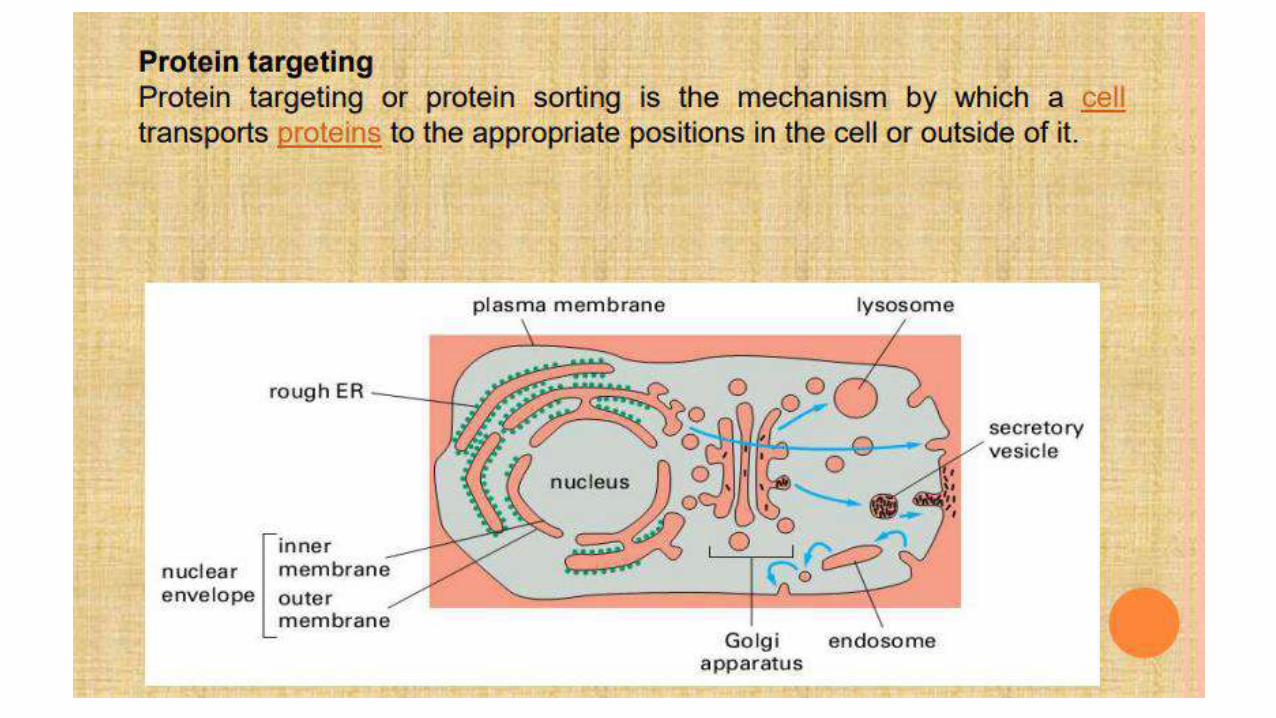
• Protein targeting or protein sorting is the biological mechanism bywhich proteins are transported to their appropriate destinations inthe cell or outside it.
• Proteins can be targeted to the inner space of an organelle, differentintracellular membranes, plasma membrane, or to exterior of the cellvia secretion.
• This delivery process is carried out based on information contained inthe protein itself. Correct sorting is crucial for the cell; errors can leadto diseases.


• Endoplasmic Reticulum- The vesicular network starts from nuclear membraneand spread throughout the cytosol constitutes endoplasmic reticulum.
• There are two different types of endoplasmic reticuli present in the cell, 1) Roughendoplasmic reticulum (RER), and 2) smooth endoplasmic reticulum (SER).
• RER has ribosome attached to it to give a rough appearance whereas smoothendoplasmic reticulum is devoid of ribosomes.
• Protein synthesis on ribosome attached to RER are sorted into 3 differentcatagories, such as integral membrane proteins, proteins for secretion andprotein destined for different organelles.
• Proteins are synthesized with a n-signal peptide and these signal peptides arerecognized by signal recognition particle on their the target organelles.
• For example, if a protein is synthesized with a signal peptide for mitochondria, itwill attach to signal recognition particle and receptor onto the outermitochondrial membrane to deliver the protein.
• The proteins without any signal peptide tags are supposed to remain in the cytsol.


Functions of endoplasmic reticulum:
• Synthesis of steroid hormone in gonad cells.
• Detoxification
• Ca2+ sequestration
• Synthesis of protein, phospholipid and carbohydrate.
• Protein sorting to different organelles.
• Protein modifications such as glycosylation etc.

Golgi Bodies-• Golgi bodies were first visualized by a metallic stain invented
by Camillo golgi and it is made of flattened, disk like cisternaearranged in a stacked manner to give 3 distinct zones.
• Cis-face receives material or vesicles from endoplasmicreticulum, medial Golgi is the actual place where protein arecovalently modified with the sugar.
• Trans Golgi is the face of Golgi towards plasma membrane and thissite sorts vesicle for their destined organelles or plasma membrane.
Functions of golgi bodies1.Protein sorting2.Protein modifications (Glycosylation)3.Proteolysis

Golgi bodies
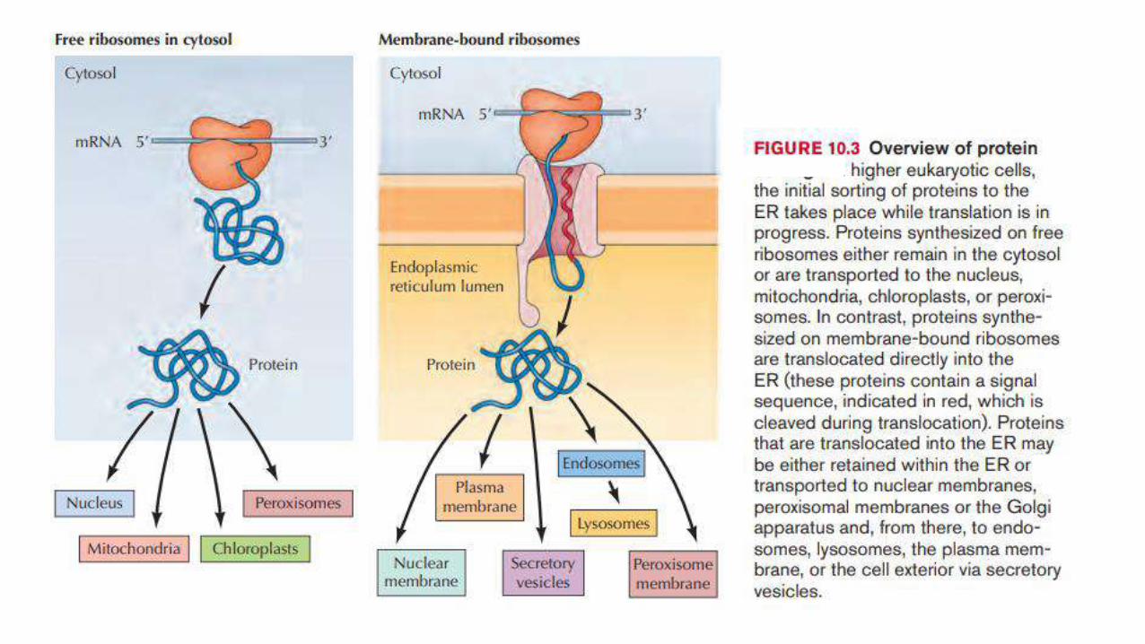

• The translocation starts while the protein is still being synthesized on the ribosome.
• Proteins targeted for ER, Golgi apparatus, plasma membrane, lysosome, vacuole andextracellular space uses the SRP-dependent pathway and are translocated co-translationally.
• The N-terminal signal sequence of these proteins, is recognized by a signal recognitionparticle (SRP), while the proteins being translated in the free ribosome.
• The ribosome-protein complex is transferred to a SRP receptor on the ER and thesynthesis pauses.
• There, the nascent protein is inserted into the the translocon that passes through theER membrane. Transfer of the ribosome-mRNA complex from the SRP to thetranslocon opens the gate on the translocon and allows the translation to resume.
• The signal sequence is immediately cleaved from the polypeptide once it has beentranslocated into the ER by signal peptidase in secretory proteins. Within the ER,chaperone helps protein to fold correctly.
• From ER, proteins are transported in vescicles to the Golgi apparatus where they arefurther processed and sorted for transport to endosomes, lysosomes, plasmamembrane or secretion from the cell.
• The proteins for ER will have various ER retention signals to keep them in the ER itself.

• Proteins are sorted to their locations with the help of an address signalpresent in the primary structure level.
• Each organelle has a mechanism to identify its own proteins.
• In this section, important protein localization sites like nucleus,mitochondrion, chloroplast, peroxisome, and secretory proteins.
The Endoplasmic Reticulum• The Endoplasmic Reticulum is the first branching point in protein sorting.• Most of the proteins targeted for secretion, Golgi apparatus, plasma membrane,
vacuole, lysosome are translated on the ribosomes bounded to the EndoplasmicReticulum and they enter into the ER co-translationally.
• Only a few proteins enter the ER posttranslationally.• The protein translation starts at the free ribosomes in the cytosol.• The synthesis continues till the sorting signal which is present in the N-terminal
emerges.• This sorting signal is recognized by signal recognition particle. The SRP binds to the
sorting signal and the translation pauses.

• The complex of SRP, ribosome, polypeptide chain and mRNA moves to the ERand the polypeptide chain enters the ER through translocon.
• The translocon is a protein complex containing various components used forprotein translocation.
• The SRP receptor of the translocon binds with the SRP, the ribosome receptorbinds with the ribosme and hold it in the correct position, the pore proteinforms the channel through which the growing polypetide enter the ER lumen,the signal peptidase cut the signal once it enters the ER.
• After the SRP and ribosomes are bound by SRP receptor and ribosme receptorrespectively, GTP binds to the the complex of SRP and SRP receptor and thetranslation resumes.
• This causes the transfer of the signal sequence into the channel of poreprotein.
• Then the GTP is hydrolysed and the SRP is released. While the sorting signalremains bound at the the pore protein, the polypeptide grows into a loop andtranslocates into the ER lumen
• When the polypetide synthesis is finished, the signal peptidase cleavesoff the sorting signal, releasing the polypeptide into the ER lumen.
• After this, the ribosome detaches from the ER and dissociate into itssubunits, and the mRNA is released.
• Inside the ER, the polypeptide chains are folded into their native formsusually with the help of molecular chaperones, which controls thequality of protein folding.
• Integral membrane proteins of the plasma membrane or themembranes of the ER, Golgi apparatus, and lysosome are first insertedinto the membrane of ER.
• These proteins do not enter the lumen cotranslationally but anchoredto the ER membrane by membrane spanning α helices that stop transferof the growing polypeptide chain across the membrane.

• Proteins travel along the secretory pathway in transport vesicle, whichbud from the membrane of one organelle and then fuse with themembrane of another.
• The proteins are exported from the ER in vesicles that bud from thetransitional ER and carry their cargo through the ER-Golgi intermediatecompartment and then to Golgi apparatus.
• The proteins targeted for the ER has a retention signal in their Cterminal that makes them come back to the ER even if they areexported from the ER.
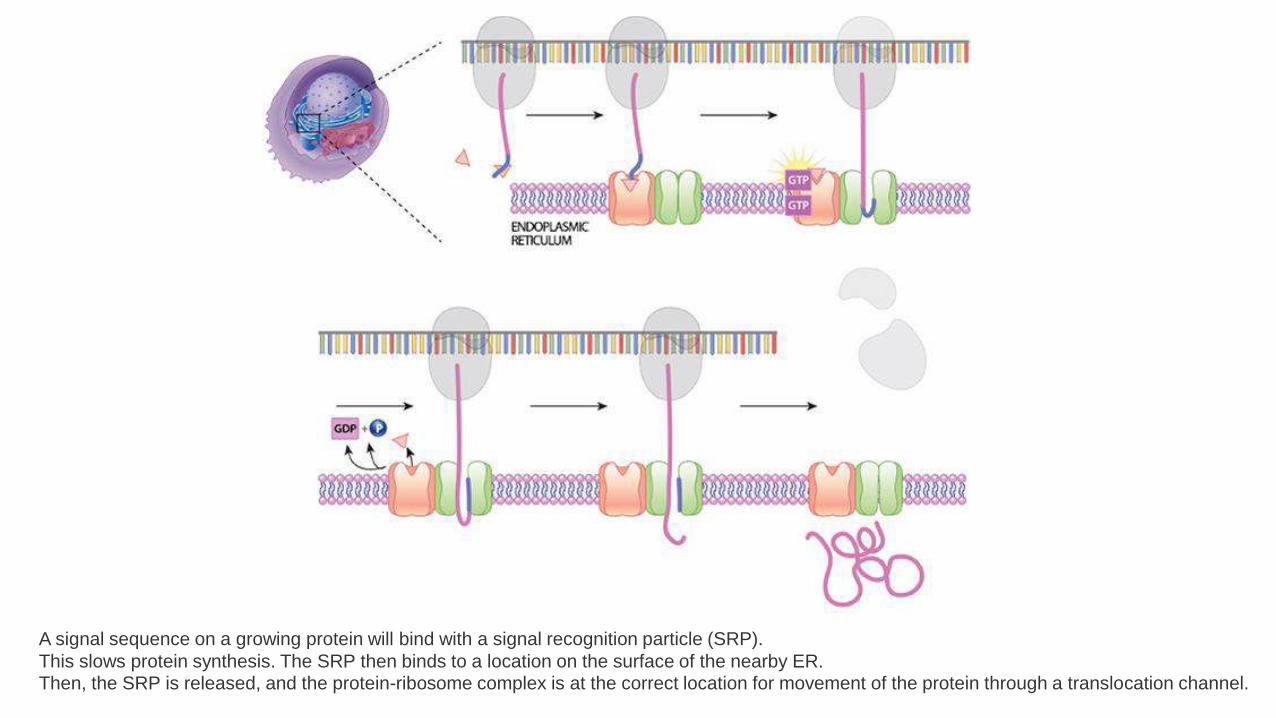
A signal sequence on a growing protein will bind with a signal recognition particle (SRP).
This slows protein synthesis. The SRP then binds to a location on the surface of the nearby ER. Then, the SRP is released, and the protein-ribosome complex is at the correct location for movement of the protein through a translocation channel.
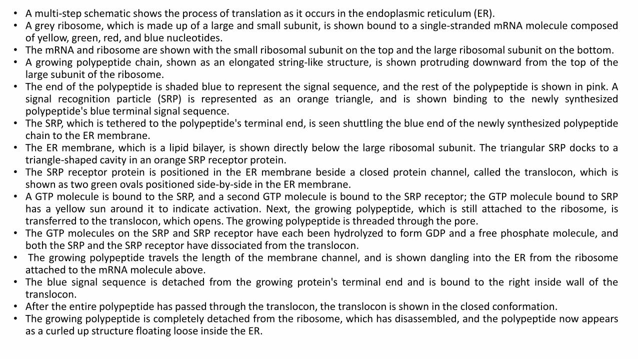
• A multi-step schematic shows the process of translation as it occurs in the endoplasmic reticulum (ER).• A grey ribosome, which is made up of a large and small subunit, is shown bound to a single-stranded mRNA molecule composed
of yellow, green, red, and blue nucleotides.• The mRNA and ribosome are shown with the small ribosomal subunit on the top and the large ribosomal subunit on the bottom.• A growing polypeptide chain, shown as an elongated string-like structure, is shown protruding downward from the top of the
large subunit of the ribosome.• The end of the polypeptide is shaded blue to represent the signal sequence, and the rest of the polypeptide is shown in pink. A
signal recognition particle (SRP) is represented as an orange triangle, and is shown binding to the newly synthesizedpolypeptide's blue terminal signal sequence.
• The SRP, which is tethered to the polypeptide's terminal end, is seen shuttling the blue end of the newly synthesized polypeptidechain to the ER membrane.
• The ER membrane, which is a lipid bilayer, is shown directly below the large ribosomal subunit. The triangular SRP docks to atriangle-shaped cavity in an orange SRP receptor protein.
• The SRP receptor protein is positioned in the ER membrane beside a closed protein channel, called the translocon, which isshown as two green ovals positioned side-by-side in the ER membrane.
• A GTP molecule is bound to the SRP, and a second GTP molecule is bound to the SRP receptor; the GTP molecule bound to SRPhas a yellow sun around it to indicate activation. Next, the growing polypeptide, which is still attached to the ribosome, istransferred to the translocon, which opens. The growing polypeptide is threaded through the pore.
• The GTP molecules on the SRP and SRP receptor have each been hydrolyzed to form GDP and a free phosphate molecule, andboth the SRP and the SRP receptor have dissociated from the translocon.
• The growing polypeptide travels the length of the membrane channel, and is shown dangling into the ER from the ribosomeattached to the mRNA molecule above.
• The blue signal sequence is detached from the growing protein's terminal end and is bound to the right inside wall of thetranslocon.
• After the entire polypeptide has passed through the translocon, the translocon is shown in the closed conformation.• The growing polypeptide is completely detached from the ribosome, which has disassembled, and the polypeptide now appears
as a curled up structure floating loose inside the ER.

• Transport of molecules within a cell and out of the cell requires a complex endomembrane system.• Endocytosis occurs when the cell membrane engulfs particles (dark blue) outside the cell, draws the contents in, and forms an intracellular vesicle called an
endosome.• This vesicle travels through the cell, and its contents are digested as it merges with vesicles containing enzymes from the Golgi. The vesicle is then known as a
lysosome when its contents have been digested by the cell.• Exocystosis is the process of membrane transport that releases cellular contents outside of the cell.• Here, a transport vesicle from the Golgi or elsewhere in the cell merges its membrane with the plasma membrane and releases its contents. In this way, membranes
are continually recycled and reused for different purposes throughout the cell.• Membrane transport also occurs between the endoplasmic reticulum and the Golgi.

The rough endoplasmic reticulum is key in multiple functions:
• Manufacture of lysosomal enzymes with a mannose-6-phosphate markeradded in the cis-Golgi network.
• Manufacture of secreted proteins, either secreted constitutively with notag or secreted in a regulatory manner involving clathrin and paired basicamino acids in the signal peptide.
• Integral membrane proteins that stay embedded in the membrane asvesicles exit and bind to new membranes. Rab proteins are key intargeting the membrane; SNAP and SNARE proteins are key in the fusionevent.
• Initial glycosylation as assembly continues. This is N-linked (O-linkingoccurs in the Golgi).• N-linked glycosylation: If the protein is properly
folded, Oligosaccharyltransferase recognizes the AA sequence NXS or NXT (withthe S/T residue phosphorylated) and adds a 14-sugar backbone (2-N-acetylglucosamine, 9-branching mannose, and 3-glucose at the end) to the side-chain nitrogen of Asn.
Smooth endoplasmic reticulum
• The smooth endoplasmic reticulum, or smooth ER, is an organellefound in both animal cells and plant cells.
• An organelle is a sub-unit within a cell that has a specialized function.
• The main function of the smooth ER is to make cellular products likehormones and lipids.
• In most cells the smooth endoplasmic reticulum (abbreviated SER) isscarce.

• Instead there are areas where the ER is partly smooth and partlyrough, this area is called the transitional ER.
• The transitional ER gets its name because it contains ER exit sites.
• These are areas where the transport vesicles that contain lipids andproteins made in the ER, detach from the ER and start moving tothe Golgi apparatus.
• Specialized cells can have a lot of smooth endoplasmic reticulum andin these cells the smooth ER has many functions.
• It synthesizes lipids, phospholipids, and steroids.

• The smooth endoplasmic reticulum plays a major role insynthesizing lipids by means of enzymes embedded in these smoothmembranes.
• It produces the phospholipids and cholesterol used in membraneformation, and along with the membrane proteins produced by therough ER it can synthesize more membrane for itself, for the Golgicomplex, the cell membrane, lysosomes, and others.
• In liver cells the smooth ER contains enzymes for the detoxification ofharmful drugs and metabolic by-products.
• In the reproductive organs, smooth ER in the cells produces thesteroid hormones testosterone and estrogen.

• Cells which secrete these products, such as those in the testes, ovaries,and sebaceous glands have an abundance of smooth endoplasmicreticulum.
• It also carries out the metabolism of carbohydrates, detoxification ofnatural metabolism products and of alcohol and drugs, attachment ofreceptors on cell membrane proteins, and steroid metabolism.
• In muscle cells, it regulates calcium ion concentration.
• The smooth endoplasmic reticulum also contains the enzyme glucose-6-phosphatase, which converts glucose-6-phosphate to glucose, a stepin gluconeogenesis.
• It is connected to the nuclear envelope and consists of tubules that arelocated near the cell periphery. These tubes sometimes branch forming anetwork that is reticular in appearance.
• The network of smooth endoplasmic reticulum allows for an increasedsurface area to be devoted to the action or storage of key enzymes andthe products of these enzymes.

• Lipids include fats, waxes, phospholipids, sterols, such as cholesterol,and fat-soluble vitamins.
• Broadly speaking, there are three possible sites where lipids aresynthesized: the smooth endoplasmic reticulum (SER), the cytosol and,in plants specifically, the chloroplast.
• In animal and yeast cells, phospholipids, fat-soluble vitamins and, inlimited types of cells, waxes are synthesized in the SER; triglycerides, orfats, are synthesized in the cytosol; and, sterols begin the process in thecytosol with the help of carrier proteins and are ultimately synthesizedin the SER.
• The SER is part of a network of membrane-enclosed tubes andcompartments, which extend from the nucleus.
• The larger cell body, or the cytoplasm, is made up of cytosol.
• Glycosylation (see also chemical glycosylation) is the reaction in whicha carbohydrate, i.e. a glycosyl donor, is attached to a hydroxyl or otherfunctional group of another molecule (a glycosyl acceptor).
• In biology, glycosylation mainly refers in particular to the enzymaticprocess that attaches glycans to proteins, or other organic molecules. Thisenzymatic process produces one of the fundamental biopolymers found incells (along with DNA, RNA, and proteins).
• Glycosylation is a form of co-translational and post-translationalmodification.
• Glycans serve a variety of structural and functional roles in membrane andsecreted proteins.
• The majority of proteins synthesized in the rough endoplasmicreticulum undergo glycosylation. It is an enzyme-directed site-specificprocess, as opposed to the non-enzymatic chemical reaction of glycation.
• Glycosylation is also present in the cytoplasm and nucleus as the O-GlcNAcmodification. Aglycosylation is a feature of engineered antibodies to bypassglycosylation.Five classes of glycans are produced:

Golgi Apparatus• Golgi apparatus is composed of flattened membrane-enclosed sacs called
cisternae and associated vesicles.• The Golgi apparatus is a main center for protein sorting.• It receives proteins from the ER and further process them and sort them to their
targeted location: lysosomes, endosomes, plasma mem brane, or extracellular.• The proteins from the ER enter the cis face of the ER which is convex in shape and
is oriented towards the nucleus.• They are transported through the Golgi and exit from its concave shaped trans
face.• The proteins that function within the Golgi has to be retained from export.• All proteins known to be retained in the Golgi complex are associated with the
Golgi membrane and their retention signals are present in the transmembranedomain.
• This prevents these proteins from being packaged in the transport vesicle thatleave trans Golgi network.
Lysosomes• A lysosome is a membrane-bound organelle found in many
animal cells.
• They are spherical vesicles that contain hydrolytic enzymes that canbreak down many kinds of biomolecules.
• A lysosome has a specific composition, of both its membraneproteins, and its lumenal proteins.
• The lumen's pH (~4.5–5.0) is optimal for the enzymes involved inhydrolysis, analogous to the activity of the stomach.
• Besides degradation of polymers, the lysosome is involved in variouscell processes, including secretion, plasma membrane repair, cellsignaling, and energy metabolism.
• Lysosomes act as the waste disposal system of the cell by digesting obsoleteor un-used materials in the cytoplasm, from both inside and outside the cell.
• Material from outside the cell is taken-up through endocytosis, whilematerial from the inside of the cell is digested through autophagy.
• The sizes of the organelles vary greatly—the larger ones can be more than 10times the size of the smaller ones.
• They were discovered and named by Belgian biologist Christian de Duve,who eventually received the Nobel Prize in Physiology or Medicine in 1974.
• Lysosomes are known to contain more than 60 different enzymes, and havemore than 50 membrane proteins.
• Enzymes of the lysosomes are synthesised in the rough endoplasmicreticulum.
• The enzymes are imported from the Golgi apparatus in small vesicles, whichfuse with larger acidic vesicles.
• Enzymes destined for a lysosome are specifically tagged with themolecule mannose 6-phosphate, so that they are properly sorted intoacidified vesicles

• Lysosomes originate by budding off from the membrane of the trans-Golginetwork, a region of the Golgi complex responsible for sorting newlysynthesized proteins, which may be designated for use in lysosomes,endosomes, or the plasma membrane.
• The lysosomes then fuse with membrane vesicles that derive from one of threepathways: endocytosis, autophagocytosis, and phagocytosis.
• In endocytosis, extracellular macromolecules are taken up into the cell to formmembrane-bound vesicles called endosomes that fuse with lysosomes.
• Autophagocytosis is the process by which old organelles and malfunctioningcellular parts are removed from a cell; they are enveloped by internalmembranes that then fuse with lysosomes.
• Phagocytosis is carried out by specialized cells (e.g., macrophages) that engulflarge extracellular particles, such as dead cells or foreign invaders(e.g., bacteria), and target them for lysosomal degradation.
• Many of the products of lysosomal digestion, such as aminoacids and nucleotides, are recycled back to the cell for use in the synthesis ofnew cellular components.

Acid hydrolases• An acid hydrolase (lysosomal acid lipase) is an enzyme that works
best at acidic pHs.
• an ENZYME that is capable of catalysing the HYDROLYSIS of compounds at acid pH (generallyaround pH 5).
• It is commonly located in lysosomes, which are acidic on the inside.
• Enzymes in this group include certain PROTEINASES, PHOSPHATASES,LIPASES and NUCLEASES.
• Acid hydrolases may be nucleases, proteases, glycosidases, lipases,phosphatases, sulfatases and phospholipases and make up theapproximately 50 degradative enzymes of the lysosome that breakapart biological matter.

• Enzymes found in the lysosome require an acidic environment tofunction properly and are called acid hydrolases.
• Although it may seem dangerous for cells to contain enzymes thatcan digest most biological molecules, the contents of the cell aredoubly protected from the digestive enzymes of the lysosome.


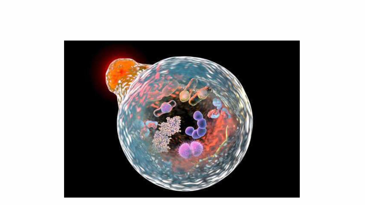
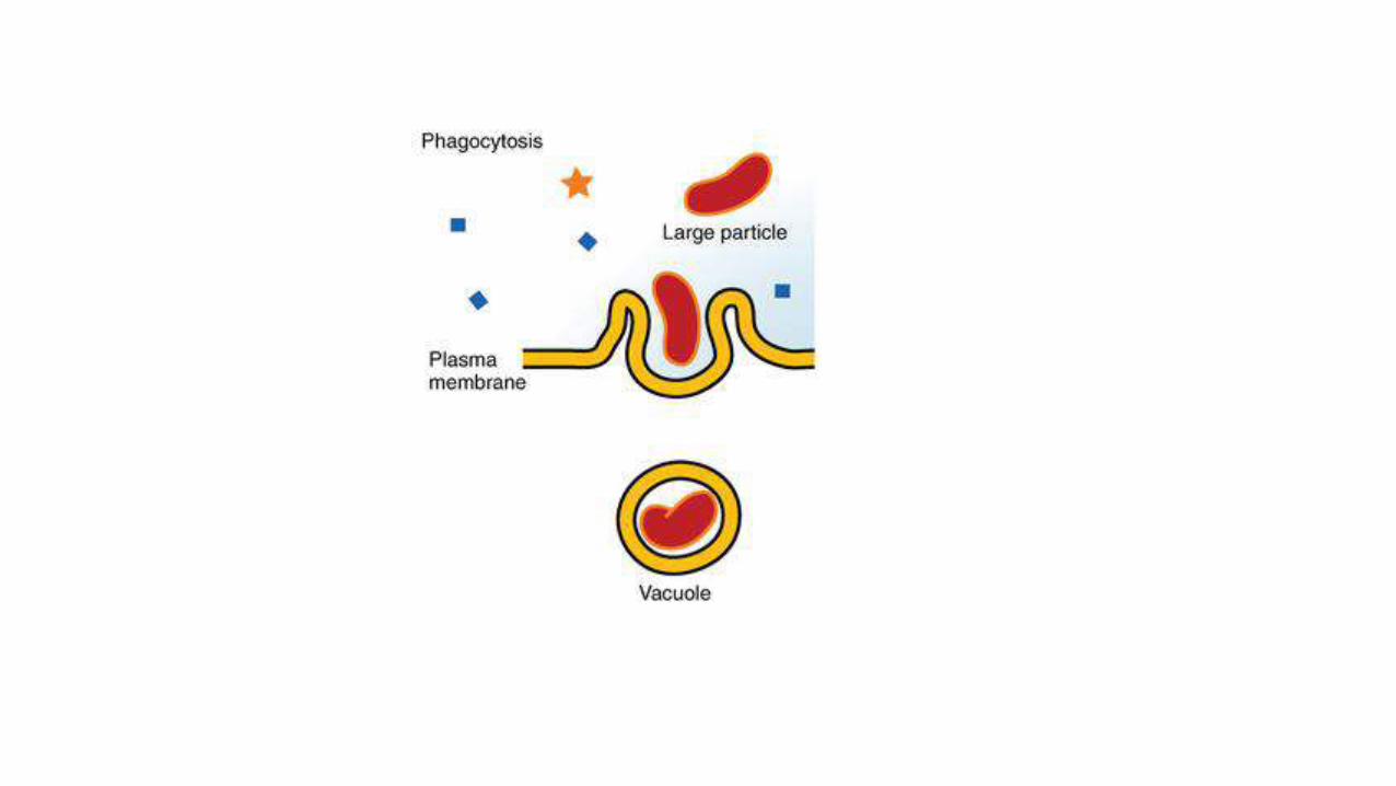
Phagocytosis
• Phagocytosis is the process by which a cell uses its plasmamembrane to engulf a large particle (≥ 0.5 μm) , giving rise to aninternal compartment called the phagosome.
• It is one type of endocytosis pinocytosis. In a multicellularorganism's immune system, phagocytosis is a major mechanism usedto remove pathogens and cell debris.
• The ingested material is then digested in the phagosome. Bacteria,dead tissue cells, and small mineral particles are all examples ofobjects that may be phagocytized.
• Some protozoa use phagocytosis as means to obtain nutrients

Professional phagocytic cells• Neutrophils, macrophages, monocytes, dendritic
cells, osteoclasts and eosinophils can be classified as professional phagocytes.
• The first three have the greatest role in immune response to most infections.
• The role of neutrophils is patrolling the bloodstream and rapid migration to thetissues in large numbers only in case of infection.
• There they have direct microbicidal effect by phagocytosis.
• After ingestion, neutrophils are efficient in intracellular killing of pathogens.
• Neutrophils phagocytose mainly via the Fcγ receptors and complement receptors 1and 3.
• The microbicidal effect of neutrophils is due to a large repertoire of moleculespresent in pre-formed granules.
• Enzymes and other molecules prepared in these granules are proteases, suchas collagenase, gelatinase or serine proteases, myeloperoxidase, lactoferrin andantibiotic proteins.
• Degranulation of these into the phagosome, accompanied by high reactive oxygenspecies production (oxidative burst) is highly microbicidal.

• Monocytes, and the macrophages that mature from them, leaveblood circulation to migrate through tissues.
• There they are resident cells and form a resting barrier.
• Macrophages initiate phagocytosis by mannose receptors, scavengerreceptors, Fcγ receptors and complement receptors 1, 3 and 4.
• Macrophages are long-lived and can continue phagocytosis byforming new lysosomes.
• Dendritic cells also reside in tissues and ingest pathogens byphagocytosis.
• Their role is not killing or clearance of microbes, but rather breakingthem down for antigen presentation to the cells of the adaptiveimmune system
Eosinophils
• Eosinophils, sometimes called eosinophiles or, lesscommonly, acidophils, are a variety of white blood cells and one ofthe immune system components responsible for combatingmulticellular parasites and certain infections in vertebrates.
• Along with mast cells and basophils, they also control mechanismsassociated with allergy and asthma.
• They are granulocytes that develop during hematopoiesis inthe bone marrow before migrating into blood, after which they areterminally differentiated and do not multiply