proteomics of protein secretion by bacillus subtilis: separating … · bacillus cereus.....228...
TRANSCRIPT
MICROBIOLOGY AND MOLECULAR BIOLOGY REVIEWS, June 2004, p. 207–233 Vol. 68, No. 21092-2172/04/$08.00�0 DOI: 10.1128/MMBR.68.2.207–233.2004Copyright © 2004, American Society for Microbiology. All Rights Reserved.
Proteomics of Protein Secretion by Bacillus subtilis: Separating the“Secrets” of the Secretome
Harold Tjalsma,1,2 Haike Antelmann,3 Jan D.H. Jongbloed,1 Peter G. Braun,4 Elise Darmon,1Ronald Dorenbos,4 Jean-Yves F. Dubois,4,5 Helga Westers,4 Geeske Zanen,4 Wim J. Quax,4
Oscar P. Kuipers,1 Sierd Bron,1* Michael Hecker,3 and Jan Maarten van Dijl4,5
Department of Genetics, Groningen Biomolecular Sciences and Biotechnology Institute, 9751 NN Haren,1 Departmentof Clinical Chemistry/564, University Medical Centre Nijmegen, 6500 HB Nijmegen,2 Department of Pharmaceutical
Biology, University of Groningen, 9713 AV Groningen,4 and Department of Molecular Bacteriology, University ofGroningen, 9700 RB Groningen,5 The Netherlands, and Institut fur Mikrobiologie und Molekularbiologie,
Ernst-Moritz-Arndt-Universiat Greifswald, D-17487 Greifswald, Germany3
GENERAL INTRODUCTION...................................................................................................................................208SCOPE OF THIS REVIEW: THE PROTEOMICS OF PROTEIN SECRETION BY B. SUBTILIS ..............208PROTEIN SORTING IN B. SUBTILIS ...................................................................................................................208
Signal Peptides........................................................................................................................................................209Signal Peptide Prediction and Classification .....................................................................................................209
Twin-arginine (RR/KR-type) signal peptides..................................................................................................209Secretory (Sec-type) signal peptides ................................................................................................................210Lipoprotein signal peptides...............................................................................................................................211Pseudopilin-like signal peptides .......................................................................................................................211Signal peptides of pheromones and bacteriocins ...........................................................................................211
Retention Signals ....................................................................................................................................................211Transmembrane domains ..................................................................................................................................211Lipid modification...............................................................................................................................................211Pseudopilin assembly .........................................................................................................................................211Cell wall-binding repeats ...................................................................................................................................212Covalent attachment to the cell wall................................................................................................................212
PROTEOMICS OF PROTEIN SECRETION BY B. SUBTILIS ..........................................................................212Extracellular Proteome of B. subtilis 168.............................................................................................................212Toward an Extracellular Zymoproteome of B. subtilis 168 ...............................................................................212Cell Wall Proteome of B. subtilis 168 ...................................................................................................................213Verification of Secretome Predictions..................................................................................................................215
CONTRIBUTION OF THE Sec MACHINERY TO THE EXTRACELLULAR PROTEOME.........................215Cytoplasmic Targeting Factors .............................................................................................................................216
Ffh depletion........................................................................................................................................................217Sec Translocase.......................................................................................................................................................218
SecA limitation ....................................................................................................................................................219SecA inhibition by sodium azide ......................................................................................................................219SecDF deletion and SpoIIIJ and YqjG depletion...........................................................................................220
Type I Signal Peptidases........................................................................................................................................220SPase I deletions.................................................................................................................................................221
Lipoprotein Modification and Processing ...........................................................................................................221SPase II deletion.................................................................................................................................................221Lgt deletion..........................................................................................................................................................221
Folding Catalysts ....................................................................................................................................................222PrsA depletion .....................................................................................................................................................222Bdb mutations .....................................................................................................................................................223
Quality Control Factors .........................................................................................................................................223Modulation of HtrA and HtrB levels ...............................................................................................................223WprA deletion......................................................................................................................................................224
Extracellular Proteases ..........................................................................................................................................224CONTRIBUTION OF Sec-INDEPENDENT PROTEIN EXPORT TO THE EXTRACELLULAR
PROTEOME........................................................................................................................................................224Twin-Arginine Translocation Machinery.............................................................................................................224
* Corresponding author. Mailing address: Department of Genetics,Groningen Biomolecular Sciences and Biotechnology Institute, Kerk-laan 30, 9751 NN Haren, The Netherlands. Phone: 31-50-3638037.Fax: 31-50-3632348. E-mail: [email protected].
207
on July 18, 2019 by guesthttp://m
mbr.asm
.org/D
ownloaded from
TatC and total-Tat deletions.............................................................................................................................225MECHANISMS FOR EXTRACELLULAR ACCUMULATION OF PROTEINS ..............................................225
Protein Secretion via the Sec and Tat Pathways ...............................................................................................225Release of Membrane Proteins by Proteolysis....................................................................................................225Release of Lipoproteins by Proteolytic Shaving and/or Shedding ...................................................................225Release of Cell Wall Proteins by Proteolytic Shaving and Cell Wall Turnover ............................................226Release of Extracellular Proteins without Typical Export Signals..................................................................227
EXTRACELLULAR PROTEOMES OF OTHER GRAM-POSITIVE BACTERIA............................................227Bacillus cereus ..........................................................................................................................................................228Clostridium difficile...................................................................................................................................................228Staphylococcus aureus ..............................................................................................................................................229Group A Streptococcus.............................................................................................................................................229Mycobacterium tuberculosis .....................................................................................................................................229
PERSPECTIVES .........................................................................................................................................................229ACKNOWLEDGMENTS ...........................................................................................................................................229REFERENCES ............................................................................................................................................................230
GENERAL INTRODUCTION
Protein export from the cytoplasm to destinations outsidethe cell is a phenomenon that takes place in all domains of life.Most bacterial proteins destined to leave the cytoplasm areexported via the highly conserved SecA-YEG (Sec) pathway.In addition, more specialized bacterial export pathways areused for the export of specific subsets of extracellular proteins.Most exported proteins are synthesized as precursors with anN-terminal signal peptide (151, 152). These preproteins arefirst recognized by soluble targeting factors for their transportto the translocation machinery in the cell membrane. Next, thepolypeptide chain is transported through a proteinacious chan-nel in the membrane, a process driven by a translocation motorthat binds and hydrolyzes nucleotide triphosphates. Finally, thesignal peptide is removed, resulting in the release of the ma-ture protein from the membrane. The mature protein foldsinto its native conformation shortly after the release from thetranslocase, unless it is translocated in a folded state. Thesebasic principles of protein transport across membranes applyto most eukaryotic and prokaryotic organisms (35, 93, 102, 111,129).
SCOPE OF THIS REVIEW: THE PROTEOMICS OFPROTEIN SECRETION BY B. SUBTILIS
Bacterial secretory proteins are known to perform severalvery important “remote-control” functions, such as the provi-sion of nutrients, cell-to-cell communication, detoxification ofthe environment, and killing of potential competitors. Morespecifically, extracellular proteins of pathogenic bacteria seemto play critical roles in virulence (53, 59, 105). The fact thatexported Bacillus subtilis proteins are not retained by an outermembrane, as encountered in gram-negative bacteria, makesthis gram-positive bacterium a preferred organism for the pro-teomic analysis of protein secretion. In addition, the availabil-ity of the complete genome sequence (58) and about 3,000“y”-mutants constructed within the Bacillus subtilis FunctionalAnalysis program (54, 115) make B. subtilis an ideal modelorganism for research on gram-positive bacteria. Furthermore,previous studies have predicted the composition of the so-called secretome of B. subtilis, which, by our definition, in-cludes both the secreted proteins and the protein secretionmachinery (129). These predictions showed that at least four
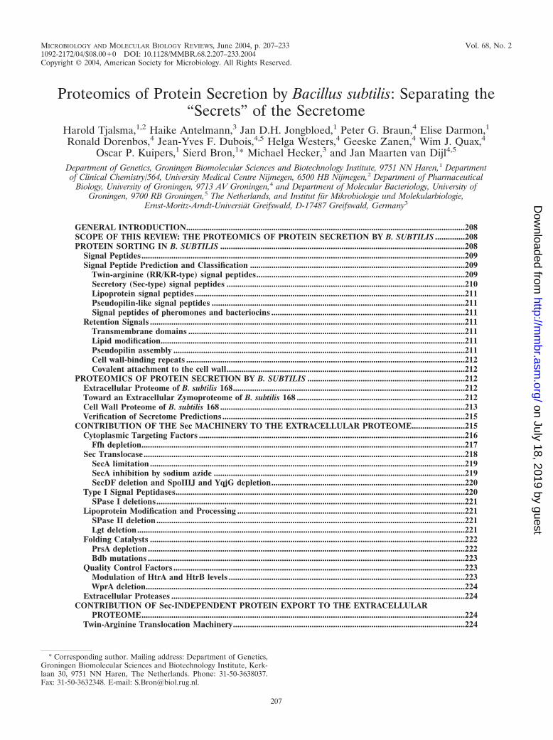
distinct pathways for protein export from the cytoplasm andapproximately 300 proteins with the potential to be exportedcould be distinguished. By far the largest number of exportedproteins was predicted to follow the major Sec pathway forprotein secretion. In contrast, the recently identified twin-argi-nine translocation Tat pathway (51, 52), a pseudopilin exportpathway for competence development, and pathways usingATP-binding cassette (ABC) transporters, can be regarded asspecial-purpose pathways through which only few proteins ap-pear to be transported (Fig. 1) (129). In this review, we discussthe latest views of protein secretion by B. subtilis as obtainedfrom recent proteomic studies that were aimed at defining theextracellular complement of the B. subtilis secretome. Usingdifferent growth conditions and mutant strains, about 200 ex-tracellular proteins could be visualized by two-dimensional(2D) gel electrophoresis, of which almost 50% could be iden-tified by mass spectrometry (3–6, 46, 51, 52). In summary, thesestudies showed that in addition to the known mechanisms forprotein export, B. subtilis also makes use of alternative mech-anisms to release proteins into the external environment. Fur-thermore, the proteomic data could be used to verify genome-wide predictions concerning the secretome. Even though theprocess of protein secretion by B. subtilis had been docu-mented fairly well by more classical genetic and biochemicalapproaches (129, 145), various secretome secrets were un-veiled by proteomic approaches. These include the apparentexport of cytoplasmic proteins, processing of native membraneproteins by type I signal peptidases (SPases), and the release ofnormally cell-associated lipoproteins and cell wall proteins intothe growth medium.
PROTEIN SORTING IN B. SUBTILIS
Although the soil bacterium B. subtilis has a relatively simplecell structure, proteins can at least be delivered to, or retainedat, five (sub)cellular locations: the cytoplasm, the cytoplasmicmembrane, the membrane/cell wall interface, the cell wall, andthe growth medium (129). The final destination of a protein isgoverned by the presence or absence of signal peptides and/orretention signals. Nearly all proteins of B. subtilis lacking trans-port signals are retained in the cytoplasm and fold, with orwithout the aid of chaperones, into their native conformation.Other proteins contain membrane-spanning domains that arerequired for their insertion into the cytoplasmic membrane.
208 TJALSMA ET AL. MICROBIOL. MOL. BIOL. REV.
on July 18, 2019 by guesthttp://m
mbr.asm
.org/D
ownloaded from
Most proteins that are completely transported across the cy-toplasmic membrane are synthesized with an N-terminal signalpeptide. Since B. subtilis lacks an outer membrane, many ofthese proteins are secreted directly into the growth medium.Other exported proteins involved in processes, such as cell wallturnover, substrate binding, and the folding and modificationof translocated secretory proteins, have to be retained at themembrane/cell wall interface to fulfill their function. In thefollowing sections, signal peptides, export routes, and retentionmechanisms that are known to be involved in protein sorting inB. subtilis are discussed in the light of recent findings fromproteomic analyses.
Signal Peptides
Three distinct domains, N, H, and C, are generally present insignal peptides (148–151). The N-domain contains at least one
arginine or lysine residue, which has been suggested to interactwith the translocation machinery and the negatively chargedphospholipids in the lipid bilayer of the membrane (1, 32). TheH-region, following the N-region, is formed by a stretch ofhydrophobic residues that can adopt an �-helical conformationin the membrane (21). In the middle of this hydrophobic core,helix-breaking glycine or proline residues are often present toallow the formation of a hairpin-like structure that can insertinto the membrane. It was proposed that unlooping of thishairpin results in insertion of the complete signal peptide intothe membrane (32). Helix-breaking residues at the end of theH-domain are thought to facilitate cleavage by a specific SPase(88). The C-domain, following the H-domain, contains thecleavage site for specific SPases that remove signal peptidesfrom the mature part of the exported protein during or shortlyafter translocation. The signal peptide is degraded by signalpeptide peptidases and removed from the membrane. Finally,the mature part of the protein is released from the membraneand can fold into its native conformation. Despite the similarstructure of signal peptides, apparently small variations canresult in transport to different destinations and/or export viadifferent pathways, as described below.
Signal Peptide Prediction and Classification
Predictions showed that 300 proteins with the potential to beexported could be distinguished in B. subtilis (129). On thebasis of SPase cleavage sites and the export pathways by whichthese preproteins are (predicted to be) exported, signal pep-tides can be divided into five distinct classes: (i) twin-arginine(RR/KR) signal peptides, (ii) secretory (Sec-type) signal pep-tides, (iii) lipoprotein signal peptides, (iv) pseudopilin-like sig-nal peptides, and (v) bacteriocin and pheromone signal pep-tides. The first group of signal peptides contains a so-calledtwin-arginine (RR/KR) motif, which serves to direct proteinsinto the Tat pathway (51). The second, and most abundant,class is composed of typical secretory signal peptides (lackingan RR/KR-motif) that direct proteins into the Sec pathway.Both the twin-arginine and secretory signal peptides appear tobe cleaved by one of the various type I SPases of B. subtilis(130). The third class of signal peptides is present at the Nterminus of prelipoproteins that are exported via the Sec path-way, lipid modified, and cleaved by the type II SPase (Lsp)(136). The fourth class is formed by signal peptides of pseu-dopilins which, in B. subtilis, are cleaved by the SPase ComC(64). Finally, the fifth class of signal peptides is found onribosomally synthesized pheromones and lantibiotics that areexported and cleaved by ABC transporters (80). It should benoted that this specific class of signal peptides is often referredto as “leader peptides.”
Twin-arginine (RR/KR-type) signal peptides. Signal peptidepredictions resulted in the identification of �180 potentialsubstrates for type I SPases. A twin-arginine motif, containingat least three residues of the consensus sequence R/K-R-X-#-# (where # is a hydrophobic residue) was found in 44 ofthese signals (12 RR and 32 KR signal peptides; [51]). Thepresence of such twin-arginine motifs was initially interpretedas an indication that the corresponding preproteins could bedirected into the Tat pathway for protein export, possibly in aSec-independent manner. The predicted twin-arginine signal
FIG. 1. Protein export pathways in B. subtilis. Ribosomally synthe-sized proteins can be sorted to various destinations depending on thepresence (�SP) or absence (�SP) of an N-terminal signal peptide andspecific retention signals. Proteins devoid of a signal peptide remain inthe cytoplasm. Proteins that have to be retained at the extracytoplas-mic side of the membrane can contain either a transmembrane seg-ment (TM) or a lipid modification (�lipobox). They are exported viathe Sec or Tat pathway. Pseudopilins are exported by the Com system.Proteins that need to be retained in the cell wall can be exported viaeither the Sec or Tat pathway. To be retained in the cell wall, themature parts of these proteins contain cell wall-binding repeats(�CWB). Proteins can be secreted into the medium via the Sec or Tatpathway or by ABC transporters.
VOL. 68, 2004 PROTEOMICS OF PROTEIN SECRETION 209
on July 18, 2019 by guesthttp://m
mbr.asm
.org/D
ownloaded from
peptides with a consensus R-R-X-#-# motif have an averagelength of 36 amino acid residues. Thus, they are significantlylonger than typical Sec-type signal peptides. This is mainlybecause the N-domains of these R-R-X-#-# containing signalpeptides have an average length of 14 amino acid residues,almost twice as long as the N-domain of the regular (Sec-type)signals (Fig. 2). Furthermore, these N-domains contain, onaverage, more positively charged residues than do those ofSec-type signal peptides (129). In contrast, the average fea-tures of predicted twin-arginine signal peptides with a K-R-X-
#-# motif are similar to those of Sec-type signal peptides (51,52, 129).
Secretory (Sec-type) signal peptides. The 135 predicted sig-nal peptides lacking RR/KR motifs have an average length of28 residues and contain two or three positively charged lysine(L) or arginine (R) residues in their N-domain. The hydropho-bic core (H-domain) has an average length of 19 residues, andabout 60% of the predicted Sec-type signal peptides contains ahelix-breaking residue (mostly glycine) in the middle of thisdomain. The C-domain of the predicted signal peptides carries
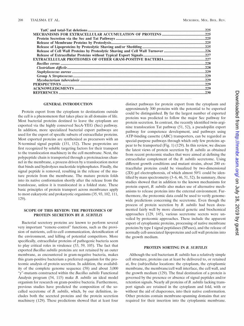
FIG. 2. Classification of cleavable N-terminal signal peptides. On the basis of SPase cleavage sites and the export pathways via which thepreproteins are exported, predicted signal peptides (129) were divided into five distinct classes: twin-arginine (RR/KR) signal peptides, secretory(Sec-type) signal peptides, lipoprotein signal peptides, pseudopilin-like signal peptides, and bacteriocin and pheromone signal peptides. The exportpathways via which the preproteins are exported and the SPases responsible for their cleavage are indicated. Most signal peptides have a tripartitestructure: a positively charged N-domain (N), containing lysine and/or arginine residues (indicated by �), a hydrophobic H-domain (H, indicatedby a gray box), and a C-domain (C) that specifies the cleavage site for their specific SPase. The length of the signal peptides and their subdomainsis drawn to the same scale. Furthermore, helix-breaking residues, mostly glycine or proline (G/P), in the H-domain of Sec-type signal peptides areindicated. These residues are, respectively, thought to facilitate loopwise membrane insertion and cleavage by SPase I (129). Finally, whereappropriate, the most frequently occurring first amino acid of the mature protein (�1) is indicated.
210 TJALSMA ET AL. MICROBIOL. MOL. BIOL. REV.
on July 18, 2019 by guesthttp://m
mbr.asm
.org/D
ownloaded from
a type I SPase cleavage site, with the consensus sequenceA-S-A at positions �3 to �1 relative to the cleavage site.About 50% of these signal peptides contain a helix-breakingresidue (proline or glycine) at positions �7 to �4 relative tothe predicted processing site for SPase I (129).
Lipoprotein signal peptides. Lipoprotein signal peptide pre-dictions resulted in the identification of 114 potential sub-strates for the lipoprotein-specific (type II) SPase (Lsp) (133).Signal peptides from lipoproteins have an average length of 19residues. These are therefore considerably shorter than RR/KR- and Sec-type signal peptides. This is because both theN-domain (average of 4 residues) and the H-domain (averageof 12 residues) are shorter than the corresponding domains inRR/KR- and Sec-type signal peptides. Furthermore, helix-breaking residues are not conserved in the H-region of lipopro-tein signal peptides. The C-domain contains a so-called lipoboxwith the consensus sequence L-(A/S)-(A/G)-C. The invariablecysteine residue of the lipobox is the target for lipid modifica-tion and the first residue of the mature lipoprotein after cleav-age by SPase II (Fig. 2) (129). In fact, this lipid modification isindispensable for signal peptide cleavage by SPase II. Finally,although some lipoprotein signal peptides contain an RR/KRmotif (51), so far export of lipoproteins via the Tat pathway hasnot been reported.
Pseudopilin-like signal peptides. Only four proteins(ComGC, ComGD, ComGE, and ComGG) with pseudopilinsignal peptides have been identified in B. subtilis (129). Thesepseudopilin signal peptides have an average length of 33 resi-dues. Strikingly, the C-domain of pseudopilin signal peptides,with the consensus sequence K-G-F at positions �2 to �1relative to the SPase cleavage site, is located between the N-and H-domains (Fig. 2). This is in line with the observationthat the pseudopilin signal peptidase (ComC) acts at the cyto-plasmic side of the membrane (64). In addition to processing,ComC is responsible for aminomethylation of the phenylala-nine at position �1 relative to the cleavage site. Althoughpseudopilin signal peptides show structural similarity to thepreviously described signal peptides, pseudopilin precursorsbypass the Tat and Sec pathways and are transported via thespecific Com pathway (26, 27, 129).
Signal peptides of pheromones and bacteriocins. Phero-mones and antimicrobial peptides form a distinct group ofexported proteins with cleavable N-terminal signal peptides,often called leader peptides. These leader peptides consist ofonly N- and C-domains and completely lack a hydrophobicH-domain (Fig. 2). It has been described that parts of themature protein are also required for export by a dedicatedABC transporter. Moreover, the leader peptide has an impor-tant function in the prevention of premature antimicrobialactivity and is required for the posttranslational modificationof lantibiotics (141, 144). The two known leader peptides ofthis type in B. subtilis 168 direct the secretion of sublancin 168(89) and ComX (67). Like leader peptides of other lantibiotics(23, 84), the sublancin 168 leader peptide contains a double-glycine motif (GS) N-terminally of the SPase cleavage site.Interestingly, the ABC transporter SunT is likely to play a dualrole in the secretion of sublancin 168 since it belongs to a classof ABC transporters that are responsible for both the removalof the leader peptide and the translocation of the maturelantibiotic across the cytoplasmic membrane (33). Although
not documented, it seems likely that an ABC transporter isalso responsible for the processing and secretion of the ComXpheromone. This pheromone is involved in the density-con-trolled onset of competence development, and, similar to sub-lancin 168, it is ribosomally synthesized as a precursor andmodified before secretion (119).
Retention Signals
In gram-negative bacteria, the outer membrane confines nu-merous proteins to the periplasm. The membrane/cell wallinterface of B. subtilis defines a cellular area that is analogousto the gram-negative periplasm and contains many proteinsthat fulfill important functions (72, 94). Proteins retained atthe membrane/cell wall interface include substrate-bindingproteins, chaperones for protein secretion, RNases, DNases,enzymes involved in the synthesis of peptidoglycan (penicillin-binding proteins), and cell wall hydrolases, which are involvedin cell wall turnover during cell growth, cell division, sporula-tion, and germination (10, 14, 39, 77, 95, 129). To prevent theloss of these proteins, various retention mechanisms are em-ployed by the cell.
Transmembrane domains. Membrane proteins with largeextracytoplasmic domains are translocated across the mem-brane by the Sec or Tat machinery. Due to the presence of oneor more transmembrane domains and the absence of an SPasecleavage site, such proteins remain anchored to the membrane.The N-terminal transmembrane domain with an Nin-Cout to-pology is regarded as an uncleaved signal peptide, and theabsence of a proper SPase I cleavage site is regarded as adeterminant for retention in the membrane. Furthermore, cer-tain proteins containing cleavable N-terminal signal peptidescontain additional transmembrane domains in their C termi-nus that can function as membrane anchors (5, 51, 129). Itshould be noted that proteins with predicted putative trans-membrane domains were regarded as nonsecretory proteins inprevious secretome predictions (129, 143).
Lipid modification. In Gram-positive bacteria, lipid modifi-cation of exported proteins can serve to retain these proteins atthe extracytoplasmic membrane surface. This may explain why32 lipoproteins of B. subtilis are homologues of periplasmichigh-affinity substrate-binding proteins from gram-negativebacteria (136). Lipid-modified proteins are synthesized as pre-lipoproteins and have to be modified by the diacylglyceryltransferase (Lgt) (62) before the lipoprotein precursor can beprocessed by SPase II. The diacylglyceryl group, attached tothe cysteine residue at position �1 of the mature lipoprotein,inserts into the lipid bilayer of the cytoplasmic membrane,preventing release of the protein into the environment. It isnoteworthy that some lipoproteins, such as CtaC (12) andQoxA (5), contain transmembrane segments in addition to alipoprotein signal peptide. In these cases, lipid modificationseems to be required for optimal functionality rather than forcell retention.
Pseudopilin assembly. A specific class of exported B. subtilisproteins that remain attached to the cytoplasmic membraneconsists of the above-mentioned pseudopilins ComGC,ComGD, ComGE, and ComGG. These proteins are requiredfor the binding and uptake of exogenous DNA during geneticcompetence (34). These resemble type IV pilins of various
VOL. 68, 2004 PROTEOMICS OF PROTEIN SECRETION 211
on July 18, 2019 by guesthttp://m
mbr.asm
.org/D
ownloaded from
gram-negative bacteria that are synthesized as precursors withcleavable signal peptides. After cleavage and modification, thehydrophobic H-domains represent the N termini of maturepseudopilins, which are thought to form pilin-like structuresthat are attached to the cytoplasmic membrane (98).
Cell wall-binding repeats. Several B. subtilis enzymes in-volved in cell wall turnover contain a variable number of re-peated domains (129) in their noncatalytic C termini, whichhave affinity for components of the cell wall (41, 69, 100).These repeats are thought to direct enzymes for cell wall as-sembly and turnover to specific sites, where cell wall synthesisand/or hydrolysis take place, as was shown for Staphylococcusaureus (8, 9). The targeting to a specific location is most prob-ably promoted by certain components of the cell wall, such ascholine, which is a receptor for several cell wall proteins ofStreptococcus pneumoniae (106, 109, 110).
Covalent attachment to the cell wall. A specific group ofsurface proteins from gram-positive organisms is covalentlyanchored to the cell wall via the C terminus (112, 113). Cellwall anchoring of a variety of surface proteins in S. aureusrequires, in addition to an N-terminal signal peptide, a C-terminal cell wall sorting signal consisting of the so-calledLPxTG motif, a C-terminal hydrophobic domain, and a posi-tively charged tail (82, 83, 114). A specific transpeptidase, thesortase A (SrtA), is responsible for both cleavage of the cellwall sorting signal (between the Thr and Gly residues of theLPxTG motif) and covalent attachment of the carboxyl groupof the Thr residue to the cell wall (137, 138). A second, andstructurally related, C-terminal cell wall sorting signal in S.aureus, Bacillus halodurans, and Bacillus anthracis contains theNPQTN motif. This sorting signal is most probably cleavedbetween the Thr and Asn residues by sortase B (SrtB), aparalogue of SrtA (70). Two sortase homologues, YhcS andYwpE, were identified in B. subtilis, suggesting that sortase-likeenzymes for the cleavage and cell wall linkage of surface pro-teins are present in B. subtilis. However, no exported B. subtilisproteins with LPxTG or NPQTN motifs were identified (129).This indicates either that B. subtilis does not use this cell wallretention mechanism or that YhcS and YwpE recognize a cellwall sorting signal with a different amino acid sequence.
PROTEOMICS OF PROTEIN SECRETION BYB. SUBTILIS
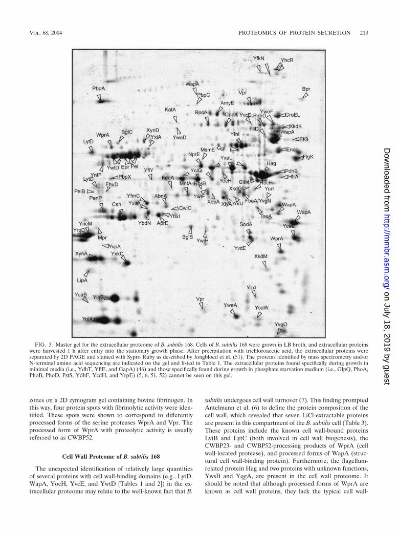
The first proteomic approaches to define the extracellularcomplement of the secretome of B. subtilis 168 were made byHirose et al. (46). In their study, cells were grown in minimalmedia with glucose, maltose, cellobiose, or starch. Extracellu-lar proteins were separated by 2D polyacrylamide gel electro-phoresis (2D PAGE) and identified by N-terminal sequencing.In subsequent studies by Antelmann et al. (3) and Jongbloed etal. (52), B. subtilis was grown under conditions of phosphatestarvation, or in Luria-Bertani (LB) broth (4, 5, 6). Extra-cellular proteins separated by 2D PAGE were identifiedby matrix-assisted laser desorption ionization/time-of-flight(MALDI-TOF) mass spectrometry. The highest levels of pro-tein secretion are usually observed when cells of B. subtilis aregrown in rich media, in particular during the postexponentialgrowth phase (see Fig. 3). Moreover, the relative amounts ofmost identified extracellular proteins were significantly in-
creased during the postexponential growth phase (5). The im-portance of protein secretion during postexponential growthwas highlighted by the fact that the extracellular levels of asubset of 13 degradative enzymes are strongly increased in theextracellular proteome of a B. subtilis degU32(hy) mutant (5,61). Recent transcript profiling experiments (66) have con-firmed that the genes encoding these degradative enzymes areindeed under the positive control of DegU-phosphate, causingtheir increased expression after the end of the exponentialgrowth phase.
Extracellular Proteome of B. subtilis 168
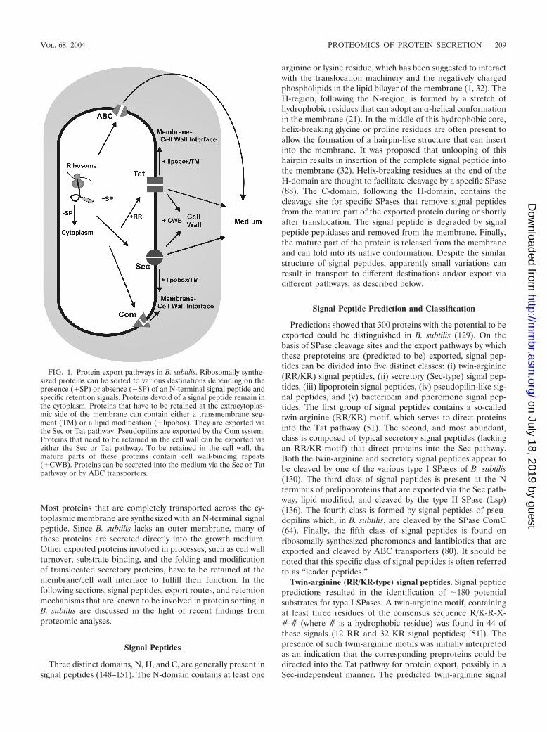
From the approximately 200 visible extracellular proteinspots, 75 different proteins could be identified as marked in the2D master gel for the extracellular proteome (Fig. 3; Tables 1and 2). Therefore, B. subtilis 168 cells were grown in Luria-Bertani broth and extracellular proteins were harvested fromthe medium 2 h after entry into the postexponential growthphase (5). In the medium of phosphate-starved cells, eightadditional extracellular proteins were identified (3, 5, 52).When B. subtilis cells were grown in minimal media, muchlower levels of extracellular proteins were detected (46). Nev-ertheless, these studies resulted in the identification of threeadditional extracellular proteins. In total, 90 extracellular pro-teins were identified, including 53 proteins to which a functionhas been assigned previously and 37 “Y-proteins” of unknownfunction (Tables 1 and 2). A possible function could be attrib-uted to 20 Y-proteins based on their amino acid sequencesimilarity to proteins with a known function. In summary, theidentified extracellular proteins of B. subtilis 168 include en-zymes related to the metabolism of carbohydrates, proteases,or peptidases, enzymes involved in the metabolism of aminoacids, enzymes involved in the decay of DNA or RNA, lipases,alkaline phosphatases, phosphodiesterases, enzymes involvedin cell wall biogenesis, lipoproteins (many of which are sub-strate-binding components of various transport systems), pro-teins involved in detoxification, flagellum-related proteins, pu-tative transcriptional regulators, proteins involved in proteinsynthesis and folding, prophage-related proteins, sporulation-specific proteins, and proteins of unknown function. In addi-tion, Chu et al. (25) identified five extracellular proteins of B.subtilis strain K-1, which were specifically induced by growth inxylan-containing medium. These are a xylose isomerase homol-ogous to XylA of B. subtilis 168, two endoxylanases homolo-gous to XynA and XynD of B. subtilis 168, a dehydroquinatedehydratase homologous to AroC of B. subtilis 168, and aregulatory protein homologous to GltC of B. subtilis 168. Thelatter proteins are not included in Tables 1 and 2, which listonly the extracellular proteins of the B. subtilis 168 strain.
Toward an Extracellular Zymoproteome of B. subtilis 168
In addition to the identification of extracellular proteins,proteomics can be used to attribute functions to extracellularproteins. An early exploration in this area was performed byPark et al. (92), who used a proteomic approach to detectfibrinolytic enzymes in the medium of B. subtilis 168. For thispurpose, images of 2D PAGE gels were superimposed to de-tect extracellular protein spots that coincided with clearing
212 TJALSMA ET AL. MICROBIOL. MOL. BIOL. REV.
on July 18, 2019 by guesthttp://m
mbr.asm
.org/D
ownloaded from
zones on a 2D zymogram gel containing bovine fibrinogen. Inthis way, four protein spots with fibrinolytic activity were iden-tified. These spots were shown to correspond to differentlyprocessed forms of the serine proteases WprA and Vpr. Theprocessed form of WprA with proteolytic activity is usuallyreferred to as CWBP52.
Cell Wall Proteome of B. subtilis 168
The unexpected identification of relatively large quantitiesof several proteins with cell wall-binding domains (e.g., LytD,WapA, YocH, YvcE, and YwtD [Tables 1 and 2]) in the ex-tracellular proteome may relate to the well-known fact that B.
subtilis undergoes cell wall turnover (7). This finding promptedAntelmann et al. (6) to define the protein composition of thecell wall, which revealed that seven LiCl-extractable proteinsare present in this compartment of the B. subtilis cell (Table 3).These proteins include the known cell wall-bound proteinsLytB and LytC (both involved in cell wall biogenesis), theCWBP23- and CWBP52-processing products of WprA (cellwall-located protease), and processed forms of WapA (struc-tural cell wall-binding protein). Furthermore, the flagellum-related protein Hag and two proteins with unknown functions,YwsB and YqgA, are present in the cell wall proteome. Itshould be noted that although processed forms of WprA areknown as cell wall proteins, they lack the typical cell wall-
FIG. 3. Master gel for the extracellular proteome of B. subtilis 168. Cells of B. subtilis 168 were grown in LB broth, and extracellular proteinswere harvested 1 h after entry into the stationary growth phase. After precipitation with trichloroacetic acid, the extracellular proteins wereseparated by 2D PAGE and stained with Sypro Ruby as described by Jongbloed et al. (51). The proteins identified by mass spectrometry and/orN-terminal amino acid sequencing are indicated on the gel and listed in Table 1. The extracellular proteins found specifically during growth inminimal media (i.e., YdhT, YflE, and GapA) (46) and those specifically found during growth in phosphate starvation medium (i.e., GlpQ, PhoA,PhoB, PhoD, PstS, YdhF, YcdH, and YrpE) (5, 6, 51, 52) cannot be seen on this gel.
VOL. 68, 2004 PROTEOMICS OF PROTEIN SECRETION 213
on July 18, 2019 by guesthttp://m
mbr.asm
.org/D
ownloaded from
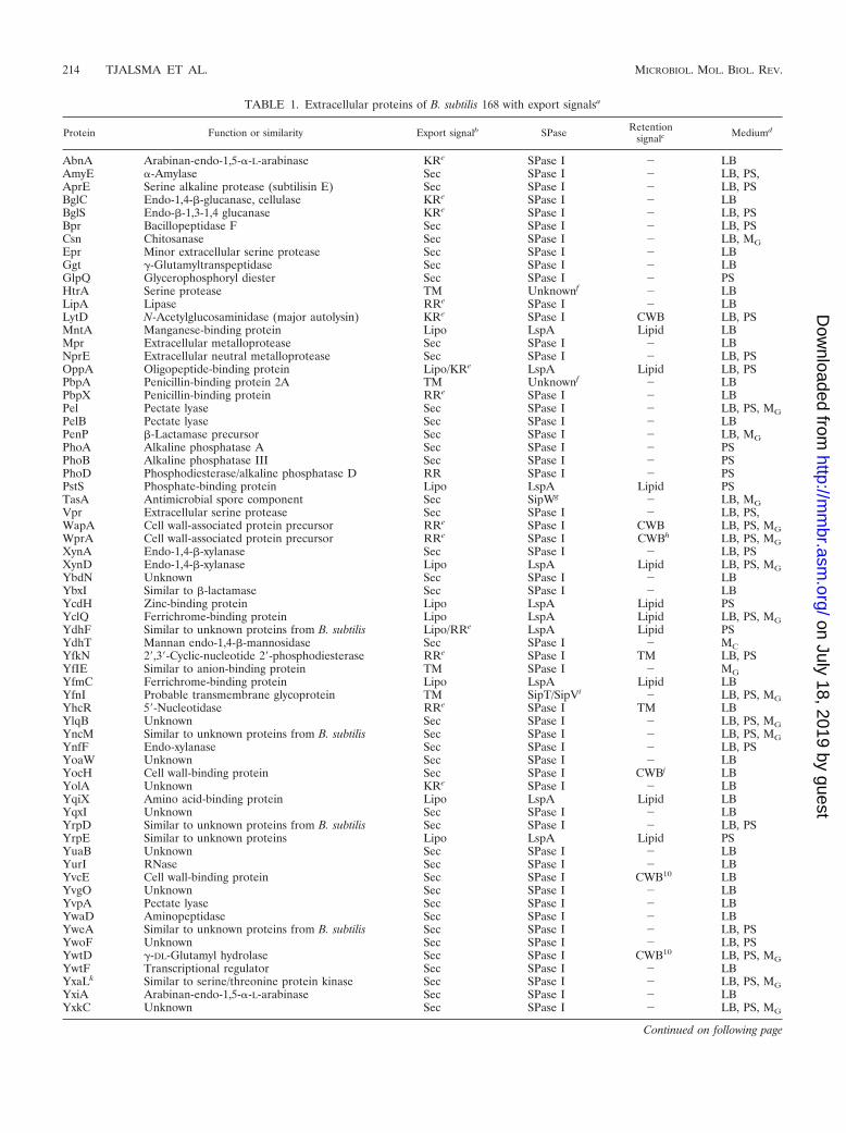
TABLE 1. Extracellular proteins of B. subtilis 168 with export signalsa
Protein Function or similarity Export signalb SPase Retentionsignalc Mediumd
AbnA Arabinan-endo-1,5-�-L-arabinase KRe SPase I � LBAmyE �-Amylase Sec SPase I � LB, PS,AprE Serine alkaline protease (subtilisin E) Sec SPase I � LB, PSBglC Endo-1,4-�-glucanase, cellulase KRe SPase I � LBBglS Endo-�-1,3-1,4 glucanase KRe SPase I � LB, PSBpr Bacillopeptidase F Sec SPase I � LB, PSCsn Chitosanase Sec SPase I � LB, MGEpr Minor extracellular serine protease Sec SPase I � LBGgt �-Glutamyltranspeptidase Sec SPase I � LBGlpQ Glycerophosphoryl diester Sec SPase I � PSHtrA Serine protease TM Unknownf � LBLipA Lipase RRe SPase I � LBLytD N-Acetylglucosaminidase (major autolysin) KRe SPase I CWB LB, PSMntA Manganese-binding protein Lipo LspA Lipid LBMpr Extracellular metalloprotease Sec SPase I � LBNprE Extracellular neutral metalloprotease Sec SPase I � LB, PSOppA Oligopeptide-binding protein Lipo/KRe LspA Lipid LB, PSPbpA Penicillin-binding protein 2A TM Unknownf � LBPbpX Penicillin-binding protein RRe SPase I � LBPel Pectate lyase Sec SPase I � LB, PS, MGPelB Pectate lyase Sec SPase I � LBPenP �-Lactamase precursor Sec SPase I � LB, MGPhoA Alkaline phosphatase A Sec SPase I � PSPhoB Alkaline phosphatase III Sec SPase I � PSPhoD Phosphodiesterase/alkaline phosphatase D RR SPase I � PSPstS Phosphate-binding protein Lipo LspA Lipid PSTasA Antimicrobial spore component Sec SipWg � LB, MGVpr Extracellular serine protease Sec SPase I � LB, PS,WapA Cell wall-associated protein precursor RRe SPase I CWB LB, PS, MGWprA Cell wall-associated protein precursor RRe SPase I CWBh LB, PS, MGXynA Endo-1,4-�-xylanase Sec SPase I � LB, PSXynD Endo-1,4-�-xylanase Lipo LspA Lipid LB, PS, MGYbdN Unknown Sec SPase I � LBYbxI Similar to �-lactamase Sec SPase I � LBYcdH Zinc-binding protein Lipo LspA Lipid PSYclQ Ferrichrome-binding protein Lipo LspA Lipid LB, PS, MGYdhF Similar to unknown proteins from B. subtilis Lipo/RRe LspA Lipid PSYdhT Mannan endo-1,4-�-mannosidase Sec SPase I � MCYfkN 2�,3�-Cyclic-nucleotide 2�-phosphodiesterase RRe SPase I TM LB, PSYfIE Similar to anion-binding protein TM SPase I � MGYfmC Ferrichrome-binding protein Lipo LspA Lipid LBYfnI Probable transmembrane glycoprotein TM SipT/SipVi � LB, PS, MGYhcR 5�-Nucleotidase RRe SPase I TM LBYlqB Unknown Sec SPase I � LB, PS, MGYncM Similar to unknown proteins from B. subtilis Sec SPase I � LB, PS, MGYnfF Endo-xylanase Sec SPase I � LB, PSYoaW Unknown Sec SPase I � LBYocH Cell wall-binding protein Sec SPase I CWBj LBYolA Unknown KRe SPase I � LBYqiX Amino acid-binding protein Lipo LspA Lipid LBYqxI Unknown Sec SPase I � LBYrpD Similar to unknown proteins from B. subtilis Sec SPase I � LB, PSYrpE Similar to unknown proteins Lipo LspA Lipid PSYuaB Unknown Sec SPase I � LBYurI RNase Sec SPase I � LBYvcE Cell wall-binding protein Sec SPase I CWB10 LBYvgO Unknown Sec SPase I � LBYvpA Pectate lyase Sec SPase I � LBYwaD Aminopeptidase Sec SPase I � LBYweA Similar to unknown proteins from B. subtilis Sec SPase I � LB, PSYwoF Unknown Sec SPase I � LB, PSYwtD �-DL-Glutamyl hydrolase Sec SPase I CWB10 LB, PS, MGYwtF Transcriptional regulator Sec SPase I � LBYxaLk Similar to serine/threonine protein kinase Sec SPase I � LB, PS, MGYxiA Arabinan-endo-1,5-�-L-arabinase Sec SPase I � LBYxkC Unknown Sec SPase I � LB, PS, MG
Continued on following page
214 TJALSMA ET AL. MICROBIOL. MOL. BIOL. REV.
on July 18, 2019 by guesthttp://m
mbr.asm
.org/D
ownloaded from

binding repeats that are present in LytB, LytC, and WapA (6, 68,129). Surprisingly, two additional cell wall-located proteins, YwsBand YqgA, also lack known cell wall retention motifs. Finally, itwas remarkable that extracellular proteins with cell wall-bindingmotifs, such as LytD, YocH, YvcE, and YwtD, were apparentlyabsent from the cell wall proteome. It has to be emphasized thatLytD, YvcE, and YwtD are abundantly present in the extracellu-lar proteome under the growth conditions used to identify cellwall-associated proteins (5), showing that the absence of theseproteins from the wall proteome is not due to a lack of expression.Probably, the same is true for YocH, but this is less clearly evidentfrom the published data (6).
Verification of Secretome Predictions
The availability of accumulating proteomic data concerningthe extracellular complement of the secretome of B. subtilis168 has allowed proteomic verification of the genome-basedpredictions of the composition of the secretome as previouslyperformed (129). Intriguingly, only 48 (53%) of the 90 identi-fied extracellular proteins are expected to be released into themedium, as judged by the presence of predicted signal peptidesand a lack of retention signals (Tables 1 and 2). A potentialRR/KR motif is present in the N-domains of 14 signal peptidesof the latter group of proteins, suggesting their potential trans-port via the Tat pathway. The remaining 34 proteins contain aSec-type signal peptide and are most probably exported by theSec pathway of B. subtilis. Strikingly, 47% of the extracellularproteome currently cannot be predicted to end up at this lo-cation (129). This unpredicted fraction consists of proteinswhich have an N-terminal lipoprotein signal peptide (cleavedby SPase II) or potential transmembrane segments accordingto the TMHMM algorithm (28). Both groups of proteins aresupposed to be retained in or at the cytoplasmic membrane. Inaddition, some predicted preproteins with a type I SPasecleavage site contain typical cell wall-binding repeats andtherefore have a predicted cell wall localization. As listed inTable 2, 24 proteins found in the medium of B. subtilis arein fact proteins that lack a typical export signal. The latterinclude flagellum-related proteins, prophage-related proteins,and proteins with known or predicted enzymatic activities in thecytoplasm. The possible mechanisms by which these proteins are
released from the cell are discussed in “Mechanisms for extracel-lular accumulation of proteins” (below).
Similarly, only about half of the identified cell wall proteinsare predicted to be retained at this subcellular location, sinceHag, the WprA-processing products CWBP23 and CWBP52,YqgA, and YwsB lack known cell wall-binding motifs. The lasttwo proteins are, in fact, found exclusively in the cell wall, likethe known cell wall-bound proteins LytB and LytC. This sug-gests that an as yet undefined cell wall retention signal ispresent in YqgA and YwsB. Conversely, the remarkable ob-servation that four proteins with typical cell wall-binding do-mains (i.e., LytD, YocH, YvcE, and YwtD) are found extra-cellularly, but not cell wall bound, might indicate that thepresence of a cell wall-binding repeat is not a guarantee forretention at this location. In this respect, it may be relevantthat YwtD exclusively cleaves extracellular �-polyglutamic acidwhereas it cannot use cell wall peptidoglycan as a substrate(127). However, the possibilities that LytD, YocH, YvcE, andYwtD are not properly extracted from the wall with LiCl, orthat these proteins are degraded during the extraction proce-dure, cannot be excluded.
CONTRIBUTION OF THE Sec MACHINERY TO THEEXTRACELLULAR PROTEOME
Protein secretion via the Sec pathway in B. subtilis can bedivided into three functional stages: targeting, translocation,and folding and release. The following components haveknown or predicted functions in these stages. Cytoplasmicchaperones, such as SRP/FtsY (47) and CsaA (75, 76), keepthe precursors in a translocation competent state and facilitatetheir targeting to the translocase in the membrane. The trans-location machinery consists of SecA (motor), SecYEG (pore),and SecDF. Possibly, YrbF and SpoIIIJ/YqjG are also part ofthis machinery (17, 129, 135, 145). During or shortly aftertranslocation, the preprotein is cleaved by one of the five typeI signal peptidases (SipS to SipW) (130) or lipid-modified bythe diacylglyceryl transferase (Lgt) (62) and cleaved by thelipoprotein-specific signal peptidase (Lsp; 133, 136). SppA andTepA may be involved in the degradation of cleaved signalpeptides (16). The folding of several secreted proteins dependson the activities of PrsA (55), BdbBCD (18, 71), and/or
TABLE 1—Continueda All listed proteins were identified by 2D PAGE and subsequent MALDI-TOF mass spectrometry and/or N-terminal amino acid sequencing as described by Hirose
et al. (46), Jongbloed et al. (51, 52), and Antelmann et al. (3–6). Putative signal peptides. SPase I or SPase II cleavage sites, transmembrane domains, and cellwall-binding domains were predicted as described by Tjalsma et al. (129) and Jongbloed et al. (51).
b Identified transient export signals are Sec-type signal peptides (Sec), twin-arginine signal peptides (RR/KR), lipoprotein signal peptides (Lipo), and transmembranedomains (TM).
c Identified retention signals present in the mature part of the protein after processing by specific SPases are lipid modifications (Lipid), transmembrane domains(TM), and cell wall-binding domains (CWB). �, absence of known retention signals.
d Proteins found in the extracellular proteomes of cells were grown in LB broth (rich medium) (4, 5, 6, 51), phosphate starvation medium (PS) (3, 5, 52), or minimalmedium with glucose (MG) or cellobiose (MC) (46).
e Despite the presence of putative RR/KR-type signal peptides, release of AbnA, BglC, BglS, LipA, LytD, OppA, PbpX, WapA, WprA, YdhF, YfkN, YhcR, andYolA into the growth medium is not Tat dependent (51).
f As pre-PbpA and pre-HtrA lack putative SPase I cleavage sites, it is unknown which protease is responsible for their cleavage and subsequent release into themedium (4, 5).
g pre-TasA processing and release of mature TasA into the medium is strictly dependent on the ER-type SPase SipW (126, 131).h WprA is known to be a major cell wall protein (6, 68), but it lacks a typical cell wall-binding motif.i Release of the C-terminal part of YfnI into the medium was shown to be dependent on the presence of SipT or SipV (5).j Despite the presence of putative cell wall-binding domains, YocH, YvcE, and YwtD are not detected in the cell wall proteome of B. subtilis 168 (6).k The protein YxaL was previously annotated as YxaK (5).
VOL. 68, 2004 PROTEOMICS OF PROTEIN SECRETION 215
on July 18, 2019 by guesthttp://m
mbr.asm
.org/D
ownloaded from
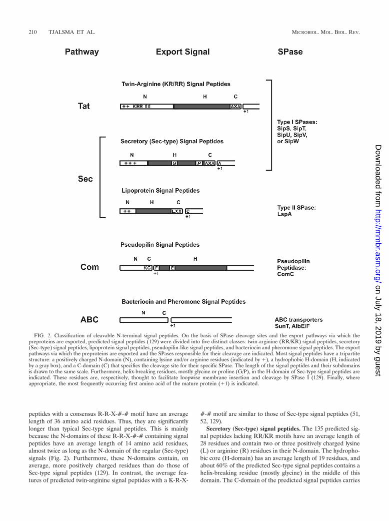
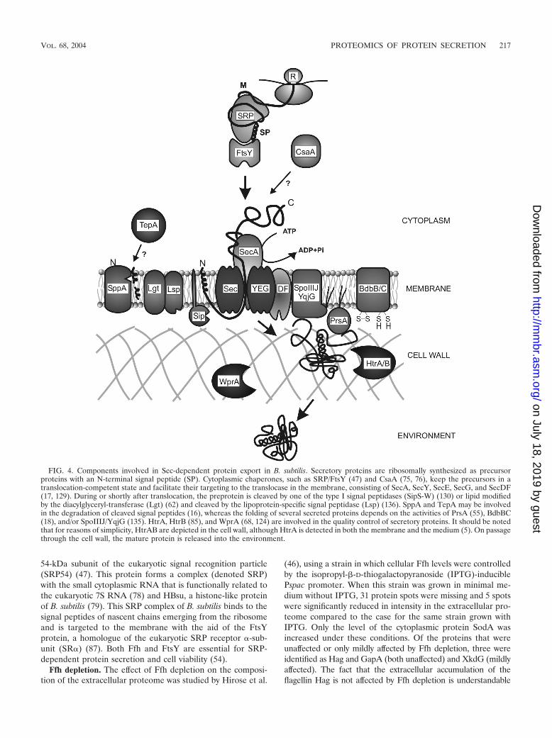
SpoIIIJ/YqjG (135). HtrA and HtrB (85), as well as WprA(68), are involved in the quality control of secretory proteins.Importantly, HtrA and HtrB have the potential to assist in thefolding or, if folding is impossible, degradation of malfoldedsecretory proteins. A model for the function of these maincomponents of the Sec machinery of B. subtilis is depicted inFig. 4. Using proteomic approaches, the extracellular pro-teomes of B. subtilis mutants that are affected in differentstages in protein secretion have been analyzed. In the followingsections, we review the currently available proteome data con-cerning B. subtilis strains lacking, or depleted of, various com-
ponents involved in Sec-dependent protein export. These dataare summarized in Table 4.
Cytoplasmic Targeting Factors
Since B. subtilis lacks a secretion-specific targeting factorsimilar to the SecB protein of Escherichia coli (99), an impor-tant role in this process has been attributed to the highlyconserved signal recognition particle (SRP) pathway (129). Animportant component of this pathway is the Ffh protein (for“fifty-four homologue”), a GTPase that is homologous to the
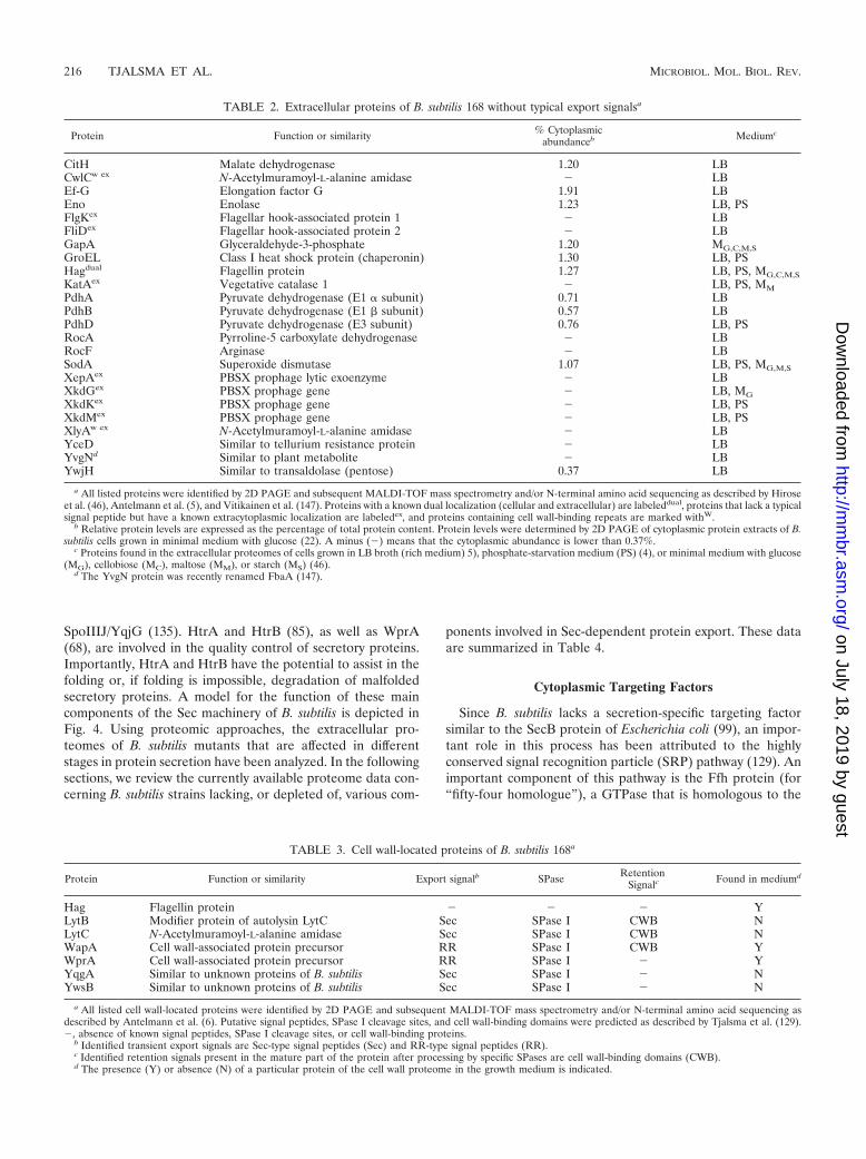
TABLE 2. Extracellular proteins of B. subtilis 168 without typical export signalsa
Protein Function or similarity % Cytoplasmicabundanceb Mediumc
CitH Malate dehydrogenase 1.20 LBCwlCw ex N-Acetylmuramoyl-L-alanine amidase � LBEf-G Elongation factor G 1.91 LBEno Enolase 1.23 LB, PSFlgKex Flagellar hook-associated protein 1 � LBFliDex Flagellar hook-associated protein 2 � LBGapA Glyceraldehyde-3-phosphate 1.20 MG,C,M,SGroEL Class I heat shock protein (chaperonin) 1.30 LB, PSHagdual Flagellin protein 1.27 LB, PS, MG,C,M,SKatAex Vegetative catalase 1 � LB, PS, MMPdhA Pyruvate dehydrogenase (E1 � subunit) 0.71 LBPdhB Pyruvate dehydrogenase (E1 � subunit) 0.57 LBPdhD Pyruvate dehydrogenase (E3 subunit) 0.76 LB, PSRocA Pyrroline-5 carboxylate dehydrogenase � LBRocF Arginase � LBSodA Superoxide dismutase 1.07 LB, PS, MG,M,SXepAex PBSX prophage lytic exoenzyme � LBXkdGex PBSX prophage gene � LB, MGXkdKex PBSX prophage gene � LB, PSXkdMex PBSX prophage gene � LB, PSXlyAw ex N-Acetylmuramoyl-L-alanine amidase � LBYceD Similar to tellurium resistance protein � LBYvgNd Similar to plant metabolite � LBYwjH Similar to transaldolase (pentose) 0.37 LB
a All listed proteins were identified by 2D PAGE and subsequent MALDI-TOF mass spectrometry and/or N-terminal amino acid sequencing as described by Hiroseet al. (46), Antelmann et al. (5), and Vitikainen et al. (147). Proteins with a known dual localization (cellular and extracellular) are labeleddual, proteins that lack a typicalsignal peptide but have a known extracytoplasmic localization are labeledex, and proteins containing cell wall-binding repeats are marked withW.
b Relative protein levels are expressed as the percentage of total protein content. Protein levels were determined by 2D PAGE of cytoplasmic protein extracts of B.subtilis cells grown in minimal medium with glucose (22). A minus (�) means that the cytoplasmic abundance is lower than 0.37%.
c Proteins found in the extracellular proteomes of cells grown in LB broth (rich medium) 5), phosphate-starvation medium (PS) (4), or minimal medium with glucose(MG), cellobiose (MC), maltose (MM), or starch (MS) (46).
d The YvgN protein was recently renamed FbaA (147).
TABLE 3. Cell wall-located proteins of B. subtilis 168a
Protein Function or similarity Export signalb SPase RetentionSignalc Found in mediumd
Hag Flagellin protein � � � YLytB Modifier protein of autolysin LytC Sec SPase I CWB NLytC N-Acetylmuramoyl-L-alanine amidase Sec SPase I CWB NWapA Cell wall-associated protein precursor RR SPase I CWB YWprA Cell wall-associated protein precursor RR SPase I � YYqgA Similar to unknown proteins of B. subtilis Sec SPase I � NYwsB Similar to unknown proteins of B. subtilis Sec SPase I � N
a All listed cell wall-located proteins were identified by 2D PAGE and subsequent MALDI-TOF mass spectrometry and/or N-terminal amino acid sequencing asdescribed by Antelmann et al. (6). Putative signal peptides, SPase I cleavage sites, and cell wall-binding domains were predicted as described by Tjalsma et al. (129).�, absence of known signal peptides, SPase I cleavage sites, or cell wall-binding proteins.
b Identified transient export signals are Sec-type signal peptides (Sec) and RR-type signal peptides (RR).c Identified retention signals present in the mature part of the protein after processing by specific SPases are cell wall-binding domains (CWB).d The presence (Y) or absence (N) of a particular protein of the cell wall proteome in the growth medium is indicated.
216 TJALSMA ET AL. MICROBIOL. MOL. BIOL. REV.
on July 18, 2019 by guesthttp://m
mbr.asm
.org/D
ownloaded from
54-kDa subunit of the eukaryotic signal recognition particle(SRP54) (47). This protein forms a complex (denoted SRP)with the small cytoplasmic RNA that is functionally related tothe eukaryotic 7S RNA (78) and HBsu, a histone-like proteinof B. subtilis (79). This SRP complex of B. subtilis binds to thesignal peptides of nascent chains emerging from the ribosomeand is targeted to the membrane with the aid of the FtsYprotein, a homologue of the eukaryotic SRP receptor �-sub-unit (SR�) (87). Both Ffh and FtsY are essential for SRP-dependent protein secretion and cell viability (54).
Ffh depletion. The effect of Ffh depletion on the composi-tion of the extracellular proteome was studied by Hirose et al.
(46), using a strain in which cellular Ffh levels were controlledby the isopropyl-�-D-thiogalactopyranoside (IPTG)-induciblePspac promoter. When this strain was grown in minimal me-dium without IPTG, 31 protein spots were missing and 5 spotswere significantly reduced in intensity in the extracellular pro-teome compared to the case for the same strain grown withIPTG. Only the level of the cytoplasmic protein SodA wasincreased under these conditions. Of the proteins that wereunaffected or only mildly affected by Ffh depletion, three wereidentified as Hag and GapA (both unaffected) and XkdG (mildlyaffected). The fact that the extracellular accumulation of theflagellin Hag is not affected by Ffh depletion is understandable
FIG. 4. Components involved in Sec-dependent protein export in B. subtilis. Secretory proteins are ribosomally synthesized as precursorproteins with an N-terminal signal peptide (SP). Cytoplasmic chaperones, such as SRP/FtsY (47) and CsaA (75, 76), keep the precursors in atranslocation-competent state and facilitate their targeting to the translocase in the membrane, consisting of SecA, SecY, SecE, SecG, and SecDF(17, 129). During or shortly after translocation, the preprotein is cleaved by one of the type I signal peptidases (SipS-W) (130) or lipid modifiedby the diacylglyceryl-transferase (Lgt) (62) and cleaved by the lipoprotein-specific signal peptidase (Lsp) (136). SppA and TepA may be involvedin the degradation of cleaved signal peptides (16), whereas the folding of several secreted proteins depends on the activities of PrsA (55), BdbBC(18), and/or SpoIIIJ/YqjG (135). HtrA, HtrB (85), and WprA (68, 124) are involved in the quality control of secretory proteins. It should be notedthat for reasons of simplicity, HtrAB are depicted in the cell wall, although HtrA is detected in both the membrane and the medium (5). On passagethrough the cell wall, the mature protein is released into the environment.
VOL. 68, 2004 PROTEOMICS OF PROTEIN SECRETION 217
on July 18, 2019 by guesthttp://m
mbr.asm
.org/D
ownloaded from
since this protein is exported by a specific flagellin assembly path-way (48). In addition, GapA is a cytoplasmic protein (44) that isreleased into the medium by an unknown mechanism. Further-more, XkdG is a prophage-related protein that is probably ex-ported by a specific prophage PBSX-encoded holin system. Thesedata suggest that flagellin assembly, release of certain cytoplasmicproteins, and export of certain phage-related proteins are not(strictly) SRP dependent. In contrast, all identified extracellularproteins with cleavable Sec-type signal peptides, Csn, Pel, PenP,TasA, WapA, WprA, XynD, YclQ, YlqB, YncM, YwtD, YxaK,and YxkC, were completely absent from the medium of Ffh-depleted cells. These data strongly suggest that signal peptide-dependent protein secretion via the Sec pathway is, directly orindirectly, SRP dependent. In this respect, it is important to con-sider the possibility that membrane proteins critical for proteintranslocation, such as certain components of the Sec translocase,could be inserted SRP dependently into the membrane. If so,SRP depletion would indirectly have a negative impact on the
secretion of proteins with Sec-type signal peptides. Finally, YfnIand YflE are two paralogous transmembrane proteins whoseC-terminal domain is released into the medium (5, 46). Therelease of YfnI was mildly affected on Ffh depletion, whereas therelease of YflE was completely inhibited under these conditions.This might indicate that compared to protein secretion, lowerlevels of SRP are required for the insertion of certain transmem-brane proteins into the cytoplasmic membrane.
Sec Translocase
The preprotein translocation machinery of the B. subtilis Secpathway consists of at least four proteins: SecA, which is thetranslocation motor, and the integral membrane proteinsSecE, SecG, and SecY. In the current model for preproteintranslocation in B. subtilis, which has many similarities to thatof E. coli, several successive steps in the translocation of pro-teins occur (36, 38, 73, 129, 145). First, SecA binds to the
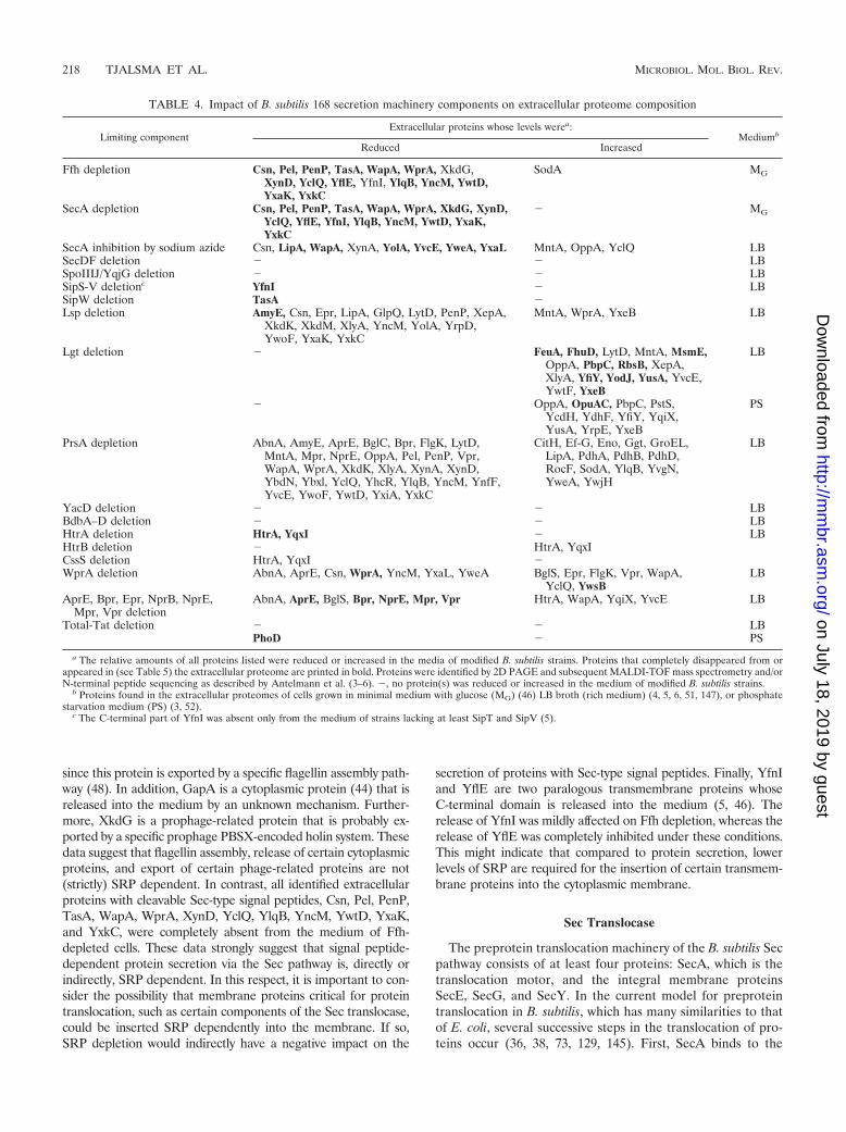
TABLE 4. Impact of B. subtilis 168 secretion machinery components on extracellular proteome composition
Limiting componentExtracellular proteins whose levels werea:
Mediumb
Reduced Increased
Ffh depletion Csn, Pel, PenP, TasA, WapA, WprA, XkdG,XynD, YclQ, YflE, YfnI, YlqB, YncM, YwtD,YxaK, YxkC
SodA MG
SecA depletion Csn, Pel, PenP, TasA, WapA, WprA, XkdG, XynD,YclQ, YflE, YfnI, YlqB, YncM, YwtD, YxaK,YxkC
� MG
SecA inhibition by sodium azide Csn, LipA, WapA, XynA, YolA, YvcE, YweA, YxaL MntA, OppA, YclQ LBSecDF deletion � � LBSpoIIIJ/YqjG deletion � � LBSipS-V deletionc YfnI � LBSipW deletion TasA �Lsp deletion AmyE, Csn, Epr, LipA, GlpQ, LytD, PenP, XepA,
XkdK, XkdM, XlyA, YncM, YolA, YrpD,YwoF, YxaK, YxkC
MntA, WprA, YxeB LB
Lgt deletion � FeuA, FhuD, LytD, MntA, MsmE,OppA, PbpC, RbsB, XepA,XlyA, YfiY, YodJ, YusA, YvcE,YwtF, YxeB
LB
� OppA, OpuAC, PbpC, PstS,YcdH, YdhF, YfiY, YqiX,YusA, YrpE, YxeB
PS
PrsA depletion AbnA, AmyE, AprE, BglC, Bpr, FlgK, LytD,MntA, Mpr, NprE, OppA, Pel, PenP, Vpr,WapA, WprA, XkdK, XlyA, XynA, XynD,YbdN, Ybxl, YclQ, YhcR, YlqB, YncM, YnfF,YvcE, YwoF, YwtD, YxiA, YxkC
CitH, Ef-G, Eno, Ggt, GroEL,LipA, PdhA, PdhB, PdhD,RocF, SodA, YlqB, YvgN,YweA, YwjH
LB
YacD deletion � � LBBdbA–D deletion � � LBHtrA deletion HtrA, YqxI � LBHtrB deletion � HtrA, YqxICssS deletion HtrA, YqxI �WprA deletion AbnA, AprE, Csn, WprA, YncM, YxaL, YweA BglS, Epr, FlgK, Vpr, WapA,
YclQ, YwsBLB
AprE, Bpr, Epr, NprB, NprE,Mpr, Vpr deletion
AbnA, AprE, BglS, Bpr, NprE, Mpr, Vpr HtrA, WapA, YqiX, YvcE LB
Total-Tat deletion � � LBPhoD � PS
a The relative amounts of all proteins listed were reduced or increased in the media of modified B. subtilis strains. Proteins that completely disappeared from orappeared in (see Table 5) the extracellular proteome are printed in bold. Proteins were identified by 2D PAGE and subsequent MALDI-TOF mass spectrometry and/orN-terminal peptide sequencing as described by Antelmann et al. (3–6). �, no protein(s) was reduced or increased in the medium of modified B. subtilis strains.
b Proteins found in the extracellular proteomes of cells grown in minimal medium with glucose (MG) (46) LB broth (rich medium) (4, 5, 6, 51, 147), or phosphatestarvation medium (PS) (3, 52).
c The C-terminal part of YfnI was absent only from the medium of strains lacking at least SipT and SipV (5).
218 TJALSMA ET AL. MICROBIOL. MOL. BIOL. REV.
on July 18, 2019 by guesthttp://m
mbr.asm
.org/D
ownloaded from
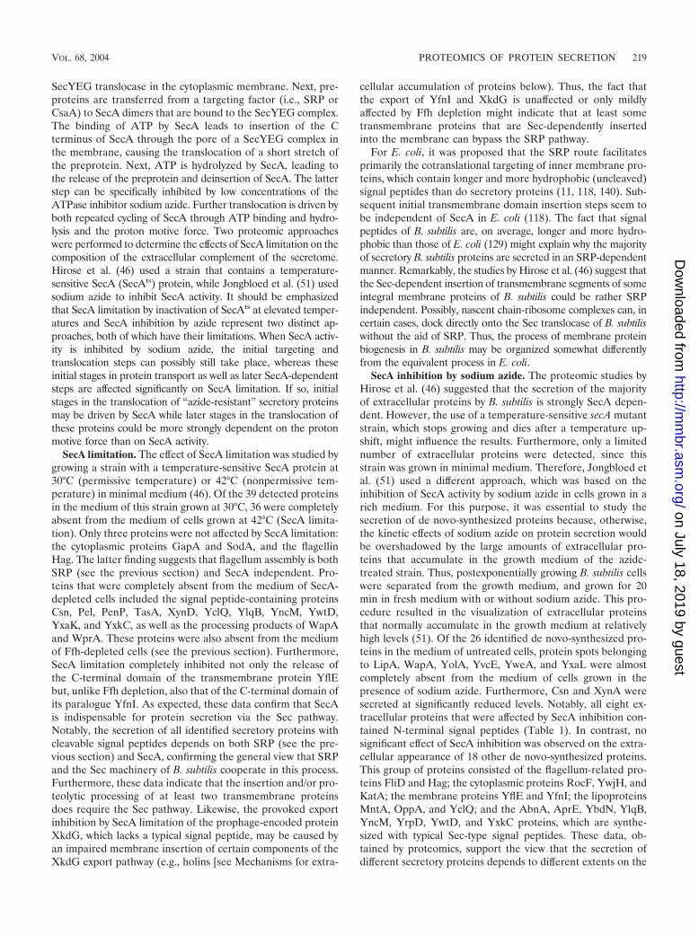
SecYEG translocase in the cytoplasmic membrane. Next, pre-proteins are transferred from a targeting factor (i.e., SRP orCsaA) to SecA dimers that are bound to the SecYEG complex.The binding of ATP by SecA leads to insertion of the Cterminus of SecA through the pore of a SecYEG complex inthe membrane, causing the translocation of a short stretch ofthe preprotein. Next, ATP is hydrolyzed by SecA, leading tothe release of the preprotein and deinsertion of SecA. The latterstep can be specifically inhibited by low concentrations of theATPase inhibitor sodium azide. Further translocation is driven byboth repeated cycling of SecA through ATP binding and hydro-lysis and the proton motive force. Two proteomic approacheswere performed to determine the effects of SecA limitation on thecomposition of the extracellular complement of the secretome.Hirose et al. (46) used a strain that contains a temperature-sensitive SecA (SecAts) protein, while Jongbloed et al. (51) usedsodium azide to inhibit SecA activity. It should be emphasizedthat SecA limitation by inactivation of SecAts at elevated temper-atures and SecA inhibition by azide represent two distinct ap-proaches, both of which have their limitations. When SecA activ-ity is inhibited by sodium azide, the initial targeting andtranslocation steps can possibly still take place, whereas theseinitial stages in protein transport as well as later SecA-dependentsteps are affected significantly on SecA limitation. If so, initialstages in the translocation of “azide-resistant” secretory proteinsmay be driven by SecA while later stages in the translocation ofthese proteins could be more strongly dependent on the protonmotive force than on SecA activity.
SecA limitation. The effect of SecA limitation was studied bygrowing a strain with a temperature-sensitive SecA protein at30°C (permissive temperature) or 42°C (nonpermissive tem-perature) in minimal medium (46). Of the 39 detected proteinsin the medium of this strain grown at 30°C, 36 were completelyabsent from the medium of cells grown at 42°C (SecA limita-tion). Only three proteins were not affected by SecA limitation:the cytoplasmic proteins GapA and SodA, and the flagellinHag. The latter finding suggests that flagellum assembly is bothSRP (see the previous section) and SecA independent. Pro-teins that were completely absent from the medium of SecA-depleted cells included the signal peptide-containing proteinsCsn, Pel, PenP, TasA, XynD, YclQ, YlqB, YncM, YwtD,YxaK, and YxkC, as well as the processing products of WapAand WprA. These proteins were also absent from the mediumof Ffh-depleted cells (see the previous section). Furthermore,SecA limitation completely inhibited not only the release ofthe C-terminal domain of the transmembrane protein YflEbut, unlike Ffh depletion, also that of the C-terminal domain ofits paralogue YfnI. As expected, these data confirm that SecAis indispensable for protein secretion via the Sec pathway.Notably, the secretion of all identified secretory proteins withcleavable signal peptides depends on both SRP (see the pre-vious section) and SecA, confirming the general view that SRPand the Sec machinery of B. subtilis cooperate in this process.Furthermore, these data indicate that the insertion and/or pro-teolytic processing of at least two transmembrane proteinsdoes require the Sec pathway. Likewise, the provoked exportinhibition by SecA limitation of the prophage-encoded proteinXkdG, which lacks a typical signal peptide, may be caused byan impaired membrane insertion of certain components of theXkdG export pathway (e.g., holins [see Mechanisms for extra-
cellular accumulation of proteins below). Thus, the fact thatthe export of YfnI and XkdG is unaffected or only mildlyaffected by Ffh depletion might indicate that at least sometransmembrane proteins that are Sec-dependently insertedinto the membrane can bypass the SRP pathway.
For E. coli, it was proposed that the SRP route facilitatesprimarily the cotranslational targeting of inner membrane pro-teins, which contain longer and more hydrophobic (uncleaved)signal peptides than do secretory proteins (11, 118, 140). Sub-sequent initial transmembrane domain insertion steps seem tobe independent of SecA in E. coli (118). The fact that signalpeptides of B. subtilis are, on average, longer and more hydro-phobic than those of E. coli (129) might explain why the majorityof secretory B. subtilis proteins are secreted in an SRP-dependentmanner. Remarkably, the studies by Hirose et al. (46) suggest thatthe Sec-dependent insertion of transmembrane segments of someintegral membrane proteins of B. subtilis could be rather SRPindependent. Possibly, nascent chain-ribosome complexes can, incertain cases, dock directly onto the Sec translocase of B. subtiliswithout the aid of SRP. Thus, the process of membrane proteinbiogenesis in B. subtilis may be organized somewhat differentlyfrom the equivalent process in E. coli.
SecA inhibition by sodium azide. The proteomic studies byHirose et al. (46) suggested that the secretion of the majorityof extracellular proteins by B. subtilis is strongly SecA depen-dent. However, the use of a temperature-sensitive secA mutantstrain, which stops growing and dies after a temperature up-shift, might influence the results. Furthermore, only a limitednumber of extracellular proteins were detected, since thisstrain was grown in minimal medium. Therefore, Jongbloed etal. (51) used a different approach, which was based on theinhibition of SecA activity by sodium azide in cells grown in arich medium. For this purpose, it was essential to study thesecretion of de novo-synthesized proteins because, otherwise,the kinetic effects of sodium azide on protein secretion wouldbe overshadowed by the large amounts of extracellular pro-teins that accumulate in the growth medium of the azide-treated strain. Thus, postexponentially growing B. subtilis cellswere separated from the growth medium, and grown for 20min in fresh medium with or without sodium azide. This pro-cedure resulted in the visualization of extracellular proteinsthat normally accumulate in the growth medium at relativelyhigh levels (51). Of the 26 identified de novo-synthesized pro-teins in the medium of untreated cells, protein spots belongingto LipA, WapA, YolA, YvcE, YweA, and YxaL were almostcompletely absent from the medium of cells grown in thepresence of sodium azide. Furthermore, Csn and XynA weresecreted at significantly reduced levels. Notably, all eight ex-tracellular proteins that were affected by SecA inhibition con-tained N-terminal signal peptides (Table 1). In contrast, nosignificant effect of SecA inhibition was observed on the extra-cellular appearance of 18 other de novo-synthesized proteins.This group of proteins consisted of the flagellum-related pro-teins FliD and Hag; the cytoplasmic proteins RocF, YwjH, andKatA; the membrane proteins YflE and YfnI; the lipoproteinsMntA, OppA, and YclQ; and the AbnA, AprE, YbdN, YlqB,YncM, YrpD, YwtD, and YxkC proteins, which are synthe-sized with typical Sec-type signal peptides. These data, ob-tained by proteomics, support the view that the secretion ofdifferent secretory proteins depends to different extents on the
VOL. 68, 2004 PROTEOMICS OF PROTEIN SECRETION 219
on July 18, 2019 by guesthttp://m
mbr.asm
.org/D
ownloaded from
activity of SecA. Accordingly, previous research has shown adifference in the SecA requirements of the �-amylase AmyE(requiring low levels of SecA activity) and the levansucraseSacB (requiring high levels of SecA activity) for secretion intothe medium of B. subtilis (60). Notably, the signal peptides ofSacB and AmyE are quite different (129, 143). The H-domainof the signal peptide of AmyE is longer than that of SacB (23and 17 residues, respectively), and its overall hydrophobicity ishigher (1.8 and 1.1 residues, respectively). It was thereforeproposed that these differences in the H-domains could beresponsible for the difference in SecA requirement of pre-AmyE and pre-SacB (60). Although AmyE and SacB were notdetected in the proteomic analysis of SecA-dependent (i.e.,azide-sensitive) protein secretion (51), these studies provided agood opportunity to evaluate the above-mentioned hypothesis.Primary amino acid sequence analysis showed, however, thatthe H-domains of the signal peptides of both azide-sensitiveand azide-resistant secretory proteins have an average lengthof 22 residues and an average hydrophobicity between 1.5 and1.6. Thus, it seems unlikely that the H-domain is the maindeterminant for the SecA requirement of a preprotein. Incontrast, the N-domains of the signal peptides of the eighthighly azide-sensitive secretory proteins are on average shorter(7 versus 11 residues) and more hydrophilic (�1.4 versus �1.1)than those of the eight azide-resistant secretory proteins. Nev-ertheless, the number of positively charged residues in theN-regions of signal peptides of azide-sensitive and azide-resis-tant secretory proteins did not significantly differ (3.6 on aver-age). Although this finding might indicate that the N-domainof signal peptides is a determinant for the SecA requirement ofa preprotein, a larger data set and site-directed mutagenesisapproaches are needed to pinpoint the relevant features ofsignal peptides in relation to the extent of SecA requirement ofthe corresponding preprotein. It is conceivable, however, thatspecific properties of the mature parts of particular secretorypreproteins are more important in determining their SecArequirement than are the properties of their signal peptides.
Another interesting observation from the azide inhibitionexperiments is that all three lipoproteins that are detectable inthe extracellular proteome of untreated cells are present inequal or even larger amounts in the medium of cells treatedwith sodium azide. This suggests that the transport of lipopro-teins via the Sec pathway requires less SecA activity than doesthe transport of secretory proteins. Furthermore, the insertionand release of the C-terminal domains of the transmembraneproteins YflE and YfnI do not seem to be affected by SecAinhibition with sodium azide. This is in marked contrast to theresults obtained by Hirose et al. (46), who showed that therelease of YflE, YfnI, and the lipoprotein YclQ into the me-dium of a temperature-sensitive secA mutant strain was com-pletely blocked by SecA limitation. Similarly, the extracellularappearance of secretory proteins whose export was not (com-pletely) inhibited by sodium azide (e.g., Csn, YwtD, YxkC,YncM and YlqB [51]) was completely blocked by SecA deple-tion (46). This shows that the secA mutation employed byHirose et al. (46) is more effective in reducing the SecA trans-location motor activity than is sodium azide.
SecDF deletion and SpoIIIJ and YqjG depletion. In additionto the heterotrimeric SecYEG subcomplex, the E. coli Secmachinery contains a second heterotrimeric subcomplex that is
composed of the SecD, SecF, and YajC proteins. This secondsubcomplex is likely to form a part of the B. subtilis Sec ma-chinery as well, although this has not been demonstrated ex-perimentally. In B. subtilis, this complex would be composed ofthe SecDF protein (a natural fusion protein of SecD and SecF)and YrbF (a homologue of E. coli YajC). The precise role ofSecDF-YajC in protein export is presently not clear, but avariety of possible functions have been proposed. These in-clude (i) removal of cleaved signal peptides or transmembranesegments from the SecYEG translocation channel; (ii) releaseof translocated proteins from the translocation channel; (iii)regulation of SecA cycling; and (iv) prevention of preproteinbacksliding (86). Unlike SecD and SecF of E. coli, SecDF of B.subtilis 168 was shown to have little impact on cell viability andprotein export, at least under standard laboratory conditions(17). A secretion defect in a secDF mutant strain was observedonly under conditions of high-level expression of secretoryproteins, such as AmyQ of Bacillus amyloliquefaciens. Accord-ingly, the disruption of secDF had no detectable influence onthe composition of the extracellular proteome (H. Antelmann,unpublished observations).
A final component that can associate with the Sec translo-case of E. coli is the YidC protein, which is involved in themembrane insertion of newly synthesized membrane proteins(65, 108, 116). Interestingly, YidC seems to be linked to theSecYEG subcomplex of the translocase through the SecDF-YajC subcomplex (86). B. subtilis contains two homologues ofYidC, known as SpollIJ and YqjG. Remarkably, the biogenesisof a variety of integral membrane proteins in B. subtilis is onlymildly affected in cells depleted of both SpoIIIJ and YqjG(135). In contrast, the simultaneous removal of SpoIIIJ andYqjG has a severe impact on (as yet undefined) posttranslo-cational stages in the secretion of proteins, such as AmyQ,LipA, and E. coli PhoA (135). Unfortunately, proteomic stud-ies with SpoIIIJ/YqjG-depleted cells turned out to be difficult,since the combined activities of these proteins are essential forcell viability. The extracellular proteomes of spoIIIJ and yqjGsingle mutants, which display no growth defects, revealed nosignificant changes compared to that of the parental strain (H.Antelmann and H. Tjalsma, unpublished observations).
Type I Signal Peptidases
SPases remove signal peptides from secretory preproteinswhen the C-domain of the signal peptide emerges at the ex-tracytoplasmic side of the membrane. This enzymatic reactionis a prerequisite for the release of the mature secretory proteinfrom the membrane (29, 30, 129). One of the most remarkablefeatures of the B. subtilis protein secretion machinery is thepresence of multiple, paralogous, type I SPases. This is incontrast to many other bacteria and archaea and the endoplas-mic reticulum (ER) of yeast, in which just one type I SPaseseems to be sufficient for the processing of secretory prepro-teins (129, 130). In B. subtilis five sip genes for type I SPases arelocated on the chromosome (denoted sipS, sipT, sipU, sipV, andsipW [130, 134]). Interestingly, SipW is homologous to SPasesfound in sporulating gram-positive bacteria, archaea, and theER membrane of eukaryotes, which, together, form the sub-family of ER-type SPases. The uniqueness of SipW was furtherunderscored by the observation that this SPase is solely re-
220 TJALSMA ET AL. MICROBIOL. MOL. BIOL. REV.
on July 18, 2019 by guesthttp://m
mbr.asm
.org/D
ownloaded from
quired for the processing of the spore-associated protein TasA(126, 131). In contrast, all other B. subtilis SPases are of theprokaryotic type (P-type). Such P-type SPases are typicallypresent in eubacteria, mitochondria, and chloroplasts (130).Although all chromosomally encoded SPases in B. subtilis canprocess secretory preproteins, only SipS and SipT are of majorimportance for preprotein processing and cell viability. In con-trast, SipU, SipV, and SipW play a minor role in proteinsecretion and have substrate specificities that differ at least inpart from those of SipS and SipT (130, 134).
SPase I deletions. The availability of proteomic techniquescreated a new opportunity to further investigate possible dif-ferences in the substrate specificities of the type I SPases of B.subtilis. Therefore, Antelmann et al. (5) analyzed the extracel-lular proteomes of single, double, triple, and quadruple SPase Imutants lacking sipS, sipT, sipU, sipV, or sipW or combinationsthereof. Surprisingly, apart from the expected absence of TasA inthe medium of a sipW mutant strain, no major differences in theextracellular protein patterns of these mutants were observed.This observation confirms the view that the presence of eitherSipS or SipT is sufficient for efficient precursor processing andthat the type I SPases of B. subtilis have largely overlappingspecificities (130). The only notable exception was the SipTV-dependent cleavage of the membrane protein YfnI. This obser-vation was remarkable not only because YfnI is a polytopic mem-brane protein but also because the cleavage site is located 44residues C-terminally of the fifth transmembrane segment of thisprotein. This suggests that despite its distant position relative tothe transmembrane segment, the SPase I cleavage site of YfnI,and possibly that of the paralogous proteins YflE, YqgS, andYvgJ (46), is accessible to the catalytic sites of SipT and SipV atthe extracytoplasmic membrane surface (5).
Lipoprotein Modification and Processing
Although lipoproteins are transported via the general Secpathway, specific enzymes for their modification (Lgt) andprocessing (SPase II) are required. In contrast to the type ISPases, B. subtilis contains only one gene for a type II SPase(lspA) (97, 136), which is specifically required for the process-ing of lipid-modified preproteins. Interestingly, B. subtilis cellslacking SPase II are viable under standard laboratory condi-tions. This indicates that processing of lipoproteins by SPase IIis not strictly required for lipoprotein function, since at leastone lipoprotein, PrsA, is essential for viability (55). The factthat processing of lipoproteins by SPase II is not strictly re-quired for lipoprotein function is probably due to activity ofuncleaved lipoproteins, as was recently shown to be the casefor lipoprotein precursors in Lactococcus lactis (146). In B.subtilis cells lacking SPase II, lipoprotein precursors are subjectto alternative N-terminal processing by as yet unidentified pro-teases (132, 136). The cumulative activity of unprocessed andalternatively processed (mature-like) lipoproteins is in manycases strongly reduced compared to that of their correspondingmature form (12, 136). In B. subtilis cells lacking SPase II, thesecretion of the nonlipoprotein AmyQ was strongly impaired,which could be attributed to malfunctioning of the precursorand mature forms of the lipoprotein PrsA, an extracytoplasmicfolding catalyst for many secretory proteins (136).
SPase II deletion. To explore the full impact of the absenceof SPase II on the extracellular proteome, Antelmann et al. (5)analyzed the extracellular proteome of an lspA mutant strain.These studies showed that two abundant extracellular proteinsof the parental strain, AmyE and YolA, were completely ab-sent from the extracellular proteome of the lspA mutant (5).Furthermore, the relative amounts of a variety of other extra-cellular proteins were strongly reduced, as exemplified by thesecretory proteins Csn, Epr, LipA, GlpQ, LytD, PenP, YncM,YrpD, YwoF, YxaK, and YxkC (Table 1) and the phage-relatedproteins XepA, XkdK, XkdM, and XlyA (Table 2). Unexpect-edly, significantly increased levels of the two processed forms(CWBP23 and CWBP52) of the cell wall protease WprA werefound in the medium of the lspA mutant strain. Similarly, theextracellular levels of two typical lipoproteins, MntA and YxeB,were strongly increased, showing that these proteins were noteffectively retained in the membrane of the lspA mutant. In con-trast, the extracellular levels of the lipoproteins OppA and YclQwere not affected by the lspA mutation. Taken together, thesefindings show that the absence of SPase II has rather pleiotropiceffects on the composition of the extracellular proteome.
Lgt deletion. Analysis of the extracellular proteome of B.subtilis 168 showed that at least nine different potential li-poproteins are released into the medium (Table 1); six of thesecan be observed when cells are grown in LB medium (MntA,OppA, YclQ, YfmC, YqiX, and YrpE), and five are presentwhen cells are grown in phosphate starvation medium (PstS,YcdH, YdhF, YqiX, and YrpE). Moreover, elevated levels ofMntA and YxeB are found in the extracellular proteome of thelspA mutant strain (see the previous section). To further in-vestigate the factors required for lipoprotein processing andretention in the cell, the composition of the extracellular pro-teome of an Igt mutant, defective in the lipid modification ofprelipoproteins (62), was analyzed by Antelmann et al. (5).Unexpectedly, the extracellular protein pattern of the Igt mu-tant grown in LB medium was completely different not onlyfrom that of the parental strain but also from the extracellularproteome of the lspA mutant. In fact, the extracellular pro-teome of the Igt mutant exhibited about 35 additional spotsthat were absent or only very weakly present in the medium ofthe parental strain. Furthermore, the extracellular levels of thepredicted lipoproteins OppA and MntA, along with severalproteins related to autolytic activities, such as the (predicted)enzymes LytD, YvcE, XepA, and XlyA and the autolysin reg-ulator YwtF (Tables 1 and 2), were significantly increased bythe Igt mutation. Of the additional extracellular proteins ap-pearing in the medium of the Igt mutant, nine were identifiedas (putative) lipoproteins. These were FeuA, FhuD, MsmE,PbpC, RbsB, YfiY, YodJ, YusA, and YxeB (Table 5). Bygrowing the Igt mutant strain in phosphate starvation medium,it was shown that the extracellular levels of the phosphatestarvation-induced lipoproteins, OpuAC, PstS, YcdH, YdhF,YqiX, and YrpE, were also significantly increased in the ab-sence of Lgt. Of the latter lipoproteins, OpuAC is the only onenot detected in the extracellular proteome of the parentalstrain, 168 (Table 5). Finally, as shown for LB medium, theextracellular levels of the putative lipoproteins OppA, PbpC,YfiY, YusA, and YxeB were also significantly increased whenthe Igt mutant was grown in phosphate starvation medium (5).Taken together, these studies showed that cells lacking Lgt
VOL. 68, 2004 PROTEOMICS OF PROTEIN SECRETION 221
on July 18, 2019 by guesthttp://m
mbr.asm
.org/D
ownloaded from
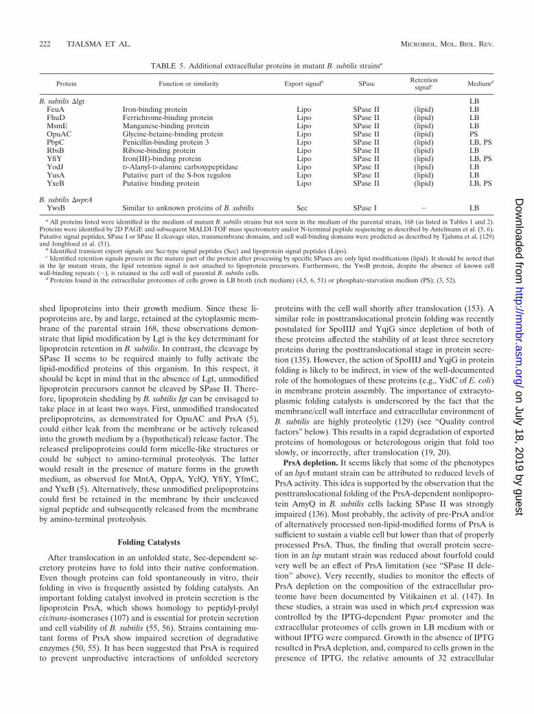
shed lipoproteins into their growth medium. Since these li-poproteins are, by and large, retained at the cytoplasmic mem-brane of the parental strain 168, these observations demon-strate that lipid modification by Lgt is the key determinant forlipoprotein retention in B. subtilis. In contrast, the cleavage bySPase II seems to be required mainly to fully activate thelipid-modified proteins of this organism. In this respect, itshould be kept in mind that in the absence of Lgt, unmodifiedlipoprotein precursors cannot be cleaved by SPase II. There-fore, lipoprotein shedding by B. subtilis Igt can be envisaged totake place in at least two ways. First, unmodified translocatedprelipoproteins, as demonstrated for OpuAC and PrsA (5),could either leak from the membrane or be actively releasedinto the growth medium by a (hypothetical) release factor. Thereleased prelipoproteins could form micelle-like structures orcould be subject to amino-terminal proteolysis. The latterwould result in the presence of mature forms in the growthmedium, as observed for MntA, OppA, YclQ, YfiY, YfmC,and YxeB (5). Alternatively, these unmodified prelipoproteinscould first be retained in the membrane by their uncleavedsignal peptide and subsequently released from the membraneby amino-terminal proteolysis.
Folding Catalysts
After translocation in an unfolded state, Sec-dependent se-cretory proteins have to fold into their native conformation.Even though proteins can fold spontaneously in vitro, theirfolding in vivo is frequently assisted by folding catalysts. Animportant folding catalyst involved in protein secretion is thelipoprotein PrsA, which shows homology to peptidyl-prolylcis/trans-isomerases (107) and is essential for protein secretionand cell viability of B. subtilis (55, 56). Strains containing mu-tant forms of PrsA show impaired secretion of degradativeenzymes (50, 55). It has been suggested that PrsA is requiredto prevent unproductive interactions of unfolded secretory
proteins with the cell wall shortly after translocation (153). Asimilar role in posttranslocational protein folding was recentlypostulated for SpoIIIJ and YqjG since depletion of both ofthese proteins affected the stability of at least three secretoryproteins during the posttranslocational stage in protein secre-tion (135). However, the action of SpoIIIJ and YqjG in proteinfolding is likely to be indirect, in view of the well-documentedrole of the homologues of these proteins (e.g., YidC of E. coli)in membrane protein assembly. The importance of extracyto-plasmic folding catalysts is underscored by the fact that themembrane/cell wall interface and extracellular environment ofB. subtilis are highly proteolytic (129) (see “Quality controlfactors” below). This results in a rapid degradation of exportedproteins of homologous or heterologous origin that fold tooslowly, or incorrectly, after translocation (19, 20).
PrsA depletion. It seems likely that some of the phenotypesof an lspA mutant strain can be attributed to reduced levels ofPrsA activity. This idea is supported by the observation that theposttranslocational folding of the PrsA-dependent nonlipopro-tein AmyQ in B. subtilis cells lacking SPase II was stronglyimpaired (136). Most probably, the activity of pre-PrsA and/orof alternatively processed non-lipid-modified forms of PrsA issufficient to sustain a viable cell but lower than that of properlyprocessed PrsA. Thus, the finding that overall protein secre-tion in an lsp mutant strain was reduced about fourfold couldvery well be an effect of PrsA limitation (see “SPase II dele-tion” above). Very recently, studies to monitor the effects ofPrsA depletion on the composition of the extracellular pro-teome have been documented by Vitikainen et al. (147). Inthese studies, a strain was used in which prsA expression wascontrolled by the IPTG-dependent Pspac promoter and theextracellular proteomes of cells grown in LB medium with orwithout IPTG were compared. Growth in the absence of IPTGresulted in PrsA depletion, and, compared to cells grown in thepresence of IPTG, the relative amounts of 32 extracellular
TABLE 5. Additional extracellular proteins in mutant B. subtilis strainsa
Protein Function or similarity Export signalb SPase Retentionsignalc Mediumd
B. subtilis �lgt LBFeuA Iron-binding protein Lipo SPase II (lipid) LBFhuD Ferrichrome-binding protein Lipo SPase II (lipid) LBMsmE Manganese-binding protein Lipo SPase II (lipid) LBOpuAC Glycine-betaine-binding protein Lipo SPase II (lipid) PSPbpC Penicillin-binding protein 3 Lipo SPase II (lipid) LB, PSRbsB Ribose-binding protein Lipo SPase II (lipid) LBYfiY Iron(III)-binding protein Lipo SPase II (lipid) LB, PSYodJ D-Alanyl-D-alanine carboxypeptidase Lipo SPase II (lipid) LBYusA Putative part of the S-box regulon Lipo SPase II (lipid) LBYxeB Putative binding protein Lipo SPase II (lipid) LB, PS
B. subtilis �wprAYwsB Similar to unknown proteins of B. subtilis Sec SPase I � LB
a All proteins listed were identified in the medium of mutant B. subtilis strains but not seen in the medium of the parental strain, 168 (as listed in Tables 1 and 2).Proteins were identified by 2D PAGE and subsequent MALDI-TOF mass spectrometry and/or N-terminal peptide sequencing as described by Antelmann et al. (5, 6).Putative signal peptides, SPase I or SPase II cleavage sites, transmembrane domains, and cell wall-binding domains were predicted as described by Tjalsma et al. (129)and Jongbloed et al. (51).
b Identified transient export signals are Sec-type signal peptides (Sec) and lipoprotein signal peptides (Lipo).c Identified retention signals present in the mature part of the protein after processing by specific SPases are only lipid modifications (lipid). It should be noted that
in the lgt mutant strain, the lipid retention signal is not attached to lipoprotein precursors. Furthermore, the YwsB protein, despite the absence of known cellwall-binding repeats (�), is retained in the cell wall of parental B. subtilis cells.
d Proteins found in the extracellular proteomes of cells grown in LB broth (rich medium) (4,5, 6, 51) or phosphate-starvation medium (PS); (3, 52).
222 TJALSMA ET AL. MICROBIOL. MOL. BIOL. REV.
on July 18, 2019 by guesthttp://m
mbr.asm
.org/D
ownloaded from
proteins were significantly reduced. Remarkably, the relativeamounts of 15 other extracellular proteins were significantlyincreased, while 6 extracellular proteins remained unaffectedby PrsA depletion. Of the proteins present in increasedamounts, 11 corresponded to cytoplasmic proteins (CitH,Ef-G, Eno, GroEL, PdhA, PdhB, PdhD, RocF, SodA, YvgN,and YwjH), which is consistent with increased cell lysis in theabsence of PrsA. In contrast, 29 of the 32 extracellular proteinspresent in decreased amounts were synthesized with an exportsignal (Table 4). The three remaining proteins present in de-creased amounts lack a typical export signal (FlgK, XkdK, andXlyA) (Table 2). These observations support the view that PrsAis of general importance for the folding of secretory proteins (50,55). Interestingly, eight proteins (Csn, Ggt, LipA, YfnI, YlqB,YweA, YxaK, and YolA) whose extracellular levels remainedunaffected or were even increased on PrsA-depletion are synthe-sized with (putative) export signals (Table 1). This indicates thata specific subset of secretory proteins does not depend on PrsAfor proper folding and secretion. Thus, the secretion of theseproteins may depend on folding factors other than PrsA. Some ofthese other folding factors could be lipoproteins, as judged by thefact that the secretion of Csn, LipA, and YxaK is strongly reducedin the absence of SPase II (see above). The PrsA paralogue YacDis dispensable for the folding of PrsA-independent extracellularproteins, because the extracellular proteome of a yacD mutantstrain was shown to be very similar to that of the parental strain168 (H. Antelmann and H. Tjalsma, unpublished). This implieseither that YacD plays no role in the biogenesis of extracellularproteins, that the specific substrates of YacD are not detectable inthe extracellular proteome, or that YacD substrates were notexpressed under the conditions tested.
Bdb mutations. Four extracytoplasmic thiol-disulfide oxi-doreductases of B. subtilis have been implicated in the forma-tion of disulfide bonds in exported proteins. Two of these,BdbC and BdbD, are required for the biogenesis of the pseu-dopilin ComGC (essential for DNA binding and uptake duringcompetence development), which contains an intramoleculardisulfide bond (71). Furthermore, BdbC and BdbD are ofmajor importance for the posttranslocational folding of a di-sulfide bond-containing heterologous protein, the alkalinephosphatase PhoA of E. coli (18, 71). A third thiol-disulfideoxidoreductase, BdbB, is involved in the production of thelantibiotic sublancin 168, which contains two disulfide bonds(33). Although BdbB and BdbC are highly similar paralogues,their substrate specificities overlap only partly. Thus, BdbCplays a minor role in the production of sublancin 168 (33) whileBdbB is only of minor importance for the secretion of E. coliPhoA by B. subtilis, and this protein is dispensable for compe-tence development. Thus far, no specific function has beenidentified for the fourth thiol-disulfide oxidoreductase of B.subtilis, which is known as BdbA. As judged by their function inthe folding of exported proteins with disulfide bonds, it isbelieved that BdbB, BdbC, and BdbD are members of anoxidation pathway. This idea is supported by the fact thatBdbB and BdbC have a high degree of similarity to DsbB of E.coli while BdbD has some similarity with DsbA of E. coli (18,71). Remarkably, neither bdbA, bdbB, bdbC, nor bdbD single-mutant strains, nor a quadruple mutant lacking all four ofthese genes, displayed detectable changes of the extracellularproteome (H. Antelmann and R. Dorenbos, unpublished ob-
servations). This suggests that very few, if any, of the nativesecreted proteins of B. subtilis contain disulfide bonds that arecritical for their stability and protease resistance and that (insome cases) Bdb-independent folding and oxidation occur.
Quality Control Factors
One important feature of B. subtilis that underscores theimportance of efficient folding of secretory proteins into theirnative, protease-resistant conformation, is the presence of atleast 27 proteases in the membrane, cell wall, and medium thatcan cleave (partially) unfolded polypeptide chains (129). Pro-teins with a known role in the quality control of secretoryproteins are two HtrA-like proteases/chaperones, HtrA andHtrB (85), which are thought to have proofreading capabilitiesfor the folding state of secretory proteins, as demonstrated forHtrA of E. coli (122). When a secretory protein is not properlyfolded, HtrAB can either assist in folding or, if that is impos-sible, degrade the malfolded secretory protein. Interestingly,transcription of the corresponding genes is induced by secre-tion stress, which is sensed and controlled by the CssR-CssStwo-component regulatory system. This system is essential forcell viability under conditions of severe secretion stress (31,49). Another protease, which seems to be involved in thequality control of secretory proteins, is WprA. For this protein,a chaperone domain (CWBP23) in addition to a protease do-main (CWBP52) has been proposed (10, 68). Notably, HtrAand WprA have a dual localization, being present both in thecell wall proteome and in the extracellular proteome (4, 6).Like HtrB, the HtrA protein has a (predicted) N-terminalmembrane anchor. As shown by N-terminal sequencing, HtrAin the medium lacks this membrane anchor domain (3). Thelocalization of HtrB has not been documented yet. The impor-tance of HtrAB and WprA in quality control was investigatedby proteomic analyses by Antelmann et al. (4, 6).
Modulation of HtrA and HtrB levels. To study the impor-tance of HtrA-like proteases for the composition of the extra-cellular proteome, cellular levels of HtrA and HtrB were mod-ulated in several ways (4). First, mutant strains wereconstructed in which htrA or htrB or both, were disrupted.Notably, disruption of one of these genes causes a secretionstress that strongly induces the activity of the promoters ofboth genes, and this response is mediated by the CssR-CssStwo-component system (31, 49, 85). Furthermore, the tran-scription of both htrA and htrB can be strongly reduced by adisruption of the cssS gene, while transcription of both htrgenes is significantly increased by overproducing the secretoryprotein AmyQ, which causes secretion stress. To monitor theimpact of HtrA and HtrB on protein secretion, Antelmann etal. analyzed the extracellular proteomes of htrA and/or htrBmutant strains (4). These analyses showed that apart from theHtrA protein, only one other protein disappeared completelyfrom the medium of an htrA mutant strain. This was YqxI, aprotein of unknown function. As expected, the HtrA spot inthe medium was increased on deletion of the htrB gene oroverproduction of AmyQ (secretion stress) but significantlydecreased in a cssS mutant strain. This pattern of HtrA ap-pearance in the media of different mutant strains closely par-alleled that of YqxI. However, in contrast to htrA, the tran-scription of yqxI was independent of CssRS and was not
VOL. 68, 2004 PROTEOMICS OF PROTEIN SECRETION 223
on July 18, 2019 by guesthttp://m
mbr.asm
.org/D
ownloaded from
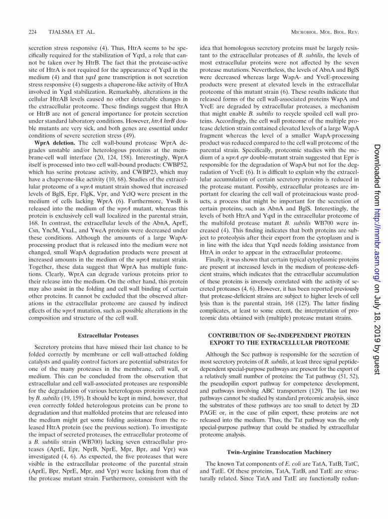
secretion stress responsive (4). Thus, HtrA seems to be spe-cifically required for the stabilization of YqxI, a role that can-not be taken over by HtrB. The fact that the protease-activesite of HtrA is not required for the appearance of YqxI in themedium (4) and that yqxI gene transcription is not secretionstress responsive (4) suggests a chaperone-like activity of HtrAinvolved in YqxI stabilization. Remarkably, alterations in thecellular HtrAB levels caused no other detectable changes inthe extracellular proteome. These findings suggest that HtrAor HtrB are not of general importance for protein secretionunder standard laboratory conditions. However, htrA htrB dou-ble mutants are very sick, and both genes are essential underconditions of severe secretion stress (49).
WprA deletion. The cell wall-bound protease WprA de-grades unstable and/or heterologous proteins at the mem-brane-cell wall interface (20, 124, 158). Interestingly, WprAitself is processed into two cell wall-bound products: CWBP52,which has serine protease activity, and CWBP23, which mayhave a chaperone-like activity (10, 68). Studies of the extracel-lular proteome of a wprA mutant strain showed that increasedlevels of BglS, Epr, FlgK, Vpr, and YclQ were present in themedium of cells lacking WprA (6). Furthermore, YwsB isreleased into the medium of the wprA mutant, whereas thisprotein is exclusively cell wall localized in the parental strain,168. In contrast, the extracellular levels of the AbnA, AprE,Csn, YncM, YxaL, and YweA proteins were decreased underthese conditions. Although the amounts of a large WapA-processing product that is released into the medium were notchanged, small WapA degradation products were present atincreased amounts in the medium of the wprA mutant strain.Together, these data suggest that WprA has multiple func-tions. Clearly, WprA can degrade various proteins prior totheir release into the medium. On the other hand, this proteinmay also assist in the folding and cell wall binding of certainother proteins. It cannot be excluded that the observed alter-ations in the extracellular proteome are caused by indirecteffects of the wprA mutation, such as possible alterations in thecomposition and structure of the cell wall.
Extracellular Proteases
Secretory proteins that have missed their last chance to befolded correctly by membrane or cell wall-attached foldingcatalysts and quality control factors are potential substrates forone of the many proteases in the membrane, cell wall, ormedium. This can be concluded from the observation thatextracellular and cell wall-associated proteases are responsiblefor the degradation of various heterologous proteins secretedby B. subtilis (19, 159). It should be kept in mind, however, thateven correctly folded heterologous proteins can be prone todegradation and that malfolded proteins that are released intothe medium might get some folding assistance from the re-leased HtrA protein (see the previous section). To investigatethe impact of secreted proteases, the extracellular proteome ofa B. subtilis strain (WB700) lacking seven extracellular pro-teases (AprE, Epr, NprB, NprE, Mpr, Bpr, and Vpr) wasinvestigated (4, 6). As expected, the five proteases that werevisible in the extracellular proteome of the parental strain(AprE, Bpr, NprE, Mpr, and Vpr) were lacking from that ofthe protease mutant strain. Furthermore, consistent with the
idea that homologous secretory proteins must be largely resis-tant to the extracellular proteases of B. subtilis, the levels ofmost extracellular proteins were not affected by the sevenprotease mutations. Nevertheless, the levels of AbnA and BglSwere decreased whereas large WapA- and YvcE-processingproducts were present at elevated levels in the extracellularproteome of this mutant strain (6). These results indicate thatreleased forms of the cell wall-associated proteins WapA andYvcE are degraded by extracellular proteases, a mechanismthat might enable B. subtilis to recycle spoiled cell wall pro-teins. Accordingly, the cell wall proteome of the multiple pro-tease deletion strain contained elevated levels of a large WapAfragment whereas the level of a smaller WapA-processingproduct was reduced compared to the cell wall proteome of theparental strain. Specifically, proteomic studies with the me-dium of a wprA epr double-mutant strain suggested that Epr isresponsible for the degradation of WapA but not for the deg-radation of YvcE (6). It is difficult to explain why the extracel-lular accumulation of certain secretory proteins is reduced inthe protease mutant. Possibly, extracellular proteases are im-portant for clearing the cell wall of proteinacious waste prod-ucts, a process that might be important for the secretion ofcertain proteins, such as AbnA and BglS. Interestingly, thelevels of both HtrA and YqxI in the extracellular proteome ofthe multifold protease mutant B. subtilis WB700 were in-creased (4). This finding indicates that both proteins are sub-ject to proteolysis after their export from the cytoplasm and isin line with the idea that YqxI needs folding assistance fromHtrA in order to appear in the extracellular proteome.
Finally, it was shown that certain typical cytoplasmic proteinsare present at increased levels in the medium of protease-defi-cient strains, which indicates that the extracellular accumulationof these proteins is inversely correlated with the activity of se-creted proteases (4, 6). However, it has been reported previouslythat protease-deficient strains are subject to higher levels of celllysis than is the parental strain, 168 (125). The latter findingcomplicates, at least to some extent, the interpretation of pro-teomic data obtained with (multiple) protease mutant strains.
CONTRIBUTION OF Sec-INDEPENDENT PROTEINEXPORT TO THE EXTRACELLULAR PROTEOME
Although the Sec pathway is responsible for the secretion ofmost secretory proteins of B. subtilis, at least three signal peptide-dependent special-purpose pathways are present for the export ofa relatively small number of proteins: the Tat pathway (51, 52),the pseudopilin export pathway for competence development,and pathways involving ABC transporters (129). The last twopathways cannot be studied by standard proteomic analysis, sincethe substrates of these pathways are too small to detect by 2DPAGE or, in the case of pilin export, these proteins are notreleased into the medium. Thus, the Tat pathway was the onlyspecial-purpose pathway that could be studied by extracellularproteome analysis.
Twin-Arginine Translocation Machinery
The known Tat components of E. coli are TatA, TatB, TatC,and TatE. Of these proteins, TatA, TatB, and TatE are struc-turally related. Since TatA and TatE are functionally redun-
224 TJALSMA ET AL. MICROBIOL. MOL. BIOL. REV.
on July 18, 2019 by guesthttp://m
mbr.asm
.org/D
ownloaded from
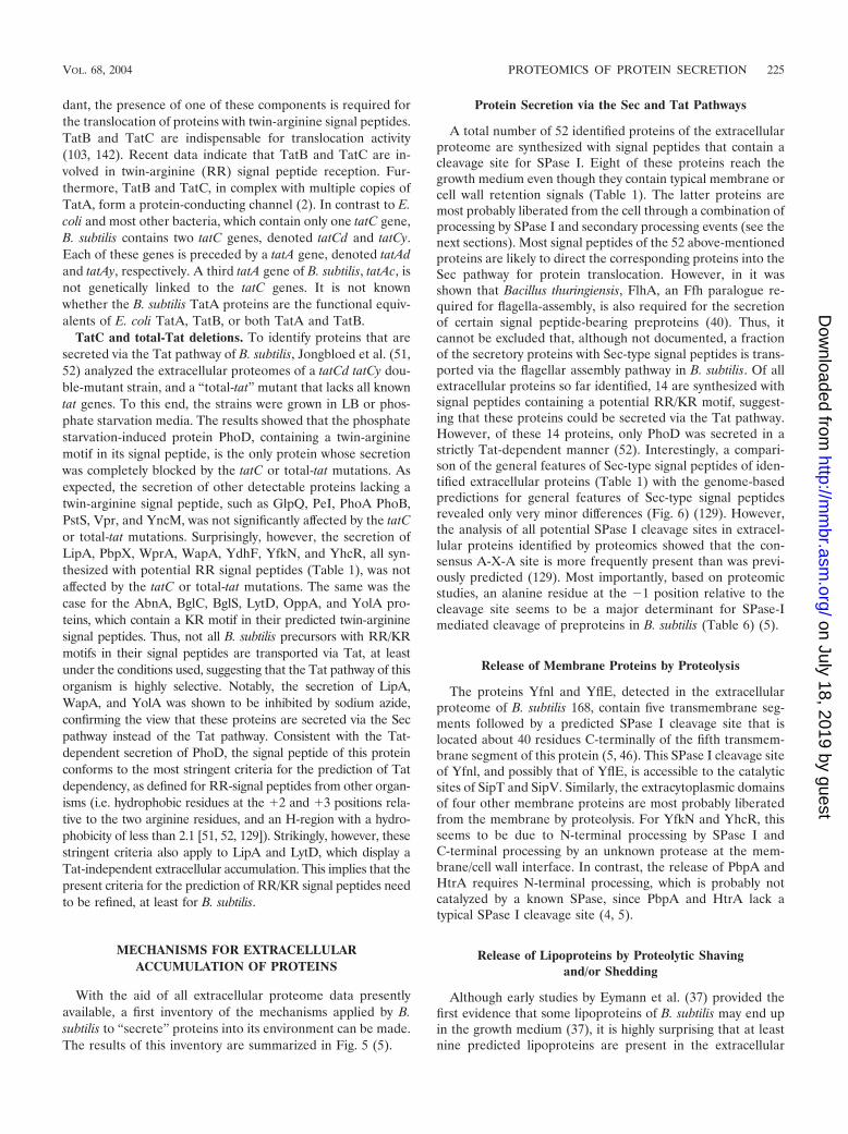
dant, the presence of one of these components is required forthe translocation of proteins with twin-arginine signal peptides.TatB and TatC are indispensable for translocation activity(103, 142). Recent data indicate that TatB and TatC are in-volved in twin-arginine (RR) signal peptide reception. Fur-thermore, TatB and TatC, in complex with multiple copies ofTatA, form a protein-conducting channel (2). In contrast to E.coli and most other bacteria, which contain only one tatC gene,B. subtilis contains two tatC genes, denoted tatCd and tatCy.Each of these genes is preceded by a tatA gene, denoted tatAdand tatAy, respectively. A third tatA gene of B. subtilis, tatAc, isnot genetically linked to the tatC genes. It is not knownwhether the B. subtilis TatA proteins are the functional equiv-alents of E. coli TatA, TatB, or both TatA and TatB.
TatC and total-Tat deletions. To identify proteins that aresecreted via the Tat pathway of B. subtilis, Jongbloed et al. (51,52) analyzed the extracellular proteomes of a tatCd tatCy dou-ble-mutant strain, and a “total-tat” mutant that lacks all knowntat genes. To this end, the strains were grown in LB or phos-phate starvation media. The results showed that the phosphatestarvation-induced protein PhoD, containing a twin-argininemotif in its signal peptide, is the only protein whose secretionwas completely blocked by the tatC or total-tat mutations. Asexpected, the secretion of other detectable proteins lacking atwin-arginine signal peptide, such as GlpQ, PeI, PhoA PhoB,PstS, Vpr, and YncM, was not significantly affected by the tatCor total-tat mutations. Surprisingly, however, the secretion ofLipA, PbpX, WprA, WapA, YdhF, YfkN, and YhcR, all syn-thesized with potential RR signal peptides (Table 1), was notaffected by the tatC or total-tat mutations. The same was thecase for the AbnA, BglC, BglS, LytD, OppA, and YolA pro-teins, which contain a KR motif in their predicted twin-argininesignal peptides. Thus, not all B. subtilis precursors with RR/KRmotifs in their signal peptides are transported via Tat, at leastunder the conditions used, suggesting that the Tat pathway of thisorganism is highly selective. Notably, the secretion of LipA,WapA, and YolA was shown to be inhibited by sodium azide,confirming the view that these proteins are secreted via the Secpathway instead of the Tat pathway. Consistent with the Tat-dependent secretion of PhoD, the signal peptide of this proteinconforms to the most stringent criteria for the prediction of Tatdependency, as defined for RR-signal peptides from other organ-isms (i.e. hydrophobic residues at the �2 and �3 positions rela-tive to the two arginine residues, and an H-region with a hydro-phobicity of less than 2.1 [51, 52, 129]). Strikingly, however, thesestringent criteria also apply to LipA and LytD, which display aTat-independent extracellular accumulation. This implies that thepresent criteria for the prediction of RR/KR signal peptides needto be refined, at least for B. subtilis.
MECHANISMS FOR EXTRACELLULARACCUMULATION OF PROTEINS
With the aid of all extracellular proteome data presentlyavailable, a first inventory of the mechanisms applied by B.subtilis to “secrete” proteins into its environment can be made.The results of this inventory are summarized in Fig. 5 (5).
Protein Secretion via the Sec and Tat Pathways
A total number of 52 identified proteins of the extracellularproteome are synthesized with signal peptides that contain acleavage site for SPase I. Eight of these proteins reach thegrowth medium even though they contain typical membrane orcell wall retention signals (Table 1). The latter proteins aremost probably liberated from the cell through a combination ofprocessing by SPase I and secondary processing events (see thenext sections). Most signal peptides of the 52 above-mentionedproteins are likely to direct the corresponding proteins into theSec pathway for protein translocation. However, in it wasshown that Bacillus thuringiensis, FlhA, an Ffh paralogue re-quired for flagella-assembly, is also required for the secretionof certain signal peptide-bearing preproteins (40). Thus, itcannot be excluded that, although not documented, a fractionof the secretory proteins with Sec-type signal peptides is trans-ported via the flagellar assembly pathway in B. subtilis. Of allextracellular proteins so far identified, 14 are synthesized withsignal peptides containing a potential RR/KR motif, suggest-ing that these proteins could be secreted via the Tat pathway.However, of these 14 proteins, only PhoD was secreted in astrictly Tat-dependent manner (52). Interestingly, a compari-son of the general features of Sec-type signal peptides of iden-tified extracellular proteins (Table 1) with the genome-basedpredictions for general features of Sec-type signal peptidesrevealed only very minor differences (Fig. 6) (129). However,the analysis of all potential SPase I cleavage sites in extracel-lular proteins identified by proteomics showed that the con-sensus A-X-A site is more frequently present than was previ-ously predicted (129). Most importantly, based on proteomicstudies, an alanine residue at the �1 position relative to thecleavage site seems to be a major determinant for SPase-Imediated cleavage of preproteins in B. subtilis (Table 6) (5).
Release of Membrane Proteins by Proteolysis
The proteins Yfnl and YflE, detected in the extracellularproteome of B. subtilis 168, contain five transmembrane seg-ments followed by a predicted SPase I cleavage site that islocated about 40 residues C-terminally of the fifth transmem-brane segment of this protein (5, 46). This SPase I cleavage siteof Yfnl, and possibly that of YflE, is accessible to the catalyticsites of SipT and SipV. Similarly, the extracytoplasmic domainsof four other membrane proteins are most probably liberatedfrom the membrane by proteolysis. For YfkN and YhcR, thisseems to be due to N-terminal processing by SPase I andC-terminal processing by an unknown protease at the mem-brane/cell wall interface. In contrast, the release of PbpA andHtrA requires N-terminal processing, which is probably notcatalyzed by a known SPase, since PbpA and HtrA lack atypical SPase I cleavage site (4, 5).
Release of Lipoproteins by Proteolytic Shavingand/or Shedding
Although early studies by Eymann et al. (37) provided thefirst evidence that some lipoproteins of B. subtilis may end upin the growth medium (37), it is highly surprising that at leastnine predicted lipoproteins are present in the extracellular
VOL. 68, 2004 PROTEOMICS OF PROTEIN SECRETION 225
on July 18, 2019 by guesthttp://m
mbr.asm
.org/D
ownloaded from
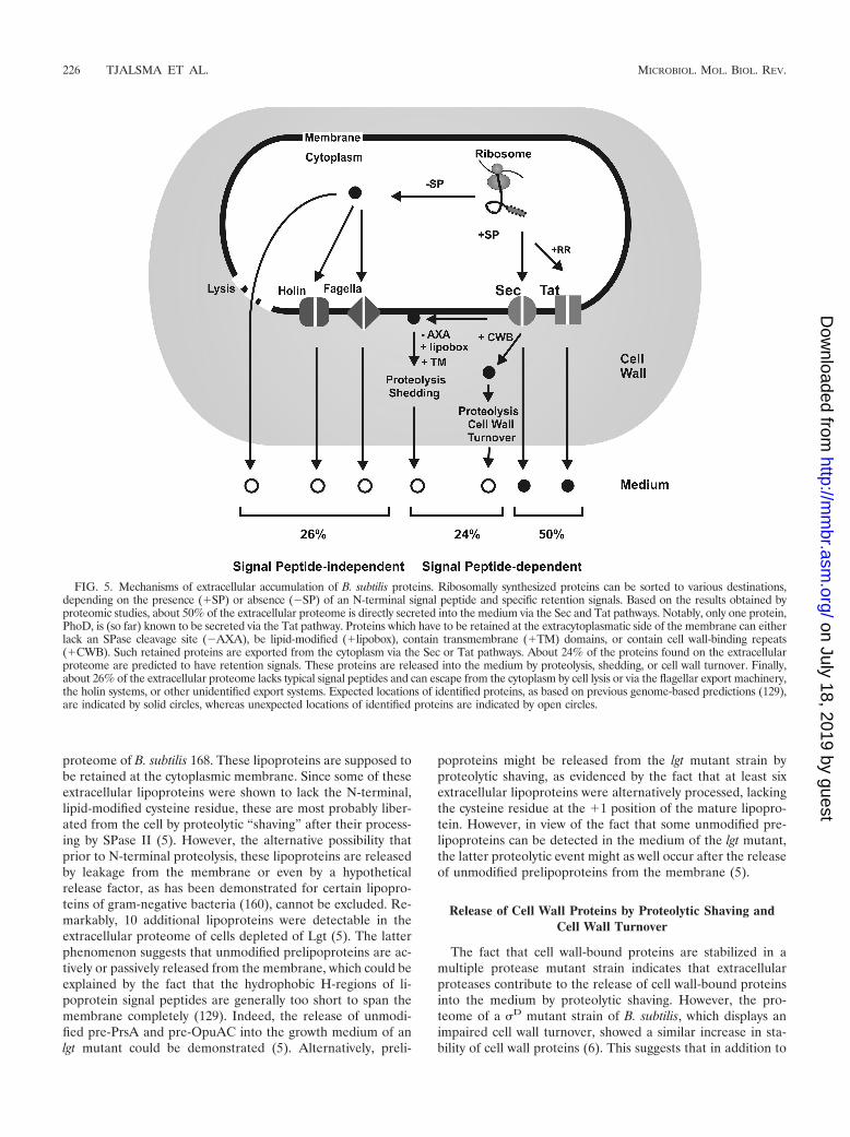
proteome of B. subtilis 168. These lipoproteins are supposed tobe retained at the cytoplasmic membrane. Since some of theseextracellular lipoproteins were shown to lack the N-terminal,lipid-modified cysteine residue, these are most probably liber-ated from the cell by proteolytic “shaving” after their process-ing by SPase II (5). However, the alternative possibility thatprior to N-terminal proteolysis, these lipoproteins are releasedby leakage from the membrane or even by a hypotheticalrelease factor, as has been demonstrated for certain lipopro-teins of gram-negative bacteria (160), cannot be excluded. Re-markably, 10 additional lipoproteins were detectable in theextracellular proteome of cells depleted of Lgt (5). The latterphenomenon suggests that unmodified prelipoproteins are ac-tively or passively released from the membrane, which could beexplained by the fact that the hydrophobic H-regions of li-poprotein signal peptides are generally too short to span themembrane completely (129). Indeed, the release of unmodi-fied pre-PrsA and pre-OpuAC into the growth medium of anlgt mutant could be demonstrated (5). Alternatively, preli-
poproteins might be released from the lgt mutant strain byproteolytic shaving, as evidenced by the fact that at least sixextracellular lipoproteins were alternatively processed, lackingthe cysteine residue at the �1 position of the mature lipopro-tein. However, in view of the fact that some unmodified pre-lipoproteins can be detected in the medium of the lgt mutant,the latter proteolytic event might as well occur after the releaseof unmodified prelipoproteins from the membrane (5).
Release of Cell Wall Proteins by Proteolytic Shaving andCell Wall Turnover
The fact that cell wall-bound proteins are stabilized in amultiple protease mutant strain indicates that extracellularproteases contribute to the release of cell wall-bound proteinsinto the medium by proteolytic shaving. However, the pro-teome of a D mutant strain of B. subtilis, which displays animpaired cell wall turnover, showed a similar increase in sta-bility of cell wall proteins (6). This suggests that in addition to
FIG. 5. Mechanisms of extracellular accumulation of B. subtilis proteins. Ribosomally synthesized proteins can be sorted to various destinations,depending on the presence (�SP) or absence (�SP) of an N-terminal signal peptide and specific retention signals. Based on the results obtained byproteomic studies, about 50% of the extracellular proteome is directly secreted into the medium via the Sec and Tat pathways. Notably, only one protein,PhoD, is (so far) known to be secreted via the Tat pathway. Proteins which have to be retained at the extracytoplasmatic side of the membrane can eitherlack an SPase cleavage site (�AXA), be lipid-modified (�lipobox), contain transmembrane (�TM) domains, or contain cell wall-binding repeats(�CWB). Such retained proteins are exported from the cytoplasm via the Sec or Tat pathways. About 24% of the proteins found on the extracellularproteome are predicted to have retention signals. These proteins are released into the medium by proteolysis, shedding, or cell wall turnover. Finally,about 26% of the extracellular proteome lacks typical signal peptides and can escape from the cytoplasm by cell lysis or via the flagellar export machinery,the holin systems, or other unidentified export systems. Expected locations of identified proteins, as based on previous genome-based predictions (129),are indicated by solid circles, whereas unexpected locations of identified proteins are indicated by open circles.
226 TJALSMA ET AL. MICROBIOL. MOL. BIOL. REV.
on July 18, 2019 by guesthttp://m
mbr.asm
.org/D
ownloaded from
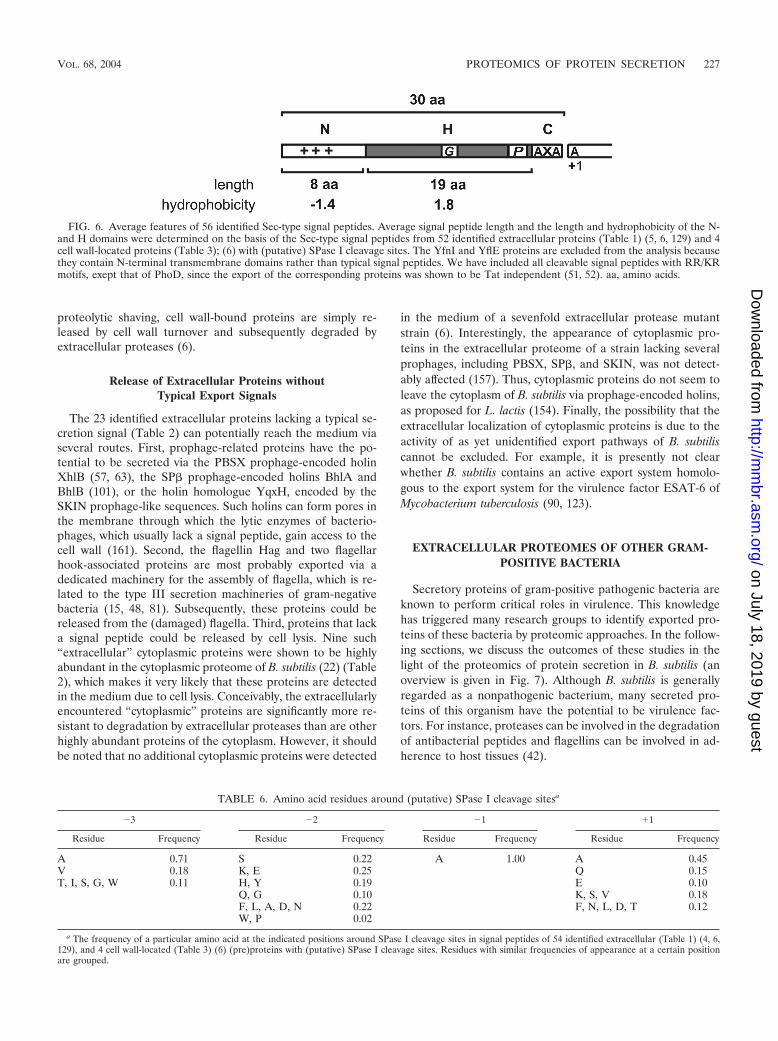
proteolytic shaving, cell wall-bound proteins are simply re-leased by cell wall turnover and subsequently degraded byextracellular proteases (6).
Release of Extracellular Proteins withoutTypical Export Signals
The 23 identified extracellular proteins lacking a typical se-cretion signal (Table 2) can potentially reach the medium viaseveral routes. First, prophage-related proteins have the po-tential to be secreted via the PBSX prophage-encoded holinXhlB (57, 63), the SP� prophage-encoded holins BhlA andBhlB (101), or the holin homologue YqxH, encoded by theSKIN prophage-like sequences. Such holins can form pores inthe membrane through which the lytic enzymes of bacterio-phages, which usually lack a signal peptide, gain access to thecell wall (161). Second, the flagellin Hag and two flagellarhook-associated proteins are most probably exported via adedicated machinery for the assembly of flagella, which is re-lated to the type III secretion machineries of gram-negativebacteria (15, 48, 81). Subsequently, these proteins could bereleased from the (damaged) flagella. Third, proteins that lacka signal peptide could be released by cell lysis. Nine such“extracellular” cytoplasmic proteins were shown to be highlyabundant in the cytoplasmic proteome of B. subtilis (22) (Table2), which makes it very likely that these proteins are detectedin the medium due to cell lysis. Conceivably, the extracellularlyencountered “cytoplasmic” proteins are significantly more re-sistant to degradation by extracellular proteases than are otherhighly abundant proteins of the cytoplasm. However, it shouldbe noted that no additional cytoplasmic proteins were detected
in the medium of a sevenfold extracellular protease mutantstrain (6). Interestingly, the appearance of cytoplasmic pro-teins in the extracellular proteome of a strain lacking severalprophages, including PBSX, SP�, and SKIN, was not detect-ably affected (157). Thus, cytoplasmic proteins do not seem toleave the cytoplasm of B. subtilis via prophage-encoded holins,as proposed for L. lactis (154). Finally, the possibility that theextracellular localization of cytoplasmic proteins is due to theactivity of as yet unidentified export pathways of B. subtiliscannot be excluded. For example, it is presently not clearwhether B. subtilis contains an active export system homolo-gous to the export system for the virulence factor ESAT-6 ofMycobacterium tuberculosis (90, 123).
EXTRACELLULAR PROTEOMES OF OTHER GRAM-POSITIVE BACTERIA
Secretory proteins of gram-positive pathogenic bacteria areknown to perform critical roles in virulence. This knowledgehas triggered many research groups to identify exported pro-teins of these bacteria by proteomic approaches. In the follow-ing sections, we discuss the outcomes of these studies in thelight of the proteomics of protein secretion in B. subtilis (anoverview is given in Fig. 7). Although B. subtilis is generallyregarded as a nonpathogenic bacterium, many secreted pro-teins of this organism have the potential to be virulence fac-tors. For instance, proteases can be involved in the degradationof antibacterial peptides and flagellins can be involved in ad-herence to host tissues (42).
FIG. 6. Average features of 56 identified Sec-type signal peptides. Average signal peptide length and the length and hydrophobicity of the N-and H domains were determined on the basis of the Sec-type signal peptides from 52 identified extracellular proteins (Table 1) (5, 6, 129) and 4cell wall-located proteins (Table 3); (6) with (putative) SPase I cleavage sites. The YfnI and YflE proteins are excluded from the analysis becausethey contain N-terminal transmembrane domains rather than typical signal peptides. We have included all cleavable signal peptides with RR/KRmotifs, exept that of PhoD, since the export of the corresponding proteins was shown to be Tat independent (51, 52). aa, amino acids.
TABLE 6. Amino acid residues around (putative) SPase I cleavage sitesa
�3 �2 �1 �1
Residue Frequency Residue Frequency Residue Frequency Residue Frequency
A 0.71 S 0.22 A 1.00 A 0.45V 0.18 K, E 0.25 Q 0.15T, I, S, G, W 0.11 H, Y 0.19 E 0.10
Q, G 0.10 K, S, V 0.18F, L, A, D, N 0.22 F, N, L, D, T 0.12W, P 0.02
a The frequency of a particular amino acid at the indicated positions around SPase I cleavage sites in signal peptides of 54 identified extracellular (Table 1) (4, 6,129), and 4 cell wall-located (Table 3) (6) (pre)proteins with (putative) SPase I cleavage sites. Residues with similar frequencies of appearance at a certain positionare grouped.
VOL. 68, 2004 PROTEOMICS OF PROTEIN SECRETION 227
on July 18, 2019 by guesthttp://m
mbr.asm
.org/D
ownloaded from
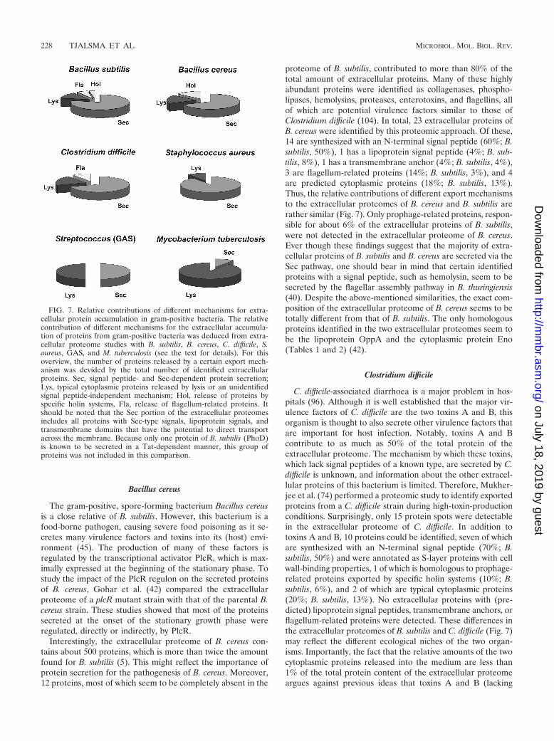
Bacillus cereus
The gram-positive, spore-forming bacterium Bacillus cereusis a close relative of B. subtilis. However, this bacterium is afood-borne pathogen, causing severe food poisoning as it se-cretes many virulence factors and toxins into its (host) envi-ronment (45). The production of many of these factors isregulated by the transcriptional activator PlcR, which is max-imally expressed at the beginning of the stationary phase. Tostudy the impact of the PlcR regulon on the secreted proteinsof B. cereus, Gohar et al. (42) compared the extracellularproteome of a plcR mutant strain with that of the parental B.cereus strain. These studies showed that most of the proteinssecreted at the onset of the stationary growth phase wereregulated, directly or indirectly, by PlcR.
Interestingly, the extracellular proteome of B. cereus con-tains about 500 proteins, which is more than twice the amountfound for B. subtilis (5). This might reflect the importance ofprotein secretion for the pathogenesis of B. cereus. Moreover,12 proteins, most of which seem to be completely absent in the
proteome of B. subtilis, contributed to more than 80% of thetotal amount of extracellular proteins. Many of these highlyabundant proteins were identified as collagenases, phospho-lipases, hemolysins, proteases, enterotoxins, and flagellins, allof which are potential virulence factors similar to those ofClostridium difficile (104). In total, 23 extracellular proteins ofB. cereus were identified by this proteomic approach. Of these,14 are synthesized with an N-terminal signal peptide (60%; B.subtilis, 50%), 1 has a lipoprotein signal peptide (4%; B. sub-tilis, 8%), 1 has a transmembrane anchor (4%; B. subtilis, 4%),3 are flagellum-related proteins (14%; B. subtilis, 3%), and 4are predicted cytoplasmic proteins (18%; B. subtilis, 13%).Thus, the relative contributions of different export mechanismsto the extracellular proteomes of B. cereus and B. subtilis arerather similar (Fig. 7). Only prophage-related proteins, respon-sible for about 6% of the extracellular proteins of B. subtilis,were not detected in the extracellular proteome of B. cereus.Ever though these findings suggest that the majority of extra-cellular proteins of B. subtilis and B. cereus are secreted via theSec pathway, one should bear in mind that certain identifiedproteins with a signal peptide, such as hemolysin, seem to besecreted by the flagellar assembly pathway in B. thuringiensis(40). Despite the above-mentioned similarities, the exact com-position of the extracellular proteome of B. cereus seems to betotally different from that of B. subtilis. The only homologousproteins identified in the two extracellular proteomes seem tobe the lipoprotein OppA and the cytoplasmic protein Eno(Tables 1 and 2) (42).
Clostridium difficile
C. difficile-associated diarrhoea is a major problem in hos-pitals (96). Although it is well established that the major vir-ulence factors of C. difficile are the two toxins A and B, thisorganism is thought to also secrete other virulence factors thatare important for host infection. Notably, toxins A and Bcontribute to as much as 50% of the total protein of theextracellular proteome. The mechanism by which these toxins,which lack signal peptides of a known type, are secreted by C.difficile is unknown, and information about the other extracel-lular proteins of this bacterium is limited. Therefore, Mukher-jee et al. (74) performed a proteomic study to identify exportedproteins from a C. difficile strain during high-toxin-productionconditions. Surprisingly, only 15 protein spots were detectablein the extracellular proteome of C. difficile. In addition totoxins A and B, 10 proteins could be identified, seven of whichare synthesized with an N-terminal signal peptide (70%; B.subtilis, 50%) and were annotated as S-layer proteins with cellwall-binding properties, 1 of which is homologous to prophage-related proteins exported by specific holin systems (10%; B.subtilis, 6%), and 2 of which are typical cytoplasmic proteins(20%; B. subtilis, 13%). No extracellular proteins with (pre-dicted) lipoprotein signal peptides, transmembrane anchors, orflagellum-related proteins were detected. These differences inthe extracellular proteomes of B. subtilis and C. difficile (Fig. 7)may reflect the different ecological niches of the two organ-isms. Importantly, the fact that the relative amounts of the twocytoplasmic proteins released into the medium are less than1% of the total protein content of the extracellular proteomeargues against previous ideas that toxins A and B (lacking
FIG. 7. Relative contributions of different mechanisms for extra-cellular protein accumulation in gram-positive bacteria. The relativecontribution of different mechanisms for the extracellular accumula-tion of proteins from gram-positive bacteria was deduced from extra-cellular proteome studies with B. subtilis, B. cereus, C. difficile, S.aureus, GAS, and M. tuberculosis (see the text for details). For thisoverview, the number of proteins released by a certain export mech-anism was devided by the total number of identified extracellularproteins. Sec, signal peptide- and Sec-dependent protein secretion;Lys, typical cytoplasmic proteins released by lysis or an unidentifiedsignal peptide-independent mechanism; Hol, release of proteins byspecific holin systems, Fla, release of flagellum-related proteins. Itshould be noted that the Sec portion of the extracellular proteomesincludes all proteins with Sec-type signals, lipoprotein signals, andtransmembrane domains that have the potential to direct transportacross the membrane. Because only one protein of B. subtilis (PhoD)is known to be secreted in a Tat-dependent manner, this group ofproteins was not included in this comparison.
228 TJALSMA ET AL. MICROBIOL. MOL. BIOL. REV.
on July 18, 2019 by guesthttp://m
mbr.asm
.org/D
ownloaded from
signal peptides) are released by cell lysis (74). In conclusion, C.difficile seems to make use of Sec-dependent and, most prob-ably, holin(-like) pathways to release a relatively small numberof proteins into its environment.
Staphylococcus aureus
S. aureus is widely recognized as a pathogen even though itis usually only a colonizer of the human host. Unfortunately, itcan switch from a commensal to a lethal pathogen (24). Patho-genesis of S. aureus involves the synthesis of cell wall-associ-ated virulence factors, a large number of extracellular proteins,and secreted toxins with damaging effects on the host cells. Toidentify extracellular proteins of S. aureus that are potentialvirulence factors, Ziebandt et al. (162) used a proteomic ap-proach to analyze the pattern of extracellular proteins of dif-ferent S. aureus strains. In total, 26 proteins of the approxi-mately 100 protein spots could be identified. Among the newlyidentified proteins were enterotoxins, a leukotoxin, serine pro-teases, a thermonuclease, and an immunoglobulin G-bindingprotein, all being potential virulence factors. Strikingly, in con-trast to B. subtilis (50%), about 90% of the identified extracel-lular proteins of S. aureus are synthesized with Sec-type signalpeptides. The remaining proteins are most probably cytoplas-mic proteins released by cell lysis. No extracellular proteinswith (predicted) lipoprotein signal peptides, transmembraneanchors, flagellum-related proteins, or phage-related proteinswere detected which, together, form a significant portion of theB. subtilis secretome (5) (Fig. 7). Interestingly, one extracellu-lar protein was found to be homologous to the transmembraneprotein YfnI of B. subtilis, whose C-terminal part is released bySPase I-mediated processing (5). However, the YfnI protein ofS. aureus seems to have a potential signal peptide instead ofthe five membrane-spanning domains in the N terminus ofYfnI from B. subtilis. Remarkably, the SPase I-processing sitesare both located about 40 residues C-terminally of the fifthtransmembrane segment of B. subtilis YfnI (AYA) and thepotential signal peptide of S. aureus YfnI (ALA).
Bernardo et al. (13) performed a proteomic study aimed atthe characterization of virulence of different S. aureus strains.Extracellular protein spots that were present in the medium ofall tested strains were identified as protein A, hemolysins,lipases, and autolysins. The fact that protein A is a cell wall-anchored protein of S. aureus shows that, similarly to B. subtilis,certain cell wall-bound proteins are released into the growthmedium. However, it is not clear whether this release of pro-tein A is important for the pathogenesis of S. aureus strains.
Group A Streptococcus
Strains of group A Streptococcus (GAS) species are a com-mon cause of severe invasive infections with unusually highrates of morbidity and mortality (155). Certain extracellularproteins from GAS strains play critical roles in human infec-tions caused by these organisms. To perform a systematic anal-ysis of these extracellular proteins, the proteins present in themedia of different GAS strains were analyzed by Lei et al.using 2D PAGE (59). Of the about 80 protein spots that wereobserved, 43 distinct proteins were identified. Strikingly, only16 of these had typical signal peptides whereas 27 proteins did
not. Among the extracellular proteins with a signal peptidewere the (putative) virulence factors streptolysin O, the M1and M3 proteins, mitogenic factor, streptococcal pyrogenicexotoxin A, streptococcal inhibitor of complement, and homo-logues of class B acid phosphatase and serine proteases. Theextracellular proteins without a typical signal peptide are pre-sumably cytoplasmic proteins, including proteins involved inglycolytic metabolism, translation, the urea cycle, and chaper-onins like GroEL. It should be noted that typical cytoplasmicproteins, such as enolases, influence pathogen-host interac-tions (91). Together, these data show that about half of theextracellular proteins from GAS strains are secreted in a signalpeptide- and Sec-dependent manner. The remaining 50% ofthe proteins are released by cell lysis or by other, as yet un-identified, Sec-independent export mechanisms (Fig. 7).
Mycobacterium tuberculosis
Mycobacterium tuberculosis causes about 8 million cases oftuberculosis worldwide each year (128). To facilitate the designof novel measures for the prevention and therapy of this healththreat, the proteomes of nonvirulent Mycobacterium bovisstrains and virulent M. tuberculosis strains were compared byJungblut et al. (53). Furthermore, Rosenkrands et al. (105)extended these proteome studies specifically for M. tuberculo-sis. The extracellular proteome of M. tuberculosis cultures con-tained between 600 and 800 protein spots, of which, in total, 84proteins could be identified (105). Of these 84 proteins, only 31were unique to the extracellular proteome since these proteinswere absent from the cytoplasmic or cell wall proteomes. Thissuggests that a large portion of the extracellular proteins arereleased by cell lysis (139). The latter view is supported by theidentification of typical cytoplasmic proteins, such as aldolases,enolases, elongation factor G, GroEL, superoxide dismutase,and various dehydrogenases, which were also detected in theextracellular proteome of B. subtilis (5). Strikingly, of the 31proteins that were found exclusively in the medium fraction,only 5 are synthesized with a putative N-terminal signal pep-tide as previously predicted (43). However, it should be notedthat proteins that contain transmembrane domains in additionto a signal peptide were excluded from the latter predictions.At least two such proteins were previously identified in theculture medium of M. tuberculosis, which shows that certainproteins are proteolytically released from the cell envelope(156), similar to the YfkN and YhcR proteins of B. subtilis (5).In addition, at least one protein with a putative lipoproteinsignal peptide was identified in both the cell wall and mediumfractions. Taken together, these data strongly suggest that al-though a small repertoire of extracellular proteins from M.tuberculosis are secreted in a signal peptide- and Sec-depen-dent manner, most proteins are released into the medium bycell lysis or via specific Sec-independent export mechanisms,such as the Snm system (117, 123).
PERSPECTIVES
The power of high-resolution proteomic techniques hasbeen effectively used to gain novel insights in the general flowof proteins into the environment of B. subtilis. Specifically,these studies shed new light on signal peptide function, the role
VOL. 68, 2004 PROTEOMICS OF PROTEIN SECRETION 229
on July 18, 2019 by guesthttp://m
mbr.asm
.org/D
ownloaded from
of signal peptide processing, the importance of signal peptide-independent protein export pathways, and the function of ex-tracellular proteins in general. This boost of information willmost certainly provide major leads for future research on pro-tein transport in B. subtilis and other gram-positive bacteria.Importantly, several extracellular and surface-exposed proteinsof gram-positive pathogens have been implicated as importantvirulence factors and mediators in the inflammatory responsein human hosts during bacterial infections. Vaccines or drugsthat inhibit export pathways for such proteins can thereforehave broad applications in human and animal health care. Thisidea is especially attractive for export pathways that are absentfrom humans and other higher eukaryotes, such as the flagel-lum, holin, or Snm pathways, which have been implicated inthe virulence of several gram-positive bacteria. Interestingly,the recent proteomic data imply that B. subtilis employs mostof the protein export pathways or mechanisms that have beendescribed for pathogenic gram-positive organisms. The factthat B. subtilis is genetically very amenable and nonpathogenic,combined with the availability of a large B. subtilis strain col-lection that contains �3,000 different isogenic mutants withsingle-gene disruptions (54) and the vast amount of readilyavailable knowledge concerning the molecular biology of B.subtilis (120), makes this organism an ideal model to studygram-positive protein secretion in a proteome-wide context.With respect to medical applications, in particular, the eluci-dation of all cellular mechanisms for the export and/or releaseof B. subtilis extracellular proteins is an important challengefor future research.
ACKNOWLEDGMENTS
We thank members of the Groningen and European Bacillus Secre-tion Groups and the ExporteRRs consortium for stimulating discus-sions.
H.T. was supported by Genencor International (Leiden, The Neth-erlands). H.A, J.D.H.J., P.G.B, J.-Y.F.D., R.D., H.W., W.J.Q., O.P.K.,S.B., and J.M.V.D. were supported by grants BIO4-CT98-0250, QLK3-CT-1999-00413, and QLK3-CT-1999-00917 from the European Union.E.D. was supported by the Ubbo Emmius foundation of the Universityof Groningen. G.Z. was supported by grant STW VBI.1837 from theStichting Technische Wetenschappen. M.H. was supported fromgrants from the Deutsche Forschungsgemeinschaft, the Bundesminis-terium fur Bildung, Wissenschaft, Forschung und Technologie and theFonds der Chemischen Industrie.
REFERENCES
1. Akita, M., S. Sasaki, S. Matsuyama, and S. Mizushima. 1990. SecA inter-acts with secretory proteins by recognizing the positive charge at the aminoterminus of the signal peptide in Escherichia coli. J. Biol. Chem. 265:8162–8169.
2. Alami, M., I. Luke, S. Deitermann, J. Brunner, and M. Muller. 2003.Differential interactions between a twin-arginine signal peptide and itstranslocase in Escherichia coli. Mol. Cell 12:937–946.
3. Antelmann, H., C. Scharf, and M. Hecker. 2000. Phosphate starvation-inducible proteins of Bacillus subtilis: proteomics and transcriptional anal-ysis. J. Bacteriol. 182:4478–4490.
4. Antelmann, H., E. Darmon, D. Noone, J. W. Veening, H. Westers, S. Bron,O. P. Kuipers, K. M. Devine, M. Hecker, and J. M. van Dijl. 2003 Theextracellular proteome of Bacillus subtilis under secretion stress conditions.Mol. Microbiol. 49:143–156.
5. Antelmann, H., H. Tjalsma, B. Voigt, S. Ohlmeier, S. Bron, J. M. van Dijl,and M. Hecker. 2001. A proteomic view on genome-based signal peptidepredictions. Genome Res. 11:14984–14502.
6. Antelmann, H., H. Yamamoto, J. Sekiguchi, and M. Hecker. 2002. Stabili-zation of cell wall proteins in Bacillus subtilis: a proteomic approach. Pro-teomics 2:591–602.
7. Archibald, A. R., I. C. Hancock, and C. R. Harwood. 1993. Cell wall struc-ture, synthesis and turnover, p. 381–410. In A. L. Sonenshein, J. A. Hoch,
and R. Losick (ed.), Bacillus subtilis and other gram-positive bacteria, ASMPress, Washington, D.C.
8. Baba, T., and O. Schneewind. 1996. Target cell specificity of a bacteriocinmolecule: a C-terminal signal directs lysostaphin to the cell wall of Staph-ylococcus aureus. EMBO J. 15:4789–4797.
9. Baba, T., and O. Schneewind. 1998. Targeting of muralytic enzymes to thecell division site of Gram-positive bacteria: repeat domains direct autolysinto the equatorial surface ring of Staphylococcus aureus. EMBO J. 17:4639–4646.
10. Babe, L. M., and B. Schmidt. 1998. Purification and biochemical analysis ofWprA, a 52-kDa serine protease secreted by Bacillus subtilis as an activecomplex with its 23- kDa propeptide. Biochim. Biophys. Acta 1386:211–219.
11. Beck, K., L. F. Wu, J. Brunner, and M. Muller. 2000. Discriminationbetween SRP- and SecA/SecB-dependent substrates involves selective rec-ognition of nascent chains by SRP and trigger factor. EMBO J. 19:134–143.
12. Bengtsson, J., H. Tjalsma, C. Rivolta, and L. Hederstedt. 1999. Subunit IIof Bacillus subtilis cytochrome c oxidase is a lipoprotein. J. Bacteriol. 181:685–688.
13. Bernardo, K., S. Fleer, N. Pakulat, O. Krut, F. Hunger, and M. Kronke.2002. Identification of Staphylococcus aureus exotoxins by combined sodiumdodecyl sulfate gel electrophoresis and matrix-assisted laser desorption/ionization-time of flight mass spectrometry. Proteomics 2:740–746.
14. Blackman, S. A., T. J. Smith, and S. J. Foster. 1998. The role of autolysinsduring vegetative growth of Bacillus subtilis 168. Microbiology 144:73–82.
15. Blocker, A., K. Komoriya, and S. I. Aizawa. 2003. Type III secretion systemsand bacterial flagella: insights into their function from structural similari-ties. Proc. Natl. Acad. Sci. USA 100:3027–3030.
16. Bolhuis, A., A. Matzen, H. L. Hyyrylaınen, V. P. Kontinen, R. Meima,J. Chapuis, G. Venema, S. Bron, R. Freudl, and J. M. van Dijl. 1999. Signalpeptide peptidase- and ClpP-like proteins of Bacillus subtilis are requiredfor efficient translocation and processing of secretory proteins. J. Biol.Chem. 274:24585–24592.
17. Bolhuis, A., C. P. Broekhuizen, A. Sorokin, M. L. van Roosmalen, G.Venema, S. Bron, W. J. Quax, and J. M. van Dijl. 1998. SecDF of Bacillussubtilis, a molecular Siamese twin required for the efficient secretion ofproteins. J. Biol. Chem. 273:21217–21224.
18. Bolhuis, A., G. Venema, W. J. Quax, S. Bron, and J. M. van Dijl. 1999.Functional analysis of paralogous thiol-disulfide oxidoreductases in Bacillussubtilis. J. Biol. Chem. 274: 24531–24538.
19. Bolhuis, A., H. Tjalsma, H. E. Smith, A. de Jong, R. Meima, G. Venema, S.Bron, and J. M. van Dijl. 1999. Evaluation of bottlenecks in the late stagesof protein secretion in Bacillus subtilis. Appl. Environ. Microbiol. 65:2934–2941.
20. Bolhuis, A., H. Tjalsma, K. Stephenson, C. R. Harwood, G. Venema, S.Bron, and J. M. van Dijl. 1999. Different mechanisms for thermal inacti-vation of Bacillus subtilis signal peptidase mutants. J. Biol. Chem. 274:15865–15868.
21. Briggs, M. S., D. G. Cornell, R. A. Dluhy, and L. M. Gierasch. 1986.Conformations of signal peptides induced by lipids suggest initial steps inprotein export. Science 233:206–208.
22. Buttner, K., J. Bernhardt, C. Scharf, R. Schmid, U. Mader, C. Eymann, H.Antelmann, A. Volker, U. Volker, and M. Hecker. 2001. A comprehensivetwo- dimensional map of cytosolic proteins of Bacillus subtilis. Electro-phoresis 22:2908–2935.
23. Chakicherla, A., and J. N. Hansen. 1995. Role of the leader and structuralregions of prelantibiotic peptides as assessed by expressing nisin-subtilinchimeras in Bacillus subtilis 168, and characterization of their physical,chemical, and antimicrobial properties. J. Biol. Chem. 270:23533–23539.
24. Cheung, A. L., S. J. Projan, and H. Gresham. 2002. The genomic aspect ofvirulence, sepsis, and resistance to killing mechanisms in Staphylococcusaureus. Curr. Infect. Dis. Rep. 4:400–410.
25. Chu, P. W., M. N. Yap, C. Y. Wu, C. M. Huang, F. M. Pan, M. J. Tseng, andS. T. Chen. 2000. A proteomic analysis of secreted proteins from xylan-induced Bacillus sp. strain K-1. Electrophoresis 21:1740–1745.
26. Chung, Y. S., and D. Dubnau. 1995. ComC is required for the processingand translocation of ComGC, a pilin-like competence protein of Bacillussubtilis. Mol. Microbiol. 15:543–551.
27. Chung, Y. S., F. Breidt, and D. Dubnau. 1998. Cell surface localization andprocessing of the ComG proteins, required for DNA binding during trans-formation of Bacillus subtilis. Mol. Microbiol. 29:905–913.
28. Cserzo, M., E. Wallin, I. Simon, G. von Heijne, and A. Elofsson. 1997.Prediction of transmembrane alpha-helices in prokaryotic membrane pro-teins: the dense alignment surface method. Protein Eng. 6:673–676.
29. Dalbey, R. E., and G. von Heijne. 1992. Signal peptidases in prokaryotesand eukaryotes: a new protease family. Trends Biochem. Sci. 17:474–478.
30. Dalbey, R. E., M. O. Lively, S. Bron, and J. M. van Dijl. 1997. The chemistryand enzymology of the type I signal peptidases. Protein Sci. 6:1129–1138.
31. Darmon, E., D. Noone, A. Masson, S. Bron, O. P. Kuipers, K. M. Devine,and J. M. van Dijl. 2002. A novel class of heat and secretion stress-responsive genes is controlled by the autoregulated CssRS two-componentsystem of Bacillus subtilis. J. Bacteriol. 184:5661–5671.
32. de Vrije, T., A. M. Batenburg, J. A. Killian, and B. De Kruijff. 1990. Lipid
230 TJALSMA ET AL. MICROBIOL. MOL. BIOL. REV.
on July 18, 2019 by guesthttp://m
mbr.asm
.org/D
ownloaded from
involvement in protein translocation in Escherichia coli. Mol. Microbiol.4:143–150.
33. Dorenbos, R., T. Stein, J. Kabel, C. Bruand, A. Bolhuis, S. Bron, W. J.Quax, and J. M. van Dijl. 2002. Thiol-disulfide oxidoreductases are essen-tial for the production of the lantibiotic sublancin 168. J. Biol. Chem.277:16682–16688.
34. Dubnau, D. 1997. Binding and transport of transforming DNA by Bacillussubtilis: the role of type-IV pilin-like proteins-a review. Gene 192:191–198.
35. Economou, A. 1998. Bacterial pre-protein translocase: mechanism and con-formational dynamics of a processive enzyme. Mol. Microbiol. 27:511–518.
36. Economou, E. 2002. Bacterial secretome: the assembly manual and oper-ating instructions. Mol. Membr. Biol. 19:159–169.
37. Eymann, C., H. Mach, C. R. Harwood, and M. Hecker. 1996. Phosphate-starvation- inducible proteins in Bacillus subtilis: a two-dimensional gelelectrophoresis study Microbiology 142:3163–3170.
38. Fekkes, P., and A. J. M. Driessen. 1999. Protein targeting to the bacterialcytoplasmic membrane. Microbiol. Mol. Biol. Rev. 63:161–173.
39. Foster, S. J. 1993. Molecular analysis of three major wall-associated pro-teins of Bacillus subtilis 168: evidence for processing of the product of agene encoding a 258 kDa precursor two-domain ligand-binding protein.Mol. Microbiol. 8:299–310.
40. Ghelardi, E., F. Celandroni, S. Salvetti, D. J. Beecher, M. Gominet, D.Lereclus, A. C. Wong, and S. Senesi. 2002. Requirement of flhA for swarm-ing differentiation, flagellin export, and secretion of virulence-associatedproteins in Bacillus thuringiensis. J. Bacteriol. 184:6424–6433.
41. Ghuysen, J. M., J. Lamotte-Brasseur, B. Joris, and G. D. Shockman. 1994.Binding site-shaped repeated sequences of bacterial wall peptidoglycanhydrolases. FEBS Lett. 342:23–28.
42. Gohar, M., O. A. Okstad, N. Gilois, V. Sanchis, A. B. Kolsto, and D.Lereclus. 2002. Two-dimensional electrophoresis analysis of the extracellu-lar proteome of Bacillus cereus reveals the importance of the PlcR regulon.Proteomics 2:784–791.
43. Gomez, M., S. Johnson, and M. L. Gennaro. 2000. Identification of secretedproteins of Mycobacterium tuberculosis by a bioinformatic approach. Infect.Immun. 68:2323–2327.
44. Graumann, P., K. Schroder, R. Schmid, and M. A. Marahiel. 1996. Coldshock stress-induced proteins in Bacillus subtilis. J. Bacteriol. 178:4611–4619.
45. Guinebretiere, M. H., V. Broussolle, and C. Nguyen-The. 2002. Enterotoxi-genic profiles of food-poisoning and food-borne Bacillus cereus strains.J. Clin. Microbiol. 40:3053–3056.
46. Hirose, I., K. Sano, I. Shiosa, M. Kumano, K. Nakamura, and K. Yamane.2000. Proteome analysis of Bacillus subtilis extracellular proteins: a two-dimensional protein electrophoretic study. Microbiology 146:65–75.
47. Honda, K., K. Nakamura, M. Nishiguchi, and K. Yamane. 1993. Cloningand characterization of a Bacillus subtilis gene encoding a homolog of the54-kilodalton subunit of mammalian signal recognition particle and Esch-erichia coli Ffh. J. Bacteriol. 175:4885–4894.
48. Hueck, C. J. 1998. Type III protein secretion systems in bacterial pathogensof animals and plants. Microbiol. Mol. Biol. Rev. 62:379–433.
49. Hyyrylaınen, H. L., A. Bolhuis, E. Darmon, L. Muukkonen, P. Koski, M.Vitikainen, M. Sarvas, Z. Pragai, S. Bron, J. M. van Dijl, and V. P. Kon-tinen. 2001. A novel two-component regulatory system in Bacillus subtilisfor the survival of severe secretion stress. Mol. Microbiol. 41:1159–1172.
50. Jacobs, M., J. B. Andersen, V. P. Kontinen, and M. Sarvas. 1993. Bacillussubtilis PrsA is required in vivo as an extracytoplasmic chaperone for se-cretion of active enzymes synthesized either with or without pro-sequences.Mol. Microbiol. 8:957–966.
51. Jongbloed, J. D. H., H. Antelmann, M. Hecker, R. Nijland, F. Pries, P.Koski, W. J. Quax, S. Bron, J. M. van Dijl, and P. G. Braun. 2002. Selectivecontribution of the twin-arginine translocation pathway to protein secretionin Bacillus subtilis. J. Biol. Chem. 277:44068–44078.
52. Jongbloed, J. D. H., U. Martin, H. Antelmann, M. Hecker, H. Tjalsma, G.Venema, S. Bron, J. M. van Dijl, and J. Muller. 2000. TatC is a specificitydeterminant for protein secretion via the twin-arginine translocation path-way. J. Biol. Chem. 275:41350–41357.
53. Jungblut, P. R., U. E. Schaible, H. J. Mollenkopf, U. Zimny-Arndt, B.Raupach, J. Mattow, P. Halada, S. Lamer, K. Hagens, and S. H. Kauf-mann. 1999. Comparative proteome analysis of Mycobacterium bovis BCGstrains: towards functional genomics of microbial pathogens. Mol. Micro-biol. 33:1103–1117.
54. Kobayashi K., S. D. Ehrlich, A. Albertini, G. Amati, K. K. Andersen, M.Arnaud, K. Asai, S. Ashikaga, S. Aymerich, P. Bessieres, et al. 2003. Es-sential Bacillus subtilis genes. Proc. Natl. Acad. Sci. USA 100:4678–4683.
55. Kontinen, V. P, and M. Sarvas. 1993. The PrsA lipoprotein is essential forprotein secretion in Bacillus subtilis and sets a limit for high-level secretion.Mol. Microbiol. 8:727–737.
56. Kontinen, V. P., P. Saris, and M. Sarvas. 1991. A gene (prsA) of Bacillussubtilis involved in a novel, late stage of protein export. Mol. Microbiol.5:1273–1283.
57. Krogh, S., S. T. Jørgensen, and K. M. Devine. 1998. Lysis genes of theBacillus subtilis defective prophage PBSX. J. Bacteriol. 180:2110–2117.
58. Kunst, F., N. Ogasawara, I. Moszer, A. M. Albertini, G. Alloni, V. Azevedo,M. G. Bertero, P. Bessieres, A. Bolotin, S. Borchert, et al. 1997. Thecomplete genome sequence of the Gram-positive bacterium Bacillus subti-lis. Nature 390:249–256.
59. Lei, B., S. Mackie, S. Lukomski, and J. M. Musser. 2000. Identification andimmunogenicity of group A Streptococcus culture supernatant proteins.Infect. Immun. 68:6807–6818.
60. Leloup, L., A. J. M. Driessen, R. Freudl, R. Chambert, and M. F. Petit-Glatron. 1999. Differential dependence of levansucrase and alpha-amylasesecretion on SecA (Div) during the exponential phase of growth of Bacillussubtilis. J. Bacteriol. 181:1820–1826.
61. Lepesant, J. A., F. Kunst, J. Lepesant-Kejzlarova, and R. Dedonder. 1972.Chromosomal location of mutations affecting sucrose metabolism in Bacil-lus subtilis Marburg. Mol. Gen. Genet. 118:135–160.
62. Leskela, S., E. Wahlstrom, V. P. Kontinen, and M. Sarvas. 1999. Lipidmodification of prelipoproteins is dispensable for growth but essential forefficient protein secretion in Bacillus subtilis: characterization of the lgtgene. Mol. Microbiol. 31:1075–1085.
63. Longchamp, P. F., C. Mauel, and D. Karamata. 1994. Lytic enzymes asso-ciated with defective prophages of Bacillus subtilis: sequencing and charac-terization of the region comprising the N-acetylmuramoyl-L-alanine ami-dase gene of prophage PBSX. Microbiology 140:1855–1867.
64. Lory, S. 1994. Leader peptidase of type IV prepilins and related proteins,pp. 17–29. In G. von Heijne (ed.), Signal Peptidases. R. G. Landes Co.,Austin, Tex.
65. Luirink, J., T. Samuelsson, and J. W. de Gier. 2001. YidC/Oxa1p/Alb3:evolutionarily conserved mediators of membrane protein assembly. FEBSLett. 501:1–5.
66. Mader, U., H. Antelmann, T. Buder, M. K. Dahl, M. Hecker, and G.Homuth. 2002. Bacillus subtilis functional genomics: genome-wide analysisof the DegS-DegU regulon by transcriptomics and proteomics. Mol. Genet.Genomics 268:455–467.
67. Magnuson, R., J. Solomon, and A. D. Grossman. 1994. Biochemical andgenetic characterization of a competence pheromone from Bacillus subtilis.Cell 77:207–216.
68. Margot, P., and D. Karamata. 1996. The wprA gene of Bacillus subtilis 168,expressed during exponential growth, encodes a cell-wall-associated pro-tease. Microbiology 142:3437–3444.
69. Margot, P., M. Wahlen, A. Gholamhoseinian, P. Piggot, and D. Karamata.1998. The lytE gene of Bacillus subtilis 168 encodes a cell wall hydrolase. J.Bacteriol. 180:749–572.
70. Mazmanian, S. K., H. Ton-That, K. Su, and O. Schneewind. 2002. An iron-regulated sortase anchors a class of surface protein during Staphylococcusaureus pathogenesis. Proc. Natl. Acad. Sci. USA 99:2293–2298.
71. Meima, R., C. Eschevins, S. Fillinger, A. Bolhuis, L. W. Hamoen, R. Doren-bos, W. J. Quax, J. M. van Dijl, R. Provvedi, I. Chen, D. Dubnau, and S.Bron. 2002. The bdbDC operon of Bacillus subtilis encodes thiol-disulphideoxidoreductases required for competence development. J. Biol. Chem. 277:6994–7001.
72. Merchante, R., H. M. Pooley, and D. Karamata. 1995. A periplasm inBacillus subtilis. J. Bacteriol. 177:6176–6183.
73. Mori, H., and K. Ito. 2001. The Sec protein-translocation pathway. TrendsMicrobiol. 9:494–500.
74. Mukherjee, K., S. Karlsson, L. G. Burman, and T. Akerlund. 2002. Proteinsreleased during high toxin production in Clostridium difficile. Microbiology148:2245–2253.
75. Muller, J., S. Bron, G. Venema, and J. M. van Dijl. 2000. Chaperone-likeactivities of the CsaA protein of Bacillus subtilis. Microbiology 146:77–88.
76. Muller, J., J. Ozegowski, S. Vettermann, J. Swaving, K. H. van Wely, andA. J. M. Driessen. 2000. Interaction of Bacillus subtilis CsaA with SecA andprecursor proteins. Biochem. J. 348:367–373.
77. Murray, T., D. L. Popham, and P. Setlow. 1997. Identification and charac-terization of pbpA encoding Bacillus subtilis penicillin-binding protein 2A. J.Bacteriol. 179:3021–3029.
78. Nakamura, K., M. Nishiguchi, K. Honda, and K. Yamane. 1994. TheBacillus subtilis SRP54 homologue, Ffh, has an intrinsic GTPase activity andforms a ribonucleoprotein complex with small cytoplasmic RNA in vivo.Biochem. Biophys. Res. Commun. 199:1394–1399.
79. Nakamura, K., S. Yahagi, T. Yamazaki, and K. Yamane. 1999. Bacillussubtilis histone-like protein, HBsu, is an integral component of a SRP-likeparticle that can bind the Alu domain of small cytoplasmic RNA. J. Biol.Chem. 274:13569–13576.
80. Nakano, M. M., and P. Zuber. 1990. Molecular biology of antibiotic pro-duction in Bacillus. Crit. Rev. Biotechnol. 10:223–240.
81. Namba, K., I. Yamashita, and F. Vonderviszt. 1989. Structure of the coreand central channel of bacterial flagella. Nature 342:648–654.
82. Navarre, W. W., and O. Schneewind. 1994. Proteolytic cleavage and cellwall anchoring at the LPXTG motif of surface proteins in Gram-positivebacteria. Mol. Microbiol. 14:115–121.
83. Navarre, W. W., S. Daefler, and O. Schneewind. 1996. Cell wall sorting oflipoproteins in Staphylococcus aureus. J. Bacteriol. 178:441–446.
VOL. 68, 2004 PROTEOMICS OF PROTEIN SECRETION 231
on July 18, 2019 by guesthttp://m
mbr.asm
.org/D
ownloaded from
84. Nes, I. F., and J. R. Tagg. 1996. Novel lantibiotics and their pre-peptides.Antonie Leeuwenhoek 69:89–97.
85. Noone, D., A. Howell, R. Collery, and K. M. Devine. 2001. YkdA and YvtA,HtrA- like serine proteases in Bacillus subtilis, engage in negative autoreg-ulation and reciprocal cross-regulation of ykdA and yvtA gene expression. J.Bacteriol. 183:654–663.
86. Nouwen, N., and A. J. M. Driessen. 2002. SecDFyajC forms a heterotet-rameric complex with YidC. Mol. Microbiol. 44:1397–1405.
87. Ogura, A., H. Kakeshita, K. Honda, H. Takamatsu, K. Nakamura, and K.Yamane. 1995. Srb: a Bacillus subtilis gene encoding a homologue of thealpha- subunit of the mammalian signal recognition particle receptor. DNARes. 2:95–100.
88. Paetzel, M., R. E. Dalbey, and N. C. Strynadka. 1998. Crystal structure ofa bacterial signal peptidase in complex with a beta-lactam inhibitor. Nature396:186–190.
89. Paik, S. H., A. Chakicherla, and J. N. Hansen. 1998. Identification andcharacterization of the structural and transporter genes for, and the chem-ical and biological properties of, sublancin 168, a novel lantibiotic producedby Bacillus subtilis 168. J. Biol. Chem. 273:23134–23142.
90. Pallen, M. J. 2002. The ESAT-6/WXG100 superfamily: a new Gram-posi-tive secretion system? Trends Microbiol. 10:209–212.
91. Pancholi, V., and V. A. Fischetti. 1998. Alpha-enolase, a novel strongplasmin(ogen) binding protein on the surface of pathogenic streptococci.J. Biol. Chem. 273:14503–14515.
92. Park, S. G., C. W. Kho, S. Cho, D. H. Lee, S. H. Kim, and B. C. Park. 2002.A functional proteomic analysis of secreted fibrinolytic enzymes from Ba-cillus subtilis 168 using a combined method of two-dimensional gel electro-phoresis and zymography. Proteomics 2:206–211.
93. Pohlschroder, M., W. A. Prinz, E. Hartmann, and J. Beckwith. 1997. Pro-tein translocation in the three domains of life: variations on a theme. Cell91:563–566.
94. Pooley, H. M., R. Merchante, and D. Karamata. 1996. Overall proteincontent and induced enzyme components of the periplasm of Bacillussubtilis. Microb. Drug. Resist. 2:9–15.
95. Popham, D. L., J. Helin, C. E. Costello, and P. Setlow. 1996. Analysis of thepeptidoglycan structure of Bacillus subtilis endospores. J. Bacteriol. 178:6451–6458.
96. Poxton, I. R., J. McCoubrey, and G. Blair. 2001. The pathogenicity ofClostridium difficile. Clin. Microbiol. Infect. 7:421–427.
97. Pragai, Z., H. Tjalsma, A. Bolhuis, J. M. van Dijl, G. Venema, and S. Bron.1997. The signal peptidase II (Isp) gene of Bacillus subtilis. Microbiology143:1327–1333.
98. Pugsley, A. P. 1993. The complete general secretory pathway in Gram-negative bacteria. Microbiol. Rev. 57:50–108.
99. Randall, L. L., and S. J. Hardy. 2002. SecB, one small chaperone in thecomplex milieu of the cell. Cell Mol. Life Sci. 59:1617–1623.
100. Rashid, M. H., M. Mori, and J. Sekiguchi. 1995. Glucosaminidase of Ba-cillus subtilis: cloning, regulation, primary structure and biochemical char-acterization. Microbiology 141:2391–2404.
101. Regamey, A., and D. Karamata. 1998. The N-acetylmuramoyl-L-alanineamidase encoded by the Bacillus subtilis 168 prophage SP�. Microbiology144:885–893.
102. Riezman, H. 1997. The ins and outs of protein translocation. Science 278:1728–1729.
103. Robinson, C., and A. Bolhuis. 2001. Protein targeting by the twin-argininetranslocation pathway. Nat. Rev. Mol. Cell. Biol. 2:350–356.
104. Rood, J. I. 1998. Virulence genes of Clostridium perfringens. Annu. Rev.Microbiol. 52:333–360.
105. Rosenkrands, I., K. Weldingh, S. Jacobsen, C. V. Hansen, W. Florio, I.Gianetri, and P. Andersen. 2000. Mapping and identification of Mycobac-terium tuberculosis proteins by two-dimensional gel electrophoresis, micro-sequencing and immunodetection. Electrophoresis 21:935–948.
106. Rosenow, C., P. Ryan, J. N. Weiser, S. Johnson, P. Fontan, A. Ortqvist, andH. R. Masure. 1997. Contribution of novel choline-binding proteins toadherence, colonization and immunogenicity of Streptococcus pneumoniae.Mol. Microbiol. 25:819–829.
107. Rudd, K. E., H. J. Sofia, E. V. Koonin, G. Plunkett, S. Lazar, and P. E.Rouviere. 1995. A new family of peptidyl-prolyl isomerases. Trends Bio-chem Sci. 20:12–14.
108. Samuelson, J. C., M. Chen, F. Jiang, I. Moller, M. Wiedmann, A. Kuhn,G. J. Phillips, and R. E. Dalbey. 2000. YidC mediates membrane proteininsertion in bacteria. Nature 406:637–641.
109. Sanchez-Puelles, J. M., C. Ronda, J. L. Garcıa, R. Lopez, and E. Garcıa.1986. Searching for autolysin functions: characterization of a pneumococcalmutant deleted in the lytA gene. Eur. J. Biochem. 158:289–293.
110. Sanchez-Puelles, J. M., J. M. Sanz, J. L. Garcıa, and E. Garcıa. 1990.Cloning and expression of gene fragments encoding the choline-bindingdomain of pneumococcal murein hydrolases. Gene 89:69–75.
111. Schatz, G., and B. Dobberstein. 1996. Common principles of protein trans-location across membranes. Science 271:1519–1526.
112. Schneewind, O., A. Fowler, and K. F. Faull. 1995. Structure of the cell wallanchor of surface proteins in Staphylococcus aureus. Science 268:103–106.
113. Schneewind, O., D. Mihaylova-Petkov, and P. Model. 1993. Cell wall sortingsignals in surface proteins of Gram-positive bacteria. EMBO J. 12:4803–4811.
114. Schneewind, O., P. Model, and V. A. Fischetti. 1992. Sorting of protein A tothe staphylococcal cell wall. Cell 70:267–281.
115. Schumann, W., S. D. Ehrlich, and N. Ogasawara. 2000. Functional analysisof bacterial genes: a practical manual. John Wiley & Sons, Inc., New York,N.Y.
116. Scotti, P. A., M. L. Urbanus, J. Brunner, J. W. de Gier, G. von Heijne, C.van der Does, A. J. M. Driessen, B. Oudega, and J. Luirink. 2000. YidC, theEscherichia coli homologue of mitochondrial Oxa1p, is a component of theSec translocase. EMBO J. 19:542–549.
117. Scotti, P. A., Q. A. Valent, E. H. Manting, M. L. Urbanus, A. J. M. Driessen,B. Oudega, and J. Luirink. 1999. SecA is not required for signal recognitionparticle- mediated targeting and initial membrane insertion of a nascentinner membrane protein. J. Biol. Chem. 274:29883–29888.
118. Seluanov, A., and E. Bibi. 1997. FtsY, the prokaryotic signal recognitionparticle receptor homologue, is essential for biogenesis of membrane pro-teins. J. Biol. Chem. 272:2053–2055.
119. Solomon, J. M., R. Magnuson, A. Srivastava, and A. D. Grossman. 1995.Convergent sensing pathways mediate response to two extracellular com-petence factors in Bacillus subtilis. Genes Dev. 9:547–558.
120. Sonenshein, A. L., J. A. Hoch, and R. Losick (ed.). 2001. Bacillus subtilis andits closest relatives: from genes to cells. ASM Press, Washington, D.C.
121. Sørensen, A. L., S. Nagai, G. Houen, P. Andersen, and A. B. Andersen.1995. Purification and characterization of a low-molecular-mass T-cell an-tigen secreted by Mycobacterium tuberculosis. Infect. Immun. 63:1710–1717.
122. Spiess, C., A. Beil, and M. Ehrmann. 1999. A temperature-dependentswitch from chaperone to protease in a widely conserved heat shock pro-tein. Cell 97:339–347.
123. Stanley, S. A., S. Raghavan, W. W. Hwang, and J. S. Cox. 2003. Acuteinfection and macrophage subversion by Mycobacterium tuberculosis requirea specialized secretion system. Proc. Natl. Acad. Sci. USA 100:13001–13006.
124. Stephenson, K., and C. R. Harwood. 1998. Influence of a cell-wall-associ-ated protease on production of alpha-amylase by Bacillus subtilis. Appl.Environ. Microbiol. 64:2875–2881.
125. Stephenson, K., S. Bron, and C. R. Harwood. 1999. Cellular lysis in Bacillussubtilis: the effect of multiple extracellular protease deficiencies. Lett. Appl.Microbiol. 29:141–145.
126. Stover, A. G., and A. Driks. 1999. Secretion, localization, and antibacterialactivity of TasA, a Bacillus subtilis spore-associated protein. J. Bacteriol.181:1664–1672.
127. Suzuki, T., and Y. Tahara. 2003. Characterization of the Bacillus subtilisywtD gene, whose product is involved in �-polyglutamic acid degradation. J.Bacteriol. 185:2379–2382.
128. Tiruviluamala, P., and L. B. Reichman. 2002. Tuberculosis. Annu. Rev.Public Health 23:403–426.
129. Tjalsma, H., A. Bolhuis, J. D. H. Jongbloed, S. Bron, and J. M. van Dijl.2000. Signal peptide-dependent protein transport in Bacillus subtilis: a ge-nome-based survey of the secretome. Microbiol. Mol. Biol. Rev. 64:515–547.
130. Tjalsma, H., A. Bolhuis, M. L. van Roosmalen, T. Wiegert, W. Schumann,C. P. Broekhuizen, W. Quax, G. Venema, S. Bron, and J. M. van Dijl. 1998.Functional analysis of the secretory precursor processing machinery ofBacillus subtilis: identification of a eubacterial homolog of archaeal andeukaryotic signal peptidases. Genes Dev. 12:2318–2331.
131. Tjalsma, H., A. G. Stover, A. Driks, G. Venema, S. Bron, and J. M. van Dijl.2000. Conserved serine and histidine residues are critical for activity of theER-type signal peptidase SipW of Bacillus subtilis. J. Biol. Chem. 275:25102–25108.
132. Tjalsma, H., G. Zanen, G. Venema, S. Bron, and J. M. van Dijl. 1999. Thepotential active site of the lipoprotein-specific (type II) signal peptidase ofBacillus subtilis. J. Biol. Chem. 274:28191–28197.
133. Tjalsma, H., G. Zanen, S. Bron, and J. M. van Dijl. 2001. The eubacteriallipoprotein-specific (type II) signal peptidase. 22:3–23.
134. Tjalsma, H., M. A. Noback, S. Bron, G. Venema, K. Yamane, and J. M. vanDijl. 1997. Bacillus subtilis contains four closely related type I signal pep-tidases with overlapping substrate specificities: constitutive and temporallycontrolled expression of different sip genes. J. Biol. Chem. 272:25983–25992.
135. Tjalsma, H., S. Bron, S., and J. M. van Dijl. 2003. Complementary impactof paralogous Oxa1-like proteins of Bacillus subtilis on post-translocationalstages in protein secretion. J. Biol. Chem. 278:15622–15632.
136. Tjalsma, H., V. P. Kontinen, Z. Pragai, H. Wu, R. Meima, G. Venema, S.Bron, M. Sarvas, and J. M. van Dijl. 1999. The role of lipoprotein process-ing by signal peptidase II in the Gram-positive eubacterium Bacillus subtilis:signal peptidase II is required for the efficient secretion of �-amylase, anon-lipoprotein. J. Biol. Chem. 274:1698–1707.
137. Ton-That, H., G. Liu, S. K. Mazmanian, K. F. Faull, and O. Schneewind.1999. Purification and characterization of sortase, the transpeptidase that
232 TJALSMA ET AL. MICROBIOL. MOL. BIOL. REV.
on July 18, 2019 by guesthttp://m
mbr.asm
.org/D
ownloaded from
cleaves surface proteins of Staphylococcus aureus at the LPXTG motif.Proc. Natl. Acad. Sci. USA 96:12424–12429.
138. Ton-That, H., K. F. Faull, and O. Schneewind. 1997. Anchor structure ofstaphylococcal surface proteins. A branched peptide that links the carboxylterminus of proteins to the cell wall. J. Biol. Chem. 272:22285–22292.
139. Tullius, M. V., G. Harth, and M. A. Horwitz. 2001. High extracellular levelsof Mycobacterium tuberculosis glutamine synthetase and superoxide dis-mutase in actively growing cultures are due to high expression and extra-cellular stability rather than to a protein-specific export mechanism. Infect.Immun. 69:6348–6363.
140. Ulbrandt, N. D., J. A. Newitt, and H. D. Bernstein. 1997. The Escherichiacoli signal recognition particle is required for the insertion of a subset ofinner membrane proteins. Cell 88:187–196.
141. van der Meer, J. R., H. S. Rollema, R. J. Siezen, M. M. Beerthuyzen, O. P.Kuipers, and W. M. de Vos. 1994. Influence of amino acid substitutions inthe nisin leader peptide on biosynthesis and secretion of nisin by Lactococ-cus lactis. J. Biol. Chem. 269:3555–3562.
142. van Dijl, J. M., P. G. Braun, C. Robinson, W. J. Quax, H. Antelmann, M.Hecker, J. Muller, H. Tjalsma, S. Bron, and J. D. H. Jongbloed. 2002.Functional genomic analysis of the Bacillus subtilis Tat pathway for proteinsecretion. J. Biotechnol. 98:243–254.
143. van Dijl, J. M., A. Bolhuis, H. Tjalsma, J. D. H. Jongbloed, A. de Jong, andS. Bron. 2001. Protein transport pathways in Bacillus subtilis: a genome-based road map, p. 337–355. In A. L. Sonenshein, J. A. Hoch, and R. Losick(ed.), Bacillus subtilis and its closest relatives from genes to cells. ASMPress, Washington, D.C.
144. van Kraaij, C., W. M. de Vos, R. J. Siezen, and O. P. Kuipers. 1999.Lantibiotics: biosynthesis, mode of action and applications. Nat. Prod. Rep.16:575–587.
145. van Wely, K. H., J. Swaving, R. Freudl, and A. J. M. Driessen. 2001.Translocation of proteins across the cell envelope of Gram-positive bacte-ria. FEMS Microbiol. Rev. 25:437–454.
146. Venema, R., H. Tjalsma, J. M. van Dijl, A. de Jong, K. Leenhouts, G. Buist,and G. Venema. 2003. Active lipoprotein precursors in the Gram-positiveeubacterium Lactococcus lactis. J. Biol. Chem. 278:14739–14746.
147. Vitikainen, M., I. Lappalainen, R. Seppala, H. Antelmann, H. Boer, S.Taira, H. Savilahti, M. Hecker, M. Vihinen, M. Sarvas, and V. P. Kontinen.2004. Structure- function analysis of PrsA reveals roles for the parvulin-likeand flanking N- and C- terminal domains in protein folding and secretionin Bacillus subtilis. J. Biol. Chem. doi:10.1074/jbc.M400861200.
148. von Heijne, G. 1984. How signal sequences maintain cleavage specific. J.Mol. Biol. 173:243–251.
149. von Heijne, G. 1989. The structure of signal peptides from bacterial lipopro-teins. Protein Eng. 2:531–534.
150. von Heijne, G. 1990a. Protein targeting signals. Curr. Opin. Cell Biol.2:604–608.
151. von Heijne, G. 1990b. The signal peptide. J. Membr. Biol. 115:195–201.152. von Heijne, G. 1998. Life and death of a signal peptide. Nature 396:111,
113.153. Wahlstrom, E., M. Vitikainen, V. P. Kontinen, and M. Sarvas. 2003. The
extracytoplasmic folding factor PrsA is required for protein secretion onlyin the presence of the cell wall in Bacillus subtilis. Microbiology 149:569–577.
154. Walker, S. A., and T. R. Klaenhammer. 2001. Leaky Lactococcus culturesthat externalize enzymes and antigens independently of culture lysis andsecretion and export pathways. Appl. Environ. Microbiol. 67:251–259.
155. Weiss, K. A., and M. Laverdiere. 1997. Group A Streptococcus invasiveinfections: a review. Can. J. Surg. 40:18–25.
156. Weldingh, K., I. Rosenkrands, S. Jacobsen, P. B. Rasmussen, M. J. Elhay,and P. Andersen. 1998. Two-dimensional electrophoresis for analysis ofMycobacterium tuberculosis culture filtrate and purification and character-ization of six novel proteins. Infect. Immun. 66:3492–3500.
157. Westers, H., R. Dorenbos, J. M. van Dijl, J. Kabel, T. Flanagan, K. M.Devine, F. Jude, S. J. Seror, A. C. Beekman, E. Darmon, C. Eschevins, A.de Jong, S. Bron, O. P. Kuipers, A. M. Albertini, H. Antelmann, M. Hecker,N. Zamboni, U. Sauer, C. Bruand, D. S. Ehrlich, J. C. Alonso, M. Salas, andW. J. Quax. 2003. Genome engineering reveals large dispensable regions inBacillus subtilis. Mol. Biol. Evol. 20:2076–2090.
158. Wu, S. C., J. C. Yeung, Y. Duan, R. Ye, S. J. Szarka, H. R. Habibi, and S. L.Wong. 2002. Functional production and characterization of a fibrin-specificsingle-chain antibody fragment from Bacillus subtilis: effects of molecularchaperones and a wall- bound protease on antibody fragment production.Appl. Environ. Microbiol. 68:3261–3269.
159. Wu, X. C., W. Lee, L. Tran, and S. L. Wong. 1991. Engineering a Bacillussubtilis expression-secretion system with a strain deficient in six extracellularproteases. J. Bacteriol. 173:4952–4958.
160. Yakushi, T., K. Masuda, S. Narita, S. Matsuyama, and H. Tokuda. 2000. Anew ABC transporter mediating the detachment of lipid-modified proteinsfrom membranes. Nat. Cell Biol. 2:212–218.
161. Young, R., and U. Blasi. 1995. Holins: form and function in bacteriophagelysis. FEMS Microbiol. Rev. 17:191–205.
162. Ziebandt, A. K., H. Weber, J. Rudolph, R. Schmid, D. Hoper, S. En-gelmann, and M. Hecker. 2001. Extracellular proteins of Staphylococcusaureus and the role of SarA and sigma B. Proteomics 1:480–493.
VOL. 68, 2004 PROTEOMICS OF PROTEIN SECRETION 233
on July 18, 2019 by guesthttp://m
mbr.asm
.org/D
ownloaded from