pulmonary system anatomy and physiology. respiratory system must work continously or death will...
TRANSCRIPT

Pulmonary SystemPulmonary System Anatomy and PhysiologyAnatomy and Physiology


RESPIRATORY SYSTEM
• MUST WORK CONTINOUSLY OR DEATH WILL OCCUR
• HOW MUCH O2 DO WE HAVE?• FOUR TO SIX MINUTES SUPPLY

RESPIRATORY SYSTEM
• NOSE• PHARYNX• LARYNX• TRACHEA• BRONCHI• ALVEOLI• LUNGS• DIAPHRAM

Nasal CavityNasal Cavity• Nostrils also
known as anterior nares
• Beginning of respiratory tract
• Warms the air
• Filters the air
• Moistens the air

NASAL SEPTUM
• PARTITION OR WALL • CARTILAGE
DIVIDES THE NOSE INTO HOLLOW SPACES

CILIA
• TINY HAIRLIKE STRUCTURES IN NASAL CAVITY
• TRAPS DIRT• TRAPS PATHOGENS• TRAPPED PARTICLES PUSHED
TOWARD ESOPHAGUS• SWALLOWED

SinusesSinuses• Hollow cavities
• Short ducts connect the sinuses to the nasal cavity
• Mucous membrane lines the sinuses to help warm and moisten the air
• Also give resonance to the voice

PharynxPharynx• Commonly known
as the “throat”
• Subdivided• Nasopharynx• Oropharynx• Laryngopharynx
• Both air AND food travel down the pharynx
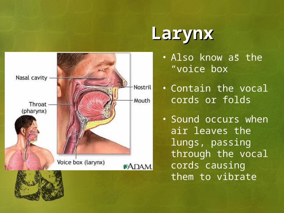
LarynxLarynx• Also know as the
“voice box”
• Contain the vocal cords or folds
• Sound occurs when air leaves the lungs, passing through the vocal cords causing them to vibrate

LarynxLarynx• Has 9 layers of
cartilage• The largest is
called the thyroid cartilage or the Adam’s apple

EpiglottisEpiglottis• Flap of cartilage lying
behind the tongue and in front of the larynx
• At rest is upright and allows air to pass through the larynx to the lungs
• During swallowing it folds back over the larynx to prevent food and liquids from getting into the airway
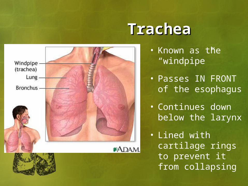
TracheaTrachea• Known as the
“windpipe”
• Passes IN FRONT of the esophagus
• Continues down below the larynx
• Lined with cartilage rings to prevent it from collapsing


CiliaCilia • Also in trachea
• Smoking is a constant irritation
• Smoking kills the cilia
• Leads to frequent infection and inflammation
• Triggers cough reflex and results in what we call the “smoker’s cough”

LUNGSLUNGS• Porous, spongy tissue
• Right lung is larger and broader than the left lung• It has three lobes
• This is because the heart lies to the left and needs room
• Left lung, therefore, only has two lobes

PLEURAPLEURA • Thin, moist, slippery membrane of tough tissue cells
• Two layers• Visceral covers just the
lung• Parietal covers lungs
and diaphragm and lines the thoracic cavity
• Pleural Fluid fills the space between the two pleural membranes

BronchiBronchi• Lower end of trachea
separates into the left and right bronchus

BronchiolesBronchioles• Bronchi subdivide into
the bronchioles

AlveoliAlveoli• About 500 million
alveoli in the adult lung
• This is 3x the amount needed to sustain life
• Inner surface are covered with a lipid substance called SURFACTANT
• Prevents alveoli from collapsing
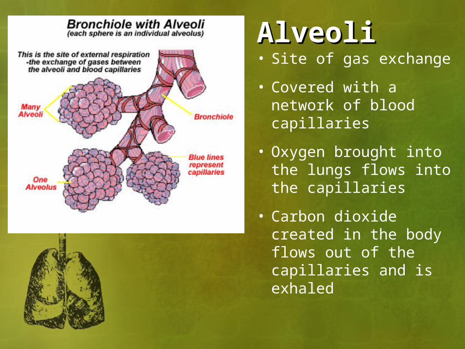
AlveoliAlveoli• Site of gas exchange
• Covered with a network of blood capillaries
• Oxygen brought into the lungs flows into the capillaries
• Carbon dioxide created in the body flows out of the capillaries and is exhaled

Process of BreathingProcess of Breathing• VENTILATION
• Mechanical process known as “breathing”
• Two phases called Inspiration and Expiration

VentilationVentilation • Inspiration• Diaphragm
contracts (moves downward)
• Intercostal muscles contract (pull ribs outward)
• Creates positive pressure and therefore air rushes into the lungs

VentilationVentilation • Expiration• Diaphragm and
intercostal muscles relax (return to resting state)
• Returns to negative pressure state
• Air is forced out of the lungs

Fun FactsFun Facts• Hiccups
• Caused by a spasm of the diaphragm believed to be the result of an irritation
• Sneezing• Air rushes out of your nose at a rate of
100 miles per second• Some people have a “photic reflex”
which makes them sneeze in response to a sudden, bright light
• Yawning• A deep, prolonged breath believed to be
caused by the need to increase oxygen in the blood

RespirationRespiration• Chemical process where oxygen
(O2) and carbon dioxide (CO2) are exchanged
• There are three types• External respiration• Internal respiration• Cellular respiration
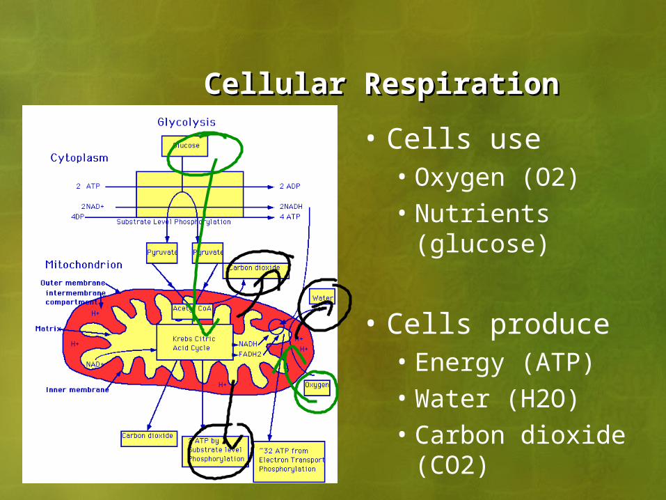
Cellular RespirationCellular Respiration
• Cells use• Oxygen (O2)• Nutrients (glucose)
• Cells produce• Energy (ATP)• Water (H2O)• Carbon dioxide
(CO2)

External RespirationExternal Respiration• Occurs in the lungs
• Between the alveoli and the blood stream
• Exchange of oxygen (O2) and carbon dioxide (CO2)

Internal RespirationInternal Respiration• Occurs in the
body• Between the blood
stream and tissue cells
• Exchange of oxygen (O2) and carbon dioxide (CO2)

Control of the Respiratory CenterControl of the Respiratory Center• Medulla oblongata controls respirations
• Located in the brain (lowest portion of the brain stem)
• Increased respirations occur if these things are happen• Decreased oxygen (O2) in the blood
stream• Increased carbon dioxide (CO2) in the
blood stream

DISEASES OF THE RESPIRATORY SYSTEM
• ASTHMA
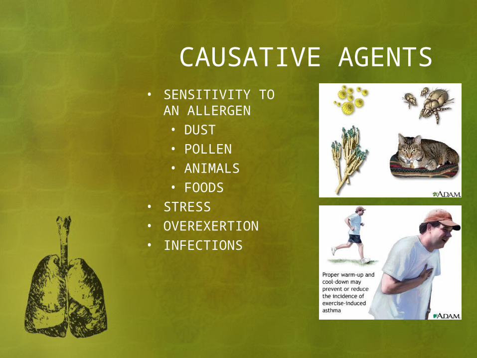
CAUSATIVE AGENTS• SENSITIVITY TO AN
ALLERGEN• DUST• POLLEN• ANIMALS• FOODS
• STRESS• OVEREXERTION• INFECTIONS

SYMPTOMS OCCUR• BRONCHOSPAS
MS NARROW OPENING OF BRONCHIOLES
• MUCUS PRODUCTION INCREASES
• EDEMA DEVELOPS IN MUCOSAL LINING
SYMPTOMS• DYSPNEA • WHEEZING• COUGHING WITH
EXPECTORATION OF SPUTUM• TIGHTNESS IN CHEST

Treatment• Bronchodilators (via rescue inhaler
or nebulizer)• Anti-inflammatory medications
(steroids)• Epinephrine• O2 Therapy

PREVENTING ASTHMA ATTACKS
• IDENTIFY ALLERGEN
• ELIMINATE ALLERGEN
• DESENSITIZATION TO ALLERGENS

BRONCHITIS
• INFLAMMATION•BRONCHI•BRONCHIAL TUBES

ACUTE BRONCHITIS• CAUSED BY
INFECTION• SYMPTOMS
• PRODUCTIVE COUGH
• DYSPNEA• CHEST PAIN• FEVER

TREATMENT
• ANTIBIOTICS• EXPECTORANTS TO
REMOVE EXCESSIVE MUCOUS

CHRONIC BRONCHITIS• OCCURS AFTER
FREQUENT ATTACKS OF ACUTE BRONCHITIS
• LONG-TERM EXPOSURE TO POLLUTANTS OR SMOKING
• CHARACTERIZED BY CHRONIC INFLAMMATION• DAMAGED CILIA• ENLARGED
MUCOUS GLANDS
SYMPTOMS• EXCESSIVE MUCUS
• PRODUCTIVE COUGH• WHEEZING & DYSPNEA• CHEST PAIN• PROLONGED EXPIRATION OF
AIR

TREATMENT
• NO CURE• ANTIBIOTICS• BRONCHODILATORS• RESPIRATORY THERAPY

LARYNGITIS
• INFLAMMATION•LARYNX•VOCAL CORDS
• MAY OCCUR WITH RESPIRATORY INFECTIONS

SYMPTOMS
• HOARSENESS • LOSS OF VOICE• SORE THROAT• DYSPHAGIA: DIFFICULTY IN
SWALLOWING
TREATMENT• REST• FLUIDS• LIMITED USE OF THE VOICE• MEDICATIONS
• INFECTION IF PRESENT

INFLUENZA (FLU)• CONTAGIOUS
VIRAL INFECTION• UPPER
RESPIRATORY SYSTEM
• SUDDEN ONSET

SYMPTOMS• CHILLS • HIGH FEVER• COUGH• SORE THROAT• RUNNY NOSE• MUSCLE PAIN• FATIGUE
TREATMENT• BED REST • FLUIDS• ANALGESICS
• PAIN• FEVER
• ANTIBIOTICS• NOT EFFECTIVE AGAINST VIRUSES• GIVEN TO AVOID SECONDARY
INFECTIONS• PNEUMONIA
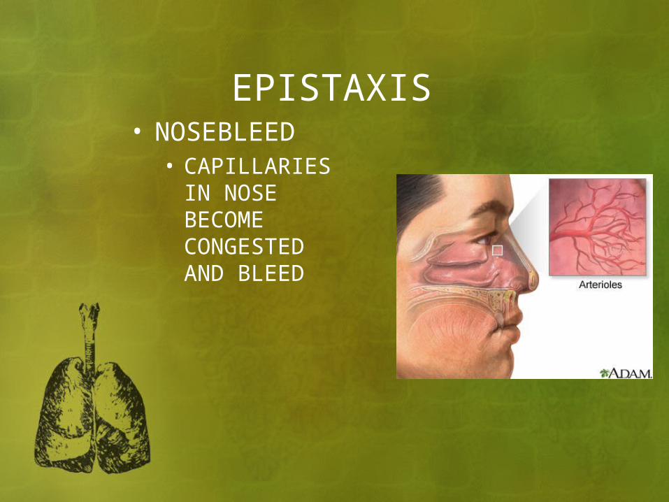
EPISTAXIS• NOSEBLEED
• CAPILLARIES IN NOSE BECOME CONGESTED AND BLEED

CAUSES• INJURY OR BLOW TO NOSE• HYPERTENSION• CHRONIC INFECTIONS• ANTICOAGULANT DRUGS• BLOOD DISEASES
• HEMOPHILIA• LEUKEMIA

TREATMENT• COMPRESS NOSTRILS• ELEVATE HEAD • TILT FORWARD
SLIGHTLY• APPLY COLD
COMPRESSES• NASAL PACKS• CAUTERIZE THE
BLEEDING VESSEL• ELIMINATE
UNDERLYING CAUSE

PNEUMONIA• INFLAMMATION • INFECTION OF
LUNGS• BUILD UP OF
EXUDATE IN ALVEOLI
• CAUSED BY BACTERIA, VIRUS, OR CHEMICALS

SYMPTOMS
• CHILLS• FEVER• CHEST PAIN• PRODUCTIVE COUGH• DYSPNEA• FATIGUE

TREATMENT• BEDREST• FLUIDS• ANTIBIOTICS IF INDICATED• RESPIRATORY THERAPY• PAIN MEDICATION

RHINITIS (URI)• INFLAMMATION OF
NASAL MUCOUS MEMBRANE• RUNNY NOSE• SORENESS• CONGESTION

COMMON CAUSES• INFECTIONS • ALLERGENS

TREATMENT
• FLUIDS• MEDICATION TO RELIEVE
CONGESTION
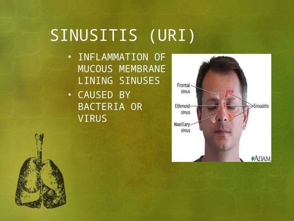
SINUSITIS (URI)• INFLAMMATION
OF MUCOUS MEMBRANE LINING SINUSES
• CAUSED BY BACTERIA OR VIRUS

SYMPTOMS• HEADACHE• PRESSURE• THICK NASAL DISCHARGE• CONGESTION• LOSS OF RESONANCE IN VOICE

TREATMENT• ANALGESICS• MEDICATIONS TO LOOSEN
SECRETIONS• MOIST INHALATIONS• SURGERY
• CHRONIC SINUSITIS • OPENS CAVITIES
• ENCOURAGE DRAINAGE

TREATMENT• ADMINISTRATION OF DRUGS
• DESTROY BACTERIA• GOOD NUTRITION• REST

TB• TUBERCULOSIS
• INFECTIOUS DISEASE OF THE LUNGS• CAUSED BY
BACTERIA• MYCOBACTERUIM
TUBERCULOSIS
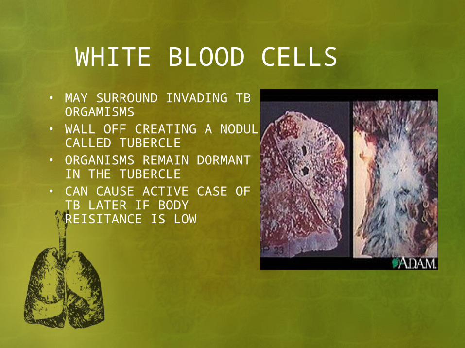
WHITE BLOOD CELLS• MAY SURROUND INVADING TB
ORGAMISMS• WALL OFF CREATING A
NODULE CALLED TUBERCLE• ORGANISMS REMAIN
DORMANT IN THE TUBERCLE• CAN CAUSE ACTIVE CASE OF
TB LATER IF BODY REISITANCE IS LOW

SYMPTOMS OF ACTIVE TB
• FATIGUE• CHEST PAIN• FEVER • NIGHT SWEATS• WEIGHT LOSS• HEMOPTYSIS
• COUGHING UP BLOOD TINGED SPUTUM

Treatment
• Medications for one or more years to destroy the bacteria
• Good nutrition• Rest
* In recent years a new strain of the TB bacteria that is resistant to drug therapy has emerged causing concern that it will become a widespread infectious disease

EMPHYSEMA• NONINFECTIOUS
CHRONIC RESPIRATORY CONDITION• WALLS OF THE
ALVEOLI DETERIORATE
• LOSE ELASTICITY• CARBON
DIOXIDE REMAINS TRAPPED IN THE ALVEOLI
• POOR EXCHANGE OF GASES
CAUSE• HEAVY SMOKING• PROLONGED EXPOSURE TO AIR
POLLUTANTS

TREATMENT• NO CURE• AVOID SMOKING• BRONCHODILATORS• PROMPT TREATMENT OF
RESPIRATORY INFECTIONS• OXYGEN THERAPY • RESPIRATORY THERAPY
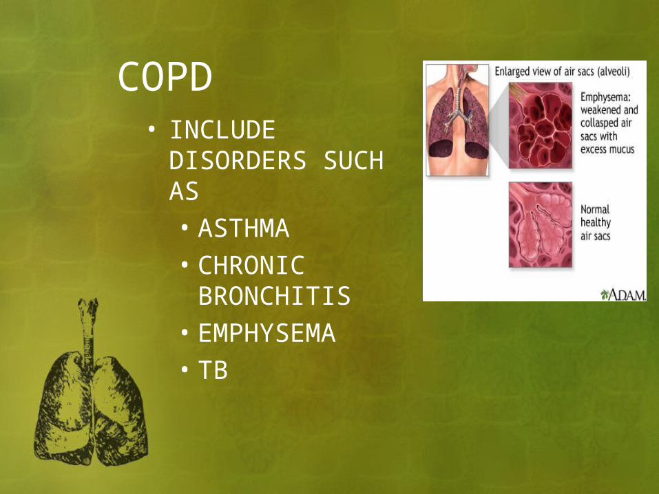
COPD• INCLUDE
DISORDERS SUCH AS• ASTHMA• CHRONIC
BRONCHITIS• EMPHYSEMA• TB

CAUSES• SMOKING IS THE PRIMARY
CAUSE• OTHER FACTORS INCLUDE
• ALLERGIES• CHRONIC RESPIRATORY
INFECTIONS
SYMPTOMS• DYSPNEA• FEELING OF SUFFOCATION• PAIN • BARREL CHEST• CHRONIC COUGH• CYANOSIS• RAPID RESPIRATIONS WITH
PROLONGED EXPIRATION• RESPIRATORY FALURE
……….DEATH

LUNG CANCER• DIAGNOSIS-
• XRAY • BRONCHOSCOPY
(flexible tube passed through mouth or nose into bronchi and lungs)
• TREATMENT• SURGERY• CHEMOTHERAPY• RADIATION

CAUSED BY• USUALLY SMOKING

LUNG CANCER XRAYS

Diagnostic Tests
• Pulmonary Function Testing (PFT)
• Bronchoscopy

Pulmonary Function Tests• Check how well your lungs work• Determine how much air your lungs can hold• Determine how quickly you can move air in and
out of your lungs• Determine how well your lungs put oxygen into
and remove carbon dioxide from your blood• The tests can diagnose lung diseases, measure
the severity of lung problems, and check to see how well treatment for a lung disease is working.
• Spirometry is the first and most commonly done lung function test. It measures how much and how quickly you can move air out of your lungs. For this test, you breathe into a mouthpiece attached to a recording device (spirometer). The information collected by the spirometer may be printed out on a chart called a spirogram.
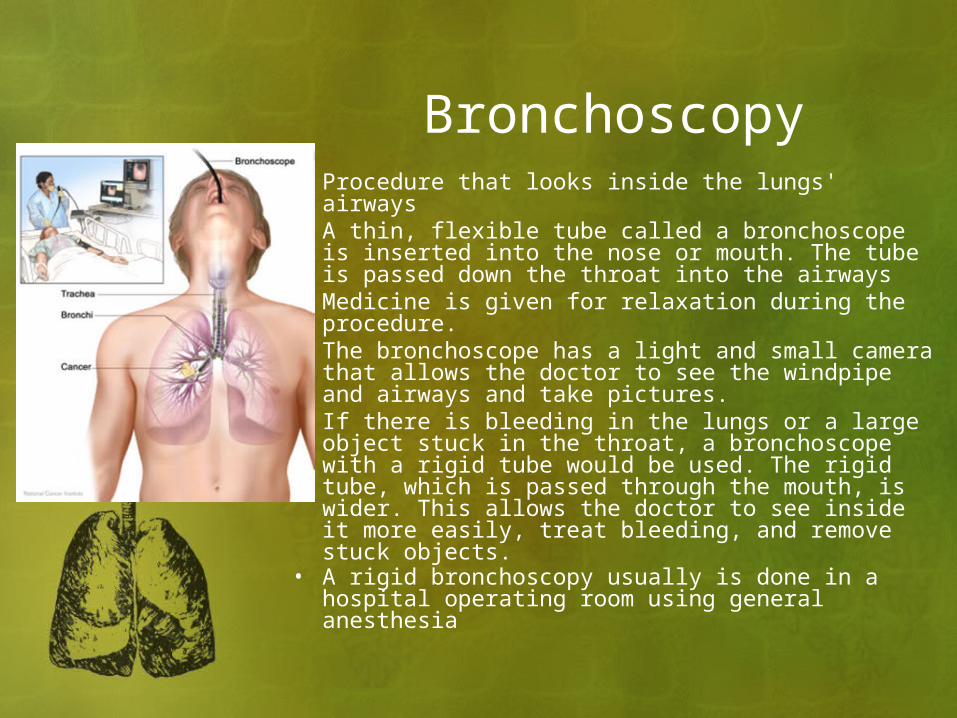
Bronchoscopy• Procedure that looks inside the lungs' airways• A thin, flexible tube called a bronchoscope is
inserted into the nose or mouth. The tube is passed down the throat into the airways
• Medicine is given for relaxation during the procedure.
• The bronchoscope has a light and small camera that allows the doctor to see the windpipe and airways and take pictures.
• If there is bleeding in the lungs or a large object stuck in the throat, a bronchoscope with a rigid tube would be used. The rigid tube, which is passed through the mouth, is wider. This allows the doctor to see inside it more easily, treat bleeding, and remove stuck objects.
• A rigid bronchoscopy usually is done in a hospital operating room using general anesthesia

Bronchoscopy