pulmonary tumor thrombotic microangiopathy associated … · pulmonary tumor thrombotic...
TRANSCRIPT
146146 THE EWHA MEDICAL JOURNALTHE EWHA MEDICAL JOURNAL
Pulmonary Tumor Thrombotic Microangiopathy Associated with Advanced Gastric Cancer Successfully Treated with Chemotherapy
Seung-Hyun Yoo, Kwonoh Park, Ji Yeon Hong, Ji Yeon Kim, Jang Won Park, Yong Won Park, Kyung-Hun Lee1, Kyung-So Jeon2
Department of Internal Medicine, KEPCO Medical Center; 1Department of Internal Medicine, Seoul National University Hospital; 2Laboratory Medicine, KEPCO Medical Center, Seoul, Korea
Introduction
Pulmonary tumor thrombotic microangiopathy (PTTM) is
known as a rare and severe cancer-related pulmonary complica-
tion that was first reported by Von Herbay et al. in 1990 [1]. In
PTTM cases, endothelial attachment of multiple microscopic tu-
mor emboli induces intimal proliferation of small pulmonary ar-
teries and arterioles combined with secondary thrombosis, which
leads to massive reduction of the pulmonary vascular bed and
pulmonary hypertension [1-3]. Due to extremely rapid progres-
sion of PTTM and non-specific clinical findings, establishing a
PTTM diagnosis is very difficult, and nearly all reported cases
are diagnosed primarily by autopsy [1,3]. There have only been
two PTTM cases reported in Korea, but they were diagnosed
by autopsy or shortly before death, and thus any effective treat-
ment was not available [4,5]. There have been only three cases
worldwide that have been diagnosed ante mortem and treated
effectively [6-8].
We report a 44-year-old woman who did not have a history
of cancer but presented with rapidly progressive dyspnea and
pulmonary hypertension. She was diagnosed as advanced gastric
cancer and related PTTM ante mortem. She was treated effec-
tively with chemotherapy for the underlying malignancy.
Case
In June 2013, a 44-year-old woman visited a primary clinic
complaining of dry cough. She was prescribed medication for
bronchitis, but the cough failed to improve. In July 2013, the
symptom worsened with progressive exertional dyspnea, and she
visited our hospital. She was evaluated using echocardiography
and the pulmonary function test (PFT). However, there was no
CaseReport
Ewha Med J 2014;37(2):146-151http://dx.doi.org/10.12771/emj.2014.37.2.146pISSN 2234-3180 • eISSN 2234-2591
Pulmonary tumor thrombotic microangiopathy (PTTM) is an uncommon and fatal malignancy-related pulmonary complication characterized by fibrocellular intimal proliferation of small pulmonary arteries and arterioles. It causes marked pulmonary hypertension, right-side heart failure, and sudden death. Diagnosis of PTTM is ex-tremely difficult while the patient is alive. Here, we report a 44-year-old woman who presented with complaining of progressing dyspnea and pulmonary hypertension but with no history of cancer. She was diagnosed with PTTM caused by advanced gastric cancer ante mortem and was treated effectively with chemotherapy. (Ewha Med J 2014;37(2):146-151)
Received February 20, 2014, Accepted May 28, 2014
Corresponding authorKwonoh ParkDepartment of Internal Medicine, KEPCO Medical Center, 308 Uicheon-ro, Dobong-gu, Seoul 132-703, KoreaTel: 82-2-901-3009, Fax: 82-2-901-3464E-mail: [email protected]
Key WordsPulmonary tumor thrombotic microangiopathy; Stomach neoplasms; Pulmonary hypertension; Chemotherapy
147THE EWHA MEDICAL JOURNAL
PTTM Associated with Advanced Gastric Cancer
significant abnormality, except diastolic dysfunction (grade I:
mild tricuspid regurgitation [TR]; right ventricular systolic pres-
sure [RVSP], 27 mmHg). Her dyspnea was aggravated, and she
developed left supraclavicular lymph node enlargement com-
bined with weight loss (3 kg in a month). She was admitted
to the department of hematology-oncology, KEPCO Medical
Center, for further examination and laboratory tests. She had
no specific medical history except for hypertension, which had
been diagnosed 7 years ago. She had never smoked.
On physical examination, the patient appeared ill and had
clear consciousness. Her blood pressure was 100/60 mmHg,
heart rate 104 beats per min, respiratory rate 23 breaths per
minute, body temperature 36.7oC, and oxygen saturation 96%
(in room air). The palpated left supraclavicular lymph node was
15 mm sized, hard and firm. The results of her arterial blood
gas analysis in room air were as follows: pH 7.47, pCO2 29
mmHg, pO2 91 mmHg, HCO3 21 mmol/L, SaO2 98%. Her
D-dimer was elevated to 2.5 μg/mL (normal, <0.23 μg/
mL), troponin I level and brain natriuretic peptide were within
the normal range, 0.00 ng/mL (normal, <0.05 ng/mL) and
45 pg/mL (normal, <100 pg/mL), respectively. Tumor marker
studies revealed that carcinoembryonic antigen (0.33 ng/mL)
and carbohydrate antigen 19-9 (36.7 ng/mL) levels were within
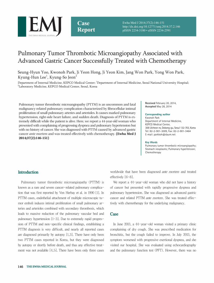
the normal range. A peripheral blood smear showed increased
numbers of schistocytes and reticulocytes consistent with mi-
croangiopathic hemolytic anemia (MAHA) (Fig. 1). A chest
radiograph showed that the lung field was clear without cardio-
megaly. Electrocardiography showed a heart rate of 104 beats
per min sinus tachycardia with negative T wave in V1-4, and
it revealed deviation of the frontal axis to the right, and Q wave
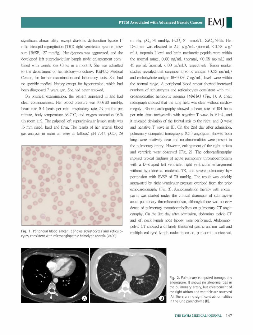
and negative T wave in III. On the 2nd day after admission,
pulmonary computed tomography (CT) angiogram showed both
lungs were relatively clear and no abnormalities were present in
the pulmonary artery. However, enlargement of the right atrium
and ventricle were observed (Fig. 2). The echocardiography
showed typical findings of acute pulmonary thromboembolism
with a D-shaped left ventricle, right ventricular enlargement
without hypokinesia, moderate TR, and severe pulmonary hy-
pertension with RVSP of 79 mmHg. The result was quickly
aggravated by right ventricular pressure overload from the prior
echocardiography (Fig. 3). Anticoagulation therapy with enoxa-
parin was started under the clinical diagnosis of submassive
acute pulmonary thromboembolism, although there was no evi-
dence of pulmonary thromboembolism on pulmonary CT angi-
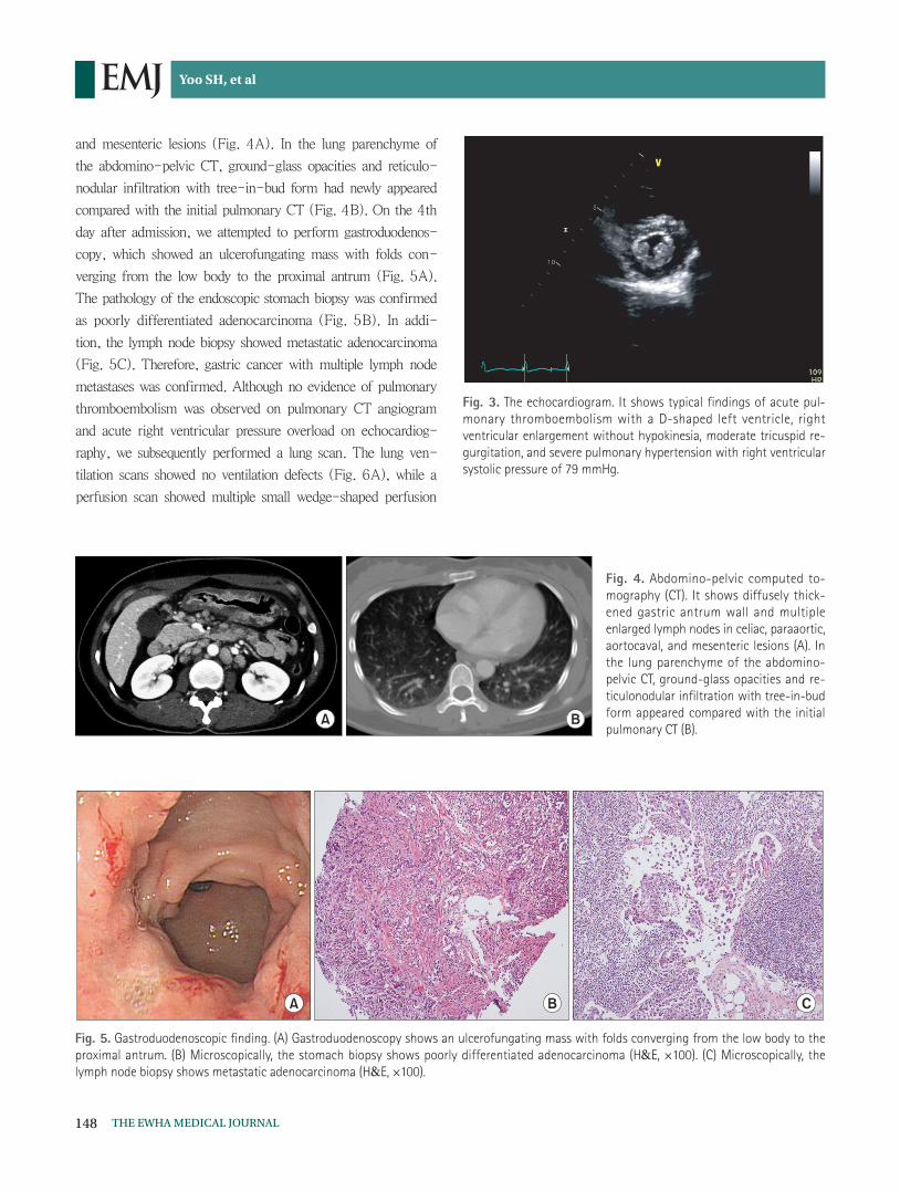
ography. On the 3rd day after admission, abdomino-pelvic CT
and left neck lymph node biopsy were performed. Abdomino-
pelvic CT showed a diffusely thickened gastric antrum wall and
multiple enlarged lymph nodes in celiac, paraaortic, aortocaval, Fig. 1. Peripheral blood smear. It shows schistocytes and reticulo-cytes, consistent with microangiopathic hemolytic anemia (×400).
Fig. 2. Pulmonary computed tomography angiogram. It shows no abnormalities in the pulmonary artery, but enlargement of the right atrium and ventricle are observed (A). There are no significant abnormalities in the lung parenchyme (B).
148 THE EWHA MEDICAL JOURNAL
Yoo SH, et al
and mesenteric lesions (Fig. 4A). In the lung parenchyme of
the abdomino-pelvic CT, ground-glass opacities and reticulo-
nodular infiltration with tree-in-bud form had newly appeared
compared with the initial pulmonary CT (Fig. 4B). On the 4th
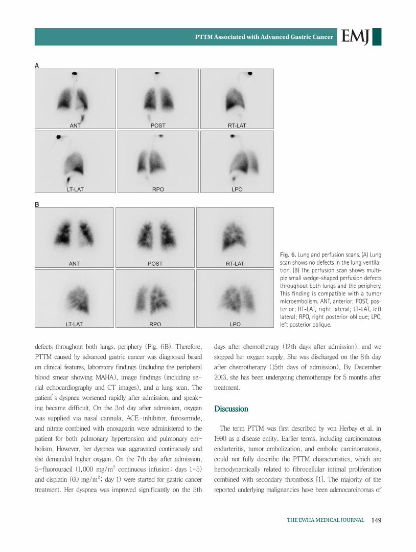
day after admission, we attempted to perform gastroduodenos-
copy, which showed an ulcerofungating mass with folds con-
verging from the low body to the proximal antrum (Fig. 5A).
The pathology of the endoscopic stomach biopsy was confirmed
as poorly differentiated adenocarcinoma (Fig. 5B). In addi-
tion, the lymph node biopsy showed metastatic adenocarcinoma
(Fig. 5C). Therefore, gastric cancer with multiple lymph node
metastases was confirmed. Although no evidence of pulmonary
thromboembolism was observed on pulmonary CT angiogram
and acute right ventricular pressure overload on echocardiog-
raphy, we subsequently performed a lung scan. The lung ven-
tilation scans showed no ventilation defects (Fig. 6A), while a
perfusion scan showed multiple small wedge-shaped perfusion
Fig. 3. The echocardiogram. It shows typical findings of acute pul-monary thromboembolism with a D-shaped left ventricle, right ventricular enlargement without hypokinesia, moderate tricuspid re-gurgitation, and severe pulmonary hypertension with right ventricular systolic pressure of 79 mmHg.
Fig. 4. Abdomino-pelvic computed to-mography (CT). It shows diffusely thick-ened gastric antrum wall and multiple enlarged lymph nodes in celiac, paraaortic, aortocaval, and mesenteric lesions (A). In the lung parenchyme of the abdomino-pelvic CT, ground-glass opacities and re-ticulonodular infiltration with tree-in-bud form appeared compared with the initial pulmonary CT (B).
Fig. 5. Gastroduodenoscopic finding. (A) Gastroduodenoscopy shows an ulcerofungating mass with folds converging from the low body to the proximal antrum. (B) Microscopically, the stomach biopsy shows poorly differentiated adenocarcinoma (H&E, ×100). (C) Microscopically, the lymph node biopsy shows metastatic adenocarcinoma (H&E, ×100).
149THE EWHA MEDICAL JOURNAL
PTTM Associated with Advanced Gastric Cancer
defects throughout both lungs, periphery (Fig. 6B). Therefore,
PTTM caused by advanced gastric cancer was diagnosed based
on clinical features, laboratory findings (including the peripheral
blood smear showing MAHA), image findings (including se-
rial echocardiography and CT images), and a lung scan. The
patient’s dyspnea worsened rapidly after admission, and speak-
ing became difficult. On the 3rd day after admission, oxygen
was supplied via nasal cannula. ACE-inhibitor, furosemide,
and nitrate combined with enoxaparin were administered to the
patient for both pulmonary hypertension and pulmonary em-
bolism. However, her dyspnea was aggravated continuously and
she demanded higher oxygen. On the 7th day after admission,
5-fluorouracil (1,000 mg/m2 continuous infusion; days 1-5)
and cisplatin (60 mg/m2; day 1) were started for gastric cancer
treatment. Her dyspnea was improved significantly on the 5th
days after chemotherapy (12th days after admission), and we
stopped her oxygen supply. She was discharged on the 8th day
after chemotherapy (15th days of admission). By December
2013, she has been undergoing chemotherapy for 5 months after
treatment.
Discussion
The term PTTM was first described by von Herbay et al. in
1990 as a disease entity. Earlier terms, including carcinomatous
endarteritis, tumor embolization, and embolic carcinomatosis,
could not fully describe the PTTM characteristics, which are
hemodynamically related to fibrocellular intimal proliferation
combined with secondary thrombosis [1]. The majority of the
reported underlying malignancies have been adenocarcinomas of
Fig. 6. Lung and perfusion scans. (A) Lung scan shows no defects in the lung ventila-tion. (B) The perfusion scan shows multi-ple small wedge-shaped perfusion defects throughout both lungs and the periphery. This finding is compatible with a tumor microembolism. ANT, anterior; POST, pos-terior; RT-LAT, right lateral; LT-LAT, left lateral; RPO, right posterior oblique; LPO, left posterior oblique.
150 THE EWHA MEDICAL JOURNAL
Yoo SH, et al
gastrointestinal origin [1-3,9]. Several cases with PTTM have
been reported in Japan, which shows a similar incidence of
gastric cancer to Korea [2,3]. However, no cases of PTTM due
to gastric cancer have been reported in Korea previously. Death
due to PTTM caused by gastric cancer may not be rare in Ko-
rea, and the few PTTM reports in Korea might be due to the
social reluctance to undergo autopsy. In our case, gastric cancer
with PTTM was identified ante mortem, and treated effectively
by chemotherapy.
PTTM is characterized by tumor emboli, accelerated co-
agulation, and intimal proliferation [1]. Tumor emboli, in the
vasculature with consequent local activation of coagulation and
combined with widespread fibrocellular and fibromuscular inti-
mal proliferation of small pulmonary arteries, leads to increased
vascular resistance, resulting in marked pulmonary hypertension
[1,3]. PTTM also shows hemodynamic effects similar to those
observed with MAHA and disseminated intravascular coagula-
tion (DIC). Although the underlying molecular mechanisms of
PTTM remain obscure, platelet-derived growth factor, tissue
factor, vascular endothelial growth factor, osteopontin and sero-
tonin likely play roles in the pathogenesis of PTTM [2,3,6].
PTTM is difficult to diagnose, and other diseases such as in-
fection, pulmonary thromboembolism, or drug-induced adverse
events, should be excluded before diagnosis. Normal chest X-
ray and chest CT findings may yield little evidence of PTTM.
However, a few reports showed characteristic tree-in-bud
upon chest CT [10]. In some cases, aggressive bronchoscopic
biopsy, transbronchial lung biopsy, and pulmonary microvas-
cular cytology using a wedged pulmonary artery catheter have
been suggested for diagnosis of PTTM [5,6,8,11]. Due to ex-
tremely rapid progression of PTTM, nearly all reported patients
have died within 1 week of dyspnea onset before differential
and confirmatory diagnostic processes [2,3,9,10]. It should be
noted that the aforementioned diagnostic procedures may not
be applicable to patients who are already experiencing severe
and progressive dyspnea. Hence, it is important for clinicians to
consider the possibility of PTTM in patients with dyspnea and
pulmonary hypertension based on clinical findings and non-
invasive diagnostic tools in order to make an early diagnosis and
provide therapeutic intervention. Peripheral blood smears for the
detection of MAHA, pulmonary artery CT angiogram to exclude
pulmonary embolism, and echocardiography for pulmonary hy-
pertension are warranted. In addition, lung scans are usually not
helpful given the diffuse involvement of the vasculature. How-
ever, they sometimes can show multiple peripheral subsegmental
perfusion defects without ventilatory abnormalities. This find-
ing is often referred to as the segmental contour pattern, which
has been described for PTTM. In our case, although pathology
was not confirmed in pulmonary vasculature, PTTM could be
diagnosed based on clinical symptoms, including a stubborn dry
cough and rapidly progressive dyspnea, MAHA from the periph-
eral blood smear, progressive changes in serial echocardiogra-
phy, and peripheral perfusion defect on the lung scan. There are
no specific diagnostic criteria without pathologic confirmation
for PTTM due to its rarity. Considering aggressiveness of PTTM
and poor tolerance of patients to invasive procedure, diagnosis
using clinical symptoms and non-invasive tools might be rather
appropriate in the perspective of the real practice.
PTTM is difficult to diagnose, and there is currently no ef-
fective management identified for PTTM. Chemotherapy, corti-
costeroids, and anticoagulation agents could be applied. Several
previous studies reported PTTM cases that were determined
through an ante mortem diagnosis, and the patients survived
after chemotherapy [6-8]. Our case also showed an improve-
ment in PTTM after appropriate management of the underlying
gastric cancer. However, further studies are needed to determine
whether early chemotherapy is effective, since some cases have
been reported of PTTM development after chemotherapy [12].
In summary, we report a case of PTTM caused by advanced
gastric cancer that improved after systemic chemotherapy. We
suggest that PTTM should be considered in the differential di-
agnosis of patients with unexplained, rapidly-worsening dyspnea
with pulmonary hypertension, even in the absence of a history
of cancer.
References
1. von Herbay A, Illes A, Waldherr R, Otto HF. Pulmonary tumor thrombotic microangiopathy with pulmonary hypertension. Cancer 1990;66:587-592.
2. Sato Y, Marutsuka K, Asada Y, Yamada M, Setoguchi T, Sumiyo-shi A. Pulmonary tumor thrombotic microangiopathy. Pathol Int 1995;45:436-440.
3. Okubo Y, Wakayama M, Kitahara K, Nemoto T, Yokose T, Abe F, et al. Pulmonary tumor thrombotic microangiopathy induced by gastric carcinoma: morphometric and immunohistochemi-cal analysis of six autopsy cases. Diagn Pathol 2011;6:27.
4. Kim HJ, Kwak MH, Kong SY, Seong MW, Kang HS, Lee KS, et al.
151THE EWHA MEDICAL JOURNAL
PTTM Associated with Advanced Gastric Cancer
A case of locally advanced breast cancer complicated by pul-monary tumor thrombotic microangiopathy. Cancer Res Treat 2012;44:267-270.
5. Yoo JH, Kim YH, Kim DY, Kang HY, Kim IJ, Lim SW, et al. Pulmo-nary tumor thrombotic microangiopathy in a patient with breast cancer. Korean J Med 2009;76(Suppl 1):S87-S92.
6. Miyano S, Izumi S, Takeda Y, Tokuhara M, Mochizuki M, Mat-subara O, et al. Pulmonary tumor thrombotic microangiopathy. J Clin Oncol 2007;25:597-599.
7. Kayatani H, Matsuo K, Ueda Y, Matsushita M, Fujiwara K, Yonei T, et al. Pulmonary tumor thrombotic microangiopathy diag-nosed antemortem and treated with combination chemothera-py. Intern Med 2012;51:2767-2770.
8. Kitamura A, Nishimura N, Jinta T, Suda R, Yamano Y, Ishikawa G, et al. A case of pulmonary tumor thrombotic microangiopathy diagnosed by transbronchial lung biopsy and treated with che-motherapy and long-term oxygen and anticoagulation therapies.
Case Rep Pulmonol 2013;2013:259080.9. Chinen K, Tokuda Y, Fujiwara M, Fujioka Y. Pulmonary tumor
thrombotic microangiopathy in patients with gastric carcinoma: an analysis of 6 autopsy cases and review of the literature. Pathol Res Pract 2010;206:682-689.
10. Franquet T, Gimenez A, Prats R, Rodriguez-Arias JM, Rodri-guez C. Thrombotic microangiopathy of pulmonary tumors: a vascular cause of tree-in-bud pattern on CT. Am J Roentgenol 2002;179:897-899.
11. Case records of the Massachusetts General Hospital. Weekly clinicopathological exercises. Case 19-1995. A 55 year-old woman with acute respiratory failure and radiographically clear lungs. N Engl J Med 1995;332:1700-1707.
12. Fisher DC, Sherrill GB, Hussein A, Rubin P, Vredenburgh JJ, El-kordy M, et al. Thrombotic microangiopathy as a complication of high-dose chemotherapy for breast cancer. Bone Marrow Transplant 1996;18:193-198.