pvc’s / pac’s what do they mean? what should … and... · pvc’s - ecg • qrs duration...
TRANSCRIPT
PVC’s / PAC’s What Do They Mean? What Should You Do?
Jeffrey H. Neuhauser, D.O.,F.A.C.C. BHHI Primary Care Symposium
February 27, 2015
Financial disclosures
Paid speaker for Pfizer
3
Learning Objectives • Understand the pathophysiology of PVC’s / PAC’s
• Understand the role for appropriate diagnostic
testing
• Differentiate the low risk from the high risk pt • Understand management options
4
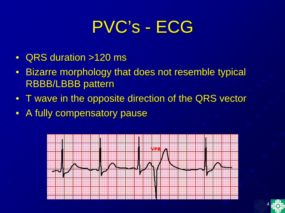
PVC’s - ECG • QRS duration >120 ms • Bizarre morphology that does not resemble typical
RBBB/LBBB pattern • T wave in the opposite direction of the QRS vector • A fully compensatory pause
5
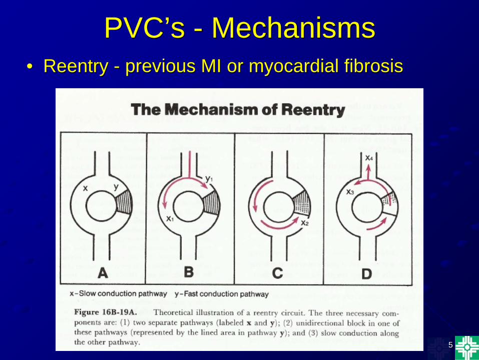
PVC’s - Mechanisms • Reentry - previous MI or myocardial fibrosis
6
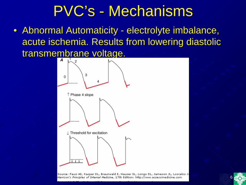
PVC’s - Mechanisms • Abnormal Automaticity - electrolyte imbalance,
acute ischemia. Results from lowering diastolic transmembrane voltage.
7
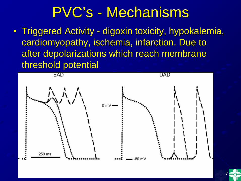
PVC’s - Mechanisms • Triggered Activity - digoxin toxicity, hypokalemia,
cardiomyopathy, ischemia, infarction. Due to after depolarizations which reach membrane threshold potential
PVC’s - Prevalence
• 1% of routine ECG’s of 30-60 sec duration • 80% incidence on 24 hr holter monitors of
healthy pts • More frequent in males & African
Americans • Prevalence increases with age,metabolic
abnormalities, & organic heart disease
9
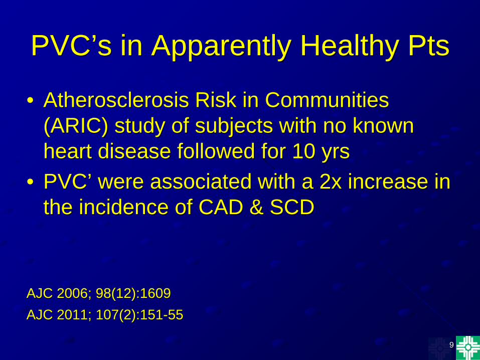
PVC’s in Apparently Healthy Pts
• Atherosclerosis Risk in Communities (ARIC) study of subjects with no known heart disease followed for 10 yrs
• PVC’ were associated with a 2x increase in the incidence of CAD & SCD
AJC 2006; 98(12):1609 AJC 2011; 107(2):151-55
10
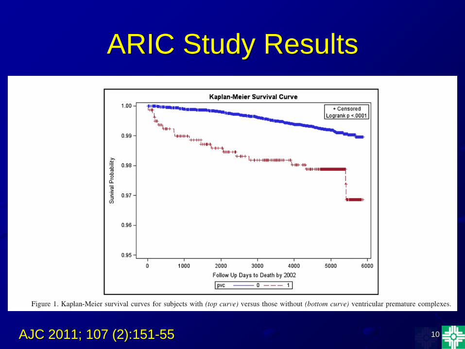
ARIC Study Results
AJC 2011; 107 (2):151-55
11
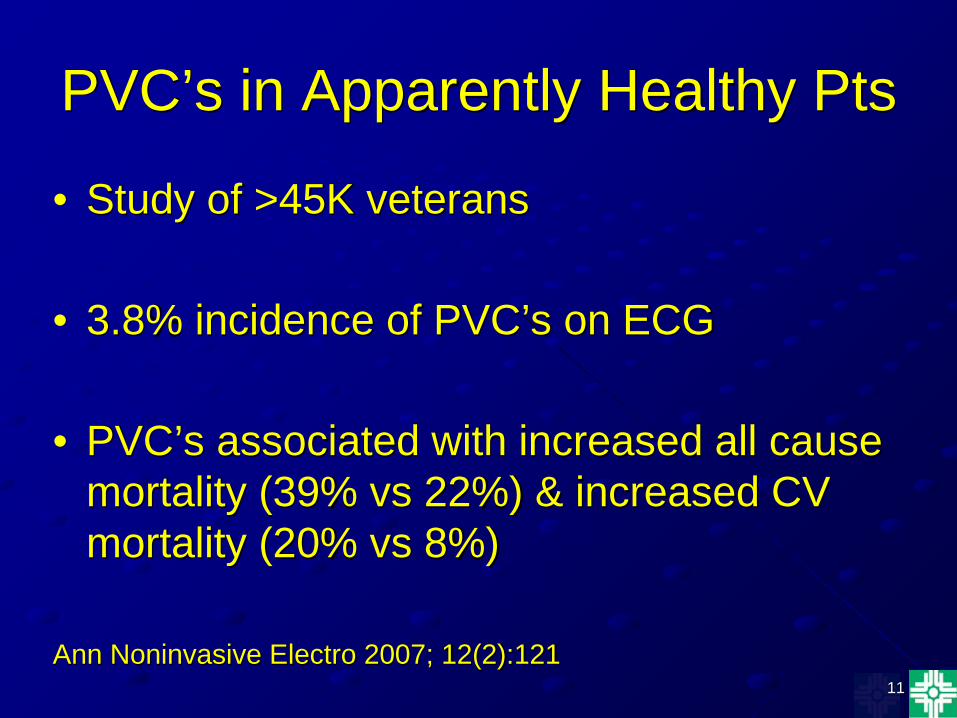
PVC’s in Apparently Healthy Pts
• Study of >45K veterans
• 3.8% incidence of PVC’s on ECG
• PVC’s associated with increased all cause mortality (39% vs 22%) & increased CV mortality (20% vs 8%)
Ann Noninvasive Electro 2007; 12(2):121
12
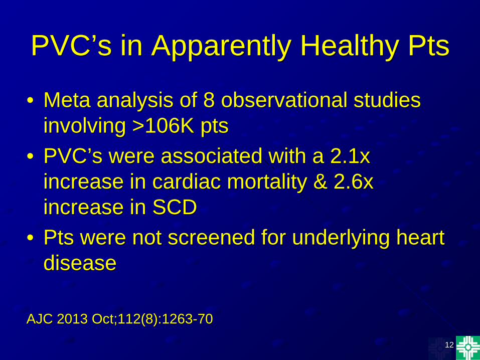
PVC’s in Apparently Healthy Pts
• Meta analysis of 8 observational studies involving >106K pts
• PVC’s were associated with a 2.1x increase in cardiac mortality & 2.6x increase in SCD
• Pts were not screened for underlying heart disease
AJC 2013 Oct;112(8):1263-70
13
PVC’s - H&P
• Few or no symptoms in most pts • Palpitations or dizziness are the most common
symptoms • Irregular pulse on exam • Variable intensity of S1 & cannon A waves • Ask about syncope, near syncope, family history
of early unexpected death, seizure disorder, or drowning
• Must differentiate the high risk from the low risk pt!
14
PVC’s - Diagnostic Evaluation
• ECG • Electrolytes, digoxin level • 24 hr Holter monitor • Echo • Exercise Treadmill Testing +/- imaging • Cardiac catheterization • Electrophysiologic study
15
Identifying the High Risk Pt • Frequent PVC’s (esp multifocal) & NSVT
• Decreased LV systolic function (LVEF </= 35%)
LVEF 35-40% is a gray zone
• Prior MI
• Ischemic burden
• LV hypertrophy with septal wall thickness >/= 3 cm • Intraventricular conduction delay on ECG
16
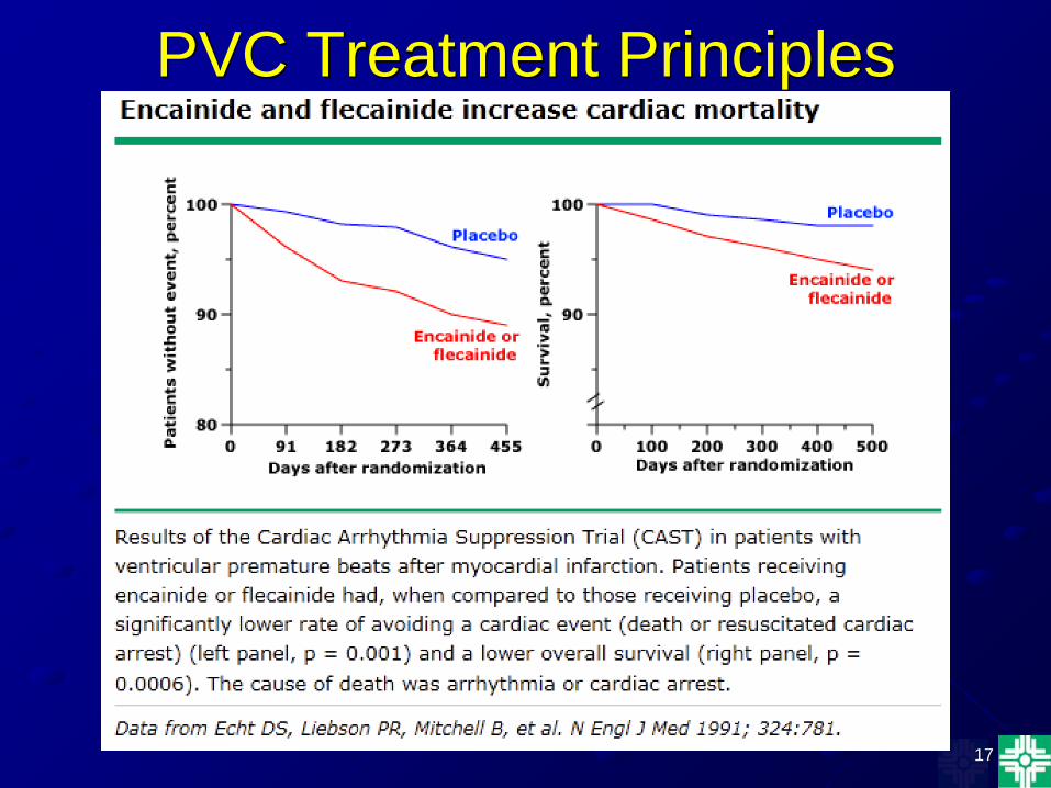
PVC’s -Treatment Principles • PVC’s should only be treated if they cause significant
symptoms • The risk depends upon the presence of underlying
structural heart disease • Suppressing PVC’s does not reduce mortality in high risk
pts • Beta blockers often 1st line therapy • If pt has a structurally normal heart, the risk for SCD is
low & catheter ablation can be considered for significant symptoms
• Sotalol, dofetilide, & amiodarone can be used for continued symptoms in pts with CAD or CM
• ICD’s are indicated for prevention of SCD in high risk pts
17
PVC Treatment Principles
18
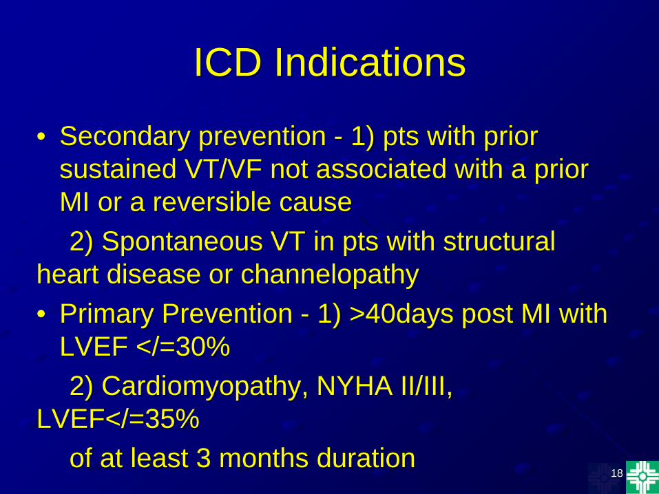
ICD Indications
• Secondary prevention - 1) pts with prior sustained VT/VF not associated with a prior MI or a reversible cause 2) Spontaneous VT in pts with structural
heart disease or channelopathy • Primary Prevention - 1) >40days post MI with
LVEF </=30% 2) Cardiomyopathy, NYHA II/III,
LVEF</=35% of at least 3 months duration
19
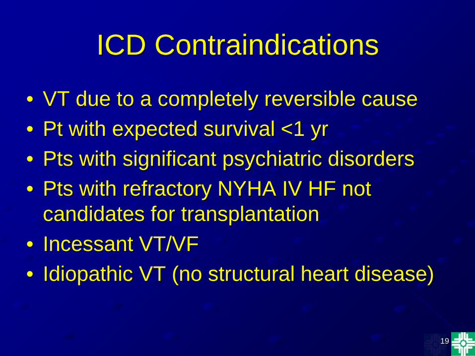
ICD Contraindications
• VT due to a completely reversible cause • Pt with expected survival <1 yr • Pts with significant psychiatric disorders • Pts with refractory NYHA IV HF not
candidates for transplantation • Incessant VT/VF • Idiopathic VT (no structural heart disease)
20
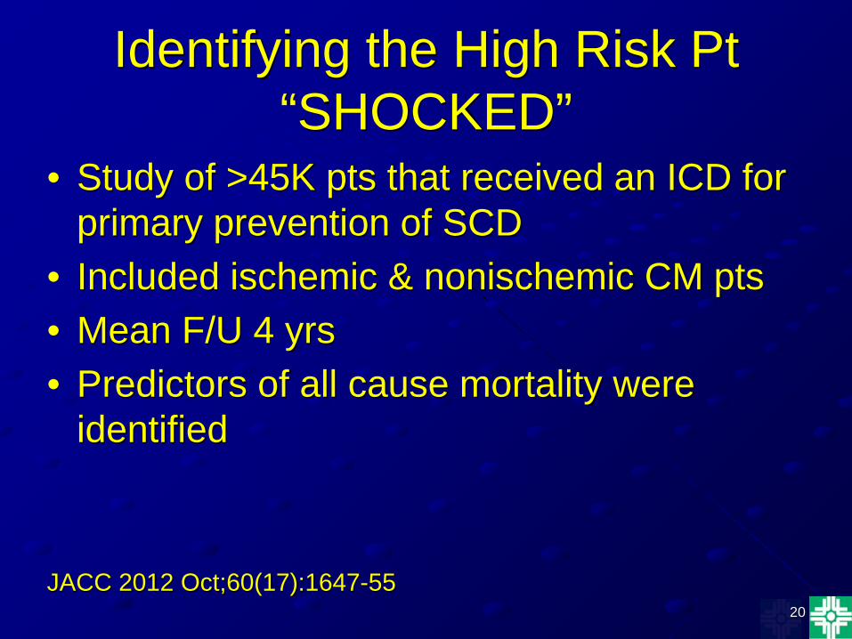
Identifying the High Risk Pt “SHOCKED”
• Study of >45K pts that received an ICD for primary prevention of SCD
• Included ischemic & nonischemic CM pts • Mean F/U 4 yrs • Predictors of all cause mortality were
identified
JACC 2012 Oct;60(17):1647-55
21
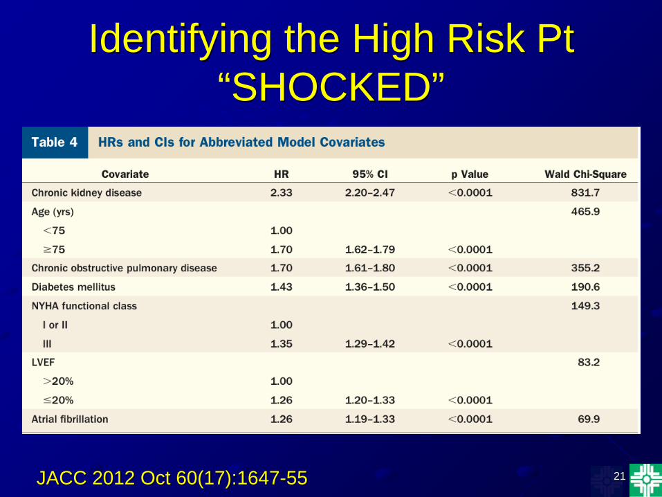
Identifying the High Risk Pt “SHOCKED”
JACC 2012 Oct 60(17):1647-55
22
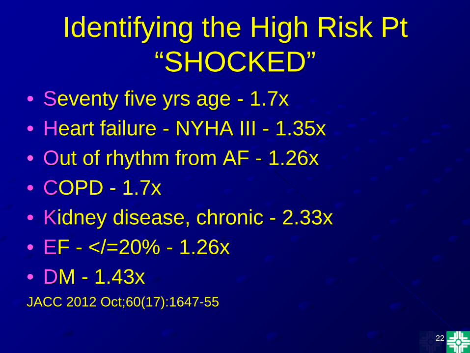
Identifying the High Risk Pt “SHOCKED”
• Seventy five yrs age - 1.7x • Heart failure - NYHA III - 1.35x • Out of rhythm from AF - 1.26x • COPD - 1.7x • Kidney disease, chronic - 2.33x • EF - </=20% - 1.26x • DM - 1.43x JACC 2012 Oct;60(17):1647-55
23
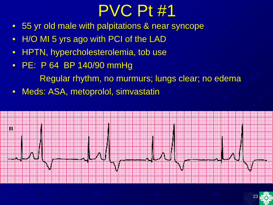
PVC Pt #1 • 55 yr old male with palpitations & near syncope • H/O MI 5 yrs ago with PCI of the LAD • HPTN, hypercholesterolemia, tob use • PE: P 64 BP 140/90 mmHg
Regular rhythm, no murmurs; lungs clear; no edema • Meds: ASA, metoprolol, simvastatin
24
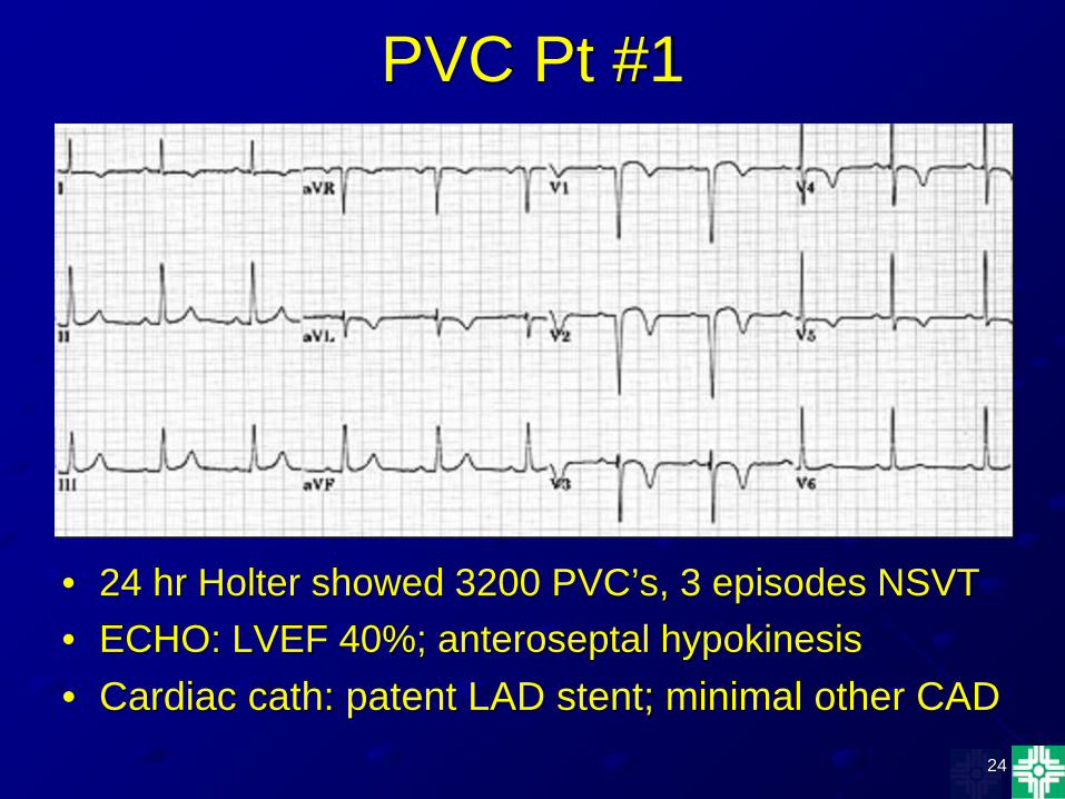
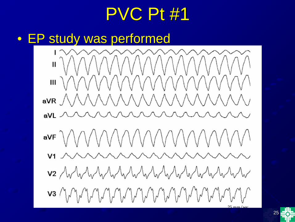
PVC Pt #1
• 24 hr Holter showed 3200 PVC’s, 3 episodes NSVT • ECHO: LVEF 40%; anteroseptal hypokinesis • Cardiac cath: patent LAD stent; minimal other CAD
25
PVC Pt #1 • EP study was performed
26
PVC Pt #2 • 23 yr old male with palpitations. No syncope or near
syncope. • No PMH • No medications • No significant family history • Consumes 3 energy drinks per day • Normal physical exam • Normal ECG • 24 hr Holter shows 550 PVC’s • Echo - normal • ETT - exercised 14 min no increase in PVC’s; no VT

27
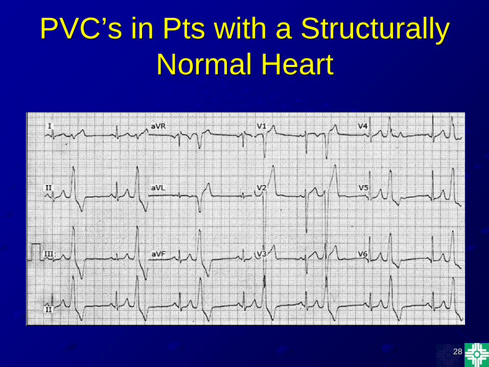
PVC’s in Pts With Structurally Normal Hearts
• Usually occurs in young to middle age pts • Catecholamine sensitive • Frequently exacerbated by exercise • Monomorphic, LBBB/RA on ECG • Originate in RVOT (occasionally LVOT) • Can be highly symptomatic, but rarely result in
SCD • Respond to beta blockers & Ca channel blockers • Can be cured by catheter ablation
28
PVC’s in Pts with a Structurally Normal Heart
29
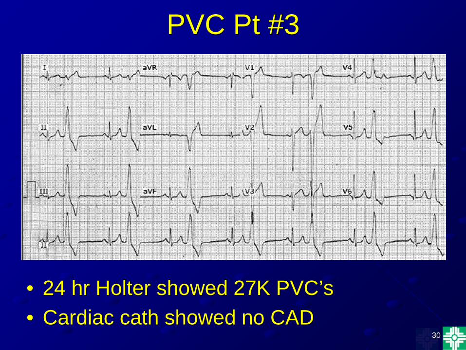
PVC Pt #3 • 40 yr old female presents with progressive
dyspnea, fatigue, & palpitations. The pt used to run, but is now currently unable to exercise.
• No PMH • No significant family history • No medications • PE: P 96 BP 100/60 mmHg
Irregular rhythm Lungs clear No edema
30
PVC Pt #3
• 24 hr Holter showed 27K PVC’s • Cardiac cath showed no CAD

31
PVC Pt #3
32
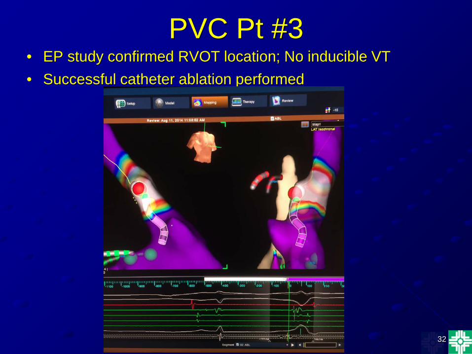
PVC Pt #3 • EP study confirmed RVOT location; No inducible VT • Successful catheter ablation performed
33
PVC Induced Cardiomyopathy
• Frequent PVC’s have been associated with reversible cardiomyopathy
• PVC burden >24% on 24 hr Holter • PVC’s >20K on 24 hr Holter • QRS duration (>160 ms) of the PVC’s may
be associated with development of CM

34
PVC Induced Cardiomyopathy • Catheter ablation of idiopathic PVC’s
(RVOT/LVOT) reported to have >80% success
• LVEF normalized within 4 months in most
• Epicardial origin of PVC’s & greater QRS duration of PVC’s associated with delayed recovery of LVF
Heart Rhythm 2013; 10(2):172
35
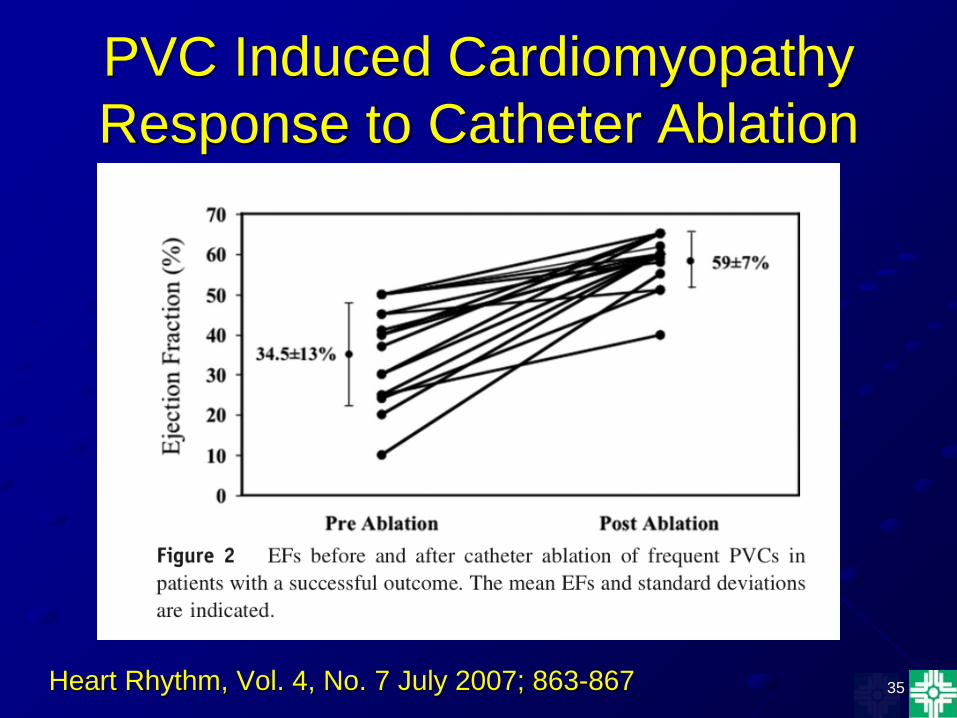
PVC Induced Cardiomyopathy Response to Catheter Ablation
Heart Rhythm, Vol. 4, No. 7 July 2007; 863-867
36
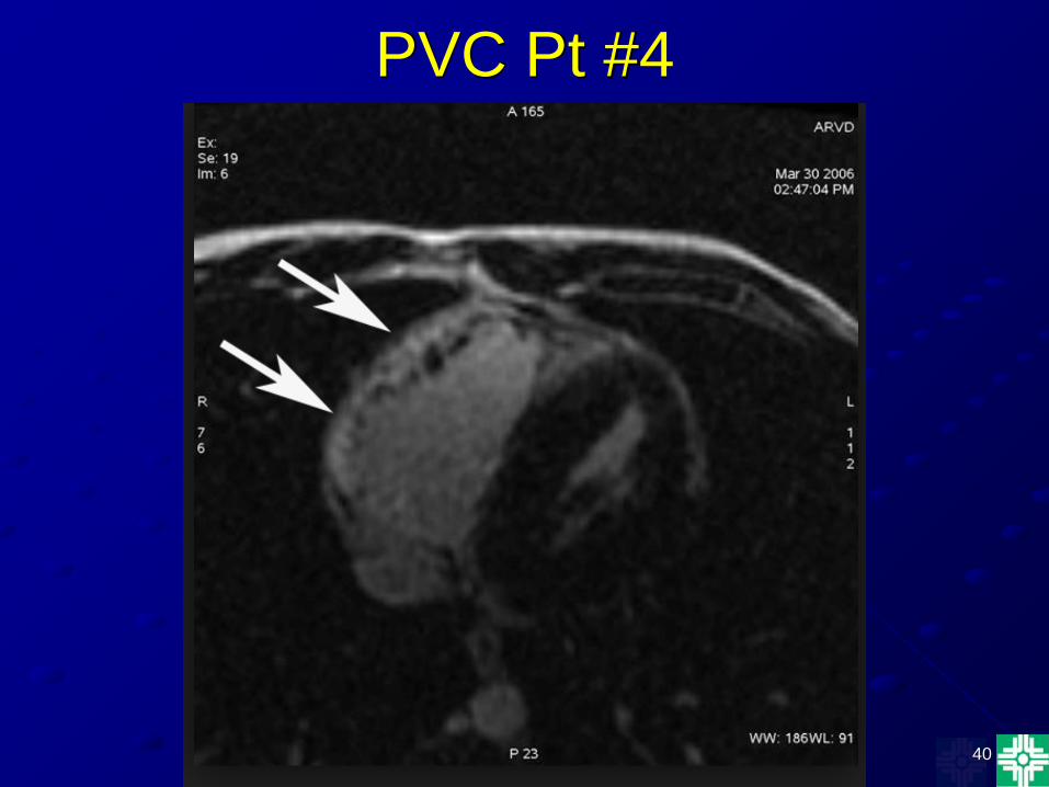
PVC Pt #4
• 20 yr old male with palpitations & near syncope. He is on the college wrestling team.
• No PMH • No medications • Father drowned. First cousin diagnosed
with a seizure disorder.
37
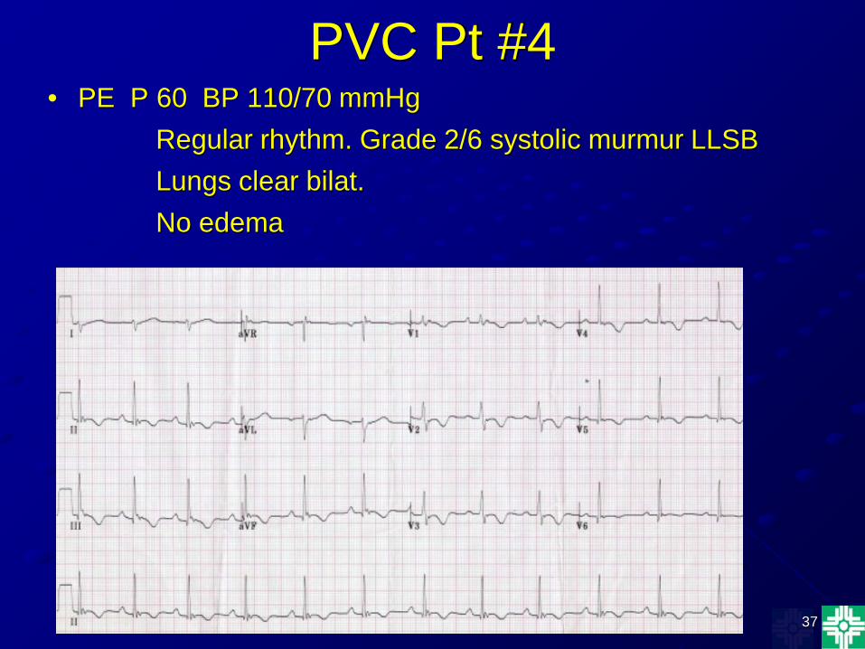
PVC Pt #4 • PE P 60 BP 110/70 mmHg
Regular rhythm. Grade 2/6 systolic murmur LLSB Lungs clear bilat. No edema
38
PVC Pt #4
• Holter monitor 3K PVC’s, 2 episodes NSVT
• Cardiac catheterization showed normal coronary anatomy & LVEF 55%
• Echo showed dilated RA/RV
39
PVC Pt #4
40
PVC Pt #4
41
Arrhythmogenic RV Dysplasia • Genetically inherited cardiomyopathy involving
predominantly the RV • Fibrofatty replacement of the RV myocardium
initially producing wall motion abnormalities & progressing to RV dilatation
• Both autosomal dominant & recessive inheritance • Prevalence 1:1000 - 1:2000 • Important cause of SCD in young adults
42
Arrhythmogenic RV Dysplasia • ECG - Incomplete RBBB, TWI V1-V3,prolonged S
wave V1-V3, Epsilon wave V1 • Echo - dilated RV with reduced RV systolic function • Cardiac MRI - dilated RV, RV wall motion
abnormalities, RVEF <40% • Electrophysiologic study may be helpful in risk
stratification • Pts should not participate in competitive sports • Beta blockers often recommended; no controlled data • ICD may be indicated for high risk pts

43
PVC’s in Athletes
• Not necessarily associated with increased risk for SCD
• Prognostic importance depends on the presence of underlying structural heart disease
• Exclusion of underlying heart disease is essential
44
PVC’s in Athletes Evaluation
• ECG • 24 hr Holter • ETT • Echo • Cardiac MRI • Cardiac catheterization in selected athletes
with abnormal echo/MRI
45
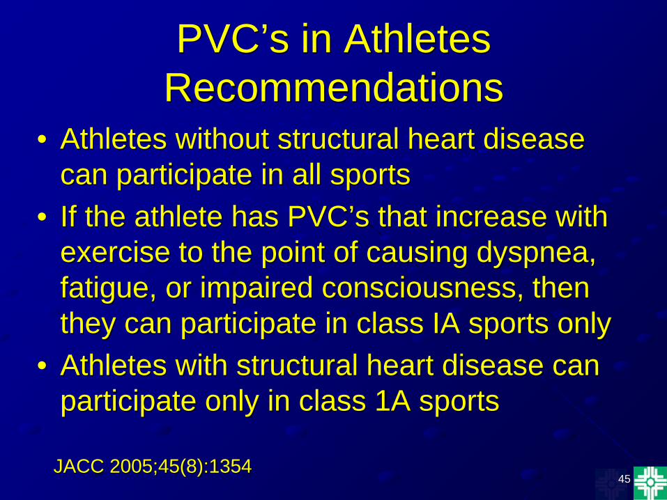
PVC’s in Athletes Recommendations
• Athletes without structural heart disease can participate in all sports
• If the athlete has PVC’s that increase with exercise to the point of causing dyspnea, fatigue, or impaired consciousness, then they can participate in class IA sports only
• Athletes with structural heart disease can participate only in class 1A sports
JACC 2005;45(8):1354
46
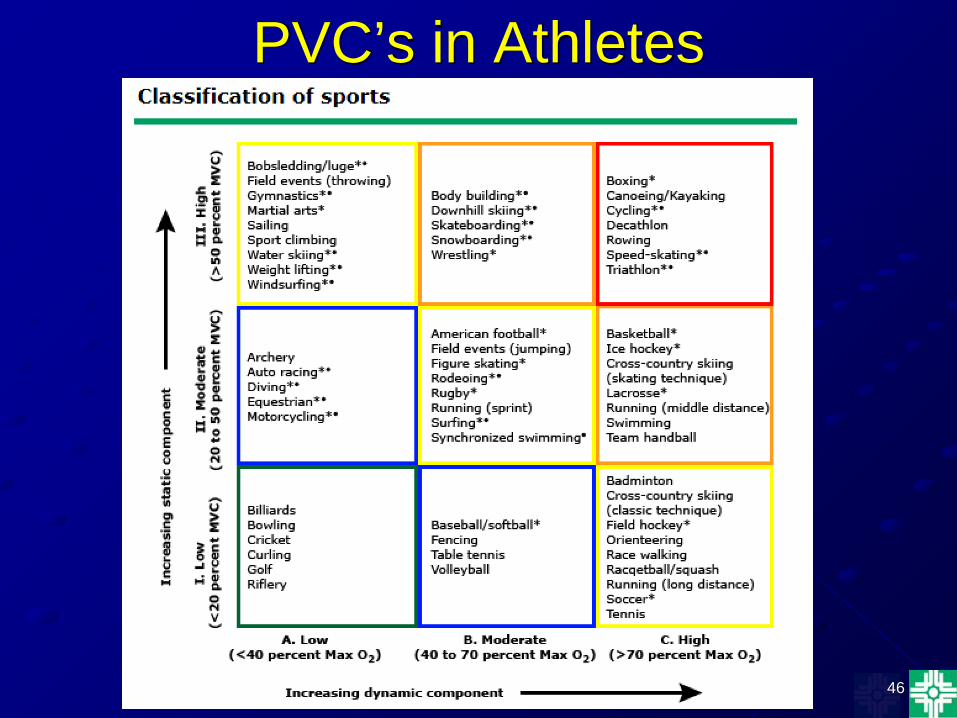
PVC’s in Athletes
47
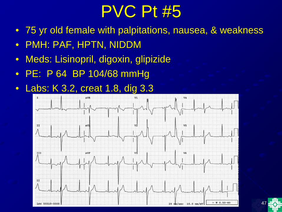
PVC Pt #5 • 75 yr old female with palpitations, nausea, & weakness • PMH: PAF, HPTN, NIDDM • Meds: Lisinopril, digoxin, glipizide • PE: P 64 BP 104/68 mmHg • Labs: K 3.2, creat 1.8, dig 3.3
48
Digoxin toxicity
• Symptoms include nausea, vomiting, anorexia, abdominal pain, confusion
• Serum dig level >2 ng/ml, however, toxicity can occur with levels in the “normal” range
• Hypokalemia, hypomagnesemia, hypercalcemia can predispose to toxicity
• PVC’s are the most common arrhythmia & often seen early in dig toxicity
• PVC’s usually occur in a bigeminal pattern
49
Digoxin Toxicity Management
• Admit pt for cardiac monitoring • If no evidence of renal failure, electrolyte
abnormality, or severe arrhythmia, pts can be safely discharged after at least 6 hours of monitoring
• Correct electrolyte abnormalities - hypo or hyperkalemia
• Fluid resuscitation if evidence of renal failure • Digoxin specific antibody fragments for life
threatening arrhythmias
50
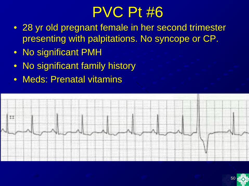
PVC Pt #6 • 28 yr old pregnant female in her second trimester
presenting with palpitations. No syncope or CP. • No significant PMH • No significant family history • Meds: Prenatal vitamins
51
PVC Pt #6
• PE: P 76 BP 108/70 Irregular rhythm. No murmurs or gallop Lungs clear Trace edema
• Normal resting ECG • Holter monitor 320 unifocal PVC’s • Echo - normal
52
PVC’s in Pregnancy
• Study of 110 symptomatic & 52 asymptomatic pregnant women
• 40% incidence in asymptomatic women • 49% incidence in symptomatic women AM J Cardiology 1997;79(8);1061
53
PVC’s in Pregnancy Evaluation
• ECG
• Electrolytes
• 24 hr Holter • Echo
54
PVC’s in Pregnancy Management
• Usually no therapy is needed
• Correct electrolyte abnormalities
• Discontinue smoking, caffeine, stimulants
• If symptoms are intolerable, metoprolol can be used
55
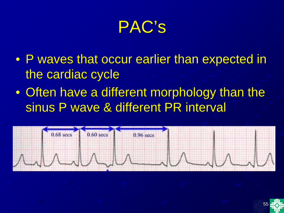
PAC’s
• P waves that occur earlier than expected in the cardiac cycle
• Often have a different morphology than the sinus P wave & different PR interval
56
PAC’s • Occurs commonly in young, elderly, pts with &
without structural heart disease • Increased incidence in pts with mitral valve disease
& LV dysfunction • Can be exacerbated by smoking, alcohol,
theophylline, & caffeine • The mechanisms - same as PVC’s
Reentry Abnormal automaticity Triggered activity
57
PAC’s • Usually produce few or no symptoms • Palpitations are the most common symptom • Rarely cause hemodynamic compromise • Irregular pulse on exam; sometimes can cause
cannon A waves • ECG obtained for evaluation • 24 hr Holter can also be used • Do not need tx • For severe symptoms beta blockers, class IC &
class III antiarrhythmic drugs can be used
58
Summary
• PAC’s occur commonly • Palpitations are the most common symptom • PAC’s may be associated with structural heart
disease but their presence alone does not signify increased risk
• PAC’s should not be treated unless the pt is severely symptomatic
• Try to identify underlying disease processes & precipitating factors
59
Summary • PVC’s can be markers for increased risk of CV events
& mortality • PVC’s can also occur in otherwise healthy pts • It is crucial to differentiate the low risk from the high risk
pt • Beta blockers & class III AAD’s can be used when it
becomes necessary to suppress symptoms • An ICD should be considered for the high risk pt • Frequent PVC’s can cause cardiomyopathy • Catheter ablation should be considered for
symptomatic PVC’s in pts with structurally normal hearts.