r i $ wr p lf % r p e 6 x uy ly r uv ±
TRANSCRIPT

Solid Cancer Incidence among the Life Span Study ofAtomic Bomb Survivors: 1958–2009
Authors: Grant, Eric J., Brenner, Alina, Sugiyama, Hiromi, Sakata,Ritsu, Sadakane, Atsuko, et al.
Source: Radiation Research, 187(5) : 513-537
Published By: Radiation Research Society
URL: https://doi.org/10.1667/RR14492.1
BioOne Complete (complete.BioOne.org) is a full-text database of 200 subscribed and open-access titlesin the biological, ecological, and environmental sciences published by nonprofit societies, associations,museums, institutions, and presses.
Your use of this PDF, the BioOne Complete website, and all posted and associated content indicates youracceptance of BioOne’s Terms of Use, available at www.bioone.org/terms-of-use.
Usage of BioOne Complete content is strictly limited to personal, educational, and non - commercial use.Commercial inquiries or rights and permissions requests should be directed to the individual publisher ascopyright holder.
BioOne sees sustainable scholarly publishing as an inherently collaborative enterprise connecting authors, nonprofitpublishers, academic institutions, research libraries, and research funders in the common goal of maximizing access tocritical research.
Downloaded From: https://bioone.org/journals/Radiation-Research on 24 Dec 2021Terms of Use: https://bioone.org/terms-of-use

RADIATION RESEARCH 187, 513–537 (2017)0033-7587/17 $15.00�2017 by Radiation Research Society.All rights of reproduction in any form reserved.DOI: 10.1667/RR14492.1
Solid Cancer Incidence among the Life Span Study of Atomic BombSurvivors: 1958–2009
Eric J. Grant,a,1 Alina Brenner,d Hiromi Sugiyama,a Ritsu Sakata,a Atsuko Sadakane,a Mai Utada,a
Elizabeth K. Cahoon,d Caitlin M. Milder,c Midori Soda,a Harry M. Cullings,b Dale L. Preston,e
Kiyohiko Mabuchid and Kotaro Ozasaa
Departments of a Epidemiology and b Statistics, and c Visiting Researcher, Radiation Effects Research Foundation, Hiroshima and Nagasaki,Japan; d Radiation Epidemiology Branch, Division of Cancer Epidemiology and Genetics, National Cancer Institute, Bethesda, Maryland; and
e Hirosoft International Corporation, Eureka, California
Grant, E. J., Brenner, A., Sugiyama, H., Sakata, R.,Sadakane, A., Utada, M., Cahoon, E. K., Milder, C. M., SodaM., Cullings, H. M., Preston, D. L., Mabuchi, K. and Ozasa,K. Solid Cancer Incidence among the Life Span Study ofAtomic Bomb Survivors: 1958–2009. Radiat. Res. 187, 513–537(2017).
This is the third analysis of solid cancer incidence amongthe Life Span Study (LSS) cohort of atomic bomb survivorsin Hiroshima and Nagasaki, adding eleven years of follow-updata since the previously reported analysis. For this analysis,several changes and improvements were implemented,including updated dose estimates (DS02R1) and adjustmentfor smoking. Here, we focus on all solid cancers in aggregate.The eligible cohort included 105,444 subjects who were aliveand had no known history of cancer at the start of follow-up.A total of 80,205 subjects had individual dose estimates and25,239 were not in either city at the time of the bombings. Thefollow-up period was 1958–2009, providing 3,079,484 person-years of follow-up. Cases were identified by linkage withpopulation-based Hiroshima and Nagasaki Cancer Registries.Poisson regression methods were used to elucidate the natureof the radiation-associated risks per Gy of weighted absorbedcolon dose using both excess relative risk (ERR) and excessabsolute risk (EAR) models adjusted for smoking. Riskestimates were reported for a person exposed at age 30 yearswith attained age of 70 years. In this study, 22,538 incidentfirst primary solid cancer cases were identified, of which 992were associated with radiation exposure. There were 5,918cases (26%) that occurred in the 11 years (1999–2009) sincethe previously reported study. For females, the dose responsewas consistent with linearity with an estimated ERR of 0.64per Gy (95% CI: 0.52 to 0.77). For males, significant upwardcurvature over the full dose range as well as restricted doseranges was observed and therefore, a linear-quadratic modelwas used, which resulted in an ERR of 0.20 (95% CI: 0.12 to0.28) at 1 Gy and an ERR of 0.010 (95% CI:�0.0003 to 0.021)at 0.1 Gy. The shape of the ERR dose response wassignificantly different among males and females (P ¼ 0.02).
While there was a significant decrease in the ERR withincreasing attained age, this decrease was more rapid inmales compared to females. The lowest dose range thatshowed a statistically significant dose response using the sex-averaged, linear ERR model was 0–100 mGy (P ¼ 0.038). Inconclusion, this analysis demonstrates that solid cancer risksremain elevated more than 60 years after exposure. Sex-averaged upward curvature was observed in the doseresponse independent of adjustment for smoking. Findingsfrom the current analysis regarding the dose-response shapewere not fully consistent with those previously reported,raising unresolved questions. At this time, uncertainties in theshape of the dose response preclude definitive conclusions toconfidently guide radiation protection policies. Upcomingresults from a series of analyses focusing on the radiationrisks for specific organs or organ families, as well ascontinued follow-up are needed to fully understand thenature of radiation-related cancer risk and its public healthsignificance. Data and analysis scripts are available fordownload at: http://www.rerf.or.jp. � 2017 by Radiation Research
Society
INTRODUCTION
The Life Span Study (LSS) of atomic bomb survivors inHiroshima and Nagasaki, Japan, provides quantitativeestimates of cancer risks associated with exposure to low-linear energy transfer (LET) radiation and is a major sourceof human data used for radiation risk assessment inestablishing radiation safety standards. Long-term follow-up of this cohort continues to provide updated informationon temporal patterns of radiation-related risk of cancer.Mortality follow-up data, based on Japan’s nationwidesystem of recording deaths, have been reported 14 timessince 1961, with the most recently reported data coveringthe follow-up period through 2003 (1). Mortality data,although highly valuable, do not provide adequate infor-mation on less fatal cancers. LSS cancer incidence dataderived from linkage with local population-based cancer
1 Address for correspondence: Radiation Effects Research Foun-dation, Epidemiology, 5-2 Hijiyama Park, Minami-ku, Hiroshima732-0815, Japan; email: [email protected].
513
Downloaded From: https://bioone.org/journals/Radiation-Research on 24 Dec 2021Terms of Use: https://bioone.org/terms-of-use

registries enable risk estimates for both fatal and nonfatalcancers with better diagnostic accuracy and disease onsetdate. Results of comprehensive analyses of solid andhematopoietic cancer incidence data among the LSS withfollow-up through 1987 were first reported in 1994 (2, 3)and updated for solid cancer incidence with follow-upthrough 1998 in 2007 (4). Incident hematopoietic cancerdata were recently updated through 2001 (5).
The principal finding regarding solid cancer risks fromthe follow-up, both incidence and mortality, of this cohorthas been a persistent increase in solid cancer risks due toradiation exposure that occurred at the time of the bombingsin 1945. The radiation dose response for all solid cancers asa group was previously observed to be linear with noevidence of a threshold. The excess relative risk (ERR) perunit dose of radiation for all solid cancers has been found todecrease with increasing attained age while the excessabsolute risks (EARs) have increased with attained agethroughout the follow-up period (4).
This article covers the third comprehensive analysis ofLSS solid cancer incidence risks, adding 11 years of follow-up to the previously reported study (4), extending theoverall follow-up period to 52 years, i.e., up to 64 yearsafter exposure. For this analysis, we have incorporatedseveral significant improvements in the data and methods.Individual radiation dose estimates have been revised, asdescribed by Cullings et al. (6). Briefly, the system forcalculating the doses is largely unchanged from DosimetrySystem 2002 (DS02) but the input parameters regarding asurvivor’s location and shielding information at the time ofthe bombing have been updated based on a thorough reviewof original materials. We updated estimates of migrationrates that account for cohort members moving out of andreturning to the cancer incidence catchment areas, re-appraised the appropriateness of cancers not clinicallyevident but identified only via the autopsy program foratomic bomb survivors and censored certain in situ cancersthat had been counted in some earlier reported studies. Wealso prepared and made use of lifestyle data, specificallysmoking data, obtained from various surveys of LSS cohortmembers.
This analysis concerns the radiation risks of all solidcancers in aggregate, focusing on the shape of the doseresponse after adjusting for age, sex, birth cohort andsmoking. Radiation-risk modifiers included attained age,age at exposure and sex. Subsequent organ or system-specific reports will follow and provide detailed dose-response analyses that address various topics of interestwhile including relevant lifestyle risk factors.
MATERIALS AND METHODS
Life Span Study Cohort
The Life Span Study cohort of 120,321 subjects includes 93,741atomic bomb survivors of Hiroshima and Nagasaki and 26,580persons who were not in either city [‘‘not in city’’ (NIC)] at the time of
the bombings. Details of the sampling methods are describedelsewhere (7, 8). Briefly, approximately 284,000 atomic bombsurvivors were enumerated at the time of the 1950 National Census.Among them, roughly 190,000 who were still living in Hiroshima orNagasaki at the time of the census served as the basis for selecting the94,000 survivors in this cohort; the cohort consists of 54,000 personswho were within 2.5 km of the hypocenter and thus exposed torelatively high doses of radiation (i.e., proximal survivors) and 40,000city, age and sex-matched survivors who were between 2.5 and 10 kmof the hypocenter who were exposed to lower or negligible doses (i.e.,distal survivors). The NIC subjects were identified by separate citycensuses and frequency matched to the survivors on city, sex and age.The NIC group was included in the risk analyses to improve estimatesof temporal and birth cohort patterns of background (baseline) cancerrates, as previously reported elsewhere (4).
Ascertainment of the vital status of LSS members was facilitated bythe Japanese national family registry system (koseki), which isvirtually complete. Since systematic solid cancer incidence ascertain-ment was not possible until the Hiroshima and Nagasaki population-based tumor registries were established in 1958, analyses of incidencedata were limited to a subset of the LSS cohort members who werealive and not known to have had cancer prior to January 1, 1958. Afterexcluding those who had died or been diagnosed with cancer prior toJanuary 1, 1958 (n¼ 8,317), along with those who could not be tracedusing koseki (n ¼ 86) and one person who was followed up induplicate, the LSS solid cancer incidence cohort consisted of 111,917(93% of the LSS cohort members). In the analysis, we also excluded6,473 survivors for whom Dosimetry System 2002 Revision 1(DSO2R1) doses (described later) could not be estimated. Thus, thetotal number of subjects considered in the current analysis was105,444 (consisting of 80,205 survivors and 25,239 NIC subjects).
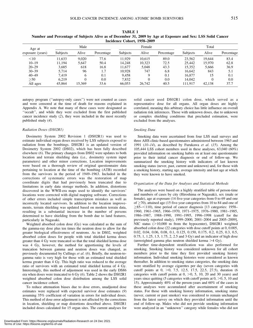
Table 1 shows distribution of the subjects in the LSS solid cancerincidence cohort by vital status and age at exposure by sex. As of theend of follow-up on December 31, 2009, 37.7% of members (33.6%of males and 40.5% of females) were alive. The majority (83.4%) ofthose alive at the end of follow-up were exposed as children (less than10 years old). Among all those exposed at less than 20 years of age,72.7% were alive at the end of follow-up while follow-up of thoseexposed after 30 years of age was virtually complete.
Ascertainment of Incident Cancer Cases
Cancer incidence follow-up of the LSS subjects is conducted usingvarious data sources with linkage to Hiroshima and Nagasaki basedcity and prefecture-wide cancer incidence registry systems. Cancerregistry data were supplemented by information from several RERFsources, including the Adult Health Study (AHS) and Atomic BombCasualty Commission/Radiation Effects Research Foundation(ABCC/RERF) surgical and autopsy programs. Members of theAHS cohort, a subset of the LSS cohort, have been invited to undergobiennial clinical health examinations since 1958. Under the ABCC/RERF autopsy program, extensive postmortem examinations wereperformed from 1948 to 1988, targeting LSS cohort members. Thecancer registries were the principal sources of cancer incidence data(.86% of cases).
The focus of the current analysis was on first primary solid cancersdiagnosed in the Hiroshima and Nagasaki cancer registry catchmentareas between 1958 and 2009. We grouped solid cancers using ICD-O-3 topography codes C00–C89 with behavior code 3 (malignant),plus brain and central nervous system tumors of benign or uncertain/unknown behavior [ICD-O-3 topography codes C70–C72, pituitarygland code C751, craniopharyngeal duct code C752, and pineal glandcode C753 with behavior code 0 (benign) or 1 (uncertain or unknownnature)]. We excluded leukemia, lymphoma, myeloma and otherlymphohematopoietic malignancies (ICD-O-3 morphology codes9590–9970). In situ cancers and intramucosal colorectal carcinomaswere ignored. In addition, otherwise eligible cases with a diagnosisbased solely on postmortem examination under the ABCC/RERF
514 GRANT ET AL.
Downloaded From: https://bioone.org/journals/Radiation-Research on 24 Dec 2021Terms of Use: https://bioone.org/terms-of-use
autopsy program (‘‘autopsy-only cases’’) were not counted as casesand were censored at the time of death for reasons explained inAppendix A. We note that many of these cases were designated as‘‘occult’’, and while they were excluded from the first publishedcancer incidence study (2), they were included in the most recentlypublished study (4).
Radiation Doses (DS02R1)
Dosimetry System 2002 Revision 1 (DS02R1) was used toestimate individual organ doses received by LSS subjects exposed toradiation from the bombings. DS02R1 is an updated version ofDosimetry System 2002 (DS02), which has been fully describedelsewhere (6). The primary changes from DS02 were updates to bothlocation and terrain shielding data (i.e., dosimetry system inputparameters) and other minor corrections. Location improvementswere based on a thorough review of original questionnaire datapertaining to location at the time of the bombing (ATB) recordedfrom the survivors in the period of 1949–1963. Included in thecorrections of systematic errors was the restoration of mapcoordinate digits that had previously been truncated due tolimitations in early data storage methods. In addition, distortionsdiscovered in the WWII-era maps used to identify the survivors’locations were corrected with digital mapping software. Correctionsof other errors included simple transcription mistakes as well asincorrectly located survivors. In addition to the location improve-ments, terrain shielding was updated based on modern terrain data,resulting in a substantial increase in the number of personsdetermined to have shielding from the bomb due to land features,particularly in Nagasaki.
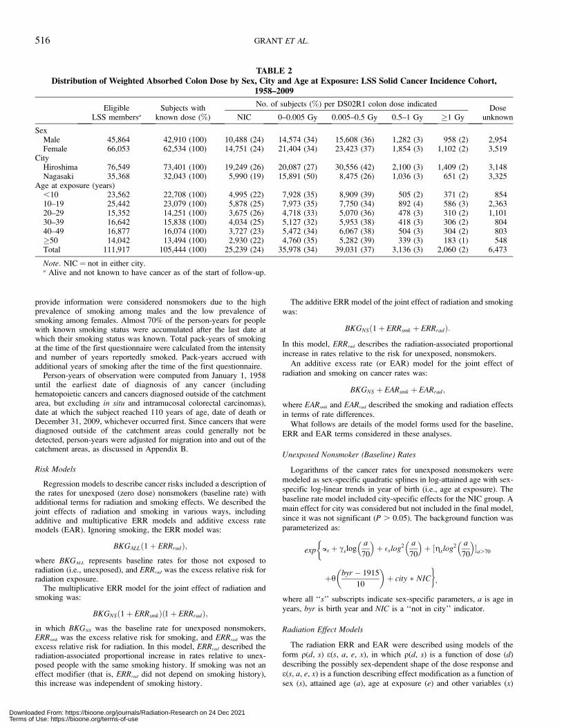
Weighted absorbed colon dose (Gy) was calculated as the sum ofthe gamma-ray dose plus ten times the neutron dose to allow for thegreater biological effectiveness of neutrons. As in DS02, weightedabsorbed colon doses for people with total shielded kerma dosesgreater than 4 Gy were truncated so that the total shielded kerma dosewas 4 Gy, however, the method for apportioning the levels oftruncation between gamma and neutron doses was changed, aspreviously documented by Cullings et al. (6). Briefly, the neutron-to-gamma ratio is very high for those with an estimated total shieldedkerma greater than 4 Gy. This high ratio was reduced to the averageratio of survivors with an estimated total shielded kerma of 4 Gy.Interestingly, this method of adjustment was used in the early DS86era when doses were truncated to 6 Gy (6). Table 2 shows the DS02R1weighted absorbed colon dose distribution among the LSS solidcancer incidence cohort.
To reduce attenuation biases due to dose errors, unadjusted doseestimates were replaced with expected survivor dose estimates (9)assuming 35% coefficient of variation in errors for individual doses.This method of dose error adjustment is not affected by the correctionsin location, shielding or map distortions described above. DS02R1included doses calculated for 15 organ sites. The current analyses for
solid cancer used DS02R1 colon dose, which served as arepresentative dose for all organs. All organ doses are highlycorrelated, meaning this arbitrary choice has little influence on overallradiation risk inferences. Those with unknown doses, due to unknownor complex shielding conditions that precluded estimation, wereexcluded from the analyses.
Smoking Data
Smoking data were ascertained from four LSS mail surveys andthree AHS clinic-based questionnaires administered between 1963 and1991 (10–14), as described by Furukawa et al. (15). Among the105,444 LSS cohort members used in these analyses, 63,040 (60%)provided information on smoking habits on at least one questionnaireprior to their initial cancer diagnosis or end of follow-up. Wesummarized the smoking history with indicators of last knownsmoking status (never, past, current and unknown) and, for those witha smoking history, starting age, average intensity and last age at whichthey were known to have smoked.
Organization of the Data for Analyses and Statistical Methods
The analyses were based on a highly stratified table of person-timeand numbers of cases by city (Hiroshima or Nagasaki), sex (male orfemale), age at exposure (14 five-year categories from 0 to 69 and oneof �70), attained age (15 five-year categories from 10 to 84 and one of�85–,110), time period of cancer diagnosis [13 categories: 1958–1960, 1961–1965, 1966–1970, 1971–1975, 1976–1980, 1981–1985,1986–1987, 1988–1990, 1991–1995, 1996–1998 (cutoff for thepreviously reported study), 1999–2000, 2001–2004 and 2005–2009],NIC status (.10,000 m from the hypocenter), DS02R1 weightedabsorbed colon dose (22 categories with dose cutoff points at 0, 0.005,0.02, 0.04, 0.06, 0.08, 0.1, 0.125, 0.150, 0.175, 0.2, 0.25, 0.3, 0.5,0.75, 1, 1.25, 1.5, 1.75, 2, 2.5 and 3 Gy) and an indicator of high dose(unweighted gamma plus neutron shielded kerma .4 Gy).
Further time-dependent stratification was also performed forsmoking. Smoking history was considered unknown for all cohortmembers prior to the time they first provided smoking historyinformation. Individual smoking histories were considered as knownthereafter. In addition to smoking status categories, the smoking datawere stratified by average cigarettes per day (seven categories withcutoff points at 0, .0, 7.5, 12.5, 17.5, 22.5, 27.5), duration (6categories with cutoff points at 0, .0, 5, 10, 20 and 30 years) andyears since quitting (5 categories with cutoff points at 0, .0, 5, 10 and15). Approximately 40% of the person-years and 60% of the cases inthese analyses were accumulated after ascertainment of smokingstatus. For those with smoking history information, smoking status(never, current or past smoker) was considered to remain unchangedfrom the latest survey on which they provided information until theend of follow-up. Males who did not provide smoking informationwere analyzed in an ‘‘unknown’’ category while females who did not
TABLE 1Number and Percentage of Subjects Alive as of December 31, 2009 by Age at Exposure and Sex: LSS Solid Cancer
Incidence Cohort, 1958–2009
Age atexposure (years)
Male Female Total
Subjects Alive Percentage Subjects Alive Percentage Subjects Alive Percentage
,10 11,633 9,020 77.6 11,929 10,615 89.0 23,562 19,644 83.410–19 11,194 5,647 50.4 14,248 10,323 72.5 25,442 15,970 62.820–29 3,685 618 16.8 11,677 5,048 43.3 15,352 5,666 36.930–39 5,714 96 1.7 10,928 747 6.8 16,642 843 5.140–49 7,419 6 0.1 9,458 9 0.1 16,877 15 0.1�50 6,219 0 0.0 7,832 0 0.0 14,042 0 0.0All ages 45,864 15,369 33.6 66,053 26,742 40.5 111,917 42,138 37.7
SOLID CANCER INCIDENCE AMONG ATOMIC BOMB SURVIVORS 515
Downloaded From: https://bioone.org/journals/Radiation-Research on 24 Dec 2021Terms of Use: https://bioone.org/terms-of-use
provide information were considered nonsmokers due to the highprevalence of smoking among males and the low prevalence ofsmoking among females. Almost 70% of the person-years for peoplewith known smoking status were accumulated after the last date atwhich their smoking status was known. Total pack-years of smokingat the time of the first questionnaire were calculated from the intensityand number of years reportedly smoked. Pack-years accrued withadditional years of smoking after the time of the first questionnaire.
Person-years of observation were computed from January 1, 1958until the earliest date of diagnosis of any cancer (includinghematopoietic cancers and cancers diagnosed outside of the catchmentarea, but excluding in situ and intramucosal colorectal carcinomas),date at which the subject reached 110 years of age, date of death orDecember 31, 2009, whichever occurred first. Since cancers that werediagnosed outside of the catchment areas could generally not bedetected, person-years were adjusted for migration into and out of thecatchment areas, as discussed in Appendix B.
Risk Models
Regression models to describe cancer risks included a description ofthe rates for unexposed (zero dose) nonsmokers (baseline rate) withadditional terms for radiation and smoking effects. We described thejoint effects of radiation and smoking in various ways, includingadditive and multiplicative ERR models and additive excess ratemodels (EAR). Ignoring smoking, the ERR model was:
BKGALLð1þ ERRradÞ;where BKGALL represents baseline rates for those not exposed toradiation (i.e., unexposed), and ERRrad was the excess relative risk forradiation exposure.
The multiplicative ERR model for the joint effect of radiation andsmoking was:
BKGNSð1þ ERRsmkÞð1þ ERRradÞ;in which BKGNS was the baseline rate for unexposed nonsmokers,ERRsmk was the excess relative risk for smoking, and ERRrad was theexcess relative risk for radiation. In this model, ERRrad described theradiation-associated proportional increase in rates relative to unex-posed people with the same smoking history. If smoking was not aneffect modifier (that is, ERRrad did not depend on smoking history),this increase was independent of smoking history.
The additive ERR model of the joint effect of radiation and smokingwas:
BKGNSð1þ ERRsmk þ ERRradÞ:
In this model, ERRrad describes the radiation-associated proportionalincrease in rates relative to the risk for unexposed, nonsmokers.
An additive excess rate (or EAR) model for the joint effect ofradiation and smoking on cancer rates was:
BKGNS þ EARsmk þ EARrad;
where EARsmk and EARrad described the smoking and radiation effectsin terms of rate differences.
What follows are details of the model forms used for the baseline,ERR and EAR terms considered in these analyses.
Unexposed Nonsmoker (Baseline) Rates
Logarithms of the cancer rates for unexposed nonsmokers weremodeled as sex-specific quadratic splines in log-attained age with sex-specific log-linear trends in year of birth (i.e., age at exposure). Thebaseline rate model included city-specific effects for the NIC group. Amain effect for city was considered but not included in the final model,since it was not significant (P . 0.05). The background function wasparameterized as:
exp as þ csloga
70
� �þ �slog2 a
70
� �þ ½gslog2 a
70
� ��a.70
�
þhbyr � 1915
10
� �þ city � NICg;
where all ‘‘s’’ subscripts indicate sex-specific parameters, a is age inyears, byr is birth year and NIC is a ‘‘not in city’’ indicator.
Radiation Effect Models
The radiation ERR and EAR were described using models of theform q(d, s) e(s, a, e, x), in which q(d, s) is a function of dose (d)describing the possibly sex-dependent shape of the dose response ande(s, a, e, x) is a function describing effect modification as a function ofsex (s), attained age (a), age at exposure (e) and other variables (x)
TABLE 2Distribution of Weighted Absorbed Colon Dose by Sex, City and Age at Exposure: LSS Solid Cancer Incidence Cohort,
1958–2009
Eligible Subjects withNo. of subjects (%) per DS02R1 colon dose indicated
DoseLSS membersa known dose (%) NIC 0–0.005 Gy 0.005–0.5 Gy 0.5–1 Gy �1 Gy unknown
SexMale 45,864 42,910 (100) 10,488 (24) 14,574 (34) 15,608 (36) 1,282 (3) 958 (2) 2,954Female 66,053 62,534 (100) 14,751 (24) 21,404 (34) 23,423 (37) 1,854 (3) 1,102 (2) 3,519
CityHiroshima 76,549 73,401 (100) 19,249 (26) 20,087 (27) 30,556 (42) 2,100 (3) 1,409 (2) 3,148Nagasaki 35,368 32,043 (100) 5,990 (19) 15,891 (50) 8,475 (26) 1,036 (3) 651 (2) 3,325
Age at exposure (years),10 23,562 22,708 (100) 4,995 (22) 7,928 (35) 8,909 (39) 505 (2) 371 (2) 85410–19 25,442 23,079 (100) 5,878 (25) 7,973 (35) 7,750 (34) 892 (4) 586 (3) 2,36320–29 15,352 14,251 (100) 3,675 (26) 4,718 (33) 5,070 (36) 478 (3) 310 (2) 1,10130–39 16,642 15,838 (100) 4,034 (25) 5,127 (32) 5,953 (38) 418 (3) 306 (2) 80440–49 16,877 16,074 (100) 3,727 (23) 5,472 (34) 6,067 (38) 504 (3) 304 (2) 803�50 14,042 13,494 (100) 2,930 (22) 4,760 (35) 5,282 (39) 339 (3) 183 (1) 548Total 111,917 105,444 (100) 25,239 (24) 35,978 (34) 39,031 (37) 3,136 (3) 2,060 (2) 6,473
Note. NIC ¼ not in either city.a Alive and not known to have cancer as of the start of follow-up.
516 GRANT ET AL.
Downloaded From: https://bioone.org/journals/Radiation-Research on 24 Dec 2021Terms of Use: https://bioone.org/terms-of-use

discussed in greater detail below. The following dose-response modelswere considered:
Linear: q(d, s) ¼ bsdLinear quadratic (more below): q(d, s) ¼ b1sd þ b2sd2
Linear threshold: q(d, s) ¼ bsðd � D1Þ d.D1
0 d � D1
� �
‘‘Nonparametric’’: q(d, s) ¼P
his I(Di � d , Di þ 1).
The sex-dependent linear-quadratic (LQ) dose-response modelcould be rewritten as b1s(d þ rsd2), where rs ¼ b2s/b1s (if b1s 6¼ 0)and was a measure of the curvature in the dose response. Linear-quadratic models in which the linear slope could depend on sex but thecurvature was independent of sex were also considered. This commoncurvature model was b1s(d þ rd2). For some analyses, focus wasplaced on the nature of the dose response over the limited dose rangefrom 0 to Dlim, where Dlim was the dose of interest. Dose variables weredefined as dlo ¼ dI(d � Dlim) and dhi ¼ dI(d . Dlim), and the doseresponse was modeled as b1sdlo þ b2sd2
loþ b3sdhi þ b4sd2
hi orreparameterized to provide estimates of the curvature parameter(s).The primary concern was the values of the parameters over the low-dose range (i.e., b1s, b2s); effect modifiers were common to the full-dose range.
In some plots showing categorical dose-response estimates, theERR estimates, plotting positions and confidence limits weresmoothed using running weighted-average smoothers. The weightsfor these smoothers were defined as the product of fixed smoothingweights and the inverses of the standard errors of the category-specific risk estimates. Three-point smoothing was used for thelowest and highest categories while five-point smoothing was usedfor all other dose categories. These values were then smoothed usinga locally weighted regression smoother (Lowess) (16) with abandwidth of 0.25 (see Appendix E). The dose categories used forthe 22 nonparametric categorical risk estimates are provided above(Organization of the Data for Analyses and Statistical Methodssection).
Effect modification of the ERR or EAR [i.e., e(s, a, e, x), fromabove] was described using log-linear models with the basic formexp d1log a
70
� þ d2
e�3010
� þ /IðK.4Þ
�, where attained age and age
at exposure were scaled so they corresponded to attained age 70 afterexposure at age 30. The last term in this model was an adjustmentintended to limit the impact of survivors with total shielded kermaestimates (K) more than 4 Gy. As in most recent LSS analyses, thisadjustment was included because it was believed such survivor doseswere erroneously high but were included to bolster the power of theeffect modifiers. In some analyses the effect modifiers could includesex-dependent effects. Effect modification by time-since-exposure andage-at-exposure was also considered [replacing the d1log a
70
� term
with d1years since exposure � 40
10
� in the preceding equation], but are only
briefly reported.
Smoking Effect Models
The ERR for smoking (ERRsmk) was modeled as linear in time-dependent pack years (a measure of cumulative number of cigarettessmoked) with allowance for additional log-linear dependence on thelog of smoking intensity and log duration. This model was chosen forits similarity to a model previously described by Furukawa et al.(15). Model values were scaled so that the reported smoking ERRestimates corresponded to the risk for a continuing 70-year-old one-pack-per-day smoker who started smoking at age 20. This modelimplied that the smoking ERR was proportional to the product ofintensity to a power and duration to a (possibly different) power. If aperson stopped smoking, duration was fixed at its value at theirreported age at smoking cessation. The smoking effect modelallowed for changes in the post-smoking ERR through the inclusionof a function of the logarithm of 1 plus years since quitting. Lettingcpd represent smoking intensity in cigarettes per day, smkdur be the
(time-dependent) duration of smoking in years, tsq be the (time-dependent) number of years after smoking cessation, and packyrs bethe total time-dependent number of pack years, the basic form ofERRsmk was:
ERRsmk ¼ bs
packyrs
50
� �eh1log
cpd20ð Þþh2log smkdur
50ð Þþh3logð1þtsqÞ;
given: packyrs ¼ smkdur � cpd
20;
ERRsmk ¼ bs
cpd
20
� �1þh1 smkdur
50
� �1þh2
ð1þ tsqÞh3
Sex effects on the smoking ERR (bs) were also considered, as wellas models that allowed for modification of the smoking effect by sex-dependent functions of attained age and birth cohort. The smokingintensity, duration and time-since-quitting effects on the smokingexcess rate (EARsmk) were described using a model with the same formas that for ERRsmk given above with additional sex-specific effectmodification by attained age and birth cohort. The smoking effectmodels also included sex-dependent effects for people with unknownsmoking status.
Radiation risks were reported per Gy (or Gy2 for quadratic terms) ofweighted absorbed colon dose. Estimated parameters, likelihood ratiotests, likelihood-based 95% confidence intervals and Wald-based 95%confidence intervals (for estimates of combined linear and quadraticterms) were computed with the AMFIT computer program from theEpicure risk regression software (17).
Ethical Considerations and Data Access
This study was approved by the Human Investigation Committee ofthe Radiation Effects Research Foundation (RP 1-75: Research planfor RERF study of Life-span of A-bomb survivors, Hiroshima andNagasaki; RP 18-61: Tumor registry study in Hiroshima andNagasaki). The Hiroshima and Nagasaki Prefectures and the city ofHiroshima approved the linkages between LSS cohort members anddata from the Cancer Registries. Data and analysis scripts are availablefor download at: http://www.rerf.or.jp.
RESULTS
Characteristics of all Solid Cancer Cases and CrudeIncidence Rates
During the study period from 1958 until the end of 2009,a total of 24,448 first primary cancers were diagnosedwithin the catchment areas among the 105,444 subjects inthe final analysis cohort. After excluding hematopoieticcancers (n¼ 1,290) and cancers diagnosed only at autopsy(n ¼ 620), 22,538 solid cancers remained for analysis.Among these eligible cases, 5,918 cases (26%) occurred inthe 11 years (1999–2009) since the end of the follow-upperiod for the previous LSS solid cancer incidence analysis(4).
The stomach was the most common cancer site for bothmales and females and accounted for 29.5% of cases amongmales and for 21.3% among females. Other commonlyoccurring cancer sites included the lung (13.8%), liver(10.7%), colon (7.5%) and rectum (4.9%) among males, andbreast (12.2%), colon (9.4%), lung (8.3%) and cervix uteri(7.3%) among females (see appendix table C1). For 76.7%
SOLID CANCER INCIDENCE AMONG ATOMIC BOMB SURVIVORS 517
Downloaded From: https://bioone.org/journals/Radiation-Research on 24 Dec 2021Terms of Use: https://bioone.org/terms-of-use
of the cases, cancer diagnosis was verified histologically(85% of cases since 1999). The percentage of histologicallyconfirmed cases was 90% or higher for cancers of the oralcavity, rectum, skin (nonmelanocytic), breast, uterinecervix, uterine corpus, prostate and thyroid. Liver cancercases had the lowest percentage of histologically confirmeddiagnosis (38.6%). For 9.2% of the cases, cancer diagnosiswas made via the death certificate only (DCO) and notconfirmed elsewhere (see appendix table C2).
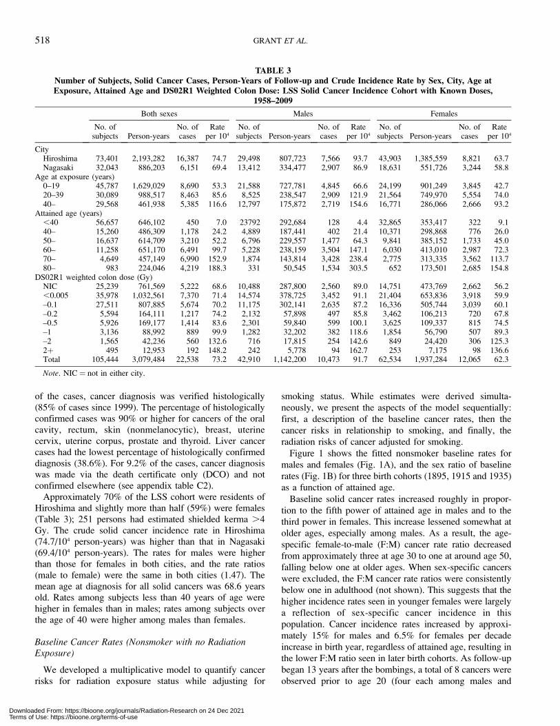
Approximately 70% of the LSS cohort were residents ofHiroshima and slightly more than half (59%) were females(Table 3); 251 persons had estimated shielded kerma .4Gy. The crude solid cancer incidence rate in Hiroshima(74.7/104 person-years) was higher than that in Nagasaki(69.4/104 person-years). The rates for males were higherthan those for females in both cities, and the rate ratios(male to female) were the same in both cities (1.47). Themean age at diagnosis for all solid cancers was 68.6 yearsold. Rates among subjects less than 40 years of age werehigher in females than in males; rates among subjects overthe age of 40 were higher among males than females.
Baseline Cancer Rates (Nonsmoker with no RadiationExposure)
We developed a multiplicative model to quantify cancerrisks for radiation exposure status while adjusting for
smoking status. While estimates were derived simulta-
neously, we present the aspects of the model sequentially:
first, a description of the baseline cancer rates, then the
cancer risks in relationship to smoking, and finally, the
radiation risks of cancer adjusted for smoking.
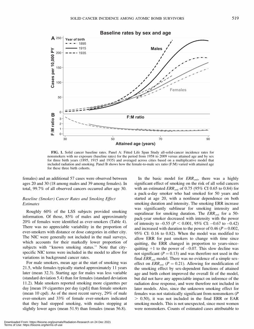
Figure 1 shows the fitted nonsmoker baseline rates for
males and females (Fig. 1A), and the sex ratio of baseline
rates (Fig. 1B) for three birth cohorts (1895, 1915 and 1935)
as a function of attained age.
Baseline solid cancer rates increased roughly in propor-
tion to the fifth power of attained age in males and to the
third power in females. This increase lessened somewhat at
older ages, especially among males. As a result, the age-
specific female-to-male (F:M) cancer rate ratio decreased
from approximately three at age 30 to one at around age 50,
falling below one at older ages. When sex-specific cancers
were excluded, the F:M cancer rate ratios were consistently
below one in adulthood (not shown). This suggests that the
higher incidence rates seen in younger females were largely
a reflection of sex-specific cancer incidence in this
population. Cancer incidence rates increased by approxi-
mately 15% for males and 6.5% for females per decade
increase in birth year, regardless of attained age, resulting in
the lower F:M ratio seen in later birth cohorts. As follow-up
began 13 years after the bombings, a total of 8 cancers were
observed prior to age 20 (four each among males and
TABLE 3Number of Subjects, Solid Cancer Cases, Person-Years of Follow-up and Crude Incidence Rate by Sex, City, Age atExposure, Attained Age and DS02R1 Weighted Colon Dose: LSS Solid Cancer Incidence Cohort with Known Doses,
1958–2009
Both sexes Males Females
No. ofsubjects Person-years
No. ofcases
Rateper 104
No. ofsubjects Person-years
No. ofcases
Rateper 104
No. ofsubjects Person-years
No. ofcases
Rateper 104
CityHiroshima 73,401 2,193,282 16,387 74.7 29,498 807,723 7,566 93.7 43,903 1,385,559 8,821 63.7Nagasaki 32,043 886,203 6,151 69.4 13,412 334,477 2,907 86.9 18,631 551,726 3,244 58.8
Age at exposure (years)0–19 45,787 1,629,029 8,690 53.3 21,588 727,781 4,845 66.6 24,199 901,249 3,845 42.720–39 30,089 988,517 8,463 85.6 8,525 238,547 2,909 121.9 21,564 749,970 5,554 74.040– 29,568 461,938 5,385 116.6 12,797 175,872 2,719 154.6 16,771 286,066 2,666 93.2
Attained age (years),40 56,657 646,102 450 7.0 23792 292,684 128 4.4 32,865 353,417 322 9.140– 15,260 486,309 1,178 24.2 4,889 187,441 402 21.4 10,371 298,868 776 26.050– 16,637 614,709 3,210 52.2 6,796 229,557 1,477 64.3 9,841 385,152 1,733 45.060– 11,258 651,170 6,491 99.7 5,228 238,159 3,504 147.1 6,030 413,010 2,987 72.370– 4,649 457,149 6,990 152.9 1,874 143,814 3,428 238.4 2,775 313,335 3,562 113.780– 983 224,046 4,219 188.3 331 50,545 1,534 303.5 652 173,501 2,685 154.8
DS02R1 weighted colon dose (Gy)NIC 25,239 761,569 5,222 68.6 10,488 287,800 2,560 89.0 14,751 473,769 2,662 56.2,0.005 35,978 1,032,561 7,370 71.4 14,574 378,725 3,452 91.1 21,404 653,836 3,918 59.9–0.1 27,511 807,885 5,674 70.2 11,175 302,141 2,635 87.2 16,336 505,744 3,039 60.1–0.2 5,594 164,111 1,217 74.2 2,132 57,898 497 85.8 3,462 106,213 720 67.8–0.5 5,926 169,177 1,414 83.6 2,301 59,840 599 100.1 3,625 109,337 815 74.5–1 3,136 88,992 889 99.9 1,282 32,202 382 118.6 1,854 56,790 507 89.3–2 1,565 42,236 560 132.6 716 17,815 254 142.6 849 24,420 306 125.32þ 495 12,953 192 148.2 242 5,778 94 162.7 253 7,175 98 136.6Total 105,444 3,079,484 22,538 73.2 42,910 1,142,200 10,473 91.7 62,534 1,937,284 12,065 62.3
Note. NIC ¼ not in either city.
518 GRANT ET AL.
Downloaded From: https://bioone.org/journals/Radiation-Research on 24 Dec 2021Terms of Use: https://bioone.org/terms-of-use
females) and an additional 57 cases were observed betweenages 20 and 30 (18 among males and 39 among females). Intotal, 99.7% of all observed cancers occurred after age 30.
Baseline (Smoker) Cancer Rates and Smoking EffectEstimates
Roughly 60% of the LSS subjects provided smokinginformation. Of those, 85% of males and approximately20% of females were identified as ever-smokers (Table 4).There was no appreciable variability in the proportion ofever-smokers with distance or dose categories in either city.The NIC were generally not included in the mail surveys,which accounts for their markedly lower proportion ofsubjects with ‘‘known smoking status.’’ Note that city-specific NIC terms were included in the model to allow forvariations in background cancer rates.
For male smokers, mean age at the start of smoking was21.5, while females typically started approximately 11 yearslater (mean 32.3). Starting age for males was less variable(standard deviation 5.4) than for females (standard deviation11.2). Male smokers reported smoking more cigarettes perday [mean 19 cigarettes per day (cpd)] than female smokers(mean 10 cpd). As of the most recent survey, 29% of maleever-smokers and 33% of female ever-smokers indicatedthat they had stopped smoking, with males stopping atslightly lower ages (mean 51.9) than females (mean 56.8).
In the basic model for ERRsmk, there was a highlysignificant effect of smoking on the risk of all solid cancerswith an estimated ERRsmk of 0.75 (95% CI 0.65 to 0.84) fora pack-a-day smoker who had smoked for 50 years andstarted at age 20, with a nonlinear dependence on bothsmoking duration and intensity. The smoking ERR increasewas significantly sublinear for smoking intensity andsupralinear for smoking duration. The ERRsmk for a 50-pack-year smoker decreased with intensity with the powerof intensity to –0.55 (P , 0.001, 95% CI:�0.67 to�0.42)and increased with duration to the power of 0.46 (P¼0.002,95% CI: 0.16 to 0.82). When the model was modified toallow ERR for past smokers to change with time sincequitting, the ERR changed in proportion to years-since-quitting þ1 to the power of �0.07. This slow decline wasnot significant (P¼ 0.13) and was therefore not used in thefinal ERRsmk model. There was no evidence of a simple sex-effect on ERRsmk (P ¼ 0.21). Allowing for modification ofthe smoking effect by sex-dependent functions of attainedage and birth cohort improved the overall fit of the model,but did not have any appreciable impact on inference of theradiation dose response, and were therefore not included inlater models. Also, since the unknown smoking effect forfemales was not statistically significant from nonsmokers (P. 0.50), it was not included in the final ERR or EARsmoking models. This is not unexpected, since most womenwere nonsmokers. Counts of estimated cases attributable to
FIG. 1. Solid cancer baseline rates. Panel A: Fitted Life Span Study all-solid-cancer incidence rates fornonsmokers with no exposure (baseline rates) for the period from 1958 to 2009 versus attained age and by sexfor three birth years (1895, 1915 and 1935) and averaged across cities based on a multiplicative model thatincluded radiation and smoking. Panel B shows how the female-to-male sex ratio (F:M) varied with attained agefor these three birth cohorts.
SOLID CANCER INCIDENCE AMONG ATOMIC BOMB SURVIVORS 519
Downloaded From: https://bioone.org/journals/Radiation-Research on 24 Dec 2021Terms of Use: https://bioone.org/terms-of-use
smoking and radiation using various models are shown in
Appendix D.
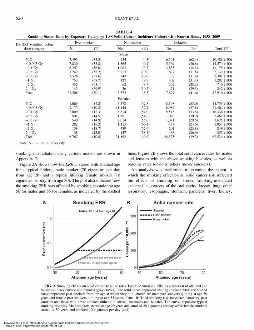
Figure 2A shows how the ERRsmk varied with attained age
for a typical lifelong male smoker (20 cigarettes per day
from age 20) and a typical lifelong female smoker (10
cigarettes per day from age 30). The plot also indicates how
the smoking ERR was affected by smoking cessation at age
50 for males and 55 for females, as indicated by the dashed
lines. Figure 2B shows the total solid cancer rates for males
and females with the above smoking histories, as well as
baseline rates for nonsmokers (never smokers).
An analysis was performed to examine the extent to
which the smoking effect on all solid cancer risk reflected
the effects of smoking on known smoking-associated
cancers (i.e., cancers of the oral cavity, larynx, lung, other
respiratory, esophagus, stomach, pancreas, liver, kidney,
TABLE 4Smoking Status Data by Exposure Category: LSS Solid Cancer Incidence Cohort with Known Doses, 1958–2009
DS02R1 weighted colondose category
Ever-smoker Nonsmoker Unknown
No. (%) No. (%) No. (%) Total (%)
Males
NIC 3,493 (33.3) 454 (4.3) 6,541 (62.4) 10,488 (100),0.005 Gy 7,830 (53.8) 1,365 (9.4) 5,369 (36.8) 14,574 (100)–0.1 Gy 6,257 (56.0) 1,083 (9.7) 3,835 (34.3) 11,175 (100)–0.2 Gy 1,242 (58.2) 213 (10.0) 677 (31.8) 2,132 (100)–0.5 Gy 1,326 (57.6) 243 (10.6) 732 (31.8) 2,301 (100)–1 Gy 753 (58.7) 127 (9.9) 402 (31.4) 1,282 (100)–2 Gy 452 (63.1) 62 (8.7) 202 (28.2) 716 (100)2þ Gy 145 (59.9) 26 (10.7) 71 (29.3) 242 (100)Total 21,508 (50.1) 3,573 (8.3) 17,829 (41.6) 42,910 (100)
Females
NIC 1,065 (7.2) 5,578 (37.8) 8,108 (55.0) 14,751 (100),0.005 Gy 2,173 (10.2) 11,144 (52.1) 8,087 (37.8) 21,404 (100)–0.1 Gy 2,009 (12.3) 8,814 (54.0) 5,513 (33.8) 16,336 (100)–0.2 Gy 501 (14.5) 1,891 (54.6) 1,070 (30.9) 3,462 (100)–0.5 Gy 540 (14.9) 2,014 (55.6) 1,071 (29.5) 3,625 (100)–1 Gy 282 (15.2) 1,115 (60.1) 457 (24.6) 1,854 (100)–2 Gy 159 (18.7) 489 (57.6) 201 (23.8) 849 (100)2þ Gy 38 (15.0) 147 (58.1) 68 (26.9) 253 (100)Total 6,767 (10.8) 31,192 (49.9) 24,575 (39.3) 62,534 (100)
Note. NIC ¼ not in either city.
FIG. 2. Smoking effects on solid cancer baseline rates. Panel A: Smoking ERR as a function of attained agefor males (black curves) and females (gray curves). The solid curves represent lifelong smokers while the dashedcurves represent past smokers from the age at which they quit (shown are male past smokers quitting at age 50years and female past smokers quitting at age 55 years). Panel B: Total smoking risk for current smokers, pastsmokers and those who never smoked (thin solid curves) for males and females. The curves represent typicalsmoking histories. Male smokers started at age 20 years and smoked 20 cigarettes per day while female smokersstarted at 30 years and smoked 10 cigarettes per day (cpd).
520 GRANT ET AL.
Downloaded From: https://bioone.org/journals/Radiation-Research on 24 Dec 2021Terms of Use: https://bioone.org/terms-of-use

bladder and other urinary and rectum), as defined by Doll etal. (18) The ERRsmk for smoking-related cancers among 70-year-old, pack-a-day smokers who smoked for 50 years was1.27 (95% CI: 1.12 to 1.44), while the ERRsmk fornonsmoking-related cancers was 0.05 and not statisticallysignificant.
Radiation Effects
ERR models. There was evidence of a statisticallysignificant all-solid-cancer dose response in a linear ERRmodel without adjustment for smoking (Table 5, top panel)like that used in the previously reported solid cancerincidence study (4). In this model, the sex-averaged ERRfor all solid cancers at attained age of 70 after exposure atage 30 was 0.50 per Gy (95% CI: 0.42 to 0.59), with theF:M ratio of 1.80 (95% CI: 1.42 to 2.33). The ERR variedsignificantly with both attained age (P , 0.001) and age atexposure (P , 0.001). Note that these values are quitesimilar to those previously reported, in which the sex-averaged ERR per Gy was estimated at 0.47 with a F:Mratio of 1.6; the modifying effect of attained age on the ERRwas a decrease with age to the power of –1.65 while theERR decreased by �17% per decade increase of age atexposure (4). When modifications by attained age and ageat exposure were assessed separately and independently,their effects were somewhat larger with the current data; i.e.,–2.02 for attained age and –28.6% for age at exposure. Thelowest dose range that showed a statistically significant doseresponse using the sex-averaged linear ERR model with noadjustment for smoking (i.e., as in the top row of Table 5)was 0–100 mGy with an ERR estimate of 0.49/Gy (95% CI:0.026 to 1.01; P ¼ 0.038).
Smoking-adjusted radiation dose-response models.Smoking-adjusted linear ERR models, assuming eitheradditive or multiplicative joint effects with radiation, fitthe data markedly better than the unadjusted models (Table
5, middle and bottom rows). Due to the difference in sex-specific smoking prevalence, smoking-adjusted modelsprimarily affected the radiation risk estimates for malesand therefore, the ERRrad sex-ratios, but there was littleimpact on the modification of the ERR per Gy estimates byattained age or age at exposure. ERR estimates weresomewhat higher for the additive joint effect model than forthe multiplicative joint effect model, especially for males.The reason for this is that in the additive model, theradiation-associated ERR was relative to the rate fornonsmokers, while in the multiplicative joint effect model,the radiation effect was measured relative to the risk forpeople with comparable smoking histories. Although anadditive ERR model for the joint effect of radiation andsmoking fit the data better than a multiplicative joint effectmodel, we used, unless explicitly noted, the results from themultiplicative joint effects model for the rest of the analyses.This decision was made for comparability to previouslyreported studies that ignored smoking, which results inradiation risk estimates relative to persons with the samesmoking history (i.e., analogous to our current multiplica-tive joint effects model). This choice has almost no effect oninference regarding the dose-response shape or age-relatedeffect modifiers, and helps to facilitate comparisons with theprevious LSS results, as well as studies of other irradiatedpopulations that did not adjust for smoking effects.
Age-at-Exposure Effects
Using the standard log-linear age-at-exposure model withattained-age effect modification and multiplicative adjust-ment for smoking, the linear ERR was estimated to decreaseby 21% (95% CI: 12% to 29%) per decade increase in age atexposure. No model improvement was found when effectmodification by age at exposure could vary by sex (P .
0.5) or city (P . 0.5). The BEIR VII model (19), whichallowed for the ERR at 1 Gy to decrease with increasing age
TABLE 5All Solid Cancer Linear ERR per Gy Adjusted for Modifying Effects of Age at Exposure and Attained Age with or
without Adjustment for Smoking: LSS Solid Cancer Incidence Cohort with Known Doses, 1958–2009
ERR per Gya
F:M ratio(95% CI)
Age at exposureb
(percentage change per10-year increase) (95% CI)
Attained agec
(power) (95% CI)Sex-averaged
(95% CI)Males
(95% CI)Females(95% CI)
Unadjusted for smoking (deviation ¼ 57,404.131, 17 parameters)
0.50 0.36 0.65 1.80 –19% –1.57(0.42 to 0.59) (0.28 to 0.45) (0.53 to 0.77) (1.42 to 2.33) (–27% to –12%) (–2.01 to –1.11)
Adjusted for smoking, additive joint effect (deviation ¼ 56,950.969, 21 parameters)
0.56 0.48 0.64 1.33 –21% –1.53(0.46 to 0.66) (0.36 to 0.61) (0.52 to 0.76) (1.04 to 1.74) (–29% to –13%) (–1.98 to –1.07)
Adjusted for smoking, multiplicative joint effect (deviation ¼ 56,959.086, 21 parameters)
0.47 0.33 0.60 1.81 –21% –1.66(0.39 to 0.55) (0.25 to 0.42) (0.49 to 0.72) (1.42 to 2.35) (–29% to –12%) (–2.11 to –1.20)
a Estimates were centered and scaled to correspond with an attained age of 70 years after exposure at age 30 years.b The age-at-exposure effect was expressed as percentage change per decade increase (e.g., in the top row, the per decade decrease is calculated
as: �19% ¼ 100*(exp[�0.21*(age exp � 30) / 10] �1), where �0.21 is the model parameter estimate and ageexp is age 40).c The effect of attained age was modeled as power of attained age (e.g., in the top row: [ageattained/70]–1.57)
SOLID CANCER INCIDENCE AMONG ATOMIC BOMB SURVIVORS 521
Downloaded From: https://bioone.org/journals/Radiation-Research on 24 Dec 2021Terms of Use: https://bioone.org/terms-of-use

at exposure up to 30 years while remaining constant
thereafter, did not fit the data significantly better than a
simple log-linear age-at-exposure model (P ¼ 0.18).
Additional details on the age-at-exposure effect and how
it is affected by inclusion of autopsy-only cases are given in
Appendix A.
Attained-Age Effects
As previously found with both the solid cancer incidence
(4) and mortality (1) data, the radiation ERR decreased
significantly with attained age even after allowing for effect
modification by age at exposure. In the basic analysis of the
current data, the decrease in the radiation ERR with attained
age was estimated to be proportional to age to the power of
–1.66 (smoking-adjusted multiplicative ERR model in
Table 5). When the model was extended to allow the
attained-age effect to differ for males and females, there was
a significant improvement in fit (P ¼ 0.016), with the
estimated decrease in radiation ERR more rapid for males
than for females. Figure 3A plots the sex-specific estimated
radiation ERR at 1 Gy as a function of attained age for three
ages at exposure. The decrease in radiation ERR was
proportional to attained age to the power of –2.56 (95% CI:
–3.41 to –1.71) for males and –1.38 (95% CI: –1.88 to
–0.86) for females. Figure 3B indicates how the female-to-
male ERR ratio varies with attained age at 1 Gy.
Time-since-Exposure Effects
The three time scales (attained age, age at exposure and
time since exposure) cannot be simultaneously modeled,
since they are colinear. We tested a model with time since
exposure and age at exposure. The radiation ERR decreased
significantly with both time since exposure (27% per
decade; P¼ 0.001) and age at exposure (43% per decade; P¼ 0.001). The Akaike information criterion (AIC) for this
model was higher than that of a similar model with age at
exposure and attained age (AIC ¼ 56,996 vs. 56,990,
respectively) and did not affect the shape of the dose
response (data not shown); time-since-exposure models
were not further considered.
City Effect
There was no evidence of a difference in effect due to city
in the baseline rates (P . 0.50). Allowing city to modify the
radiation effect resulted in little improvement in fit (P ¼0.28). The radiation effect for Nagasaki was estimated to be
12% lower than that in Hiroshima (95% CI: –30% to 10%).
Dose-Response Shape
Assuming a linear dose response for both males and
females with sex-common age at exposure but sex-
dependent attained-age effect modification, and multiplica-
FIG. 3. Age-at-exposure and attained-age effects on solid cancer ERRs at 1 Gy by age at exposure and sex.Panel A shows how the radiation ERRs varied with attained age by sex (gray for females and black for males)and by age of exposure. This is a linear ERR model with multiplicative adjustment for smoking, sex-averagedage-at-exposure modification and sex-specific attained-age modification. Panel B shows how the female-to-male(F:M) ERR ratio varies with attained age at 1 Gy.
522 GRANT ET AL.
Downloaded From: https://bioone.org/journals/Radiation-Research on 24 Dec 2021Terms of Use: https://bioone.org/terms-of-use
tive adjustment for smoking, the estimated linear ERR per
Gy was 0.27 (95% CI: 0.19 to 0.37) for males and 0.64
(95% CI: 0.52 to 0.77) for females (Table 6). Of note, these
values differ slightly from Table 5 due to the added sex-
specific effect modification by attained age. The dose
response, however, exhibited statistically significant (P ¼0.03) upward curvature (i.e., the ratio of quadratic to linear
terms) in a linear-quadratic dose-response model that
assumed common curvature for males and females. The
common curvature (r) was estimated to be 0.22 per Gy
(95% CI: 0.01 to 0.60). The linear dose coefficients for
males and females were 0.21 (95% CI: 0.12 to 0.31) and
0.49 (95% CI: 0.33 to 0.67), respectively (data not shown).
Allowing the curvature to differ for males and females led
to a further statistically significant improvement in fit (P¼0.02 compared to the common curvature model and P ¼0.007 compared to the linear model). For males, the linear
dose coefficient was 0.087 (95% CI: –0.03 to 0.23) with a
quadratic estimate of 0.11 (95% CI: 0.04 to 0.20) resulting
in a curvature estimate of 1.3 (Pcurve¼ 0.002). For females,
the linear estimate was 0.57 (95% CI: 0.40 to 0.77) with a
quadratic estimate of 0.049 (95% CI –0.06 to 0.16) and a
curvature estimate of 0.084 (Pcurve ¼ 0.39). Thus, while the
dose response for females was consistent with linearity, for
males it exhibited significant upward curvature. The plots in
Fig. 4 compare the sex-specific fitted linear and linear-
quadratic dose-response functions for males and females
over the full range of doses. The plots also include
nonparametric estimates of the ERR for the 22 dose
categories (with the ,0.005 category used as the baseline),
along with smoothed nonparametric estimates with point-
wise confidence bounds (the sex-specific categorical ERR
estimates and 95% CIs are shown in Appendix E). Figure 5
shows the same data restricted to doses less than 1 Gy. In
males, but not females, the ERR at low doses is markedly
less than that predicted by the linear model.
As in earlier LSS reports (1, 2, 4), a series of analyses
were performed to investigate the low-dose linear slope and
evidence of curvature in data restricted over various dose
ranges. Table 6 summarizes the results of these analyses
separately for males and females.
For females, the ERR per Gy estimates were quite similar
for all the dose ranges considered. For males, the linear
model ERR estimate on the 0 to 0.1 Gy range (0.33), while
quite uncertain, was higher than the estimate over the full
range (0.27) and had the highest point estimate of any dose
range. This suggests that the upward curvature in the dose
response for males is largely driven by the rather flat dose
response in the range of 0.20–0.75 Gy; the linear ERR per
Gy estimates were 0.02 for the 0–0.25 Gy range and 0.07
for the 0–0.5 Gy range. This pattern can be seen in the
categorical and smoothed dose-response estimates illustrat-
ed in Fig. 5. The linear-quadratic model in men offered no
statistical improvement over a purely quadratic model over
the full dose range (P ¼ 0.11).
Examination of Threshold
The evidence of a threshold dose below which there was
no dose response was examined using linear-quadratic
threshold models for males and linear threshold models for
females. There was no evidence of a threshold for females
(estimated threshold dose of 0.08 Gy). This was not
significantly different from 0 (P ¼ 0.18) and the upper
95% confidence bound was 0.2 Gy. For males, the best
estimate for a threshold dose was 0.75 Gy. Similarly, this
was not significantly different from 0 (P¼ 0.49). However,
the upper 95% confidence bound for the male threshold was
considerably larger than that for females (0.8 Gy). The
proximity of the best estimate and upper bound among
males reflects a bimodal likelihood profile that declines
rapidly after the higher dose peak.
TABLE 6Estimated Sex-Specific ERR Linear Dose Coefficients and Confidence Intervals (and for Males, Linear-Quadratic Dose
Coefficients) over Selected Dose Ranges
Doserange
Lineara Linear-quadratic:b males only
Females(95% CI)
Males(95% CI)
Linear(95% CI)
Quadratic(95% CI)
Curvature (r)(95% CI)
Full range 0.64 (0.52 to 0.77) 0.27 (0.19 to 0.37) 0.09 (–0.03 to 0.23) 0.11 (0.04 to 0.20) 1.3 (Pcurve ¼ 0.002c)0–2 Gy 0.65 (0.52 to 0.78) 0.25 (0.17 to 0.36) 0.02 (,–0.05 to 0.18) 0.18 (0.07 to 0.30) 7.2 (Pcurve , 0.001)0–1 Gy 0.58 (0.44 to 0.74) 0.19 (0.09 to 0.30) –0.09 (,–0.10 to 0.11) 0.38 (0.12 to .0.41) –4.4 (Pcurve ¼ 0.004)0–0.5 Gy 0.53 (0.34 to 0.75) 0.07 (,–0.05 to 0.22) 0.02 (,–0.09 to 0.38) 0.13 (,–0.17 to .0.62) 5.6 (Pcurve . 0.5)0–0.25 Gy 0.55 (0.24 to 0.92) 0.02 (,–0.18 to 0.25) Pcurve . 0.5d
0–0.1 Gy 0.39 (–0.27 to 1.1) 0.33 (,–0.10 to 0.89) Pcurve ¼ 0.08d
a Estimated sex-specific excess relative risks (ERR) per Gy using a linear dose-response model over the dose range. All estimates in this tablewere based on models that included radiation effect modification by attained age (sex-specific), and age at exposure (common to both sexes) andwere adjusted for smoking using a multiplicative ERR model for the joint effect of radiation and smoking.
b Linear (per Gy) and quadratic (per Gy2) dose effect estimates in a linear quadratic dose-response model. Only males were allowed to varyusing the quadratic model term over the dose range.
c P value for a likelihood ratio test of curvature in the male dose response.d Linear-quadratic model parameter estimates unstable due to limited data, results not shown.
SOLID CANCER INCIDENCE AMONG ATOMIC BOMB SURVIVORS 523
Downloaded From: https://bioone.org/journals/Radiation-Research on 24 Dec 2021Terms of Use: https://bioone.org/terms-of-use

EAR Models
Smoking EAR model. Both radiation and smoking effectscan also be described using the excess (absolute) risk (ratedifference). To adequately model the EAR for smoking, itwas necessary to include attained age, sex and birth cohorteffects in the smoking term; these effect modifiers were notnecessary in the ERRsmk model.
Radiation EAR model. Table 7 provides the excess-rate-model parameter estimates and confidence bounds for theEAR model. The radiation EAR for both males and femalesincreased with increasing attained age but the sex difference
was only marginally significant (P ¼ 0.08). However, to be
consistent with the ERR model, we allowed for sex-specific
attained-age modifiers. Figure 6A shows the pattern of the
excess rates with attained age for males and females exposed
at ages 10, 30 and 50, while Fig. 6B displays the age
dependence of the female-to-male EAR ratio at 1 Gy. This
ratio tended to decrease with increasing attained age. For the
same age, the female-to-male EAR ratio also varied with
dose due to the nonlinear dose response for males. The latter
variability was similar to that seen for the ERR (Fig. 7). The
female EAR estimate was 54.7 excess cases per 10,000
FIG. 4. Panels A and B: Solid cancer dose-response functions for males and females (full dose range). Fittedlinear (black dashed line) and linear-quadratic (black solid curve) ERRs for all solid cancers using linear andlinear-quadratic dose-response functions for males and females. Also shown are ERR estimates for all 22 dosecategories (points) and a nonparametric smoothed estimate (solid gray curve) with point-wise 95% confidenceintervals (dashed gray curves). The ERRs are given for subjects at attained age of 70 years after exposure at age30 years.
FIG. 5. Panels A and B: Solid cancer dose-response functions for males and females (0–1 Gy). Fitted linear(black dashed line) and linear-quadratic (black solid curve) ERRs for all solid cancers using linear and linear-quadratic dose-response functions for males and females over the range of 0–1 Gy. Also shown are ERRestimates for 15 visible dose categories (points) and a nonparametric smoothed estimate (solid gray curve) withpoint-wise 95% confidence intervals (dashed gray curves). The ERRs are given for subjects at attained age of 70years after exposure at age 30 years.
524 GRANT ET AL.
Downloaded From: https://bioone.org/journals/Radiation-Research on 24 Dec 2021Terms of Use: https://bioone.org/terms-of-use
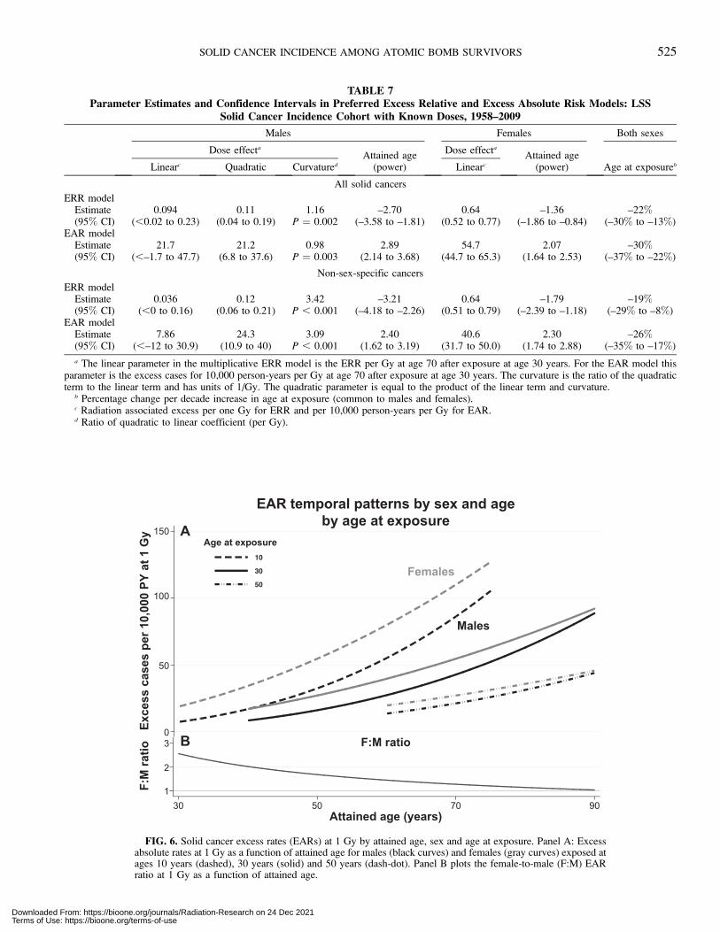
TABLE 7Parameter Estimates and Confidence Intervals in Preferred Excess Relative and Excess Absolute Risk Models: LSS
Solid Cancer Incidence Cohort with Known Doses, 1958–2009
Males Females Both sexes
Dose effecta
Attained age(power)
Dose effecta
Attained age(power) Age at exposurebLinearc Quadratic Curvatured Linearc
All solid cancers
ERR modelEstimate 0.094 0.11 1.16 –2.70 0.64 –1.36 –22%(95% CI) (,0.02 to 0.23) (0.04 to 0.19) P ¼ 0.002 (–3.58 to –1.81) (0.52 to 0.77) (–1.86 to –0.84) (–30% to –13%)
EAR modelEstimate 21.7 21.2 0.98 2.89 54.7 2.07 –30%(95% CI) (,–1.7 to 47.7) (6.8 to 37.6) P ¼ 0.003 (2.14 to 3.68) (44.7 to 65.3) (1.64 to 2.53) (–37% to –22%)
Non-sex-specific cancers
ERR modelEstimate 0.036 0.12 3.42 –3.21 0.64 –1.79 –19%(95% CI) (,0 to 0.16) (0.06 to 0.21) P , 0.001 (–4.18 to –2.26) (0.51 to 0.79) (–2.39 to –1.18) (–29% to –8%)
EAR modelEstimate 7.86 24.3 3.09 2.40 40.6 2.30 –26%(95% CI) (,–12 to 30.9) (10.9 to 40) P , 0.001 (1.62 to 3.19) (31.7 to 50.0) (1.74 to 2.88) (–35% to –17%)
a The linear parameter in the multiplicative ERR model is the ERR per Gy at age 70 after exposure at age 30 years. For the EAR model thisparameter is the excess cases for 10,000 person-years per Gy at age 70 after exposure at age 30 years. The curvature is the ratio of the quadraticterm to the linear term and has units of 1/Gy. The quadratic parameter is equal to the product of the linear term and curvature.
b Percentage change per decade increase in age at exposure (common to males and females).c Radiation associated excess per one Gy for ERR and per 10,000 person-years per Gy for EAR.d Ratio of quadratic to linear coefficient (per Gy).
FIG. 6. Solid cancer excess rates (EARs) at 1 Gy by attained age, sex and age at exposure. Panel A: Excessabsolute rates at 1 Gy as a function of attained age for males (black curves) and females (gray curves) exposed atages 10 years (dashed), 30 years (solid) and 50 years (dash-dot). Panel B plots the female-to-male (F:M) EARratio at 1 Gy as a function of attained age.
SOLID CANCER INCIDENCE AMONG ATOMIC BOMB SURVIVORS 525
Downloaded From: https://bioone.org/journals/Radiation-Research on 24 Dec 2021Terms of Use: https://bioone.org/terms-of-use
person-year-Gy (95% CI: 44.7 to 65.3), while for males thetotal of the linear and quadratic excess cases at 1 Gy was 42.9(21.7 3 1 Gy þ 21.2 3 1 Gy2) per 10,000 person-year-Gy.These values were very similar to the 2007 analysis, whichreported 60 and 43 excess cases per 10,000 person-year-Gyfor females and males, respectively. The previously reportedage-at-exposure modifier was –24% per decade increasewhile attained age was modified to the power of 2.38 (4) andwere similar to the current estimates.
As with the ERR model, there was evidence ofstatistically significant upward curvature in the EAR dose-response model for males (P¼ 0.003), but no indication ofsuch curvature for females (P¼ 0.38). The magnitude of thecurvature parameter in the male dose response was 0.98,which was similar to that seen in the ERR model (1.16). Thecurvature in the EAR dose response for males differedsignificantly from that for females (P ¼ 0.04).
Summary of Preferred Models
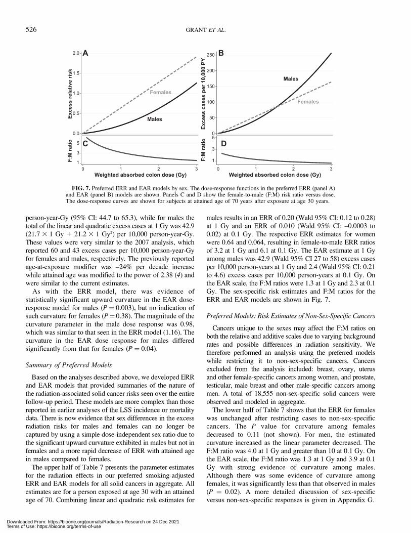
Based on the analyses described above, we developed ERRand EAR models that provided summaries of the nature ofthe radiation-associated solid cancer risks seen over the entirefollow-up period. These models are more complex than thosereported in earlier analyses of the LSS incidence or mortalitydata. There is now evidence that sex differences in the excessradiation risks for males and females can no longer becaptured by using a simple dose-independent sex ratio due tothe significant upward curvature exhibited in males but not infemales and a more rapid decrease of ERR with attained agein males compared to females.
The upper half of Table 7 presents the parameter estimatesfor the radiation effects in our preferred smoking-adjustedERR and EAR models for all solid cancers in aggregate. Allestimates are for a person exposed at age 30 with an attainedage of 70. Combining linear and quadratic risk estimates for
males results in an ERR of 0.20 (Wald 95% CI: 0.12 to 0.28)at 1 Gy and an ERR of 0.010 (Wald 95% CI: –0.0003 to0.02) at 0.1 Gy. The respective ERR estimates for womenwere 0.64 and 0.064, resulting in female-to-male ERR ratiosof 3.2 at 1 Gy and 6.1 at 0.1 Gy. The EAR estimate at 1 Gyamong males was 42.9 (Wald 95% CI 27 to 58) excess casesper 10,000 person-years at 1 Gy and 2.4 (Wald 95% CI: 0.21to 4.6) excess cases per 10,000 person-years at 0.1 Gy. Onthe EAR scale, the F:M ratios were 1.3 at 1 Gy and 2.3 at 0.1Gy. The sex-specific risk estimates and F:M ratios for theERR and EAR models are shown in Fig. 7.
Preferred Models: Risk Estimates of Non-Sex-Specific Cancers
Cancers unique to the sexes may affect the F:M ratios onboth the relative and additive scales due to varying backgroundrates and possible differences in radiation sensitivity. Wetherefore performed an analysis using the preferred modelswhile restricting it to non-sex-specific cancers. Cancersexcluded from the analysis included: breast, ovary, uterusand other female-specific cancers among women, and prostate,testicular, male breast and other male-specific cancers amongmen. A total of 18,555 non-sex-specific solid cancers wereobserved and modeled in aggregate.
The lower half of Table 7 shows that the ERR for femaleswas unchanged after restricting cases to non-sex-specificcancers. The P value for curvature among femalesdecreased to 0.11 (not shown). For men, the estimatedcurvature increased as the linear parameter decreased. TheF:M ratio was 4.0 at 1 Gy and greater than 10 at 0.1 Gy. Onthe EAR scale, the F:M ratio was 1.3 at 1 Gy and 3.9 at 0.1Gy with strong evidence of curvature among males.Although there was some evidence of curvature amongfemales, it was significantly less than that observed in males(P ¼ 0.02). A more detailed discussion of sex-specificversus non-sex-specific responses is given in Appendix G.
FIG. 7. Preferred ERR and EAR models by sex. The dose-response functions in the preferred ERR (panel A)and EAR (panel B) models are shown. Panels C and D show the female-to-male (F:M) risk ratio versus dose.The dose-response curves are shown for subjects at attained age of 70 years after exposure at age 30 years.
526 GRANT ET AL.
Downloaded From: https://bioone.org/journals/Radiation-Research on 24 Dec 2021Terms of Use: https://bioone.org/terms-of-use
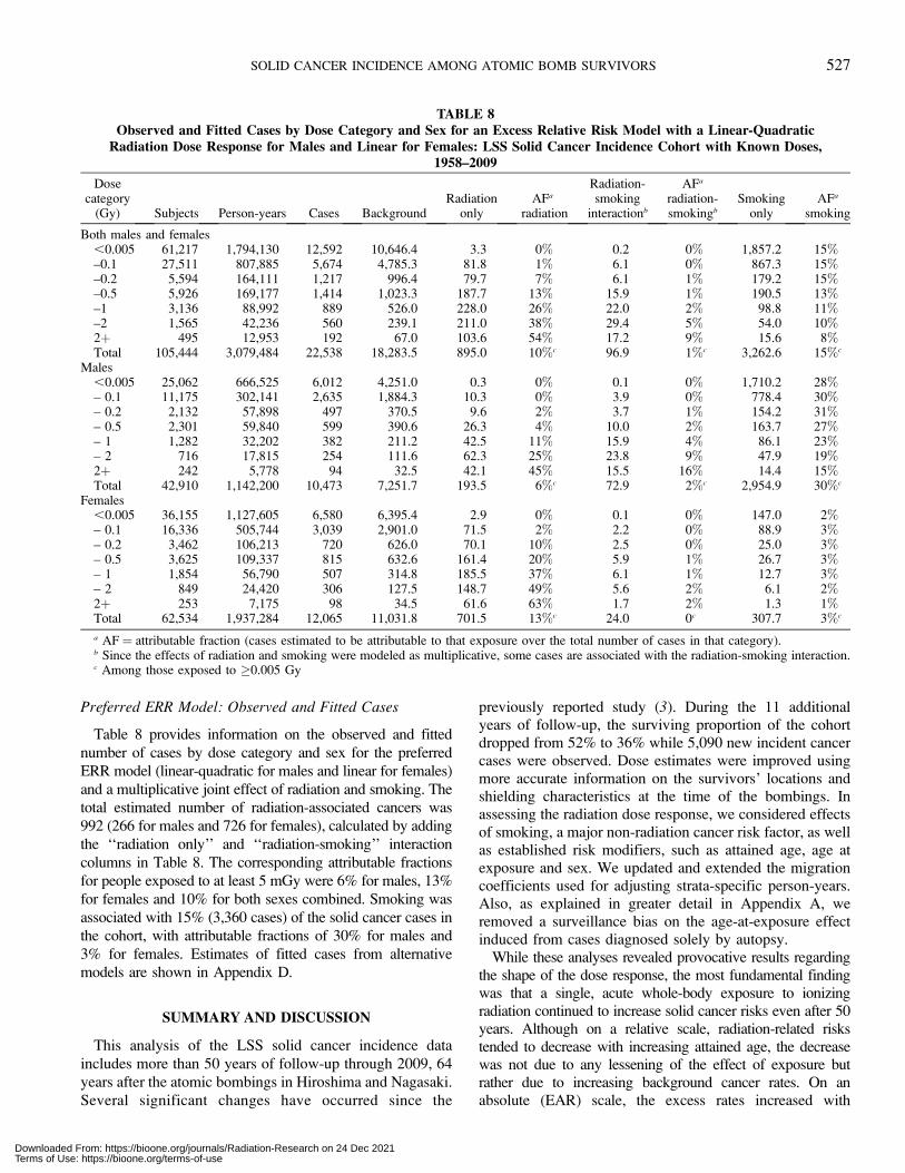
Preferred ERR Model: Observed and Fitted Cases
Table 8 provides information on the observed and fitted
number of cases by dose category and sex for the preferred
ERR model (linear-quadratic for males and linear for females)
and a multiplicative joint effect of radiation and smoking. Thetotal estimated number of radiation-associated cancers was
992 (266 for males and 726 for females), calculated by adding
the ‘‘radiation only’’ and ‘‘radiation-smoking’’ interaction
columns in Table 8. The corresponding attributable fractions
for people exposed to at least 5 mGy were 6% for males, 13%
for females and 10% for both sexes combined. Smoking was
associated with 15% (3,360 cases) of the solid cancer cases in
the cohort, with attributable fractions of 30% for males and
3% for females. Estimates of fitted cases from alternativemodels are shown in Appendix D.
SUMMARY AND DISCUSSION
This analysis of the LSS solid cancer incidence data
includes more than 50 years of follow-up through 2009, 64
years after the atomic bombings in Hiroshima and Nagasaki.
Several significant changes have occurred since the
previously reported study (3). During the 11 additionalyears of follow-up, the surviving proportion of the cohortdropped from 52% to 36% while 5,090 new incident cancercases were observed. Dose estimates were improved usingmore accurate information on the survivors’ locations andshielding characteristics at the time of the bombings. Inassessing the radiation dose response, we considered effectsof smoking, a major non-radiation cancer risk factor, as wellas established risk modifiers, such as attained age, age atexposure and sex. We updated and extended the migrationcoefficients used for adjusting strata-specific person-years.Also, as explained in greater detail in Appendix A, weremoved a surveillance bias on the age-at-exposure effectinduced from cases diagnosed solely by autopsy.
While these analyses revealed provocative results regardingthe shape of the dose response, the most fundamental findingwas that a single, acute whole-body exposure to ionizingradiation continued to increase solid cancer risks even after 50years. Although on a relative scale, radiation-related riskstended to decrease with increasing attained age, the decreasewas not due to any lessening of the effect of exposure butrather due to increasing background cancer rates. On anabsolute (EAR) scale, the excess rates increased with
TABLE 8Observed and Fitted Cases by Dose Category and Sex for an Excess Relative Risk Model with a Linear-Quadratic
Radiation Dose Response for Males and Linear for Females: LSS Solid Cancer Incidence Cohort with Known Doses,1958–2009
Dosecategory
(Gy) Subjects Person-years Cases BackgroundRadiation
onlyAFa
radiation
Radiation-smoking
interactionb
AFa
radiation-smokingb
Smokingonly
AFa
smoking
Both males and females,0.005 61,217 1,794,130 12,592 10,646.4 3.3 0% 0.2 0% 1,857.2 15%–0.1 27,511 807,885 5,674 4,785.3 81.8 1% 6.1 0% 867.3 15%–0.2 5,594 164,111 1,217 996.4 79.7 7% 6.1 1% 179.2 15%–0.5 5,926 169,177 1,414 1,023.3 187.7 13% 15.9 1% 190.5 13%–1 3,136 88,992 889 526.0 228.0 26% 22.0 2% 98.8 11%–2 1,565 42,236 560 239.1 211.0 38% 29.4 5% 54.0 10%2þ 495 12,953 192 67.0 103.6 54% 17.2 9% 15.6 8%Total 105,444 3,079,484 22,538 18,283.5 895.0 10%c 96.9 1%c 3,262.6 15%c
Males,0.005 25,062 666,525 6,012 4,251.0 0.3 0% 0.1 0% 1,710.2 28%– 0.1 11,175 302,141 2,635 1,884.3 10.3 0% 3.9 0% 778.4 30%– 0.2 2,132 57,898 497 370.5 9.6 2% 3.7 1% 154.2 31%– 0.5 2,301 59,840 599 390.6 26.3 4% 10.0 2% 163.7 27%– 1 1,282 32,202 382 211.2 42.5 11% 15.9 4% 86.1 23%– 2 716 17,815 254 111.6 62.3 25% 23.8 9% 47.9 19%2þ 242 5,778 94 32.5 42.1 45% 15.5 16% 14.4 15%Total 42,910 1,142,200 10,473 7,251.7 193.5 6%c 72.9 2%c 2,954.9 30%c
Females,0.005 36,155 1,127,605 6,580 6,395.4 2.9 0% 0.1 0% 147.0 2%– 0.1 16,336 505,744 3,039 2,901.0 71.5 2% 2.2 0% 88.9 3%– 0.2 3,462 106,213 720 626.0 70.1 10% 2.5 0% 25.0 3%– 0.5 3,625 109,337 815 632.6 161.4 20% 5.9 1% 26.7 3%– 1 1,854 56,790 507 314.8 185.5 37% 6.1 1% 12.7 3%– 2 849 24,420 306 127.5 148.7 49% 5.6 2% 6.1 2%2þ 253 7,175 98 34.5 61.6 63% 1.7 2% 1.3 1%Total 62,534 1,937,284 12,065 11,031.8 701.5 13%c 24.0 0c 307.7 3%c
a AF ¼ attributable fraction (cases estimated to be attributable to that exposure over the total number of cases in that category).b Since the effects of radiation and smoking were modeled as multiplicative, some cases are associated with the radiation-smoking interaction.c Among those exposed to �0.005 Gy
SOLID CANCER INCIDENCE AMONG ATOMIC BOMB SURVIVORS 527
Downloaded From: https://bioone.org/journals/Radiation-Research on 24 Dec 2021Terms of Use: https://bioone.org/terms-of-use
increasing attained age in both males and female (Fig. 7). Theoverall attributable fraction of cases due to radiation exposurewas 10%, which is very similar to the value (11%) reported byboth Preston et al. in 2007 (4) and Thompson et al. in 1994(2). These values are a few percentage points higher than forthe attributable fraction observed in the previous mortalitystudies, which have also been quite consistent at approximate-ly 8% among those with non-zero doses (1, 19–21).
While previous LSS solid cancer incidence data demon-strated linear dose responses for both males and females, thecurrent analyses demonstrated significant upward curvature formales with little indication of nonlinearity for females. Itshould be noted that the latest published LSS mortality report,by Ozasa et al. (1), presented evidence of curvature in the ERRdose response over the dose range 0–2 Gy for all solid cancer,which was not evident over the full dose range. Ozasa et al.further reported that the evidence of curvature under 2 Gy hadincreased with the longer follow-up periods, with the mostrecent eight years of follow-up between 1995 and 2003changing the P value for curvature from 0.16 to the statisticallysignificant value of 0.02. Preliminary analyses of more recentsolid cancer mortality data continue to suggest curvature,perhaps in both sexes. Due to the differences in fatality forsome cancers, there are inherent differences in the mix ofcancer types between incidence and mortality data, but resultsfrom dose-response analyses that have aggregated all solidcancers have been broadly comparable (22).
We investigated several factors that may explain thecurrent curvature findings in the dose response for solidcancer incidence, particularly among males. First, since thecurrent cancer incidence data differed from the previousdata (4) in several ways, we compared results of the currentanalysis with those from analyses with no smokingadjustment, with follow-up restricted through 1998 andwith autopsy-only cases included as in the previouslyreported study (4). We further tested the impact of removingthe NIC group from the analysis. These comparisons weredone using both the DS02R1 and DS02 doses, and for thefull dose range and the 0–2 Gy dose range. The detailedresults of these comparisons are given in Appendix F. Therevised dose estimates consistently strengthened the evi-dence of curvature in all the analyses and generally hadmore impact on curvature than other changes to the data.We note that all changes made to update the doses weredone without regard to the sex of the survivor. Amongmales, regardless of the dosimetry version used, theextended follow-up also strengthened the evidence ofcurvature over both the full and restricted-dose ranges.However, evidence of curvature was already present inanalysis restricted to 1998 when the new doses were used.Censoring the autopsy-only cases slightly strengthened theevidence of curvature. Excluding the NIC cohort membershad little effect on either the risk estimates or curvatureinferences. Regardless of the dosimetry version used,adjustment for smoking had virtually no effect on curvatureover the full or restricted dose ranges. Among females, there
was no statistical evidence of curvature in any of theseanalyses, however, the updated DS02R1 dosimetry as wellas analyses restricted to the 0–2 Gy range generally tendedto decrease P values when testing curvature.
We also investigated the extent to which different cancertypes may influence the overall and sex-specific shape of ERRdose responses for solid cancer incidence. Appendix G detailsthe shape of ERR dose responses in several subsets, including:sex-specific/non-sex-specific cancers; smoking-related/non-smoking-related cancers; and gastrointestinal (GI) tractcancers/non-GI cancers. Removal of sex-specific cancerstended to strengthen the evidence of curvature among females,especially in the 0–2 Gy range where we observed statisticallysignificant upward curvature (Pcurve¼ 0.01). The dose responsefor sex-specific cancers showed no evidence of curvature ineither sex over the full or restricted dose ranges. Theproportion of sex-specific cancers differed in males andfemales (9 vs. 26%, respectively). For smoking-related cancersthere was no evidence of curvature in either sex over the fullrange but evidence of curvature in both sexes over therestricted range. For nonsmoking-related cancers, only malesshowed evidence of curvature while there was none amongwomen. Over the full dose range, there was evidence ofcurvature in males for both GI and non-GI cancers but notamong females. Over the 0–2 Gy dose range the evidence ofcurvature in males was not significant for GI cancers (P ¼0.27). Male non-GI cancers included a smaller proportion ofsex-specific cancers (21%) than female non-GI cancers (47%).These findings suggest that the sex difference in the dose-response shape for all solid cancer as a group may more likelybe a consequence of heterogeneity in the shape of the doseresponse for different cancer sites coupled with a differentialdistribution of the sites by sex, than to reflect some moregeneral sex-related mechanism. Site-specific radiation dose-response shapes may vary by cancer site because of theinvolvement of risk modifiers, known or unknown, or possiblyreflect different biological responses of organs/tissue involved.
Pooling all solid cancers offers the advantage of largenumbers to enhance statistical precision when assessing thedose response, especially at low doses, and investigatingeffect modification of the radiation risk by age, time, sexand other factors. Pooling of all solid cancers is particularlyrelevant for the atomic bomb survivors, who receivedwhole-body exposure and among whom radiation effectsare indicated for virtually all organ sites. Aggregate solidcancer risks have been traditionally reported in both cancerincidence and cancer mortality risks from the LSS and othercohorts. However, there are also limitations to such anapproach because there may be real differences in themagnitude of radiation risk and nature of effect modificationacross different cancer sites. Subsequent LSS solid cancerreports in this series will provide detailed analyses ofradiation dose responses for site-specific cancers and relatedcancer types, focusing on how the dose response andmodifying effects are affected by smoking and otherrelevant lifestyle factors. Additional reports will include
528 GRANT ET AL.
Downloaded From: https://bioone.org/journals/Radiation-Research on 24 Dec 2021Terms of Use: https://bioone.org/terms-of-use

respiratory cancers, upper and lower digestive cancers, maleand female sex-specific cancers, among others. Lifestylefactors for these analyses include self-reported alcoholconsumption, educational background, reproductive history,medical history and dietary intake.
The current data also provide new insights into the temporalpattern of the all-solid cancer risk. That is, the ERR per Gydecreased with attained age more rapidly for males thanfemales. Also, the EAR per 10,000 person-year-Gy increasedslightly more rapidly for males than females. Consequently,sex ratios for the ERR and EAR were attained-age dependentas well as dose dependent because of the curvature in doseresponse for males. Both ERR and EAR decreased withincreasing age at exposure. The ERR for males wasconsiderably lower than that for females at any dose, whichmay be due primarily to lower background rates of cancerincidence among females. On the other hand, while excessrates for males were lower than those for females for doses lessthan approximately 1.5 Gy, males had higher excess rates thanfemales at higher doses. This comparison of alternativemeasures of the radiation excess highlights the importance ofconsidering the effects of radiation on both relative andabsolute scales. The analyses of non-sex-specific cancers as asubgroup showed that the EARs were consistently lower formales compared to females, particularly at lower doses. Thelatter finding is a slight departure from the previously reportedstudy, which showed similar EARs across the sexes.
In analyzing the modifying effect of age at exposure on theradiation risk, special attention was given to the potential effectof cancers diagnosed only through autopsy examinations(‘‘autopsy-only cases’’). The investigation was prompted by theU-shaped pattern of the ERR per 1 Gy for solid cancer by age atexposure, as reported in our previous published study (4) andrecognized by the BEIR VII (23). As explained in Appendix A,‘‘occult’’ cases had been censored in the report by Thompson etal. (2), while no such censoring was performed by Preston et al.(4). The occult cases were most often detected at autopsy. Wemade a more general decision to censor all cases that had beendetermined only due to postmortem exam. After censoringthese autopsy-only cases, the U-shaped response no longerappeared and could be rejected statistically. Instead, a simplelog-linear model was used.
Although we decided to use a simple multiplicative modelfor the radiation-smoking joint effects on the ERR, thechoice of smoking adjustment model (multiplicative oradditive) had little impact on the shape of radiation doseresponse or modifying effect of age at exposure or attainedage, which indicates that the radiation risk estimates are notstrongly confounded or otherwise modified by smoking.The use of the multiplicative model for smoking adjustmentallows for comparison with the previous LSS dataunadjusted for smoking. The current estimate of 992radiation-associated solid cancer cases among the 22,538eligible first primary solid cancers is 139 more than in thepreviously reported analysis. Approximately 3,360 of thesolid cancers were estimated to be associated with smoking.
The current LSS solid cancer incidence risk estimates can becompared to those from other populations with whole-bodyexposure. The latest analysis of mostly male nuclear workers(mean dose, 0.021 Gy) in France, the United Kingdom and theUnited States (INWORKS) reported a linear estimate of ERRper Gy of 0.47 (90% CI: 0.18 to 0.79) for solid cancermortality, unadjusted for smoking (24). This is comparable toour linear ERR estimate of 0.36 per Gy (95% CI: 0.28 to 0.45)for males at age 70 after exposure at age 30 (Table 5, firstline). However, the male risk in the LSS at 100 mGy using ourpreferred linear-quadratic ERR model (Table 7) was estimatedto be 0.01, which was lower than that of 0.047 linearly scaledto 100 mGy from the INWORKS data. In the Techa Rivercohort of residents with low-dose exposure to radioactivematerials from contaminated river and soil, the sex-averagedERR for solid cancer incidence was estimated to be 0.077 per100 mGy (95% CI: 0.013 to 0.150) after adjustment forsmoking (25). Again, this was comparable to our linear,smoking adjusted, sex-averaged ERR estimate of 0.047 per100 mGy (95% CI: 0.039 to 0.055) (Table 5, last line; scaledto 100 mGy). The Mayak Production Association workers(75% male) had mixed exposure to gamma rays andplutonium (Pu) but had fractionated gamma exposures in thedose range similar to the LSS; the linearly estimated ERR forexternal gamma-ray exposure adjusted for Pu dose was 0.12per Gy (95% CI: 0.03 to 0.21) for solid cancer mortality (26)or an ERR of 0.07 (95% CI: 0.01 to 0.19) per Gy for solidcancer unadjusted for Pu exposure (27). Both studies excludedcancer sites primarily related to Pu exposure (i.e., lung, liverand bone). These estimates are lower than our linear estimates.We note the LSS male dose response over the lowest doserange considered (0–100 mGy) tended to be considerablygreater than the estimates that consider broader dose ranges(Table 6). This highlights the uncertainties in the shape of thedose response in the current analyses. These uncertaintiestaken together with inconsistencies with prior LSS analysesand the findings from other studies precludes definitiveconclusions that might confidently guide the development ofmodified radiation protection policies at this time.
More than six decades after the atomic bombs, solidcancer continues to be the major documented healthdetriment attributed to radiation exposure in the atomicbomb survivors. The excess risk of solid cancer persists andwill likely persist throughout the atomic bomb survivors’lifetimes. As of 2009, the average age of the LSS cohortwas 78 years, with those still alive exposed at the youngestages. Many incident cancers are predicted in the next 10–15years. Critical questions regarding the long-term risk amongthe youngest survivors are yet to be answered, and mayhave significant bearing on the dose response and temporalpatterns of radiation risk. New trends may have begun toemerge. Upward curvature in the dose response, previouslyobserved in the mortality data, are now evident in theincidence data, especially in males. Females also showsome evidence of curvature in the dose response of non-sex-specific cancers, particularly over the 0–2 Gy range. There
SOLID CANCER INCIDENCE AMONG ATOMIC BOMB SURVIVORS 529
Downloaded From: https://bioone.org/journals/Radiation-Research on 24 Dec 2021Terms of Use: https://bioone.org/terms-of-use
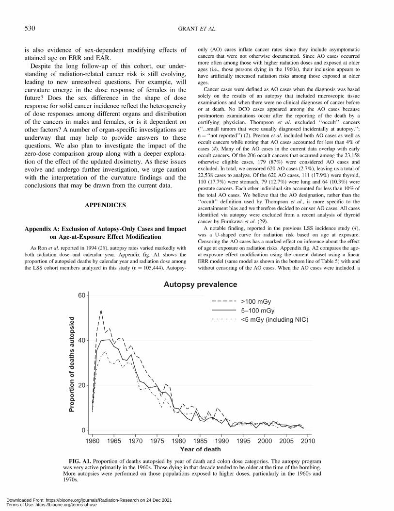
is also evidence of sex-dependent modifying effects ofattained age on ERR and EAR.
Despite the long follow-up of this cohort, our under-standing of radiation-related cancer risk is still evolving,
leading to new unresolved questions. For example, will
curvature emerge in the dose response of females in thefuture? Does the sex difference in the shape of dose
response for solid cancer incidence reflect the heterogeneityof dose responses among different organs and distribution
of the cancers in males and females, or is it dependent onother factors? A number of organ-specific investigations are
underway that may help to provide answers to these
questions. We also plan to investigate the impact of thezero-dose comparison group along with a deeper explora-
tion of the effect of the updated dosimetry. As these issuesevolve and undergo further investigation, we urge caution
with the interpretation of the curvature findings and theconclusions that may be drawn from the current data.
APPENDICES
Appendix A: Exclusion of Autopsy-Only Cases and Impacton Age-at-Exposure Effect Modification
As Ron et al. reported in 1994 (28), autopsy rates varied markedly with
both radiation dose and calendar year. Appendix fig. A1 shows the
proportion of autopsied deaths by calendar year and radiation dose among
the LSS cohort members analyzed in this study (n ¼ 105,444). Autopsy-
only (AO) cases inflate cancer rates since they include asymptomatic
cancers that were not otherwise documented. Since AO cases occurred
more often among those with higher radiation doses and exposed at older
ages (i.e., those persons dying in the 1960s), their inclusion appears to
have artificially increased radiation risks among those exposed at older
ages.
Cancer cases were defined as AO cases when the diagnosis was based
solely on the results of an autopsy that included microscopic tissue
examinations and when there were no clinical diagnoses of cancer before
or at death. No DCO cases appeared among the AO cases because
postmortem examinations occur after the reporting of the death by a
certifying physician. Thompson et al. excluded ‘‘occult’’ cancers
(‘‘...small tumors that were usually diagnosed incidentally at autopsy.’’;
n¼ ‘‘not reported’’) (2). Preston et al. included both AO cases as well as
occult cancers while noting that AO cases accounted for less than 4% of
cases (4). Many of the AO cases in the current data overlap with early
occult cancers. Of the 206 occult cancers that occurred among the 23,158
otherwise eligible cases, 179 (87%) were considered AO cases and
excluded. In total, we censored 620 AO cases (2.7%), leaving us a total of
22,538 cases to analyze. Of the 620 AO cases, 111 (17.9%) were thyroid,
110 (17.7%) were stomach, 79 (12.7%) were lung and 64 (10.3%) were
prostate cancers. Each other individual site accounted for less than 10% of
the total AO cases. We believe that the AO designation, rather than the
‘‘occult’’ definition used by Thompson et al., is more specific to the
ascertainment bias and we therefore decided to censor AO cases. All cases
identified via autopsy were excluded from a recent analysis of thyroid
cancer by Furukawa et al. (29).
A notable finding, reported in the previous LSS incidence study (4),
was a U-shaped curve for radiation risk based on age at exposure.
Censoring the AO cases has a marked effect on inference about the effect
of age at exposure on radiation risks. Appendix fig. A2 compares the age-
at-exposure effect modification using the current dataset using a linear
ERR model (same model as shown in the bottom line of Table 5) with and
without censoring of the AO cases. When the AO cases were included, a
FIG. A1. Proportion of deaths autopsied by year of death and colon dose categories. The autopsy programwas very active primarily in the 1960s. Those dying in that decade tended to be older at the time of the bombing.More autopsies were performed on those populations exposed to higher doses, particularly in the 1960s and1970s.
530 GRANT ET AL.
Downloaded From: https://bioone.org/journals/Radiation-Research on 24 Dec 2021Terms of Use: https://bioone.org/terms-of-use
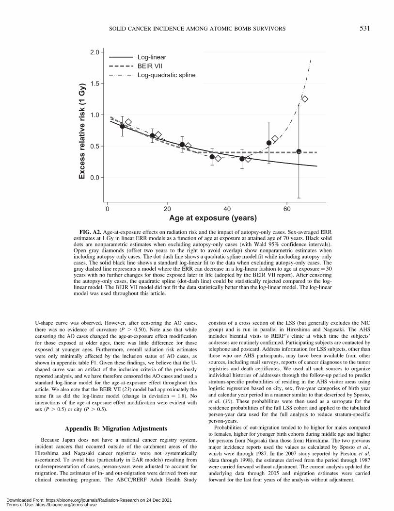
U-shape curve was observed. However, after censoring the AO cases,
there was no evidence of curvature (P . 0.50). Note also that while
censoring the AO cases changed the age-at-exposure effect modification
for those exposed at older ages, there was little difference for those
exposed at younger ages. Furthermore, overall radiation risk estimates
were only minimally affected by the inclusion status of AO cases, as
shown in appendix table F1. Given these findings, we believe that the U-
shaped curve was an artifact of the inclusion criteria of the previously
reported analysis, and we have therefore censored the AO cases and used a
standard log-linear model for the age-at-exposure effect throughout this
article. We also note that the BEIR VII (23) model had approximately the
same fit as did the log-linear model (change in deviation ¼ 1.8). No
interactions of the age-at-exposure effect modification were evident with
sex (P . 0.5) or city (P . 0.5).
Appendix B: Migration Adjustments
Because Japan does not have a national cancer registry system,
incident cancers that occurred outside of the catchment areas of the
Hiroshima and Nagasaki cancer registries were not systematically
ascertained. To avoid bias (particularly in EAR models) resulting from
underrepresentation of cases, person-years were adjusted to account for
migration. The estimates of in- and out-migration were derived from our
clinical contacting program. The ABCC/RERF Adult Health Study
consists of a cross section of the LSS (but generally excludes the NIC
group) and is run in parallel in Hiroshima and Nagasaki. The AHS
includes biennial visits to RERF’s clinic at which time the subjects’
addresses are routinely confirmed. Participating subjects are contacted by
telephone and postcard. Address information for LSS subjects, other than
those who are AHS participants, may have been available from other
sources, including mail surveys, reports of cancer diagnoses to the tumor
registries and death certificates. We used all such sources to organize
individual histories of addresses through the follow-up period to predict
stratum-specific probabilities of residing in the AHS visitor areas using
logistic regression based on city, sex, five-year categories of birth year
and calendar year period in a manner similar to that described by Sposto,
et al. (30). These probabilities were then used as a surrogate for the
residence probabilities of the full LSS cohort and applied to the tabulated
person-year data used for the full analysis to reduce stratum-specific
person-years.
Probabilities of out-migration tended to be higher for males compared
to females, higher for younger birth cohorts during middle age and higher
for persons from Nagasaki than those from Hiroshima. The two previous
major incidence reports used the values as calculated by Sposto et al.,which were through 1987. In the 2007 study reported by Preston et al.(data through 1998), the estimates derived from the period through 1987
were carried forward without adjustment. The current analysis updated the
underlying data through 2005 and migration estimates were carried
forward for the last four years of the analysis without adjustment.
FIG. A2. Age-at-exposure effects on radiation risk and the impact of autopsy-only cases. Sex-averaged ERRestimates at 1 Gy in linear ERR models as a function of age at exposure at attained age of 70 years. Black soliddots are nonparametric estimates when excluding autopsy-only cases (with Wald 95% confidence intervals).Open gray diamonds (offset two years to the right to avoid overlap) show nonparametric estimates whenincluding autopsy-only cases. The dot-dash line shows a quadratic spline model fit while including autopsy-onlycases. The solid black line shows a standard log-linear fit to the data when excluding autopsy-only cases. Thegray dashed line represents a model where the ERR can decrease in a log-linear fashion to age at exposure¼ 30years with no further changes for those exposed later in life (adopted by the BEIR VII report). After censoringthe autopsy-only cases, the quadratic spline (dot-dash line) could be statistically rejected compared to the log-linear model. The BEIR VII model did not fit the data statistically better than the log-linear model. The log-linearmodel was used throughout this article.
SOLID CANCER INCIDENCE AMONG ATOMIC BOMB SURVIVORS 531
Downloaded From: https://bioone.org/journals/Radiation-Research on 24 Dec 2021Terms of Use: https://bioone.org/terms-of-use

Appendix C: Cancer Diagnoses
Appendix Table C1Number of Solid Cancer Cases by Cancer Site and Sex among the LSS Subjects, 1958–2009
Cancer site
Sex
TotalMale Female
No. of cases Percentage No. of cases Percentage No. of cases Percentage
All solid cancers 10,473 100% 12,065 100% 22,538 100Oral cavity and pharynx
Lip 2 0.02% 3 0.02% 5 0.02%Tongue 63 0.6% 56 0.5% 119 0.53%Salivary gland 31 0.3% 19 0.2% 50 0.22%Floor of mouth 10 0.1% 7 0.1% 17 0.08%Gum and other mouth 38 0.4% 53 0.4% 91 0.40%Nasopharynx 11 0.1% 9 0.1% 20 0.09%Tonsil 9 0.1% 3 0.02% 12 0.05%Oropharynx 14 0.1% 5 0.04% 19 0.08%Hypopharynx 53 0.5% 3 0.02% 56 0.25%Other oral cavity and pharynx 5 0.05% 0 0% 5 0.02%
Digestive systemEsophagus 394 3.8% 92 0.8% 486 2.2%Stomach 3,090 29.5% 2,571 21.3% 5,661 25.1%Small intestine 15 0.1% 24 0.2% 39 0.2%Colon 782 7.5% 1132 9.4% 1,914 8.5%Rectum 512 4.9% 510 4.2% 1,022 4.5%Anus, anal canal and anorectum 6 0.1% 18 0.1% 24 0.1%Liver 1,122 10.7% 763 6.3% 1,885 8.4%Intrahepatic bile duct 44 0.4% 87 0.7% 131 0.6%Gallbladder 84 0.8% 270 2.2% 354 1.6%Other biliary 136 1.3% 204 1.7% 340 1.5%Pancreas 306 2.9% 417 3.5% 723 3.2%Retroperitoneum 3 0.03% 8 0.1% 11 0.0%Peritoneum, omentum and mesentery 0 0% 3 0.02% 3 0.01%Other digestive organs 8 0.1% 18 0.2% 26 0.1%
Respiratory systemNose, nasal cavity and middle ear 48 0.5% 50 0.4% 98 0.4%Larynx 154 1.5% 26 0.2% 180 0.8%Lung and bronchus 1,445 13.8% 1,001 8.3% 2,446 10.9%Pleura 1 0.01% 0 0% 1 0.00%Trachea, mediastinum and other respiratory 3 0.03% 13 0.1% 16 0.1%
Bones and jointsBones and joints 13 0.1% 12 0.10% 25 0.11%
Mesothelioma and soft tissueMesothelioma 17 0.2% 10 0.1% 27 0.1%Soft tissue including heart 21 0.2% 26 0.2% 47 0.2%
SkinMelanoma of the skin 10 0.1% 12 0.1% 22 0.1%Other non-melanoma skin 195 1.9% 321 2.7% 516 2.3%
BreastBreast 10 0.1% 1,470 12.2% 1,480 6.6%
Female genital systemCervix uteri – – 886 7.3% 886 3.9%Corpus uteri – – 244 2.0% 244 1.1%Uterus, NOS – – 121 1.0% 121 0.5%Ovary – – 288 2.4% 288 1.3%Vagina – – 22 0.2% 22 0.1%Vulva – – 37 0.3% 37 0.2%Other female genital – – 11 0.1% 11 0.05%
Male genital systemProstate 851 8.1% – – 851 3.8%Testis 18 0.2% – – 18 0.1%Penis 12 0.1% – – 12 0.1%Other male genital 13 0.1% – – 13 0.1%
Continued on next page
532 GRANT ET AL.
Downloaded From: https://bioone.org/journals/Radiation-Research on 24 Dec 2021Terms of Use: https://bioone.org/terms-of-use
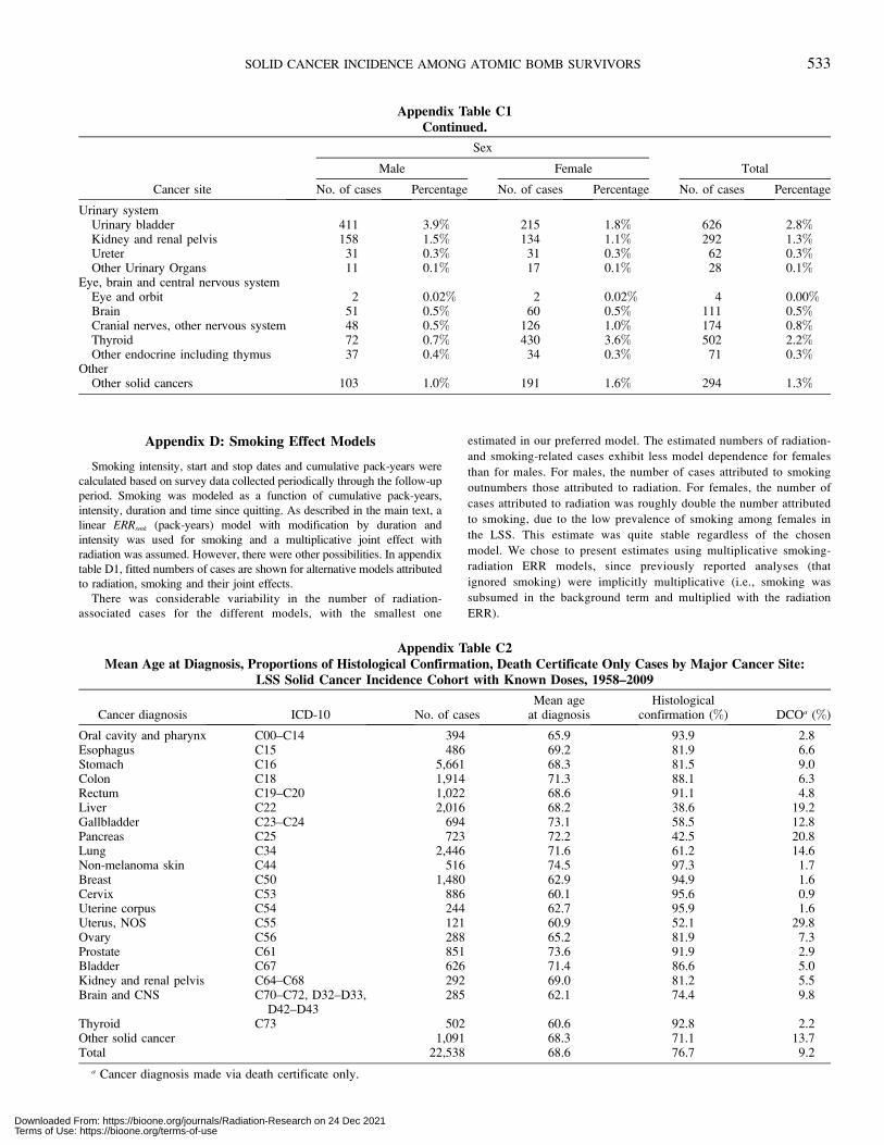
Appendix D: Smoking Effect Models
Smoking intensity, start and stop dates and cumulative pack-years were
calculated based on survey data collected periodically through the follow-up
period. Smoking was modeled as a function of cumulative pack-years,
intensity, duration and time since quitting. As described in the main text, a
linear ERRsmk (pack-years) model with modification by duration and
intensity was used for smoking and a multiplicative joint effect with
radiation was assumed. However, there were other possibilities. In appendix
table D1, fitted numbers of cases are shown for alternative models attributed
to radiation, smoking and their joint effects.
There was considerable variability in the number of radiation-
associated cases for the different models, with the smallest one
estimated in our preferred model. The estimated numbers of radiation-
and smoking-related cases exhibit less model dependence for females
than for males. For males, the number of cases attributed to smoking
outnumbers those attributed to radiation. For females, the number of
cases attributed to radiation was roughly double the number attributed
to smoking, due to the low prevalence of smoking among females in
the LSS. This estimate was quite stable regardless of the chosen
model. We chose to present estimates using multiplicative smoking-
radiation ERR models, since previously reported analyses (that
ignored smoking) were implicitly multiplicative (i.e., smoking was
subsumed in the background term and multiplied with the radiation
ERR).
Appendix Table C1Continued.
Cancer site
Sex
TotalMale Female
No. of cases Percentage No. of cases Percentage No. of cases Percentage
Urinary systemUrinary bladder 411 3.9% 215 1.8% 626 2.8%Kidney and renal pelvis 158 1.5% 134 1.1% 292 1.3%Ureter 31 0.3% 31 0.3% 62 0.3%Other Urinary Organs 11 0.1% 17 0.1% 28 0.1%
Eye, brain and central nervous systemEye and orbit 2 0.02% 2 0.02% 4 0.00%Brain 51 0.5% 60 0.5% 111 0.5%Cranial nerves, other nervous system 48 0.5% 126 1.0% 174 0.8%Thyroid 72 0.7% 430 3.6% 502 2.2%Other endocrine including thymus 37 0.4% 34 0.3% 71 0.3%
OtherOther solid cancers 103 1.0% 191 1.6% 294 1.3%
Appendix Table C2Mean Age at Diagnosis, Proportions of Histological Confirmation, Death Certificate Only Cases by Major Cancer Site:
LSS Solid Cancer Incidence Cohort with Known Doses, 1958–2009
Cancer diagnosis ICD-10 No. of casesMean age
at diagnosisHistological
confirmation (%) DCOa (%)
Oral cavity and pharynx C00–C14 394 65.9 93.9 2.8Esophagus C15 486 69.2 81.9 6.6Stomach C16 5,661 68.3 81.5 9.0Colon C18 1,914 71.3 88.1 6.3Rectum C19–C20 1,022 68.6 91.1 4.8Liver C22 2,016 68.2 38.6 19.2Gallbladder C23–C24 694 73.1 58.5 12.8Pancreas C25 723 72.2 42.5 20.8Lung C34 2,446 71.6 61.2 14.6Non-melanoma skin C44 516 74.5 97.3 1.7Breast C50 1,480 62.9 94.9 1.6Cervix C53 886 60.1 95.6 0.9Uterine corpus C54 244 62.7 95.9 1.6Uterus, NOS C55 121 60.9 52.1 29.8Ovary C56 288 65.2 81.9 7.3Prostate C61 851 73.6 91.9 2.9Bladder C67 626 71.4 86.6 5.0Kidney and renal pelvis C64–C68 292 69.0 81.2 5.5Brain and CNS C70–C72, D32–D33,
D42–D43285 62.1 74.4 9.8
Thyroid C73 502 60.6 92.8 2.2Other solid cancer 1,091 68.3 71.1 13.7Total 22,538 68.6 76.7 9.2
a Cancer diagnosis made via death certificate only.
SOLID CANCER INCIDENCE AMONG ATOMIC BOMB SURVIVORS 533
Downloaded From: https://bioone.org/journals/Radiation-Research on 24 Dec 2021Terms of Use: https://bioone.org/terms-of-use

Appendix E: Sex-Specific ERR Estimates by Dose
Category
Appendix table E1 presents sex-specific ERR estimates and 95%
confidence intervals for each of 22 dose categories relative to the risk
observed in the 0–5 Gy category for males and females with
likelihood-based 95% confidence bounds. The table also includes the
sampling weights for the nonparametric smoothing algorithm used in
Figs. 4 and 5.
Appendix F: Effects of Updated Data and Data Subsets on
Model Inference
As discussed in the main text, a number of changes with the underlying
data were incorporated since the last study published in 2007 (4). In
addition to the data accumulated since 1998, DS02 doses were revised to
DS02R1 (7), autopsy-only cases were censored and smoking was included
as an adjustment factor. As the findings, particularly regarding curvature,
were different in this study compared to the last published study, we
Appendix Table D1Fitted Cases for Alternative Radiation and Smoking Models: LSS Solid Cancer Incidence Cohort with Known Doses,
1958–2009
Joint effect modelDose-response
shape
Fitted cases
Baseline(nonsmoker)
Radiationonly
Radiation-smokingjoint effect
Smokingonly
Radiationtotala
Smokingtotalb
MalesMultiplicative ERR LQc 7,251.7 193.5 72.9 2,954.9 266.4 3,027.8Multiplicative ERR L 7,187.0 251.5 96.1 2,938.5 347.6 3,034.6Additive ERR LQ 7,106.9 268.9 - 3,097.2 268.9 3097.2Additive EAR LQ 6,817.8 283.9 - 3371.3 283.9 3371.3Unadjusted EAR LQ 10,185.9d 287.1 - - 287.1 -
FemalesMultiplicative ERR Lc 11,031.8 701.6 24.0 307.7 725.6 331.7Additive ERR L 11,012.1 727.7 - 325.2 727.7 325.2Additive EAR L 11054.3 718.3 - 292.4 718.3 292.4Unadjusted EAR L 11,325.5d 739.5 - - 739.5 -
a Sum of radiation- and radiation-smoking-joint-effect associated cases.b Sum of smoking- and radiation-smoking-joint-effect associated cases.c Preferred model.d Smoking cases implicitly included within baseline.
Appendix Table E1ERR Estimates by Dose Category and Sampling Weights Used for Smoothing: LSS Solid Cancer Incidence Cohort with
Known Doses, 1958–2009
DoseCategory
(Gy)
Males Females
Meandose (Gy) ERR SE 95% CI
Samplingweight
Meandose (Gy) ERR SE 95% CI
Samplingweight
0– 0 0 — ref 59.66 0 0 — ref 59.040.005– 0.01 –0.003 0.011 (,–0.01 to 0.02) 14.92 0.01 –0.002 0.017 (,–0.01 to 0.03) 14.760.02– 0.03 –0.005 0.017 (,–0.01 to 0.02) 6.80 0.03 0.003 0.025 (,–0.01 to 0.06) 6.530.04– 0.05 0.018 0.023 (,0.01 to 0.07) 3.87 0.05 0.014 0.033 (,–0.01 to 0.08)) 3.980.06– 0.07 0.014 0.027 (,0.01 to 0.07) 2.62 0.07 0.014 0.041 (,–0.01 to 0.10) 2.520.08– 0.09 0.069 0.037 (0.004 to 0.15) 1.41 0.09 0.089 0.053 (,–0.01 to 0.20) 1.480.10– 0.11 –0.006 0.040 (,–0.01 to 0.04) 1.25 0.11 0.002 0.048 (,–0.01 to 0.10) 1.860.125– 0.14 –0.006 0.040 (,–0.01 to 0.04) 1.25 0.14 0.117 0.057 (0.01 to 0.24) 1.320.15– 0.16 0.027 0.042 (,–0.01 to 0.12) 1.11 0.16 0.235 0.073 (0.10 to 0.38) 0.800.175– 0.19 0.050 0.052 (,–0.01 to 0.17) 0.73 0.19 0.007 0.060 (,–0.01 to 0.14) 1.170.20– 0.22 –0.006 0.049 (,–0.01 to 0.08) 0.82 0.22 0.143 0.061 (0.03 to 0.27) 1.140.25– 0.27 0.058 0.046 (,–0.01 to 0.17) 0.94 0.28 0.164 0.065 (0.04 to 0.30) 1.010.30– 0.39 0.030 0.030 (,–0.01 to 0.10) 2.26 0.39 0.203 0.044 (0.12 to 0.29) 2.160.50– 0.61 0.052 0.039 (,0 to 0.14) 1.28 0.62 0.371 0.060 (0.26 to 0.49) 1.170.75– 0.86 0.319 0.069 (0.19 to 0.46) 0.42 0.86 0.559 0.090 (0.39 to 0.75) 0.521.00– 1.11 0.276 0.084 (0.12 to 0.46) 0.28 1.12 0.838 0.142 (0.58 to 1.13) 0.211.25– 1.36 0.262 0.102 (0.10 to 0.47) 0.19 1.36 0.790 0.161 (0.50 to 1.13) 0.161.50 1.61 0.666 0.167 (0.37 to 1.03) 0.07 1.61 1.453 0.304 (0.91 to 2.11) 0.051.75– 1.87 0.720 0.212 (0.35 to 1.20) 0.04 1.87 1.397 0.293 (0.87 to 2.03) 0.052.0– 2.26 0.670 0.166 (0.37 to 1.04) 0.07 2.27 1.533 0.278 (1.04 to 2.12) 0.052.5– 2.65 0.824 0.293 (0.35 to 1.46) 0.02 2.67 1.368 0.453 (0.60 to 2.39) 0.023.0– 3.15 1.828 1.132 (0.27 to 4.86) 0.002 3.15 0.959 1.033 (,0 to 6.07) 0.004
Notes. Excess relative risk estimates are relative to rates for the 0–5 mGy dose category. ERR estimates shown are for a person with attained age70 after exposure at age 30. The joint effect of radiation and smoking was modeled as multiplicative.
534 GRANT ET AL.
Downloaded From: https://bioone.org/journals/Radiation-Research on 24 Dec 2021Terms of Use: https://bioone.org/terms-of-use

investigated each of these changes independently to determine if any of
them were primarily responsible for the new findings of curvature.
Appendix table F1 shows male ERR (linear and quadratic) and female
ERR estimates (linear) along with P values for nonlinearity. The first line
shows the preferred ERR model estimates; these match the estimates
shown in Table 7. Main sections of the table are by dose range (full or 0–2
Gy) and by dosimetry version (DS02R1 versus the previously-used DS02).
For females, no iteration shows significant evidence of curvature, although
suggestive P values were observed over the range of 0–2 Gy using the
updated DS02R1 doses. For males, additional follow-up and updated
doses appeared to have the largest impact on curvature inference, while
other factors, including adjustment for smoking, had little effect.
Exclusion of the NIC group had virtually no impact on the risk estimates
or curvature inferences.
Appendix G: Common and Sex-Specific Dose-ResponseCurvature for Selected Subsets of the Solid Cancer Cases
Appendix table G1 provides information on the sex-specific linear-
quadratic fits for all solid cancers and several families of cancer subsets.
These subsets are:
� Sex-specific cancers (904 males and 3,079 females) (breast, ovary,
uterus and other female cancers along with prostate, testicular and other
male cancers) and non-sex-specific cancers (9,569 males and 8,986
females);� Smoking-related cancers (7,928 males and 6,016 females) (oral cavity,
larynx, lung, other respiratory, esophagus, stomach, pancreas, liver,
kidney, bladder and other urinary and rectum) and nonsmoking-related
cancers (2,545 males and 6,049 females);� Gastrointestinal tract (GI) cancers (6,212 males and 5,503 females)
(esophagus, stomach, colon, rectum, anus, liver and pancreas) and non-
GI cancers (4,261 males and 6,562 females).
Appendix table G1 includes the results for all solid cancers and for each
of the subsets. Note that there was no evidence of a difference of curvature
in the subset of smoking cancers only and that both males and females
displayed no evidence of curvature when sex-specific cancers were
analyzed. This was consistent with the data shown in Appendix table F1
(above) where removal of adjustment for smoking did not change the
curvature inference for males. However, in the subset of nonsmoking
cancers, only males showed evidence of curvature (P ¼ 0.008 for males
and P . 0.50 in females). The risks for smoking were not significant when
the subset of nonsmoking cancers was analyzed.
Appendix Table F1Sex-Specific Linear (and Quadratic for Men) ERR Estimates and Tests for Curvature by Dose Range, Dosimetry System
and Various Exclusion Criteria: LSS Solid Cancer Incidence Cohort with Known Doses, 1958–2009
Male Female
Dose Dose2 Pcurve Dose Pcurve
DS02R1 full range
Current analysisa 0.094 0.109 0.002 0.638 0.392No smoking adjustment 0.119 0.116 0.003 0.681 0.500Follow-up through 1998 0.088 0.090 0.018 0.618 0.217Including autopsy-only cases 0.106 0.102 0.004 0.633 0.395With autopsy only through 1998 0.107 0.085 0.029 0.627 0.219Excluding high dose individuals 0.125 0.094 0.031 0.635 0.195Excluding NIC 0.090 0.106 0.003 0.627 0.456
DS02R1 dose range of 0–2 Gy
Current analysisa 0.025 0.178 ,0.001 0.635 0.073No smoking adjustment 0.043 0.191 0.001 0.678 0.105Follow-up through 1998 0.001 0.178 0.002 0.612 0.064Including autopsy-only cases 0.041 0.168 0.002 0.629 0.079With autopsy only through 1998 0.018 0.173 0.004 0.620 0.071Excluding high dose individuals 0.036 0.182 0.001 0.631 0.072Excluding NIC 0.023 0.174 ,0.001 0.625 0.092
DS02 full range
Current analysisa 0.147 0.073 0.036 0.622 .0.5No smoking adjustment 0.173 0.078 0.041 0.661 .0.5Follow-up through 1998 0.138 0.061 0.099 0.604 0.283Including autopsy-only cases 0.157 0.068 0.050 0.618 .0.5With autopsy only through 1998 0.157 0.057 0.135 0.613 0.293Excluding high dose individuals 0.188 0.052 0.227 0.621 0.234Excluding NIC 0.143 0.071 0.038 0.610 .0.5
DS02 dose range of 0–2 Gy
Current analysisa 0.052 0.162 0.004 0.602 .0.5No smoking adjustment 0.070 0.174 0.005 0.642 . 0.5Follow-up through 1998 0.045 0.150 0.014 0.582 .0.5Including autopsy-only cases 0.071 0.148 0.008 0.599 .0.5With autopsy only through 1998 0.069 0.139 0.029 0.592 .0.5Excluding high dose individuals 0.066 0.163 0.006 0.599 .0.5Excluding NIC 0.050 0.158 0.004 0.592 .0.5
a Adjusted for smoking, follow-up through 2009, excluding autopsy-only cases, including high-dose survivors, including NIC.NIC: Not in the cities of Hiroshima or Nagasaki at the time of bombing.
SOLID CANCER INCIDENCE AMONG ATOMIC BOMB SURVIVORS 535
Downloaded From: https://bioone.org/journals/Radiation-Research on 24 Dec 2021Terms of Use: https://bioone.org/terms-of-use

Non-sex-specific cancers. Differences in sex-specific curvature were
pronounced for the sex-specific vs. non-sex-specific cancer groups shown
in appendix table G1. There was no indication of curvature among sex-
specific cancers (P . 0.5) for either males (P ¼ 0.22) or females (P ¼0.22). However, for non-sex-specific cancers (i.e., cancers that occur in
both men and women) as a group, there was a strong indication of upward
curvature (P ¼ 0.02) that appeared to be largely driven by the male dose
response (P , 0.001) with a suggestion of upward curvature for women (P
¼ 0.11).
Appendix table G2 provides more detail on the ERR and EAR dose-
response estimates for non-sex-specific cancers in males and females as
well as point estimates of the risks at 1 Gy and 0.1 Gy (after exposure at
age 30 years with attained age of 70 years). The evidence of upward
curvature in the dose response for both males and females is stronger when
focus is placed on the 0–2 Gy dose range. Over the restricted 0–2 Gy dose
range, there is evidence of curvature among females in both the ERR and
EAR models.
ACKNOWLEDGMENTS
We thank the LSS cohort members for their longstanding cooperation.
We also thank Drs. Donald Pierce, Ethel Gilbert, Mark Little, Roy Shore
and the anonymous reviewers for their valuable comments and
suggestions, as well as the support from the Hiroshima and Nagasaki
cancer registries. The Radiation Effects Research Foundation (RERF),
Hiroshima and Nagasaki, Japan is a public interest foundation funded by
the Japanese Ministry of Health, Labour and Welfare (MHLW) and the
U.S. Department of Energy (DOE). The research was also funded in part
through DOE award DE-HS0000031 to the National Academy of Sciences
and contract HHSN261201400009C through the U.S. National Cancer
Appendix Table G1Sex-Specific Linear-Quadratic ERR Dose-Response Model Parameter Estimates for All Solid Cancers and Various
Cancer Subsets: LSS Solid Cancer Incidence Cohort with Known Doses, 1958–2009
Outcome
Commona
Sex differenceb Males Females
Curvature Pcurve Pdiff Linearc Quadraticd Pcurve Linearc Quadraticd Pcurve
Full RangeAll solid 0.22 0.034 0.02 0.09 0.11 0.002 0.57 0.05 0.39Sex-specific cancers only –0.17 0.14 .0.5 0.77 –0.17 0.22 0.83 –0.13 0.22Non-sex-specific cancers 0.51 0.001 0.02 0.03 0.13 ,0.001 0.50 0.11 0.11Nonsmoking cancers 0.16 0.18 0.02 0.21 0.26 0.008 0.59 0.01 .0.50Smoking cancers only 0.35 0.07 0.43 0.08 0.07 0.09 0.53 0.13 0.22Non-GI cancers 0.14 0.20 0.04 0.13 0.15 0.02 0.72 0.03 .0.50GI cancers only 0.46 0.07 0.29 0.08 0.11 0.04 0.38 0.08 0.41
Radiation dose ,2 GyAll solid 0.54 0.002 0.03 0.02 0.18 ,0.001 0.48 0.14 0.07Sex-specific cancers only –0.12 0.48 .0.5 0.74 –0.13 .0.5 0.79 –0.08 .0.5Non-sex-specific cancers 1.2 ,0.001 .0.5 –0.02 0.19 ,0.001 0.38 0.23 0.01Nonsmoking cancers 0.31 0.09 0.05 0.11 0.35 0.01 0.54 0.05 .0.5Smoking cancers only 1.06 0.005 0.39 0.02 0.14 0.04 0.38 0.28 0.04Non-GI cancers 0.50 0.008 0.19 –0.01 0.31 ,0.001 0.63 0.12 0.24GI cancers only 0.71 0.08 .0.5 0.09 0.09 0.27 0.28 0.18 0.16
a Quadratic-to-linear coefficient ratio in an ERR dose-response model that allows the linear coefficient to depend on sex but constrains thecurvature to be equal for males and females.
b P value of a test of the hypothesis of equal curvature in males and females.c Linear dose coefficient in a linear-quadratic ERR model with dose in units of weighted colon dose in Gy.d Quadratic dose coefficient in a linear-quadratic ERR model with dose in units of weighted colon dose in Gy.
Appendix Table G2ERR and EAR Dose-Response Model Parameter Estimates for Non-Sex-Specific Solid Cancers by Sex Using all Doses
and the 0–2 Gy Dose Range: LSS Solid Cancer Incidence Cohort with Known Doses, 1958–2009
Model(dose range)
Linear(95% CI)
Quadratic(95% CI)
CurvatureP value
Risk at 1 Gy(95% CI)a
Risk at 0.1 Gy(95% CI)a
MaleERR (all doses) 0.027 (,–0.03 to 0.10) 0.13 (0.06 to 0.22) ,0.001 0.16 (0.08 to 0.23) 0.004 (–0.005 to 0.01)ERR (0–2 Gy) –0.023 (,–0.03 to 0.11) 0.18 (0.09 to .0.25) ,0.001 0.16 (0.09 to 0.23) –0.0004 (–0.01 to 0.01)EAR (all doses) 6.1 (–14 to 29) 25.7 (12 to 42) ,0.001 31.8 (18 to 45) 0.86 (–0.96 to 2.7)EAR (0–2 Gy) –4.2 (,–17 to 21) 36.3 (17 to 58) ,0.001 32.1 (19 to 45) –0.06 (–2.0 to 1.9)
FemaleERR (all doses) 0.50 (0.29 to 0.73) 0.11 (–0.02 to 0.26) 0.11 0.61 (0.47 to 0.75) 0.051 (0.03 to 0.07)ERR (0–2 Gy) 0.38 (0.16 to 0.63) 0.23 (0.04 to 0.42) 0.01 0.61 (0.47 to 0.76) 0.04 (0.02 to 0.06)EAR (all doses) 31.3 (18 to 46) 7.3 (–1.5 to 17) 0.11 38.7 (29.6 to 47.7) 3.2 (1.9 to 4.8)EAR (0–2 Gy) 24.0 (10 to 40) 14.9 (3.0 to 28) 0.01 38.9 (30 to 48) 2.5 (1.2 to 4.0)
a Wald bounds.
536 GRANT ET AL.
Downloaded From: https://bioone.org/journals/Radiation-Research on 24 Dec 2021Terms of Use: https://bioone.org/terms-of-use

Institute (NCI), with additional support from the Division of Cancer
Epidemiology and Genetics in the NCI Intramural Research Program. This
publication was supported by RERF Research Protocols 1-75 and 18-61.
The views of the authors do not necessarily reflect those of the two
governments.
Received: April 21, 2016; accepted: February 4, 2017; published online:
March 20, 2017
REFERENCES
1. Ozasa K, Shimizu Y, Suyama A, Kasagi F, Soda M, Grant EJ, etal. Studies of the mortality of atomic bomb survivors, Report 14,1950–2003: an overview of cancer and noncancer diseases. RadiatRes 2012; 177:229–43.
2. Thompson DE, Mabuchi K, Ron E, Soda M, Tokunaga M,Ochikubo S, et al. Cancer incidence in atomic bomb survivors. PartII: Solid tumors, 1958–1987. Radiat Res 1994; 137:S17–67.
3. Preston DL, Kusumi S, Tomonaga M, Izumi S, Ron E, KuramotoA, et al. Cancer incidence in atomic bomb survivors. Part III.Leukemia, lymphoma and multiple myeloma, 1950–1987. RadiatRes 1994; 137:S68–97.
4. Preston DL, Ron E, Tokuoka S, Funamoto S, Nishi N, Soda M, etal. Solid cancer incidence in atomic bomb survivors: 1958–1998.Radiat Res 2007; 168:1–64.
5. Hsu W-L, Preston DL, Soda M, Sugiyama H, Funamoto S,Kodama K, et al. The incidence of leukemia, lymphoma andmultiple myeloma among atomic bomb survivors: 1950–2001.Radiat Res 2013; 179:361–82.
6. Cullings HM, Grant EJ, Egbert SD, Watanabe T, Oda T,Nakamura F, et al. DS02R1: Improvements to atomic bombsurvivors’ input data and implementation of dosimetry system2002 (DS02) and resulting changes in estimated doses. HealthPhys 2017; 112:56–97.
7. Cullings HM, Fujita S, Funamoto S, Grant EJ, Kerr GD, PrestonDL. Dose estimation for atomic bomb survivor studies: itsevolution and present status. Radiat Res 2006; 166 (1 Pt 2):219–54.
8. Preston DL, Kato H, Kopecky K, Fujita S. Studies of the mortalityof A-bomb survivors. 8. Cancer mortality, 1950–1982. Radiat Res1987; 111:151–78.
9. Pierce DA, Preston DL, Stram DO, Vaeth M. Allowing for dose-estimation errors for the A-bomb survivor data. Radiat Res 1991;32:108–21.
10. Epidemiologic survey, Adult Health Study sample, Hiroshima andNagasaki study of cardiovascular disease Hiroshima and Nagasaki:mortality related to family history and habits. Hiroshima/Nagasaki,Japan: Atomic Bomb Casualty Commission (now the RadiationEffects Research Foundation); 1963.
11. Study of cardiovascular disease Hiroshima and Nagasaki: mortalityrelated to family history and habits. Research Protocol No. 9-65.Hiroshima/Nagasaki, Japan: Atomic Bomb Casualty Commission(now the Radiation Effects Research Foundation); 1965.
12. Mail questionnaire survey for epidemiologic data on females in theJNIH-ABCC Life Span Study sample. Research Protocol No. 11–69. Hiroshima/Nagasaki, Japan: Atomic Bomb Casualty Commis-sion (now the Radiation Effects Research Foundation); 1969.
13. Mail questionnaire survey for epidemiologic data on the Life SpanStudy extended sample, 1978. RERF Research Protocol No. 14–78. Hiroshima/Nagasaki, Japan: Radiation Effects ResearchFoundation; 1978.
14. Mail survey on epidemiologic factors in the Extended Life SpanStudy sample, 1991. RERF Research Protocol No. 4–91.Hiroshima/Nagasaki, Japan: Radiation Effects Research Founda-tion; 1991.
15. Furukawa K, Preston DL, Lonn S, Funamoto S, Yonehara S,Matsuo T, et al. Radiation and smoking effects on lung cancerincidence among atomic bomb survivors. Radiat Res 2010;174:72–82.
16. Cleveland WS, Devlin SJ. Locally weighted regression: anapproach to regression analysis by local fitting. JASA 1988;83:596–610.
17. Preston DL, Lubin J, Pierce DA, McConney ME, Shilnikova NS.Epicure User Guide. Vol. 2.01. Ottawa: Risk Sciences Interna-tional; 2015.
18. Doll R, Peto R, Boreham J, Sutherland I. Mortality from cancer inrelation to smoking: 50 years observations on British doctors. Br JCancer 2005; 92:426–9.
19. Shimizu Y, Kato H, Schull WJ, Preston DL, Fujita S, Pierce DA.Studies of the mortality of A-bomb survivors. 9. Mortality, 1950–1985: Part 1. Comparison of risk coefficients for site-specificcancer mortality based on the DS86 and T65DR shielded kermaand organ doses. Radiat Res 1989; 118:502–24.
20. Pierce DA, Shimizu Y, Preston DL, Vaeth M, Mabuchi K. Studiesof the mortality of atomic bomb survivors. Report 12, Part I.Cancer: 1950–1990. Radiat Res 1996; 146:1–27.
21. Preston DL, Shimizu Y, Pierce DA, Suyama A, Mabuchi K.Studies of mortality of atomic bomb survivors. Report 13: Solidcancer and noncancer disease mortality: 1950–1997. Radiat Res2003; 160:381–407.
22. Ron E, Preston DL, Mabuchi K, Thompson DE, Soda M. Cancerincidence in atomic bomb survivors. Part IV: Comparison ofcancer incidence and mortality. Radiat Res 1994; 137:S98–112.
23. Health Risks from Exposure to Low Levels of Ionizing Radiation:BEIR VII - Phase 2. Washington, DC: The National AcademiesPress; 2006.
24. Richardson DB, Cardis E, Daniels RD, Gillies M, O’Hagan JA,Hamra GB, et al. Risk of cancer from occupational exposure toionising radiation: retrospective cohort study of workers in France,the United Kingdom, and the United States (INWORKS). BMJ.2015; 351:h5359.
25. Davis FG, Yu KL, Preston DL, Epifanova S, Degteva M, AkleyevAV. Solid Cancer Incidence in the Techa River Incidence Cohort:1956–2007. Radiat Res 2015; 184:56–65.
26. Sokolnikov M, Preston DL, Gilbert E, Schonfeld S, KoshurnikovaN. Radiation effects on mortality from solid cancers other thanlung, liver, and bone cancer in the Mayak worker cohort: 1948–2008. PLOS One 2015; 10:e0117784.
27. Hunter N, Kuznetsova IS, Labutina EV, Harrison JD. Solid cancerincidence other than lung, liver and bone in Mayak workers: 1948–2004. Brit J Cancer 2013; 109:1989–96.
28. Ron E, Carter R, Jablon S, Mabuchi K. Agreement between deathcertificate and autopsy diagnoses among atomic bomb survivors.Epidemiology 1994; 5:48–56.
29. Furukawa K, Preston DL, Funamoto S, Yonehara S, Ito M,Tokuoka S, et al. Long-term trend of thyroid cancer risk amongJapanese atomic-bomb survivors: 60 years after exposure. Int JCancer 2013; 132:1222–6.
30. Sposto R, Preston DL. Correcting for catchment area nonresidencyin studies based on tumor-registry data. RERF CR 1-92;Hiroshima: Radiation Effects Research Foundation, 1992. p. 1–16.
SOLID CANCER INCIDENCE AMONG ATOMIC BOMB SURVIVORS 537
Downloaded From: https://bioone.org/journals/Radiation-Research on 24 Dec 2021Terms of Use: https://bioone.org/terms-of-use