r. - whoapps.who.int/iris/bitstream/10665/37156/1/9241544899_eng.pdf · dr sri oemijati...
TRANSCRIPT


These bench aids were planned and developed by Dr Thomas C. Orihel. William Vincent Professor of Tropical Diseases. Tulane University.
School of Public Health and Tropical Medicine. New Orleans. LA. USA; Dr Lawrence R. Ash. Professor Emeritus of Infectious and Tropical Diseases. School of Public Health.
University of California. Los Angeles, CA. USA; Dr C. P. Ramachandran, Chief, and Dr Eric Ottesen, Medical Officer. Filariasis Control.
Division of Control of Tropical Diseases. World Health Organization. Geneva, Switzerland.
Acknowledgements Thanks are due to:
Mme Francoise Ardoin (France), Dr Odile Bain (France). Dr John Cross (USA). Dr Vida Dennis (USA), Dr Mark Eberhard (USA). Dr Robert Lowrie Jr (USA),
Dr Sri Oemijati (Indonesia). Dr Jean-Claude Petithory (France), Purnomo (Indonesia). and Dr LQrenzo Savioli (WHO),
who either provided materials from which the photomicrographs were prepared or contributed in some other way to
the development of these bench aids.
The American Society of Clinical Pathologists Press (USA) graciously permitted reproduction
of images 4f and 5f-h. j.
WHO Library Cataloguing in Publication Data
Bench aids for the diagnosis of filarial infections. 1.Filariasis - diagnosis
ISBN 92 41544899 (NLM Classification: WC 880)
The World Health Organization welcomes requests for permission to reproduce or translate its publications, in part or in full. Applications and enquiries should be addressed
to the Office of Publications. World Health Organization. Geneva. Switzerland. which will be glad to provide the latest information on any changes made to the text.
plans for new editions, and reprints and translations already available.
@ World Health Organization 1997
Publications of the World Health Organization enjoy copyright protection in accordance with the provisions of Protocol 2 of the Universal Copyright Convention.
All rights reserved.
The mention of specific companies or of certain manufacturers' products does not imply that they are endorsed or recommended by the World Health Organization
in preference to others of a similar nature that are not mentioned. Errors and omissions excepted. the names of proprietary
products are distinguished by initial capital letters.
Front cover: Living microfilaria of Loa loa in a drop of fresh blood. Back cover: Microfilariae of Wuchereria bancrofti in a haem8;toxylin-stained thick blood film.
Design and illustration by WHO Graphics. Printed in France

• _B_en_c_h_A_i_ds_fo_r_t_h_e_d_ia ___ n_o_si_s_o_f_fe_lIa_r_ia_l_i_nf_e_ct_io_n_s ___ ~ " ~ __ ---.;.In~tr.;.;od;.;.uct;,;,;.io_n ~ . rJl
© World Heallh Organization 1997
Introduct-on Several species of filar ial worms infect humans in the tropical and subtropical regions of the world (Table 1, overleaf) . The adult worms inhabit various tissues and organs of the body and are inaccessible for identification. Consequently, diagnosis of filarial infections depends primarily on the identification of the larval stage of the parasite (microfilaria). Most species of microfilaria circulate in peripheral blood ; however, some are found in the skin .
The miaofilaria
--~
At the light-microscopic level and with the aid of a variety of stains, a microfilaria appears Fig. 1 Typical microfilaria as a primitive organism, serpentine in shape and filled with the nuclei of many cells . Figure 1 is a diagram of a typical microfilaria. In many, but not all, species, the body may be enveloped in a membrane called a sheath (sh) . Where a sheath is present it may extend a short or long distance beyond either extremity of the microfilaria. In some species, depending on the stain used, the sheath displays a characteristic staining quality which aids in species identification. The nuclei of the cells that fill the body are usually darkly stained and may be crowded together or dispersed . The anterior extremity is typically devoid of nuclei and is called the cephalic or head space (hs) ; it may be short or long. Along the body of the microfilaria there are additional spaces and cells that serve as anatomical landmarks. These include the nerve ring (nr) , excretory pore (ep), excretory cell (ee), and anal pore (ap) . In some species, an amorphous mass called the innerbody (ib) and four small cells called the rectal cells (R-1 , R-2, R-3, R-4) can be seen , usually with the aid of special stains. These structures and their positions are sometimes useful for species identification. The shape of the tail and the presence or absence and distribution of nuclei within it are also important in species identification.
Periodidty Some species of microfilariae circulate in peripheral blood at all hours of the day and night , while others are present only during certain periods. The fluctuation in numbers of microfilariae present in peripheral blood during a 24-hour period is referred to as periodiCity (Fig. 2). Species that are found in the blood during night-time hours but are absent at other times are designated nocturnally periodic (e.g. Wuchereria bancrafti, Brugia malayi) ; those that are present only during certain daytime hours are designated diurnally periodic (e.g . Loa loa). Microfilariae that are normally present in the blood at all hours but whose density increases significantly during either the night or the day are referred to as subperiodic. Microfilariae that circulate in the blood throughout a 24-hour period without significant changes in their numbers are referred to as nonperiodic or aperiodic (e .g. Mansonella spp .).
The periodicity of a given species or geographical variant is especially useful in determining the best time of day to collect blood samples for examination. To determine microfilarial periodicity in an individual, it is necessary to examine measured quantities of peripheral blood collected at consecutive intervals of 2 or 4 hours over a period of 24-30 hours.
Further reading Basic laboratory methods in medical parasitology. Geneva, World Health Organization, 1991 . Ash LR, Orihel TC. Atlas of human parasitology, 4th ed. Chicago, ASCP Press (in press). Ash LR, Orihel TC . Parasites: a guide to laboratory procedures and identification.
Chicago, ASCP Press , 1991 . Orihel TC , Ash LR. Parasites in human tissues. Chicago, ASCP Press, 1995.
Fig. 2 Patterns of periodicity
100 /". .' ',\
/ . : \ I ' : \ I '
100
Hours
_. Nocturnal periodicity - Diurnal periodicity
E ,""" ....... " .............................. . ""....... ___
\
E .. ···.... / ................ , '"", .~ 50 ._ .. -..... / /' / ..•............... ..><.
/ ' .~
.-'
0 ~-8~-1~2~1~6~20~~24~~4~~8~~
Hours
......... Diurnally subperiodic - - Nocturnally subperiodic WHO 95669

lillie 1. a..teristics of ( ........... ,,"15 ® [ ~ Q 0: I
Species Wuch.r.ri. ,,,,,I, 'rll,l. Loa loa M.",o",lIa M,,,,o,,,I1. M.",o".I1. Ottchoc,rc, (!) tI>
J 9' bancroftl mal.yl tlmorl ozzardi p,rsta", slr.'oc,rc, volllulus 0 a ~ r South-east ASia, Indonesian West and Central Caribbean, Africa and West and Africa, Yemen, N' ~.
Indian archipelago, Africa Central and South America Central AfJica Central and 0 ::l
f ~
Lesser South America South America (0 (0
Sunda Islands ....
f Vectors Mosauitos: Mosauitos: MOSQuitos: Tabanid flies: Biting midges: Biting midges: Biting midges: Black flies: Chrysops Culicoides Culicoides Culicoides Simulium
Black flies: Simuliuma
Adult habitat Lymphatic system Lymphatic system Lymphatic system SUbcutaneous Subcutaneous Mesenteries, Dermis Subcutaneous .. and deeper
I tissues of tissues
Habitat of Blood Blood Blood Blood Blood Blood Skin Skin microfilaria
J Periodicity Nocturnalb NocturnalC Nocturnal Diurnal Aperiodic Aperiodic
Sheath Present Present Present Present Absent Absent Absent Absent
Length (J.i11)d
.. smears 244-296 (260) 177-230 (220) 265-323 (287) 231-250 (238) 163-203 (183) 190-200 (195) 2% formalin 275-317 (298) 240-298 (270) 332-383 (358) 270-300 (281) 203-254 (224) 183-225 (203)
180-240 (210) 304-315 (309)
5.0-7.0 3.0-5.0 4.0-5.0 5.0-6.0 5.0-9.0
Tapered; nuclei Long, slender, Bluntly rounded; Bluntly rounded; Typically irregularly pOinted; nuclei to bent into hook; flexed; tapered spaced to anucleate end of tail nuclei to end to a point; end of tail of tail anucleate
Key features of Short head space; Long head space; Long head space; Single row of Small size; Small size; Slender shape; Flexed tail; occurs microfilaria dispersed nuclei; sheath stains sheath unstained nuclei to end long slender blunt tail filled hooked tail in skin, occasion-
sheath unstained pink in Glemsa; in Giemsa; of tail; sheath tail; aperiodic with nuclei; filled with nuclei; ally in urine or if! Gfeffisa; ooey in terminal and terminal and unstained in aperiodic occurs in skin blood after smooth curves subterminal nuclei subterminal nuclei Giemsa treatment
a Reported in Brazil, Guyana, and the Amazon region of Colombia. b Diurnally subperiodic in New Caledonian and Polynesian regions; nocturnally subperiodic in rural areas of Thailand. C Nocturnally subperiodic in parts of Indonesia, Malaysia, Philippines, and Thailand. d Mean values given in parentheses.
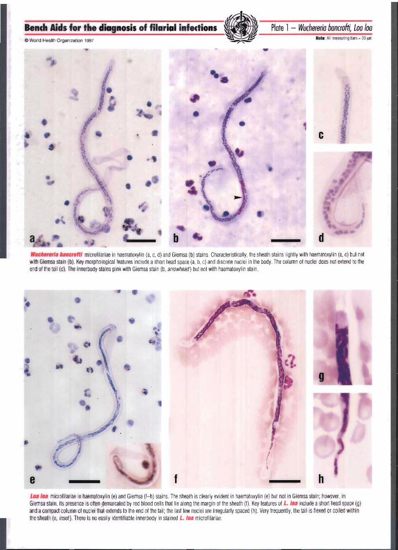
Bench Aids for the diagnosis of filarial infections Plate 1 - Wuchereria bancrofti, Loa loa © World Health Organization 1997 Note: All measuring bars ~ 30 ~ -
-, c
b Wuchereria bancroftl microfilariae in haematoxylin (a, c, d) and Giemsa (b) stains. Characteristically, the sheath stains lightly with haematoxylin (a, c) but not with Giemsa stain (b) Key morphological features include a short head space (a, b, c) and discrete nuclei in the body. The co lumn of nuclei does not extend to the end of the tail (d). The innerbody stains pink with Giemsa stain (b, arrowhead) but not with haematoxylin stain.
e f
1 f I , \
g
h Loa loa microfilariae in haematoxylin (e) and Giemsa (I-h) stains. The sheath is clearly evident in haematoxylin (e) but not in Giemsa stain; however, in Giemsa stain, its presence is often demarcated by red blood cells that lie along the margin of the sheath (f) Key features of L. loa include a short head space (g) and a compact column of nuclei tha't extends to the end of the tail ; the last few nuclei are irregularly spaced (h) Very frequently, the tail is flexed or coiled within the sheath (e, inset). There is no easily identifiable innerbody in stained L. loa microfilariae.

.~ _B_en_c_h_li_·d_s_f_o_' _'h_e_di_a ...... no_s_is_o_f_f_il_a_'i_a_I i_n_fe_c_'i_on_s ___ ~ . ~ . ~ _____ Plo_te_l © World Healtn Organization 1997 ~ .fJ!j
D· is of marl inlecf 0 s As well as in blood and skin , microfilariae may occasionally be found in bone marrow preparations, fine-needle biopsy aspirates, cervical smears contaminated with blood , hydrocele fluid, chylous urine, and normal urine following treatment with diethylcarbamazine. Methods commonly used for the detection of microfilariae include
Blood examination stained thick blood films
• direct examination of capillary blood • membrane filtration (fresh or preserved blood) • haemolysed venous blood concentration (Knott concentration method)
TIssue examination • skin snips
Other body fluid examination urine
• hydrocele fluid
CautiDIJ: Standard biosafety guidelines should be followed in obtaining blood and tissue samples. Disposable or sterile lancets, syringes, and needles should be used for all laboratory procedures. These guidelines are summarized in Biosafety guidelines for diagnostic and research laboratories working with HIV (Geneva, World Health Organization , 1991; WHO AIDS Series , No.9) .
Preparation of thkk blood films The examination of thick blood films is the most widely used method in field surveys of filarial infection. Properly done, it is a reliable procedure for both identification of microfilariae and enumeration studies. Carefully measured samples of at least 20 ~ll and preferably 60 III in volume are recommended.
1. Thoroughly clean the microscope slides (including factory "pre-cleaned " slides) before use. Dust, grease, detergent, or cotton 'lint and threads may cause the blood film to lift off the slide.
2. Clean the finger tip (or ear lobe) from which the blood will be taken with a cotton ball soaked in alcohol.
3. Prick the finger tip or ear lobe with a sterile lancet and allow the blood to ooze freely . 4. Draw the required volume of blood into a disposable or sterile calibrated capillary pipette. 5. Expel the blood onto a microscope slide and smear the sample uniformly in a circular
or rectangular shape ; avoid creating any bubbles. 6. Allow the slide to dry at room temperature in a horizontal position. 7. Label and store the slide in a dust-free environment until staining . It is also important
to protect unfixed blood films from damage by insects.
NDte: Excess alcohol on the skin may partial1ly fix the blood sample; squeezing the finger or ear lobe may dilute the sample with tissue fluids . Films that are too thick tend to lift off the slide. Blood films must be thoroughly dried before dehaemoglobinization; this may require 12- 48 hours, depending on humidity . If blood is collected in a heparinized capillary pipette, or if the film is made from blood containing an anticoagulant, drying requires at least 48- 72 hours. Thin blood films are of little value because the volume of blood examined is small. However, when microfilariae are found in thin films they tend to be concentrated at the "feathered " end and at the margins of the film. The morphology of microfilariae found in thin films tends to be good since the films are routinely fixed before staining.
(apt1lary blood examination Microscopic examination of fresh blood has limited utility . It can reveal the presence of microfilariae actively moving among the red blood cells (see front cover), but species identification is not possible. However , in regions where only one species of microfilaria is found, its presence and density in the blood can be determined with reasonable accuracy by this means.
'"?~

Bench Aids for the diagnosis of filarial infections ft.-~ ~ 'I· W --~I ------~-------~I 'iII,'iZ Pate 2 - 8rugia malayi, 8rugia timori ~ ·1 rtp
© World Health Organization 1997 ~"?~ Note: All measuring bars = 30 jJrT1
11
•
c
•
a b d Brugiaalall microfilariae in haematoxylin (a) and Giemsa (b-d) stains. In haematoxylin, the sheath does not stain but may be fainlly visible (a, arrow). This contrasts with the pink-stained sheath seen in Giemsa preparations (b, c) The column of nuclei is compact, and the widely separated subterminal and terminal nuclei in the tall are key diagnostic features (a, arrowheads, d) Nuclei are sparse in the region of the innerbody (a)
• B. malay; (upper) and W. bancroft/ (l ower) microfilariae in the same field of a Giemsa-stained blood film (e). The pink-stained sheath and the darkly stained, compact column of nuclei identify B. malayi and distinguish it from W. banr:roftl
f h
• • •
Brugia limori microfilariae in haematoxylin (f) and Giemsa (g-i) stains. B. timor; is larger than B. malsyl and the sheath does not stain pink (g, arrowhead) with Giemsa stain . The long head :,pace and the subterminal and terminal nuclei are conspicuous features (I-i)

• _Be_n_c_h_Ai_·d_s_'_o_r_th_e_di_G ...... no_s_is_o_'_'_il_G_ri_G_li_n_fe_c_ti_on_s ___ ~ . ~f ~ __ ~_Pla_te_2 © World H "al t~ Organizati on 1997 ~ ~, ii
DIOOG fi 5
Giemsa and haematoxylin are the preferred and most widely used stains for preparing permanently stained blood films. Each has its advantages, bl1t Giemsa stain is used most often. Slides can be processed in either small or large numbers using stain less steel , glass, or plastic staining racks and dishes.
Before staining , thoroughly dried films must be dehaemoglobinized and fi xed. Immerse slides in tap or distilled water until the haemoglobin leaches out of the film , which becomes whitish in colour; this requires about 3-5 minutes . Films that are prepared from blood containing an anticoagulant and that have dried for more than a few days will dehaemoglobinize slowly, usual ly in 8-10 minutes. Allow dehaemoglobinized films to airdry thoroughly . Fix the films in methanol for 30- 60 seconds and air-dry
Note: In the event that the same films are being used for malaria surveys , they should be stained without dehaemoglobinization or fi xation. Microfilariae found in these preparations usually appear slightly swollen , and the nuclei are not sharply demarcated (Plate 3b) .
Giemsa stein Stain blood films for 45 minutes in a 1 :50 dilut ion of Giemsa stain (or 20 minutes in a 1 :20 dilution) at a pH of 6.8 - 7.2; wash films for 3- 5 minutes in neutral buffered water or under running tap water . Dry films in a vertical position.
Note: The staining dilution and procedure used for processing malaria films can be used here with the expectation of acceptable results . Nuclei of microfilariae will stain blue to purple in colour. A sheath , if present, will stain pink (B. malayi) or not at all. The innerbody of W bancroftiwill stain a bright pink colour , but that of most other species does not stain.
Haematoxyhn stain Various haematoxylin stains are used as alternat ives to Giemsa stain; Delafield 's haematoxylin is recommended and is widely used. It enhances nuclear detail in the microfilaria and stains the sheath , when present, a greyish-blue colour. For preparation of Delafield 's haematoxylin and details of another useful staining procedure, consult the WHO publication Basic laboratory methods in medical parasitology (1991). It is also acceptable to use other available stains and procedures .
Procedure 1. Thick blood films should be dried thoroughly , dehaemoglobinized , and fi xed as
describEld above. If films are prepared from sedimented Knott concentration material , dehaem globinization and fi xation are omitted .
2. Stain slides for 10 - 15 minutes in Delafield 's haematoxylin solution . Rinse in distilled water to remove excess stain.
3. Destain in 0.1 % (1 g/I) aqueous hydrochloric or acetic acid for approximately 1 minute. Rinse sl ides in distilled water for 1 minute .
4. Place the slides in tap water containing several drops of ammonia for 3-5 minutes . The films will become dark blue in colour .
5. Rinse in tap water for 2- 5 minutes and allow to dry.
Note: Films may be made permanent by adding a synthetic mounting medium and a coverslip . Alternatively , simply clarify with a drop of immersion oil , add a coverslip, and examine under low magnification.
~~

Bench Aids for the diagnosis of filarial illfections Plote 3 - Mansonella perstans, Mansonella ozzardi © World Health Organization 1997 lote: All measuring bars; 30 IfT1
c
a b Manso ella persians microfilariae in haematoxylin (a, c, d) and Giemsa-stained (b) blood films. M. persians is small, has a short head space (c), lacks a sheath, and is readily recognized by the blunt tail that is filled by the column of nuclei (a, b, d). In th ick blood films stained with Giemsa stain without fixation, the body usually appears thickened, and individual nuclei may be indistinct (b).
g
e f h Mansonella ozzardi microfilariae in haematoxylin (e, g, h) and Giemsa (I) stains. Key features of this small, unsheathed microfi laria include a compact column of nuclei, a head space that is slightly longer than it is wide (g) and, most importantly, a tail that is long, slender, and devoid of nuclei (h). The appearance is the same in haematoxylin and Giemsa stains (e, I).
•
-M. persians and M. ozzardi are often lound in individuals inlected with other lilariae in areas where species overlap. It is not uncommon to see, as in (i), M. perstans (upper) with ! . Ioa or, more rarely, as in (j), fl. perslass wilh Mic'olilaria semlclarum (lower).' In the Americas, as shown in (k), mixed inlections 01 W bancroft} (upper) and M. ozzardiare often seen. Mixes 01 • persians ,lnd M. olZ8ldi are also common. Microfilariae (i-k) stained in haematoxylin. , Microfifaria SlIIIfelllnl111 (j) has been found in Ihe blood of people in Zaire (Fain A. DipetalonEma semiclarum sp. nov. Irom the btood of man in the Republic of Zaire (Nematoda: Fi tarioidea ). Annales de la Societe beige de MMecine tropicale,1974, 54:195-207.). A valid genus name ha i not been assigned to this species of filarial worm.

Bench Aids for the dia nosis of filarial infections II· .'~\\ ~ ,' . ' ~ ____ Pla_te_3 ~ rJ1 © World Hr,allt·, Organization 1997
~"'--
(onc of n roced es The detecti ')n of microfilariae in peripheral blood when few are present is best accomplished by concentration procedures , which allow for the examination of a larger volume of blood. The use of membrane il tration and the Knott concentration method are the most widely used procedures.
Membrane filtration Membrane !iltration allows for removal of elements in the blood by filtration through a membrane of desired por :3 size. Membrane filtration is more effectively used to determine microfilarial density than as a means of microfilaria identification. Cellulose- mixed-ester filters (e.g. Mlilipore filters) and polycarbon te filters (e.g. Nuclepore fil ters) are the most common membrane fi lters used. Formerly , fresh blood samples required processing soon after they were obtained. Recently, however, a procedure for membrane filtration of preserved blood has been published (1). Both are described below.
Filtration of fresh whole blood Materials nd reagents 1. Sodium citrate solution, 3.8% (38 gil) or EDTA (ethylenediaminetetraacetic acid) solution, 7.5% (75 gil). 2. Teepol- saline solution, 10% (prepare by adding 50 g Teepol concentrate to 450 ml sal ine). 3. Saline, 0.85% (8.5 g/I). 4. Giemsa stain. 5. Syringe (disposable polypropylene with rubber plunger tip), 20-ml capacity. 6. Membrane filter holder (e.g. Swinnex type) . 7. Membrane filter, 3-5-l1m porosity, 25-mm diameter .
Note: Although a pore size of 5 11m is ideal for L. loa microfllariae, 4 11m is more efficient for filtration of W. bancrofti and other smaller species of microfilariae such as Mansonella perstans.
8. Absolute methanol.
Procedure 1. Collect a fresh blood sample in sodium citrate or EDT A solution. 2. Add 1 m of citrated or EDTA-preserved blood to 10 ml of Teepol-saline solution. 3, Place mQistened membrane filter, secured with a rubber gasket, into filter holder (Fig. 3). 4, Remove plunger from barrel of 20-ml syringe and connect barrel of syringe to filter holder, 5. Pour the blood- Teepol mixture (from step 2) into barrel of syringe, replace plunger in syringe and ,
by appl)'ing gentle, even pressure, force solution through filter (Fig. 4). Discard blood into disinfectant for disposal.
Note: Some workers prefer to push a 1-ml blood sample directly through the filter followed by 20-35 ml of water or saline to wash out the remaining blood . Others suspend the blood in 10 ml of water , a itate, and allow the mixture to stand for several minutes before passage through the filter.
6. Remove syringe from filter holder , draw up 10 ml of water into syringe , reattach filter holder, and gently w sh filter by flushing the solution through it.
7. Force tWo) syringe-volumes of air through filter to expel excess water and make microfilariae more adherent to filter.
Note: Procedures may be modified at this point depending on the type of preparation desired ,
Mluol1lariae may be fixed and stained on the filter as loRows: 8. For permanent, stained preparations:
a. Pass :3 ml of methanol through filter to fix microfilariae. b. Pass ,'3 ir through filter to expel residual methanol. c. Remove filter from the holder and place on a glass slide; allow it to dry thoroughly, d. Stain the preparation in Giemsa stain as for a blood film . e, Rinse in tap water and allow to dry, f. Dip the slide in toluene to avoid bubbles in or under the filter. Add a drop of synthetic mounting
mediL m and a coverslip. The slide may be examined in the same manner as any blood film and stored as a permanent preparation for future reference .
Mluolilariae lIIay be examined alive as fo'ows: 9. Alternmively, following step 7, remove syringe from filter holder, carefully unscrew top from filter,
and, Ll slng forceps , remove rubber gasket. 10. Use fin forceps to transfer wet fil ter to a slide, with the residue on the membrane facing upwards. 11. Add a drop of saline to the membrane and cover with a coverslip. Examine under the microscope
with 10:( objective; microfilariae will be seen actively moving .
Fig. 3
--r -'"~ I .---- Filter holder ~- Membrane filter __ 4 Rubber gasket
S? Filter holder base
WHO 95670
Fig. 4

it.-~ Bench Aids for the diagnosis of filarial infections ~ : . .~ ~ · rJR
Plate 4 - Techniques, artefacts, oddities © World Health Organization 1997 ?~ Note: All measuring bars; 30 ~
• • _t. " • .. -.~ .. ... ~
!~ \ ~ ...
; ,
. b -L. loa microfilariae in a Knott concentration. They are easily enumerated at low magnification (a). At high magnification, features such as size, shape, and the presence or absence of a sheath are evident (b). Note the sheath extensions (arrowheads) at both ends (b).
e Microfilaria semlclarumsuperficially resembles M. perstans; it is similar in size but has a sparsely nucleated area (arrowhead) in the posterior half of the body. The adult worms and vectors have not been identified. Preparation stained in haematoxylin.
L. loa microfi lariae collected on a polycarbonate filter and stai ned with Giemsa stain. Microfilariae are easily enumerated at low magnification (c); the distribution of nuclei in the tail allows the identification of the micro filaria at high magnification (d) .
•
•
O. volvulus microfilariae in a section of skin stained with haematoxylin and eosin. Only portions of the microfilariae are visible (arrowheads) .
j
-
Fibres (g, h), unidentified elements (i), and fungi (Hel/cosporium ) (j) found on Giemsa-stained blood films are often conlused with microfi lariae. In spite of similar size, the presence of a darkly stained core and/or vacuoles, the absence of nuclei, and jagged or broken ends rule out identification as microfilariae.
Bench Aids for the dia nosis of filarial infections © World Healtll Organization 1997
(once of n DrCM:eaures (continued)
Filtration of preserved blood If it is not possible to process fresh blood immediately, the following procedure may be used.
Materials and reagents Materials required are the same as for processing whole blood, except that 2 ml of 37% (370 g/I) formaldehyde solution is added to 10 ml of Teepol concentrate and 88 ml of distilled water to make 100 ml of Teepol-formalin solution.
Procedure 1. Blood specimens preserved in the Teepol-formalin solution (1 ml of blood should be
added to 10 ml of Teepol-formalin solution) are filtered through a membrane filter in the same manner as described in steps 3-7, above.
2. Filters can be examined wet with or without the addition of Giemsa, haematoxylin, or other stains, to allow for enumeration of the microfilariae or study of their morphology.
3. Alternati \; ely, the wet filter can be placed on a slide, allowed to dry, and stained as desired. drop of synthetic mounting medium and a coverslip can be added to make a permanent preparation.
Note: Teepot lyses blood and formaldehyde preserves the morphological features of microfilaria .. Blood specimens in the Teepol-formalin solution can be retained for 9 months or longer before examination, without marked deterioration of the microfilariae. Blood specimens in Teepol only, or in a Teepol-sodium azide solution, are not useful for long-term storage since microfilariae undergo degenerative changes within a week or less.
KnoH concentration method The Knott concentration method is very sensitive and relatively inexpensive to perform.
Materials ad reagents 1. Centrifuge tubes, 1S-ml capacity. 2. Formalin , 2% (2 ml of 37% (370 g/I) formaldehyde solution + 98 ml of distilled water) . 3. Slides and coverslips. 4. Needles and syringes. 5. Centrifuge (hand- or electric-powered).
Procedure 1. Collect 1 ml of blood (whole or citrated) by venepuncture and place in a 1S-ml
centrifuge tube containing at least 10 ml of formalin; shake vigorously. Red cells are lysed by the formalin solution.
2. Centrifuge at approximately 3009 for 2 minutes. If a centrifuge is not available, place the tube In an upright position for 12 hours for gravitational sedimentation.
3. Decant the supernatant fluid (the small amount remaining in the tube is allowed to flow back on 0 the sediment).
4. Examine a drop of the sediment on a slide under a coverslip with the low-power objective of the microscope.
5. A portion of the sediment may be spread on a slide as a thick film and allowed to dry thoroughly. Stain the film with Giemsa or haematoxylin stain.
Note: Avoid ' dding more than 1 ml of blood to 10 ml of formalin; as much as 12-14 ml of formalin is desirable for each 1 ml of blood. Only microfilariae and white blood cells are found in the sediment; microfilariae are fixed without significant shrinkage and are easy to count accurately. A sheath, if present, is also easy to see. The technique is useful for quantification of microfilaraemia. Samples need not be examined immediately and can be stored in thl3 laboratory for several weeks. Microfilariae present in the stained sediment will show details of internal structure.
Plate 4

Bench Aids for the diagnosis of filarial infections • ~ .. ~ ~ote 5 - Onchocerca volvulus, Mansonella streptocerca ~ (JJ! Iote: All measuring bars = 30 ~ ~-
© World Health Organization 1997
c
•
a b d Onchocerca ~o/rulus microfilariae from skin snips in haematoxylin (a, c, d) and Giemsa stains (b). This microfilaria is large and has no sheath, a long head space (c) and, typically, a flexed tail (d). The column of body nuclei is only moderately compact. The most important diagnostic feature is that O. volvulus is found in the skin and only rarely in the blood.
g
e f h Man oneil a streptocerca microfilariae from skin snips in haematoxyl in (e,g,h) and Giemsa stains (t) . M streptocerca is readily distinguished from O. volvulus by its very slender shape and "crooked" tail (e , f, h) . Note that the column of nuclei starts in the anterior extremity as a single row of 10-12 (or more) nuclei (g) and extends to the end of the ta il (e,f,h).
• ~. . ~ ____ PI_Qte_5 ~ . rI ~~~
Bench · ds for the dia nosis of filarial infections © World Health Organization 1997
Skin snips The microfilariae of Onchocerca volvulus and Mansonella streptocerca that reside in the skin are best detected by looking for their presence in skin snips. Intensity of infection is reflected in the numbers of microfilariae emerging from the snips. Skin snips are obtained in one of two ways:
1. Skin snips can be standardized in both size and weight through the use of sclerocorneal punches of either the Holth or Walser type. These instruments take snips of uniform d iameter (approximately 2.3-2.5 mm). This is the preferred method.
2. A needle can be used to raise the skin and a razor blade to cut off the raised area; forceps and curved scissors can also be used . Such skin snips vary in size, shape, and the depth of the cut. When snips are cut too deeply, small capi llaries may be lacerated and the snip may be contaminated by microfilariae that might be present in the patient's blood.
Coution: It is of the utmost importance that all instruments used for each patient are sterile in order to avoid transmission of viral hepatitis Band HIV infections.
Procedure 1. Skin snips should be taken from selected sites on the body. In Africa , the preferred site is the iliac crest; in Central and South
America, the iliac crest or the scapular area; and in Yemen, the lower calf. In surveys, ideally two snips should be taken from all three of these sites on each side of the body of the individual.
2. Transfer skin snips from each site to a drop of normal saline, distilled water, or tissue culture medium in a well of a 96-well, flat-bottom, tissue culture tray ; or place snips on a microscope slide in one of the fluids . It is not necessary to tease the snips.
3. Examine after 30 minutes to 3 hours. (Tissue culture trays may be covered with plastic wrap or similar material, and slides placed in a covered Petri dish. to retard evaporation.) If the wells or slides are negative for microfilariae, allow the snips to remain overnight in an incubator at 37°C or at room temperature and examine them again. If microfilariae are present they will be apparent in the fluid. The morp'lological features of 0. volvulus and M. streptocerca are so distinct that differentiation of microfilariae is quite easy.
4. To make permanent preparations of microfilariae. remove the skin snips, transfer the fluid to a slide if necessary , and allow the flu id to evaporate. When the slide is thoroughly dry, fix the microfilariae in methanol and stain with Giemsa or haematoxylin stain.
Urine and y.ocele fluid Pour 15 ml of urine or hydrocele fluid into a conical centrifuge tube and centrifuge for 5 minutes at 350g or more. Pour off supernatant and examine sediment for microfilariae. Slides can be stained and/or fixed as described for blood samples.
Other diagnostic methods Microhaematocrit Originally used for diagnosis of trypanosomiasis, the microhaematocrit procedure is equally useful for the diagnosis of filarial infections, especially when the numbers of microfilariae present are too small for efficient detection by thick blood films. Only a small amount of blood is needed. so that one or two drops obtained by finger-prick can be used when venepuncture cannot be performed (2).
Quantitative buHy coat The utilizatio'1 of the quantitative buffy coat tube (microhaematocrit tube recoated with acridine orange) has been reported to be an acceptable rapid diagnostic test for the detection of microfilariae, with a sensitivity equivalent to that of the thick blood film (3).
Micro'ilariae counts Accurate cc·unts of microfilariae can be made from stained thick blood films of measured volume. Counting requires carefu l systemat ic ~; canning of the blood film with the low-power objective of the microscope. The stained slides can be kept as a permanent record. Equally reliable counts can be made from membrane filters which, if mounted with a coverslip , can be retained as a perrlarent record. Some investigators prefer using a counting-chamber technique , which is very reliable but does not lend itself to species identification or permanence (4).
References 1. Dickerson JW, Eberhard ML, Lammie PJ. A technique for microfilarial detection in preserved blood using Nuclepore filters.
Journal of parasitology, 1990,76:829-833. 2. Control of lymphatic filariasis. A manual for health personnel. Geneva, World Health Organization, 1987. 3. Freedman DO, Berry RS. Rapid diagnosis of bancroftian fil ariasis by acridine orange staining of centrifuged parasites.
American journal of tropical medicine and hygiene, 1992, 47:787-793. 4. Fleck SL, Moody AH . Diagnostic techniques in medical parasitology. London, Butterworth, 1988.

These bench aids, pan ion to Bench Aids the diagnosis of intestinal parasites, are intended
both as a guide for laboratory and field workers in countries d as a teaching ai gf ~ud_
1IUt~S. They p de guidance on th ~reparat"'" Italn"~lck blood films, microfilarial concen tlon
IN)c~eaUlre!lllana examination of skin for the di sis 1"~tICI1& Plhot(lIllM~"bs demonstrate
the various
The prod weatherproo plastic-sealed format that is
d easy to use in the field and at the laboratory They are recommended for use by all health workers engaged in the routine diagnosis of fllartat Infections.
ISBN 9241