radiation and the atom robert metzger, ph.d. course objectives prepare for the physics portion of...
TRANSCRIPT
Radiation and the Atom
Robert Metzger, Ph.D.
Course Objectives
Prepare for the Physics portion of the ABR Boards.Understand image formation and image quality for all standard imaging techniques.Assess patient dose and risks.Accrue the 200 hours required by NRC for licensing.
Course Outline
Approximately 100 hours of lecture, lab, and problem solving from July 2005 to June, 2006.
Another 50 200 hours is acquired by reading Bushberg and Huda, and solving problems from Huda.
The final 50 hours are from preparation for the boards using old exams. This occurs from July 2006 to exam time in September.
Course Structure
Each Thursday we will meet for approximately 2 hours. The first 1 to 1.5 hours will be lecture and the remainder will be taken up with problem solving and labs. When labs occur at other times (e.g. equipment surveys, image quality evaluations), the class time will be shortened accordingly.
Course Structure
As the class develops, these slide programs and worked problems will be posted on the web. If you miss a class you can obtain all of the information from the web (www.radsafe.com).
Quarterly practice tests will be offered over the materials covered the previous quarter, with questions drawn from previous board exam questions.

Textbooks
Bushberg, J, Seibert, A, Leidholdt, E., Boone M., The Essential Physics of Medical Imaging, 2nd Ed., Lippincott Williams, & Wilkins, 2002.
Huda, W., Slone, R., Review of Radiologic Physics, 2nd Ed., Lippincott Williams, & Wilkins, 2003.

Example Problems
RAPHEX Exams 2001 – 2004. 2001 to 2003 exams will be used for example problems while the 2004 exam will beused for the quarterly practice exams.Nickoloff, E., Radiology Review, Radiologic Physics, Elseveir Saunders, 2005.RSNA website (www.rsna.org).
Fast Moving Technology
Radiology is moving faster than the textbooks can keep up. Academics create the board exam questions and include areas not covered in the textbooks (e.g. radiobiology, fluoro safety, multislice CT, digital mammo, etc, etc, etc.
RSNA has excellent tutorials in areas where there is insufficient treatment in the textbooks. Read them!
ABR
ABR has indicated that they are going to toughen up the physics requirements for Radiology Residents.
They have already added questions in areas previously uncovered and indicate (see handout) that they intend to increase the passing percentage for the test.

NRC/ARRA
The NRC has established training requirements for Radiologist that read Nuclear Medicine or are Radiation Safety Officers (see 10 CFR 35).200 hours of physics and a preceptor statement are required.Preceptors are hard to obtain the further you are from your residency. Many older radiologists are not able to be licensed as they never had their preceptor filled out and were listed on a license.
NRC/ARRA
One of the objectives of this course is to complete the 200 required hours of physics training and fill out the preceptor statement immediately.
Try to get listed on the Radioactive Materials license the first place you work.
Once licensed, the preceptor is no longer required. You are good for life!
Basic Physics Review
Describe the basic characteristics of electromagnetic (EM) Describe the basic characteristics of electromagnetic (EM) radiation and how they are mathematically related radiation and how they are mathematically related
Describe how atomic electronic structure determines the Describe how atomic electronic structure determines the characteristics of emitted EM radiation characteristics of emitted EM radiation
Describe the various ways x-rays can interact with and are Describe the various ways x-rays can interact with and are attenuated in matter Describe the energy dependence of these attenuated in matter Describe the energy dependence of these interactions interactions
Describe and calculate the various quantitative parameters Describe and calculate the various quantitative parameters used to characterize x-ray attenuation used to characterize x-ray attenuation
Differentiate between radiographic exposure absorbed dose Differentiate between radiographic exposure absorbed dose and equivalent dose as well as use the correct radiological unitsand equivalent dose as well as use the correct radiological units

Radiation
Propagation of Energy Through Space or Matter
Particulate Radiation: Electrons, Protons, Alpha Particles, Beta Particles, Neutrons, etc.
Electromagnetic Radiation. No Particle With Mass. See electromagnetic spectrum.
Acoustic Radiation: Ultrasound (reviewed later.

Ionizing Radiation
Radiation with sufficient energy to ionize human tissue. That is, it must impart enough energy to clip an electron off a water molecule and produce an ion pair (free electron + positively charged nucleus. Requires about 10 to 20 eV/ion pair.
Radiations that do not impart enough energy are called non-ionizing radiations. Both are used in medical imaging.
Non-Ionizing Radiations
MR Imaging (FM Region)
Ultrasound.
Microwave Diathermy.
Lasers used for various treatments.
Visible Light to read images.
Characteristics of Waves
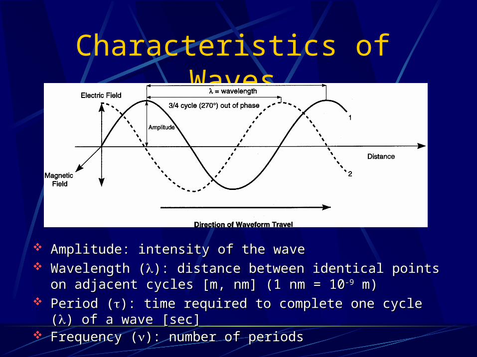
Amplitude: intensity of the wave Amplitude: intensity of the wave Wavelength (Wavelength (): distance between identical points on adjacent ): distance between identical points on adjacent
cycles [m, nm] (1 nm = 10cycles [m, nm] (1 nm = 10-9-9 m) m) Period (Period (): time required to complete one cycle (): time required to complete one cycle () of a wave [sec] ) of a wave [sec] Frequency (Frequency (): number of periods ): number of periods
Electromagnetic Radiation
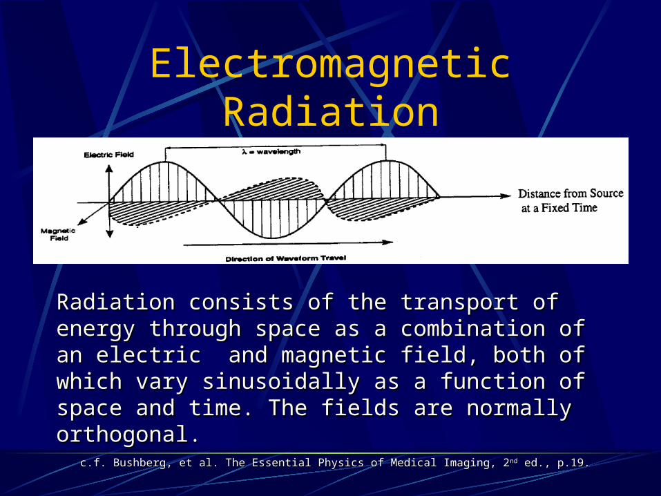
c.f. Bushberg, et al. The Essential Physics of Medical Imaging, 2c.f. Bushberg, et al. The Essential Physics of Medical Imaging, 2ndnd ed., p.19. ed., p.19.
Radiation consists of the transport of energy through Radiation consists of the transport of energy through space as a combination of an electric and magnetic space as a combination of an electric and magnetic field, both of which vary sinusoidally as a function of field, both of which vary sinusoidally as a function of space and time. The fields are normally orthogonal.space and time. The fields are normally orthogonal.
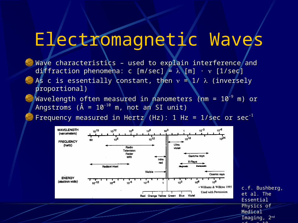
Electromagnetic WavesWave characteristics – used to explain interference and diffraction Wave characteristics – used to explain interference and diffraction phenomena: c [m/sec] = phenomena: c [m/sec] = [m] ∙ [m] ∙ [1/sec] [1/sec]
As c is essentially constant, then As c is essentially constant, then = 1/ = 1/ (inversely proportional) (inversely proportional)
Wavelength often measured in nanometers (nm = 10Wavelength often measured in nanometers (nm = 10-9-9 m) or Angstroms m) or Angstroms (Å = 10(Å = 10-10-10 m, not an SI unit) m, not an SI unit)
Frequency measured in Hertz (Hz): 1 Hz = 1/sec or secFrequency measured in Hertz (Hz): 1 Hz = 1/sec or sec-1-1
c.f. Bushberg, et al. c.f. Bushberg, et al. The Essential The Essential Physics of Medical Physics of Medical Imaging, 2Imaging, 2ndnd ed., ed., p.18.p.18.
Particle Characteristics
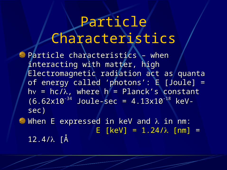
Particle characteristics – when interacting with Particle characteristics – when interacting with matter, high Electromagnetic radiation act as quanta matter, high Electromagnetic radiation act as quanta of energy called ‘photons’: E [Joule] = hof energy called ‘photons’: E [Joule] = h = hc/ = hc/, , where h = Planck’s constant (6.62x10where h = Planck’s constant (6.62x10⁻³⁴-34 Joule-sec = Joule-sec = 4.13x104.13x10-18-18 keV-sec) keV-sec)
When E expressed in keV and When E expressed in keV and in nm: in nm: E [keV] = 1.24/E [keV] = 1.24/ [nm] [nm] = 12.4/ = 12.4/ [Å [Å
An X-Ray
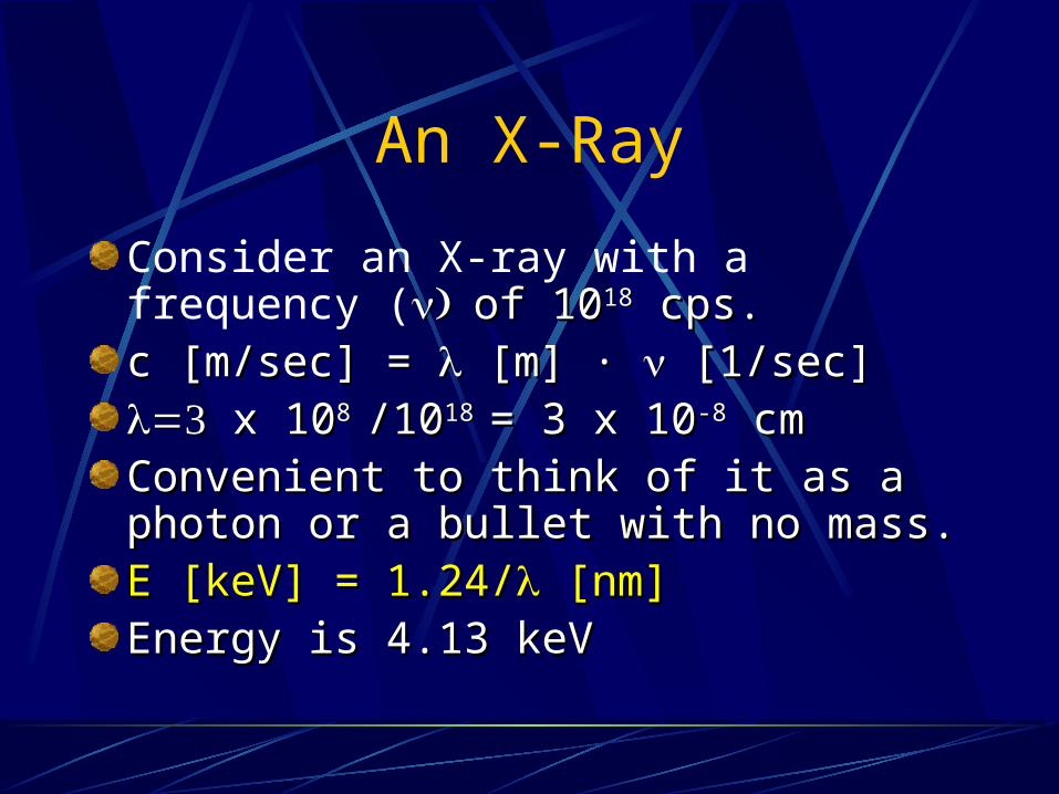
Consider an X-ray with a frequency (of 10of 101818 cps. cps.c [m/sec] = c [m/sec] = [m] ∙ [m] ∙ [1/sec] [1/sec] x 10x 108 8 /10/1018 18 = 3 x 10= 3 x 10⁻⁸-8 cm cmConvenient to think of it as a photon or Convenient to think of it as a photon or a bullet with no mass.a bullet with no mass.E [keV] = 1.24/E [keV] = 1.24/ [nm] [nm]Energy is 4.13 keVEnergy is 4.13 keV
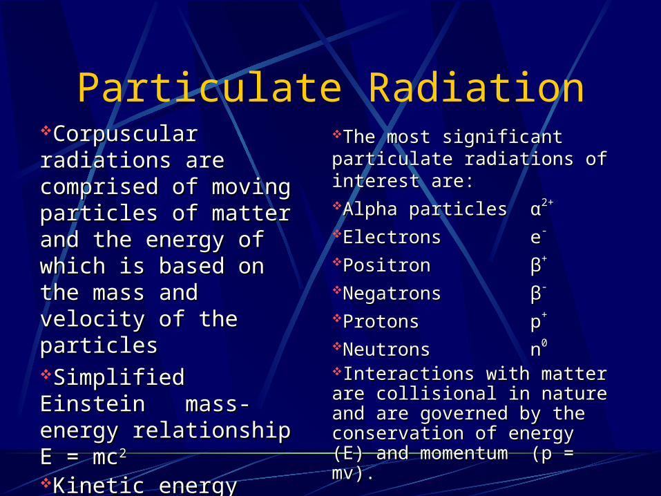
Particulate RadiationCorpuscular radiations Corpuscular radiations are comprised of moving are comprised of moving particles of matter and the particles of matter and the energy of which is based energy of which is based on the mass and velocity on the mass and velocity of the particles of the particles Simplified Einstein Simplified Einstein mass-energy relationship mass-energy relationship E = mcE = mc2 2
Kinetic energy (KE) Kinetic energy (KE) = ½ mv= ½ mv22 (for non- (for non-relativistic velocities)relativistic velocities)
The most significant particulate The most significant particulate radiations of interest are: radiations of interest are: Alpha particlesAlpha particlesαα2+2+
ElectronsElectrons ee--
PositronPositron ββ++
NegatronsNegatrons ββ-- ProtonsProtons pp++ NeutronsNeutrons nn00
Interactions with matter are Interactions with matter are collisional in nature and are collisional in nature and are governed by the conservation of governed by the conservation of energy (E) and momentumenergy (E) and momentum (p (p = mv).= mv).

Pauli exclusion principle Pauli exclusion principle No two electrons can have the No two electrons can have the
same energy same energy → → 2n2n22 electrons per shell electrons per shell
quantum numbers quantum numbers nn: principal q.n. – which e: principal q.n. – which e-- shell shell ℓℓ: azimuthal – angular momentum : azimuthal – angular momentum
q.n. (ℓ = 0, 1, ... , n-1)q.n. (ℓ = 0, 1, ... , n-1) mmℓℓ: magnetic q.n. – orientation of : magnetic q.n. – orientation of
the ethe e-- magnetic moment in a magnetic moment in a magnetic field (mmagnetic field (mℓℓ = -ℓ, -ℓ+1, ..., = -ℓ, -ℓ+1, ...,
0, ... ℓ-1, ℓ)0, ... ℓ-1, ℓ) mmss: spin q.n. – direction of the e: spin q.n. – direction of the e--
spin (mspin (mss = +½ or -½) = +½ or -½)
c.f. Bushberg, et al. The Essential Physics of Medical Imaging, 2c.f. Bushberg, et al. The Essential Physics of Medical Imaging, 2ndnd ed., p.21. ed., p.21.
Electronic Structure – Electron OrbitsElectronic Structure – Electron Orbits
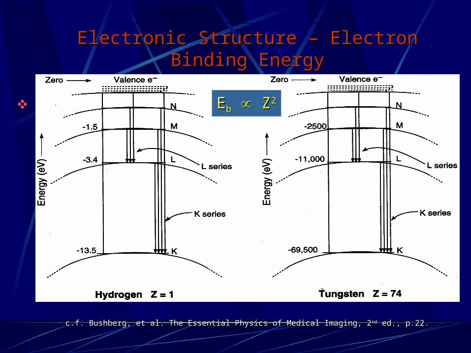
Electronic Structure – Electron Binding EnergyElectronic Structure – Electron Binding Energy
c.f. Bushberg, et al. The Essential Physics of Medical Imaging, 2c.f. Bushberg, et al. The Essential Physics of Medical Imaging, 2ndnd ed., p.22. ed., p.22.
EEbb Z Z22

Radiation from Electron TransitionsRadiation from Electron Transitions
Characteristic X-rays Characteristic X-rays Auger Electrons and Fluorescent Yield (Auger Electrons and Fluorescent Yield (KK): ):
(characteristic x-rays/total) (characteristic x-rays/total) Preference for Auger ePreference for Auger e-- for low Z for low Z
c.f. Bushberg, et al. The Essential Physics c.f. Bushberg, et al. The Essential Physics of Medical Imaging, 2of Medical Imaging, 2ndnd ed., p.23. ed., p.23.
c.f. Sorenson, et al. Physics in Nuclear c.f. Sorenson, et al. Physics in Nuclear Medicine, 1Medicine, 1stst ed., p.8. ed., p.8.
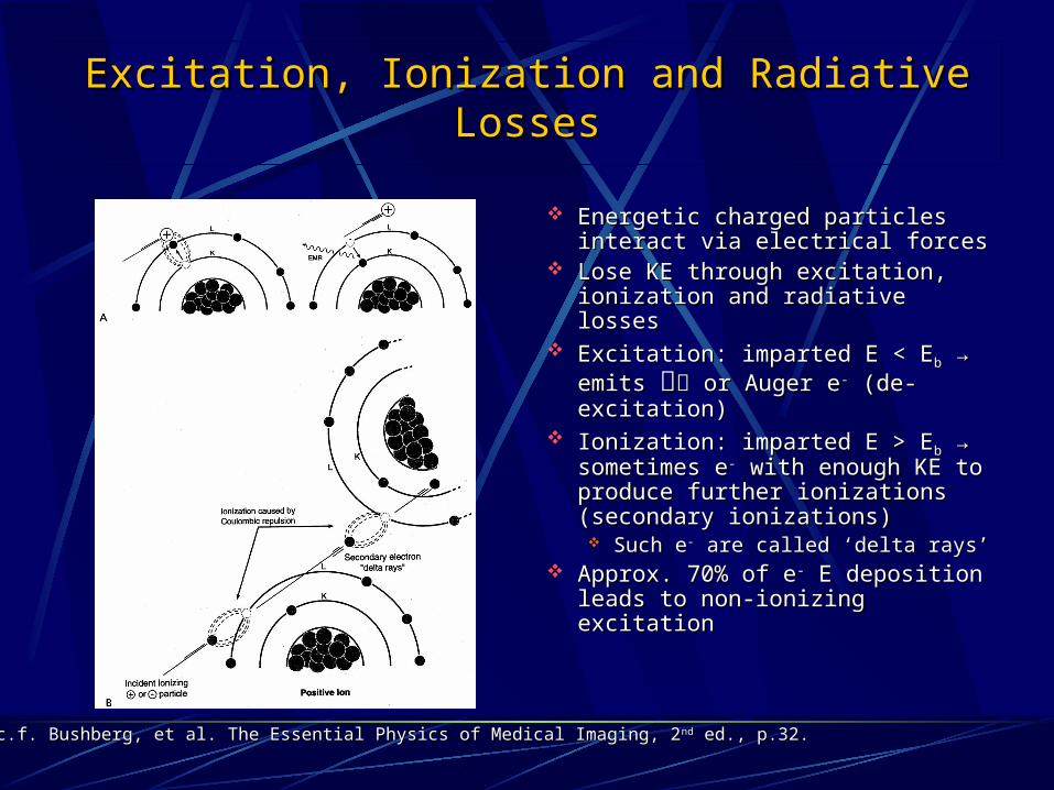
Excitation, Ionization and Radiative LossesExcitation, Ionization and Radiative Losses
Energetic charged particles Energetic charged particles interact via electrical forces interact via electrical forces
Lose KE through excitation, Lose KE through excitation, ionization and radiative losses ionization and radiative losses
Excitation: imparted E < EExcitation: imparted E < Ebb → → emits emits or Auger e or Auger e-- (de- (de-excitation) excitation)
Ionization: imparted E > EIonization: imparted E > Ebb → → sometimes esometimes e-- with enough KE with enough KE to produce further ionizations to produce further ionizations (secondary ionizations) (secondary ionizations) Such eSuch e-- are called ‘delta rays’ are called ‘delta rays’
Approx. 70% of eApprox. 70% of e-- E deposition E deposition leads to non-ionizing excitation leads to non-ionizing excitation
c.f. Bushberg, et al. The Essential Physics of Medical Imaging, 2c.f. Bushberg, et al. The Essential Physics of Medical Imaging, 2ndnd ed., p.32. ed., p.32.
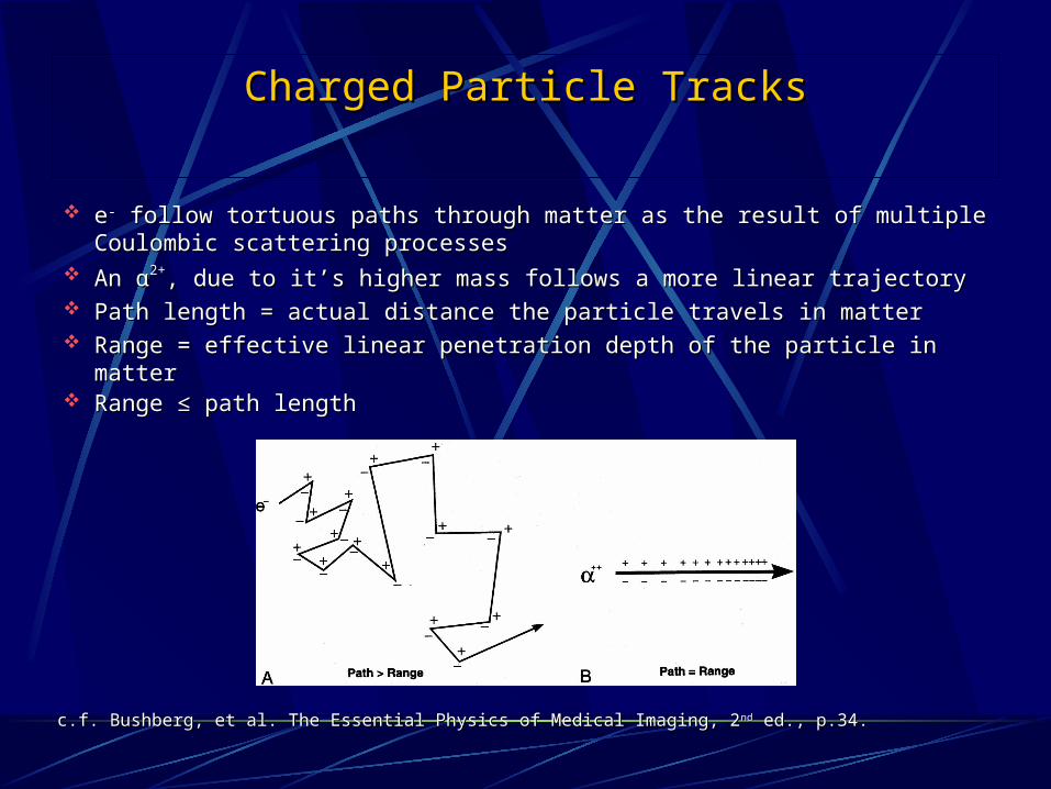
Charged Particle TracksCharged Particle Tracks
ee-- follow tortuous paths through matter as the result of multiple follow tortuous paths through matter as the result of multiple Coulombic scattering processes Coulombic scattering processes
An An αα2+2+, due to it’s higher mass follows a more linear trajectory, due to it’s higher mass follows a more linear trajectory Path length = actual distance the particle travels in matter Path length = actual distance the particle travels in matter Range = effective linear penetration depth of the particle in matter Range = effective linear penetration depth of the particle in matter Range ≤ path lengthRange ≤ path length
c.f. Bushberg, et al. The Essential Physics of Medical Imaging, 2c.f. Bushberg, et al. The Essential Physics of Medical Imaging, 2ndnd ed., p.34. ed., p.34.
The Atomic NucleusThe Atomic Nucleus
Covered in Nuclear Medicine course (August 2005) Covered in Nuclear Medicine course (August 2005) Composition of the Nucleus Composition of the Nucleus
Protons and Neutron Protons and Neutron Number of protons = Z Number of protons = Z Number of neutrons = N Number of neutrons = N Mass number = A = Z + N Mass number = A = Z + N Chemical symbol = X Chemical symbol = X Isotopes: same Z, but different A Isotopes: same Z, but different A Notation: Notation: AA
ZZXXNN, but , but AAX uniquely defines an isotope (also written X uniquely defines an isotope (also written
as X-A) as X → Z and N = A - Z as X-A) as X → Z and N = A - Z For example For example 131131I or I-131I or I-131
Linear Energy Transfer (LET)Linear Energy Transfer (LET)
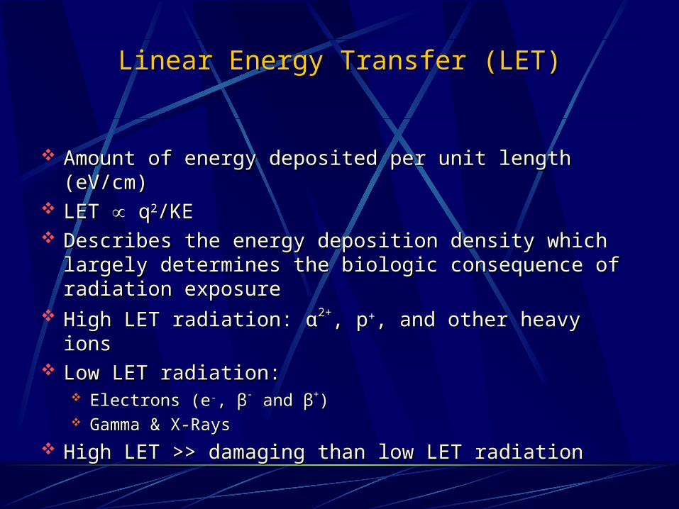
Amount of energy deposited per unit length (eV/cm) Amount of energy deposited per unit length (eV/cm) LET LET q q22/KE /KE Describes the energy deposition density which largely Describes the energy deposition density which largely
determines the biologic consequence of radiation determines the biologic consequence of radiation exposure exposure
High LET radiation: High LET radiation: αα2+2+, p, p++, and other heavy ions , and other heavy ions Low LET radiation: Low LET radiation:
Electrons (eElectrons (e--, , ββ-- and and ββ++) ) Gamma & X-RaysGamma & X-Rays
High LET >> damaging than low LET radiationHigh LET >> damaging than low LET radiation
Atomic Nucleus
Consists of Protons and Neutrons held together by the exchange of pions amongst the nucleons (strong forces).
The number of protons is the Atomic Number (Z).
The total number of protons and neutrons is the mass number (A).
Atomic Structure
Nuclei with the same number of protons, but different number of neutrons are said to be isotopes.Isotopes of an element have the same electron structure (same No. of Protons)and therefore the same chemical properties.Isotopes of an element may have very different nuclear properties. Stability is governed by N and Z ratios.
Radioactivity
Process by which an unstable nucleus decays by one or more discreet steps until a stable state is reached.
May occur in one step (e.g. 3H)
Or may require many steps. 238U heads a chain of 14 isotopes before stopping at stable 206Pb.
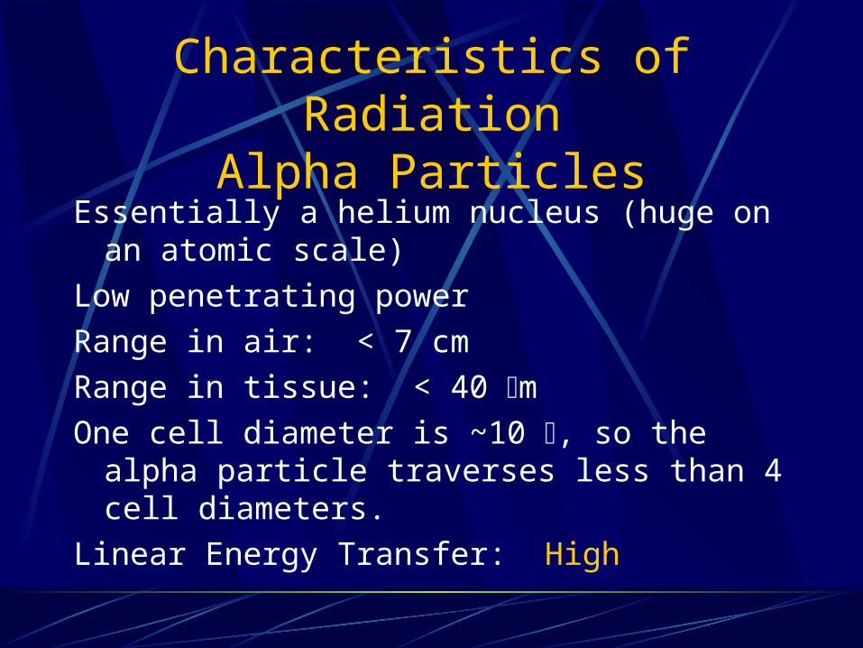
Characteristics of RadiationAlpha Particles
Essentially a helium nucleus (huge on an atomic scale)
Low penetrating power
Range in air: < 7 cm
Range in tissue: < 40 mOne cell diameter is ~10 , so the alpha particle
traverses less than 4 cell diameters.
Linear Energy Transfer: High

Radiation CharacteristicsBeta Particles
Electrons ejected from the nucleus of the radioactive atom.
Range in air: < 1 meter
Range in tissue: < 1 cm
LET: Low
They can be an external skin hazard and an internal hazard.
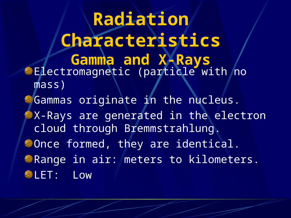
Radiation CharacteristicsGamma and X-Rays
Electromagnetic (particle with no mass)
Gammas originate in the nucleus.
X-Rays are generated in the electron cloud through Bremmstrahlung.
Once formed, they are identical.
Range in air: meters to kilometers.
LET: Low

Radiation CharacteristicsNeutrons
Only one neutron emitting isotope – Cf-252Range in air – meters to kilometers.Range in tissue – centimeters.High LET – note short range in tissue.Best shielding is hydrogenous materials such as polyethylene, wax, paraffin, and water.

Neutrons
Nuclear weapons run on neutrons.
Fissile materials (e.g. Pu-239, Uranium enriched in U-235) fission when struck by a neutron. Fission produces two smaller nuclei, 1 to 3 additional neutrons, and a release of a significant amount of energy.

Radiation Characteristics
Type Alpha Beta Gamma Neutron
External No <1 m Yes Yes
Internal Yes Yes Yes Cf-252
Skin No Yes Yes Cf-252
Pathway Yes Yes Yes Cf-252
LET High Low Low High
Shield Paper Plastic Lead Water
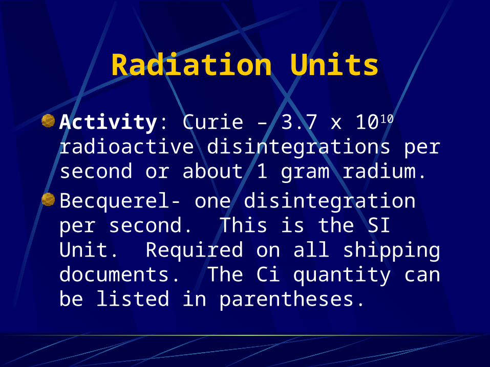
Radiation Units
Activity: Curie – 3.7 x 10¹⁰ radioactive disintegrations per second or about 1 gram radium.
Becquerel- one disintegration per second. This is the SI Unit. Required on all shipping documents. The Ci quantity can be listed in parentheses.
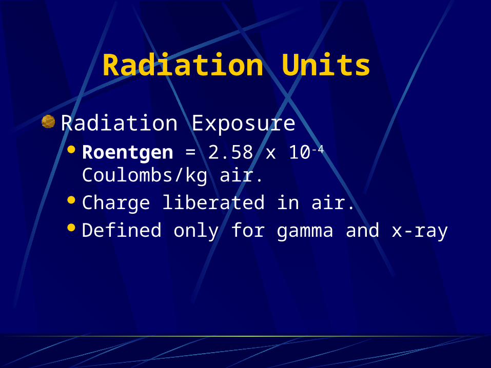
Radiation Units
Radiation Exposure Roentgen = 2.58 x 10⁻⁴ Coulombs/kg air.
Charge liberated in air.Defined only for gamma and x-ray

Radiation Units
Radiation Absorbed Dose rad = 0.01 Joules/kgEnergy Absorbed per gram.Defined for all radiations & all tissue
Radiation Units
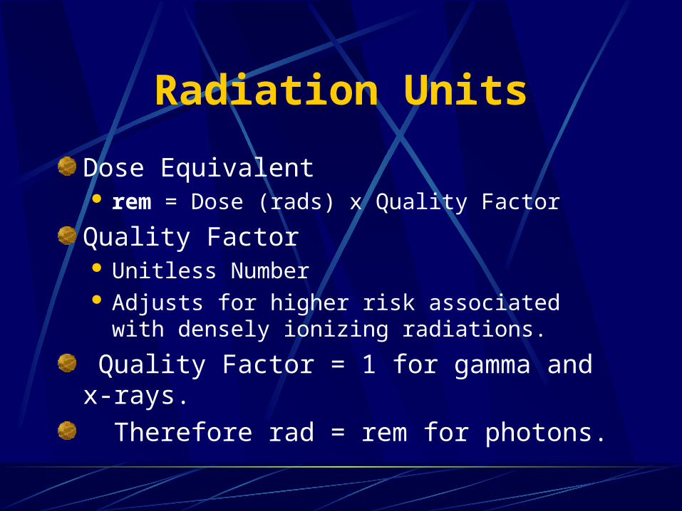
Dose Equivalent rem = Dose (rads) x Quality Factor
Quality Factor Unitless Number Adjusts for higher risk associated with densely
ionizing radiations.
Quality Factor = 1 for gamma and x-rays.
Therefore rad = rem for photons.
Radiation Units
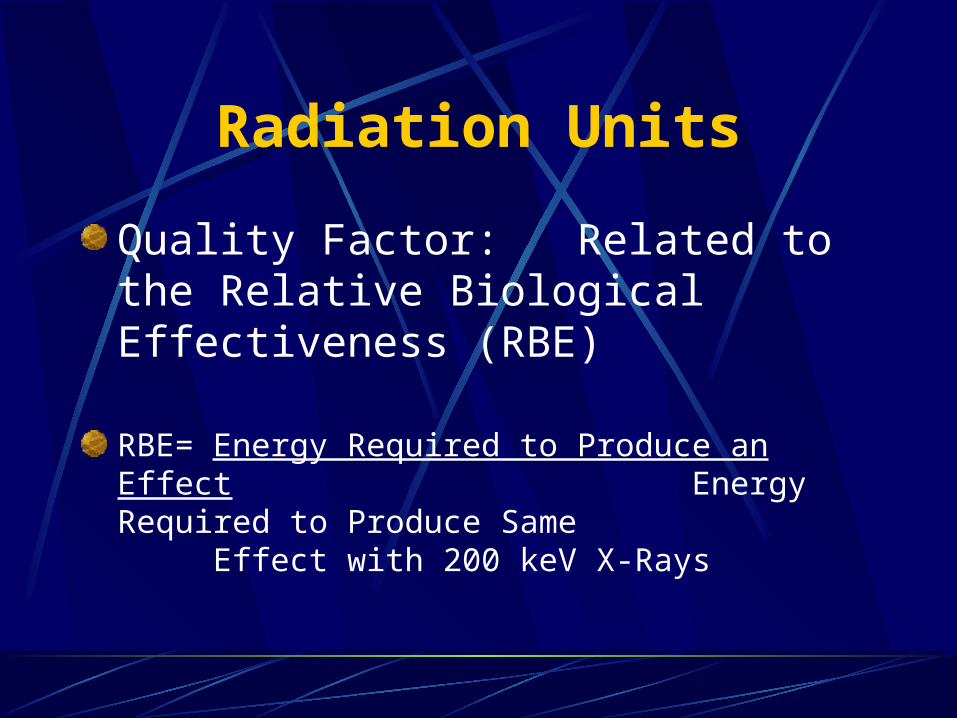
Quality Factor: Related to the Relative Biological Effectiveness (RBE)
RBE= Energy Required to Produce an Effect Energy Required to Produce
Same Effect with 200 keV X-Rays
Radiation Units
Relative Biological EffectivenessVaries with End Point and Tissue TypeQuality Factor is Average RBE
Quality Factors
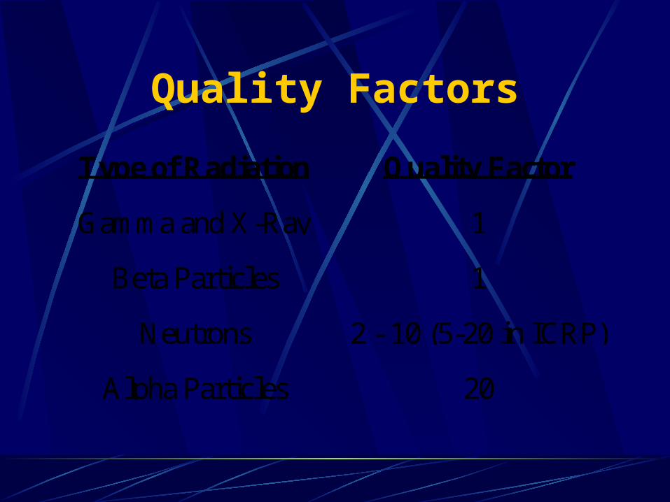
Type of Radiation Quality Factor
Gamma and X-Ray 1
Beta Particles 1
Neutrons 2 - 10 (5-20 in ICRP)
Alpha Particles 20
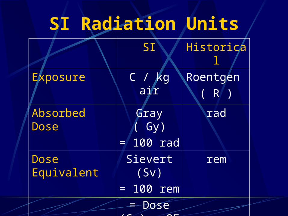
SI Radiation UnitsSI Historical
Exposure C / kg air Roentgen
( R )
Absorbed Dose Gray ( Gy)
= 100 rad
rad
Dose Equivalent
Sievert (Sv)
= 100 rem
= Dose (Gy) x QF
rem
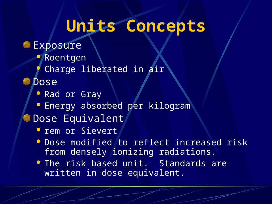
Units ConceptsExposure Roentgen Charge liberated in air
Dose Rad or Gray Energy absorbed per kilogram
Dose Equivalent rem or Sievert Dose modified to reflect increased risk from
densely ionizing radiations. The risk based unit. Standards are written in dose
equivalent.
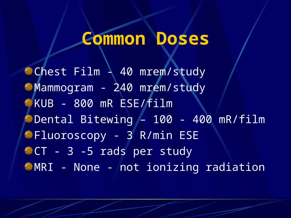
Common Doses
Chest Film - 40 mrem/study
Mammogram - 240 mrem/study
KUB - 800 mR ESE/film
Dental Bitewing – 100 - 400 mR/film
Fluoroscopy - 3 R/min ESE
CT - 3 -5 rads per study
MRI - None - not ionizing radiation

Annual Dose Equivalent360 mrem
11%
11%
8%
8%54%
1%3% 4%
X-Rays Internal Terrestrial Cosmic
Radon Other Consumer Nuc Med
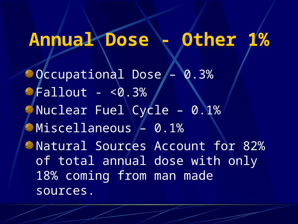
Annual Dose - Other 1%
Occupational Dose – 0.3%
Fallout - <0.3%
Nuclear Fuel Cycle – 0.1%
Miscellaneous – 0.1%
Natural Sources Account for 82% of total annual dose with only 18% coming from man made sources.