rady 401 case presentation: spontaneous perinephric hematoma
TRANSCRIPT
Megan Gurjar, MS4 8/17/20
RADY 401 Case Presentation:
Spontaneous Perinephric Hematoma

History of Present Illness
• 49 yo F presented 2 mo ago to OSH with L-sided flank pain, found to have 11cm L-sided renal hematoma on CT A/P; followed by local urologist outpatient
• Presented to UNC from OSH due to worsening flank pain, decreased appetite, 40lb weight loss, subjective low-grade fevers
Past Medical History
• T2DM, HTN, recurrent UTI, atrophic right renal unit, and right duplex collecting system s/p open ureteroureterostomy (1984)
Vitals
HR 103 | BP 136/79 | Temp 36.9 °C | SpO2 97%
Pertinent Physical Exam
Cardiac: Mild tachycardia.
Abdomen: Soft, nontender, nondistended.
GU: Left CVA tenderness.
Pertinent Labs
WBC 11.7
H&H 9.9/31.4
Cr 0.66
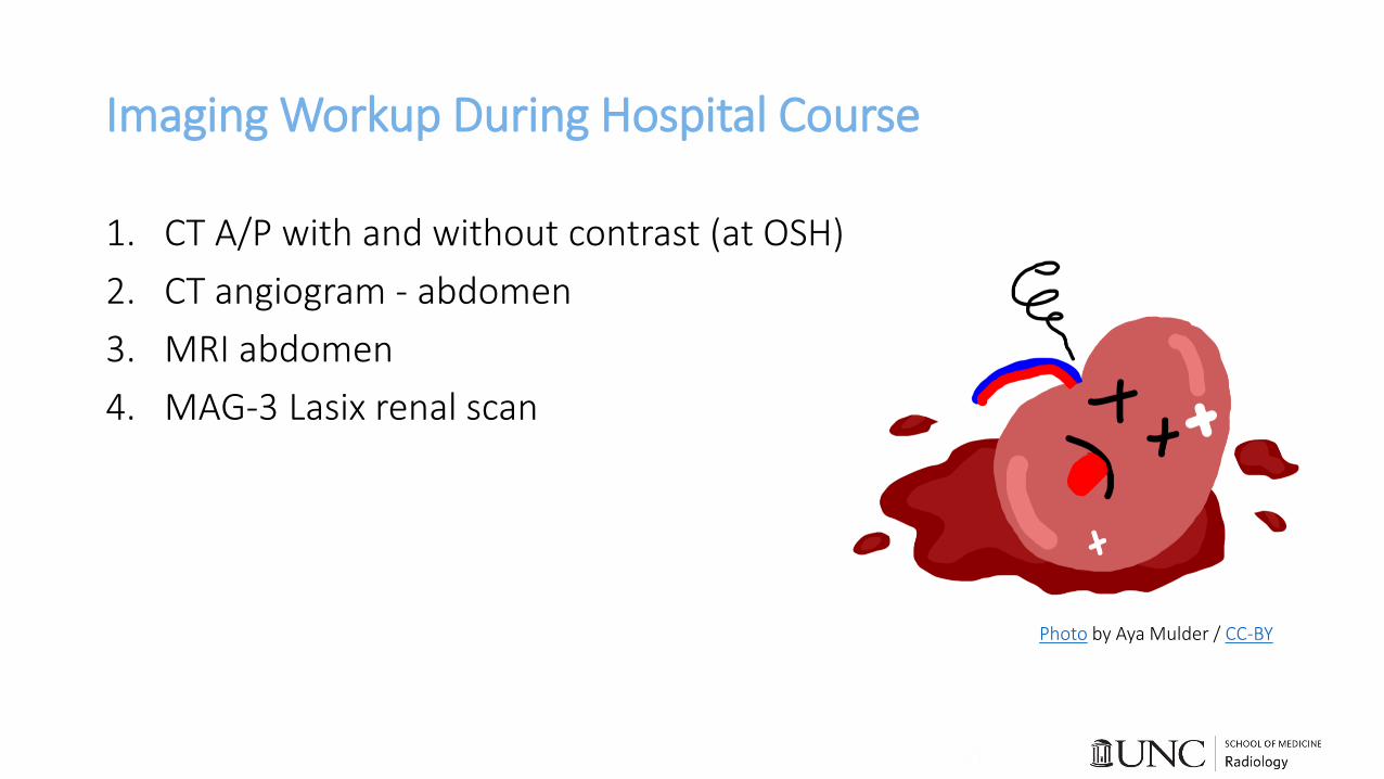
Imaging Workup During Hospital Course
1. CT A/P with and without contrast (at OSH)
2. CT angiogram - abdomen
3. MRI abdomen
4. MAG-3 Lasix renal scan
Photo by Aya Mulder / CC-BY

Initial CT A/P
• Left perinephric hematoma 15.3cm of unknown etiology

CTA Abdomen – 2 days later
• Interval enlargement of fluid collection to 15.7cm
• No foci of active extravasation

MRI Abdomen
• Following CT findings, IR consulted for evaluation and drainage; requested MRI for further characterization
• T1 hypointense, T2 hyperintense heterogeneous mass with enhancing internal septations
• Concern for possible cystic malignancy; features less typical of subcapsular/perinephric hematoma

Lasix Renal Scan
• Reason for exam:
history of atrophic R
renal unit and
consideration surgical
intervention for L
kidney
• Split function
• 77.3% R kidney
• 22.7% L kidney
Patient Outcome
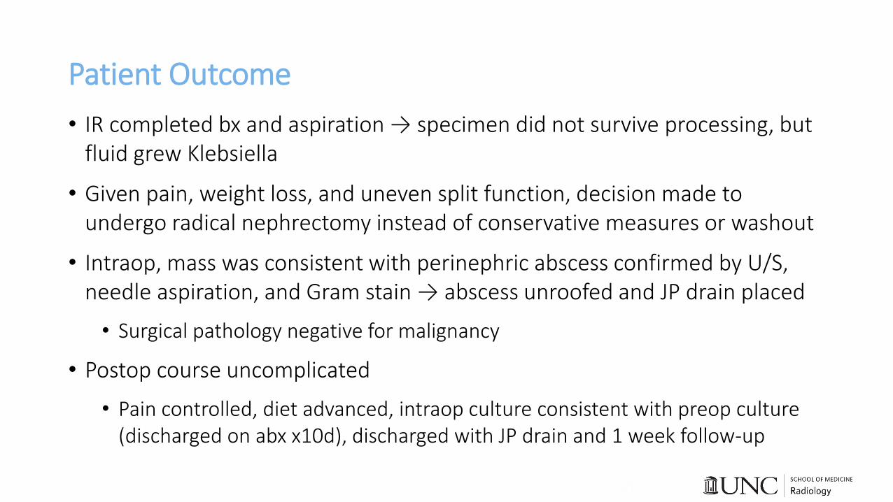
• IR completed bx and aspiration → specimen did not survive processing, but fluid grew Klebsiella
• Given pain, weight loss, and uneven split function, decision made to undergo radical nephrectomy instead of conservative measures or washout
• Intraop, mass was consistent with perinephric abscess confirmed by U/S, needle aspiration, and Gram stain → abscess unroofed and JP drain placed
• Surgical pathology negative for malignancy
• Postop course uncomplicated
• Pain controlled, diet advanced, intraop culture consistent with preop culture (discharged on abx x10d), discharged with JP drain and 1 week follow-up
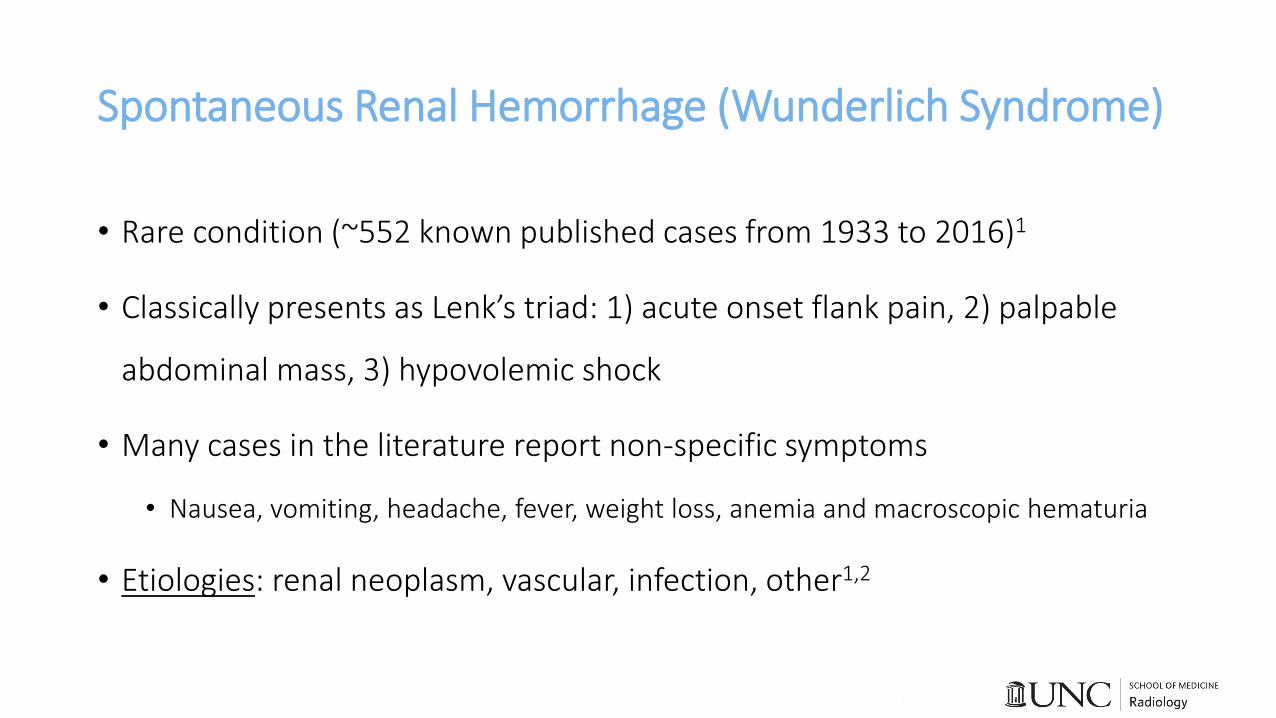
Spontaneous Renal Hemorrhage (Wunderlich Syndrome)
• Rare condition (~552 known published cases from 1933 to 2016)1
• Classically presents as Lenk’s triad: 1) acute onset flank pain, 2) palpable
abdominal mass, 3) hypovolemic shock
• Many cases in the literature report non-specific symptoms
• Nausea, vomiting, headache, fever, weight loss, anemia and macroscopic hematuria
• Etiologies: renal neoplasm, vascular, infection, other1,2

Preferred Imaging of Renal Hematoma
• Ultrasound found to have 56% sensitivity for identifying spontaneous
hemorrhage compared to CT with 100% sensitivity2
• Etiology correctly identified with sensitivity and specificity of 11% and 33% for
ultrasound and 57% and 82% for CT, respectively
• CT angiography recommended if initial CT scan fails to reveal the underlying
cause, especially given likelihood of vascular etiology3
• MRI may have advantage over CT for diagnosing small tumors4

Classic CT Findings of Perinephric Hematoma
Video scan clip case courtesy of Dr Mohammad TaghiNiknejad, Radiopaedia.org, rID: 20509
https://radiopaedia.org/cases/renal-haematoma-in-a-patient-with-angiomyolipoma-wunderlich-syndrome?lang=us

Imaging Costs & Relevant Radiation Concerns
1. CT A/P with and without contrast at OSH
• $3208 at UNC5
• ~20 mSv (7 years of background radiation)6
2. CT angiogram abdomen
• $3,322 at UNC5
• ~10 mSv (3 years of background radiation)6
3. MRI abdomen
• $3,520 at UNC5
4. MAG-3 Lasix renal scan
• $2,984 at UNC5
Photo by George Hodan / CC0 1.0

Management of Perinephric Hematoma
Hemodynamically unstable7
Trans-arterial angioembolization
Radical nephrectomy
Partial nephrectomy
Exploration/washout
Hemodynamically stable7
Conservative management with fluids, transfusion, and observation

Top 3 Take Home Points
• CT is the most appropriate imaging tool for identifying spontaneous renal hemorrhage and associated etiology.
• Interventional radiology’s expertise is highly valued in management of patients with suspected renal bleeds.
• Imaging may not give the whole story.Photo by Megan Gurjar

References1. Ahn T, Roberts MJ, Navaratnam A, Chung E, Wood S. Changing etiology and management
patterns for spontaneous renal hemorrhage: a systematic review of contemporary series. International Urology and Nephrology. 2017 Nov 1;49(11):1897-905.
2. Zhang JQ, Fielding JR, Zou KH. Etiology of spontaneous perirenal hemorrhage: a meta-analysis. J Urol. 2002;167(4):1593-1596.
3. Brkovic D, Moehring K, Doersam J, et al. Aetiology, diagnosis and management of spontaneous perirenal haematomas. Eur Urol. 1996;29(3):302-307.
4. Kendall AR, Senay BA, Coll ME. Spontaneous subcapsular renal hematoma: diagnosis and management. J Urol. 1988;139(2):246-250.
5. UNC Chargemaster. https://www.unchealthcare.org/about-us/billing-and-financial-assistance/chargemaster/
6. American College of Radiology. Radiation Dose to Adults From Common Imaging Examinations.
7. Baishya RK, Dhawan DR, Sabnis RB, Desai MR. Spontaneous subcapsular renal hematoma: a case report and review of literature. Urology annals. 2011 Jan;3(1):44.