rady 413 case presentation
TRANSCRIPT

RADY 413 Case Presentation
Edited by John Lilly, MD

54-year-old man presents with right axillary lymphadenopathy

54-year-old man with no significant past medical history presents to his primary care physician for 6 months of daily fevers, night sweats, and a 50-pound unintentional weight loss. He has also noted right axillary swelling that has increased over the past few months. He has had no recent international travel or exposure to sick individuals. Physical exam is notable for a >5 cm fixed mass in the right axilla with irregular borders. The mass is not tender to palpation, there is no associated skin discoloration, and no masses are palpated in the left axilla.
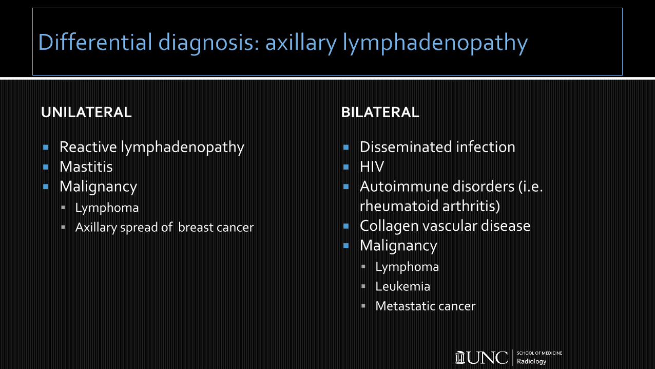
UNILATERAL
Reactive lymphadenopathy Mastitis Malignancy
Lymphoma
Axillary spread of breast cancer
BILATERAL
Disseminated infection HIV Autoimmune disorders (i.e.
rheumatoid arthritis) Collagen vascular disease Malignancy
Lymphoma
Leukemia
Metastatic cancer

Targeted ultrasound of bilateral axilla Bilateral diagnostic mammogram
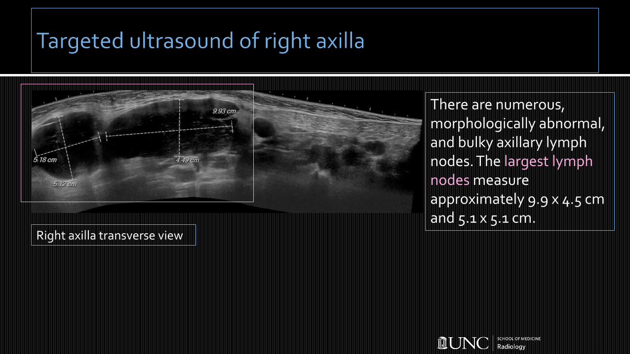
There are numerous, morphologically abnormal, and bulky axillary lymph nodes. The largest lymph nodes measure approximately 9.9 x 4.5 cm and 5.1 x 5.1 cm.
Right axilla transverse view

Due to the presence of bilateral enlarged lymph nodes, a bilateral diagnostic mammogram was performed.
Targeted ultrasound of the left axilla demonstrates a mildly prominent left axillary lymph node.
Left axilla longitudinal view
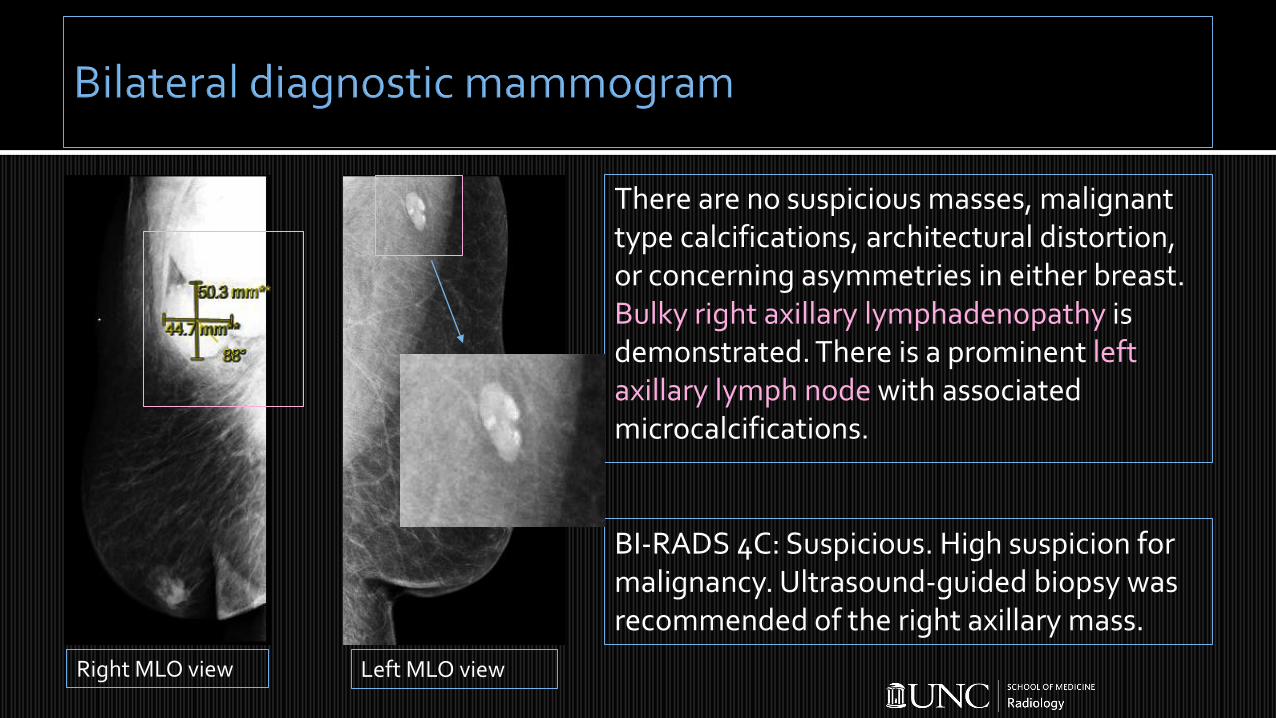
BI-RADS 4C: Suspicious. High suspicion for malignancy. Ultrasound-guided biopsy was recommended of the right axillary mass.
There are no suspicious masses, malignant type calcifications, architectural distortion, or concerning asymmetries in either breast. Bulky right axillary lymphadenopathy is demonstrated. There is a prominent left axillary lymph node with associated microcalcifications.
Right MLO view Left MLO view

Core needle biopsy of the axillary mass was performed with ultrasound guidance, aseptic technique, and 1% lidocaine as the local anesthetic. Three core samples were obtained with a 14-gauge Achieve biopsy device. The samples were placed in formalin and a hydromark biopsy marking clip was placed at the biopsy site.
needlepost fire
needlepre-fire
clippost bx

Hematopathology: Classic Hodgkin lymphoma Staging:
B symptoms (i.e. fever, night sweats, weight loss)
Further imaging: CXR, CT chest/abdomen/pelvis, PET scan, +/- bone marrow aspiration and biopsy
Follow-up with hematology/oncology 5-year survival for all patients diagnosed with HL ~86%
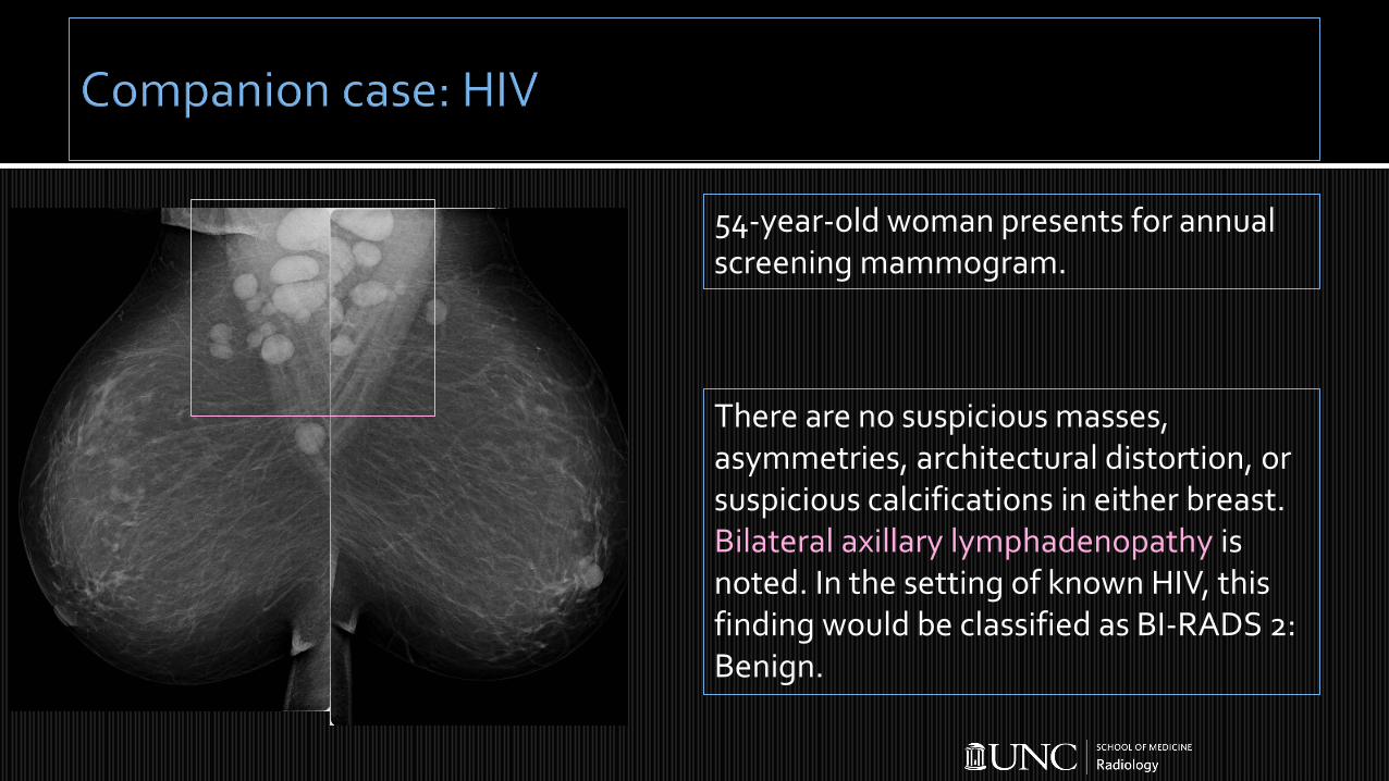
54-year-old woman presents for annual screening mammogram.
There are no suspicious masses, asymmetries, architectural distortion, or suspicious calcifications in either breast. Bilateral axillary lymphadenopathy is noted. In the setting of known HIV, this finding would be classified as BI-RADS 2: Benign.

52-year-old female undergoes screening mammogram and is found to have bilateral axillary lymphadenopathy, which is re-demonstrated on targeted ultrasound of bilateral axilla (left axillary ultrasound image shown).
Review of her history reveals diagnosis of sarcoidosis.

67-year-old female undergoes screening mammogram and is found to have bilateral axillary lymphadenopathy, for which she is called back for diagnostic workup confirming lymphadenopathy of unknown etiology.
She is found to have chronic lymphocytic leukemia (CLL) on core needle biopsy.

UNILATERAL
Reactive lymphadenopathy Mastitis Malignancy
Lymphoma
Axillary spread of breast cancer
BILATERAL
Disseminated infection HIV Autoimmune disorders (i.e.
rheumatoid arthritis) Collagen vascular disease Malignancy
Lymphoma
Leukemia
Metastatic cancer
Initial laboratory evaluation of axillary lymphadenopathy should include CBC with differential, CMP, HIV Ag/Ab, LDH, uric acid
The axillary lymph nodes are involved in up to 50% of patients with symptomatic breast cancer and in 10-20% of patients with breast cancers identified by screening
Axillary lymphadenopathy can also be a benign finding on screening mammography associated with many systemic diseases
1. Ikeda DM and Miyake KK. (2017). Clinical breast problems and unusual breast conditions. In Breast imaging: The requisites (pp. 397-438). Retrieved from https://www-clinicalkey-com.
2. Hacker NF and Friedlander ML. (2016). Breast disease: a gynecologic perspective. In Hacker & Moore’s Essentials of Obstetrics and Gynecology (pp. 348-355). Retrieved from https://www-clinicalkey-com.
3. American Cancer Society. (2019 Jan 8). Key statistics for Hodgkin lymphoma. Retrieved from https://www.cancer.org/cancer/hodgkin-lymphoma/about/key-statistics.html.