rapidthin-layerchromatographicmethodforassessing ... · 1370 clinicalchemistry. vol.20,no.10,1974...
TRANSCRIPT
1368 CLINICALCHEMISTRY, Vol. 20, No. 10, 1974
Rapid Thin-Layer Chromatographic Method for Assessingthe Lecithin/Sphingomyelin Ratio in Amniotic Fluid
Submitters: Emily H. Coch, Gerald Kessler, and John S.Meyer, Department of Pathology and Lab-oratory Medicine, The Jewish Hospital ofSt. Louis, St. Louis, Mo. 63110
Evaluators: Paul T. Russell, Department of Obstetricsand Gynecology, University of CincinnatiMedical Center, Cincinnati, Ohio 54229
Donald T. Forman, Division of Clinical Bio-chemistry, Evanston Hospital, Evanston,
Ill. 60201
Laszlo Sarkozi and Hahn N. Kovacs, MountSinai School of Medicine, New York, N. Y.10029
Introduction
The method described here is essentially that of
Gluck (1), for assessing the lecithin/sphingomyelin(L/S) ratio in amniotic fluid. It is modified to de-crease assay time and allow easier estimation of theL/S ratio in laboratories that do not possess densito-
metric scanning equipment.The ratio is a useful and practical index of fetal
pulmonary maturity (1-6). Lecithin is a principalcomponent of pulmonary surfactant, a material that
coats the air sacs of the lungs and lowers surface ten-sion, thereby allowing the lungs to maintain suffi-cient residual air volume upon expiration to prevent
alveolar collapse. Synthesis of this pulmonary surfac-tant by the fetus first becomes significant (and it firstappears in amniotic fluid) sometime during the 32nd
to 36th week of gestation. Infants born after this timeusually will have no difficulty with breathing. If preg-
nancy is terminated prior to this time, however, thereis a high risk of development of the respiratory dis-tress syndrome, which involves the collapse of pul-monary alveoli despite intense inspiratory effort andpossible formation of hyaline membranes (1).
The lecithins involved are mainly dipalmitoyl leci-thin and a-palmitoyl, 3-myristy1 lecithin, but therapid increase in surface-active lecithin observed be-
tween 32-36 weeks of gestation is mainly an increasein dipalmitoyl lecithin (1). The changing concentra-tions of lecithin in the amniotic fluid are measuredand are most conveniently expressed as a ratio of lec-ithin to another phospholipid, sphingomyelin, whichis present in much more nearly constant concentra-
tions [until about the 35th week of gestation, when itgradually decreases (1)].
Gluck et al. (1 ) described a method for extractinglecithin and sphingoymelin from amniotic fluid, con-centrating the “surface-active” lecithin and sphin-gomyelin by cold acetone precipitation, separatingthe two phospholipids by thin-layer chromatographyon glass plates coated with silica gel, and then mak-
ing them visible by an acid-spray, heat-char tech-
nique (1 ). They have demonstrated, by densitometric
quantitation of the lecithin and sphingomyelin inmore than 300 amniotic fluid specimens, that the L/S
ratio is 1 or less before 32 weeks gestation, and thatthe ratio increases between the 32nd and 36th week,corresponding to the sharp surge in fetal synthesis of
pulmonary surfactant. By this procedure, an L/Sratio of 2 or greater indicates fetal pulmonary matu-rity, with minimal risk of respiratory distress syn-drome. These criteria have been corroborated in
other laboratories (3, 4, 6).In the procedure we describe here, the phospholip-
id fraction is directly extracted and concentrated, butno precipitation with cold acetone is used and thetwo phospholipids are separated more quickly by use
of a different thin-layer chromatographic supportmedium. The visualization technique described here
CLINICAL CHEMISTRY, Vol. 20. No. 10, 1974 1369
yields higher values for the L/S ratio for specimensfrom immature and mature subjects than doesGluck’s procedure, does not require a densitometer,and it differentiates those lecithins peculiar to am-niotic fluid from those peculiar to blood (and thuscan indicate when there is significant contaminationof amniotic fluid with blood).
PrinciplePhospholipids are extracted from amniotic fluid
into chloroform and concentrated by evaporation. Inthe concentrated extract, lecithin is separated fromsphingomyelin by thin-layer chromatography onglass-fiber sheets impregnated with silica gel. Phos-pholipid standards are run concurrently. After chro-
matography, the phospholipids are made visible bythe independent use of two different reagents. Onereagent, containing bismuth subnitrate, is relatively
specific for lecithin and sphingomyelin, because itproduces an orange color with compounds containinga choline residue. Thus dipalmitoyl lecithin and
sphingomyelin appear as bright yellow-orange spots(7). In the second technique, the developed chro-matogram is exposed to iodine vapor, whereupon allphospholipids appear as yellow to brown spots (8).
The latter procedure is less specific but is aboutthreefold as sensitive as the former. It is nondestruc-tive; that is to say, the part of the chromatogram ex-
posed to iodine vapor can also later be used for otherdetection methods, if desired.
The size of the lecithin spot is compared visually to
that of the sphingomyelin spot (or the amounts canbe semi-quantitated by measuring the areas with agrid). The ratios are reported as whole numbers andare interpreted as described below.
MaterialsReagents
Methanol (“Spectranalyzed”; Fisher Scientific Co.,Philadelphia, Pa. 15219).
Ammonium hydroxide, reagent grade (Fisher Sci-
entific Co.)Chloroform (“Baker analyzed”; J. T. Baker Chemi-
cal Co., Phillipsburg, N. J. 08865).Bismuth subnitrate, powder (Matheson, Coleman
and Bell, Norwood, Ohio 45212).Potassium iodide, certified A.C.S. (Fisher Scientif-
ic Co.).Iodine crystals, U.S.P. resublimed (Fisher Scien-
tific Co.).Bismuth spray reagent:
Solution A: 3.48 mol/liter acetic acid. Dilute 200 mlof glacial acetic acid (17.4 mol/liter) to one liter withdistilled water.
Solution B: 1.7 g bismuth subnitrate in 100 ml ofsolution A. Store in a brown bottle. The compoundrequires about 3-4 h to dissolve.
Solution C: 40 g of KI in 100 ml of water (this mustbe stored in a brown bottle). If the solution is not col-orless, discard it and prepare a fresh solution.
Final working spray (to be stored in a brown bot-tie):
70 ml
20 ml
5 ml
solution A
solution B
solution C
Phospholipid stock standard (General Biochemi-
cals, Chagrin Falls, Ohio 44022). L-a-Lecithin (dipal-mitoyl, synthetic), sphingomyelin, phosphatidyletha-nolamine, and lysolecithin. Store at -20 #{176}C.
Thin-layer chromatography working standards:
Store at -20 #{176}C.Mix No. 1: lysolecithin and phosphatidylethanola-
mine, each at 2.0 glliter of chloroform.
Mix No. 2: L/S ratio, 2:1; lecithin and sphingomye-un at 4.0 g/liter and 2.0 g/liter of chloroform, respec-
tively.Mix No. 3: L/S ratio, 5:1; lecithin and sphingomye-
tin at 10.0 and 2.0 g/Iiter of chloroform, respectively.
Apparatus
Thin-layer chromatography chamber: Chromatog-raphy Kit (Eastman Kodak Co., Rochester, N. Y.14650).
Thin-layer chromatography sheets: 20 X 20 cm
silica gel impregnated glass fiber sheets (ITLC-SG)(Gelman Instrument Co., Ann Arbor, Mich. 48106).
Gelman spotting guide (Geiman Instrument Co.).“ Yankee Disposable Micropets, “ 5 tl (Clay-
Adams, Inc., New York, N. Y. 10010).Extraction tubes, Pyrex, 25 X 150 mm, with Tef-
lon-lined screw tops.Chromatography spraying vessel (Scientific Prod-
ucts, St. Louis, Mo. 63043).Evaporating dish, Pyrex, 80 X 45 mm.
Collection and Handlingof Specimens
When received, amniotic fluid is centrifuged for 10mm at 800-1100 X g (2300 rpm in a Sorval GLC-1centrifuge with Type HL4 head or at about 1600 rpmin an International Model V with Type 240 head).
Specimens are not assayed if the specimen appearsvisibly bloody, with a hematocrit greater than 1.5%
(see below), or if meconium is present, because otherforms of lecithin are present in these contaminants(1, 2, 5, 13, 16, 17).
Notg: Centrifugation of material for 10 mm increases thetemperature of the material by less than 5 “C aboveambient.
The supernatant fluid is removed by pipet andused immediately for the L/S assay and any otherchemical determinations (25). The sedimented pelletmay be used for staining with Nile Blue Sulfate (9,25). If the L/S assay cannot be performed the sameday, the supernatant material is frozen at -20 #{176}C.Ifthe assay is only delayed for 1-2 h, the supernate isrefrigerated.
1370 CLINICAL CHEMISTRY. Vol. 20, No. 10, 1974
ProcedureMix 5 ml of the amniotic fluid supernate with 5 ml
of methanol in one of the extraction tubes. When lessmaterial is available, 3 or 4 ml can be assayed, but all
reagent volumes are reduced proportionately. For themost meaningful interpretations, however, 5 ml
should be extracted.Add 10 ml of chloroform, cap tightly, and shake for
5 mm to extract. Centrifuge at 600 X g (approximate-ly 1700 rpm with the International Model V withtype 240 head) for 5 mm.
Remove and discard the aqueous (upper) layer.Filter the lower (chloroform) phase through What-man No. 1 paper into an evaporating dish. Alterna-tively, filter through Whatman No. 1 PS (silicone-
treated phase-separating 1 or through anhy-drous magnesium sulfate (6) to remove the maximumamount of water before the evaporation.
Concentrate the material to about 0.5 ml by usinga gentle stream of clean air flowing over the solventsurface. (The material may be allowed to go to dry-ness, but exposure to room temperature should belimited to 5-10 mm.) This evaporation step requires20 to 30 mm.
Note: Some workers prefer to use a stream of nitrogenrather than air (1, 2, 4) or to use a rotary-type evaporator(at low pressure and elevated temperature) (6). These al-ternatives may be used if the temperature is not allowedto rise above 60 #{176}C.Use of a stream of air as describedabove keeps the temperature of the evaporating chioro-form extract below 4 #{176}C,and there appears to be no Se-lective loss of phospholipid over the time period as as-sessed by relative recoveries of standard L/S mixes car-ned through this procedure.
Rinse the dish with 1 to 2 ml of chloroform and
transfer the rinsings to a 13 X 100 mm tube. Further
evaporate to about 50 l (i.e., 1-2 drops).The concentrated extract can be stored at -20 #{176}C
at this point if the chromatography cannot be donethe same day.
Note: If it is desired to include a precipitation step withcold acetone, as recommended by Gluck (1 ), evaporatethe chloroform extract to 0.1-0.2 ml in a conical-tipglass-stoppered centrifuge tube. Add 10 ml of ice-coldacetone dropwise with gentle mixing of tube. Stopper thetube and chill it at -20 “C for 1 h. Centrifuge at 600 X gfor 5 mm (do not allow the temperature of material intube to exceed 8 #{176}C).Immediately and carefully draw offthe supernate, which can be concentrated by evaporationfor thin-layer chromatographic analysis. The precipitat-ed material is redissolved in 2 ml of chloroform and con-centrated by evaporation to about 50 l for thin-layerchromatographic analysis. We do not find this precipita-tion necessary, as will be discussed below.
Place a 20 X 20 cm chromatographic sheet on the
Gelman spotting guide and mark off 11 points. Thesepoints are approximately 2 cm apart and 2.5 cm fromthe bottom edge of sheet.
1 Personal communication, Evaluator P. T. R.
At points 1 and 11, apply 5 sl of standard mix No.1 (lysolecithin and phosphatidylethanolamine, 1:1).At points 3 and 7, apply 5 I of mix No. 2 (L/S = 2).
At points 5 and 9, apply 5 l of mix No. 3 (L/S 5).There is sufficient space on the sheet for duplicate
applications of samples from two patients, or of theacetone precipitate and acetone supernate of thesame patient, if desired. Divide one sample equallybetween points 2 and 8 and a second sample betweenpoints 4 and 10. Leave point No. 6 empty. Apply 5 lat a time to ensure spot diameters of less than 4 mm.
Use the entire extract.Prepare the solvent freshly each time: 170 ml of
chloroform, 20 ml of absolute methanol, and 3 ml ofconcentrated ammonium hydroxide.
Assemble the Eastman Kodak chromatography
easel and trough. Pour the solvent into the trough upto the etched line. Place the thin-layer chromatogra-phy sheet into the Eastman Kodak chromatographychamber, clip the sides, and set the chamber into thetrough. Allow development to proceed for 15 mm.
Remove the sheet and allow it to air dry for 5 mm.Carefully divide lengthwise along the center (point
No. 6).Place one half of the sheet into a closed chamber
containing iodine crystals for at least 5 mm. Yellowto brown spots will be observed. Remove the sheetand immediately circle the spots lightly with a pencil.(If the spots should fade too quickly, they will appearagain upon re-exposure to iodine vapor.)
Spray the other half of the sheet thoroughly withthe bismuth spray until the standards appear aswhitish spots against an orange background, andallow it to air dry for several minutes.
Place the bismuth-stained sheet in a pan contain-ing solution A (3.48 mol/liter acetic acid) and swirl
the pan gently until the background is again white.The lecithin and sphingomyelin standards will ap-
pear as yellow-orange spots. Remove the sheet fromthe pan and circle the spots lightly in pencil. Theyare better seen by holding the sheet up to a light. Ob-servations should be made as soon as possible after
destaining, because the background color gradually(within 15 mm) reappears.
Visually compare the size of lecithin and sphin-
gomyelin spots, and report the results as listed under“Interpretation.”
When the sheets are dry, wrap completely in cellu-lose sheeting (“Saran Wrap”) and tape to a sheet
containing all pertinent information.After the iodine-stained spots have been allowed to
fade, the iodine-stained half of the sheet may be used
for several other types of visualization techniques, in-cluding 8-anilino-1-naphthalene sulfonic acid (ANS),phosphomolybdic acid-stannous chloride, or acid
spray and heat char as described below.
Other techniques that may be used to make thespots visible are:
(a) ANS: Spray the developed and dried chromato-
gram with a 0.25 g/liter aqueous solution of ANS
CLINICAL CHEMISTRY, Vol. 20, No. 10, 1974 1371
(Sigma) . When viewed under ultraviolet light, phos-pholipids appear as bright-blue fluorescent spots (10).
(b) Rhodamine: Spray the dried chromatogram
with Rhodamine B spray (Gelman) and view underultraviolet light at 30-mm intervals. (After 12 stainingit is preferable to wait 12 h before this spraying.) Lip-ids appear as bright-orange spots against a pinkbackground (8).
(c) Acid spray or dip, heat char: Spray the chro-
matogram either with 9 mol/liter H2S04 (1) or withaqueous ammonium sulfate (300 g/liter) acidifiedwith 12 ml of concentrated sulfuric acid per deciliter(6). Heat for 5 mm over a hot plate set on high heat(do not allow sheet to touch surface of plate). An at-
ternative spray consists of phosphoric acid (18 g/dl)containing cupric sulfate pentahydrate (8 g/dl),which reportedly gives charred spots within 15 mm ata lower temperature (100 #{176}C)(Evaluator D. T. F.).
(d) Phosphomolybdic acid-stannous chloride: Dis-solve 1 g of phosphomolybdic acid in 50 ml of chloro-form and 50 ml of methanol. Spray the sheet thor-oughly. Overspray with a solution containing 1 g ofstannous chloride per deciliter of dilute (2 mol/liter)HC1. Heat the sheet over a hot plate until blue spotsappear.
Note: The following improvement has been reported foruse with thin-layer chromatography plates (22). It hasnot been tried by the Submitters on Gelman ITLCsheets. The following solutions are needed: Solution A. 4g of molybdenum trioxide is added to enough 25 mol!liter H2S04 to make a final volume of 1 dl. Boil the mix-ture gently until all trioxide salt is dissolved. Solution B.Add 175 mg of powdered molybdenum to about 50 ml of
Solution A. Boil gently for 15 mm and cool. Carefullymix solutions A and B and dilute to 1 dl with distilledwater. This reagent is stable at room temperature for atleast six months in an amber-colored bottle. It reported-ly can be sprayed on thin-layer chromatography platesand blue spots will develop at room temperature imme-diately.
Interpretation
Immature (high risk of respiratory distress syn-
drome): L/S ratio, 2 or less.Lecithin and sphingomyelin appear as faint traces
or as approximately equal but small spots by bismuthstain. By iodine stain, L/S ratio is 2 or less.
Transitional (some risk of respiratory distress
syndrome but survival probable): L/S ratio between2 and 5.
Definite but small lecithin spot and only a trace orno sphingomyelin by bismuth (size of lecithin usuallyless than that in the 2:1 standard). By iodine stain-ing, the ratio of spot sizes is greater than 2 but lessthan 5.
Mature (minimal risk of respiratory distress syn-drome): L/S ratio, 5 or higher.
Definite lecithin and no sphingomyelin by bismuth(size of lecithin usually greater than the lecithin spotof the 2:1 standard). By iodine staining, the ratio is 5or greater. (No sphingomyelin may be seen, even withiodine staining, in many cases.)
DiscussionThis method includes several modifications of
Gluck’s original assay (1 ), all designed to reduce
assay time and facilitate interpretation in the ab-sence of densitometric scanning equipment.
Use of glass-fiber sheets impregnated with silica
gel rather than coated glass plates or Mylar sheetsdecreases solvent development time from at least 1 hto only 15 mm and makes it easier to spray and thendestain by immersion. The Eastman Kodak chroma-tography chamber, with which the sandwich platetechnique is used, accommodates the Gelman ITLC
sheets easily and obviates the need to pre-equilibratethe chamber with solvent. The sheets are easy to di-vide for different visualization techniques after sol-vent development and easy to store. We observeclean separation of lecithin from sphingomyelin andclean separation of both from other phospholipidsthat might be present in amniotic fluid (11). Repre-sensitive R F values are: phosphatidyl ethanolamine
0.72; lecithin, 0.67; sphingomyelin 0.48; lysolecithin,0.32; phosphatidyl serine, 0.30; and phosphatidyli-
nositide (or phosphatylinositol), 0.23. (The latterthree standards were obtained from General Bio-chemicals Div., Mogul Corp., Chagrin Falls, Ohio44022; Cat. No. 100130, 100120, and 800120, respec-tively.) Both cholesterol and triglyceride (triolein)migrate with or just behind the solvent front in thissystem. These lipids are present in the “surface-ac-
tive” fraction of rabbit lung washings (23).
Evaluation of the L/S ratio is based on the use of areagent that makes visible both dipalmitoyl lecithinand sphingomyelin as bright yellow-orange spotsagainst a white background. This modified Dragen-
dorff stain (7), containing bismuth subnitrate andpotassium iodide, reacts with compounds possessinga choline residue. Because sphingomyelin and leci-
thin each contain one choline moiety per mole andare of similar molecular weight, equal concentrationswill display spots of similar size and color intensity.Lysolecithin is also made visible, but the spot is adarker shade of orange and is well resolved from leci-thin and sphingomyelin; in our experience, it is onlyseen in amniotic fluids from term or near-term in-fants (such fluids contain large amounts of lecithin)
or in specimens contaminated with blood. The bis-muth subnitrate spray can detect sphingomyelin at aconcentration of 1 mg/dl (dipalmitoyl lecithin stainsslightly more than does sphingomyelin with this
stain, and therefore its detection limits are a littlelower). From analyses of 103 amniotic fluids, we con-
dude that the concentration of sphingomyelin in am-niotic fluid is usually less than 2 mg/dl when corn-pared with extracted L/S standard mixes of knownconcentration.
Most specimens from mature fetuses do not show
any sphingomyelin by this stain, and many speci-mens from immature fetuses show only the faintesttraces of both lecithin and sphingornyelin. Thereforean alternative, more sensitive stain is also needed
1372 CLINICAL CHEMISTRY, Vol. 20, No. 10, 1974
that will always make visible the lecithin and sphin-gomyelin in early amniotic fluid specimens, i.e., that
can detect concentrations of 0.5 mg/dl or less. Iodinevapor, although less specific (8), has the necessarysensitivity. Although pure dipalmitoyl lecithin doesnot stain with the same color intensity as does sphin-gomyelin when exposed to iodine vapor, we observedthat dipalmitoyl lecithin added to amniotic fluid pre-viously shown to have only traces of lecithin andsphingomyelin did exhibit a color intensity similar tothat of sphingomyelin; we have no explanation forthis phenomenon. At this time we have performedover 200 analyses of the kind described here, and in
over 95% of these assays the ratio estimated on thebasis of the bismuth stain correlated with that ob-served by iodine vapor according to the scheme listedunder “Interpretation.” The only cases in which theresults of the two techniques did not agree at all in-volved bloody taps, and the significance of blood con-taminatin will be discussed briefly below. The iodine-vapor stain is of greatest use in confirming an inter-pretation of immaturity or transitional maturity:
both lecithin and sphingomyelin spots may be verysmall when made visible by the bismuth stain, butboth lecithin and sphingomyelin always appear withiodine vapor in both these categories. Specimens forwhich a substantial lecithin spot, but no sphin-gornyelin, was revealed by bismuth (larger than thelecithin standards) are easily interpreted as matureand display large lecithin spots with only faint trace,if any, of sphingomyelin by iodine vapor.
The recoveries of lecithin and sphingomyelin werefound to be equivalent by extracting various dipalmi-
toyl L/S standard mixes (12). The concentration ofsphingomyelin was 2 mg/dl and the concentration oflecithin ranged from 2 to 12 mg/dl. Relative recov-eries were similar when sphingomyelin was decreased
to 1 mg/dl.The cold-acetone precipitation step recommended
by Gluck to eliminate “non-surface-active” lecithins(1) is routinely omitted, but instructions for the pre-cipitation have been included so that the reader maymake his own comparison. The interpretation of 10
different amniotic fluid specimens in this laboratoryremained the same whether or not this precipitationstep was included [eight of these were interpreted as“mature” and two as “immature” (12)]. There was aloss in sensitivity noted, however (i.e., smaller thin-layer chromatographic spots for the specimens car-ned through the precipitation step). In addition, a 1:1 and a 4:1 L/S standard mix were extracted with and
without the precipitation step. There was no differ-ence in the results obtained, but again the sensitivitywas lower after precipitation. When the acetone su-pernates were assayed, both dipalmitoyl lecithin andsphingomyelin could be observed. Sarkozi et al. (6)and Forman et al.,2 who utilize the same extraction
2 Forman, D. T., Grayson, S. H., and Friederici, H., Presentation
at 24th National Meeting, American Association of Clinical Chem-ists, Cincinnati, Ohio, August 10-24, 1972.
and almost the same thin-layer chromatography andvisualization techniques as Gluck, also have reportedthat the precipitation with cold acetone is unneces-sary and does not alter the interpretation. It is criti-cal that great care be exercised to ensure that the rel-ative recoveries of dipalmitoyl lecithin and sphin-gomyelin are the same, if this step is used (26).
Although the bismuth subnitrate and iodine vaportechniques do not readily lend themselves to quanti-tation by densitometry, because the spots fade on the12 sheet and gradual background color develops onthe bismuth-sprayed sheet, the clinical followup in-formation available indicates that the L/S assay asdescribed (i.e., involving visual determination only)reliably indicate fetal pulmonary maturity. Of 86 pa-tients for whom follow-up information was available,the last L/S ratio was measured within 24 h of deliv-ery on 28. Of these 28 patients, the interpretation was“immature” (L/S < 2) for two; both infants devel-oped respiratory distress syndrome, one “moderate”and one “severe” with immediate death. In addition,there were three “transitional” cases; two of these(each L/S = 3) survived with no evidence of respira-tory distress syndrome and one (L/S 4) developed“mild” respiratory distress syndrome but survived;the latter case involved moderate to severe Rh isoim-
munization. Of the 23 cases for whom the interpreta-tion was “mature” (results obtained within 24 h ofdelivery), two exhibited “mild” respiratory distresssyndrome, but both survived (25). One of these cases(L/S = 5) involved a diabetic mother; the second
mother underwent a cesarean section. A frozen ali-quot of the second specimen (L/S = 10) was sent to
Dr. Gluck’s laboratory;3 the value they obtained was3.2, which according to their criteria (1) was also in-
terpreted as mature. In summary, of the total of 71cases interpreted as “mature,” two showed “mild” re-spiratory distress syndrome and both survived (theinterval between the last assessment of L/S ratio anddelivery ranged from 0 to 29 days). Of a total of four
“transitional” results, all survived, with one case ofrespiratory distress syndrome (intervals were 24-48h). Of a total of 1 1 “immature” results there were five
cases of respiratory distress syndrome, four of them“severe,” and three deaths (intervals were 1, 3, 3, 23,and 60 days).
There have been other cases cited in the literaturein which respiratory distress syndrome developed
after a “mature” interpretation was made (13). Aprognosis of “maturity” based on the L/S ratio doesnot invariably assure that respiratory distress syn-drome will not develop, especially in pregnancies in-volving maternal toxemia and diabetes mellitus. Con-versely, an L/S interpreted as “immature” within 48
h of birth is not an infallible indicator that respirato-ry distress syndrome wilt develop (2-4, 14).
The L/S range indicated in the interpretationscheme is considerably wider than that of Gluck et al.
3 We would like to thank Dr. Louis Gluck and Mrs. Marie Kulo-vich for performing this analysis for us.
CLINICALCHEMISTRY,VoI. 20, No. 10, 1974 1373
(1), who report an L/S of less than 1.5 as immature,1.5-1.8 as transitional, and 2 or greater as mature.The variation in range is mainly due to the differencein visualization techniques. The bismuth, 12, andANS reagents give L/S ratios that more closely corre-late to the actual weight ratio of the two phospholip-ids. The acid spray or dip-and-heat-char techniqueemphasizes sphingomyelin much more than lecithin.Sarkozi et at. (6) have illustrated that, with the latterprocedure, the densitometric L/S ratio increases atonly about a half to a third the rate of the weightratio. Gluck and Kulovich (15) state that an L/S of 2obtained by their densitometric method actually re-flects a molar ratio of L/S concentrations of about 4.The difference in visualization techniques also ex-plains why sphingomyelin is rarely seen in maturespecimens, even with iodine-vapor staining, whereasa sphingomyelin spot is usually noticeable with theheat-char technique.
Dipalmitoyl lecithin is routinely used as the stan-dard. If some other lecithin is used, its behavior withrespect to sphingomyelin in the various visualizationtechniques must be evaluated. Egg lecithin, whichcontains the unsaturated fatty acid, oleic, and possi-
bly its saturated counterpart, stearic, behaves differ-ently with the bismuth stain and the acid spray andheat-char technique. Whereas dipalmitoyl lecithin,upon spraying with the bismuth reagent, appears
bright yellow-orange, similar to an equivalentamount of sphingomyelin, the spot produced with anequal amount of egg lecithin is distinctly brownerand much fainter. With the acid spray and heat-chartechnique, dipalmitoyl lecithin does not char as in-tensely as does sphingomyelin and egg lecithin: 5 tgof egg lecithin chars identically with 5 ig of sphin-
gomyelin, but both char much more darkly than 5 tgof dipalmitoyl lecithin. In addition, dipalmitoyl leci-thin is relatively insoluble in cold acetone, but egglecithin is soluble. Kits are supplied commercially(Supelco, Bellefonte, Pa. 16823) that contain individ-ual vials with various ratios of L/S specified (1:1, 1.5:
1, and 2:1). These kits are intended for use with theacid-treated, heat-char technique (1, 6). The lecithinin these vials behaves similarly to egg lecithin with
the bismuth reagent, the acid-spray, heat-char tech-nique, and with respect to its solubility in cold ace-tone (12). The 1:1 L/S mixture, for example, will givetwo spots of equal size and intensity when charredafter acid treatment (1, 6), but cannot be used as astandard for the bismuth spray or to test the recover-ability of “lecithin” by the cold acetone precipitationtechnique. Therefore, such kits should only be usedwith the specific technique against which they werestandardized.
An additional advantage of the bismuth stain is
that it distinguishes dipalmitoyl lecithin from thelecithins in serum and erythrocytes and will thereforeindicate if there is significant contamination of thesample with blood. The latter class of lecithins be-haves similarly to egg lecithin; i.e., when sprayed with
the bismuth reagent, they stain a distinctly darkershade of orange (almost brown). Iodine vapor will notdistinguish the various forms of lecithin.
Extraction of standard mixes of known L/S ratio,each titrated with increasing amounts of serum, dem-
onstrated that serum in excess of 0. 1 ml per 5 ml ofamniotic fluid may distort the L/S ratio (12). (Wehave also used plasma and hemolyzed erythrocytes,and the results were similar.) The extent or direction
of this distortion is not easy to predict. In general, ifthe specimen is contaminated with blood, the iodinevapor appears to give a larger ratio than does the bis-muth spray. Both lecithin and sphingomyelin are car-ned over from serum; therefore, whenever a sphin-gomyelin spot equal to or greater than the sphin-
gomyelin standard spot is seen and a dark browncolor in the lecithin area also appears, blood contami-nation of the specimen is to be suspected and an in-terpretation cannot be made. Precipitation with coldacetone will not compensate for blood contamination;although serum lecithin will not be precipitated, thesphingomyelin component will be recovered in theprecipitate. A hematocrit greater than about 1.5% inamniotic fluid usually significantly distorts the L/S
ratio. Other workers have also commented on the ef-fects ofserum contamination (2, 5, 13, 16).
The iodine vapor stain, in addition to being easy toread and sensitive to concentrations of 0.2 mg of
sphingomyelin or 0.5 mg dipalmitoyl lecithin per de-ciliter of amniotic fluid, is essentially a nondestruc-tive stain. After the iodine-stained spots fade, thesame sheet can be used for visualization by other
techniques described at the end of the Procedure sec-tion.
1. Acid spray or dip, and heat char (1, 6): As al-ready described, this technique gives blacker andsomewhat larger spots for sphingomyelin than forequivalent concentrations of dipalmitoyl lecithin andlower ratios for a given L/S mix than the method pre-sented above. Quantitation by densitometry would bethe method of choice for obtaining a useable L/S
ratio. Sphingomyelin is detectable at amniotic fluidconcentrations well below 0.2 mg per deciliter. Dipal-
mitoyl lecithin is detectable at about 0.5 mg per 100ml.
2. ANS spray (10): Lipids fluoresce a bright blueunder ultraviolet light immediately after spraying.This method makes visible equal quantities of leci-thin and sphingomyelin and detects them at concen-trations of 0.2 mg/dl. The fluorescence begins to fade
perceptibly within 12 h.3. Rhodamine B spray (8): Lipids fluoresce a
bright orange against a pink background under ultra-violet light about 60 mm after spraying. The fluores-cenceis stable for at least 2 weeks and concentrationsof sphingomyelin and lecithin as small as 0.2 mg/dlcan be detected.
4. Phosphomolybdate-stannous chloride: Phos-pholipids appear as dark-blue spots against a lighter
blue background. About 0.2-0.5 mg/dl of both phos-
1374 CLINICAL CHEMISTRY, Vol. 20, No. 10, 1974
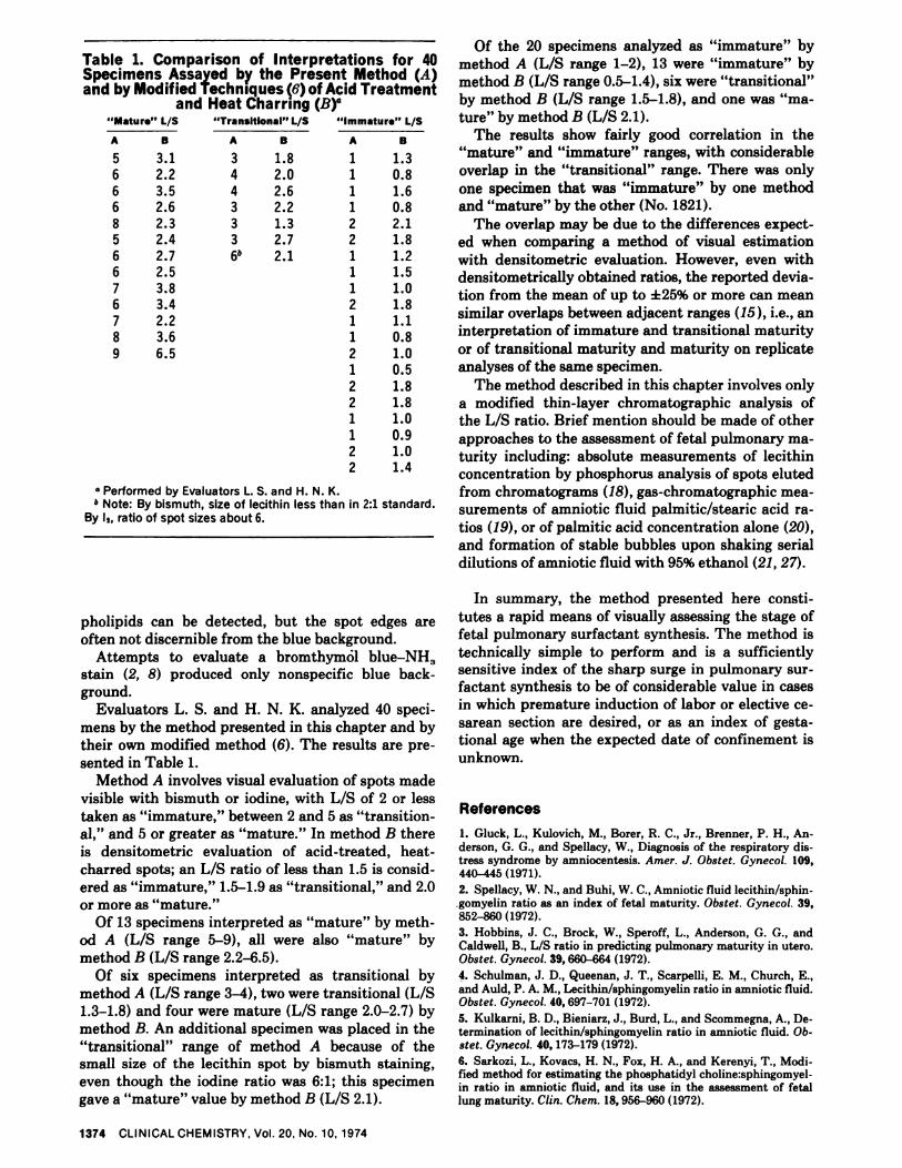
Table 1. Comparison of Interpretations for 40Specimens Assayed by the Present Method (A)and by Modified Techniques (6) of Acid Treatment
and Heat Charring (B)“Mature” L/S “TransItIonal” 1/S
A B A B
5 3.1 3 1.8
6 2.2 4 2.06 3.5 4 2.6
6 2.6 3 2.28 2.3 3 1.3
5 2.4 3 2.76 2.7 6 2.16 2.57 3.86 3.47 2.28 3.6
9 6.5
“Immature” 1/S
A B
1 1.31 0.8
1 1.61 0.82 2.12 1.81 1.21 1.51 1.02 1.81 1.11 0.82 1.01 0.52 1.82 1.81 1.01 0.92 1.02 1.4
Performed by Evaluators L. S. and H. N. K.b Note: By bismuth, size of lecithin iess than in 2:1 standard.
By i,, ratio of spot sizes about 6.
Of the 20 specimens analyzed as “immature” bymethod A (L/S range 1-2), 13 were “immature” bymethod B (L/S range 0.5-1.4), six were “transitional”by method B (L/S range 1.5-1.8), and one was “ma-ture” by method B (L/S 2.1).
The results show fairly good correlation in the“mature” and “immature” ranges, with considerableoverlap in the “transitional” range. There was onlyone specimen that was “immature” by one methodand “mature” by the other (No. 1821).
The overlap may be due to the differences expect-ed when comparing a method of visual estimationwith densitometric evaluation. However, even withdensitometrically obtained ratios, the reported devia-tion from the mean of up to ±25% or more can meansimilar overlaps between adjacent ranges (15), i.e., aninterpretation of immature and transitional maturityor of transitional maturity and maturity on replicate
analyses of the same specimen.The method described in this chapter involves only
a modified thin-layer chromatographic analysis ofthe L/S ratio. Brief mention should be made of otherapproaches to the assessment of fetal pulmonary ma-turity including: absolute measurements of lecithinconcentration by phosphorus analysis of spots elutedfrom chromatograms (18), gas-chromatographic mea-surements of amniotic fluid palmitic/stearic acid ra-tios (19), or of palmitic acid concentration alone (20),
and formation of stable bubbles upon shaking serialdilutions of amniotic fluid with 95% ethanol (21, 27).
pholipids can be detected, but the spot edges areoften not discernible from the blue background.
Attempts to evaluate a bromthym#{243}l blue-NH3stain (2, 8) produced only nonspecific blue back-ground.
Evaluators L. S. and H. N. K. analyzed 40 speci-mens by the method presented in this chapter and by
their own modified method (6). The results are pre-sented in Table 1.
Method A involves visual evaluation of spots madevisible with bismuth or iodine, with L/S of 2 or lesstaken as “immature,” between 2 and 5 as “transition-al,” and 5 or greater as “mature.” In method B thereis densitometric evaluation of acid-treated, heat-
charred spots; an L/S ratio of less than 1.5 is consid-ered as “immature,” 1.5-1.9 as “transitional,” and 2.0or more as “mature.”
Of 13 specimens interpreted as “mature” by meth-od A (L/S range 5-9), all were also “mature” bymethod B (L/S range 2.2-6.5).
Of six specimens interpreted as transitional bymethod A (L/S range 3-4), two were transitional (L/S1.3-1.8) and four were mature (L/S range 2.0-2.7) bymethod B. An additional specimen was placed in the“transitional” range of method A because of thesmall size of the lecithin spot by bismuth staining,even though the iodine ratio was 6:1; this specimengave a “mature” value by method B (L/S 2.1).
In summary, the method presented here consti-tutes a rapid means of visually assessing the stage offetal pulmonary surfactant synthesis. The method istechnically simple to perform and is a sufficientlysensitive index of the sharp surge in pulmonary sur-factant synthesis to be of considerable value in casesin which premature induction of labor or elective ce-sarean section are desired, or as an index of gesta-tional age when the expected date of confinement is
unknown.
References1. Gluck, L., Kulovich, M., Borer, R. C., Jr., Brenner, P. H., An-derson, G. G., and Spellacy, W., Diagnosis of the respiratory dis-tress syndrome by amniocentesis. Amer. ,J. Obstet. Gynecol. 109,440-445 (1971).
2. Spellacy, W. N., and Buhi, W. C., Amniotic fluid lecithin/sphin-gomyelin ratio as an index of fetal maturity. Obstet. Gynecol. 39,852-860 (1972).
3. Hobbins, J. C., Brock, W., Speroff, L., Anderson, G. G., andCaIdwell, B., L/S ratio in predicting pulmonary maturity in utero.Obstet. Gynecol. 39, 660-664 (1972).
4. Schulman, J. D., Queenan, J. T., Scarpelli, E. M., Church, E.,and Auld, P. A. M., Lecithin/sphingomyelin ratio in amniotic fluid.Obstet. Gynecol. 40, 697-701 (1972).
5. Kulkarni, B. D., Bieniarz, J., Burd, L., and Scommegna, A., De-termination of lecithin/sphingomyelin ratio in amniotic fluid. Ob-stet. Gynecol. 40, 173-179 (1972).6. Sarkozi, L., Kovacs, H. N., Fox, H. A., and Kerenyi, T., Modi-fied method for estimating the phosphatidyl choline:sphingomyel-in ratio in amniotic fluid, and its use in the assessment of fetallung maturity. Clin. Chem. 18, 956-960 (1972).
CLINICALCHEMISTRY, Vol. 20, No. 10, 1974 1375
7. Haer, F. C., An Introduction to Chromatography on Impreg-nated Glass Fiber, Ann Arbor Science Publ. Inc., Ann Arbor,Mich., 1969, pp 87-88.
8. MaIms, D. C., and Manfold, H. K., Thin layer chromatography.Stand. Methods Chem. Anal. 3, 738 (1966).
9. Barnett, H. R., and Nevin, M., The value of the Nile Blue testin estimating fetal maturity in normal and complicated pregnan-cies. J. Obstet. Gynecol. Brit. Commonw. 77, 151-155 (1970).
10. Heyneman, R. A., Bernard, D. M., and Vercauteren, R. E., Di-rect fluorometric microdetermination of phospholipids on thin-layer chromatograms. J. Chromatogr. 68, 285-288 (1972).11. Gluck, L., Kulovich, M., and Brody, S. J., Rapid quantitativemeasurements of lung tissue phospholipids. J. Lipid Res. 7, 570-574 (1966).
12. Coch, E., Meyer, J. S., Goldman, G., and Kessler, G., A modi-fled procedure for evaluation of the lecithin/sphingomyelin ratio inamniotic fluid. Clin. Chem. 19, 967-972 (1973).
13. Lemons, J. A., and Jaffe, R. B., Amniotic fluid lecithin/sphin-gomyelin ratio in the diagnosis of hyaline membrane disease.Amer. J. Obstet. Gynecol. 115, 233-237 (1973).
14. Cedard, L., Centene, J., Amiel-Tison, D., and Henrion, R., As-sessment of fetal lung maturity by amniocentesis with the lecithin!sphingomyelin ratio. Amer. J. Obstet. Gynecol. 115, 275-276(1973).
15. Gluck, L., and Kulovich, M., L/S ratios in amniotic fluid innormal and abnormal pregnancy. Amer. J. Obstet. Gynecol. 11,539-546 (1973).
16. Bryan, R., Validity of the Iecithin/sphingomyelin (L!S) ratiofor amniotic fluid containing blood. Clin. Chem. 15, 1551 (1972).
17. Whitfield, C. R., Chan, W. H., Sproule, W. B., and Stewart, A.D., Amniotic fluid lecithin:sphingomyelin ratio and fetal lung de-velopment. Brit. Med. J. 2, 85-86 (1972).
18. Bhagwanani, S. G., Fahmy, D. aid Turnbull, A. C., Prediction
of neonatal respiratory distress by estimation of amniotic-fluid lec-ithin. Lancet i 159-169 (1972).19. Alcindor, L. G., Bereziat, G., Vielh, J. P., and Gautray, J. P.,La rapport de concentration acid palmitique sur acide stearique,indicateur de Ia maturite pulmonaire foetale. Clin. Chim. Acta 50,31-34 (1974).
20. Warren, C., Allen, J. T., and Holton, J. B., Assessment of foe-tel lung maturity by amniotic fluid fatty acid analysis. Clin. Chim.Acta 44, 457-459 (1973).
21. Clements, J. A., Platzker, C. G., Tierney, D. F., Hobel, C. J.,Creasy, R. K., Margolis, A. J., Thibeault, D. W., Tooley, W. H., andOh, W., Assessment of the risk of the respiratory-distress syn-drome by a rapid test for surfactant in amniotic fluid. New EngI.J. Med. 286, 1077-1081 (1972).
22. Giridhar, V., Dalal, F. R., and Winsten, S., Molybdenum bluereagent used in determination of the lecithin!sphingomyelin ratioin amniotic fluid. Clin. Chem. 20, 513-514 (1974). Letter to theEditor.
23. Balis, J. U., and Shelley, S. A., Quantitative evaluation of thesurfactant system of the lung. Ann. Clin. Lab. Sci. 2, 410-419(1972).
24. Kiyasu, J., and Mathur, R., Considerations in determining lec-ithin!sphingomyelin ratio in amniotic fluid. Clin. Chem. 19,1224-1225 (1973).
25. Meyer, J. S., Coch, E. H., Goldman, G. M., and Kessler, G.,Amniotic fluid lecithin/sphingomyelin ratio and other tests forfetal maturity. S. Med. J. 67, 431-436 (1974).
26. Verder, H., and Clausen, J., Idiopathic respiratory distresssyndrome and the phospholipids in the amniotic fluid. Clin. Chim.Acta 51, 271-275 (1974).
27. Shephard, B., Buhi, M. S., and Spellacy, W., Critical analysisof the amniotic fluid shake test. Obstet. Gynecol. 43, 558-562(1974).
No reprints of this paper will be available.