redox signaling, oxidative stress and neurodegenerative...
TRANSCRIPT

Redox Signaling, Oxidative Stress
and Neurodegenerative
Diseases
Rodrigo Franco
Redox Biology Center and
School of Veterinary Medicine and Biomedical
Sciences

Redox Signaling, Oxidative Stress and
Neurodegenerative Diseases
• Oxidative stress happens in Neurodegeneration:
Cause, Contributor or Consequence
– Alzheimer’s disease
– Huntington’s disease
– Amyotrophic Lateral Sclerosis
– Parkinson’s disease
• Parkinson’s disease
– Energy/redox metabolism
– Gene-environment interactions
– Signaling and Redox modulation

Neurodegenerative diseases
• Neurodegenerative diseases are
defined as hereditary and sporadic
conditions which are characterized
by progressive nervous system
dysfunction.
• These disorders are often associated
with atrophy of the affected central or
peripheral structures of the nervous
system and include: Alzheimer's
Disease and other dementias,
Parkinson's Disease, Multiple Sclerosis,
Amyotrophic Lateral Sclerosis (ALS or
Lou Gehrig's Disease), Huntington's
Disease, Prion Diseases, and others.
• There is no cure for these diseases.
Nature Reviews Neuroscience 7, 278-294

Main mechanisms associated with
neurodegeneration • Energy and Metabolism dysfunction
– Oxidative Phosphorylation, Glycolysis
– Impaired ATP-dependent processes (Synaptic transmission, protein quality control
mechanisms)
– ATP is required to maintain antioxidant systems
– Glucose metabolism regulates the PPP (NADPH), antioxidant precursors and
glycative stress
• Mitochondrial dysfunction
– Energy failure
– Signaling (Calcium, pro-cell death molecules)
– Increase steady state levels of reactive oxygen species (oxidative damage)
• Dysfunction in Protein Quality Control Mechanisms.
– Protein Folding
– Protein degradation
• Proteasome
• Autophagy
– Impaired mitochondria turnover and accumulation of oxidized biomolecules

– Parkinson's disease (PD)
• Mitochondrial toxins, dopamine oxidation
• Decreased antioxidant levels (GSH)
• Lipid peroxidation, protein (carbonyls and nitrotyrosines) and
nucleic acid oxidation
– Alzheimer's disease (AD) and mild cognitive impairment.
• Increased lipid-peroxidation (CSF), protein (carbonyls and
nitrotyrosines) and nucleic acid oxidation
• Oxidative stress influences Aβ formation and Aβ has pro-
oxidant effects
– Huntington's disease (HD) – Energy metabolism
Oxidative stress is a common event in the
pathogenesis of neurodegenerative diseases


ROS and oxidative stress during the aging
process

Cell Metabolism. 2011 14, 724–738
CNS complexity

Alzheimer’s disease
• The two core pathological hallmarks of Alzheimer's disease are
amyloid plaques and neurofibrillary tangles.
• The amyloid cascade hypothesis suggests that deposition of
amyloid β (Aβ) triggers neuronal dysfunction and death in the
brain.
• Genetic causes of Alzheimer's disease include mutations of the
genes encoding amyloid precursor protein (APP) and presenilin
1 (PSEN1) and PSEN2. PSEN1 and PSEN2 mutations affect
concentrations of Aβ1–42 because the presenilin proteins form
part of γ secretase, which cleaves APP to produce Aβ.
• Many treatable medical conditions are also associated with an
increased risk of Alzheimer's disease, including stroke, diabetes
(inflammation).

Amyloid beta peptide A) Amyloid precursor protein (APP) is an
integral membrane protein expressed in
many tissues and concentrated in the
synapses of neurons. Its primary function is
not known, though it has been implicated as a
regulator of synapse formation, neural
plasticity and iron export.
Amyloidogenic processing is initiated by β-
secretase beta-site amyloid precursor
protein–cleaving enzyme 1 (BACE-1),
releasing a shortened sAPPβ. The C99
fragment is a γ-secretase substrate,
generating Aβ and AICD (amyloid precursor
protein intracellular domain).
B) Soluble Aβ is prone to aggregation.
Protofibrils (upper) and annular or pore-like
profiles (lower) are intermediate aggregates.
Self-association of Aβ monomers into
oligomers (right) is dependent on
concentration (left IB) and is promoted by
oxidizing conditions (lane 2) and divalent
metal conditions (lane 3, right IB).
NEJM Volume 362:329-344

Tau hyperphosphorylation
• Tau proteins are proteins that stabilize
microtubules. Four repeat sequences (R1-
R4) make up the microtubule-binding
domain (MBD).
• Normal phosphorylation of tau occurs on
serine and threonine residues.
• When followed by proline (P), these amino
acids are phosphorylated by glycogen
synthase kinase 3 (GSK-3β), cyclin-
dependent kinase (cdk5), or mitogen-
activated protein kinase (MAPK).
Nonproline-directed kinases
phophorylating tau are Akt, Fyn, protein
kinase A (PKA), calcium–calmodulin protein
kinase 2 (CaMKII), and microtubule affinity-
regulating kinase (MARK).
• Excessive kinase, reduced phosphatase
activities, or both cause
hyperphosphorylated tau to detach and
self-aggregate and to destabilize
microtubules.
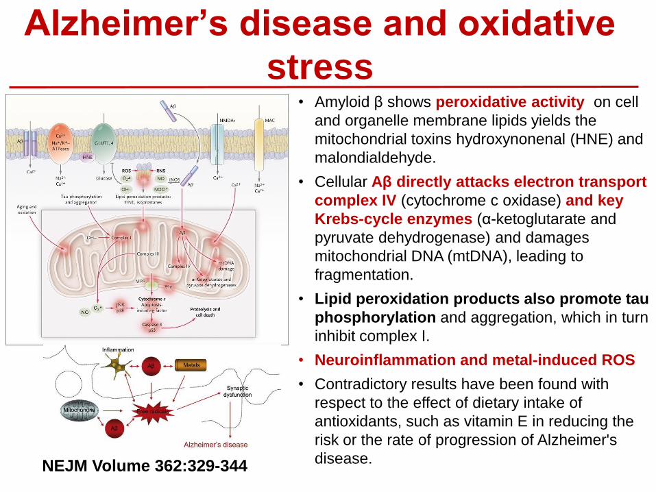
Alzheimer’s disease and oxidative
stress • Amyloid β shows peroxidative activity on cell
and organelle membrane lipids yields the
mitochondrial toxins hydroxynonenal (HNE) and
malondialdehyde.
• Cellular Aβ directly attacks electron transport
complex IV (cytochrome c oxidase) and key
Krebs-cycle enzymes (α-ketoglutarate and
pyruvate dehydrogenase) and damages
mitochondrial DNA (mtDNA), leading to
fragmentation.
• Lipid peroxidation products also promote tau
phosphorylation and aggregation, which in turn
inhibit complex I.
• Neuroinflammation and metal-induced ROS
• Contradictory results have been found with
respect to the effect of dietary intake of
antioxidants, such as vitamin E in reducing the
risk or the rate of progression of Alzheimer's
disease. NEJM Volume 362:329-344

Alzheimer’s disease and RNS
• NMDAR hyperactivation triggers
generation of NO and subsequent
S-nitrosylation of neuronal proteins,
contributing to synaptic damage and
eventually neuronal death.
• Soluble oligomers of Aβ oligomers,
can facilitate neuronal NO production
in both NMDAR-dependent and -
independent manners.
• S-Nitrosylation of the fission-inducing
protein Drp1 (dynamin-related
protein 1, forming SNO-Drp1) can
contribute to synaptic damage and
neuronal cell death by triggering
excessive mitochondrial fission and
bioenergetic impairment
Apoptosis. 2010 Nov;15(11):1354-63.
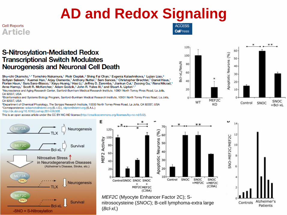
AD and Redox Signaling
MEF2C (Myocyte Enhancer Factor 2C); S-
nitrosocysteine (SNOC); B-cell lymphoma-extra large
(Bcl-xl,)

Huntington’s disease and oxidative stress • Huntington disease (HD) is an autosomal
dominant neurodegenerative disease.
• HD is caused by a CAG (glutamine)
repeat expansion in the first exon of the
HTT gene that encodes huntingtin (Htt).
• γ-Aminobutyric acid (GABAergic) medium
spiny neurons of the striatum that contain
enkephalin or substance P and project to
the globus pallidus and substantia nigra are
particularly vulnerable in HD.
• Defects in energy metabolism,
particularly mitochondrial function,
represent a common thread in studies of
HD pathogenesis in humans and animal
models
• Impaired oxidative phosphorylation,
oxidative stress , impaired mitochondrial
calcium handling , abnormal mitochondria
trafficking, deregulation of key factors of
mitochondrial biogenesis, such as the
transcriptional coactivator PPARγ
coactivator-1α (PGC-1α) , and decreased
glycolisis.
J Clin Invest. 2011;121(2):493–499.
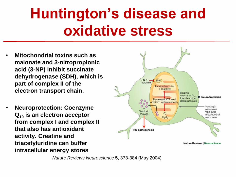
Huntington’s disease and
oxidative stress
• Mitochondrial toxins such as
malonate and 3-nitropropionic
acid (3-NP) inhibit succinate
dehydrogenase (SDH), which is
part of complex II of the
electron transport chain.
• Neuroprotection: Coenzyme
Q10 is an electron acceptor
from complex I and complex II
that also has antioxidant
activity. Creatine and
triacetyluridine can buffer
intracellular energy stores
Nature Reviews Neuroscience 5, 373-384 (May 2004)

HD and Redox Homeostasis
HD; Clinical staging with increasing severity
of the disease . Specificity protein 1 (Sp1),
a known transcription factor for CSE and
co-activator transcription initiation factor
TFIID subunit 4 (TAF4)

Amyotrophic lateral sclerosis
• Amyotrophic lateral sclerosis (ALS) is a paralytic disorder caused by motor
neuron degeneration.
• The causes of most cases of ALS are as yet undefined. Excessive excitatory
tone, protein misfolding, impaired energy production, abnormal calcium
metabolism, altered axonal transport and activation of proteases and nucleases.
• Several factors are proposed to instigate these phenomena, including latent
infections by viral and non-viral agents , toxins (for example, insecticides and
pesticides) and autoimmune reactions.
• The protein products of these mutated genes are cytosolic Cu/Zn superoxide
dismutase (SOD1), alsin, senataxin (SETX), synaptobrevin/VAMP (vesicle-
associated membrane protein)-associated protein B (VAPB) and dynactin have
been reported to cause ALS .
• About 20–25% of all familial ALS cases arise because of mutations in
SOD1, the protein product of which accounts for 0.1–0.2% of the cellular
proteins in the CNS.
• TDP-43 protein (known as TARDBP) accounts for approximately 2-6% of all
familial ALS cases and is found aggregated in sporadic cases

Amyotrophic
lateral sclerosis
• In the aberrant redox chemistry model,
mutant superoxide dismutase 1 (SOD1) is
unstable and aberrant chemistry is mediated
by promiscuous interaction with non-
conventional substrates.
– Hydrogen peroxide (H2O2) or peroxynitrite
(ONOO-) can react with reduced SOD1
(SOD1-Cu+).
– Molecular oxygen (O2) can react
aberrantly with Zn-deficient SOD1 to
generate an excess of superoxide anion
(O2-).
– The unstable protein can also release free
copper and/or zinc, which might be toxic.
• In the protein toxicity (proteotoxic) model,
conformationally altered mutant SOD1 forms
toxic, proteinaceous deposits.
– Aggregated SOD1 inhibits chaperone
and/or proteasome activity, with
subsequent misfolding and insufficient
clearance of numerous proteins.
– Alternatively, these aggregates could
sequester, inactivate or enhance the
toxicity of other proteins crucial for
cellular processes.
Nat Rev Neurosci. 2006 Sep;7(9):710-23.

ALS, Protein Oxidation and Aggregation
TDP-43 has been shown to bind both DNA and
RNA and have multiple functions in transcriptional
repression, pre-mRNA splicing and translational
regulation. T-cell intracellular antigen (TIA)-1 and
TIA-1-related protein (TIAR) are mRNA-binding
proteins that can aggregate within granules under
specific stress conditions. Frontotemporal lobar
degeneration (FTLD) characterized by TDP‐43
pathology (FTLD‐TDP).
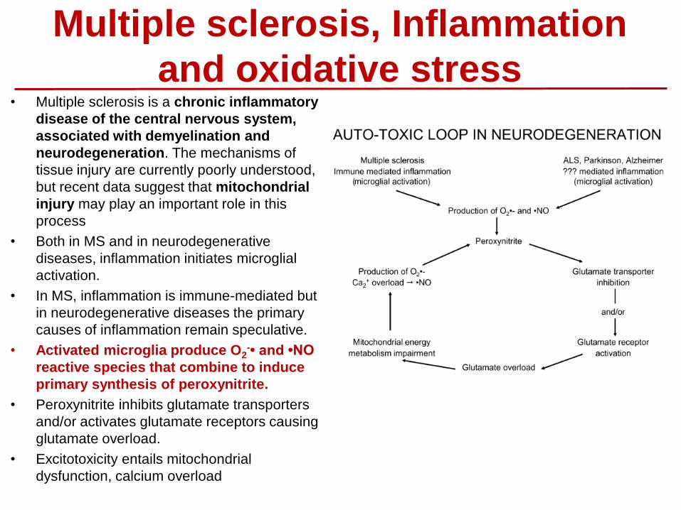
Multiple sclerosis, Inflammation
and oxidative stress • Multiple sclerosis is a chronic inflammatory
disease of the central nervous system,
associated with demyelination and
neurodegeneration. The mechanisms of
tissue injury are currently poorly understood,
but recent data suggest that mitochondrial
injury may play an important role in this
process
• Both in MS and in neurodegenerative
diseases, inflammation initiates microglial
activation.
• In MS, inflammation is immune-mediated but
in neurodegenerative diseases the primary
causes of inflammation remain speculative.
• Activated microglia produce O2-• and •NO
reactive species that combine to induce
primary synthesis of peroxynitrite.
• Peroxynitrite inhibits glutamate transporters
and/or activates glutamate receptors causing
glutamate overload.
• Excitotoxicity entails mitochondrial
dysfunction, calcium overload

Redox control of prion and disease
pathogenesis
• The underlying cause of brain pathology in all
prion disorders is PrP-scrapie (PrP(Sc)), a
beta-sheet-rich conformation of a normal
glycoprotein, the prion protein (PrP(C)).
• In prion disorders, imbalance of brain-iron
homeostasis is observed before end-stage
disease and worsens with disease
progression, implicating iron-induced
oxidative stress in disease pathogenesis.
• Increased oxidation, glycoxidation, and
lipoxidation of brain proteins in prion
disease. Free Radic Biol Med. 2008 Oct
15;45(8):1159-66.
• Acute exposure to prion infection induces
transient oxidative stress progressing to be
cumulatively deleterious with chronic
propagation in vitro. Free Radic Biol Med. 2011
Apr 3.
• Cellular prion protein protects against
reactive-oxygen-species-induced DNA
damage. Free Radic Biol Med. 2007 43(6):959-
67.

What causes Parkinson’s
disease
• Parkinson’s disease (PD) is a chronic progressive
neurodegenerative disorder that leads to shaking
(tremors) and difficulty with walking, movement,
and coordination. Parkinson's may lead to a
deterioration of all brain functions, and an early
death.
• Loss of dopamine neurons from the substantia
nigra pars compacta leads to deficiency of
dopamine in the caudate and putamen (“striatum”).
• Currently, there is no treatment to cure or stop PD
progression.
• The exact cause of PD is unknown.
Familial (Hereditary) forms of PD (~10%)
α-synuclein, Parkin, DJ-1, PINK1 and LRRK2
genes.
Sporadic (Idiopathic) PD
~5% are linked to genetic alterations
Environmental or occupational factors.
PD
Aging Genetics
Environment

Products of PD-associated genes that affect
mitochondrial function and oxidative stress
Rare inherited mutations in genes encoding electron
transport chain components have been associated
with parkinsonism.
• Parkin is partially localized to the outer mitochondrial
membrane,
• PINK1 is a mitochondrial serine–threonine kinase that
affords protection against oxidative stress and acts
with Parkin to regulate the balance of mitochondrial
fission and fusion.
• DJ-1 is relocated to mitochondria under conditions of
oxidative stress and is thought to be neuroprotective
under such conditions.
• The α-synuclein protein has an amino-terminal
mitochondrial targeting sequence and, when
overexpressed or under conditions of acidification, is
at least partially associated with the inner
mitochondrial membrane, where it might cause direct
damage.
• LRRK2 associates, at least in part, with the outer
mitochondrial membrane
• HTRA2 is a mitochondrial serine protease, the release
of which might be involved in apoptotic cell death.
Nature Clinical Practice Neurology (2008) 4, 600-609

DJ-1 in Parkinson’s disease
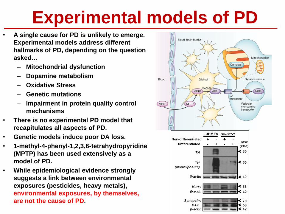
Experimental models of PD • A single cause for PD is unlikely to emerge.
Experimental models address different
hallmarks of PD, depending on the question
asked…
– Mitochondrial dysfunction
– Dopamine metabolism
– Oxidative Stress
– Genetic mutations
– Impairment in protein quality control
mechanisms
• There is no experimental PD model that
recapitulates all aspects of PD.
• Genetic models induce poor DA loss.
• 1-methyl-4-phenyl-1,2,3,6-tetrahydropyridine
(MPTP) has been used extensively as a
model of PD.
• While epidemiological evidence strongly
suggests a link between environmental
exposures (pesticides, heavy metals),
environmental exposures, by themselves,
are not the cause of PD.

What makes a dopaminergic neuron
the target?
• Dopamine and Iron (pro-
oxidant)
• Dopaminergic neurons
consume a significant
amount of energy due to:
– Pacemaking activity
– Vesicle transport
– Action potential and
membrane potential
maintenance through the
unmyelinated axon
The axonal arbors of single nigrostriatal dopaminergic
neurons in rat brain. The image shows axon fibers in the
neostriatum. Red and blue lines indicate the axon fibers
located in the striosome and matrix compartments of the
neostriatum. A single dopamine neuron can influence up to 5%
of all neurons in the neostraitum or ~75,000 neurons.
The Journal of Neuroscience, 14 January 2009, 29(2): 444-453
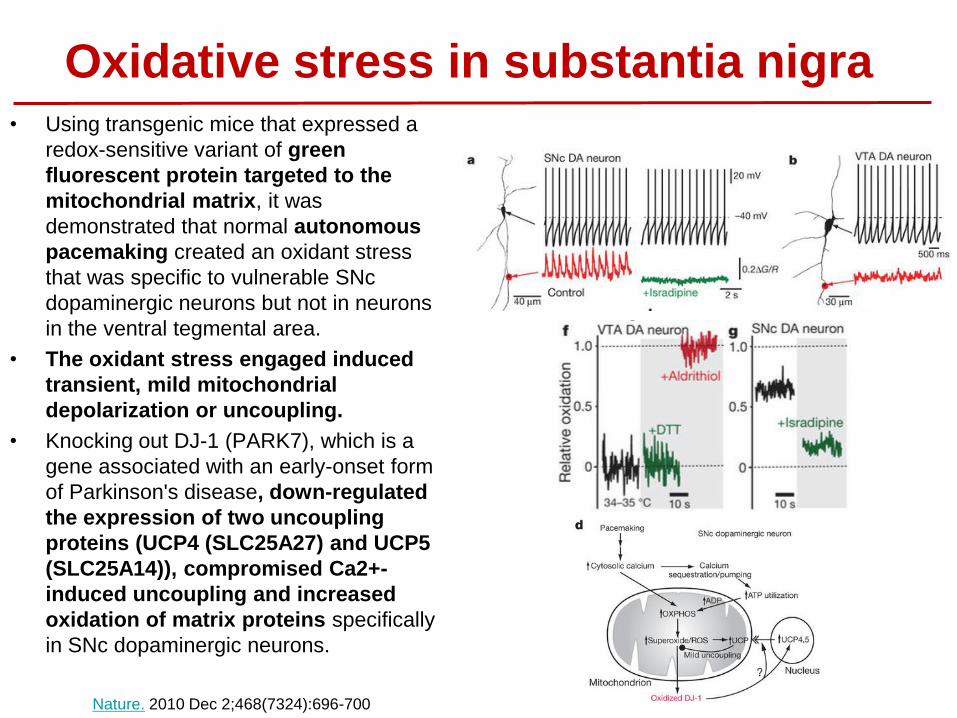
Oxidative stress in substantia nigra • Using transgenic mice that expressed a
redox-sensitive variant of green
fluorescent protein targeted to the
mitochondrial matrix, it was
demonstrated that normal autonomous
pacemaking created an oxidant stress
that was specific to vulnerable SNc
dopaminergic neurons but not in neurons
in the ventral tegmental area.
• The oxidant stress engaged induced
transient, mild mitochondrial
depolarization or uncoupling.
• Knocking out DJ-1 (PARK7), which is a
gene associated with an early-onset form
of Parkinson's disease, down-regulated
the expression of two uncoupling
proteins (UCP4 (SLC25A27) and UCP5
(SLC25A14)), compromised Ca2+-
induced uncoupling and increased
oxidation of matrix proteins specifically
in SNc dopaminergic neurons.
Nature. 2010 Dec 2;468(7324):696-700

• Redox cycling
• Thiols • GSH
• Cysteine modifications
• Trx/Prx/Srx
• Oxidative Stress
• Hydrogen sulfide
• Redox regulated
transcription factors
• Mitochondria
• Inflammation
Redox regulation of Parkinson’s Disease

• Antioxidant supplementation has for the most part failed to achieve
meaningful effects in PD.
o Lack of specificity
o Administration of antioxidants might not have been given early
enough
o Not all cases arise from the same causes
o Wrong experimental models
o Oxidative stress is not the main/single cause in
neurodegeneration
Why have antioxidant clinical trials failed?

• Environmental agents linked to increased
incidence/risk to develop Parkinson’s disease
• Pesticides
• Heavy Metals
• Infectious agents: Proc Natl Acad Sci. 2009,
106:14063-8.
• Industrialization: Sci Rep. 2013, 3:1395.
• Dietary factors: Toxicol Sci. 2014, 140:179.
• The U.S. is the country as a whole with the
world's highest prevalence of PD.
• For residents of highly agricultural areas,
pesticides are environmental risk factors of major
public health concern for populations directly
working with them or indirectly exposed by
residential proximity to crop fields.
• Epidemiological evidence demonstrates that
pesticide exposure (paraquat and rotenone)
contributes to PD progression (Environ Health
Perspect. 2011 Jun;119(6):866-72).
• Not a single environmental toxicant can be the
cause of sporadic PD.
Environmental agents and Parkinson’s disease
Prevalence of Parkinson's disease in U.S.A

Mitochondrial Dysfunction: Redox Homeostasis vs Bioenergetics
Redox homeostasis Bioenergetics

Paraquat toxicity in vivo
C
PBS MPTP
TH
+ n
eu
ron
s (
co
un
ts)
0
2000
4000
6000
8000
10000
12000
*
MPTP
PBS
A
TH PSSG Merge
PBS
PQ
* *
B
Control
PQ
Antioxid Redox Signal 2012; 17(12):1676-93.

In vivo metabolic dysfunction induced by
PQ
Striat Mid Cereb Cort
PQ - + - + - + - +
vs Cont 1 1.9 1 3.5 1 0.6 1 0.7
vs Cont 1 1.6 1 5 1 1.2 1 0.9
AMPK 1
pAMPK T172
-actin
pACC
Anandhan et al., Mol Neurobiol. 2016 (accepted)

Gene-Environment Interactions in PD
The Multi-hit Hypothesis

Gene-Environment Interactions in
Parkinson’s Disease
• Parkinson’s disease is a multifactorial disease: ageing, genetics and environment.
• The formation of intracellular aggregates (Lewy bodies) of which a major component is the protein α-synuclein, is a pathological hallmark.
• Mitochondrial dysfunction and energy failure induced by environmental toxicants can lead to α-synuclein misfolding and aggregation by an impairment in protein quality control mechanisms.

MOI
Empty 15 - - - -
α-synuclein - 2.5 5 10 15
C
A
Monomer
-synuclein
-Actin
HMW
aggregates
-synuclein
B
-synuclein Potentiates PQ Toxicity and
Metabolic Dysfunction
PQ [M] 0 50
Ce
ll s
urv
iva
l (%
)
0
20
40
60
80
100
Empty
-synuclein
A53T
a, b a
Anandhan et al., Mol Neurobiol. 2016 (accepted)

A
-synuclein Potentiates PQ-induced Metabolic
Dysfunction
PQ [M] - 50 100
Empty + - - + - - + - -
-synuclein - + - - + - - + -
A53T - - + - - + - - +
vs Cont 1 1.1 1.4 3.5 182 513 61 249 689
vs Empty 1 51 143 1 4 11
pAMPK T172
AMPK1
- PQ + PQ
OC
R /
EC
AR
0
1
2
3
4 Empty
-synuclein
B
Time (min)
0 20 40 60 80 100
EC
AR
(v
s c
on
tro
l)
0
5
10
15
20
Empty
+ PQ
-synuclein
-synuclein + PQ
NG +G
+O +2-DG
D C
*
*
Anandhan et al., Mol Neurobiol. 2016 (accepted)

Empty Alpha syn A53T
Cell s
urv
ival (%
)
0
20
40
60
80
100
Control
PQ
STF-31
PQ + STF-31
* *
Paraquat [M] 0 50 100
Cell s
urv
ival (%
)
0
20
40
60
80
100
Empty
-syn
Empty+ 6AN
-syn + 6AN
*
*
Paraquat [M] 0 50 100
Cell s
urv
ival (%
)
0
20
40
60
80
100
Empty
-syn
Empty - Glucose
-syn - Glucose
A
*
*
B
Glucose metabolism regulates α-synuclein-
PQ toxic interaction
-synuclein
Paraquat [M] 0 50 100
Cell s
urv
ival (%
)
0
20
40
60
80
100
Empty
-Syn
Empty + Ascorbic acid
a-Syn + Ascorbic acid
*
*
C D
Anandhan et al., Mol Neurobiol. 2016 (accepted)

Glucose metabolism and PQ + -synuclein
toxic interaction in PD
NADP +
NADPH
PQ +
PQ O2 •-
O2
Ascorbic acid
STF-31
AMPK
AMP (ADP)/ATP
Glucose
Glucose
GLUT
PPP
Cell death
+ -synuclein
6-AN
Glutamine
Glycolysis
Glutaminolysis
P
2-DG
Galactose
iNOS
?
?
Glucose metabolism regulates α-synuclein + PQ toxicity. • PQ and -synuclein impair glucose
metabolism. • PQ increases glucose transport and
translocation of glucose transporters. Inhibition of GLUT-like transporters prevents α-synuclein + PQ toxicity.
• Inhibition of PPP protects against α-synuclein + PQ.
AMPK signaling regulates α-synuclein + PQ toxicity. • Activation of AMPK and cell death are
mediated by the iNOS. • AMPK protects against α-synuclein + PQ. • α-synuclein and impairment of AMPK
signaling and induce an overall metabolic dysfunction that enhances PQ toxicity . Anandhan et al., Mol Neurobiol. 2016 (accepted)

Summary • Three main mechanisms linked to neurodegeneration.
– Energy Failure
– Mitochondrial dysfunction
– Dysfunction in Protein Quality Control Mechanisms.
• Three major risk factors involved in neurodegeneration: Age, genes and environment.
• Alterations in redox homeostasis in neurodegeneration are not isolated events.
– Cell signaling
– Bioenergetics

Post-doctoral fellows: Anandhan Annadurai Pablo Hernandez-Franco Laura Zavala-Flores Aracely Garcia-Garcia Humberto Rodriguez-Rocha Sumin Li Graduate Students: Shulei Lei Juliana Navarro-Yepes Undergraduate / PPVM students: Jianhui Li (UCARE) Chris Beasley (UCARE) Reilly Grealish (UCARE) Mallory Sea (REU) Rachel Foguth (REU) Erin Bradley (CMV) Arisdelsy Cervantes (REU) Alice Hackett (REU) Chillian Picket (REU) Michaela Burns (UCARE)
Funding:
• Centers of Biomedical Research Excellence (COBRE/NIH). Redox Biology Center.
• Office of Research. University of Nebraska-Lincoln.
• Alzheimer’s Association. International Research Grant Program.
• Scientist Development Grant, American Heart Association
• Science and Technology National Council (Mexico) CONACYT
Collaborators (RBC): Dr. Robert Powers (Chemistry) Dr. Oleh Khalimonchuk (Biochemistry) Dr. Jaekwon Lee (Biochemistry) Dr. You Zhou (Biochemistry) Dr. Matthew Zimmerman (UNMC) Collaborators (UNL/UNMC): Dr. Lee Mosley (UNMC) Dr. Ming Li (Psychology) Dr. Matthew C. Zimmerman (UNMC) Collaborators External:
Dr. Jean-Christophe Rochet (Purdue University)
Dr. Robert C. Stanton (Joslin Diabetes Center)
Dr. Betzabet Quintanilla-Vega (CINVESTAV, Mexico)
Dr. Reto Asmis (UTHSC)