reduction of intra-operative blood loss by temporary ... · neck defect left after surgery. 2 ......
TRANSCRIPT
1
Reduction of intra-operative blood loss by temporary control of external
carotid artery in advanced head and neck malignancies
Hosam Abd El-Kader El-Fol, M.D 1
Mamdouh S. Ahmed, DDS2
Author Affiliation: 1 Lecturer, Department of Surgical Oncology, Faculty of Medicine, Menofia University 2 Lecturer, Department of Oral & Maxillofacial Surgery, Faculty of Oral & Dental Medicine,
Cairo University
ABSTRACT:
Objective: The study introduces a simple vascular control procedure to minimize
intraoperative blood loss during resection of advanced head and neck malignancies
through clamping to the external carotid artery (ECA).
Patients and Methods: This prospective study included 20 patients with different
operable head and neck cancer randomized to perform vascular clamping of ECA during
neck dissection before tumor resection (group A, n = 11) or classical neck dissection and
resection of the tumors without vascular control (group B, n = 9).
Results: There was no significant difference between the 2 groups regarding
demographic and disease characteristics. Statistically significant decrease of blood loss
was observed in the vascular control group. Blood loss in group A was nearly a quarter of
that in group B.
Conclusion: Temporary intraoperative clamping of the ECA minimizes blood loss, and
consequently, the need for blood transfusion with all its complications. In addition, it
ensures more optimum survival of the full-thickness graft used for coverage of the head
neck defect left after surgery.

2
INTRODUCTION:
Head and neck cancer represents 5% of all malignant tumors in US and 17% in Egypt [1].
The majority of patients present with locally advanced lesions necessitating major
surgical excisions and challenging reconstruction. Major hemorrhage during surgery for
advanced head and neck tumors is life threatening problem. The control of bleeding
through the ligation of the offending artery was clearly the treatment of choice [2].
Identifying the offending vessel and gaining adequate access to it are often difficult to
perform although temporary arrest of the bleeding is usually possible with packing and
pressure. Definitive control of the bleeding has historically been achieved with either
ligation of the external carotid artery (ECA) or selective embolization [2].
Ligation of several branches of the ECA reduces collateral blood flow and hence
contributes significantly to the arrest of bleeding. Selective embolization of bleeding
vessels requires catheterization of the femoral artery, angiography to identify the bleeding
vessel and deposition of a thrombogenic agent. Embolization may not be possible if the
bleeding vessel is excessively tortuous, small or in vasospasm. Embolization needs a
skilled interventional radiologist, professional in angiography and embolization [2].
Complications of blood transfusion as hemolytic reactions, febrile and allergic reactions,
bacterial sepsis, embolism, overtransfusion and pulmonary edema and transmission of
diseases (malaria, Chagas' disease, brucellosis and transmission of hepatitis C and HIV-1)
have been dramatically minimized by the introduction of better antibody and nucleic acid
screening for these pathogens [3].
The external carotid artery begins at the bifurcation of the common carotid artery at C4. It
continues upward to a point posterior to the neck of the mandible (approximately 1.5 cm
below the zygomatic arch) where it bifurcates to form the maxillary and superficial
temporal arteries. The superior thyroid, lingual, and facial arteries arise from the ventral
aspect near the origin of the external carotid; the ascending pharyngeal, occipital and
posterior auricular branches arise from the dorsal side of the external carotid [4]. The
3
common carotid may bifurcate high at the level of the hyoid bone, or lower at the level of
the cricoid cartilage [5]. External carotid artery also delivers blood to the internal carotid
(by virtue of the anastomoses of the two). But, even if there is no vascular disease, it is
sufficient in only 50% of the cases. [6]
The normal relationship of external to internal carotid artery may be altered the
terminology of these vessels in no way represents their relationship in the neck but
simply refers to their ultimate distribution. Except for rare circumstances, the internal
carotid artery has no branches in the neck, whereas the external carotid artery has three or
four branches within several centimeters of the bifurcation. It is best to expose both
internal and external carotid arteries for at least 2.5 cm and ascertain the presence or
absence of branches. Avoid injury to the superior laryngeal nerve, which passes deep to
external carotid artery [7].
In this study, we introduce the results of a simple vascular control procedure to minimize
intraoperative blood loss, and avoid blood transfusion during resection of advanced head
and neck tumors without the need for embolization or ligation of the external carotid
artery through vascular clamping to the external carotid artery.
PATIENTS AND METHODS:
This prospective study was performed in Surgical Oncology Department, Faculty of
Medicine, Menofia University and Oral and Maxilliofacial Surgery Department, Faculty
of Oral and Dental Medicine, Cairo University between May 2006 and March 2010.
Following thorough clinical examination and routine preoperative laboratory tests, a
search of locoregional and distant metastases were done with computed tomography (CT)
scan, magnetic resonance imaging (MRI), bone scan and abdominal ultrasonography.
Operable head and neck tumors were included and randomized into two groups:
Group A (Table 1) includes 11 patients receiving vascular controls to the external
carotid artery during neck dissection by vascular clamp before resection of the

4
tumors. Diagnosis of these patients was recurrent mucoepidermoid carcinoma of the
parotid (n = 1),sqauamous cell carcinoma of maxillary alveolar margins (n = 2),
retromolar trigone (n = 2), mandibular alveolar margins (n = 3) and buccal mucosa (n
= 3).
Group B (Table 2) includes 9 patients received classical neck dissection and resection
of the tumors without vascular control. They suffered squamous cell carcinoma of
retromolar trigone (n = 2), mandibular alveolar margin (n = 5) and buccal mucosa (n
= 2).
Table (1): Detailed data of Group (A)
Serial Pathology
+ Grade
Age Sex Site TNM Procedure Blood
loss
1 Mucoepidermoid
Carcinoma grade
2
58 male Parotid region & Buccal
mucosa
T4 invade
mandible
N+ve
Hemimand. +full thickness skin, buccal
mucosa + buccal fat pad +
Parotidectomy + RND + Reconstruction
PMMF
300 cc
2 SCC grade 2 53 male Upper alveolar margin T2 N0 Hemimaxillectomy+ supraomohyoid ND 100 cc
3 SCC grade 2 46 female Upper alveolar margin T2N0 Hemimaxillectomy+ supraomohyoid ND 90 cc
4 SCC grade 1 65 male Right Retromolar trigone T2 N 0 Excision + Right Hemimandibulectomy +
FND
200 cc
5 SCC grade 2 46 male Right Retromolar trigone T2N 0 Hemimandibulectomy. +FND 200 cc
6 SCC grade 1 68 female Left Lower alveolar
margin
T2 N 0 Excision + Left hemimandibulectomy +
FND
180 cc
7 SCC grade 2 59 male Left Lower alveolar
margin
T2 N 0 Excision + Left hemimandibulectomy +
FND
170 cc
8 SCC grade 2 68 male Left Lower alveolar
margin
T3 N +ve Left hemimandibulectomy +FND 200 cc
9 SCC grade 1 57 female Right Buccal mucosa T1 N0 Right bExcision+FND 100 cc
10 SCC grade 3 51 male Right Buccal mucosa T2 N0 Excision with skin+ RND + reconstruction
PMMF
250 cc
11 SCC grade 3 45 male Left Buccal mucosa T4 N+ve Full thickness resection
+hemimandbulectomy +RND + reconst.
PMCF(Double island)
270 cc
5
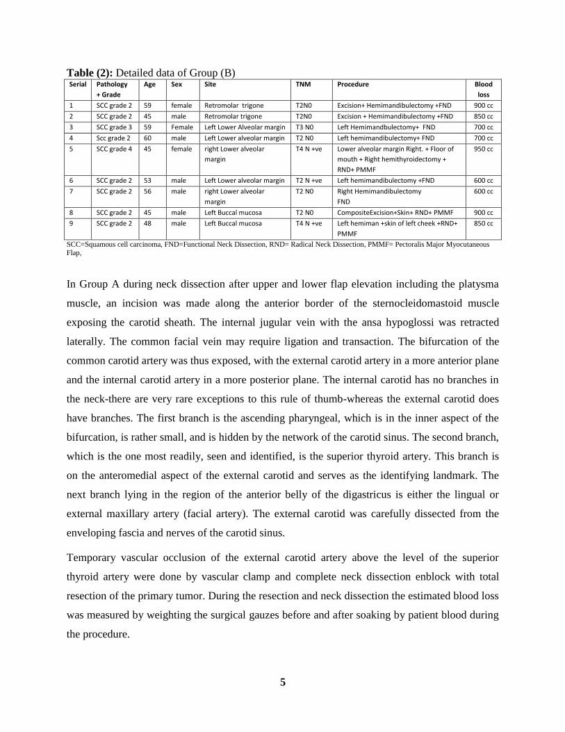
Table (2): Detailed data of Group (B) Serial Pathology
+ Grade
Age Sex Site TNM Procedure Blood
loss
1 SCC grade 2 59 female Retromolar trigone T2N0 Excision+ Hemimandibulectomy +FND 900 cc
2 SCC grade 2 45 male Retromolar trigone T2N0 Excision + Hemimandibulectomy +FND 850 cc
3 SCC grade 3 59 Female Left Lower Alveolar margin T3 N0 Left Hemimandbulectomy+ FND 700 cc
4 Scc grade 2 60 male Left Lower alveolar margin T2 N0 Left hemimandibulectomy+ FND 700 cc
5 SCC grade 4 45 female right Lower alveolar
margin
T4 N +ve Lower alveolar margin Right. + Floor of
mouth + Right hemithyroidectomy +
RND+ PMMF
950 cc
6 SCC grade 2 53 male Left Lower alveolar margin T2 N +ve Left hemimandibulectomy +FND 600 cc
7 SCC grade 2 56 male right Lower alveolar
margin
T2 N0 Right Hemimandibulectomy
FND
600 cc
8 SCC grade 2 45 male Left Buccal mucosa T2 N0 CompositeExcision+Skin+ RND+ PMMF 900 cc
9 SCC grade 2 48 male Left Buccal mucosa T4 N +ve Left hemiman +skin of left cheek +RND+
PMMF
850 cc
SCC=Squamous cell carcinoma, FND=Functional Neck Dissection, RND= Radical Neck Dissection, PMMF= Pectoralis Major Myocutaneous Flap,
In Group A during neck dissection after upper and lower flap elevation including the platysma
muscle, an incision was made along the anterior border of the sternocleidomastoid muscle
exposing the carotid sheath. The internal jugular vein with the ansa hypoglossi was retracted
laterally. The common facial vein may require ligation and transaction. The bifurcation of the
common carotid artery was thus exposed, with the external carotid artery in a more anterior plane
and the internal carotid artery in a more posterior plane. The internal carotid has no branches in
the neck-there are very rare exceptions to this rule of thumb-whereas the external carotid does
have branches. The first branch is the ascending pharyngeal, which is in the inner aspect of the
bifurcation, is rather small, and is hidden by the network of the carotid sinus. The second branch,
which is the one most readily, seen and identified, is the superior thyroid artery. This branch is
on the anteromedial aspect of the external carotid and serves as the identifying landmark. The
next branch lying in the region of the anterior belly of the digastricus is either the lingual or
external maxillary artery (facial artery). The external carotid was carefully dissected from the
enveloping fascia and nerves of the carotid sinus.
Temporary vascular occlusion of the external carotid artery above the level of the superior
thyroid artery were done by vascular clamp and complete neck dissection enblock with total
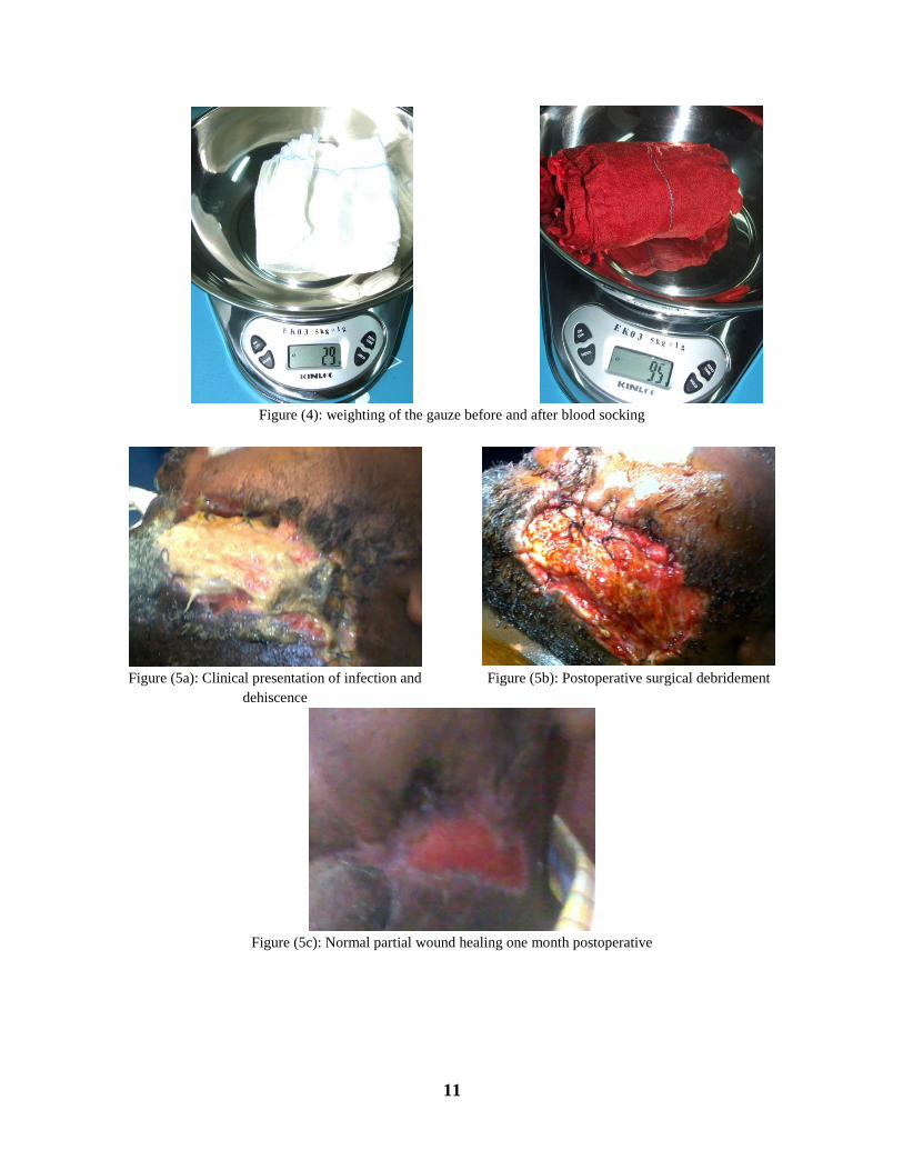
resection of the primary tumor. During the resection and neck dissection the estimated blood loss
was measured by weighting the surgical gauzes before and after soaking by patient blood during
the procedure.
6
Statistical analysis:
Data was analyzed using SPSSwin statistical package version 15 (SPSS Inc., Chicago, IL).
Numerical data were expressed as mean and standard deviation. Qualitative data were expressed
as frequency and percentage. Fisher’s exact test was used to examine the difference between the
two groups in qualitative variables. For quantitative data, comparison between two groups was
done using Mann-Whitney test. A p-value < 0.05 was considered significant.
RESULTS:
Table 3 summarizes the results of this study. There was no significant difference between group
A and group B regarding demographic and disease characteristics. Statistically significant
decrease of blood loss was observed in group A (vascular control group). Blood loss in group A
was nearly a quarter of that in group B (without vascular control).
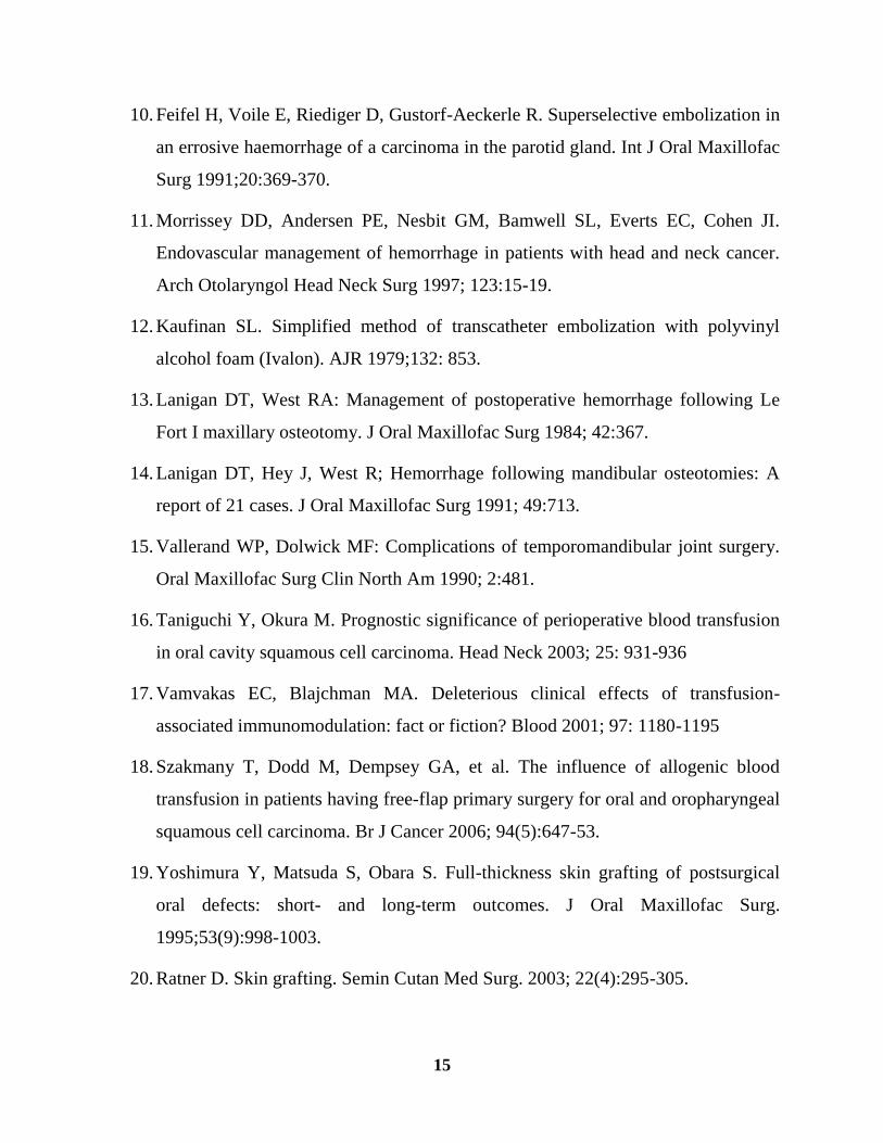
All cases showed uneventful postoperative healing except three cases. In group B, case no., 8
there was wound infection and dehiscence one week postoperatively. Antibiotic administration,
surgical debridement and re-suturing of the gapped areas showed normal healing. In the same
group, case no. 9 showed small salivary fistula which was managed conservatively by Ryle
feeding and antibiotic administration. While in group A, case no., 11, partial loss of the skin of
PMMF was observed 5 days postoperative and managed by surgical debridement and frequent
dressing.

7
Table 3: Demographic and disease characteristics and intraoperative blood loss of the two
studied groups
Group A
n = 11
Group B
n = 9
P value
Age 56.0±8.6 52.2±6.5 0.370
Sex (male/female) 8/3 6/3 0.769
Site
Buccal mucosa 3 (27.3%) 2 (22.2%)
Lower Alveolar margin 3 (27.3%) 5 (55.6%)
Retromolar trigone 2 (18.2%) 2 (22.2%)
Upper alveolar margin 2 (18.2%) 0 (0.0%)
Parotid region 1 (9.1%) 0 (0.0%)
T stage
Stage 1 & 2 6 (54.5%) 6 (66.7%) 0.670
Stage 3 & 4 5 (45.5%) 3 (33.3%)
N stage
N0 8 (72.7%) 6 (66.7%) 1.000
N1 3 (27.3%) 3 (33.3%)
Differentiation grade
Grade 1 & 2 9 (81.8%) 7 (77.8%) 1.000
Grade 3 & 4 2 (18.2%) 2 (22.2%)
Intraoperative Blood Loss 187.3±70.0 783.3±134.6 < 0.001
8
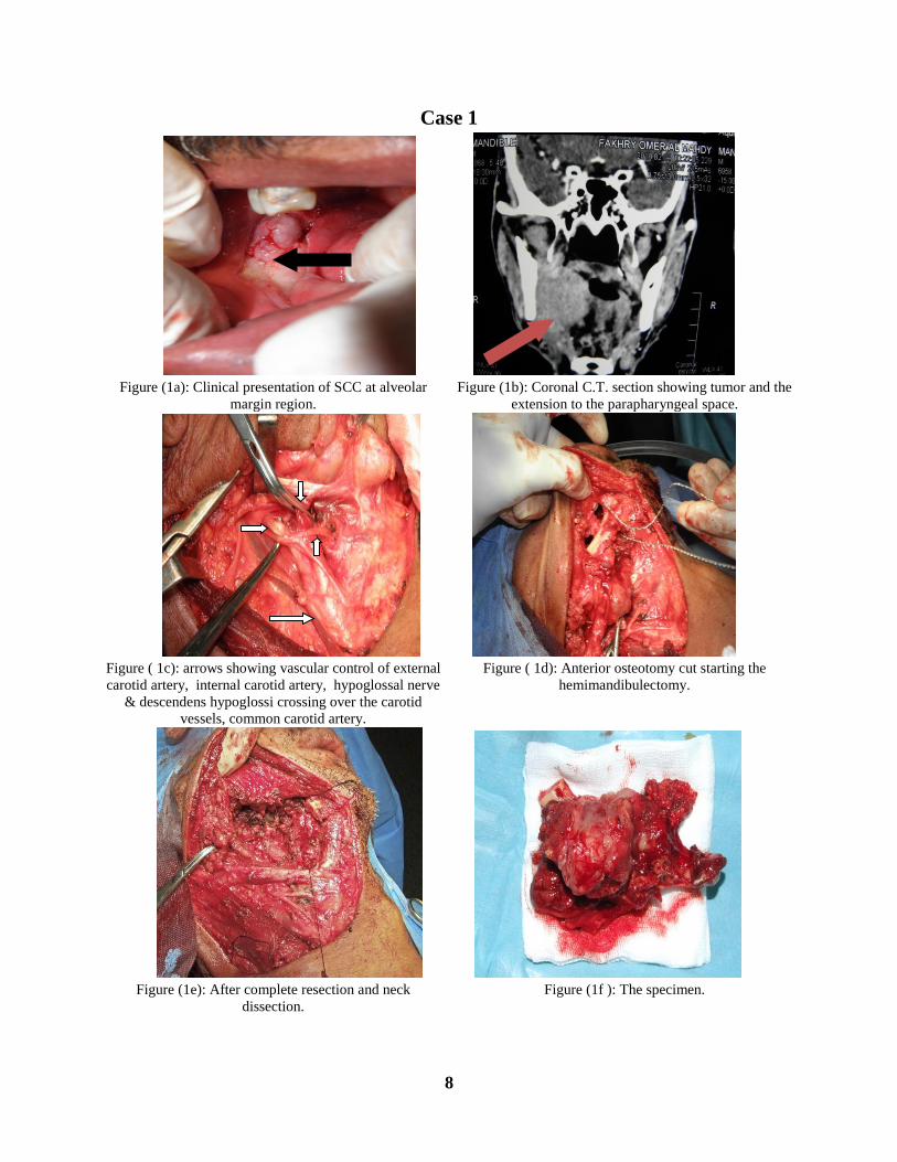
Case 1
Figure (1a): Clinical presentation of SCC at alveolar
margin region.
Figure (1b): Coronal C.T. section showing tumor and the
extension to the parapharyngeal space.
Figure ( 1c): arrows showing vascular control of external
carotid artery, internal carotid artery, hypoglossal nerve
& descendens hypoglossi crossing over the carotid
vessels, common carotid artery.
Figure ( 1d): Anterior osteotomy cut starting the
hemimandibulectomy.
Figure (1e): After complete resection and neck
dissection.
Figure (1f ): The specimen.

9
Case 2
Figure (2a): Clinical presentation of SCC at buccal
mucosa.
Figure (2b): Incision design to include the overlying skin
and incision for neck dissection.
Figure (2c) Identification of external carotid Figure (2d) Vascular control of external
Carotid artery before resection
Figure (2e): Completed resection and neck dissection
notice the clean and dry field after resection.
Figure (2f): The specimen.
10
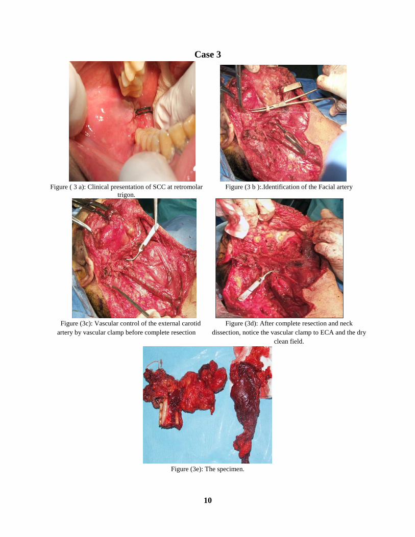
Case 3
Figure ( 3 a): Clinical presentation of SCC at retromolar
trigon.
Figure (3 b ):.Identification of the Facial artery
Figure (3c): Vascular control of the external carotid
artery by vascular clamp before complete resection
Figure (3d): After complete resection and neck
dissection, notice the vascular clamp to ECA and the dry
clean field.
Figure (3e): The specimen.
11
Figure (4): weighting of the gauze before and after blood socking
Figure (5a): Clinical presentation of infection and
dehiscence
Figure (5b): Postoperative surgical debridement
Figure (5c): Normal partial wound healing one month postoperative

12
DISCUSSION:
Patients with advanced head and neck malignancies are prone to many problems,
including bleeding, infection, fistula, and uncontrollable pain. Massive hemorrhage is the
most serious and immediate of these complications. Bleeding from an unresectable or
advanced, local recurrent malignancies of the head and neck, which can occur either from
the tumor bed or from erosion into a large vessel, represents a difficult management
problem [8]. Past management of tumor hemorrhage has included ligation of the carotid
artery and transarterial embolization using polyvinyl alcohol (PVA) and a detachable
balloon [8-12].
Ligation of the external carotid artery (ECA) is a relatively simple procedure with
minimal morbidity, however, its efficacy in arresting ongoing blood loss is questionable.
It was reported to be successful in 2 of 3 cases of hemorrhage after Le Fort osteotomy
[13], in 4 of 5 patients having hemorrhage during mandibular osteotomies [14] and in
bleeding after temporo-mandibular joint surgery [15].
In this study, we question the necessity of ligation of ECA to minimize intraoperative
blood loss during resection of advanced head and neck tumors. Ligation is a permanent
procedure that is undesirable in these cases. We need a temporary procedure for
intraoperative vascular control to allow good operative field during surgery and well
perfused bed which in turn ascertains a better healing during the postoperative period.
Using temporary intraoperative clamping of the ECA, we found a statistically significant
decrease of blood loss mounting to nearly a quarter of blood loss in patients where
carotid arteries were left unclamped.
In the control group with unclamped ECA, the estimated blood loss reached nearly 1 liter.
The management of oral and oropharyngeal cancer involves prolonged surgical
procedures for tumour resection and flap reconstruction which result in significant
intraoperative blood loss. The reported transfusion rates varied from 32% to 81% [16].
Vamvakas & Blajchman [17] suggest that transfusion of 3 or more units of blood is

13
associated with a worse outcome in oropharyngeal cancer patients, with a reduction in
survival and an increase in recurrence rates. Other investigators reported an almost five-
fold increase in the risk of death among patients transfused more than 3 units of blood
compared to nontransfused patients [16, 18].
Thus, clamping of ECA to minimize blood loss and hence blood transfusion may have an
impact on both short- and long term treatment outcome in advanced head and neck
malignancies. This needs to be proved in longer follow-up studies.
Our protocol of management of head and neck malignancies involved covering large
defects with locoregional flaps such as pectoralis major myocutaneous falp. Vascularity
of the grafted bed has a considerable role in graft healing. In a study evaluating the use of
full-thickness skin grafts following excision of precancerous and cancerous oral lesions,
the authors concluded that the scarcity of the blood supply in the grafted bed, and the
uneven pressure and immobilization of the grafted skin, influence the success of the
procedure [19].
Insufficient recipient bed vascularity is a well known cause of graft failure in addition to
hematoma, seroma, infection, excessive tension, mechanical shearing forces, and
improper postoperative care. This tends to affect full thickness skin grafts, which have a
greater surface area to nourish and support, more than split thickness skin grafts [20].
These effects occur early in the ischemic period. Even after that factors that decrease the
blood supply nourishing the graft as cigarette smoking and diabetes mellitus, may pose
more complications to the grafted site [21-23].
We believe that permanent ligation of the external carotid artery as well as arterial
emobolization may add to these factors causing graft failure. Consequently, temporary
clamping of ECA seems a better way to optimize graft survival.

14
CONCLUSION:
In summary, temporary intraoperative clamping of the ECA minimizes blood loss, and
need for blood transfusion with all its complications. In addition, it ensures more
optimum survival of the full-thickness graft used for coverage of the head neck defect left
after surgery.
REFERENCES:
1. El-Bolkainy N. (2000): Head and Neck Cancer. In topographic pathology of
cancer NCI, Cairo University.
2. Bouloux GF and Perciaccante VJ: Massive Hemorrhage During Oral and
Maxillofacial Surgery: Ligation of the External Carotid Artery or Embolization. J
Oral Maxillofac Surg; 67, 1547-1551; 2009.
3. Schwartz: Principles of Surgery. McGraw-Hill’s, Chp 3; Oct 2007.
4. Skandalakis' Surgical Anatomy. Chap 1; 2004.
5. Wind GG, Valentine RJ. Anatomic Exposures in Vascular Surgery. Baltimore:
Williams & Wilkins, 1991.
6. Roberts B, Hardesty WH, Holling HE, Reivich M, Toole JF. Studies on
extracranial cerebral blood flow. Surgery 1964:56:826.
7. Lore & Medina an atlas of Head & Neck Surgery. 4th edi; Pp. 1336, 2009.
8. Bhansali S, Wilner H, Jacobs JR. Arterial embolization for control of bleeding in
advanced head and neck carcinoma. J Laryngol Otol 1986; 100:1289-1293.
9. Wilner HI, Lazo A, Metes JJ, Beil KA, Nowack P, Jacobs J. Embolization in
cataclysmal hemorrhage caused by Squamous cell carcinomas of the head and
neck. Radiology 1987;163:759-762.
15
10. Feifel H, Voile E, Riediger D, Gustorf-Aeckerle R. Superselective embolization in
an errosive haemorrhage of a carcinoma in the parotid gland. Int J Oral Maxillofac
Surg 1991;20:369-370.
11. Morrissey DD, Andersen PE, Nesbit GM, Bamwell SL, Everts EC, Cohen JI.
Endovascular management of hemorrhage in patients with head and neck cancer.
Arch Otolaryngol Head Neck Surg 1997; 123:15-19.
12. Kaufinan SL. Simplified method of transcatheter embolization with polyvinyl
alcohol foam (Ivalon). AJR 1979;132: 853.
13. Lanigan DT, West RA: Management of postoperative hemorrhage following Le
Fort I maxillary osteotomy. J Oral Maxillofac Surg 1984; 42:367.
14. Lanigan DT, Hey J, West R; Hemorrhage following mandibular osteotomies: A
report of 21 cases. J Oral Maxillofac Surg 1991; 49:713.
15. Vallerand WP, Dolwick MF: Complications of temporomandibular joint surgery.
Oral Maxillofac Surg Clin North Am 1990; 2:481.
16. Taniguchi Y, Okura M. Prognostic significance of perioperative blood transfusion
in oral cavity squamous cell carcinoma. Head Neck 2003; 25: 931-936
17. Vamvakas EC, Blajchman MA. Deleterious clinical effects of transfusion-
associated immunomodulation: fact or fiction? Blood 2001; 97: 1180-1195
18. Szakmany T, Dodd M, Dempsey GA, et al. The influence of allogenic blood
transfusion in patients having free-flap primary surgery for oral and oropharyngeal
squamous cell carcinoma. Br J Cancer 2006; 94(5):647-53.
19. Yoshimura Y, Matsuda S, Obara S. Full-thickness skin grafting of postsurgical
oral defects: short- and long-term outcomes. J Oral Maxillofac Surg.
1995;53(9):998-1003.
20. Ratner D. Skin grafting. Semin Cutan Med Surg. 2003; 22(4):295-305.

16
21. Goldminz D, Bennett R: Cigarette smoking and flap and full-thickness graft
necrosis. Arch Dermatol 1991; 127:1012-1015.
22. Johnson T, Ratner D, Nelson B: Soft tissue reconstruction with skin grafting. J Am
Acad Dermatol 1992; 27(2 Pt 1):151-165.
23. Johnson T, Ratner D: Skin Grafts, in Ratz JL, (ed.): Textbook of Dermatologic
Surgery. Philadelphia, PA, Lippincott-Raven, 1998, pp 201-221.