regulation of transactivation-independent proapoptotic
TRANSCRIPT
Regulation of transactivation-independentproapoptotic activity of p53 by FOXO3aHan You, Kazuo Yamamoto, and Tak Wah Mak*
The Campbell Family Institute for Breast Cancer Research, University Health Network, 620 University Avenue, Suite 706, Toronto, ON, Canada M5G 2C1;and Department of Medical Biophysics and Immunology, University of Toronto, Toronto, ON, Canada M5G 2C1
Contributed by Tak Wah Mak, February 5, 2006
The tumor suppressor p53 can trigger cell death independently ofits transcriptional activity through subcellular translocation andactivation of proapoptotic Bcl-2 family members. The regulation ofsuch activity of endogenous p53 in response to stress remainslargely unknown. Here we show that nuclear, activated FOXO3acould impair p53 transcriptional activity. However, activation ofFOXO3a either on serum starvation or by expressing a constitu-tively active form of FOXO3a could induce p53-dependent apopto-sis, even in cells bearing a transcriptionally inactive form of p53.Furthermore, FOXO3a could promote p53 cytoplasmic accumula-tion by increasing its association with nuclear exporting machin-ery. Our data also suggest that PUMA and Bax are required forp53-dependent apoptosis in manner that is independent of p53transcriptional activity.
apoptosis
Members of mammalian FOXO family of forkhead transcrip-tion factors are critical positive regulators of longevity in
species as diverse as worms and flies (1–3). Regulation of resistanceto cellular oxidative stress has been suggested as the mechanism bywhich FOXO factors exert their antiaging function (4). However,FOXO factors can also play a proapoptotic role in neurons orhematopoietic cells subjected to growth factor or cytokine with-drawal. In this situation, the survival kinase AKT is inactive, andFOXO remains in the nucleus where it induces p27 and possiblyother unidentified downstream targets to induce apoptosis (5, 6). Itis clear that transcriptional regulation of multiple downstreamtargets plays important roles in proapoptotic or antiapoptoticfunction of FOXO. However, the way in which FOXO communi-cates and coordinates with other proapoptotic or antiapoptoticsignaling pathways in response to genotoxic stress remains largelyunexplored. In this report, we demonstrate that nuclear, activatedFOXO3a (one of the FOXO family members) can inhibit theactivation of p53 by DNA damage. Paradoxically, FOXO3a cantrigger p53-dependent cell death, and the transcriptional activity ofp53 is dispensable for such forms of apoptosis. Our results suggestthat FOXO3a could regulate p53 subcellular localization, which inturn induces cell death through PUMA–Bax axis.
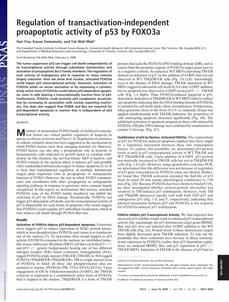
ResultsActivation of FOXO3a Induces p53-Dependent Apoptosis. Genotoxicstress triggers p53 to induce expression of SGK1 protein kinase,which in turn phosphorylates FOXO3a and causes it to translocateout of the nucleus (7). To determine what would happen to p53activity if FOXO3a remained in the nucleus, we established induc-ible mouse embryonic fibroblast (MEF) cell lines (in both p53���and p53��� genetic backgrounds) bearing one of two differentestrogen receptor (ER) fusion constructs: hemagglutinin (HA)-tagged FOXO3a-triple mutant (TM)-ER (TM-ER) or HA-taggedFOXO3a-TM�DB-ER (TM�DB-ER). TM is a triple mutant formof FOXO3a in which all three Akt phosphorylation sites aremutated to alanine (FOXO3a-TM, T32A�S253A�S315A). Uponengagement of ER by 4-hydroxytamoxifen (4-OHT), the TM-ERconstruct is expressed as a constitutively active form of FOXO3athat is trapped in the nucleus. TM�DB-ER is a form of TM-ER
protein that lacks the FOXO3a DNA binding domain (DB), and socannot bind the promoter regions of FOXO3a target genes (servesas a negative control). As expected, WT MEFs expressing TM-ERshowed an induction of p27 on the addition of 4-OHT that was notobserved in WT TM�DB-ER cells (Fig. 1A Left). Interestingly,even in the absence of DNA damage, TM-ER expression in WTMEFs triggered substantial cell death by 8 h after 4-OHT addition,but no apoptosis was observed in 4-OHT-treated p53��� TM-ERcells (Fig. 1A Right). Thus, FOXO3a-induced apoptosis is p53-dependent. Induction of TM�DB-ER in WT MEFs failed to induceany apoptosis, indicating that the DNA binding domain of FOXO3ais essential for cell death under these circumstances. Furthermore,when genotoxic stress in the form of UV or etoposide (Etop) wasapplied simultaneously with TM-ER induction, the proportion ofcells undergoing apoptosis increased significantly (Fig. 1B). Theadditional activation of apoptotic program in these cells induced byFOXO3a-TM plus DNA damage was confirmed by examination ofcaspase-3 cleavage (Fig. 1C).
Stabilization of p53 by Nuclear, Activated FOXO3a. The requirementof p53 for FOXO3a-induced apoptosis implied that there mightbe a functional interaction between these two transcriptionfactors. To explore this possibility, we determined p53 proteinlevels as well as p53 transcriptional activity in WT TM-ER andWT TM�DB-ER cells. Upon addition of 4-OHT, p53 proteinwas markedly increased in TM-ER cells but not in TM�DB-ERcells (Fig. 1 A Left). However, using quantitative real-time PCR,we determined that this difference was not because of regulationof p53 gene transcription by FOXO3a (data not shown). Rather,we found that TM-ER activation extended the half-life of p53from its usual 20 min (under physiological conditions) to �90min (Fig. 1D). Because FOXO3a has been shown to bind p53 (8),we then investigated whether protein-protein interaction wasinvolved in TM-induced p53 stabilization. However, both TMand TM�DB interacted equally well with both GST-p53 andendogenous p53 (Fig. 1 E and F, respectively), indicating thatphysical interaction between p53 and FOXO3a is not requiredfor FOXO3a-induced p53 stabilization.
FOXO3a Inhibits p53 Transcriptional Activity. We had expected thatincreased p53 stability would result in enhanced p53 transcriptionalactivity, but, surprisingly, the p53 downstream target genes MDM2,Bax, and p21 were not induced after 4-OHT addition to the WTTM-ER cells (Fig. 2A). Protein levels of these downstream targetswere slightly decreased upon TM-ER induction. To rule out thepossibility that these reductions were because of direct transcrip-tional repression by FOXO3a (rather than p53-dependent regula-tion), we analyzed MDM2, Bax, and p21 expression in p53���TM-ER MEFs. Induction of TM-ER in the absence of p53 had no
Conflict of interest statement: No conflicts declared.
Abbreviations: 4-OHT, 4-hydroxytamoxifen; ER, estrogen receptor; Etop, etoposide; HA,hemagglutinin; MEF, mouse embryonic fibroblast; NES, nuclear export signal; TM, triplemutant.
*To whom correspondence should be addressed. E-mail: [email protected].
© 2006 by The National Academy of Sciences of the USA
www.pnas.org�cgi�doi�10.1073�pnas.0600889103 PNAS � June 13, 2006 � vol. 103 � no. 24 � 9051–9056
CELL
BIO
LOG
Y
Dow
nloa
ded
by g
uest
on
Feb
ruar
y 6,
202
2

impact on the expression of MDM2 and Bax (Fig. 2B), but p21levels were slightly elevated. This latter result is consistent with arecent report that p21 is a direct downstream target of FOXO (9).
We next determined whether FOXO3a-TM could affect p53transcriptional activity in the presence of DNA damage. Uponadding 4-OHT, p53 activation in response to DNA damage signals
was inhibited. Western blotting showed that the inductions ofMDM2, p21, and Bax in response to UV or Etop treatment wereall decreased upon activation of TM-ER (Fig. 2A) but not after theactivation of TM�DB-ER (Fig. 2C). These results were confirmedand extended for WT TM-ER MEFs by using quantitative real-timePCR to analyze mdm2, bax, and noxa mRNA expression (Fig. 2D).Taken together, these data indicate that constitutively activeFOXO3a impairs p53 transcriptional activity. Moreover, TM-
Fig. 1. Regulation of p53 protein stability and p53-dependent apoptosis byFOXO3a. Nuclear, activated FOXO3a induces p53-dependent apoptosis in theabsence (A) or presence (B) of DNA damage. (A Left) Western blot of lysatesof cells exposed to 4-OHT for 6 h probed with antibodies against the indicatedproteins. (A Right) WT, WT TM�DB–ER, p53��� TM-ER, and WT TM-ER MEFswere exposed to 4-OHT (0.5 �M) for 8 or 16 h. Percentages of apoptotic cellsin the cultures were determined by flow cytometry. (B) WT TM-ER cells wereleft untreated or treated with UV (30J�m2) or Etop (0.5 �M) in the presence orabsence of 4-OHT (0.5 �M) for 8 h. Percentages of apoptotic cells weredetermined as in A. (C) Confirmation of apoptosis by caspase-3 cleavage. WTTM-ER cells were treated as in B and the cleavage of caspase-3 in cell lysateswas examined by Western blotting. (D) FOXO3a-TM stabilizes steady-statelevels of endogenous p53 protein. WT MEFs expressing TM-ER were leftuntreated or treated with 0.5 �M 4-OHT along with cycloheximide (CHX; 40�g�ml) for indicated time points. Cell extracts were subjected to Westernblotting. (E and F) In vitro and in vivo interaction between p53 and FOXO3a.(E) The p53 DNA binding domain is required for in vitro interaction withFOXO3a. (F) GST or GST-full-length p53, the p53 N terminus (N), the p53 middleregion (M), or the p53 C terminus (C) was incubated with in vitro-translatedHA-FOXO3a-TM or HA-FOXO3a-TM�DB. Bound FOXO3a was detected byanti-HA immunoblotting. Both FOXO3a-TM and TM�DB were able to interactwith endogenous p53. (F) In vivo association of p53 and FOXO3a. WT MEFsexpressing TM-ER or TM�DB-ER were left untreated or treated with 4-OHT for8 h. Immunoprecipitates obtained by using anti-p53 Ab were immunoblottedwith anti-HA and anti-p53 Abs as indicated.
Fig. 2. Activation of FOXO3a-TM inhibits p53 transcriptional activity. (A)Inhibition of p53-induced target protein expression. WT TM-ER cells weretreated for 8 h with 4-OHT (0.5 �M), UV (30J�m2), or Etop (0.5 �M) alone, orin the indicated combinations. Western blotting was performed to assessprotein levels of p53 and its downstream targets, MDM2, Bax, and p21. (B andC) p53��� TM-ER MEF cells (B), as well as WT TM�DB–ER MEFs (C), weretreated with 4-OHT (0.5 �M) or Etop (0.5 �M) alone, or treated with Etopalong with 4-OHT for 8 h. Western blotting was performed for antibodies asindicated. (D) Inhibition of p53 target gene mRNA expression. WT TM-ER MEFswere treated as in A, and total RNA was extracted. Levels of mdm2, bax, andnoxa mRNA were assessed by quantitative real-time PCR and normalized tocontrol TBP mRNA. (E and F) Inhibition of p53 DNA binding activity. (E) Gelshift assays were performed by using nuclear extracts from WT TM-ER orTM�DB-ER cells treated as in A. p53-DNA complex formation was visualized byautoradiography. (F) Chromatin immunoprecipitation (ChIP) assays to detectassociation between p53 and mdm2 or bax promoter were performed asdescribed in Supporting Text, which is published as supporting information onthe PNAS web site, by using WT TM-ER MEFs treated as indicated for 8 h. DNAfrom input samples was amplified for normalization. Results shown are onetrial representative of three independent experiments.
9052 � www.pnas.org�cgi�doi�10.1073�pnas.0600889103 You et al.
Dow
nloa
ded
by g
uest
on
Feb
ruar
y 6,
202
2
induced p53 protein stabilization might be partly because of theattenuated MDM2 mediated-ubiquitination via the decreased ex-pression of MDM2 that resulted from impaired p53 transactivation.
To investigate whether the observed reduction in p53 transcrip-tional activity was because of a decrease in its DNA bindingcapacity, we performed both gel shift and chromatin immunopre-cipitation (ChIP) assays. Nuclear extracts were prepared from WTTM-ER or TM�DB-ER MEFs that had been left untreated ortreated with either UV or Etop, in the presence or absence of4-OHT. The binding of p53 in these extracts to a DNA fragmentcontaining a consensus p53-binding element was then analyzed bygel shift assay. p53-DNA complex formation was significantlydecreased in TM-ER, but not in TM�DB-ER, cells when 4-OHTwas added to the culture (Fig. 2E), demonstrating that the TM formof FOXO3a is able to prevent p53 from binding to the p53-bindingelement in vitro. To examine p53-DNA binding in vivo, we per-formed ChIP assays by using primers flanking the p53 binding sitesin the promoter regions of mdm2 and bax genes. Consistent with thegel shift results, the ChIP assays showed that FOXO3a-TM acti-vation reduced the association of p53 with the endogenous mdm2or bax promoters on Etop treatment (Fig. 2F). Thus, the in vivoDNA binding activity of p53 is inhibited by FOXO3a-TM, even inthe presence of DNA damage signals.
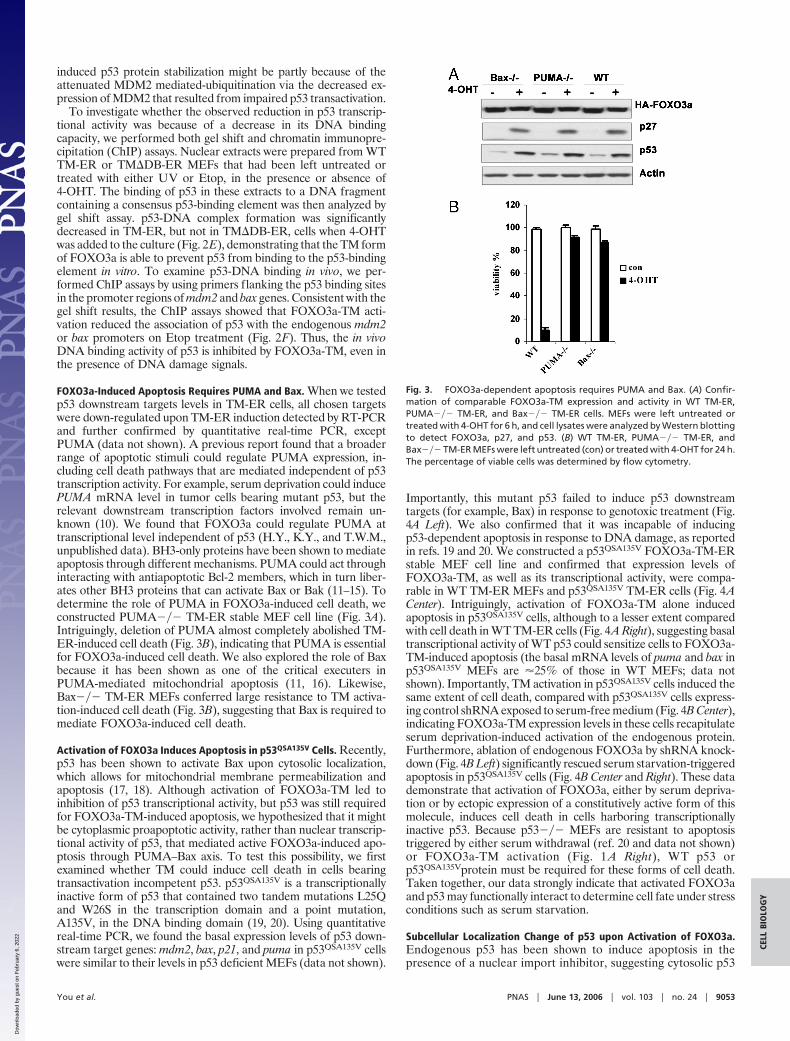
FOXO3a-Induced Apoptosis Requires PUMA and Bax. When we testedp53 downstream targets levels in TM-ER cells, all chosen targetswere down-regulated upon TM-ER induction detected by RT-PCRand further confirmed by quantitative real-time PCR, exceptPUMA (data not shown). A previous report found that a broaderrange of apoptotic stimuli could regulate PUMA expression, in-cluding cell death pathways that are mediated independent of p53transcription activity. For example, serum deprivation could inducePUMA mRNA level in tumor cells bearing mutant p53, but therelevant downstream transcription factors involved remain un-known (10). We found that FOXO3a could regulate PUMA attranscriptional level independent of p53 (H.Y., K.Y., and T.W.M.,unpublished data). BH3-only proteins have been shown to mediateapoptosis through different mechanisms. PUMA could act throughinteracting with antiapoptotic Bcl-2 members, which in turn liber-ates other BH3 proteins that can activate Bax or Bak (11–15). Todetermine the role of PUMA in FOXO3a-induced cell death, weconstructed PUMA��� TM-ER stable MEF cell line (Fig. 3A).Intriguingly, deletion of PUMA almost completely abolished TM-ER-induced cell death (Fig. 3B), indicating that PUMA is essentialfor FOXO3a-induced cell death. We also explored the role of Baxbecause it has been shown as one of the critical executers inPUMA-mediated mitochondrial apoptosis (11, 16). Likewise,Bax��� TM-ER MEFs conferred large resistance to TM activa-tion-induced cell death (Fig. 3B), suggesting that Bax is required tomediate FOXO3a-induced cell death.
Activation of FOXO3a Induces Apoptosis in p53QSA135V Cells. Recently,p53 has been shown to activate Bax upon cytosolic localization,which allows for mitochondrial membrane permeabilization andapoptosis (17, 18). Although activation of FOXO3a-TM led toinhibition of p53 transcriptional activity, but p53 was still requiredfor FOXO3a-TM-induced apoptosis, we hypothesized that it mightbe cytoplasmic proapoptotic activity, rather than nuclear transcrip-tional activity of p53, that mediated active FOXO3a-induced apo-ptosis through PUMA–Bax axis. To test this possibility, we firstexamined whether TM could induce cell death in cells bearingtransactivation incompetent p53. p53QSA135V is a transcriptionallyinactive form of p53 that contained two tandem mutations L25Qand W26S in the transcription domain and a point mutation,A135V, in the DNA binding domain (19, 20). Using quantitativereal-time PCR, we found the basal expression levels of p53 down-stream target genes: mdm2, bax, p21, and puma in p53QSA135V cellswere similar to their levels in p53 deficient MEFs (data not shown).
Importantly, this mutant p53 failed to induce p53 downstreamtargets (for example, Bax) in response to genotoxic treatment (Fig.4A Left). We also confirmed that it was incapable of inducingp53-dependent apoptosis in response to DNA damage, as reportedin refs. 19 and 20. We constructed a p53QSA135V FOXO3a-TM-ERstable MEF cell line and confirmed that expression levels ofFOXO3a-TM, as well as its transcriptional activity, were compa-rable in WT TM-ER MEFs and p53QSA135V TM-ER cells (Fig. 4ACenter). Intriguingly, activation of FOXO3a-TM alone inducedapoptosis in p53QSA135V cells, although to a lesser extent comparedwith cell death in WT TM-ER cells (Fig. 4A Right), suggesting basaltranscriptional activity of WT p53 could sensitize cells to FOXO3a-TM-induced apoptosis (the basal mRNA levels of puma and bax inp53QSA135V MEFs are �25% of those in WT MEFs; data notshown). Importantly, TM activation in p53QSA135V cells induced thesame extent of cell death, compared with p53QSA135V cells express-ing control shRNA exposed to serum-free medium (Fig. 4B Center),indicating FOXO3a-TM expression levels in these cells recapitulateserum deprivation-induced activation of the endogenous protein.Furthermore, ablation of endogenous FOXO3a by shRNA knock-down (Fig. 4B Left) significantly rescued serum starvation-triggeredapoptosis in p53QSA135V cells (Fig. 4B Center and Right). These datademonstrate that activation of FOXO3a, either by serum depriva-tion or by ectopic expression of a constitutively active form of thismolecule, induces cell death in cells harboring transcriptionallyinactive p53. Because p53��� MEFs are resistant to apoptosistriggered by either serum withdrawal (ref. 20 and data not shown)or FOXO3a-TM activation (Fig. 1 A Right), WT p53 orp53QSA135Vprotein must be required for these forms of cell death.Taken together, our data strongly indicate that activated FOXO3aand p53 may functionally interact to determine cell fate under stressconditions such as serum starvation.
Subcellular Localization Change of p53 upon Activation of FOXO3a.Endogenous p53 has been shown to induce apoptosis in thepresence of a nuclear import inhibitor, suggesting cytosolic p53
Fig. 3. FOXO3a-dependent apoptosis requires PUMA and Bax. (A) Confir-mation of comparable FOXO3a-TM expression and activity in WT TM-ER,PUMA��� TM-ER, and Bax��� TM-ER cells. MEFs were left untreated ortreated with 4-OHT for 6 h, and cell lysates were analyzed by Western blottingto detect FOXO3a, p27, and p53. (B) WT TM-ER, PUMA��� TM-ER, andBax��� TM-ER MEFs were left untreated (con) or treated with 4-OHT for 24 h.The percentage of viable cells was determined by flow cytometry.
You et al. PNAS � June 13, 2006 � vol. 103 � no. 24 � 9053
CELL
BIO
LOG
Y
Dow
nloa
ded
by g
uest
on
Feb
ruar
y 6,
202
2

may retain some proapoptotic activity even when its nuclearactivity is impaired (17). To determine how p53 is regulated byFOXO3a during FOXO3a-triggered apoptosis, we first exam-ined WT p53 subcellular localization in response to TM-ERactivation. Upon addition of 4-OHT, cytosolic p53 accumulationwas observed in WT TM-ER cells (Fig. 5A). Intriguingly,cytosolic p53 levels correlated with Bax oligomerization status.TM activation, along with UV damage, induced more cytosolicp53 accumulation compared with TM activation alone, and theupregulation of cytosolic p53 correlated with increased Baxoligomerization even when PUMA induction was achieved to thesame levels (Fig. 5 B and C), indicating cytosolic p53 might havethe potential to activate Bax, as suggested by others (17, 21).Because FOXO3a-TM activation also increased p53 proteinstability in WT cells (Fig. 2 A), the increase in cytosolic p53 inWT TM-ER cells then might be because of changes in the totalprotein level. To further confirm the role of FOXO3a in inducingp53 accumulation in cytosol, we performed cytosolic fraction-ation to detect p53 levels in p53QSA135V TM-ER cells before andafter the addition of 4-OHT, or in p53QSA135V MEFs before andafter serum starvation. Consistent with ref. 19, we found thatp53QSA135V protein displayed heterogeneous distribution underuntreated conditions. Although a large portion of p53 localizedin the nucleus (data not shown), a small percentage of cytoplas-mic p53 proteins were also observed. Activation of TM-ER orserum withdrawal significantly increased cytosolic p53 levels inS-100 fraction (Fig. 5D) without affecting total p53 proteinexpression (Fig. 4A Center). Cytosolic p53 accumulation was alsodetected when p53QSA135V cells expressing control shRNA weredeprived of serum for 6 h (Fig. 5D). This translocation of p53 wasimpaired when endogenous FOXO3a was ablated by shRNA
knockdown. Taken together, our results suggest that activationof FOXO3a, either by serum starvation or expressing TMmutant, can drive changes in p53 subcellular localization.
The enhanced cytoplasmic localization of p53 protein inp53QSA135V MEFs in response to activation of FOXO3a suggestedthat greater nuclear export of p53 might occur under these condi-tions. We hypothesized that p53 protein might be more efficientlyassociated with the key nuclear-export receptor CRM1 upon acti-vation of FOXO3a. Immunoprecipitation and Western blot analysisby using cell lysates from p53QSA135V TM-ER or p53QSA135V MEFsindicated that there was increased association of CRM1 with p53when 4-OHT or serum deprivation was applied (Fig. 5E). Intrigu-ingly, knockdown of FOXO3a significantly impaired CRM1-p53interaction after serum withdrawal (Fig. 5F), suggesting a criticalrole of FOXO3a in mediating p53 nuclear-cytoplasm shuttling.Posttranslational modifications of p53, for instance, ubiquitinationof p53 by MDM2, have been reported to contribute to p53 nuclearexport (22–24). However, the nuclear export signal (NES) of p53itself is capable of mediating cytoplasmic localization of p53 inMDM2-independent manner (25). Because p53QSA135V is defectivein MDM2 binding (19), the increased nuclear-cytoplasm shuttlingof p53 in response to FOXO3a activation might be independent ofMDM2 (see Discussion).
Discussionp53 is the most commonly mutated gene in human cancers (26, 27).The tumor suppressor function of p53 has been viewed exclusivelyas a transcription factor that conducts its many functions bytransactivating downstream targets involved in cell cycle arrest orapoptosis. Thus, p53 protein carrying loss-of-function mutations
Fig. 4. FOXO3a activation promotes cell death in p53QSA135V cells. (A) FOXO3a-TM induces apoptosis in p53QSA135V cells. (A Left) WT and p53QSA135V MEFs weretreated with doxorubicin (0.2 �g�ml) for 8 h. Western blotting was used to detect p53 and Bax. (A Center) Cell lysates from WT TM-ER and p53QSA135V TM-ERcells were subjected to Western blotting. (A Right) WT TM-ER and p53QSA135V TM-ER MEFs were treated with 4-OHT (0.5 �M) for 24 h and analyzed for apoptosisby flow cytometry. Results shown are the mean � SD of duplicate samples and are representative of three independent experiments. (B) FOXO3a is required forserum starvation-induced apoptosis in p53QSA135V cells. (B Left) Western blot showing that FOXO3a shRNA successfully blocked FOXO3a expression. (B Center)Left column, crystal violet staining to detect apoptosis in cultures of p53QSA135V TM-ER cells that were left untreated (con) or treated with 4-OHT for 48 h; centercolumn, serum starvation (48 h) induced apoptosis in p53QSA135V cells expressing control shRNA; right column, shRNA knockdown of FOXO3a prevents serumstarvation-induced cell death in p53QSA135V cells. Results shown are representative of three independent experiments. (B Right) Quantification of results fromCenter. Untreated, cells grow in medium containing 10%FBS; treated, cells were exposed to serum starvation. The difference of cell death induced by serumstarvation between control shRNA and FOXO3a-shRNA MEFs is statistically significant (P�0.001, t test).
9054 � www.pnas.org�cgi�doi�10.1073�pnas.0600889103 You et al.
Dow
nloa
ded
by g
uest
on
Feb
ruar
y 6,
202
2

has been considered as ‘‘dead’’ p53 in chemotherapy, because thesemutants confer resistance to anticancer reagents.
During the past decades, several groups reported transcriptionindependent proapoptotic activity of p53. The proposed mecha-nisms include that p53 can function at the mitochondrial (28, 29) orthat p53 can act as a BH3 protein to induce Bax activation if forcedto translocate into cytoplasm by pharmacological inhibitors (17).Here, our data presented a model that activation of FOXOtranscription factor could regulate the transactivation-independentproapoptotic activity of endogenous p53 (Fig. 6). Ectopic express-
ing active form of FOXO3a has negative effect on nuclear p53activation, even in the presence of DNA damage signals. Paradox-ically, FOXO3a is still able to induce apoptosis in p53-dependentmanner. To explore which activity of p53 contributes to FOXO3a-triggered cell death, we took advantage of a transactivation incom-petent mutant form of p53: p53QSA135V. A human p53 mutantcarrying the same mutations in residues 22 and 23 has been shownto trigger apoptosis upon overexpression, despite failing to inducesignificant activation of relevant p53 target promoters (30). Inter-estingly, in our system, activation of exogenously expressedFOXO3a induced apoptosis in p53QSA135V MEF cells. Further-more, in p53QSA135V MEFs, constitutively active FOXO3a couldmimic serum starvation-induced FOXO3a activation in inducingapoptosis, indicating that the gene dosage of exogenously expressedFOXO3a could recapitulate activation of endogenous FOXO3aunder physiological conditions.
The pathway by which p53 regulates serum starvation re-sponse has not been well characterized. Our finding thatp53QSA135V retains partial apoptotic activity under conditions ofserum starvation, whereas p53 null cells confer resistance to suchstress, suggests that a transactivation-independent activity of p53is required to induce cell death in this situation. Acute loss ofFOXO3a by shRNA knockdown significantly rescued apoptosisin p53QSA135V cells, indicating that crosstalk between the endog-enous FOXO3a and p53QSA135V proteins may determine cell fateunder such stress conditions. Moreover, we investigated themechanism by which activated FOXO3a (present either as aresult of the TM mutation or serum starvation) might regulatep53, and we found that FOXO3a induced changes to p53’s
Fig. 5. FOXO3a activation drives p53 cytoplasmic localization in p53QSA135V cells. (A) FOXO3a-TM activation induces accumulation of p53 in the cytosol. WTTM-ER MEFs were treated with UV in the presence or absence of 4-OHT for 6 h. S-100 fractions were subjected to Western blotting by using antibodies againstp53 and tubulin (cytosolic fraction indicator). Nuclear protein poly(ADP ribose) polymerase (Parp) was used to control the purity of cytosolic fractions (PC, positivecontrol). (B) WT TM-ER cells were treated as in A. PUMA expression levels were detected by quantitative real-time PCR. (C) Activation of TM induces Baxoligomerization at the mitochondrial. WT TM-ER cells were treated as in A. Mitochondrial fractions were extracted as described in ref. 39 and were subjectedto Western blotting. COX IV was used for loading control. (D) p53QSA135V MEFs expressing TM-ER, control shRNA, or FOXO3a-shRNA were treated as indicated.Western blotting was performed by using S-100 fraction. Parp was used to monitor cytoplasm fraction contamination (PC, positive control). (E) Immunopre-cipitation (IP) of p53 followed by Western blot analysis for associated nuclear export receptor CRM1 in p53QSA135V TM-ER MEFs or p53QSA135V MEFs treated asindicated. CRM1 was presented by using different exposure time (top panel, longer exposure; second panel from top, shorter exposure). p53 and CRM1 levelswere also detected by Western blotting in whole cell extracts (WCE). (F) p53QSA135V MEFs expressing control shRNA or FOXO3a shRNA were serum starved for6 h; IP was conducted as in E. CRM1 and p53 levels in either immune complexes or WCE were detected by using specific antibodies by Western blotting.
Fig. 6. Model of apoptosis induced by nuclear activated FOXO3a andmediated by cytosolic p53 and PUMA–Bax axis.
You et al. PNAS � June 13, 2006 � vol. 103 � no. 24 � 9055
CELL
BIO
LOG
Y
Dow
nloa
ded
by g
uest
on
Feb
ruar
y 6,
202
2

subcellular localization. Importantly, the impairment of cytoso-lic p53 localization in p53QSA135V cells that occurred uponFOXO3a shRNA knockdown correlated with the decrease incell death observed after serum deprivation. We also performedserum starvation by using WT MEFs to see whether activationof endogenous FOXO3a could mimic FOXO3a-TM in regulat-ing WT p53. Surprisingly, serum withdrawal led to significantstabilization of p53, which subsequently induced apoptosis (H.Y.and T.W.M., unpublished data). Our preliminary data suggestposttranslational modifications of p53 may play an importantrole in p53 stabilization after serum deprivation. Taken together,it is likely that both transactivation-dependent and transactiva-tion-independent activities of p53 are involved in regulatingserum starvation response. Our results provided evidence thatFOXO3a could regulate the latter activity of p53 throughsubcellular localization change-mediated pathway.
How does FOXO3a regulate p53 subcellular translocation? Ahighly conserved leucine-rich NES has been identified in thetetramerization domain of p53. This intrinsic NES is both necessaryand sufficient to direct p53 nuclear export (25). Interestingly, underconditions when FOXO3a was activated by 4-OHT or serumstarvation, we found an increased association between p53 proteinand the nuclear-export receptor CRM1. Tetramerization of p53 hasbeen proposed as a potential mechanism leading to p53 nuclearaccumulation and activation by masking the NES and preventingaccess to the nuclear export machinery (31–33). Furthermore, it iswell known that tetrameric p53 binds its DNA response elementsmost efficiently and is most effective for transactivation (34–38).Thus, we hypothesize that activation of FOXO3a may interfere withp53 oligomerization, which in turn unmasks p53 NES, and resultsin increased nuclear export, as well as impairment of p53 DNAbinding and transactivation.
Materials and MethodsCell Culture. p53QSA135V primary MEF cells were provided byGeoffrey Wahl (The Salk Institute, La Jolla, CA). Primary PU-
MA��� MEFs were the generous gift of Andreas Strasser (TheWalter and Eliza Hall Institute, Melbourne, Australia). Bax���MEFs were the kind gift of Doug Green (St. Jude Children’sResearch Hospital, Memphis, TN). All primary MEFs were trans-formed by E1A�ras G12V, followed by puromycin selection. ForFOXO3a-related stable cell lines, MEFs were cotransfected withpECE HA-FOXO3a (TM or DB) and a GFP plasmid in 15:1 ratio.Cells were selected by GFP sorting after 48 h, and positive cells werepooled together for use in experiments.
RT-PCR and Real-Time PCR. Total RNA was extracted withTRIZOL (Invitrogen) and purified by using the RNeasy kit(Qiagen) according to the manufacturer’s protocol, with theaddition of on-column DNase treatment (Qiagen). RNA (4 �g)was reverse-transcribed in a 20-�l reaction by using the Super-script first strand RT-PCR kit (Invitrogen). After RNase Htreatment at 37°C for 20 min, followed by inactivation at 95°C for2 min, the real-time reaction was diluted. cDNA was used forRT-PCR or real-time PCR. Primer sequences were as follows:Noxa, 5�-CCACCTGAGTTCGCAGCTCAA and 5�-GTT-GAGCACACTCGTCCTTCAA; and p27, 5�-TCAAACGT-GAGAGTGTCTAACGG and 5�-AGGGGCTTATGATTCT-GAAAGTCG. MDM2, p21, and Bax primers were from ref. 17.
We thank A. Levine (Institute for Advanced Study, Princeton), M.Greenberg (Harvard Medical School, Boston), S. Lowe (Cold SpringHarbor Laboratory, Cold Spring Harbor, NY), G. Wahl (The SalkInstitute, La Jolla, CA), D. Green (St. Jude Children’s ResearchHospital, Memphis, TN), B. Vogelstein (Johns Hopkins Medical Insti-tutions, Baltimore), A. Strasser (The Walter and Eliza Hall Institute ofMedical Research, Melbourne), W. Gu (Columbia University, NewYork), and H. Lu (Oregon Health and Science University, Portland, OR)for reagents; K. Vousden (The Beatson Institute, Glasgow, U.K.) and S.Benchimol (University of Toronto, Toronto) for critical discussions; andM. Saunders for scientific editing. This work was supported by the TerryFox Cancer Foundation of the National Cancer Institute of Canada. H.Y.is a recipient of a postdoctoral fellowship from the Cancer ResearchInstitute (New York).
1. Lin, K., Hsin, H., Libina, N. & Kenyon, C. (2001) Nat. Genet. 28, 139–145.2. Hwangbo, D. S., Gersham, B., Tu, M. P., Palmer, M. & Tatar, M. (2004) Nature
429, 562–566.3. Giannakou, M. E., Goss, M., Junger, M. A., Hafen, E., Leevers, S. J. &
Partridge, L. (2004) Science 305, 361.4. Nemoto, S. & Finkel, T. (2002) Science 295, 2450–2452.5. Stahl, M., Dijkers, P. F., Kops, G. J., Lens, S. M., Coffer, P. J., Burgering, B. M.
& Medema, R. H. (2002) J. Immunol. 168, 5024–5031.6. Gilley, J., Coffer, P. J. & Ham, J. (2003) J. Cell Biol. 162, 613–622.7. You, H., Jang, Y., You-Ten, A. I., Okada, H., Liepa, J., Wakeham, A., Zaugg,
K. & Mak, T. W. (2004) Proc. Natl. Acad. Sci. USA 101, 14057–14062.8. Brunet, A., Sweeney, L. B., Sturgill, J. F., Chua, K. F., Greer, P. L., Lin, Y., Tran,
H., Ross, S. E., Mostoslavsky, R., Cohen, H. Y., et al. (2004) Science 303,2011–2015.
9. Seoane, J., Le, H. V., Shen, L., Anderson, S. A. & Massague, J. (2004) Cell 117,211–223.
10. Han, J., Flemington, C., Houghton, A. B., Gu, Z., Zambetti, G. P., Lutz, R. J.,Zhu, L. & Chittenden, T. (2001) Proc. Natl. Acad. Sci. USA 98, 11318–11323.
11. Yu, J., Wang, Z., Kinzler, K. W., Vogelstein, B. & Zhang, L. (2003) Proc. Natl.Acad. Sci. USA 100, 1931–1936.
12. Yu, J., Zhang, L., Hwang, P. M., Kinzler, K. W. & Vogelstein, B. (2001) Mol.Cell 7, 673–682.
13. Nakano, K. & Vousden, K. H. (2001) Mol. Cell 7, 683–694.14. Cheng, E. H., Wei, M. C., Weiler, S., Flavell, R. A., Mak, T. W., Lindsten, T.
& Korsmeyer, S. J. (2001) Mol. Cell 8, 705–711.15. Korsmeyer, S. J., Wei, M. C., Saito, M., Weiler, S., Oh, K. J. & Schlesinger, P. H.
(2000) Cell Death Differ. 7, 1166–1173.16. Melino, G., Bernassola, F., Ranalli, M., Yee, K., Zong, W. X., Corazzari, M.,
Knight, R. A., Green, D. R., Thompson, C. & Vousden, K. H. (2004) J. Biol.Chem. 279, 8076–8083.
17. Chipuk, J. E., Kuwana, T., Bouchier-Hayes, L., Droin, N. M., Newmeyer, D. D.,Schuler, M. & Green, D. R. (2004) Science 303, 1010–1014.
18. Chipuk, J. E., Bouchier-Hayes, L., Kuwana, T., Newmeyer, D. D. & Green,D. R. (2005) Science 309, 1732–1735.
19. Jimenez, G. S., Nister, M., Stommel, J. M., Beeche, M., Barcarse, E. A., Zhang,X. Q., O’Gorman, S. & Wahl, G. M. (2000) Nat. Genet. 26, 37–43.
20. Johnson, T. M., Hammond, E. M., Giaccia, A. & Attardi, L. D. (2005) Nat.Genet. 37, 145–152.
21. Erster, S., Mihara, M., Kim, R. H., Petrenko, O. & Moll, U. M. (2004) Mol. Cell.Biol. 24, 6728–6741.
22. Gottifredi, V. & Prives, C. (2001) Science 292, 1851–1852.23. Dumont, P., Leu, J. I., Della Pietra, A. C., III, George, D. L. & Murphy, M.
(2003) Nat. Genet. 33, 357–365.24. Lohrum, M. A., Woods, D. B., Ludwig, R. L., Balint, E. & Vousden, K. H.
(2001) Mol. Cell. Biol. 21, 8521–8532.25. Stommel, J. M., Marchenko, N. D., Jimenez, G. S., Moll, U. M., Hope, T. J.
& Wahl, G. M. (1999) EMBO J. 18, 1660–1672.26. Vogelstein, B., Lane, D. & Levine, A. J. (2000) Nature 408, 307–310.27. Levine, A. J. (1997) Cell 88, 323–331.28. Mihara, M., Erster, S., Zaika, A., Petrenko, O., Chittenden, T., Pancoska, P.
& Moll, U. M. (2003) Mol. Cell 11, 577–590.29. Leu, J. I., Dumont, P., Hafey, M., Murphy, M. E. & George, D. L. (2004) Nat.
Cell Biol. 6, 443–450.30. Haupt, Y., Rowan, S., Shaulian, E., Vousden, K. H. & Oren, M. (1995) Genes.
Dev. 9, 2170–2183.31. Lee, W., Harvey, T. S., Yin, Y., Yau, P., Litchfield, D. & Arrowsmith, C. H.
(1994) Nat. Struct. Biol. 1, 877–890.32. Clore, G. M., Ernst, J., Clubb, R., Omichinski, J. G., Kennedy, W. M.,
Sakaguchi, K., Appella, E. & Gronenborn, A. M. (1995) Nat. Struct. Biol. 2,321–333.
33. Jeffrey, P. D., Gorina, S. & Pavletich, N. P. (1995) Science 267, 1498–1502.34. McLure, K. G. & Lee, P. W. (1998) EMBO J. 17, 3342–3350.35. Hupp, T. R. & Lane, D. P. (1994) Curr. Biol. 4, 865–875.36. Friedman, P. N., Chen, X., Bargonetti, J. & Prives, C. (1993) Proc. Natl. Acad.
Sci. USA 90, 3319–3323.37. Hainaut, P., Hall, A. & Milner, J. (1994) Oncogene 9, 299–303.38. Halazonetis, T. D. & Kandil, A. N. (1993) EMBO J. 12, 5057–5064.39. Zong, W. X., Li, C., Hatzivassiliou, G., Lindsten, T., Yu, Q. C., Yuan, J. &
Thompson, C. B. (2003) J. Cell Biol. 162, 59–69.
9056 � www.pnas.org�cgi�doi�10.1073�pnas.0600889103 You et al.
Dow
nloa
ded
by g
uest
on
Feb
ruar
y 6,
202
2