renal cancer - baus.org.uk€¦ · renal cancer tom walton january 2011 1 renal cancer demographics...
TRANSCRIPT

Renal cancer
Tom Walton January 2011 1
Renal Cancer
Demographics 3% of all cancers in US. 7th leading cancer in men and 9th leading cancer in females Peak age 60-70 Male-female ratio ~2:1 Western World Increasing incidence associated with improved 5YS – presumed 2’ to more imaging Aetiology Smoking – RR 1.4-2.3 Obesity Hypertension Dialysis – with native kidneys in situ (3-6x increased risk) Heredity (2% of all RCC) VHL disease Hereditary Papillary Renal Carcinoma (HPRC) Hereditary Leiomyomatosis & Papillary RCC (HLRCC) Birt-Hogg Dube syndrome Presentation Incidental finding in over 50% [Novick 1999] Haematuria Loin Pain Palpable Mass Classical Triad of haematuria, loin pain, mass in 10% Paraneoplastic syndromes Hypercalcaemia (PTHrP) – seen in approximately 25% Anaemia Hyponatraemia Raised ESR Polycythaemia - may be associated with inceased response to IFN Stauffer’s syndrome Abnormal LFTs Hepatic necrosis Thrombocytopenia Neutropenia Fever Weight loss ? secondary to elevated IL-6. Majority of cases respond to nephrectomy but poor prognostic sign Occasionally metastatic disease (found in 30% when looked for)

Renal cancer
Tom Walton January 2011 2
Pathology Heidelberg Classification 1997 Sporadic Renal Cell Carcinoma:
Conventional clear-cell 75% (60% VHL mutation) Papillary 12% (13% MET oncogene mutation) Chromophobe 4% Collecting Duct 1% Unclassified* 3-5%
* One recently characterised tumour is renal medullary carcinoma Features Blacks Almost exclusively with sickle cell disease Third decade of life Usually widespread metastasis Highly aggressive and resistant to conventional Rx Life expectancy a few months RCC as an inherited syndrome:
VHL disease (clear-cell) VHL gene mutation/deletion Familial clear cell renal cancer chromosome 3p translocation HPRCC (papillary) MET oncogene HLRCC (papillary) Fumarate hydratase gene Birt-Hogg-Dube (chromophobe or BHD gene oncocytoma)
Conventional or clear-cell RCC Derived from cells of proximal convoluted tubule. Large cells typically full of glycogen and cholesterol, do not take up stain, thus clear. Almost always positive for cytokeratib 8/18 and 50% positive for vimentin. Abundant mitochondria on EM. Associated with VHL gene defects in up to 60% of sporadic cases. Presence of spindle cells (sarcomatoid variant – 5% CCRCC) associated with worse prognosis.
Renal cancer
Tom Walton January 2011 3
VHL disease and the hypoxia-inducible factor pathway Rare autosomal dominant genetic disease 1 in 36,000 live births Characterised by inheritance of mutated VHL tumour suppressor gene on short arm of chromosome 3 (3p25-26) 50% chance of offspring inheriting defective gene Disease results from inactivation or silencing of normal (wild-type) allele 25-50% of VHL ‘carriers’ will get RCC in lifetime. 70% risk aged 60. 3 major manifestations of VHL disease: Clear-cell RCC; CNS haemangio-blastomas [retina, cerebellum]; phaeochromocytoma Also hepatic and pancreatic cysts VHL classification Type 1 Most common Due to major disrupting mutation e.g. truncation
Associated HIF-α upregulation Clear-cell RCC with CNS haemangioblastoma Type 2 Uncommon More subtle mutations e.g. missense mutations Phaeochromocytoma +/- other 2a Phaeo and CNS haemangioblastoma 2b Phaeo, clear-cell RCC and CNS haemangioblastoma 2c Phaeo alone Hypoxia inducible factor is a crucial regulator of the hypoxic response. Normally, in conditions of low oxygen tension, HIFα binds to HIFβ, translocates to the nucleus and binds to a specific HIF response element (HRE) on the promoter of of a number of hypoxia inducible genes. Examples include VEGF, PDGF, Epo, TGF-α, GLUT-1 (glucose transport) and carbonic anhydrase 9. HIF-α subunits (three have been described to date, HIF-1α, HIF-2α and HIF-3α) appear to be subject to regulation by VHL, rather than constitutively expressed HIF-β. VHL functions to inhibit HIF-1α. In normoxia, HIF-1α is hydroxylated (-OH added) by proline and asparagine hydroxylases. VHL binds to hydroxylated proline residues on HIF-α, and in association with elongins B and C and Cul2, marks it for ubiquitination and subsequent destruction by the 26S proteosome. VHL defects thus lead to inappropriate HIF-1α upregulation in normoxic conditions. Papillary RCC (chromophilic) Associated with loss of chromosome 4 and Y, trisomy 7 &17 Mutation of C-MET proto-oncogene (C-MET gene identified on chromosome 7q31.1-35) in 13%. Associated with aquired cystic disease and ESRF Multicentricity in 40%

Renal cancer
Tom Walton January 2011 4
Cell of origin controversial. Campbells - proximal convoluted tubule. Others think derived from distal convoluted tubule. Two types described: Type 1 (figure B) Basal cells Basophilic cytoplasm (H&E) Benign cf. type 2 Type 2 (figure C) Extra cell layers Eosinophilic cytoplasm Earlier metastasis cf. type 1 Clinical behaviour of papillary RCC controversial. Some authors report improved behaviour vs. CCRCC whilst other do not. Probably due to failure to subdivide papillary RCC into type 1 and 2.
Hereditary papillary RCC (HPRCC) First reported in 1995 (Zbar 1995) Type 1 papillary RCC Autosomal dominant transmission C-MET gene mutation on chromosome 7q31.1-35 (protein product = tyrosine kinase receptor for hepatocyte growth factor) Early onset, multifocality and aggression common features Also associated with ca breast, pancreas, lung (SCC) Hereditary leiomyomatosis and renal cell cancer (HLRCC) Aka Lynch syndrome Type 2 papillary RCC – most aggressive inherited RCC cancer syndrome Autosomal dominant transmission (mutated Fumarate Hydratase (FH) gene on 1q42.3-q43) Cutaneous leiomyomata, uterine fibroids, papillary RCC Chromophobe RCC First reported in 1985 Derived from cortical portion of collecting ducts

Renal cancer
Tom Walton January 2011 5
Typically large polygonal cells with relatively unstained cytoplasm (chromophobe) and reticular growth pattern reminscent of plant cells (figure D) Eosinophilic variant which can be difficult to distinguish from oncocytoma, but Ultrastructurally packed with microtubules (cf. mitochondria in oncocytoma) Associated with LOH 3p, 5p and 17p Clinical behaviour poorly characterised. Thought to be of less malignant potential than conventional clear cell RCC and papillary RCC.
Birt-Hogg-Dube syndrome BHD gene identified on chromosome 17p11.2 Gene encodes ubiquitously expressed protein (folliculin) Hair follicle tumours (forehead, ears, nose, cheeks and neck) Spontaneous pneumothorax Renal tumours: oncocytoma; chromophobe, clear cell, papillary RCC Collecting Duct Arise from medullary collecting duct Usually younger patients Aggressive growth Positive staining for ulex eropeus lectin differentiates collectind duct carcinoma from TCC Oncocytoma Benign growth Mahogany brown tumour Derived from distal convoluted tubule Uniform round, polygonal cells with dense eosinophilic staining Packed with mitochondria at ultrastructural level Different genetic profile for sporadic oncocytoma cf. BHD. Defects on chromosome 1, Y, and the long arms of chromosomes 11 and 14 Little cytological atypia May have central necrosis and stellate scar on CT but predictive value poor Difficult to differentiate from granular clear-cell RCC or eosinophilic chromophobe RCC on biopsy
Renal cancer
Tom Walton January 2011 6
Co-exists with RCC in 7-32% of cases [Campbells]. Therefore surgical treatment advocated – partial if strongly suspected (BHD and features) and favourable location

Renal cancer
Tom Walton January 2011 7
Radiologic evaluation Ultrasound Simple Non-ionising Good for distinguishing solid, cystic and complex masses Features of benign cyst: Round/oval shape No internal echoes Smooth well-defined wall Acoustic shadow from wall edges only Good US transmission US combined with CT characteristics formed the basis of original Bosniak cyst classification (Bosniak 1986). No problems assigning cysts to Category 1 & 4 assuming good quality USS/CT, but due to difficulties characterising 2 & 3, led to the introduction of intermediate cataegory of 2F (Bosniak 1997)
Warren 2004

Renal cancer
Tom Walton January 2011 8
Meta-analysis of all studies validating Bosniak classification shown above. NB. All suffer from retrospective selection bias and surgery selection bias. Thus, values in category 1 and 2 likely to be an over-estimation as do not account for patients entering surveillance without biopsy/extirpation. Also reference 17 above is likely to give an artificially high rate of maligancy for category 2 tumours. In fact Campbell’s quotes quite different rates of malignancy:
Other techniques for evaluating indeterminate (category 2/3) cysts? CT-guided biopsy: Lang 2002 – Bosniak 2F/3. 199 biopsies. Adequate material in 90%. Sensitivity for malignancy only 70%, positive predictive value 91%, specificity & negative predictive value (reported as sensitivity and PPV for benign disease approximately 90%); Harisinghani 2003. Bosniak 3, n= 28. adequacy in 100%, incidence of malignancy 61%. No follow-up tumours. Surgery therefore avoided in 39%. Laparoscopic biopsy: Limb 2002 – Described feasibility (n=57 Bosniak 2&3). 8 (20%) cancers detected. No port site recurrences. Computed tomography Investigation of choice for staging of renal tumours CT protocol: Unenhanced scan IV contrast runs Arterial phase 15-30s Late arterial (portal venous)* 45-60s Nephrographic phase 75-90s Excretory phase** 180s

Renal cancer
Tom Walton January 2011 9
*evaluation of liver metastases/other organ mets in this phase ** evaluation of urinary tract through to bladder in this phase 3D multiplanar reformats typically 1.5mm slices with 50% overlap Unenhanced scans important to exclude hepatic mets. RCC mets typically hypervascular. Therefore enhance with rest of liver after IV contrast. CCRCC enhances > other histologic subtypes: Kim 2002 = increased attenuation of >= 84HU highly specific (100%) for clear cell RCC cf. other subtypes Central stellate scar not unique to oncocytomas – can be seen in necrotic CCRCC Oral contrast typically given to identify duodenum Many centres give 2 doses of IV contrast 3-5 mins apart (double-dose contrast scanning) and scan after second dose, thereby gaining portal venous and excretory information in one scan, limiting radiation dose. NB. Metastatic non-renal cancer involving kidney 8-13% in some series. Enhancement and absence of other non-renal mets increases likelihood of primary RCC. In one series of 100 patients with primary extrarenal cancer and a lump in the kidney, 86% of non-enhancing tumours and 31% on enhancing tumours were secondary deposits. All patients had evidence of other non-renal mets. In those with a primary non-metastatic cancer and solitary renal mass, the solitary renal mass was a RCC in all cases (Campbell’s). CT very good for determining renal vein/IVC involvement and involvement of adjacent organs. Higher false positives for lymph node metastases and capsular breach. MRI Equivalent staging accuracy to CT (Beer 2006) Less sensitive for solid lesion < 3 cm Better cf. CT and Doppler USS for determining superior extent of IVC involvement (coronal plane) Clinically used for patients with renal insufficiency/pregnancy in whom CT/iodinated contrast contraindicated NB. Increasing awareness of risk of nephrogenic systemic fibrosis (NSF) with gadolinium based contrast agents: severe multisystem disease associated with fibrosis and skin rashes – usually in patients with significantly reduced GFR and multiple exposure. Renal mass biopsy for non-cystic mass lesions Limited usage until recently for a number of reasons:
High false negative rates High perceived rate of complications 80-90% solid enhancing renal masses malignant No suitable alternative to surgical extirpation so would not change Mx Recent advances have led to a desire for pre-operative diagnosis
Renal cancer
Tom Walton January 2011 10
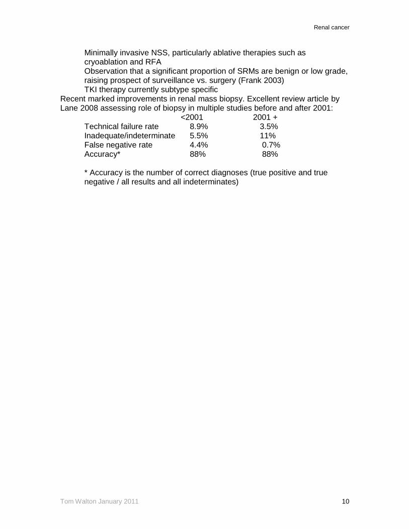
Minimally invasive NSS, particularly ablative therapies such as cryoablation and RFA Observation that a significant proportion of SRMs are benign or low grade, raising prospect of surveillance vs. surgery (Frank 2003) TKI therapy currently subtype specific
Recent marked improvements in renal mass biopsy. Excellent review article by Lane 2008 assessing role of biopsy in multiple studies before and after 2001: <2001 2001 + Technical failure rate 8.9% 3.5% Inadequate/indeterminate 5.5% 11%
False negative rate 4.4% 0.7% Accuracy* 88% 88% * Accuracy is the number of correct diagnoses (true positive and true negative / all results and all indeterminates)
Renal cancer
Tom Walton January 2011 11
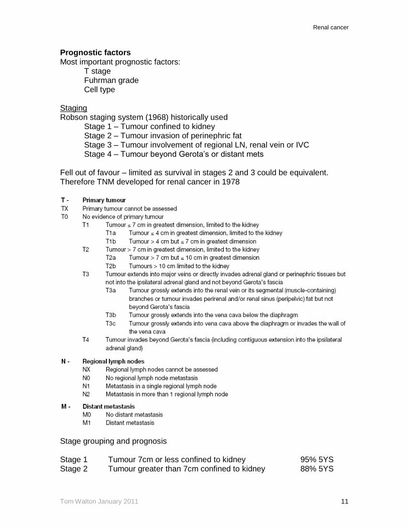
Prognostic factors Most important prognostic factors: T stage
Fuhrman grade Cell type Staging Robson staging system (1968) historically used Stage 1 – Tumour confined to kidney Stage 2 – Tumour invasion of perinephric fat Stage 3 – Tumour involvement of regional LN, renal vein or IVC Stage 4 – Tumour beyond Gerota’s or distant mets Fell out of favour – limited as survival in stages 2 and 3 could be equivalent. Therefore TNM developed for renal cancer in 1978
Stage grouping and prognosis Stage 1 Tumour 7cm or less confined to kidney 95% 5YS Stage 2 Tumour greater than 7cm confined to kidney 88% 5YS
Renal cancer
Tom Walton January 2011 12
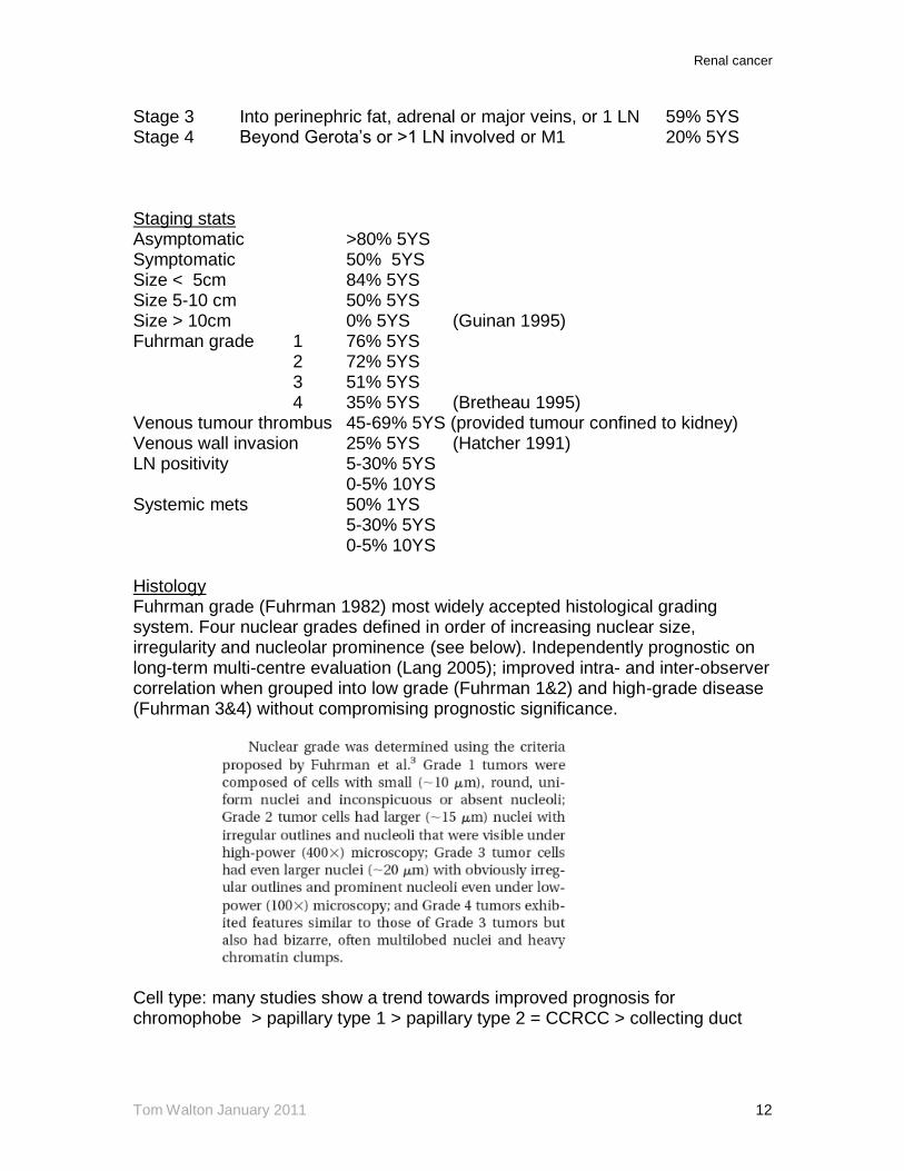
Stage 3 Into perinephric fat, adrenal or major veins, or 1 LN 59% 5YS Stage 4 Beyond Gerota’s or >1 LN involved or M1 20% 5YS Staging stats Asymptomatic >80% 5YS Symptomatic 50% 5YS Size < 5cm 84% 5YS Size 5-10 cm 50% 5YS Size > 10cm 0% 5YS (Guinan 1995) Fuhrman grade 1 76% 5YS 2 72% 5YS 3 51% 5YS 4 35% 5YS (Bretheau 1995) Venous tumour thrombus 45-69% 5YS (provided tumour confined to kidney) Venous wall invasion 25% 5YS (Hatcher 1991) LN positivity 5-30% 5YS
0-5% 10YS Systemic mets 50% 1YS 5-30% 5YS
0-5% 10YS Histology Fuhrman grade (Fuhrman 1982) most widely accepted histological grading system. Four nuclear grades defined in order of increasing nuclear size, irregularity and nucleolar prominence (see below). Independently prognostic on long-term multi-centre evaluation (Lang 2005); improved intra- and inter-observer correlation when grouped into low grade (Fuhrman 1&2) and high-grade disease (Fuhrman 3&4) without compromising prognostic significance.
Cell type: many studies show a trend towards improved prognosis for chromophobe > papillary type 1 > papillary type 2 = CCRCC > collecting duct

Renal cancer
Tom Walton January 2011 13
carcinoma. NB. Prognostic effect of cell type disappears when controlled for T-stage. Spindle cell component also confers worse prognosis. ECOG performance status, symptoms and presence of systemic features associated with a worse prognosis (multiple EAU guidelines references) Molecular markers reported to be associated with a worse prognosis are carbonic anhydrase 9, Ki67 proliferation index, HIF, VEGF, p53, PTEN and E-cadherin. However, none achieved widespread acceptance as not independently prognostic. DNA ploidy. Potentially useful in organ-confined disease. For stage 1 tumours diploid tumours associated with improved prognosis. Diploid 92% 10YS vs. 63% 10YS Natural history of small renal masses Overall 20-30% SRM benign (women > men) and often slow growth Few studies have assessed natural Hx, as once detected, surgery has usually been performed: Bennington 1987: incidence of mets in tumours < 3cm ~1-3% Bosniak 1995: 43 tumours < 3cm. Slow growth up to 1.3 cm/yr. Once > 3cm excised (all RCC, all stage 1, most grade1). *no mets during observation period of 3.5 years mean follow-up. Volpe 2004: 32 tumours < 4cm. Significant growth considered to be achieving 4cm or volume doubling within 12 months. Only one-third (11) met criteria at 3yr follow-up. Of nine tumours removed 8 = RCC and 1 oncocytoma. No patient had disease progression during study. Chawla 2006: Metaanalysis and review of literature. Large number of patients (n=286) but mean follow-up only 36 months. Slow growth rate of 0.28cm yr and low rate of metastasis 1.1%, but mets unpredictable based on growth rate alone. Features such as Fuhrmann grade would therefore be very useful if renal mass biopsy reliable.

Renal cancer
Tom Walton January 2011 14
Prognostic nomograms A number of prognostic scoring systems have been proposed in the past, including the SSIGN score, used to predict likelihood of death. Currently there are two pre-eminent scoring systems with the end-point of the development of metastasis: UISS and Mayo clinic prognostic algorithms. Both based on clinico-pathological (post-nephrectomy data). Used to predict likelihood of metastases and thus influence follow-up protocols. SSIGN score Mayo clinic scoring system to predict risk of death. Used Stage (T,N &M), Size, Grade and necrosis to assign score. Very good prediction of death from metastases. (see below). However due to increasing survival with TKIs and good survival post-metastasectomy, endpoint of development of metastasis becomes more appropriate.
Renal cancer
Tom Walton January 2011 15
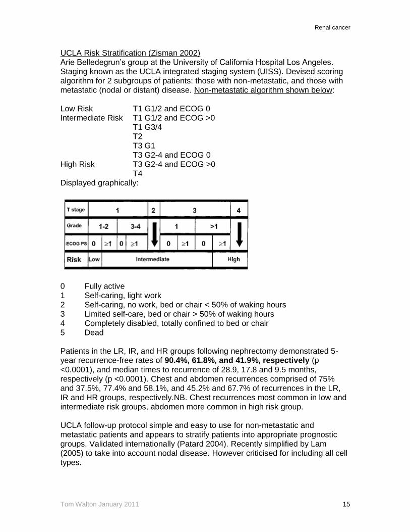
UCLA Risk Stratification (Zisman 2002) Arie Belledegrun’s group at the University of California Hospital Los Angeles. Staging known as the UCLA integrated staging system (UISS). Devised scoring algorithm for 2 subgroups of patients: those with non-metastatic, and those with metastatic (nodal or distant) disease. Non-metastatic algorithm shown below: Low Risk T1 G1/2 and ECOG 0 Intermediate Risk T1 G1/2 and ECOG >0 T1 G3/4 T2 T3 G1 T3 G2-4 and ECOG 0 High Risk T3 G2-4 and ECOG >0 T4 Displayed graphically:
0 Fully active 1 Self-caring, light work 2 Self-caring, no work, bed or chair < 50% of waking hours 3 Limited self-care, bed or chair > 50% of waking hours 4 Completely disabled, totally confined to bed or chair 5 Dead Patients in the LR, IR, and HR groups following nephrectomy demonstrated 5-year recurrence-free rates of 90.4%, 61.8%, and 41.9%, respectively (p <0.0001), and median times to recurrence of 28.9, 17.8 and 9.5 months, respectively (p <0.0001). Chest and abdomen recurrences comprised of 75% and 37.5%, 77.4% and 58.1%, and 45.2% and 67.7% of recurrences in the LR, IR and HR groups, respectively.NB. Chest recurrences most common in low and intermediate risk groups, abdomen more common in high risk group. UCLA follow-up protocol simple and easy to use for non-metastatic and metastatic patients and appears to stratify patients into appropriate prognostic groups. Validated internationally (Patard 2004). Recently simplified by Lam (2005) to take into account nodal disease. However criticised for including all cell types.

Renal cancer
Tom Walton January 2011 16
Leibovich Cancer 2003; Mayo Clinic scoring system – Clear-cell RCC only. Modified SSIGN score (Blute 2002) No specific post-operative surveillance protocol recommended in this paper.

Renal cancer
Tom Walton January 2011 17
Management of locally-confined disease Radical Nephrectomy RCC requires surgical cure – chemotherapy resistant (MDR p-glycoprotein ) and poor response to immunotherapy. Reasonable response to radiotherapy but in-situ disease not generally amenable to treatment due to risk of malignant hypertension and innocent bystander effects (small bowel etc.) Robson first described radical nephrectomy in 1969 Early vessel ligation to limit tumour emboli* Resection outside Gerota’s fascia** Ipsilateral adrenalectomy***
Complete ipsilateral lymphadenectomy from crus diaphragm to aortic bifurcation****
* advantageous, but not always possible ** valid: 25% of localised tumours show perinephric involvement *** not routinely necessary unless adrenal involvement of upper pole tumour
(Sagalowsky 1994; Tsui 2000; Siemer 2004). Not recomended by EAU unless contiguous involvement cannot be ruled out by imaging/palpation
**** Controversial for a number of reasons: Equal frequency of LN and haematogenous spread LN drainage extremely variable No evidence that routine complete LND improves survival. Recently EORTC 30881 (Blom et al) found low incidence (4%) of occult LN metastases in clinically staged N0M0 patients. No survival advantage for routine lymphadenectomy However: Useful for staging and prognostication
May have some benefit in selected patients with micrometastases (Giuliani 1990) and with enlarged regional nodes. Golimbu (1986) showed a 10-15% increased 5YS in patients with T3 disease. Also reported improved survival in the setting of cytoreductive Nx (Pantuck 2003)
No apparent value of pre-operative tumour embolisation in terms of reducing blood transfusion requirement. One Polish case-control study (Zielinski 2000) reported significantly improved survival for pre-op embolisation: The overall 5- and 10-year survival for 118 patients embolized before nephrectomy was 62% and 47%, respectively, and it was 35% and 23%, respectively, for the matched group of 116 patients treated with surgery alone (p = 0.01). Results controversial however and not repeated. Bowel injury at laparoscopic radical nephrectomy 1.3 per 1000 cases (0.1%) 70% unrecognised; 60% small bowel; 50% by electrocautery Typically pain over neaqrest port site

Renal cancer
Tom Walton January 2011 18
N+V, diarrhoea, low grade fever, low or normal WCC Nephron-sparing surgery First described by Czerny in 1890 but high morbidity Revived by Vermooten in 1950 Indications for nephron-sparing surgery: Anatomical or functional single kidney* Bilateral renal cell carcinoma
Pre-existing condition likely to lead to future loss of nephrons (high risk of metachronous multifocal tumour, renal disease)
* 20% of one kidney or GFR < 25 ml/min usually sufficient to avoid ESRF Previously normal contralateral kidney was considered to be a debatable indication for NSS. However recent studies suggest equivalent OS and DSS with radical nephrectomy, albeit with increased local recurrence rates (thought to be a manifestation of undetected multifocal tumour). Open partial nephrectomy Equivalent outcomes for patients undergoing OPN vs. ORN at least for patients with tumours < 4cm (Lerner 1996). Initial studies with <= 5 years follow-up indicate DSS 78-100% with local recurrence up to 10%. In the study with longest follow-up from Cleveland Clinic Fergany (2000) reported on 107 spontaneous tumours (mean size 4.7 cm) 10YR DSS 73%. Overall local recurrence 10.5%. Local recurrence by stage: T1a - 2%; T1b – 5%; T2+ 10-20%. NB. No minimum resection margin – as long as clear good enough. Positive margins appear to have negligible impact on subsequent survival (Bensaleh Eur Urol 2010) Laparoscopic partial nephrectomy First transperitoneal LPN performed by Winfield 1993 Relatively few studies with long term follow-up. Longest is Lane 2007: 56 patients, mean size 2.9 cm, T1a 86%. 66% turned out to be malignant. OS and DSS 86% and 100%. Main difference from OPN is that ischaemia time is limited. Accepted warm ischaemia time 30 mins. Important as WIT significant predictor of post-op impaired renal function. Complication rate certainly appears higher in LPN – approx 20% (6% bleeding, 2.5% urinary leak; Turna 2008 n =507). Recently, reduced overall (16 vs 37%) and haemorrhagic (3 vs 12%) complication rate associated with use of Floseal (Berger 2008) Best study comparing LPN and OPN (Gill 2007): 3-centre comparison of 1800 patients. Hospital stay and operative time were shorter in the LPN group. Overall postoperative complications were higher in the LPN group (18.6 vs. 13.7%), particularly hemorrhagic complications (4.2 vs. 1.6%). Positive margins for cancer were similar (1.6 vs. 1%). Local (1.4 vs. 1.5%) and distant (0.9 vs. 2.1%) recurrences were also equivalent. Cancer specific

Renal cancer
Tom Walton January 2011 19
survival at 3 years was 99.3 and 99.2%, respectively. Probe ablative therapies Minimally invasive techniques appropriate for small tumours (4cm or less) in the infirm and in those with a predisposition to forming multiple tumours. Contraindicatied in those with a bleeding diathesis. Caution should be exercised in those with central tumours encroaching on collecting system. RFA in particular associated with urinary fistulae. Radiofrequency ablation First utilised in 1911, when electrical current used to heat probes for treating bladder tumours. First use of RFA to treat renal tumours 1997 (Zlotta 1997) Performed either percutaneously or laparoscopically: px = percutaneously; ax = laparoscopically to avoid visceral injury Longest follow-up 55 months. (McDougal 2005). Ablative defect gradually fills in – approx 90% at 4 years. Area of ablation can be increased by injecting conductive agent into tissue to decrease impedance. However eschar tends to decrease impedance. One advantage of RFA is that radiation directly induced mitoses by a thermal independent mechanism. Therefore no loss of efficacy near to large vessels. Largest study to date Zagoria et al 2007; A total of 104 patients with 125 biopsy-proven RCC were treated with CT-guided percutaneous RFA. Patients were followed with contrast-enhanced CT or MRI and 93% complete ablation rate was noted over a mean follow-up of 13.8 months. Single-session percutaneous RFA was possible in 109 tumors. 7/16 failures in the study group were eventually salvaged by repeat RFA or cryoablation. When tumors were stratified by size, all 95 tumors smaller than 3.7 cm were completely ablated after a single session (100%). In comparison, only 14/30 tumors larger than 3.7 cm were treated successfully by a single RFA session.
Cryoablation First used for palliation of maliganancy in 1850 by James Arnott (Lancet 1850) Dr Irving Cooper neurosurgeon father of modern cryoablation Beneficial effects of very localised tissue damage with minimal collateral damage now fully appreciated.

Renal cancer
Tom Walton January 2011 20
First used in urology to treat BPH (Gonder 1965) Percutaneous for px tumours, laparoscopic for ax tumours. Two cycles of freeze thaw using argon/helium. Temperatures < -20 degrees necessary, < -40 considered ideal. Intra-operative monitoring using intra-op USS (lap USS for anterior tumours). Results: Largest study to date Gill 2005. mean tumour size 3.3cm (n=39). Careful MRI follow-up and repeat Bx at 6 months. 2/39 had residual RCC at Bx. Resolution of defect in 62% at 3 years. 3 yr DSS 98%.
RFA vs. Cryo No direct comparative prospective randomised controlled trials to date Retrospective single institution comparison (Hegarty 2006 Cleveland Clinic);246 pts; 164 cryo with 3 years f/up, 82 RFA with one year f/up. DSS 98% and 100% respectively. Larger ablation zone achievable with cryo, higher re-treatment rate with RFA Best meta-analysis to date Kunkle 2008: 47 studies; 1375 kidney lesions. Pre-Rx bx in approx 70%. Matched for age size of lesion (mean 2.6 cm) and follow-up. Cryo better than RFA for: Repeat Rx 1.3% vs 8.5% Local tumour progression 5.2% vs. 12.9% Metastasis 1.0% vs. 2.5% Cryo also has advantage over RFA that it allows real-time monitoring of ice-ball formation whereas RFA does not allow intra-operative measurement of treatment effect and relies on post-operative imaging. Broadly equivalent complication rates: more urinary fistulae with RFA (major); more skin paraesthesia and pain with cryo (minor and improved with insulated needles) Adjuvant treatment for high-risk locally-confined disease Neoadjuvant and adjuvant radiotherapy has previously been used for patients with RCC. No evidence to support its use but studies old (Finney 1978) and have not been repeated recently. Currently no evidence to recommend routine adjuvant treatment for high risk locally-confined disease. Also no definite consensus on what constitutes high risk disease, although a number of studies adopting Leibovich’s prognostic score.
Renal cancer
Tom Walton January 2011 21
EORTC 30955: Phase III Randomized Study of Adjuvant Interleukin-2, Interferon alfa, and Fluorouracil Versus Observation Only in Patients With Renal Cell Carcinoma at High Risk of Relapse After Surgical Resection (Closed/unreported) Inclusion criteria:
Histologically confirmed primary renal cell carcinoma meeting 1 of the following criteria:
Stage T3b, T3c, or T4 tumor Any pT stage and nodal status pN 1 or 2 Any pT stage and microscopic positive margins Presence of any microscopic vascular invasion
Underwent surgical resection of primary tumor within the past month Removal of clinical N+ disease required No evidence of metastatic disease No evidence of macroscopic residual disease
RE05/SORCE: A phase III Randomised Controlled Study Comparing Sorafenib With Placebo In Patients With Resected Primary Renal Cell Carcinoma at High or Intermediate Risk of Relapse (EORTC participation protocol #30072) - Recruiting Inclusion criteria: All kidney tumour sub-types No mets on post-Nx CT
Leibovich score 3-11 [most minimal disease = tumour > 4cm with necrosis or Fuhrman 3]
Between 4 weeks and 3 months of surgery Trial design
Patients will be randomly assigned to 3 years of placebo (Arm A), 1 year sorafenib 400mg bd & 2 years placebo (Arm B) or 3 years sorafenib 400mg bd (Arm C).
Management of locally-advanced disease IVC involvement 4-10% of patients Classification system I Adjacent to ostium of renal vein – able to be milked back II Up to lower margin of caudate lobe III Behind liver IV Above diaphragm a/w lower extremity oedema, renal dysfunction, hepatic dysfunction (Budd-Chiari) ascites, non-emptying varicocoele, caput medusae, PE, cardiac failure, SVC hypertension.
Renal cancer
Tom Walton January 2011 22
Coronal MRI investigation of choice for determining cephalad extension (Choyke 1997) – tumour thrombus enhances with gadolinium cf. simple/bland thrombus ~ 40% of cases have arterialised thrombus. Simultaneous MR angiography should therefore be performed as pre-op arterial embolisation may cause tumour shrinkage (Campbells but ? increased likelihood of tumour embolus) IVC involvement without other mets a/w overall survival of ~60%. However; IVC tumour with other mets = overall 10% 5YS (in studies after Nx – Campbells; largest study Swierzewski 1994), but probably much worse as only fit patients included. Therefore only consider ECOG 0 patients with low volume mets (eg. one or two lung nodules). Locally invasive, non-metastatic RCC Relatively uncommon Often present with pain due to invasion of px abdo wall & nerve roots Mesenteric organs usually first involved (small bowel/colon) / due to parasitisation of feeding vessels. Liver, duodenum and pancreatic invasion very uncommon – much more likely to involve liver via haematogenous seeding. Overall prognosis poor Complete excision 5% 5YS Incomplete excision 12% 1YS (deKernion 1978) No role for neoadjuvant or adjuvant radiotherapy
Renal cancer
Tom Walton January 2011 23
Management of metastatic disease (mRCC) Management of the primary tumour – cytoreductive Nx Patients with mRCC often have acquired immune dysfunction T-cell dysfunction Increased T-cell apoptosis Impaired proliferative responses Tumour associated immunosuppression Cytoreductive Nx improves all of the above. In 2001, the results of the European Organization for Research and Treatment of Cancer (EORTC) Genitourinary Group trial 30947 (Mickish 2001) and the similarly designed Southwest Oncology Group (SWOG) trial 8949 (Flanigan 2001) documented improved overall survival for patients who underwent cytoreductive nephrectomy before systemic immunotherapy with interferon-a (IFN-a) when compared with patients treated with immunotherapy alone. In a combined analysis of these two trials (326 patients), Flanigan et al. (Flanigan 2004) demonstrated a median survival duration of 13.6 months for patients who underwent cytoreductive nephrectomy and IFN-a versus 7.8 months (approx 6 months survival advantage) for patients treated with IFN-a alone. However, patient selection paramount as: Up to 25% of patients in each arm died within 4 months of enrolment* Up to 60% of patients unable to tolerate systemic Rx after CNx * Pre-CNx systemic therapy has been used as a ‘litmus test’ select out responders from non-responders. Bex (2002) used 2 cycles of IFN-a and low dose IL-2 prior to CNx. Responders/stable disease underwent CNx. Non-responders did not have CNx, rapidly progressed and died. Survival post-CNx equivalent to SWOG trial. Currently a number of phase 2 trials investigating value of pre-CNx SMIs (bevacizumab and sunitinib). No evidence that pre-surgery Rx increases morbidity (Margulis 2008) Value of cytoreductive Nx questioned in era of SMIs. New European trial of CNx + sunitinib vs. sunitinib alone about to start recruiting (? EORTC). Management of the primary tumour – tumour embolisation Simple to perform transarterially following groin puncture. Agents: absolute ethanol, gelfoam, acrylic microspheres (embospheres) and metallic coils. Coils and embospheres most popular, and often used in tandem to achieve large artery and capillary embolisation respectively. Few large studies of efficacy in palliative setting however. Schwartz recently reported experience of 121 RAEs (55% pre-op, 20% bleeding, 12.5% AML, 8% advanced tumour). Complications significant as shown below.

Renal cancer
Tom Walton January 2011 24
Post-infarction syndrome comprising pain, fever and nausea in three-quarters. Reportedly mild and self-limiting however. Metastasectomy (Rasco 2006) Number of reports of long-term survival after metastasectomy, some up to 10 years. Usually after lung/bone mets. Largest pulmonary study with longest follow-up Piltz 2002: 105 patients; 150 lung resections. 5YS 40%; 10YS 33%. Similar 44% 5YS from MSKCC for all locations. Favourable prognostic factors - DFI of longer than 12 months, a solitary metastasis and glandular/soft tissue location. OS did not appear to decrease with recurrent metastases and repeat metastasectomies. Survival by location:
Bone 15% 5YS; complication rate 40% with prior RT vs. 15%. No studies comparing RT vs. metastasectomy.
Lung Combined analysis of over 400 pts – 5YS consistently ~40%. Better with longer disease-free interval and few mets. Complications 6-12%. Mortality 1-3%.
Liver Few studies, small numbers. 56% 2YS. High complication/mortality rates. Fewer studies of pancreas but some reported long-term survival, even after Whipple’s.
Brain Traditionally whole brain irradiation +/- stereotactic RT. Largest series of conventional surgery Wronski 1996: n=50, half had WBRT after surgery (no difference in OS/DSS). Mortality 10%. Median survival 12 months. 5YS 8.5%.
Systemic therapy Multiple studies have reported improved response rates in trials of immunotherapy and chemotherapy in metastatic RCC. However response rates thought to poorly correlate with overall survival - thus only overall survival should be primary endpoint. Medroxyprogesterone acetate Early reports from animal studies and subsequent patient studies of MPA therapy in 1970s and early 1980s suggested a treatment response of 10-15% with occasional dramatic long-term responses. However a number of subsequent trials failed to demonstrate a definite benefit:

Renal cancer
Tom Walton January 2011 25
Two multicenter randomized trials (Medical Research Council Renal Cancer Collaborators, 1999 ; Negrier et al, 2005 ) tested oral medroxyprogesterone acetate as initial therapy for patients with metastatic RCC. In these reports, 174 and 123 patients received medroxyprogesterone acetate, 300 mg/day and 200 mg/day, respectively. Response rates to medroxyprogesterone acetate were uniformly low (2.0% and 2.5%, respectively), and overall median survival of the patients varied from 6.0 months ( Medical Research Council Renal Cancer Collaborators, 1999 ) to more than 15.0 months ( Negrier et al, 2005 ). The difference in survival probably reflects patient entry criteria used in the two studies rather than a true difference in biologic responsiveness. Therefore, whereas progestational agents may be useful for symptom palliation, they do not appear to have any significant value in the treatment of patients with metastatic RCC. One prospective RCT of MPA (500mg thrice weekly for 12 months) following nephrectomy (Pizzocaro 1987). N= 136, 5 yr follow-up. No survival benefit seen and significant side-effect in 57% of treatment arm due to MPA – predominantly hormonal toxicity. This and the finding of improved survival with IFN-a vs. MPA killed off MPA as a treatment. Occasionally reserved for patients unable to tolerate IFN-a or systemic therapy. Interferon alpha-2a Mechanism of action poorly understood. 4 RCTs shown increased survival advantage with IFN in metastatic renal cell cancer. Usually vs. medroxyprogesterone. Pooled median survival 3.8 months. Interestingly no difference in partial or complete response rates between groups. Largest trial MRCRCC trial 1999, no author. IFN following cytoreductive nephrectomy associated with prolonged survival in 2 trials compared with IFN alone (see above) Elevated CRP possibly an independent predictor of reduced survival in interferon therapy Blay 1994. Interleukin-2 No evidence that IL-2 associated with prolonged survival in randomised trials. From Cochrane systematic review: “We were unable to identify any published randomized study of high-dose interleukin-2 versus a non-immunotherapy control, or of high-dose interleukin-2 versus interferon-alfa reporting survival”. Is however associated with overall response rates of 15%. Sometimes prolonged and sustained complete remission in up to 7% of patients [Fyfe 1996; Yang 2003] - reason for drug approval by FDA. Significant toxicity with high dose treatment, reduced responses with low dose treatment. No survival advantage for combination interferon and IL-2 over monotherapy. Negrier 2001/2005.
Renal cancer
Tom Walton January 2011 26
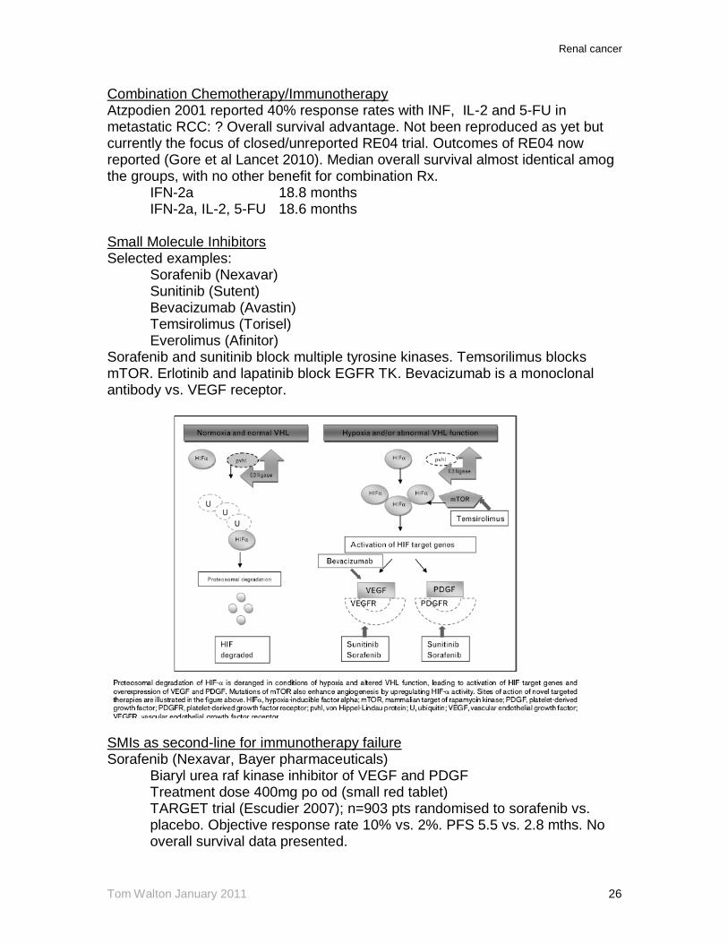
Combination Chemotherapy/Immunotherapy Atzpodien 2001 reported 40% response rates with INF, IL-2 and 5-FU in metastatic RCC: ? Overall survival advantage. Not been reproduced as yet but currently the focus of closed/unreported RE04 trial. Outcomes of RE04 now reported (Gore et al Lancet 2010). Median overall survival almost identical amog the groups, with no other benefit for combination Rx. IFN-2a 18.8 months IFN-2a, IL-2, 5-FU 18.6 months Small Molecule Inhibitors Selected examples: Sorafenib (Nexavar) Sunitinib (Sutent) Bevacizumab (Avastin) Temsirolimus (Torisel) Everolimus (Afinitor) Sorafenib and sunitinib block multiple tyrosine kinases. Temsorilimus blocks mTOR. Erlotinib and lapatinib block EGFR TK. Bevacizumab is a monoclonal antibody vs. VEGF receptor.
SMIs as second-line for immunotherapy failure Sorafenib (Nexavar, Bayer pharmaceuticals)
Biaryl urea raf kinase inhibitor of VEGF and PDGF Treatment dose 400mg po od (small red tablet) TARGET trial (Escudier 2007); n=903 pts randomised to sorafenib vs. placebo. Objective response rate 10% vs. 2%. PFS 5.5 vs. 2.8 mths. No overall survival data presented.

Renal cancer
Tom Walton January 2011 27
Side effects – diarrhoea, rash, fatigue, hand-foot reactions. Sunitinib (Sutent, Pfizer)
Inhibitor of VEGF and PDGF Treatment dose 50mg po od for 4 wks out of six Biggest trial to date Motzer 2006: 106 patients who underwent prior nephrectomy and cytokine therapy. Overall response rate of 33%. with a 14-month median duration of response. Median PFS and OS were 8.8 and 23.9 months. Side effects – diarrhoea, rash, fatigue, hand-foot reactions.
NB. Sorafenib and sunitinib most effective vs. CCRCC. Overall response rate 25% in chromophobe and only 5% in papillary RCC (Chouieri 2008) First line monotherapy Sorafenib
No trials to date Sunitinib
Recent phase 3 trial of sunitinib vs. interferon in patients with metastatic disease (90% in each arm had Nx) [Motzer 2006 n=750]. Either 50mg sunitinib od 4wks on, 2wks off, or 9MU interferon x3/wk. Primary endpoint PFS [assessment of imaging studies by independent review, performed cycles 1-4, then every 2 cycles thereafter]. Median PFS 11 mo in sunitinib group vs. 5 mo in interferon group. Trend towards increased overall survival in sunitinib group but not enough patients died*. Improved QOL and SE profile in sunitinib group. * Overall survival advantage over IFN-a of 4.6 months recently reported (Figlin 2008). Median survival 26.4 mo. vs. 21.8 mo. (Only 6% MSKCC poor prognosis patients in the trial; 60% ECOG 0)
Temsirolimus (Torisel, Wyeth pharmaceuticals) mTOR inhibitor Relatively gentle toxicity profile cf. bevacizumab and TKIs – mainly rash
oedema, hyperglycaemia and hyperlipidaemia. Global ARCC trial (Hudes 2007): n=626 poor prognosis ARCC. 25mg
temsirolimus weekly vs. combination Rx with IFNa and IFNa alone. Overall survival with temsirolimus 10.9 mths vs 7.3 for IFNa alone (combination 8.4 mths). Objective response rate only 8.5% however with stable disease in 30%.
First-line combination therapy Sorafenib and IFN-alpha 2B
Minimal improved survival vs. either agents but toxicity also synergistic – not currently recommended.
Bevacizumab (Avastin, Roche pharmaceuticals)
Renal cancer
Tom Walton January 2011 28
AVOREN trial (Escudier 2007): 649 patients were randomized to receive bevacizumab (10 mg/kg IV q 2 weeks) and IFN-a (9MU sc thrice weekly) or placebo and IFN-a. Median PFS 10.2 vs. 5.4 mths. Side-effects fatigue asthenia, hypertension and proteinuria. Similar results obtained by CALBG 90206.
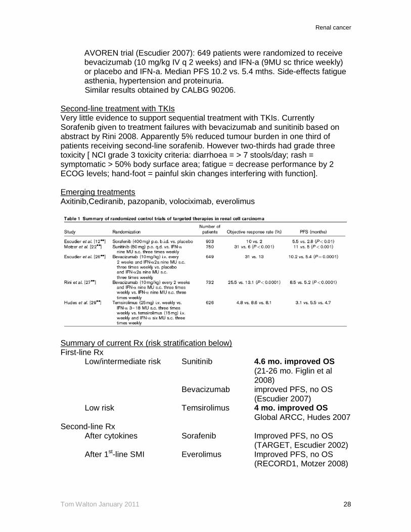
Second-line treatment with TKIs Very little evidence to support sequential treatment with TKIs. Currently Sorafenib given to treatment failures with bevacizumab and sunitinib based on abstract by Rini 2008. Apparently 5% reduced tumour burden in one third of patients receiving second-line sorafenib. However two-thirds had grade three toxicity [ NCI grade 3 toxicity criteria: diarrhoea = > 7 stools/day; rash = symptomatic > 50% body surface area; fatigue = decrease performance by 2 ECOG levels; hand-foot = painful skin changes interfering with function]. Emerging treatments Axitinib,Cediranib, pazopanib, volociximab, everolimus
Summary of current Rx (risk stratification below) First-line Rx Low/intermediate risk Sunitinib 4.6 mo. improved OS (21-26 mo. Figlin et al 2008) Bevacizumab improved PFS, no OS (Escudier 2007) Low risk Temsirolimus 4 mo. improved OS Global ARCC, Hudes 2007 Second-line Rx After cytokines Sorafenib Improved PFS, no OS (TARGET, Escudier 2002) After 1st-line SMI Everolimus Improved PFS, no OS (RECORD1, Motzer 2008)
Renal cancer
Tom Walton January 2011 29
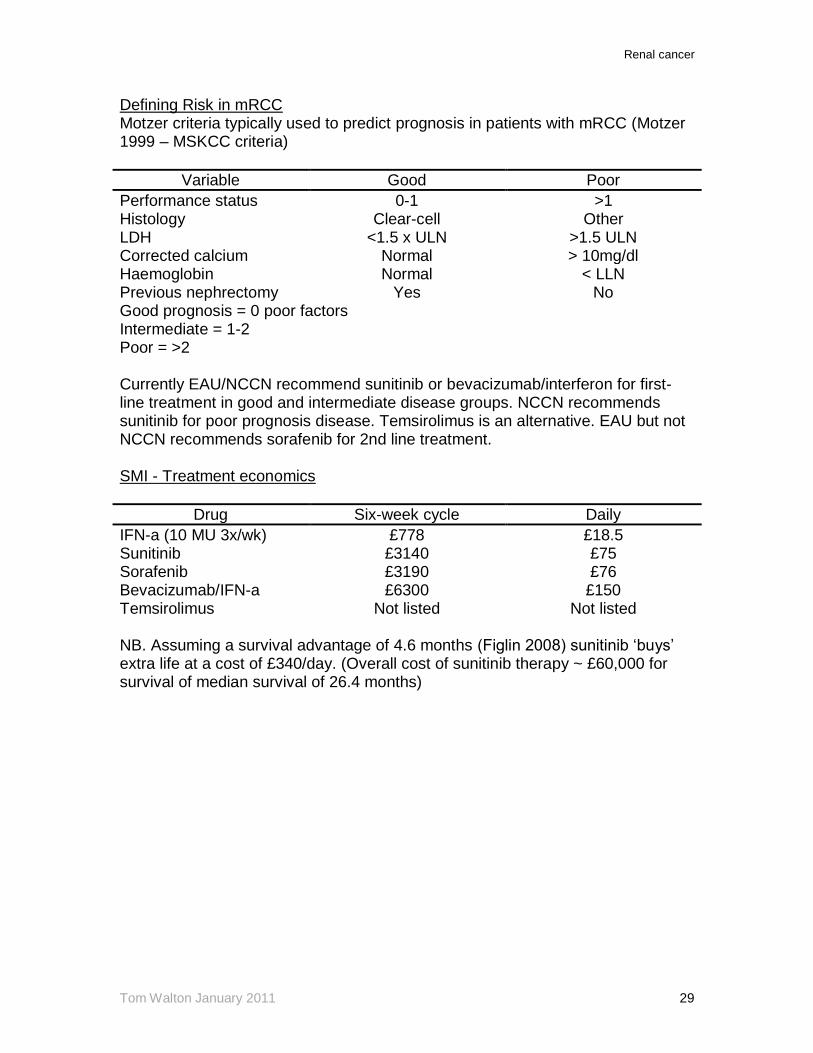
Defining Risk in mRCC Motzer criteria typically used to predict prognosis in patients with mRCC (Motzer 1999 – MSKCC criteria)
Variable Good Poor
Performance status 0-1 >1 Histology Clear-cell Other LDH <1.5 x ULN >1.5 ULN Corrected calcium Normal > 10mg/dl Haemoglobin Normal < LLN Previous nephrectomy Yes No Good prognosis = 0 poor factors Intermediate = 1-2 Poor = >2 Currently EAU/NCCN recommend sunitinib or bevacizumab/interferon for first-line treatment in good and intermediate disease groups. NCCN recommends sunitinib for poor prognosis disease. Temsirolimus is an alternative. EAU but not NCCN recommends sorafenib for 2nd line treatment. SMI - Treatment economics
Drug Six-week cycle Daily
IFN-a (10 MU 3x/wk) £778 £18.5 Sunitinib £3140 £75 Sorafenib £3190 £76 Bevacizumab/IFN-a £6300 £150 Temsirolimus Not listed Not listed NB. Assuming a survival advantage of 4.6 months (Figlin 2008) sunitinib ‘buys’ extra life at a cost of £340/day. (Overall cost of sunitinib therapy ~ £60,000 for survival of median survival of 26.4 months)

Renal cancer
Tom Walton January 2011 30
Follow-up Suggested UCLA follow-up protocol (Lam J Urol 2005) Low Risk Yearly Review Hx & Exam FBC/UE/LFTs CXR (CT chest) [Abdo CT @ 2 & 4 yrs - USA] Discharge @ 5 yrs Intermediate Risk 6 monthly review to 5 yrs,then @ 7yrs and 9 yrs Hx & Exam FBC/UE/LFTs CXR (CT chest) Yearly CT Chest/Abdo Discharge @ 10 yrs High Risk 6 monthly review to 5 yrs,then @ 7yrs and 9 yrs Hx & Exam FBC/UE/LFTs CT Chest/Abdo Discharge @ 10 yrs Displayed graphically: