repair of lumbar hernia originating from autogenous … · abdominal wall.1 the anatomy of the...
TRANSCRIPT

Formosan Journal of Surgery (2012) 45, 59e62
Available online at www.sciencedirect.com
journal homepage: www.e-f js .com
CASE REPORT
Repair of lumbar hernia originating fromautogenous iliac bone graft with bilayer mesh
Chun-Min Su a, Ching-Wen Hsu a, Yu-Chiuan Wu a,b,Wen-Yen Chang a, Wen-Ching Kung a,*
aDivision of General Surgery, Department of Surgery, Kaohsiung Armed Forces General Hospital, Kaohsiung, TaiwanbDepartment of Biomedical Engineering, I-SHOU University, Kaohsiung, Taiwan
Received 8 February 2011; received in revised form 18 April 2011; accepted 14 August 2011Available online 21 March 2012
KEYWORDSlumbar hernias;lumbar incisionalhernia;
surgical repair
* Corresponding author. Division of Gof Surgery, Kaohsiung Armed Forces GChung-Cheng 1 Road, Kaohsiung, Taiw
E-mail address: kung5437788@live
1682-606X/$ - see front matter Copyrdoi:10.1016/j.fjs.2011.12.006
Summary A 60-year-old female patient, who had previously undergone an iliac bone graftharvest, was diagnosed with the aid of an abdominal CT scan to have an inferior lumbar hernia,complicated with bowel incarceration. Reduction of the protruding sac was performed. Theweak point of the inferior lumbar hernia was reinforced by hernioplasty with a bilayer mesh.No evidence of recurrence was noted 16 months after surgery. The iliac bone graftis often adopted for various purposes by orthopedists and neurosurgeons, which maycontribute to an unusual lumbar hernia. The purpose of this communication is to emphasizethe possibility of complications following such surgical procedures and to provide useful infor-mation, including radiographic images and photographs of this extraordinary type of hernia.Hernioplasty with a bilayer mesh is safe, effective and convenient, in spite of a variety ofcommercially available meshes.Copyright ª 2012, Taiwan Surgical Association. Published by Elsevier Taiwan LLC. All rightsreserved.
1. Introduction
Lumbar hernia is a protrusion of intraperitoneal or extrap-eritoneal contents through a defect of the posterolateral
eneral Surgery, Departmenteneral Hospital, Number 2,an, ROC.mail.tw (W.-C. Kung).
ight ª 2012, Taiwan Surgical Asso
abdominal wall.1 The anatomy of the lumbar region hasbeen well documented since the first case reported byGarangeot in 1731, in which there are two well-definedweak areas: the inferior lumbar triangle, first describedby Petit in 1774,2 and the superior lumbar triangle, firstdescribed by Grynfelt in 1866.3 Lumbar hernias are classi-fied as congenital (during infancy) and acquired, accordingto their etiology. Acquired lumbar hernias are furtherclassified as primary (spontaneous) or secondary (a causalfactor existing).1 Lumbar incisional hernia belongs to the
ciation. Published by Elsevier Taiwan LLC. All rights reserved.

Figure 1 Abdominal CT scan revealed a right lumbar hernia involving extraperitoneal fat and ascending colon.
60 C.-M. Su et al.
secondary lumbar hernia, especially that occurring afteriatrogenic incisions, and the others are related to trauma orlumbar infection. We will hereby present a case of lumbarincisional hernia repaired with prosthetic meshes.
2. Case report
A 60-year-old woman, with a 20 year history of fracture ofher right femur, underwent an open reduction with internal
Figure 2 (A) Lateral decubitus position, incision planning; (B)operative wound; (C) Underlay Mesh insertion (LZ latissimus doApproximation of latissimus dorsi muscle and external oblique mus
fixation. A right iliac bone graft was used to enhance bonehealing.
No postoperative sequela was noted. However, sheexperienced right lumbar and lower abdominal pain,accompanied by an intermittent bulging sensation in theright lumbar area in the last six weeks. Due to the persis-tent symptoms, she visited our general surgery section. Onexamination, an old surgical scar, 4 cm in length, in herright supra-iliac region was noted, and a smooth, mildlytender mass about 5� 5� 3 cm in size was palpated
Sac herniating through a lumbar defect beneath the previousrsi muscle, EZ external oblique muscle, IZ iliac crest); (D)cle.
Bilayer mesh repair of lumbar hernia 61
beneath the scar. In the mass, bowel sounds were audible.The abdominal CT scan revealed a right lumbar herniainvolving extraperitoneal fat and the ascending colon onthe posterior aspect of the right lower abdomen (Fig. 1).
3. Surgical repair
The operation was performed under general anesthesia.The patient was placed in a lateral decubitus position, lyingon the opposite side of the hernia [Fig. 2 (A)]. An incisionwas made on the previous surgical scar. After dissecting thesubcutaneous tissue and surrounding muscles, a 5� 5 cmsac containing the ascending colon was noted herniatingthrough a lumbar defect located above the iliac crestbetween the latissimus dorsi muscle and the externaloblique muscle (Petit type) [Fig. 2 (B)]. The preperitonealand retroperitoneal spaces were created and then a poly-propylene mesh (GORE DUALMESH Biomaterial; W. L. Gore& Associates, Inc., Medical Products Division, Flagstaff, AZ,USA), named “Underlay Mesh”, 10� 15 cm, was placed andextended into the created preperitoneal space to cover thehernia defect. Five loop sutures (nonabsorbable 3-0 Pro-lene) were made to secure the mesh medially to the lat-issimus dorsi muscle, externally to the internal obliquemuscle and external oblique muscle, and inferiorly to theperiosteum of the iliac crest [Fig. 2 (C)]. Then, the lat-issimus dorsi muscle and external oblique muscle were
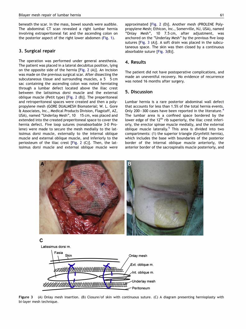
Figure 3 (A) Onlay mesh insertion. (B) Closure/of skin with cobi-layer mesh technique.
approximated [Fig. 2 (D)]. Another mesh (PROLENE Poly-propylene Mesh; Ethicon, Inc., Somerville, NJ, USA), named“Onlay Mesh”, 10� 7.5 cm, after adjustment, wasanchored on the “Underlay Mesh” by the previous five loopsutures [Fig. 3 (A)]. A soft drain was placed in the subcu-taneous space. The skin was then closed by a continuousabsorbable suture [Fig. 3(B)].
4. Results
The patient did not have postoperative complications, andmade an uneventful recovery. No evidence of recurrencewas noted 16 months after surgery.
5. Discussion
Lumbar hernia is a rare posterior abdominal wall defectthat accounts for less than 1.5% of the total hernia events.Only 200e300 cases have been reported in the literature.4
The lumbar area is a confined space bordered by thelower edge of the 12th rib superiorly, the iliac crest inferi-orly, the erector spinae muscle medially, and the externaloblique muscle laterally.5 This area is divided into twocompartments: (1) the superior triangle (Grynfeltt hernia),which includes the base with boundaries of the posteriorborder of the internal oblique muscle anteriorly, theanterior border of the sacrospinalis muscle posteriorly, and
ntinuous suture. (C) A diagram presenting hernioplasty with

62 C.-M. Su et al.
the 12th rib and serratus posterior inferior muscle superi-orly, the roof with the latissimus dorsi muscle, and the floorwith the aponeurosis of the transversus abdominis muscle;and (2) the inferior triangle (Petit hernia), which includesthe base with boundaries of the posterior border of theexternal oblique muscle anteriorly, the anterior border ofthe latissimus dorsi muscle posteriorly, and the iliac crestinferiorly, its roof with the superficial fascia, and its floorwith the internal oblique muscle, formed from the trans-versus abdominis muscle and the posterior lamina of thelumbodorsal fascia.1 Any operation on these weak areasmay evoke occurrence of a lumbar hernia.
The first study of postoperative lumbar hernias wasreported by Kretchmer in 1951. He reported 11 casesdeveloping lumbar hernias after renal surgery.6 The condi-tion may be related to injuries of the subcostal nerve, withresultant muscular atrophy on dissection.7 Lumbar inci-sional hernia is the subtype of the secondary lumbar hernia,especially well-known after iatrogenic incisions. In theliterature, reported operations that cause lumbar incisionalhernias include nephrectomy (most common), aorticaneurysm repair, latissimus dorsi myocutaneous flap andiliac bone graft harvest.8e10 The challenge of lumbar inci-sional hernia repair exists in altered anatomy of the lumbarregion, with diffuse fascial defects usually difficult toapproach.4,11 The diagnosis could be made mainly throughclinical manifestations. However, ultrasonography and a CTscan can serve to exclude some unusual causative lesions,such as abscesses or renal tumors, and can help designsurgical plans.11,12 Because of its rarity, treatment oflumbar hernia is still controversial. The techniques oflumbar hernia repair are divided into two categories: (1)the open approaches, including primary closure, coveragewith rotational flaps or onlay fascial flaps, and the use ofprosthetic meshes; (2) the laparoscopic techniques.11 Theselection of the technique should be based on the size ofthe hernia defect. Since secondary hernias are often largerthan primary hernias, Moreno-Egea A et al. concluded thatlaparoscopy is preferable to open surgery in terms ofresults, hospital stay, and costs.13
However, the laparoscopic repair requires a longerlearning curve, equipment demand and increased involve-ment of visceral structures. The open approach withtension-free bilayer meshes closes the hernia defectwithout involving the peritoneal contents. The preper-itoneal space was created. The underlay mesh is secured tothe latissimus dorsi muscle superiorly and medially, theexternal oblique muscle laterally and the iliac crest peri-osteum inferiorly. The onlay mesh is secured in the sameloop sutures of the underlay mesh. A concept of the sand-wich method is to enforce the hernia defect repair and toprevent mesh migration [Fig. 3 (C)]. This technique is an
alternative therapeutic choice for lumbar incisional hernia,and also produces good results.
6. Conclusion
A 60-year-old woman, with a 20 year history of fracture ofher right femur, underwent an operation of open reductionwith internal fixation and autogenous iliac bone graft.However, she developed a lumbar hernia originating fromthe previous bone graft site, which was successfullyrepaired with a bilayer mesh.
The iliac bone graft is often taken by orthopedists andneurosurgeons, which may contribute to an unusual lumbarhernia. Our purpose is to emphasize the possibility ofcomplications of the iliac bone graft and to provide usefulinformation, including radiographic images and photo-graphs of this extraordinary type of hernia. This bilayermesh technique is quite safe and yields good results.
References
1. Stamatiou D, Skandalakis JE, Skandalakis LJ, Mirilas P. Lumbarhernia: surgical anatomy, embryology, and technique of repair.Am Sur. 2009;75:202e207.
2. Petit JL. Traite des maladies chirurgicales, et des operationsqui leur conviennet, vol. 2. Paris: Didot; 1774. 256.
3. Grynfelt J. Quelques mots sur la hernie lombaire. MontpellierMed. 1866;16:323.
4. Palanivelu C, Rangarajan M, John SJ, Madankumar MV,Senthikumar K. Laparoscopic transperitoneal repair of lumbarincisional hernias: a combined suture and ‘double-mesh’techinique. Hernia. 2008;12:27e31.
5. Moreno-Egea A, Baena EG, Calle MC, Martinez JA, Albasini JL.Controversies in the current management of lumbar hernias.Arch Surg. 2007;142:82e88.
6. Kretchmer HL. Hernia of the kidney. J Urol. 1951;65:944e949.7. Orcutt TW. Hernia of the superior lumbar triangle. Ann Surg.
1971;173:294e297.8. Salameh JR, Salloum EJ. Lumbar incisional hernias: diagnostic
and management dilemma. JSLS. 2004;8:391e394.9. Reid RL. Hernia through an iliac bone-graft donor site. A case
report. J Bone Joint Surg Am. 1968;50:757e760.10. Stevens KJ, Banuls M. Iliolumbar hernia following bone graft-
ing. Eur Spine J. 1994;3:118e119.11. Cavallaro G, Sadighi A, Miceli M, Burza A, Carbone G,
Cavallaro A. Primary lumbar hernia repair: the open approach.Eur Surg Res. 2007;39:88e92.
12. Solaini L, di Francesco F, Gourgiotis S, Solaini L. A very simpletechnique to repair Grynfeltt-Lesshaft hernia. Hernia. 2010;14:439e441.
13. Moreno-Egea A, Torralba-Martinez JA, Morales G, Fernandez T,Girela E, Aguayo-Albasini JL. Open vs. laparoscopic repair ofsecondary lumbar hernias: a prospective nonrandomized study.Surg Endosc. 2005;19:184e187.