replication fork reversal occurs spontaneously...
TRANSCRIPT
REPLICATION FORK REVERSAL OCCURS SPONTANEOUSLY AFTER DIGESTION BUT IS CONSTRAINED IN SUPERCOILED DOMAINS*
Marta Fierro-Fernández, Pablo Hernández, Dora B. Krimer and Jorge B. Schvartzman
From the Departamento de Biología Celular y del Desarrollo, Centro de Investigaciones Biológicas (CSIC), Ramiro de Maeztu 9, 28040 Madrid, Spain.
Running title: Replication Fork Reversal
Address correspondence to: Jorge B. Schvartzman. Centro de Investigaciones
Biológicas (CSIC). Ramiro de Maeztu 9, 28040 Madrid, Spain. Phone: +34 91 837 3112, Fax: +34 91 536 0432, e-mail: [email protected]
Replication fork reversal (RFR) was
investigated in undigested and linearized replication intermediates (RIs) of bacterial DNA plasmids containing a stalled fork. Two-dimensional (2D) agarose gel electrophoresis, a branch migration and extrusion assay, electron microscopy (EM) and DNA-psoralen crosslinking were used to show that extensive RFR and extrusion of the nascent-nascent duplex occurs spontaneously after DNA nicking and restriction enzyme digestion but fork retreat is severely limited in covalently closed supercoiled domains.
Replication fork reversal (RFR) defines a condition that occurs in vivo when a replication fork encounters an obstacle such as a DNA lesion. In these cases it is thought that the replication fork halts and retreats to impede its collapse (1,2). The nascent strands separate from their corresponding parentals and anneal to each other to form a fourth arm. The resulting structure resembles a Holliday junction that behaves as a substrate for recombination enzymes (3-6). It was also found that for covalently closed circles (CCCs) of bacterial plasmids exposed to moderate concentrations of intercalating agents in vitro, while unreplicated forms acquire positive (+) supercoiling after all their native negative (-) supercoiling was removed, partially replicated forms appear unable to acquire (+) supercoiling and keep the same electrophoretic mobility as their nicked counterparts (7,8). These results were
interpreted as an indication that partially replicated plasmids containing a fork are unable to acquire (+) supercoiling as all of it is adsorbed by RFR. Curiously, in this case RFR does not affect the electrophoretic mobility of RIs as in the presence of intercalating agents, these CCRIs show the same electrophoretic mobility as their nicked counterparts (7,8).
Two-dimensional (2D) agarose gel electrophoresis (9) is increasingly used to analyze linearized replication intermediates (RIs) isolated from cells that have been exposed to different types of DNA damaging agents (10-13). In the autoradiograms of these 2D gels, the identification of a diffused pattern named “cone-signal” led some authors claim that the molecules responsible for this pattern contained reversed forks formed in vivo (13). In fact, the detection of such a pattern is currently viewed as a direct evidence for the occurrence of RFR in vivo (10-15). However, whether or not the cone signal detected in repair-deficient mutants is due to RIs containing reversed forks that formed in vivo is uncertain, as an identical pattern previously described as a “triangular smear” was also detected in 2D gels of DNA isolated from undamaged wild-type cells of a number of different species and interpreted as indicative for delocalized termination of DNA replication (16-26). An important drawback of all these studies is the random location of the event under study, namely a DNA lesion or the site where two forks growing in opposite directions meet. To overcome this potential problem, here we

2
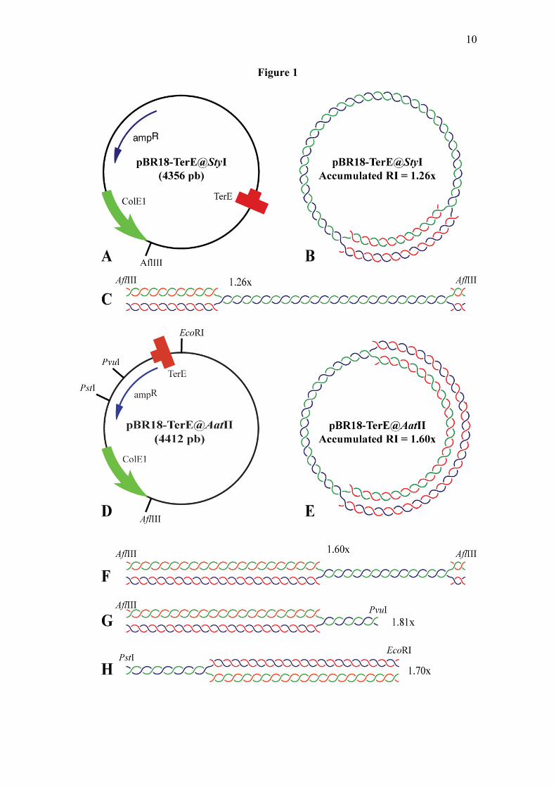
studied RFR in RIs containing a fork stalled at a specific site (7). pBR18-TerE@StyI and pBR18-TerE@AatII (Figure 1) contain the Escherichia coli polar replication terminator TerE (27,28) cloned at different distances from the unidirectional ColE1 replication origin in pBR18 (29). Digestion of the RIs of these plasmids with restriction enzymes that cut inside as well as inside and outside the replication bubble generates double and simple-Ys, respectively (Figure 1). The RIs of plasmids where the fork stalls at a specific site accumulate generating a prominent signal in 2D gel autoradiograms that can be readily distinguished on top of the simple or double-Y patterns (7,30-32). Analysis of these plasmids undigested and after restriction enzyme digestion revealed that RFR readily forms in vitro but fork retreat is severely limited in covalently closed domains (33). Extensive retreat and total extrusion of the nascent-nascent duplex, on the other hand, occurs spontaneously after nicking or restriction enzyme digestion.
EXPERIMENTAL PROCEDURES Bacterial strains and culture medium - The E. coli strain used in this study was DH5αF´. Competent cells were transformed with monomeric forms of the plasmids as described before (7,29). Cells were grown in LB medium containing 100 µg/ml ampicillin at 37ºC. Plasmid DNA isolation was performed as described previously (7,29). Two-dimensional agarose gel electrophoresis and Southern transfer - The 1st dimension was in a 0.4% agarose gel in TBE buffer at 1V/cm at room temperature for 22 h. The agarose lane containing the λ DNA/HindIII marker was excised, stained with 0.3 µg/ml EthBr and photographed. During this period, the agarose lane containing the DNA sample was kept in the dark. The 2nd dimension was in 1% agarose gel in TBE buffer containing 0.3 µg/ml EthBr run perpendicular with respect to the 1st dimension. The dissolved agarose was poured around the excised agarose lane from
the 1st dimension and electrophoresis was at 5V/cm in a 4ºC cold chamber for 10 h. Southern transfer was performed as described before (7,29). Psoralen cross-linking - To perform psoralen cross-linking, 50-100 ng of DNA in a total volume of 20 µl was incubated with 10 µg/ml 4,5’,8 trimethylpsoralen (Sigma) for 1 h at room temperature in the dark in a 96-well open plate and subsequently irradiated with a 500W high-pressure mercury lamp (model TQ 700; Original Hanau) on ice in a open plastic dish for 15 min. The lamp was placed 7 cm above the plastic dish and the light was filtered thru a Pyrex glass to eliminate radiation below 300 nm. Psoralen stock solution was prepared in 100% ethanol. This procedure was performed either before or after DNA digestion. Subsequently, the 2D agarose gel electrophoresis was carried out as described before. Non-radioactive hybridization - pBR322 that only hybridizes to the plasmid was used as a probe. DNA was labelled with the Random Primer Fluorescein kit (NEN Life Sciences Products). Membranes were prehybridized in a 20 ml prehybrization solution (2x SSPE, 0.5% Blotto, 1% SDS, 10% dextran sulphate and 0.5 mg/ml sonicated and denaturated salmon sperm DNA) at 65ºC for 4-6 h. Labelled DNA was added and hybridization lasted for 12-16 h. Hybridized membranes were sequentially washed with 2x SSC and 0.1% SDS, 0.5x SSC and 0.1% SDS, 0.1x SSC and 0.1% SDS for 15 min each at room temperature except for the last wash, which took place at 65ºC. Detection was performed with an antifluorescein-AP conjugate and CDP-Star (NEN) according to the instructions provided by the manufacturer. Preparation of DNA samples enriched for specific RIs - Specific molecules were isolated from agarose gels following the procedure described by Olavarrieta and coworkers with minor modifications (7).

3
After restriction digestion, DNA isolated from exponentially growing cells was analyzed in a one-dimensional agarose gel, the lane was cut, incubated with 0.1 M NaCl in TNE buffer (10mM Tris-HCl, pH 8.0/0.1mM EDTA/100mM NaCl) at 65ºC for 4 h and the selected DNA sample was electroeluted out of the agarose gel and resuspended in distilled water. Electron microscopy - The purified DNA sample was spread on EM grids under non-denaturating conditions in redistilled water by the BAC method (34). Branch migration and extrusion assay - The agarose lane of the 1st dimension containing the DNA sample was incubated with 0.1 M NaCl in TNE buffer (10mM Tris-HCl, pH 8.0/0.1mM EDTA/100mM NaCl) at 65ºC for 4 h without or in the presence of 0.3µg/ml EthBr. Subsequently, the 2nd dimension was performed as described before.
RESULTS AND DISCUSSION
It is generally thought that RFR is repressed in (-) supercoiled molecules and favored by (+) supercoiling (7,8,35). It was recently shown, however, that formation of Holliday-like junctions at both forks of a replication bubble creates a topological constrain that prevents further regression of the forks regardless of whether the DNA is (-) or (+) supercoiled (33). To confirm this observation for a different plasmid we used a modification of 2D gels where the agarose lane containing the DNA that came out from the 1st dimension was heated at 65ºC in the presence of 0.1M NaCl for 4 hr before the 2nd dimension took place (33,36). This condition favors branch migration and extrusion of the fourth arm of Holliday junctions in vitro (37,38). The corresponding autoradiograms are shown in figure 2 together with interpretative diagrams. Detection of two novel signals (marked with black arrows in the diagrams of figure 2) and their electrophoretic mobility during the 2nd
dimension clearly indicated that in some, although not all DNA molecules, heating caused extrusion of the two nascent strands (nascent-nascent duplex). During the 2nd dimension the new molecular species migrated as OCs and linear forms of 2627 bp, precisely the distance between the ColE1 origin and the TerE site in pBR18-TerE@AatII, indicating that the new linear fragment corresponded in fact to the extruded double-stranded fourth arm. In the very same autoradiograms, however, no extrusion occurred for CCRIs. Therefore, we concluded that extensive branch migration and extrusion of the fourth arm was impeded in CCRIs regardless of whether the DNA was (-) or (+) supercoiled. This was unexpected as it is generally thought that (+) supercoiling actually favors RFR and complete extrusion of the nascent-nascent duplex (8,35). This observation, on the other hand, agrees with the finding that RIs are able to acquire electrophoretic mobility and become (+) supercoiled when exposed to very high (0.3µg/ml and above) concentrations of EthBr (7,33) suggesting that RFR is favored by low and moderate levels of (+) supercoiling but is inhibited when the torsional stress reaches certain threshold. Postow and coworkers (8) used atomic force microscopy to study the topology of RIs containing stalled forks in the presence of 5µM EthBr (equivalent to 1.97µg/ml). They noticed that under these conditions RIs become heavily supercoiled but interpreted this supercoiling was an artifact induced during deposition of the molecules onto mica. It was later shown, however, that RIs recover electrophoretic mobility and are able to acquire (+) supercoiling when exposed to 0.3µg/ml and higher concentrations of EthBr (7) due to the topological locking mechanism activated as soon as RFR forms at both forks of a replication bubble (33). The observation that extensive RFR and complete extrusion of the nascent-nascent duplex are prevented in (-) as well as (+) supercoiled RIs prompted us to re-investigate whether or not the DNA

4
molecules containing reversed forks that are putatively responsible for the “cone signal” identified in 2D gels indeed form in vivo (10-13). To this end we combined 2D gels with psoralen crosslinking, the branch migration and extrusion assay described above and EM. If RFR occurs in vivo, the signal should be detected in restriction fragments of all sizes regardless of the extent of replication (see figure 3). Moreover, if RFR forms and retreats unconstrained once a replication fork stalls at a DNA lesion and these lesions occur at random, the mass of the RI with the stalled fork would vary between 1.0 and 2.0x. As clearly depicted in figure 3, the signal expected for a mix of molecules where the fork stalls and retreats from different sites is not the cone depicted as a gray triangle in figure 3B but rather a smear covering the whole area limited by X-shaped recombinants to the left and the ascending portion of the simple-Y pattern to the right, as also painted in gray in figure 3D. We used 2D gels to examine restriction fragments of different sizes (4.3, 4.4, 3.1 and 3.6 kb, respectively) where the RIs containing stalled forks were double-Ys of 1.26x (1.26 times the mass of unreplicated fragments) for pBR18-TerE@StyI digested with AflIII and 1.60x for pBR18-TerE@AatII also digested with AflIII. We examined also simple-Ys of 1.81x for pBR18-TerE@AatII digested with AflIII and PvuI and 1.70x for the same plasmid digested with PstI and EcoRI (see circular and linear maps in figure 1). The spikes emanating from the prominent spot on top of the simple or double-Y arcs (represented in red in the diagrams of figure 4) were easily recognized although their location, intensity and extension varied from one gel to the other. Moreover, they did not necessarily fit into the so-called cone signal described elsewhere (10-13). This spike extended almost exclusively below the accumulated spot for the 1.26x RI, it also extended below the accumulated spot but showed a small bulge above it for the 1.60x RI and extended both above and below the accumulated spot for the 1.81x RI. Similar signals have been observed for RIs of
specific masses in other systems as well (15,39). It is important to note, though, that only a small fraction of the accumulated RIs experienced RFR (see figure 4). The strength of the spot generated by accumulated RIs indicated that most of them were pretty stable and a discrete signal for molecules that experienced RFR was detected only for RIs that accumulated due to fork stalling. To confirm that these signals were generated by molecules displaying reversed forks, the agarose lane containing the DNA that came out of the 1st dimension was heated at 65ºC in the presence of 0.1M NaCl for 4 hr before the 2nd dimension took place. The results obtained are shown in the middle vertical panel of figure 4 with corresponding interpretative diagrams. Note that heating between the 1st and 2nd dimensions eliminated the original diagonal signals and generated novel ones that ran perpendicular to the 1st dimension in all cases (this was remarkable for the 1.81x RI) and are represented in red in the middle vertical diagrams of figure 3. It seems likely that the bulk of material that run at the position of the TerE-stalled RIs contained molecules where the fork retreated to some extent but where the nascent-nascent duplexes were too small to affect their mobility in an appreciable manner. These molecules were nevertheless susceptible to branch migration and extrusion by heat. This interpretation would account for both the new vertical spike, if not all such molecules completely extruded in response to heat, as well as the new spots. We speculated that molecules where the replication fork stalled at TerE and have undergone various extents of RFR, which in turn altered both their first and second dimension mobilities, generated the original diagonal spikes. As DNA heating was performed after the first dimension was completed, it could not alter 1st dimension mobilities, but after total extrusion of the nascent-nascent duplexes, the molecules that originally gave rise to the diagonal spikes would now yield a horizontal bulge that should be detected aside of the new signals. In fact, this was clearly the case for both of
5
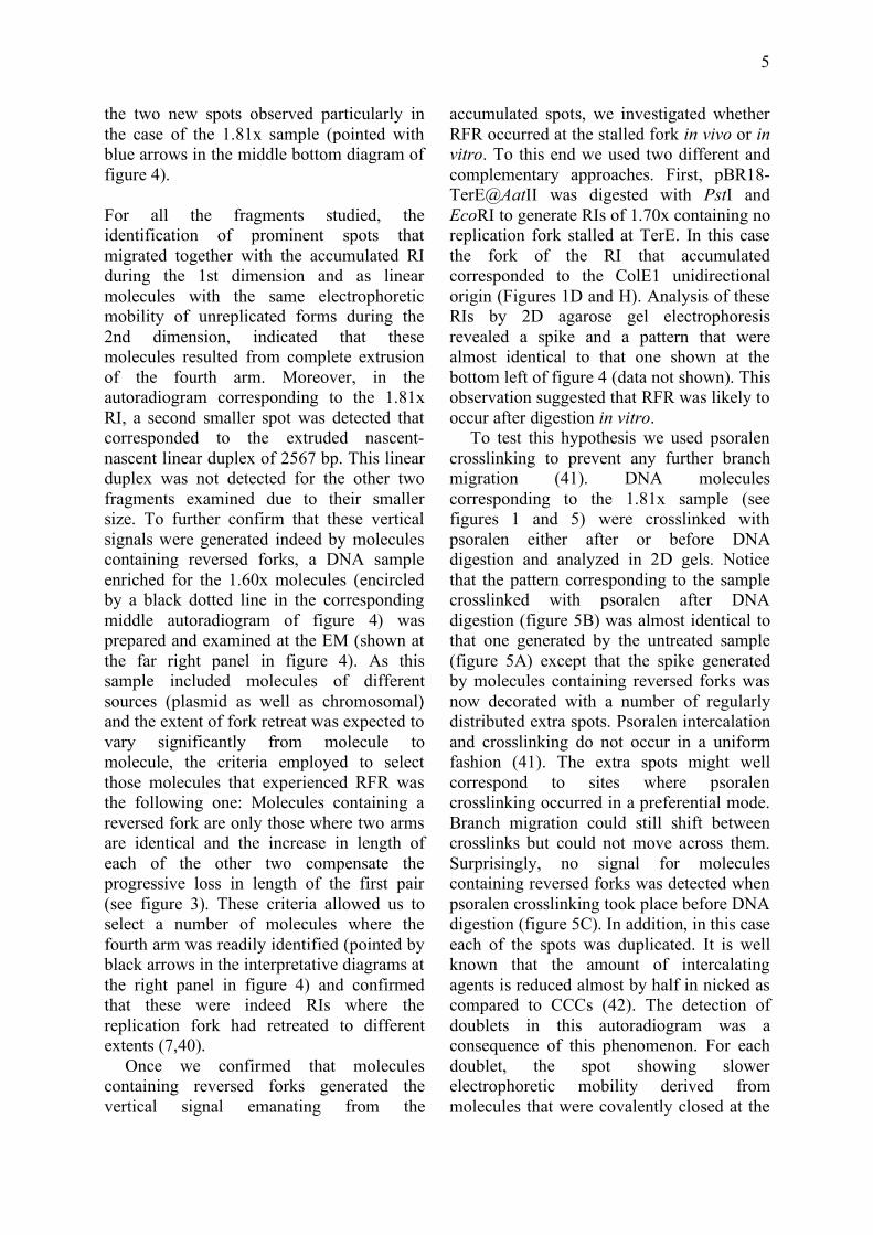
the two new spots observed particularly in the case of the 1.81x sample (pointed with blue arrows in the middle bottom diagram of figure 4). For all the fragments studied, the identification of prominent spots that migrated together with the accumulated RI during the 1st dimension and as linear molecules with the same electrophoretic mobility of unreplicated forms during the 2nd dimension, indicated that these molecules resulted from complete extrusion of the fourth arm. Moreover, in the autoradiogram corresponding to the 1.81x RI, a second smaller spot was detected that corresponded to the extruded nascent-nascent linear duplex of 2567 bp. This linear duplex was not detected for the other two fragments examined due to their smaller size. To further confirm that these vertical signals were generated indeed by molecules containing reversed forks, a DNA sample enriched for the 1.60x molecules (encircled by a black dotted line in the corresponding middle autoradiogram of figure 4) was prepared and examined at the EM (shown at the far right panel in figure 4). As this sample included molecules of different sources (plasmid as well as chromosomal) and the extent of fork retreat was expected to vary significantly from molecule to molecule, the criteria employed to select those molecules that experienced RFR was the following one: Molecules containing a reversed fork are only those where two arms are identical and the increase in length of each of the other two compensate the progressive loss in length of the first pair (see figure 3). These criteria allowed us to select a number of molecules where the fourth arm was readily identified (pointed by black arrows in the interpretative diagrams at the right panel in figure 4) and confirmed that these were indeed RIs where the replication fork had retreated to different extents (7,40).
Once we confirmed that molecules containing reversed forks generated the vertical signal emanating from the
accumulated spots, we investigated whether RFR occurred at the stalled fork in vivo or in vitro. To this end we used two different and complementary approaches. First, pBR18-TerE@AatII was digested with PstI and EcoRI to generate RIs of 1.70x containing no replication fork stalled at TerE. In this case the fork of the RI that accumulated corresponded to the ColE1 unidirectional origin (Figures 1D and H). Analysis of these RIs by 2D agarose gel electrophoresis revealed a spike and a pattern that were almost identical to that one shown at the bottom left of figure 4 (data not shown). This observation suggested that RFR was likely to occur after digestion in vitro.
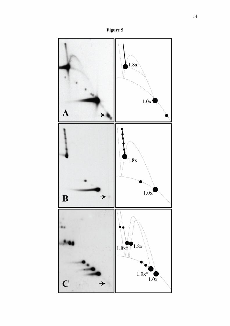
To test this hypothesis we used psoralen crosslinking to prevent any further branch migration (41). DNA molecules corresponding to the 1.81x sample (see figures 1 and 5) were crosslinked with psoralen either after or before DNA digestion and analyzed in 2D gels. Notice that the pattern corresponding to the sample crosslinked with psoralen after DNA digestion (figure 5B) was almost identical to that one generated by the untreated sample (figure 5A) except that the spike generated by molecules containing reversed forks was now decorated with a number of regularly distributed extra spots. Psoralen intercalation and crosslinking do not occur in a uniform fashion (41). The extra spots might well correspond to sites where psoralen crosslinking occurred in a preferential mode. Branch migration could still shift between crosslinks but could not move across them. Surprisingly, no signal for molecules containing reversed forks was detected when psoralen crosslinking took place before DNA digestion (figure 5C). In addition, in this case each of the spots was duplicated. It is well known that the amount of intercalating agents is reduced almost by half in nicked as compared to CCCs (42). The detection of doublets in this autoradiogram was a consequence of this phenomenon. For each doublet, the spot showing slower electrophoretic mobility derived from molecules that were covalently closed at the

6
time of psoralen crosslinking and, in consequence, captured almost double the amount of psoralen (43,44).
It is important to note, though, our results do not indicate RFR does not occur in vivo. It has been well established that in E. coli once a replication fork hits a DNA lesion, RecA (45) and/or RecG (4) promotes regression of the stalled fork generating a HLJ that is subsequently processed by the RuvABC complex to allow replication restart by PriA (3,46,47). This retreat of the forks that occurs in vivo, however, is severely constrained by DNA supercoiling and probably cannot extend very long (33). The results we showed here question to what extent the cone signal detected in 2D gels (10-13) reflects the limited retreat of the forks that may occur in vivo as opposed to
the extensive branch migration that takes place in vitro after restriction enzyme digestion. Furthermore, delocalized termination of DNA replication, which also generates a “triangular smear” (16-26) could be enhanced by fork stalling at DNA lesions and might certainly contribute to the so-called “cone-signal”.
In summary, here we showed that extensive RFR and extrusion of the fourth arm occurs spontaneously but only after nicking or DNA restriction enzyme digestion. These results strengthen the observation that the extent of fork retreat is severely constrained in supercoiled domains probably due to the topological locking that triggers when RFR forms at both forks of a replication bubble (33).
REFERENCES
1. Higgins, N. P., Kato, K., and Strauss, B. (1976) J Mol Biol 101, 417-425 2. Cox, M. M., Goodman, M. F., Kreuzer, K. N., Sherratt, D. J., Sandler, S. J., and
Marians, K. J. (2000) Nature 404, 37-41 3. Michel, B., Grompone, G., Flores, M. J., and Bidnenko, V. (2004) Proc Natl Acad
Sci USA 101, 12783-12788 4. McGlynn, P., Lloyd, R. G., and Marians, K. J. (2001) Proc Natl Acad Sci USA 98,
8235-8240 5. Schvartzman, J. B., and Stasiak, A. (2004) EMBO Reports 5, 256-261 6. Bates, A. D., and Maxwell, A. (2005) DNA Topology, Oxford University Press,
Oxford 7. Olavarrieta, L., Martínez-Robles, M. L., Sogo, J. M., Stasiak, A., Hernández, P.,
Krimer, D. B., and Schvartzman, J. B. (2002) Nucl Acids Res 30, 656-666 8. Postow, L., Ullsperger, C., Keller, R. W., Bustamante, C., Vologodskii, A. V., and
Cozzarelli, N. R. (2001) J Biol Chem 276, 2790-2796 9. Brewer, B. J., and Fangman, W. L. (1987) Cell 51, 463-471 10. Chow, K. H., and Courcelle, J. (2004) J Biol Chem 279, 3492-3496 11. Courcelle, J., Donaldson, J. R., Chow, K. H., and Courcelle, C. T. (2003) Science
299, 1064-1067 12. Lopes, M., Cotta-Ramusino, C., Liberi, G., and Foiani, M. (2003) Molecular Cell
12, 1499-1510 13. Lopes, M., CottaRamusino, C., Pellicioli, A., Liberi, G., Plevani, P., MuziFalconi,
M., Newlon, C. S., and Foiani, M. (2001) Nature 412, 557-561 14. Noguchi, E., Noguchi, C., Du, L. L., and Russell, P. (2003) Mol Cell Biol 23, 7861-
7874 15. Vengrova, S., and Dalgaard, J. Z. (2004) Gene Develop 18, 794-804 16. Dijkwel, P. A., Vaughn, J. P., and Hamlin, J. L. (1991) Mol Cell Biol 11, 3850-
3859

7
17. Duncker, B. P., Pasero, P., Braguglia, D., Heun, P., Weinreich, M., and Gasser, S. M. (1999) Mol Cell Biol 19, 1226-1241
18. Hyrien, O., and Mechali, M. (1992) Nucleic Acids Res 20, 1463-1469 19. Hyrien, O., and Mechali, M. (1993) EMBO J 12, 4511-4520 20. Little, R. D., Platt, T. H. K., and Schildkraut, C. L. (1993) Mol Cell Biol 13, 6600-
6613 21. Mahbubani, H. M., Paull, T., Elder, J. K., and Blow, J. J. (1992) Nucleic Acids Res
20, 1457-1462 22. Santamaría, D., Viguera, E., Martínez-Robles, M. L., Hyrien, O., Hernández, P.,
Krimer, D. B., and Schvartzman, J. B. (2000) Nucleic Acids Res 28, 2099-2107 23. Schvartzman, J. B., Adolph, S., Martín-Parras, L., and Schildkraut, C. L. (1990)
Mol Cell Biol 10, 3078-3086 24. Vaughn, J. P., Dijkwel, P. A., and Hamlin, J. L. (1990) Cell 61, 1075-1087 25. Veaute, X., and Sarasin, A. (1997) J Biol Chem 272, 15351-15357 26. Zhu, J., Newlon, C. S., and Huberman, J. A. (1992) Mol Cell Biol 12, 4733-4741 27. Bastia, D., and Mohanty, B. K. (1996) Mechanisms for completing DNA
replication. In: DePamphilis, M. L. (ed). DNA replication in eukaryotic cells, Cold Spring Harbor Laboratory Press, New York
28. Hill, T. M. (1992) Ann Rev Microbiol 46, 603-633 29. Santamaría, D., Hernández, P., Martínez-Robles, M. L., Krimer, D. B., and
Schvartzman, J. B. (2000) J Mol Biol 300, 75-82 30. Mirkin, E. V., Roa, D. C., Nudler, E., and Mirkin, S. M. (2006) Proc Natl Acad Sci
USA 103, 7276-7281 31. Mohanty, B. K., and Bastia, D. (2004) J Biol Chem 279, 1932-1941 32. Pohlhaus, J. R., and Kreuzer, K. N. (2005) Mol Microbiol 56, 1416-1429 33. Fierro-Fernández, M., Hernandez, P., Krimer, D. B., Stasiak, A., and Schvartzman,
J. B. (2007) Proc Natl Acad Sci USA 104, 1500-1505 34. Sogo, J. M., and Thoma, F. (1989) Methods Enzymol 170, 142-165 35. Espeli, O., and Marians, K. J. (2004) Mol Microbiol 52, 925-931 36. Noguchi, E., Noguchi, C., McDonald, W. H., Yates, J. R., 3rd, and Russell, P.
(2004) Mol Cell Biol 24, 8342-8355 37. Allers, T., and Lichten, M. (2000) Nucleic Acids Res 28, e6 38. Panyutin, I. G., and Hsieh, P. (1994) Proc Natl Acad Sci USA 91, 2021-2025 39. Bessler, J. B., and Zakian, V. A. (2004) Genetics 168, 1205-1218 40. Sogo, J. M., Lopes, M., and Foiani, M. (2002) Science 297, 599-602 41. Cimino, G. D., Gamper, H. B., Isaacs, S. T., and Hearst, J. E. (1985) Annu Rev
Biochem 54, 1151-1193 42. Sinden, R. R., Carlson, J. O., and Pettijohn, D. E. (1980) Cell 21, 773-783 43. Conconi, A., Widmer, R. M., Koller, T., and Sogo, J. M. (1989) Cell 57, 753-761 44. Lucchini, R., and Sogo, J. M. (1995) Nature 374, 276-280 45. Robu, M. E., Inman, R. B., and Cox, M. M. (2001) Proc Natl Acad Sci USA 98,
8211-8218 46. Baharoglu, Z., Petranovic, M., Flores, M. J., and Michel, B. (2006) EMBO J 25,
596-604 47. Flores, M. J., Sanchez, N., and Michel, B. (2005) Mol Microbiol 57, 1664-1675 48. Hill, T. M., and Marians, K. J. (1990) Proc Natl Acad Sci USA 87, 2481-2485 49. Martín-Parras, L., Hernández, P., Martínez-Robles, M. L., and Schvartzman, J. B.
(1992) J Biol Chem 267, 22496-22505 50. Friedman, K. L., and Brewer, B. J. (1995) Analysis of replication intermediates by
two-dimensional agarose gel electrophoresis. In: Campbell, J. L. (ed). DNA

8
Replication, Academic Press Inc., 525 B Street, Suite 1900, San Diego, CA 92101-4495
51. Martín-Parras, L., Hernández, P., Martínez-Robles, M. L., and Schvartzman, J. B. (1991) J Mol Biol 220, 843-853
52. Sogo, J. M., Stasiak, A., De Bernardin, W., Losa, R., and Koller, T. (1987) Binding of proteins to nucleic acids. In: Sommerville, J., and Scheer, U. (eds). Electron Microscopy in Molecular Biology. A Practical Approach, IRL Press, Oxford
FOOTNOTES
* We are grateful to María Luisa Martínez for technical assistance. We also thank Crisanto Gutiérrez and Maite Rejas for their help in EM and José Manuel Sogo, José Luis Díez and Amelia Partearroyo for their advice in psoralen crosslinking. This work was supported in part by grants Nº BIO2005-02224 to JBS and BFU2004-00125 to PH from the Spanish Ministerio de Educación y Ciencia. 1 The abbreviations used are: RFR, Replication fork reversal; RI, replication intermediate; CCC, covalently close circle; CCRI, covalently closed replication intermediate; OC, open circle; OCRI, open circle replication intermediate; L, linear; EthBr, ethidium bromide. EM, electron microscopy.
FIGURE LEGENDS Figure 1: Map of the plasmids. (A) Map of pBR18-TerE@StyI showing the relative position of its most relevant features (7): the ColE1 unidirectional origin, the E. coli terminator sequence TerE and the ampicillin resistance gene. (B) The Terminator Utilization Substance (TUS) binds Ter sites and the Ter-TUS complex acts as a polar replication fork barrier (27,48). In consequence, blockage of the replication fork at TerE leads to the accumulation of a specific RI containing an internal bubble and a total mass 1.26 times the mass of unreplicated plasmids. (C) Digestion of this RI containing a stalled fork with AflIII generates a double-Y of 1.26x. (D) Map of pBR18-TerE@AatII showing the relative position of its most relevant features. (E) In this case, blockage of the replication fork at TerE leads to the accumulation of a specific RI containing a larger internal bubble and a total mass of 1.60x. (F) Digestion of this RI with AflIII generates a double-Y of 1.60x, whereas the larger fragment resulting from its double digestion with AflIII and PvuI corresponds to a simple-Y of 1.81x (G). Finally, double digestion of the same RI with PstI and EcoRI generates a simple-Y of 1.70x (H). Figure 2: Exposure of undigested partially replicated plasmids to 65ºC in the presence of 0.1M NaCl enhances branch migration and leads to total extrusion of the nascent-nascent duplex, but only for nicked forms. Autoradiograms of 2D gel corresponding to pBR18-TerE@AatII where the 2nd dimension occurred either without or in the presence of 0.3µg/ml EthBr. For the autoradiograms shown to the right, the agarose lane of the 1st dimension containing the DNA sample was incubated at 65ºC with 0.1M NaCl in TNE for 4 hr either without (on top) or in the presence of 0.3µg/ml EthBr (bottom) before proceeding with the 2nd dimension. A diagrammatic interpretation is shown to the right of each autoradiogram. The signals resulting from total extrusion of the nascent-nascent duplex are depicted in gray and pointed by arrows. Dotted lines indicate the relative position of OCRIs after the 1st and 2nd dimensions.
9
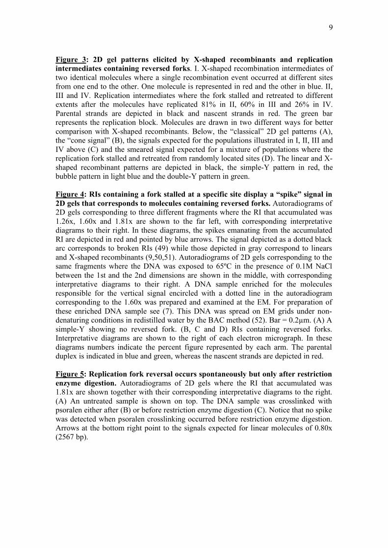
Figure 3: 2D gel patterns elicited by X-shaped recombinants and replication intermediates containing reversed forks. I. X-shaped recombination intermediates of two identical molecules where a single recombination event occurred at different sites from one end to the other. One molecule is represented in red and the other in blue. II, III and IV. Replication intermediates where the fork stalled and retreated to different extents after the molecules have replicated 81% in II, 60% in III and 26% in IV. Parental strands are depicted in black and nascent strands in red. The green bar represents the replication block. Molecules are drawn in two different ways for better comparison with X-shaped recombinants. Below, the “classical” 2D gel patterns (A), the “cone signal” (B), the signals expected for the populations illustrated in I, II, III and IV above (C) and the smeared signal expected for a mixture of populations where the replication fork stalled and retreated from randomly located sites (D). The linear and X-shaped recombinant patterns are depicted in black, the simple-Y pattern in red, the bubble pattern in light blue and the double-Y pattern in green. Figure 4: RIs containing a fork stalled at a specific site display a “spike” signal in 2D gels that corresponds to molecules containing reversed forks. Autoradiograms of 2D gels corresponding to three different fragments where the RI that accumulated was 1.26x, 1.60x and 1.81x are shown to the far left, with corresponding interpretative diagrams to their right. In these diagrams, the spikes emanating from the accumulated RI are depicted in red and pointed by blue arrows. The signal depicted as a dotted black arc corresponds to broken RIs (49) while those depicted in gray correspond to linears and X-shaped recombinants (9,50,51). Autoradiograms of 2D gels corresponding to the same fragments where the DNA was exposed to 65ºC in the presence of 0.1M NaCl between the 1st and the 2nd dimensions are shown in the middle, with corresponding interpretative diagrams to their right. A DNA sample enriched for the molecules responsible for the vertical signal encircled with a dotted line in the autoradiogram corresponding to the 1.60x was prepared and examined at the EM. For preparation of these enriched DNA sample see (7). This DNA was spread on EM grids under non-denaturing conditions in redistilled water by the BAC method (52). Bar = 0.2µm. (A) A simple-Y showing no reversed fork. (B, C and D) RIs containing reversed forks. Interpretative diagrams are shown to the right of each electron micrograph. In these diagrams numbers indicate the percent figure represented by each arm. The parental duplex is indicated in blue and green, whereas the nascent strands are depicted in red. Figure 5: Replication fork reversal occurs spontaneously but only after restriction enzyme digestion. Autoradiograms of 2D gels where the RI that accumulated was 1.81x are shown together with their corresponding interpretative diagrams to the right. (A) An untreated sample is shown on top. The DNA sample was crosslinked with psoralen either after (B) or before restriction enzyme digestion (C). Notice that no spike was detected when psoralen crosslinking occurred before restriction enzyme digestion. Arrows at the bottom right point to the signals expected for linear molecules of 0.80x (2567 bp).
10
Figure 1

Figure 2

Figure 3

13
Figure 4
14
Figure 5

15