report to the patient safety research programme … · monitoring the incidence of neonatal...
TRANSCRIPT

Report to the Patient Safety Research Programme (Policy Research Programme of the
Department Of Health)
Monitoring the incidence of neonatal encephalopathy - what next?
January 2005
Report to the Patient Safety Research Programme
(Policy Research Programme of the Department Of Health)
Monitoring the incidence of neonatal encephalopathy - what next?
Jennifer J Kurinczuk
Jennifer H Barralet
Maggie Redshaw
Peter Brocklehurst
National Perinatal Epidemiology Unit, University of Oxford, Old Road Campus, Headington, Oxford, OX3 7LF Tel: 01865 226654 Fax: 01865 227002 Email: [email protected] site: http://www.npeu.ox.ac.uk
January 2005
2

Contents Page Executive summary and recommendations 4 1. Background 6 2. Aims 7 3. Defining neonatal encephalopathy including severity criteria – aim (i) 7 3.1. Literature review 7 3.2. A consensus approach to defining neonatal encephalopathy 11 3.2.1 Consensus process objective 13 3.2.2 Consensus process results 13 3.3. Discussion and recommendations 13 4. Estimating trends in the incidence of neonatal encephalopathy and intrapartum stillbirths – aim (ii) 16
4.1. Literature review 16 4.2. Data sources 17
4.2.1. Unit-based data sources 17 4.2.2. Trent Neonatal Survey and Northern Region data 18
4.2.3. Trent CESDI data and Northern Region Perinatal Survey data – term intrapartum stillbirths 20
4.3. Neonatal encephalopathy 21 4.3.1. Rates 21 4.3.2. Severity 23 4.3.3. Demographic and clinical characteristics 24 4.4. Intrapartum stillbirths 31 4.5. Discussion and recommendations 32 5. The contribution of intrapartum events and potentially preventable mechanisms related to labour and delivery – aim (iii) 36
5.1. Literature review 36 5.2. Results from the Trent Neonatal Survey data 40 5.3. Discussion and recommendations 42
6. Conclusions – aim (iv) 43 7. Recommendations about future surveillance and research - aim (iv) 44 Acknowledgments 47 References 48 Appendix A – List of full definitions from key publications Appendix B – Preliminary Stage 1 consensus document – Defining
neonatal encephalopathy for surveillance purposes Appendix C – Additional tables and figures Appendix D – Summary of the literature review
3

Executive summary and recommendations
· Neonatal encephalopathy is a clinically-defined syndrome of impaired
neurological function that is most commonly found and best described in the
term infant. Its manifestations include difficulty initiating and maintaining
respiration, depression of tone and reflexes, subnormal levels of
consciousness, and seizures.
· At present there is no universally agreed and accepted definition of
neonatal encephalopathy in general clinical or research use.
· Currently available rates of neonatal encephalopathy are difficult to
interpret as the incidence measurement appears highly sensitive to
definitional changes. Given the problems of definition it is not possible to
provide, with any confidence, an estimate of the current rate of neonatal
encephalopathy in the UK. However, there is no evidence, from the most
recent data available, that there has been a decline in the rate of neonatal
encephalopathy since the mid-1990s, nor is there evidence of a decline in the
intrapartum stillbirth rate.
· A universally agreed definition of neonatal encephalopathy which does not
presume aetiology and that can be applied easily is urgently required. We
recommend that a consensus building approach is taken in order to develop a
definition that is usable and which would be adopted by the relevant
professions. We have embarked upon this process.
· The introduction of an agreed definition for neonatal encephalopathy into
the data collection processes of neonatal units would enable national
surveillance and local audit comparisons. Ensuring the adoption of both the
definition and the data items for universal data collection is more likely to be
achieved if endorsed by national professional organisations such as the
British Association of Perinatal Medicine, bodies responsible for governance
arrangements such as the Health Care Commission and influential individuals
from the Department of Health such as the Chief Medical Officer and his staff.
4

· The contribution of events and care provided during the intrapartum period
to the aetiology of neonatal encephalopathy is difficult to ascertain. Both the
available data and information from the literature are insufficient to derive a
robust estimate about the likely contribution of intrapartum events to the
neonatal encephalopathy rate in the UK.
· Given the complexity of data needed it may not be possible to collect the
necessary level of detail required by current criteria to identify the
contribution of intrapartum events and hypoxia in every neonatal unit. To
enable the collection of some level of information it may be necessary to
define an abbreviated set of explicit criteria and/or collect information from a
random sample of units on a rolling basis or from neonatal networks
designated as sentinel networks. As part of the consensus building process
we are using to define neonatal encephalopathy we are also including
discussions about the criteria for use to define the intrapartum contribution.
· The use of terms historically regarded as synonymous with neonatal
encephalopathy, such as birth asphyxia, perinatal asphyxia, hypoxic-
ischaemic encephalopathy and post-asphyxial encephalopathy continue to be
used in clinical parlance, medical records and the research literature. This is
unhelpful to both the understanding of aetiology and parental interpretation
of the cause of their infant’s condition. We strongly recommend that the use
of these terms is discontinued.
· A specific mesh term for neonatal encephalopathy would improve greatly
the ability of clinicians and researchers to locate relevant literature and would
also help discourage the continuing use of inappropriate terminology. We
have made a recommendation to the US National Library of Medicine (NIH) to
include neonatal encephalopathy as a mesh term and await the outcome.
· Urgent research is required in the UK to improve our understanding of the
aetiology of neonatal encephalopathy and the role that events and care
during pregnancy and delivery have in its genesis. This research will be
facilitated greatly by the availability of a universally agreed definition of
neonatal encephalopathy.
5

1. Background
In March 2004 the National Perinatal Epidemiology Unit (NPEU) was
approached by the Patient Safety Research Programme to tender for a brief
to carry out a six month project to estimate recent trends in the incidence of
neonatal encephalopathy in the UK, to explore the contribution of intrapartum
events and to make recommendations about future monitoring.1
The interest of the Patient Safety Research Programme in this issue arose
from the policy imperative of the Department of Health report ‘An
Organisation with a Memory.’ 2 This was a report from an expert group
chaired by the Chief Medical Officer of England set up to consider the extent
and nature of serious failures in NHS care. “Brain damage to babies at the
time of birth” was highlighted for discussion by the committee and it was
noted that average sums awarded following litigation are around £1.5 million
with some awards as high as £4 million. One of the recommendations
included a target for a reduction, of 25% by 2005, in the number of instances
of negligent harm in the field of obstetrics and gynaecology which result in
litigation. This target involves monitoring litigation claims, which is simple to
measure, however it highlighted the lack of national information in the UK
about trends in neonatal encephalopathy. Since a proportion of
encephalopathic cases arise or are exacerbated by intrapartum hypoxic
cerebral compromise, many authorities would regard the neonatal
encephalopathy rate as an indicator of intrapartum compromise. The lack of
national encephalopathy monitoring data prompted the development of the
tender brief by the Patient Safety Research Programme.
In addition to making recommendations to the Patient Safety Research
Programme about future monitoring, the opportunity also arose for the NPEU
to contribute to the discussions about the data items for inclusion in the
proposed national neonatal audit system currently being commissioned by
the Health Care Commission.3 The British Association of Perinatal Medicine
(BAPM) produced a recommended neonatal dataset in 1997.4 Because of the
difficulties agreeing to a definition for neonatal encephalopathy the current
version (2002 revision) of the dataset does not include neonatal
6
encephalopathy in its 41 items. At the completion of the work reported here a
definition will be recommended to the BAPM neonatal dataset working party
for possible inclusion in the BAPM dataset.
2. Aims
The aims of this six month project were to:
(i) Review the literature to fully explore the definitional issues
surrounding the diagnosis of neonatal encephalopathy and its
severity criteria (Section 3).
(ii) Estimate the trends in the incidence of neonatal encephalopathy
and intrapartum stillbirths over the last decade in the UK
(Section 4).
(iii) Explore the literature and existing datasets to identify the
contribution of intrapartum events and potentially preventable
mechanisms to the aetiology of neonatal encephalopathy
(Section 5).
(iv) Make recommendations about future monitoring and research,
particularly the value of neonatal encephalopathy as a surrogate
measure of potentially preventable intrapartum events
(Sections 6 and 7).
(v) Provide sufficient data upon which to base future sample size
calculations for both observational studies and trials (Section 6,
point v).
3. Defining neonatal encephalopathy including severity criteria – aim (i) 3.1. Literature review
An extensive search of the English language literature was carried out to
identify publications that either discussed and defined neonatal
encephalopathy or carried out studies in which it was necessary to use a case
definition of neonatal encephalopathy. Searches were carried out in Medline,
Embase, PsycINFO, Biological Abstracts and CINAHL from 1966 to November
2004. As there is no single mesh term for neonatal encephalopathy, a
7

number of search terms were used (Table 1). Using these terms 2,794
articles were identified, the abstracts were scanned and relevant articles were
extracted. Further searches were also carried out using key words, title
search, known author names and by searching reference sections of
previously identified articles and relevant books. Publications were also
extracted from frequently referenced publications and the review focused on
articles published after 1980. Summaries of relevant publications are given in
Appendix D.
Table 1. Terms used in the literature search for publications dealing with the definition of neonatal encephalopathy 1966-2004
Asphyxia neonatorum
Hypoxia-ischemia-brain
Foetal/fetal distress
Foetal/fetal asphyx*
Birth asphyx*
Newborn asphyx*
Perinatal asphyx*
Neonatal asphyx*
Hypoxic ischaemic/ischemic encephalopathy
Newborn encephalopath*
Perinatal encephalopath*
Neonat* encephalopath*
Infant encephalopath*
Twelve key publications were identified as having defined neonatal
encephalopathy using a unique combination of criteria (Table 2). Other
studies in this area, of which there are many, whilst acknowledged as
important were not included in the key publication list as they generally used
definitions drawn from the key publications or minor modifications thereof.
8

Table 2. Key publications identified on the basis as having defined neonatal encephalopathy using a unique combination of criteria
Authors
Cowan et al14
American College of Obstet &Gynecol15
Evans et al8
Ellis et al7
MacLennan
et al19
Year of publication
2003 2002 2001 2000 1999
Term in use NE NE Intrapartum hypoxic
event
NE NE Acute intrapartum
hypoxic event
Gestation (weeks) or birth weight > 36 ≥ 34 > 37or >2500g
> 37 ≥ 34
Time period after birth 72 hrs 24 hrs 96 hours 6-24 hrs 24 hrs Criteria Sentinel/acute hypoxic event ± ± Consciousness ● ● ± ● Respiration ± ± ± Tone ● ● ± ± Posture Reflexes ● ± Seizures/Convulsions/Fits ± ± ± Sucking/swallowing ± Feeding ● ± Heart rate (inutero) ± ± ± Apgar score ± ± ± Metabolic acidosis/Cord blood pH ± ● ● Cord blood pH ± ● ● Base deficit ● ● Blood pressure Segmental myoclonus Meconium staining ± Bronchial salivary secretions Pupillary abnormalities ● Brainstem function/autonomic function/cranial nerve function
● ±
EEG findings Abnormalities in imaging ± ± Brainstem Auditory Evoked Response
ECG findings Multi-organ failure/multisystem involvement
± ± ±
Renal complications Gastrointestinal motility Severe or moderate NE ● ● Cerebral palsy ● ● Neonatal Death Exclusion of other aetiologies ● Decreased activity and incubator care
NE Neonatal Encephalopathy ● Must be present ± May be present (usually in specified combinations)
9

Table 2 contd. Key publications identified on the basis as having
defined neonatal encephalopathy using a unique combination of criteria
Authors
Badawi
et al6
Low13
Nelson &
Ellenberg9
Amiel-
Tison & Ellison11
Levene
et al5
Fenichel12
Sarnat & Sarnat10
Year of publication
1998 1997 1987 1986 1985 1983 1976
Term in use NE (mod. or severe)
Intrapartum fetal
asphyxia
HIE Birth asphyxia
Post-asphyxial enceph.
HIE Postanoxic enceph.
Gestation (weeks) or birth weight ≥ 37 NS >2500g full-term 37-42 term > 36 Time period after birth 1 week NS NS NS 24-48 hrs NS 7 days* Criteria Sentinel/acute hypoxic event Consciousness ± ● ● ● ● ● Respiration ± ± ● ± ± ± Tone ± ± ● ● ± ± Posture ● Reflexes ± ± ± ● Seizures/Convulsions/Fits ± ± ± ± ± ± ± Sucking/swallowing ● ± ● ± ● Feeding ± ● ± Heart rate (inutero) ± ● Apgar score ● Metabolic acidosis/Cord blood pH ● Cord blood pH Base deficit ● Blood pressure ± Segmental myoclonus ± Meconium staining Bronchial salivary secretions ● Pupillary abnormalities ± ● Brainstem function/autonomic function/cranial nerve function
± ± ●
EEG findings ± ± ± ± Abnormalities in imaging ± Brainstem Auditory Evoked Response
±
ECG findings ± Multi-organ failure/multisystem involvement
Renal complications ± Gastrointestinal motility ± Severe or moderate NE Cerebral palsy Neonatal Death ± Exclusion of other aetiologies Decreased activity and incubator care
●
NE Neonatal Encephalopathy ● Must be present HIE Hypoxic Ischaemic Encephalopathy ± May be present (usually in specified combinations) Enceph. Encephalopathy * neurological examinations continued until discharge (usually 2-3 weeks) NS Not specified mod. Moderate
10

As can be seen from Table 2 none of the key publications used exactly the
same combination of clinical features, gestational age and timing of onset of
signs to define the presence of neonatal encephalopathy. The definitions used
by Levene et al,5 Badawi et al,6 Ellis et al,7 Evans et al,8 and Nelson and
Ellenberg 9 were the most similar. Notably the definitions from these five key
publications used fewer clinical criteria than the remaining seven.
Many studies describe using modifications of the Sarnat and Sarnat 10
criteria, the criteria defined by Levene et al 5 or Amiel-Tison and Ellison.11 It
is also notable that the terminology used to describe the condition has varied
with hypoxic-ischaemic encephalopathy,9,12 birth/intrapartum fetal
asphyxia,11,13 postanoxic encephalopathy,10 and post-asphyxial
encephalopathy5 used in the past. In recent times the term neonatal
encephalopathy has been more commonly adopted.6,7,8, ,14 15 The full version of
the definitions summarised in Table 2 is given in Appendix A.
3.2. A consensus approach to defining neonatal encephalopathy
In view of the short time-scale for this project, in our original proposal we
indicated that we would make recommendations about how future monitoring
could be carried out based primarily on a literature review. However, having
conducted the review and following discussions with clinicians about the data
they are currently collecting, it became clear that there is a lack of clarity
about how to define neonatal encephalopathy for the purposes of general
monitoring and surveillance, and a wide variation in practice. We therefore
concluded that in order to reach a rational and usable recommendation which
might be adopted in practice, a consensus approach to defining neonatal
encephalopathy would be a more appropriate approach to take.
The process we adopted to reach consensus about the definition of neonatal
encephalopathy, for monitoring, surveillance and related purposes was
modified from the nominal groups and consensus conference methods.16, ,17 18
It is also similar to the method used by MacLennan et al 19 for the purposes
of defining a causal relationship between acute intrapartum events and
cerebral palsy.
11

The consensus process, which is still underway, is as follows:
Stage 1: To convene a small meeting of experts to discuss, reach
consensus and propose a series of relevant definitions.
Stage 2: To synthesise the conclusions of this meeting in a discussion
document.
To circulate the report of the meeting and the discussion document to
the members of the meeting for correction and additions.
To meet with the small group again to finalise the proposed
definitions and reach consensus about issues that remained
unresolved after the initial meeting.
Stage 3: To circulate the discussion document to a wider group of
experts and interested parties.
To synthesize the comments arising from this wider consultation.
To undertake further iterations of the consultation process with both
the small and wider groups as necessary to reach consensus.
Stage 4: To produce a consensus document that outlines the agreed
statements and recommendations.
To make a final report to the experts, interested parties, the Patient
Safety Research Programme, the Neonatal Audit Standards Working
Group and the Neonatal Dataset Working Party of the British
Association of Perinatal Medicine, as and when final consensus is
reached.
To publish the findings in a peer reviewed journal with due
acknowledgment of the contribution of everyone involved in this
process.
12

3.2.1.Consensus process objective
The objective of the consensus process was to reach agreement about how
to:
1. Define neonatal encephalopathy for surveillance* purposes.
2. Define a severity grading for surveillance purposes.
3. Define the group of encephalopathy cases which has suffered probable
acute intrapartum or peripartum cerebral compromise for surveillance
purposes.
4. Identify the information needed to collect 1-3 above in a systematic
and consistent manner nationally.
3.2.2. Consensus process results
The final part of Stage 2 of the process is still ongoing. The preliminary
confidential discussion document produced from the first part of Stage 2 is
given in Appendix B. This will be further modified before being circulated for
Stage 3 of the process. The agreed definitions will be forwarded to the
Patient Safety Research Programme when they are finalised.
3.3. Discussion and recommendations
Neonatal encephalopathy is a clinically-defined syndrome of impaired
neurological function that is most commonly found and best described in the
term infant. It is manifest by difficulty with initiating and maintaining
respiration, depression of tone and reflexes, subnormal levels of
consciousness, and often by seizures.20 Historically neonatal encephalopathy
has been defined in a way which suggests that its aetiology is known in all
cases and is largely intrapartum in origin.21 Thus, terms such as post-
asphyxial encephalopathy or hypoxic-ischaemic encephalopathy have
commonly been used in the past to describe this condition. Furthermore,
many investigators have been drawn into the tautology of using definitions of
‘neonatal encephalopathy’ that assume an intrapartum aetiology (by
including in the case definition features indicative of intrapartum
compromise) 14, , , , ,22 23 24 25 26 and thus associations between neonatal
* Of note it was agreed early in the process of Stage 1 that we should use the term ‘surveillance’ instead of monitoring. This is to avoid confusion with clinical monitoring that might take place as part of the clinical care and diagnosis of infants with neonatal encephalopathy.
13

encephalopathy and intrapartum events have been found which almost
certainly overestimate the intrapartum contribution.20
The elucidation of the aetiology of neonatal encephalopathy has been held
back by a lack of methods that enable the direct measurement of the
physiological functioning of the fetal brain in utero and during labour and
delivery. Furthermore, the available measures of neonatal brain function
following delivery are imperfect and are only indicative of cellular level
respiration and the impact of hypoxia and ischaemia.
Reaching a full description of the risk factors for neonatal encephalopathy has
been hindered by the lack of a universally accepted case definition which
does not presume aetiology, and with few notable exceptions,6-8 a paucity of
population-based studies that include appropriately selected controls. In
general, referral biases will tend to lead to the inclusion of the more severely
affected cases. This can be a problem in studies which identify cases from
referral units.14 Lack of sufficient controls selected in an unbiased fashion
leads to a lack of comparison data which can render the information about
the cases difficult to interpret.5,8,14
In general, aetiologically based definitions of disease are important because
they enable the instigation of appropriate therapy eg. Tuberculosis.21
However, aetiologically based definitions become unhelpful when
inappropriately applied for example, the diagnosis of bacterial vaginosis as a
cause of preterm birth has not been confirmed by trials of antibiotic
treatment in early pregnancy.27 This type of attribution becomes particularly
unhelpful when litigation is instigated and the courts are left to assign clinical
blame.19,21 Despite the efforts by leading researchers in the field to encourage
obstetricians and neonatologists to avoid using terms such as birth asphyxia,
perinatal asphyxia and hypoxic-ischaemic encephalopathy 15,19, , ,28 29 30 these
terms remain in common clinical parlance, are still found written in medical
records and discussed in the research literature.31,32
14

It is clear that the lack of a commonly agreed and accepted definition for
neonatal encephalopathy greatly inhibits our ability to both measure and
monitor the incidence of neonatal encephalopathy, to investigate its causes
and thus prevent its occurrence in those circumstances when it is possible to
do so.
It is our view that the only practical mechanism for reaching a commonly
agreed definition for this clinically defined condition is through a consensus
building approach. The consensus approach was successfully used by
MacLennan and the International Cerebral Palsy Task Force to develop a
template for defining a causal relation between acute intrapartum events and
cerebral palsy following which an international consensus statement was
issued.19 By discussing the issues of definition with experts and opinion
leaders and consulting with the broader group of interested parties we
believe that it may be possible to influence clinical practice and data
collection, such that in the future neonatal units will collect information about
neonatal encephalopathy that is clearly and universally defined. Following
which they will be able to produce information that is comparable over time,
across neonatal networks and between units.
We are continuing to pursue the consensus building approach to define
neonatal encephalopathy for monitoring and surveillance purposes and will
make recommendations to both the Patient Safety Research Programme and
the BAPM Dataset working party when this process is complete. The latter,
together with the actual consensus process itself will, we hope, encourage
the national dissemination of the agreed terminology and definition of
neonatal encephalopathy. We strongly recommend that the use of all other
terms historically regarded as synonymous with neonatal encephalopathy,
including birth asphyxia, perinatal asphyxia, hypoxic-ischaemic
encephalopathy and post-asphyxial encephalopathy, is discontinued.
15

4. Estimating trends in the incidence of neonatal encephalopathy and intrapartum stillbirth – aim (ii)
4.1. Literature review
We identified only two relatively recent UK-based published studies that
provide an estimate of the incidence of neonatal encephalopathy. Smith et al
conducted a unit-based retrospective case note review of hypoxic-ischaemic
encephalopathy (HIE) for births from 1992 to 1996 in Derby 32 and compared
the results with a previous case note review performed in the same unit.24
Having identified cases from the medical records they retrospectively applied
the Levene et al 5 criteria to define cases with post-asphyxial encephalopathy.
Although explicit criteria to determine an ‘asphyxial’ cause were not applied
“Infants with encephalopathy clearly attributable to a cause other than
asphyxia were excluded.”32 Three time periods were compared: (A) 1976 to
1980, (B) 1984 to 1988, and (C) 1992 to 1996. The incidence rates for HIE
grades II and III were 2.6 (95%CI 1.95 to 3.25), 1.9 (1.32 to 2.43) and 1.2
(0.78 to 1.68) respectively. The authors concluded that for grades I, II and
III combined there was “a further significant fall in the incidence of hypoxic-
ischaemic encephalopathy in term infants in the period 1992-1996 compared
with the earlier study periods. In addition there was a statistically significant
difference in the incidence of those moderately and severely affected when
comparing periods A and C, and a downward trend when comparing B and
C.” This study provides evidence of a decline in the incidence of ‘HIE’ over
the 21 year period from 1976 to 1996. For our purposes, the limitations of
the results of this study are that it does not include recent cases, it relies on
retrospective case ascertainment and only cases assumed to have an
“asphyxial” cause are included. Furthermore the whole analysis is based on
only 136 cases over 21 years.
Evans et al 8 carried out a population-based case control study of 150 infants
with neonatal encephalopathy and 154 controls born between 1993 and 1995
in the South West Thames region. They used a broad definition of neonatal
encephalopathy (see Table 2) which did not assume an intrapartum aetiology
and they were confident that they had ascertained all eligible cases. The
16

results from this study do not allow trends to be examined. However, in
contrast to the ‘HIE’ rate of 1.2 (95%CI 0.78 to 1.68) for the period 1992 to
1996 described by Smith et al 32 for the period 1993 to 1995 Evans et al
found a significantly higher NE rate of 2.6 (95%CI 2.22 to 3.08).8 Whilst it is
possible that this difference represents a real change in incidence it is also
likely to be in part, if not wholly, a result of comparing results when a
different case definition has been used and illustrates the sensitivity of the
incidence measurement of this condition to definitional changes.
4.2. Data sources
From the outset we were aware of two regional population-based data
collection systems that would allow us to examine the neonatal
encephalopathy rate over the last decade.
4.2.1. Unit-based data sources
In the absence of other long standing regional data collection systems† we
also attempted to identify hospital-based data collection systems from other
parts of the country. To do this we used the report from the study conducted
as part of the assessment of the feasibility of establishing a national neonatal
audit programme 33 to identify units which appeared, on the basis of the
survey conducted to compile this report, to have neonatal audit data systems
that had been in operation for at least five years. On this basis we wrote to
the clinical directors of 28 neonatal units. We received 15 letters in reply, two
of which included unit-based data. From the outset, given the relative rarity
of neonatal encephalopathy, the intention was that these unit-based sources
of data would provide a limited comparator for the regionally derived rates. It
was intended that this would provide some reassurance that the regional
rates were not widely different from elsewhere. However, when we were able
to discuss our data requirements with the clinicians running the unit-based
data collection systems it became clear that even within units there was
rarely a clear definition of what constituted neonatal encephalopathy and
thus what cases they were counting; convulsions or seizures alone were often
† Of note the Manners neonatal data collection system came into operation in the West Midlands Region in September 2002 – the definition of encephalopathy is based on the Sarnat & Sarnat (1976) criteria.
17

used as a proxy; in addition there were data coding and data extraction
problems and often an inability to define the live birth denominator. In view
of these limitations the results reported here are based solely on the two
regional sources of data.
4.2.2. Trent Neonatal Survey and Northern Region Data
Under the auspices of the Trent Neonatal Survey data have been collected
from all neonatal units in the former Trent health region (now East Midlands
and South Yorkshire) since February 1990.34 The total number of live births
per annum in the former region ranged from about 63,000 in 1991 to just
less than 55,000 in 2002. The criteria for inclusion in the survey are all
babies who meet one or more of the following:
· born at less than or equal to 32 weeks gestation;
· less than or equal to 1500 grams birth weight;
· involved in transfers between units;
· who receive any intensive care;
· who died in a neonatal unit;
· at term showed signs of severe hypoxic ischaemic encephalopathy.34
The data are collected from the units by trained research nurses employed
for this purpose who visit the units regularly. Data have been collected using
the same procedures since 1990. The data from the Trent Neonatal Survey
are essentially population-based although a small number of cross-boundary
referrals out of Trent occur each year and data are not collected for these
babies unless some part of their care is provided in a unit in Trent; their
impact in relation to the data reported here is negligible. Two specific (tick
box) data items are collected which allow neonates with ‘hypoxic-ischaemic
encephalopathy II’ or ‘hypoxic-ischaemic encephalopathy III’ as their primary
reason for admission to be identified (Table 3). The data custodians
(Professor David Field and Mrs Elizabeth Draper) provided us with a de-
identified dataset containing information about all term (≥ 37 weeks
gestation) neonates born in Trent between 1991 and 2002 whose primary
reason for admission to a neonatal unit was ‘HIE II’ or ‘HIE III’.
18

Table 3. Definitions used to identify ‘encephalopathic’ neonates from Trent Neonatal Survey data and the Northern region neonatal unit
data collection
Data source
Criteria used to identify
‘encephalopathic’ neonates
Trent Neonatal Survey, term births 1991 to 2002
Mildly affected babies are not collected by the survey as their signs are too subjective.34
Infants as a minimum criteria have to suffer fits whilst others, more severely affected, will also require ventilation.34
“Included are any encephalopathic infants that have fits and where the story is broadly supportive of HIE and where there is no alternative diagnosis (eg. meningitis)” [David Field – pers. comm.].
Northern Region Neonatal Provider Consortium data, term births 1996 to 2003
No single specific defining data item [Alan Fenton – pers. comm.]. The following constellation of criteria were used as indicative of the presence of encephalopathy:
• Convulsions (tick-box data item)
• Required anti-convulsant medication (tick-box data item)
• Free text statement indicative of encephalopathy
[Alan Fenton – pers. comm.].
Data relating to all admissions for neonatal intensive care have been
collected from the four main neonatal intensive care units in the Northern
health region since 1996. The data collection is co-ordinated by Dr Alan
Fenton under the auspices of the Neonatal Provider Consortium (Managed
Clinical Network).35 The total number of live births in the region ranged from
19

about 33,000 in 1996 to just over 30,000 in 2003. Individual units are
responsible for collecting and uploading their own unit’s data. No single
specific data item is collected that enables neonates with neonatal
encephalopathy to be identified. Thus, the data custodian (Dr Alan Fenton)
searched the database to identify all neonates born between 1996 and 2003
who had convulsions (tick box item), required treatment with an anti-
convulsant medication (tick box item), had a free text statement which
indicated that the neonate had been encephalopathic (Table 3). Dr Fenton
provided us with a de-identified dataset containing information about all term
neonates born in the Northern region between 1996 and 2003 who were
identified as meeting the constellation of criteria indicative of the presence of
encephalopathy.
4.2.3. Trent CESDI data and Northern perinatal mortality survey data – intrapartum stillbirths Intrapartum stillbirth data for deliveries at term (≥ 37 weeks gestation) were
obtained for comparison with the rate of neonatal encephalopathy. This was
in order to identify whether any changes in the rate of term encephalopathy
were reflected in contrary changes in the term intrapartum stillbirth rate.
Aggregated data relating to intrapartum stillbirths delivered at term were
obtained from the Trent Regional Confidential Enquiry into Stillbirths and
Deaths in Infancy (CESDI) which started data collection in 1993.36 Term
intrapartum stillbirths were identified using the data collected by a specific
(tick box) question(s) which asked about the timing of death. Of note, the
form of the question used to collect this information changed in 2000 which
led to a proportion of stillbirths reported where the timing of the death was
reported as uncertain [Elizabeth Draper – pers. comm.]. Aggregated data
relating to term intrapartum stillbirths in the Northern region, for the period
1996 to 2003, were obtained from the Northern Region Perinatal Mortality
Survey which covers the resident population of the Northern Health
region.35,37 All intrapartum stillbirths delivered at term were included
regardless of the cause of death.
20

4.3. Neonatal encephalopathy
4.3.1. Rates
Over the 12 year period 1991 to 2002 there were 704,130 live births in the
Trent region. During this time a total of 808 neonates born at term (≥ 37
weeks gestation) were admitted to Trent neonatal units with ‘HIE II’ or ‘HIE
III’ as their primary reason for admission; an overall rate of 1.2 per 1,000
total live births (95%CI 1.07 to 1.23). Figure 1 illustrates the year by year
variation (the data relating to Figure 1 are given in Appendix C, Table C1).
Whilst the rate of ‘HIE’ appeared to decline over the period 1991 to 1996, for
the whole period 1991 to 2002 there is little evidence of an overall trend.
There is no evidence of a decline in the rate of ‘HIE’ in the most recent six
year period (1997 to 2002). Of note case definitions and data collection
procedures remained unchanged over the whole period (1991 to 2002).
In the Northern region, over the eight years from 1996 to 2003, there were
247,480 live births. In this time a total of 450 term neonates were defined as
meeting the ‘encephalopathic’ criteria; an overall rate of 1.8 per 1,000 total
live births (95%CI 1.65 to 1.99). Figure 1 illustrates the year by year
variation.
Figure 1. Rate* of ‘HIE’ in Trent Region (1991 to 2002) and rate* of ‘encephalopathy’ in Northern Region 1996 to 2003, term births (≥ 37
weeks gestation) per 1,000 total live births
Trent Region Northern Region
0.0
0.5
1.0
1.5
2.0
2.5
3.0
1991 1992 1993 1994 1995 1996 1997 1998 1999 2000 2001 2002
Year of birth
Rat
e pe
r 100
0 liv
e bi
rths
0.0
0.5
1.0
1.5
2.0
2.5
3.0
1996 1997 1998 1999 2000 2001 2002 2003
Year of birth
Rat
e pe
r 100
0 liv
e bi
rths
* 95% confidence intervals are indicated by the vertical lines
21

As can be seen from Figure 1, apart from in 2002 the annual rates of ‘HIE’ in
Trent region were consistently lower than the rates of ‘encephalopathy’ in the
Northern region. However, in view of the differences in the definitions used to
derive these data it is likely that they are counting slightly different groups of
neonates. The value of these data lie in their use for monitoring changes in
the rate of conditions they describe over time in their own specific population
rather than as a measure of absolute disease rates. The important point is
that neither of the datasets shows evidence of a decline in the
encephalopathy rate over time.
Maternal age is an identified risk factor for neonatal encephalopathy (see
Appendix C, Figure C1).6,7 In view of the change in maternal age distribution
over this period (Figure 2) the rates of ‘HIE’ in Trent region were adjusted for
the effects of maternal age using a conventional poisson regression model.
However, this had no material effect on the encephalopathy rates (see
Appendix C, Table C2).
Figure 2. Maternal age group-specific proportions of live births, Trent Region 1995 to 2001
<20
20-24
30-34
25-29
35+
0
5
10
15
20
25
30
35
40
1995 1996 1997 1998 1999 2000 2001
Year of birth
Perc
enta
ge
The effect of other known and potential risk factors, such as ethnicity and
parity, could not be adjusted for as either the numerator or denominator data
or both were not available.
22

4.3.2. Severity
Disease severity was determined for Trent data on the basis of the ‘HIE II’ or
‘HIE III’ indicators (Figure 3) (Table C.3, Appendix C). It was not possible to
derive a severity indicator from the data available for the Northern region.
For the period 1991 to 2002 the overall rate of severe ‘HIE’ was 0.67 per
1,000 total live births (95%CI 0.61 to 0.74) and the rate of moderate ‘HIE’
was 0.47 per 1,000 total live births (95%CI 0.42 to 0.53). Overall, 41% of
the Trent ‘HIE’ cases were defined as moderate (‘HIE II’) and 59% were
defined as severe (‘HIE III’). Whilst the proportions varied over time (Figure
3) the only years in which there was a greater proportion of moderate than
severe cases were in 1991 and 1998. Of note in 1995 the overall rate of ‘HIE’
was the second lowest recorded in the period 1991 to 2002 at 0.69 per 1,000
total live births; in this year 80% of the cases had severe (‘HIE III’) HIE. In
comparison, in 1992 (the year in which the overall rate was highest) only
56% of the cases fell into the more severe (‘HIE III’) category. In addition to
random variation one plausible interpretation of this variation is under-
counting of the moderate (‘HIE II’) cases in the years in which the overall
rates were low, although lack of consistency renders this interpretation
uncertain. Transfer between diagnostic categories for borderline cases on the
basis of variations in management (eg. whether or not ventilation was
instigated) is also a possibility.
23

Figure 3. Rate* of ‘HIE’ in term live births per 1,000 total live births by severity indicator, Trent Region 1991 to 2002
‘HIE II’ – moderate ‘HIE III’ – severe
0.0
0.2
0.4
0.6
0.8
1.0
1.2
1.4
1991 1992 1993 1994 1995 1996 1997 1998 1999 2000 2001 2002
Year of birth
Rat
e pe
r 100
0 liv
e bi
rths
0.0
0.2
0.4
0.6
0.8
1.0
1.2
1.4
1991 1992 1993 1994 1995 1996 1997 1998 1999 2000 2001 2002
Year of birth
Rat
e pe
r 100
0 liv
e bi
rths
* 95% confidence intervals are indicated by the vertical lines
There is no evidence of a decline in the rate of severe ‘HIE’ over the period
1991 to 2002. Overall there is limited evidence of a decline in the rate of
moderate ‘HIE’, although as noted above this must be interpreted with
caution as under-counting remains a plausible explanation.
4.3.3. Demographic and clinical characteristics
The demographic characteristics of the cases with ‘HIE’ and ‘encephalopathy’
are given in Table 4. Lack of regional live birth comparison data limits the
interpretation of these results.
Regional parity data are only available for live births within marriage which
limit their utility. However, in 2000/01 the data collected as part of the
National Sentinel Caesarean Section Audit included parity data for a sample of
deliveries in the East Midlands.38 Just over forty percent (41.5%) of the
maternities included in the Sentinel Audit data were to primiparous women
compared with 54% of the Trent ‘HIE’ infants. Caution must be used when
interpreting this difference since the Sentinel Audit included all maternities
and not just term live births. However, on this basis, there does appear to be
some evidence of an excess of ‘HIE’ infants born to primiparous women.
24
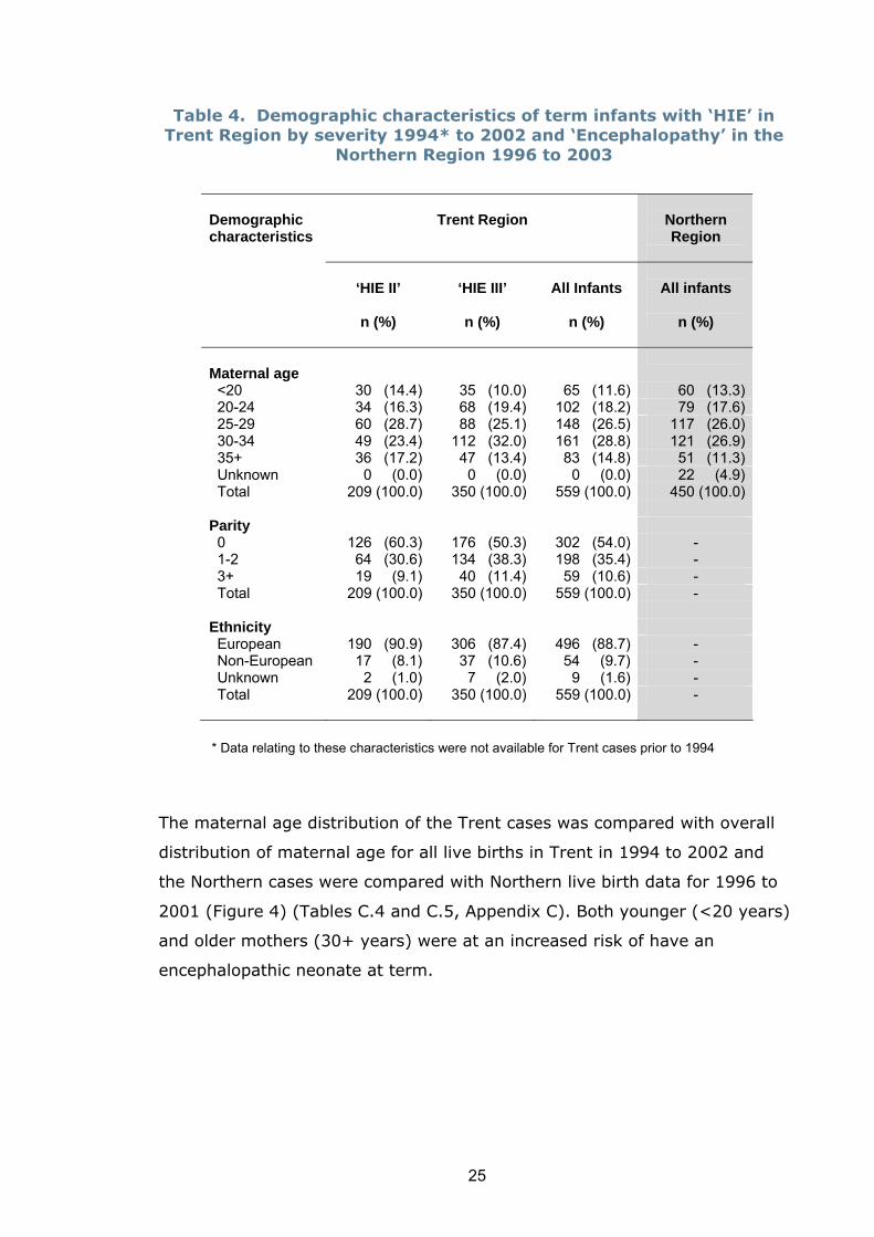
Table 4. Demographic characteristics of term infants with ‘HIE’ in Trent Region by severity 1994* to 2002 and ‘Encephalopathy’ in the
Northern Region 1996 to 2003
Demographic characteristics
Trent Region
Northern Region
‘HIE II’
n (%)
‘HIE III’
n (%)
All Infants
n (%)
All infants
n (%)
Maternal age
<20 30 (14.4) 35 (10.0) 65 (11.6) 60 (13.3) 20-24 34 (16.3) 68 (19.4) 102 (18.2) 79 (17.6) 25-29 60 (28.7) 88 (25.1) 148 (26.5) 117 (26.0) 30-34 49 (23.4) 112 (32.0) 161 (28.8) 121 (26.9) 35+ 36 (17.2) 47 (13.4) 83 (14.8) 51 (11.3) Unknown 0 (0.0) 0 (0.0) 0 (0.0) 22 (4.9) Total 209 (100.0) 350 (100.0) 559 (100.0) 450 (100.0) Parity 0 126 (60.3) 176 (50.3) 302 (54.0) - 1-2 64 (30.6) 134 (38.3) 198 (35.4) - 3+ 19 (9.1) 40 (11.4) 59 (10.6) - Total 209 (100.0) 350 (100.0) 559 (100.0) - Ethnicity European 190 (90.9) 306 (87.4) 496 (88.7) - Non-European 17 (8.1) 37 (10.6) 54 (9.7) - Unknown 2 (1.0) 7 (2.0) 9 (1.6) - Total 209 (100.0) 350 (100.0) 559 (100.0)
-
* Data relating to these characteristics were not available for Trent cases prior to 1994
The maternal age distribution of the Trent cases was compared with overall
distribution of maternal age for all live births in Trent in 1994 to 2002 and
the Northern cases were compared with Northern live birth data for 1996 to
2001 (Figure 4) (Tables C.4 and C.5, Appendix C). Both younger (<20 years)
and older mothers (30+ years) were at an increased risk of have an
encephalopathic neonate at term.
25
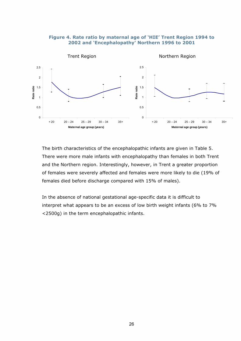
Figure 4. Rate ratio by maternal age of ‘HIE’ Trent Region 1994 to
2002 and ‘Encephalopathy’ Northern 1996 to 2001
Trent Region Northern Region
0
0.5
1
1.5
2
2.5
< 20 20 – 24 25 – 29 30 – 34 35+
Maternal age group (years)
Rat
e ra
tio
0
0.5
1
1.5
2
2.5
< 20 20 – 24 25 – 29 30 – 34 35+
Maternal age group (years)
Rat
e ra
tio
The birth characteristics of the encephalopathic infants are given in Table 5.
There were more male infants with encephalopathy than females in both Trent
and the Northern region. Interestingly, however, in Trent a greater proportion
of females were severely affected and females were more likely to die (19% of
females died before discharge compared with 15% of males).
In the absence of national gestational age-specific data it is difficult to
interpret what appears to be an excess of low birth weight infants (6% to 7%
<2500g) in the term encephalopathic infants.
26

Table 5. Birth characteristics of term infants with ‘HIE’, Trent Region 1991 to 2002 and ‘Encephalopathy’, Northern Region 1996 to 2003
Birth characteristics
Trent Region
Northern Region
‘HIE II’
n (%) ‘HIE III’ n (%)
All infants n (%)
All infants n (%)
Labour induction*
Yes 64 (30.6) 99 (28.3) 163 (29.2) 91 (20.2) No 144 (68.9) 246 (70.3) 390 (69.8) 357 (79.3) Unknown 1 (0.5) 5 (1.4) 6 (1.1) 2 (0.4) Total 209 (100.0) 350 (100.0) 559 (100.0) 450 (100.0) Mode of delivery Normal 126 (37.7) 160 (33.8) 286 (35.4) 163 (36.2) Forceps or ventouse 94 (28.1) 85 (17.9) 179 (22.2) 99 (22.0) Breech/assisted breech 6 (1.8) 13 (2.7) 19 (2.4) 6 (1.3) Caesarean section 179 (39.8) Emergency CS 100 (29.9) 203 (42.8) 303 (37.5) - In labour 87 (26.0) 144 (30.4) 231 (28.6) - Not in labour 13 (3.9) 59 (12.4) 72 (8.9) - Elective CS 7 (2.1) 13 (2.7) 20 (2.5) - Unknown 1 (0.3) 0 (0.0) 1 (0.1) 3 (0.6) Total 334 (100.0) 474 (100.0) 808 (100.0) 450 (100.0) Plurality 1 307 (91.9) 448 (94.5) 755 (93.4) 445 (98.9) 2 7 (2.1) 13 (2.7) 20 (2.5) 5 (1.1) Unknown 20 (6.0) 13 (2.7) 33 (4.1) 0 (0.0) Total 334 (100.0) 474 (100.0) 808 (100.0) 450 (100.0) Birth weight (grams) <1500 0 (0.0) 1 (0.2) 1 (0.1) 0 (0.0) 1500-2499 20 (6.0) 26 (5.5) 46 (5.7) 32 (7.1) 2500-3999 274 (82.0) 391 (82.5) 665 (82.3) 351 (78.0) 4000+ 40 (12.0) 56 (11.8) 96 (11.9) 67 (14.9) Total 334 (100.0) 474 (100.0) 808 (100.0) 450 (100.0) Gestational age (weeks) 37 27 (8.1) 45 (9.5) 72 (8.9) 49 (10.9) 38 34 (10.2) 61 (12.9) 95 (11.8) 73 (16.2) 39 60 (18.0) 67 (14.1) 127 (15.7) 76 (16.9) 40 130 (38.9) 167 (35.2) 297 (36.8) 120 (26.7) 41 65 (19.5) 92 (19.4) 157 (19.4) 109 (24.2) 42 17 (5.1) 41 (8.6) 58 (7.2) 23 (5.1) 43 1 (0.3) 1 (0.2) 2 (0.2) 0 (0.0) Total 334 (100.0) 474 (100.0) 808 (100.0) 450 (100.0) Infant sex Male 214 (64.1) 249 (52.5) 463 (57.3) 277 (61.6) Female 120 (35.9) 222 (46.8) 342 (42.3) 173 (38.4) Unknown 0 (0.0) 3 (0.6) 3 (0.4) 0 (0.0) Total 334 (100.0) 474 (100.0) 808 (100.0)
450 (100.0)
*Data about induction of labour were not collected prior to 1994
27

The mode of delivery characteristics were compared with data from the
National Sentinel Caesarean Section Audit 38 (Table 6).
Table 6. Delivery characteristics of encephalopathy cases compared with National Sentinel Audit 38 results (proportions)
Delivery characteristics
Trent ‘HIE’
cases 1991-2002
East Midlands Sentinel Audit
2000-2001
Northern ‘enceph’*
cases 1996-2003
North Eastern
Sentinel Audit
2000-2001
Induction of labour Yes
29.2%
24.1%
20.2%
23.0%
Mode of delivery Spontaneous
35.4%
67.1%
36.2%
70.8%
Instrumental** 24.6% 12.3% 23.3% 9.6% Caesarean section: Emergency CS Elective CS
40.0% 37.5%
2.5%
20.4% 12.5%
7.9%
39.8% -- --
19.3% 12.0%
7.3% Not known 0.1% -- 0.6% --
*Encephalopathy
** Includes forceps, ventouse and breech extraction
Care must be exercised when comparing the neonatal encephalopathy results
with the results from the National Sentinel Audit since the Audit data relate to
a sample of all deliveries, not just term live births. Furthermore, the Audit
data were collected towards the latter period of the data collection for the
encephalopathy data. Nevertheless, it would appear that a similar proportion
of encephalopathy cases were induced compared to all deliveries. In contrast
there is a striking difference in the distribution of mode of delivery with the
encephalopathy cases being twice as likely to be delivered by forceps,
ventouse or as a breech extraction and twice as likely to be delivered by
caesarean section. In the case of the Trent ‘HIE’ cases, when performed,
caesarean sections were more likely to be carried out as emergency
procedures compared with the general population of deliveries (94% versus
61% respectively). Only 6% of caesarean sections for the Trent ‘HIE’ cases
28

were elective compared with 39% of the East Midlands caesarean deliveries
in the Sentinel Audit. Given the increasing trends in operative and
instrumental deliveries over the last decade, this comparison, which uses
more recent population data, is likely to under-estimate the true differences
in mode of delivery between the encephalopathy cases and the general
population of births from the earlier period.
29

Information about the condition of the infant at delivery and the number of
deaths is given in Table 7.
Table 7. Condition at delivery and deaths, term infants with ‘HIE’ in Trent Region by severity 1994 to 2002 and ‘Encephalopathy’ in the
Northern Region 1996 to 2003
Condition
Trent Region
Northern Region
‘HIE II’
n (%) ‘HIE III’ n (%)
All infants n (%)
All infants n (%)
Apgar score at 1 minute
<3 53 (25.4) 187 (53.4) 240 (42.9) 109 (24.2) 3-6 77 (36.8) 103 (29.4) 180 (32.2) 90 (20.0) 7-10 78 (37.3) 50 (14.3) 128 (22.9) 105 (23.3) Unknown 1 (0.5) 10 (2.9) 11 (2.0) 146 (32.4) Total 209 (100.0) 350 (100.0) 559 (100.0) 450 (100.0) Apgar score at 5 minutes
<3 8 (3.8) 92 (26.3) 100 (17.9) 36 (8.0) 3-6 64 (30.6) 134 (38.3) 198 (35.4) 83 (18.4) 7-10 136 (65.1) 104 (29.7) 240 (42.9) 177 (39.3) Unknown 1 (0.5) 20 (5.7) 21 (3.8) 154 (34.2) Total 209 (100.0) 350 (100.0) 559 (100.0) 450 (100.0) Abnormal scalp pH Yes 9 (4.3) 16 (4.6) 25 (4.5) - No 187 (89.5) 295 (84.3) 482 (86.2) - Unknown 13 (6.2) 39 (11.1) 52 (9.3) - Total 209 (100.0) 350 (100.0) 559 (100.0) - Cord pH <7.1 - - -
78 (17.3)
7.1+ - - - 116 (25.8) Unknown - - - 256 (56.9) Total - - - 450 (100.0) Deaths Died 1 (0.3) 139 (29.3) 140 (17.3) 61 (13.6) Discharged alive 333 (99.9) 334 (70.5) 667 (82.5) 389 (86.4) Unknown 0 (0.0) 1 (0.2) 1 (0.1) 0 (0.0) Total 334 (100.0) 474 (100.0) 808 (100.0) 450 (100.0)
The absence of suitable comparison data makes it difficult to interpret the
information about the condition of the neonates at birth. The vast majority of
30

the moderate (‘HIE II’) cases in Trent survived to discharge, whereas, just
less than a third of the severe cases died before they could be discharged
home. Overall one in seven of the ‘Encephalopathic’ cases in the Northern
region died before discharge.
4.4. Intrapartum stillbirths
Intrapartum stillbirth rates for term pregnancies are given in Figure 5 (the
data for these figures are given in Tables C.6 and C.7, Appendix C).
Figure 5. Term intrapartum stillbirth rates, Trent Region, 1993 to 2001 and Northern Region 1996 to 2003
Trent Region Northern Region
0.0
0.1
0.2
0.3
0.4
0.5
0.6
0.7
0.8
1996 1997 1998 1999 2000 2001 2002 2003
Year of birth
Rat
e pe
r 100
0 to
tal b
irths
**
0.0
0.1
0.2
0.3
0.4
0.5
0.6
0.7
0.8
1993 1994 1995 1996 1997 1998 1999 2000 2001
Year of birth
Rat
e pe
r 100
0 to
tal b
irths
*In these years the form of the question used to collect data about the timing of the death was changed leading to a large proportion of cases with an unknown timing which may have been intrapartum. The hashed areas denote the rates if these unknown cases are included as intrapartum deaths.
In contrast to the neonatal encephalopathy data it is possible to make direct
comparisons between the term intrapartum stillbirth rates in Trent and the
Northern region since the data were collected using similar definitions and
procedures. The overall intrapartum stillbirth rate for pregnancies at term for
the period 1996 to 2001 (the period for which data are available for both
regions) was 0.30 per 1,000 total births in the Northern region and 0.22 per
1,000 total births in Trent excluding the deaths with an uncertain timing and
0.30 per 1,000 when they were included. There is no evidence of a trend in
the stillbirth rate in the Northern region. Similarly when the deaths with an
uncertain timing were included there is no evidence of a change in the
stillbirth rate in Trent.
31

4.5. Discussion and recommendations
One of the aims of this report was to provide a current estimate of the
national rate of neonatal encephalopathy. In the absence of a universally
agreed definition for neonatal encephalopathy it is not possible to do this. The
UK encephalopathy statistics are relatively limited and thus our analysis was
based on available data from the former Trent and Northern health regions.
These areas have the advantage of having region-wide neonatal data
collections which have used the same definitions and procedures for data
collection since 1991 and 1996 respectively. However, the definition for
encephalopathy (‘HIE’) used in Trent is based on the Sarnat and Sarnat 10
criteria and requires all infants as a minimum to have seizures. Furthermore,
‘HIE’ had to be the primary reason for admission for the infant to be included
in the dataset. There is some evidence from our analysis that under-counting
of cases may have occurred. The data collection system in the Northern
region does not include a specific data item which allows cases of neonatal
encephalopathy to be identified. We therefore had to rely on using a
constellation of criteria to identify probable cases. As a consequence it is
likely that under-counting of cases also occurred in the Northern region data.
These limitations mean that we are unable to either quote, with confidence, a
neonatal encephalopathy rate that might be regarded as a valid estimate of
the national rate or to compare the rates in the two regions. However,
because the same definitions and methods of data collection were used in
each region for the period of the data collection we are able to examine ‘HIE’
and ‘encephalopathy’ trends over time. From our analysis there is no
evidence of a decline in the rate of ‘HIE’ and ‘encephalopathy’ since the mid-
1990s in either of the two regions.
Whilst we cannot make direct comparisons between studies mainly because
different definitions of neonatal encephalopathy have been used, Table 8
places the findings from Trent and Northern into a broader context.
As can be seen from Table 8 both population-based studies which were
conducted to investigate the causes and consequences of neonatal
encephalopathy (Western Australia and South West Thames region) 6,8 used a
32

broad definition and found a higher rate of encephalopathy than the regional
(Trent, Northern) or unit-based (Derby) 32 analyses. Also of note the unit-
based data from Derby were derived from a series of retrospective case note
reviews. Whilst the Levene et al 5 criteria were then applied to the identified
cases the authors also noted that: “Infants with encephalopathy clearly
attributable to a cause other than asphyxia were excluded.” Whilst the
difference in rates illustrated in Table 8 might represent true differences in
disease risk between populations it is also plausible that the differences
reflect the sensitivity to changes in definition and the differing purposes and
methods of data collection.39
In population terms there have been enormous changes in the pattern of
maternal age at delivery during the 1990s and early 2000s, however,
adjusting for the effects of maternal age on the annual encephalopathy rate
had no effect on the rate.
We were only able to obtain a measure of severity from the Trent data.
Overall there was a greater proportion of severe cases than moderate cases.
This is surprising since it does not reflect the findings from the few other
recent population-based studies which are available for comparison, where
the reverse was true.6,7,32 As discussed above we are concerned about the
possibility of the under-counting of cases which would be more likely to affect
moderate rather than severe cases. We are therefore, cautious in our
interpretation of what appears to be a modest decline in the rate of moderate
‘HIE’ (HIE II). Severely affected cases are less likely to be subject to under-
counting as the primary reason for admission is more likely to have been
given as ‘HIE’ than another condition. There is no evidence of a systematic
decline in the rate of severe ‘HIE’ (HIE III) in Trent from 1991 to 2002.
33
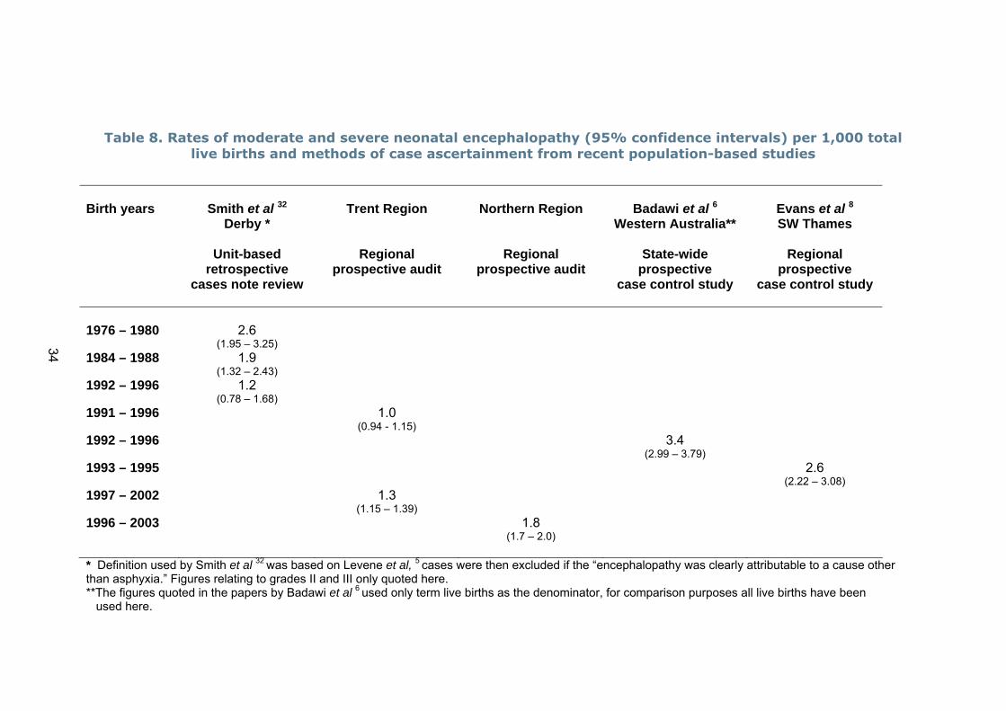
Table 8. Rates of moderate and severe neonatal encephalopathy (95% confidence intervals) per 1,000 total live births and methods of case ascertainment from recent population-based studies
Birth years
Smith et al 32
Derby *
Unit-based retrospective
cases note review
Trent Region
Regional prospective audit
Northern Region
Regional prospective audit
Badawi et al 6
Western Australia**
State-wide prospective
case control study
Evans et al 8
SW Thames
Regional prospective
case control study
1976 – 1980
2.6
(1.95 – 3.25)
1984 – 1988 1.9 (1.32 – 2.43)
1992 – 1996 1.2 (0.78 – 1.68)
1991 – 1996 1.0(0.94 - 1.15)
1992 – 1996 3.4(2.99 – 3.79)
1993 – 1995 2.6(2.22 – 3.08)
1997 – 2002 1.3(1.15 – 1.39)
1996 – 2003 1.8(1.7 – 2.0)
34
* Definition used by Smith et al 32 was based on Levene et al, 5 cases were then excluded if the “encephalopathy was clearly attributable to a cause other than asphyxia.” Figures relating to grades II and III only quoted here. **The figures quoted in the papers by Badawi et al 6 used only term live births as the denominator, for comparison purposes all live births have been used here.

Lack of regional comparison data limited our ability to interpret the findings
relating to the characteristics of the cases. However, with the combination of
the limited regional data that are available and the results from the National
Sentinel Caesarean Section Audit 38 we were able to make some comparisons
for maternal age, parity, infant sex, induction of labour and mode of delivery.
Again these comparisons must be approached with caution as in the absence
of gestation-specific data both the regional data and the Sentinel Audit data
relate to either all live births or all births/confinements whereas the
encephalopathy data relate to only term live births.
Both younger (<20 years) and older mother (30+ years) were at an
increased risk of having an encephalopathic neonate at term. This is in
contrast to the finding in the Western Australian population where the highest
risk was in the age group 20 to 29 years.6 Whilst it is difficult to compare
results from a study carried out in Nepal, Ellis et al 7 also found the highest
risk of neonatal encephalopathy in women 30 years and older. An excess of
primiparous women is in keeping with data from elsewhere as is the excess of
affected male infants.6-8
The results relating to induction and mode of delivery must also be
interpreted with care. High rates of emergency caesarean sections and low
rates of elective sections have been described in other studies.7,40 Neither
general causality nor an intrapartum cause should be inferred from the high
rate of emergency sections. Of note emergency sections were performed
prior to the onset of labour in a quarter of Trent cases overall and nearly a
third of the severe cases. We have no information about which cases were
planned as elective sections and where events overtook the planning either
leading to a spontaneous delivery or an emergency section. Much more
information is needed to unravel the complicated chain of causal events
relating to mode of delivery than is available in the two datasets analysed for
this report.
The findings relating to the characteristics of the cases have greatest utility in
the reassurance they provide that we are dealing with a similar spectrum of
cases as other investigators. The actual results should, however, not be over
35

interpreted because of the limitations in terms of appropriate comparator
data.
In contrast to the neonatal encephalopathy data the information relating to
term intrapartum stillbirths is collected using similar definitions and
procedures in the two regions. Our a priori interest in the term intrapartum
stillbirth rate was in the event that we observed a decline in the neonatal
encephalopathy rate. We wished to investigate whether, if there had been a
decline in encephalopathy, there had been an increase in the intrapartum
stillbirth rate as it might be anticipated that a small proportion of cases would
be transferred between these two diagnostic categories. As it transpired there
was no evidence of a decline in the rate of neonatal encephalopathy and the
rates of intrapartum stillbirths in term births in the two regions were similar
at about 0.3 per 1,000 total births in the period 1996 to 2001. There was no
evidence of a change in the rate of term intrapartum stillbirths in either
region from the mid-1990s onwards.
5. The contribution of intrapartum events and potentially preventable mechanisms related to labour and delivery – aim (iii) 5.1. Literature review
As part of the general literature review (see section 3.1) studies which
specifically investigated the contribution of acute intrapartum adverse events
or evidence of intrapartum hypoxia to the aetiology of neonatal
encephalopathy were identified. Given the changing patterns of the
management of labour and delivery this review concentrated on publications
from the mid-1990s onwards. These are summarised in Appendix D.
There have only been four recent UK-based studies that enable the question
of the role of acute intrapartum events and hypoxia in the aetiology of
neonatal encephalopathy to be considered.
As described earlier Smith et al 32 conducted a unit-based retrospective case
note review of hypoxic-ischaemic encephalopathy for births from 1992 to
36

1996 in Derby. Whilst they applied the criteria described by Levene et al 5 to
identify cases they also noted that “Infants with encephalopathy clearly
attributable to a cause other than asphyxia were excluded.” This implies that
all 29 cases meeting the Levene el al “post-asphyxial encephalopathy” criteria
grades II and III were attributable to an intrapartum cause and that the rate
of intrapartum encephalopathy was 1.2 per 1,000 total live births (95%CI
0.78 to 1.68). The limitations of this study are that it does not include recent
cases, it relies on retrospective case ascertainment, and the criteria to
determine an intrapartum cause for the encephalopathy were not defined,
were determined from medical records and do not appear to be based on
explicit criteria.
Evans et al 8 carried out a case control study of 150 cases of neonatal
encephalopathy and 154 controls born between 1993 and 1995 in the South
West Thames region. Seven of the cases had “well-defined conditions that
were clearly unrelated to intrapartum asphyxia. The remaining 143 cases,
where asphyxia was a possible diagnosis” were included in the analysis.
Clinical raters were used to determine the presence of hypoxic–ischaemic
encephalopathy (HIE). However, whilst various criteria for the acid-base
status of cord blood samples were defined, HIE remained undefined. The
findings were then reported for only those 16 infants who subsequently
developed cerebral palsy by the age of one. Interestingly three of the 16 had
a history “suggestive of pre-existing abnormality or vulnerability.” (eg
abnormal CTG at the onset of labour). Consequently, from the data reported
by Evans et al, it is not possible to estimate the role of intrapartum events in
the aetiology of encephalopathy where cerebral palsy was not subsequently
diagnosed.
Draper and colleagues reported the findings of a confidential enquiry, using
the CESDI enquiry methodology, into 49 survivors of ‘HIE II’ and ‘HIE III’
born at ≥ 35 weeks gestation in 1997 and identified from the Trent Neonatal
Survey.41 The timing of any apparent “asphyxial episode” was ascribed to the
appropriate period using a series of questions‡ derived from the CESDI
‡ (i) Is there evidence of peripartum insult? (ii) Does the peripartum insult alone account for the clinical condition of the baby? (iii) Is there evidence of an antepartum insult?
37
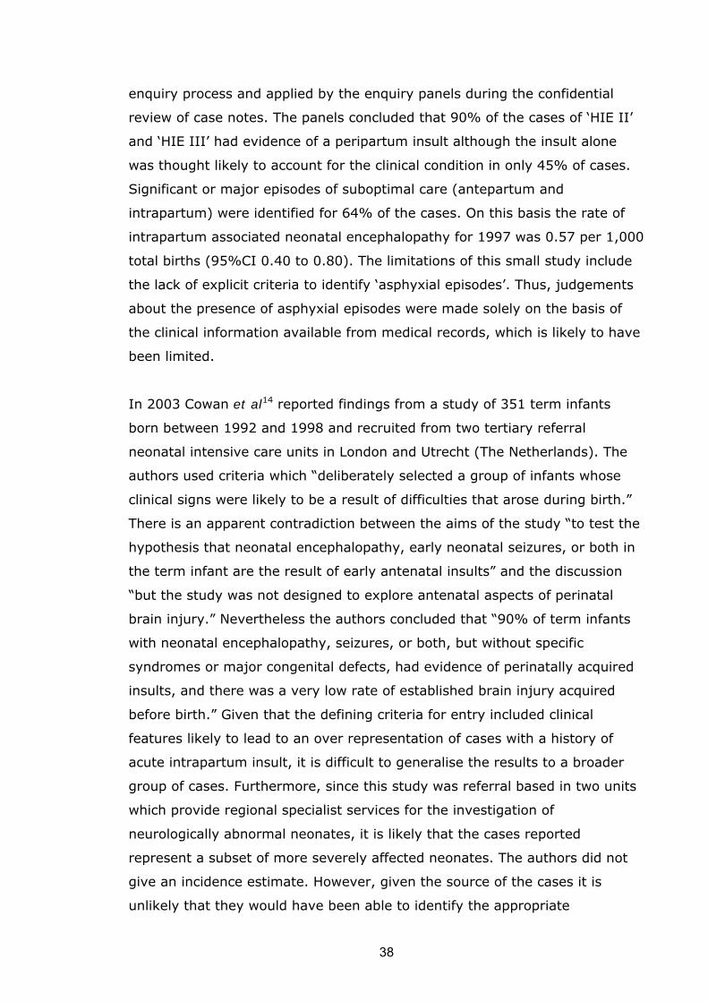
enquiry process and applied by the enquiry panels during the confidential
review of case notes. The panels concluded that 90% of the cases of ‘HIE II’
and ‘HIE III’ had evidence of a peripartum insult although the insult alone
was thought likely to account for the clinical condition in only 45% of cases.
Significant or major episodes of suboptimal care (antepartum and
intrapartum) were identified for 64% of the cases. On this basis the rate of
intrapartum associated neonatal encephalopathy for 1997 was 0.57 per 1,000
total births (95%CI 0.40 to 0.80). The limitations of this small study include
the lack of explicit criteria to identify ‘asphyxial episodes’. Thus, judgements
about the presence of asphyxial episodes were made solely on the basis of
the clinical information available from medical records, which is likely to have
been limited.
In 2003 Cowan et al14 reported findings from a study of 351 term infants
born between 1992 and 1998 and recruited from two tertiary referral
neonatal intensive care units in London and Utrecht (The Netherlands). The
authors used criteria which “deliberately selected a group of infants whose
clinical signs were likely to be a result of difficulties that arose during birth.”
There is an apparent contradiction between the aims of the study “to test the
hypothesis that neonatal encephalopathy, early neonatal seizures, or both in
the term infant are the result of early antenatal insults” and the discussion
“but the study was not designed to explore antenatal aspects of perinatal
brain injury.” Nevertheless the authors concluded that “90% of term infants
with neonatal encephalopathy, seizures, or both, but without specific
syndromes or major congenital defects, had evidence of perinatally acquired
insults, and there was a very low rate of established brain injury acquired
before birth.” Given that the defining criteria for entry included clinical
features likely to lead to an over representation of cases with a history of
acute intrapartum insult, it is difficult to generalise the results to a broader
group of cases. Furthermore, since this study was referral based in two units
which provide regional specialist services for the investigation of
neurologically abnormal neonates, it is likely that the cases reported
represent a subset of more severely affected neonates. The authors did not
give an incidence estimate. However, given the source of the cases it is
unlikely that they would have been able to identify the appropriate
38

denominator population to calculate a rate. Hence, it is not possible to
estimate the rate of encephalopathy associated with an intrapartum cause
from these data.
There have been a small number of studies based elsewhere in the world that
merit consideration. These include the Western Australian population-based
case control study of moderate and severe neonatal encephalopathy in births
from 1993 to 1995.6,39 In this study the authors specifically attempted to
estimate the contribution of adverse intrapartum events and the occurrence
of intrapartum hypoxia using the 1996 American Academy of Pediatrics and
American College of Obstetricians and Gynecologists criteria.42 The authors
used a broad definition of neonatal encephalopathy which did not presume an
intrapartum aetiology and estimated that 29% of the 164 cases were
associated with intrapartum hypoxia. However, it should be noted that 25%
of the cases with evidence of intrapartum hypoxia also had antepartum risk
factors and only 4% of the cases had evidence of intrapartum hypoxia in the
absence of other risk factors. These findings translate into a rate of
encephalopathy associated with an intrapartum cause of about 1.0 per 1,000
total live births.
Ellis and colleagues conducted a case control study in Kathmandu, Nepal,7
the data from which are probably less applicable to the UK situation than the
data from Australia. Overall 60% of the 131 mild, moderate and severe cases
born in 1995 and 1996 met the criteria to suggest “evidence of intrapartum
compromise or were born after an intrapartum difficulty likely to result in
fetal compromise.” 6
There are wide variations in the purposes, design and conduct of the small
number of UK-based studies. The applicability of the results of the non-UK
based studies to the UK situation is difficult to assess. The problems arising
from the variations in criteria used to define neonatal encephalopathy were
compounded by variations in how the intrapartum contribution was assessed.
Given these limitations we concluded that it is not possible to use the data
from these published studies to confidently estimate the contribution of
39

intrapartum events and the care provided during labour and delivery to the
aetiology of neonatal encephalopathy.
5.2. Results from the Trent Neonatal Survey data
We attempted to assess the contribution of intrapartum factors to the rate of
neonatal encephalopathy using data from the Trent Neonatal Survey. To do
this we applied the MacLennan criteria 19 (for which we had data available) to
estimate the proportion of infants with ‘HIE II’ or ‘HIE III’ with evidence of
exposure to possible intrapartum hypoxia. In the absence of information
about cord blood pH, fetal heart rate, multiorgan involvement and sentinel
events during labour and delivery we had to rely on the combination of Apgar
scores and the report of an abnormal intrapartum CTG (data collected as:
Yes/No). We used the MacLennan criterion of an Apgar score of 0-6 for longer
than five minutes (which we interpreted to mean a score at both one and five
minutes of 0-6) combined with the response of ‘Yes’ to the abnormal
intrapartum CTG data item (Table 9). Of note the 2002 American College of
Obstetrics and Gynecology criteria used the more stringent criterion for Apgar
score of 0-3 beyond five minutes.15
The use of only two criteria from a possible list of one essential and five that
suggest an intrapartum timing is clearly a considerable limitation of this
analysis. If only one criterion were to be applied, between 53% and 75% of
cases would be classified as having an intrapartum origin. As the results of
additional criteria are added the proportion falls. Having included both one
and five minute Apgar scores together with reported CTG abnormalities the
proportion is, in comparison, only 38%. If it were possible to add data about
further criteria, including the results of cord arterial samples, information
about sentinel events during labour and delivery, evidence of multi-organ
involvement and more details about CTG abnormalities it is likely that the
proportion would reduce further. Thus, it is probable that the figure of 38%,
which represents a rate of 0.41 per 1,000 total live births (95%CI 0.36 to
0.47) is an over-estimate of the intrapartum contribution. At the same time
the criteria used to define neonatal encephalopathy in the first instance are
also likely to lead to an over-estimate of the intrapartum contribution. Since
40

Table 9. Assessing the contribution of intrapartum events and hypoxia based on Apgar scores and CTG abnormalities*,
Trent 1994 to 2002
Criteria
Frequency
N=559
n %
Apgar score at 1 minute:
0 - 6
420
75.1
7 - 10 128 22.9 Missing 11 2.0 Apgar score at 5 minutes: 0 – 6 298 53.3 7 – 10 240 42.9 Missing 21 3.8 CTG abnormality reported**: Yes 376 67.3 No 167 29.9 Missing 16 2.9 Apgar score at 1 minute 0-6 and Apgar score at 5 minutes 0-6:
Yes
295
52.8
No 253 45.3 Missing 11 1.9 Apgar score at 1 minute 0-6 and Apgar score at 5 minutes 0-6*** and CTG abnormality reported**:
Yes
213
38.1 No 313 56.0 Missing 33 5.9
*Based on an abbreviated version of the MacLennan criteria.19
**These data came from a data item relating to intrapartum monitoring: CTG abnormality (Yes/No). No further details are available and it is not possible to validate the responses. ***Includes 1 case with a missing one minute Apgar score and a five minute Apgar score of 0-6.
it is not possible to quantify the combined effect of these two factors these
findings cannot be relied upon to give a robust estimate of the intrapartum
contribution. At best they can be interpreted as suggestive of a depressed
state at birth that may be related to recent acute hypoxia or ischaemia.
Consequently the results should not be regarded as confirmatory of
intrapartum hypoxia/ischaemia nor should they be regarded as predictive of
poor later outcome.20
41

5.3. Discussion and recommendations
One of the aims of this report was to provide a current estimate of the
contribution of intrapartum events and hypoxia to the neonatal
encephalopathy rate. It is not possible to do this. In the few available studies
the difficulties arising from the effects of variations in the definition of
neonatal encephalopathy used to identify cases are compounded by a lack of
consistently applied criteria to estimate the effects of intrapartum events and
possible hypoxia. Given the limitations of the information available, both from
the published literature and the analysis we carried out using data from the
Trent Neonatal Survey, we are unable with any confidence to estimate the
contribution of intrapartum factors to the overall neonatal encephalopathy
rate.
Until a universally agreed definition for neonatal encephalopathy is applied
consistently in national data collection systems and data about explicit
criteria are collected to assess the potential intrapartum contribution it will
not be possible to either estimate the rate of neonatal encephalopathy nor to
assess the contribution of intrapartum events and possible hypoxia. Existing
criteria for assessing the intrapartum contribution 15,19 were designed to
retrospectively assess the contribution of intrapartum events and hypoxia in
cases of cerebral palsy and were not designed for prospective and routine
application. To apply these criteria in full would require a complex level of
data collection. For example, information is needed about investigations that
are not universally performed eg. umbilical artery pH and base deficit.
Complex and detailed information about multiple systems would be required
to assess multi-organ involvement; these data will be difficult to collect
systematically and assess externally. Given these complexities it may never
be possible to collect this level of detail in every neonatal unit.
To collect some level of information it may be necessary to reach an agreed
abbreviated set of explicit defining criteria and/or collect information from a
random sample of neonatal units on a rolling basis or from neonatal networks
designated as sentinel reporting networks. As part of the consensus process
we are using to define neonatal encephalopathy for surveillance purposes we
42

are also including discussions about the criteria to define the role of
intrapartum events and hypoxia.
6. Conclusions – aim (iv)
i. Neonatal encephalopathy is a clinically defined condition most commonly
seen and best described in the term neonate. Its manifestations include
difficulty with initiating and maintaining respiration, depression of tone and
reflexes, subnormal levels of consciousness, and often seizures.20 There is at
present no single universally agreed definition to either monitor the rate of
this condition in the UK or to use for research into the aetiology, prevention
and treatment of encephalopathy.
ii. It is our view that the most appropriate method for reaching a universally
agreed definition for neonatal encephalopathy is a consensus approach and
we have embarked upon this process.
iii. Terms historically regarded as synonymous with neonatal encephalopathy,
such as birth asphyxia, perinatal asphyxia, hypoxic-ischaemic encephalopathy
and post-asphyxial encephalopathy continue to be used in clinical parlance,
medical records and the research literature. This is unhelpful to both our
understanding of the aetiology of the condition and the interpretation by
parents of the possible cause of their infant’s condition. The lack of a specific
medical database mesh term for neonatal encephalopathy contributes to the
continuing use of these terms.
iv. Currently available rates of neonatal encephalopathy are difficult to
interpret as, not surprisingly, the incidence measurement appears highly
sensitive to definitional changes. Given the problems of definition it is not
possible to give, with any confidence, an estimate of the current rate of
neonatal encephalopathy in the UK. However, there is no evidence, from the
most recent data available that there has been a decline in the rate of
neonatal encephalopathy since the mid-1990s, nor is there evidence of a
decline in the term intrapartum stillbirth rate.
43

v. It is not possible to recommend a sufficiently robust estimate of the
incidence of neonatal encephalopathy for use in sample size calculations for
trials and observational studies – aim (v).
vi. From the data currently available it was not possible to determine, with
any confidence, the contribution of intrapartum events and the presence of
probable hypoxia to the rate of neonatal encephalopathy. Nor is it possible to
obtain recent and credible estimates from the UK-based literature. The
difficulties arising from the variations in the definition of neonatal
encephalopathy are compounded by the lack of consistently applied criteria to
identify probable intrapartum hypoxia and sentinel events.
vii. Until a universally agreed definition for neonatal encephalopathy is
applied consistently, in national data collection systems and data about
explicit criteria are collected to allow the quantification of the effects of
intrapartum events and hypoxia, it will not be possible to either estimate the
rate of neonatal encephalopathy, nor to assess the contribution of
intrapartum events and possible hypoxia.
viii. It may not be possible to collect the necessary level of detail required by
current criteria to identify the contribution of intrapartum events and hypoxia
in every neonatal unit. To enable the collection of some information it may be
necessary to define an abbreviated set of explicit criteria and/or collect
information from a random sample of units on a rolling basis or from neonatal
networks designated as sentinel reporting networks. As part of the consensus
process we are using to define neonatal encephalopathy we are also including
discussions about the criteria for use to define the intrapartum contribution.
7. Recommendations about future surveillance and research – aim (iv)
i. A universally agreed definition of neonatal encephalopathy which does not
presume aetiology and that can be applied easily is urgently required. We
recommend that a consensus building approach is taken in order to develop a
definition that is usable and which would be adopted by the relevant
professionals. We have embarked upon this process.
44

ii. The identification of criteria indicative of an intrapartum contribution which
can be easily and universally applied is required. In view of the complexity of
current criteria we recommend that the identification of an abbreviated
version is considered. We have included this process as part of the consensus
building process which is already under way. It may be necessary to consider
collecting this level of information from a limited number of neonatal units
identified either randomly on a rolling basis or from neonatal networks
designated as sentinel reporting networks.
iii. The introduction of an agreed definition for neonatal encephalopathy into
the data collection process for neonatal units would enable national
surveillance to be undertaken. It would also allow comparison of local audit
data. Ensuring the adoption of both the definition and the data items for
universal data collection will be encouraged by the consensus building
process. However, adoption of the terminology would be more likely to result
if the definition were to be endorsed by national professional organisations
such as the British Association of Perinatal Medicine, bodies with
responsibility for governance arrangements such as the Health Care
Commission and influential individuals from the Department of Health such as
the Chief Medical Officer and his staff.
iv. We strongly recommend that the use of terms historically regarded as
synonymous with neonatal encephalopathy, such as birth asphyxia, perinatal
asphyxia and hypoxic-ischaemic encephalopathy, is discontinued.
v. A mesh term for neonatal encephalopathy would improve greatly the
ability of clinicians and researchers to locate the relevant literature from
medical databases and would also help discourage the continuing use of
inappropriate terminology. To this end we have made a recommendation to
the US National Library of Medicine (NIH) to include neonatal encephalopathy
as a mesh term and we await the outcome.
vi. Urgent research is required in the UK to improve our understanding of the
aetiology of neonatal encephalopathy and the role that events and care
45

provided during pregnancy and delivery have in its genesis. Such research
would be facilitated greatly by the availability of a universally agreed
definition of neonatal encephalopathy.
46

Acknowledgments
Our grateful thanks go to Professor David Field, Mrs Elizabeth Draper, Mr
Bradley Manktelow and all the staff in the former Trent region involved in
providing, collecting and processing the Trent Neonatal Survey data. Our
grateful thanks also go to Dr Alan Fenton, Dr Martin Ward Platt, Mrs Marjorie
Renwick and all the staff in the Northern region involved in providing,
collecting and processing the Northern region Neonatal Intensive Care data
and the Regional Perinatal Mortality Survey data. Our particular thanks go to
the data custodians of these two datasets who made their data available to
us for analysis.
We are also very grateful to all the members of the consensus panel who
attended and contributed so enthusiastically to the discussions at the Stage 1
consensus meeting. We are particularly grateful to the experts who presented
the four different perspectives about neonatal encephalopathy at the start of
the meeting. Miss Lynne Roberts made the arrangements for this meeting
and she and Mrs Ann Kennedy kindly attended to make notes. We are very
grateful for their written contributions which were very helpful in the drafting
of the Stage 1 consensus discussion document.
47

References
1 Patient Safety Research Programme (Policy Research Programme of the Department of Health). Call for proposal (single tender action) – Neonatal Encephalopathy. Birmingham: PSRP, University of Birmingham. March 2004. 2 Department of Health. An Organisation with a Memory. Report of an expert group on learning from adverse events in the NHS, chaired by the Chief Medical Officer. London: The Stationery Office. 2000. 3 http://www.rcpch.ac.uk/publications/recent_publications/ExecSummarry%20.pdf (accessed 21st November 2004) 4 http://www.bapm.org/documents/publications/dataset_fullreview_20040300.pdf (accessed 23rd December 2004) 5 Levene ML, Kornberg J, Williams THC. The incidence and severity of post-asphyxial encephalopathy in full-term infants. Early Hum Dev 1985; 11: 21-26. 6 Badawi N, Kurinczuk JJ, Keogh JM, Alessandri LM, O’Sullivan F, Burton PR, Pemberton PJ, Stanley FJ. Antepartum risk factors for newborn encephalopathy: The Western Australian case-control study. Br Med J 1998; 317: 1549-53. 7 Ellis M, Manandhar N, Manadhar DS, Costello AM. Risk factors for neonatal encephalopathy in Kathmandu, Nepal, a developing country: unmatched case-control study. Br Med J 2000; 320: 1229-1236. 8 Evans K, Rigby AS, Hamilton P, Titchiner N, Hall DMB. The relationship between neonatal encephalopathy and cerebral palsy: a cohort study. J Obstet Gynaecol 2001; 21(2): 114-120. 9 Nelson KB, Ellenberg JH. The asymptomatic newborn and risk of cerebral palsy. Am J Dis Child 1987; 141: 1333-1335. 10 Sarnat HB, Sarnat MS. Neonatal encephalopathy following fetal distress: A clinical electroencephalopgraphic study. Arch Neurol 1976; 33: 696-705. 11 Amiel-Tison C, Ellison P. Birth asphyxia in the fullterm newborn: Early assessment and outcome. Dev Med Child Neurol 1986; 28: 671-682. 12 Fenichel GM. Hypoxic-ischaemic encephalopathy in the newborn. Arch Neurol 1983; 40: 261-266. 13 Low JA. Intrapartum fetal asphyxia: Definition, diagnosis and classification. Am J Obstet Gynecol 1997; 176: 957-959. 14 Cowan F, Rutherford M, Groenendaal F, Eken P, Mercuri E, Bydder GM, Meiners LC, Dubowitz LM, de Vries LS. Origin and timing of brain lesions in term infants with neonatal encephalopathy. Lancet 2003; 361: 736-42. 15 American College of Obstetricians and Gynecologist and American Academy of Pediatrics. Neonatal Encephalopathy and Cerebral Palsy: Defining the Pathogenesis and Pathophysiology. Washington DC: American College of Obstetricians and Gynecologists. 2002. 16 Murphy MK, Black NA, Lamping DL et al. Consensus development methods, and their uses in clinical guideline development. Health Technology Assessment 1998; 2: i-iv, 1-88. 17 Susskind L, McKearnans S, Thomas-Larmer J. The consensus building handbook. London: Sage Publications. 1999.
48

18 Fink A, Kosecoff J, Chassin M, Brook RH. Consensus methods: characteristics and guidelines for use. Am J Public Health 1984; 74: 979-983. 19 MacLennan A for the International Cerebral Palsy Task Force. A template for defining a causal relation between acute intrapartum events and cerebral palsy: international consensus statement. Br Med J 1999; 319: 1054-9. 20 Nelson KB, Leviton A. How much of neonatal encephalopathy is due to birth asphyxia? Am J Dis Child 1991; 145: 1325-1331. 21 Leviton A, Nelson KB. Problems with definitions and classifications of newborn encephalopathy. Pediatric Neurol 1992; 8: 85-90. 22 Brown JK, Purvis RJ, Forfar JO, Cockburn F. Neurological aspects of perinatal asphyxia. Dev Med Child Neurol 1974; 16: 567-580. 23 Finer NN, Robertsons CM, Richards RT, Pinnell LE, Peters KL. Hypoxic-ischaemic encephalopathy in term infants: perinatal factors and outcome. Pediatrics 1981; 98: 112-117. 24 Hull J, Dodd KL. Falling incidence of hypoxic-ischaemic encephalopathy in term infants. Br J Obstet Gynaecol 1992; 99: 386-391. 25 De Souza SW, Richards B. Neurological sequelae in newborn babies after perinatal asphyxia. Arch Dis Child 1978; 53: 564-569. 26 Thornberg E. Thiringer J, Odeback A, Milsom I. Birth asphyxia: incidence, clinical course and outcome in a Swedish population. Acta Paed Scand 1995; 84: 927-932. 27 Brocklehurst P, Hannah M, McDonald H. Interventions for bacterial vaginosis in pregnancy (Cochrane Review). In: The Cochrane Library. Oxford: Update Software. 28 Johnson P. Birth asphyxia: Should it be redefined or abandoned? In: Intrapartum fetal surveillance. Eds: Spencer JAD, Ward RHT. London: RCOG Press. 1993. 29 Hankins GDV. The long journey: defining the true pathogenesis and pathophysiology of neonatal encephalopathy and cerebral palsy. Obstetrical & Gynecological Survey 2003; 58(7): 435-437. 30 American College of Obstetrics & Gynecology. Committee Opinion No 303 (replaces No 197, February 1998): Inappropriate use of the terms fetal distress and birth asphyxia. Obstet Gynecol 2004; 104(4): 903-904. 31 Depp R. Perinatal asphyxia: assessing its causal role and timing. Semin Ped Neurol 1995; 2(1): 3-36. 32 Smith J, Wells L, Dodd K. The continuing fall in incidence of hypoxic-ischaemic encephalopathy in term infants. Br J Obstet & Gynaecol 2000; 107: 461-466. 33 Hubbard M, Haines L. NCAP: Neonatal Clinical Audit Programme. A national programme of neonatal audit: report of a feasibility study. London: Royal College of Paediatrics and Child Health. 2004. (http://www.ncap.nhs.uk/reports.htm - accessed 31.12.2004) 34 Trent Neonatal Survey Report 2002. Leicester: Department of Health Sciences. 2003. 35 Northern RSMO Annual Report 2001, 2002 (provisional). Newcastle upon Tyne: RMSO/North East Public Health Observatory. 2003. 36 Trent Confidential Enquiry into Stillbirths and Deaths in Infancy – report 1998. Leicester: Department of Epidemiology & Public Health. 1999.
49

37 Bell R, Glinianaia SV, Rankin J, Wright C, Pearce MS, Parker L. Changing patterns of perinatal death, 1982-2000: a retrospective cohort study. Arch Dis Child Fetal Neonatal Ed 2004; 89: F531-F536. 38 RCOG Clinical Effectiveness Support Unit. The National Sentinel Caesarean Section Audit Report. London, Royal College of Obstetricians and Gynaecologists. 2001. 39 Badawi N, Kurinczuk JJ, Hall D, Field D, Pemberton PJ, Stanley FJ. Newborn encephalopathy in term infants: three approaches to population-based investigation. Semin Neonatol 1997; 2: 181-188. 40 Badawi N, Kurinczuk JJ, Keogh JM, Alessandri LM, O’Sullivan F, Burton PR, Pemberton PJ, Stanley FJ. Intrapartum risk factors for newborn encephalopathy: The Western Australian case-control study. Br Med J 1998; 317: 1554-1558. 41 Draper ES, Kurinczuk JJ, Lamming CR, Clarke M, James D, Field DJ. A confidential enquiry into cases of neonatal encephalopathy: clinical governance in practice. Arch Dis Child, Fetal Neonatal Ed 2002; 87: F176-F180. 42 American Academy of Pediatrics and American College of Obstetricians and Gynecologists. Use and abuse of the Apgar score. Pediatrics 1996; 98(1): 141-142.
50

APPENDIX A

1
APPENDIX A
Definitions of Neonatal Encephalopathy and Severity from 12 Key Publications
Author and year of publication
Page
Cowan, Rutherford, Groenendaal et al (2003)…………….…………...……………
2
American College of Obstetricians & Gynecologists (2002)……………………….
3
Evans, Rigby, Hamilton, Titchiner & Hall (2001)……………………………………. Ellis, Manandhar, Manandhar & Costello (2000)……………………………...…….
5 6
MacLennan (1999)……………………………………….……….……………….……
7
Badawi, Kurinczuk & Keogh et al (1999)……………….……………………….……
8
Low (1997)………………….…………………………………………….…………….
9
Nelson & Ellenberg (1987)………………………………………………...…………..
10
Amiel-Tison & Ellison (1986)………………………………………………...…………
11
Levene, Kornberg & Williams (1985)…………………………………………………
13
Fenichel (1983)……………………………………………….…………………………
14
Sarnat & Sarnat (1976)………………………….…………..…………………………
16

2
Cowan F, Rutherford M, Groenendaal F, Eken P, Mercuri E, Bydder GM, Meiners LC, Dubowitz LM & de Vries LS (2003) Origin and timing of brain lesions in term infants with neonatal encephalopathy. Lancet, 361, 736-42.
Neonatal encephalopathy: Abnormal tone pattern, feeding difficulties, altered alertness, and at least three of the following criteria: 1. Late decelerations on fetal monitoring or meconium staining 2. Delayed onset of respiration 3. Arterial cord blood pH less than 7.1 4. Apgar scores less than 7 at 5 minutes, and 5. Multi organ failure.

3
American College of Obstetricians & Gynecologists (2002) Neonatal Encephalopathy and Cerebral Palsy: Defining the Pathogenesis and Pathophysiology. Washington: American College of Obstetricians & Gynecologists.
Criteria to define an acute intrapartum hypoxic event as sufficient to cause cerebral palsy
1.1: Essential criteria (must meet all four) 1. Evidence of a metabolic acidosis in fetal umbilical cord arterial blood obtained at delivery (pH <7 and base deficit ≥ 12 mmol/L) 2. Early onset of severe or moderate neonatal encephalopathy in infants born at 34 or more weeks of gestation 3. Cerebral palsy of the spastic quadriplegic or dyskinetic type* 4. Exclusion of other identifiable etiologies, such as trauma, coagulation disorders, infectious conditions, or genetic disorders
1.2: Criteria that collectively suggest an intrapartum timing (within close proximity to labour and delivery, eg, 0-48 hours) but are nonspecific to asphyxial insults
1. A sentinel (signal) hypoxic event occurring immediately before or during labour 2. A sudden and sustained fetal bradycardia or the absence of fetal heart rate variability in the presence of persistent, late, or variable decelerations, usually after a hypoxic sentinel event when the pattern was previously normal 3. Apgar scores of 0-3 beyond 5 minutes 4. Onset of multisystem involvement within 72 hours of birth 5. Early imaging study showing evidence of acute nonfocal cerebral abnormality.
* Spastic quadriplegia, and less commonly, dyskinetic cerebral palsy are the only types of cerebral palsy associated with acute hypoxic intrapartum events. Spastic quadriplegia is not specific to intrapartum hypoxia. Hemiparetic cerebral palsy, hemiplegic cerebral palsy, spastic diplegia, and ataxia are unlikely to result from acute intrapartum hypoxia.
Neonatal encephalopathy: (references Sarnat and Volpe) A clinically defined syndrome of disturbed neurological function in the earliest days of life in the near-term and term infant. If an intrapartum insult is severe enough to result in ischemic cerebral injury, abnormalities will be noted in the neurological examination within 24 hours after birth. The examination is characterised by abnormalities in: 1. cortical function (lethargy, stupor, coma with or without seizures), 2. brainstem function (ie. Pupillary and cranial nerve abnormalities), 3. tone (hypotonia) and 4. reflexes (absent, hyporeflexia). Outcome is related to the maximum grade of severity. Separate criteria to define an acute intrapartum hypoxic event as sufficient to cause cerebral palsy.

4
“Encephalopathy in the term or near-term newborn infant is manifested by serious neurologic depression in the delivery room and nursery. Clinical signs observable in the delivery room include low apgar score and its components and correlates, such as hypotonia; depressed reflexes including cry, suck, absent Moro’s reflex; decreased consciousness; difficulty in initiating and maintaining respiration; poor colour; and bradycardia.” p.53

5
Evans AS, Rigby P, Hamilton N, Titchiner D & Hall K (2001) The relationships between neonatal encephalopathy and cerebral palsy: a cohort study. Journal of Obstetrics and Gynaecology, 21, 114-120.
Neonatal encephalopathy: An illness occurring at any time in the first 4 days (96 hours) of life and consisting of one or more of: • Definite or probable fits (whatever the clinical manifestation) and including apnoeic attacks and
cyanotic spells • Increased muscle tone • Decreased muscle tone • Abnormal neurological behaviour such as reduced responsiveness, coma, hyper-alertness,
jitteriness • Unable to feed orally for a period of at least 24 hours (unless caused by respiratory problems,
structural lesions, eg. cleft palate or oesophageal atresia, or by the need for neonatal surgery).
Severity: No staging system was used due to disagreement on classification, therefore cases were subdivided according to whether or not they had neonatal fits.
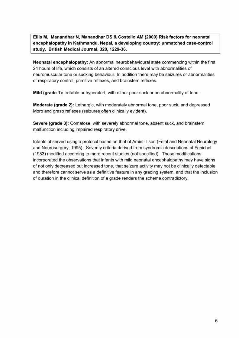
6
Ellis M, Manandhar N, Manandhar DS & Costello AM (2000) Risk factors for neonatal encephalopathy in Kathmandu, Nepal, a developing country: unmatched case-control study. British Medical Journal, 320, 1229-36. Neonatal encephalopathy: An abnormal neurobehavioural state commencing within the first 24 hours of life, which consists of an altered conscious level with abnormalities of neuromuscular tone or sucking behaviour. In addition there may be seizures or abnormalities of respiratory control, primitive reflexes, and brainstem reflexes. Mild (grade 1): Irritable or hyperalert, with either poor suck or an abnormality of tone. Moderate (grade 2): Lethargic, with moderately abnormal tone, poor suck, and depressed Moro and grasp reflexes (seizures often clinically evident). Severe (grade 3): Comatose, with severely abnormal tone, absent suck, and brainstem malfunction including impaired respiratory drive. Infants observed using a protocol based on that of Amiel-Tison (Fetal and Neonatal Neurology and Neurosurgery, 1995). Severity criteria derived from syndromic descriptions of Fenichel (1983) modified according to more recent studies (not specified). These modifications incorporated the observations that infants with mild neonatal encephalopathy may have signs of not only decreased but increased tone, that seizure activity may not be clinically detectable and therefore cannot serve as a definitive feature in any grading system, and that the inclusion of duration in the clinical definition of a grade renders the scheme contradictory.

7
MacLennan A (1999) A template for defining a causal relation between acute intrapartum events and cerebral palsy: International consensus statement. British Medical Journal, 319, 1054-9.
Criteria to define an acute intrapartum hypoxic event
Essential criteria
1. Evidence of a metabolic acidosis in intrapartum fetal, umbilical arterial cord, or very early neonatal blood samples (pH <7.00 and base deficit ≥ 12 mmol/l)
2. Early onset of severe or moderate neonatal encephalopathy in infants of ≥ 34 weeks’ gestation
3. Cerebral palsy of the spastic quadriplegic or dyskinetic type
Criteria that together suggest an intrapartum timing but by themselves are non-specific
4. A sentinel (signal) hypoxic event occurring immediately before or during labour
5. A sudden, rapid, and sustained deterioration of the fetal heart rate pattern usually after the hypoxic sentinel event where the pattern was previously normal
6. Apgar scores of 0-6 for longer than 5 minutes
7. Early evidence of multisystem involvement
8. Early imaging evidence of acute cerebral abnormality All three of the essential criteria are necessary before an intrapartum hypoxic cause of cerebral palsy can begin to be considered. If blood gas data are not available, it cannot be assumed from other signs that hypoxia was present at birth since these signs lack specificity either individually or as a group. When all three essential criteria are met it is necessary to determine whether the hypoxia was acute or chronic. If evidence for some of criteria 4 to 8 is missing or contradictory, the timing of the onset of the neuropathology becomes increasingly in doubt. Individually these latter criteria are only weakly associated with an acute intrapartum damaging hypoxic event because, with the exception of criterion 4 (a sentinel hypoxic event), they may be caused by other factors such as infection. Logically, most of the final five criteria would have to be present for the balance of probabilities to suggest an acute timing of the hypoxic event. Contrary evidence, rather than missing evidence – for example, a normal Apgar score at 5 minutes – would weigh against a serious acute event.
Neonatal encephalopathy: Neonatal encephalopathy is a clinically defined syndrome of disturbed neurological function in the infant at or near term during the first week after birth, manifested by difficulty with initiating and maintaining respiration, depression of tone and reflexes, altered level of consciousness, and often seizures. Levels of severity are not defined.

8
Badawi N, Kurinczuk JJ, Keogh JM, Alessandri LM, O'Sullivan F, Burton PR, Pemberton PJ & Stanley FJ (1998) Antepartum risk factors for newborn encephalopathy: The Western Australian case-control study. British Medical Journal, 317, 1549-53.
Moderate or severe newborn encephalopathy: • Either seizures alone
OR • Any two of the following lasting for longer than 24 hours: Abnormal consciousness Difficulty maintaining respiration (of presumed central origin) Difficulty feeding (of presumed central origin) Abnormal tone and reflexes
Criteria for severe encephalopathy: Infants that fulfilled one or more of the following criteria (the remainder defined as moderate): • Ventilation for > 24 hours • Two or more anticonvulsant treatments • Comatose or stuporous • Died in the neonatal period
Severity criteria modified from Sarnat & Sarnat (1976).

9
Low JA (1997) Intrapartum fetal asphyxia: Definition, diagnosis, and classification. American Journal of Obstetrics and Gynecology, 176, 957-9.
This classification proposes that exposure to asphyxia is confirmed by a blood gas and acid-base assessment with evidence of significant metabolic acidosis and the severity is defined by newborn encephalopathy and other organ system complications. The authors state that severity of intrapartum fetal asphyxia can be classified by determining the short-term outcome as expressed by newborn encephalopathy and other newborn organ system complications. Classification of mild, moderate and severe intrapartum fetal asphyxia
Encephalopathy
Cardiovascular, respiratory & renal complications
Asphyxia
Metabolic acidosis at delivery* Minor Moderate Severe Minor Mod/Severe
Mild + ± ± Moderate + + ± Severe + + +
* Umbilical artery base deficits ≥ 12 mmol/L The clinical signs of newborn encephalopathy associated with intrapartum fetal asphyxia occur more often on day 1 after delivery, with decreasing frequency on days 2 and 3.
Newborn encephalopathy: Minor: jitteriness and irritability Moderate: lethargy or abnormal tone Severe: coma or abnormal tone and multiple seizures.
Cardiovascular complications: Minor: bradycardia or tachycardia (determined by the 95% confidence limits for heart rate for term and preterm newborns) Moderate: hypertension or hypotension (defined by the 95% confidence limits for blood pressure for term and preterm newborns) Severe: abnormal electrocardiographic or echocardiographic findings
Respiratory complications: Minor: requiring supplementary oxygen Moderate: requiring continuous positive airway pressure or transient ventilation (<24 hours) Severe: requiring mechanical ventilation for > 24 hours
Renal complications: Minor: hematuria was observed Moderate: elevation of serum creatinine (>100 µmol/L) Severe: clinical evidence of oliguria (<1ml/kg/hr) or anuria

10
Nelson KB & Ellenberg JH (1987) The asymptomatic newborn and risk of cerebral palsy. American Journal of Diseases of Children, 141, 1333-5.
Hypoxic-ischemic encephalopathy: • Decreased activity after the first day of life • need for incubator care for three or more days • feeding problems • poor suck • respiratory difficulty, and/or • neonatal seizure.
Based on Sarnat & Sarnat (1976), Volpe (1976), Fenichel (1983) and Finer (1981).

11
Amiel-Tison C & Ellison P (1986) Birth asphyxia in the fullterm newborn: Early assessment and outcome. Developmental Medicine and Child Neurology, 28, 671-682. Asphyxia/HIE
In the immediate newborn period, asphyxia is indicated by low Apgar scores, especially five minutes or more, and the need for mask or intubation resuscitation. The use of an Apgar score of ≤ 6 at five minutes for moderate asphyxia and ≤ 3 for severe asphyxia is generally accepted.
Stage I: Characterised by hyperexcitability and mild abnormalities of tone. The subdivision for severity is the length of time that symptoms are present. A cut-off point of 7 days is based on extensive clinical experience: neonates whose signs and symptoms clear by 7 days have been normal at follow-up.
Stage II: Characterised by a deepening CNS depression, defined as either lethargy or light coma. A subdivision in severity is made between neonates with and without seizures. Those with seizures tend to have somewhat more CNS depression and further decrease in primitive reflexes.
Stage III: Characterised by deep coma. In contrast to Sarnat and Sarnat (1976), the authors associate repetitive seizures or status epilepticus with these neonates. In their experience these seizures may be very difficult to control. A subdivision is made according to the presence or absence of brainstem signs, particularly the oculovestibular response. Other brainstem responses may be difficult to evaluate in neonates on respiratory support, particularly those on pancuronium or curare. Neonates could be scored from 0 to 6 (normal to severely abnormal) with such a system: (0 = normal, 1 = stage Ia, 2 = stage Ib, 3 = stage IIa, 4 = stage IIb, 5= stage IIIa, 6 = stage IIIb).

12
Clinical changes in hypoxic-ischemic encephalopathy
A ¦ B A ¦ B A ¦ B¦ ¦ ¦
< 7 days > 7 days
Seizures Seizuresabsent isolated
Oculovestibular BRAIN STEM SIGNSpresent Oculovestibular Oculovestibular
present absentSpontaneousrespirations Spontaneous Spontaneous
present respirations respirationspresent depressed or absent± apnea
A B A B A B
NMR Normal ¦ Possibly abnormalProbably abnormal
Recovery within weeks
Abnormal Recovery more prolonged
CT Scan NormalCerebral Ultrasound NormalEEG NormalBAER Normal Normal ± Abnormal Wave V
Abnormal Normal
Moderately to severely abnormal
Moderately to severely abnormal
Severe abnormalities; Seizures
Abnormal tone
exaggerated¦¦
Spontaneous respirationspresent
¦
Normal
± Abnormal
± Mild abnormalities; Seizures
Hypotonic scarf sign,poor head control
or hyperextended neck¦
¦Swallowing normal
Stretch reflexes
Seizures absent¦
Oculovestibularpresent
Abnormal toneHypotonic
global or restricted to upper body
¦
¦Moro normal or
exaggerated¦
¦Suck normal
light coma
Stage 1 Stage 2
¦
HyperexcitabilitySleeping or fussy
¦
Stage 3
¦Stretch reflexes
diminished
global, moderate to severe± Opisthotonus
¦¦
± Opisthotonus
Lethargy or
C
NS
DEP
RE
SSIO
N
Deep coma
Abnormal toneHypotonic:
¦
¦
Seizures generally
¦
¦
¦
Suck weak or absent
Moro weak
SEI
ZUR
ESSEIZURES
Suck weak
Swallowing ± difficulty Swallowing weak or absent
repetitive or status epilepticus
BR
AIN
STE
M S
IGN
S
N
O S
EIZU
RE
S
N
O C
NS
DEP
RE
SSIO
N
¦
¦Moro absent
Stretch reflexesdiminshed or absent
BAER =Brainstem auditory evoked response NMR = nuclear magnetic resonance

13
Levene ML, Kornberg J & Williams THC (1985) The incidence and severity of post-asphyxial encephalopathy in full-term infants. Early Human Development, 11, 21-6.
Post-asphyxic encephalopathy (PAE): Clinical severity of post-asphyxial encephalopathy modified from Fenichel (1983) Grade I (mild)
Grade II (moderate)
Grade III (severe)
Irritability ‘Hyperalert’
Lethargic Comatose
Mild hypotonia
Seizures Marked abnormalities of tone
Prolonged seizures Severe hypotonia
Poor sucking Requires tube feeding Failure to maintain spontaneous respiration
A diagnosis of PAE was made on the basis of clinical symptoms. The clinical features of PAE usually progressed in severity before recovery or death occurred and the grading refers to the most severe abnormalities the infants showed. Not all features for each grade were necessary for diagnosis, but a progression of symptoms was required over the first 24 or 48 hours of the babies’ lives which then subsequently improved. Grade I: Clinical features of grade I were increasing irritability with some degree of hypotonia together with poor sucking which recovered completely by 3 days of age. These infants often appeared to be ‘hyperalert’, a state in which they seemed hungry but fed poorly and responded vigorously to minimal stimuli. Other causes of irritability, particularly hypoglycemia, were excluded wherever possible and any infant who showed persistent neurological abnormalities were not included in grade I PAE. Grade II: Convulsions, usually developing between 12 and 24 h, together with abnormalities of tone and marked lethargy usually with disinterest in feeding between seizures were necessary for a diagnosis of grade II PAE. Improvement in these symptoms was also essential before allocation to the group could be made. Grade III: In grade III the infants all required ventilatory support, were comatose and had multiple seizures. In these infants recovery was very slow or usually not seen in those who died.

14
Fenichel GM (1983) Hypoxic-ischemic encephalopathy in the newborn. Archives of Neurology, 40, 261-266.
Clinical Syndromes of HIE:
The newborns at risk for major neurologic handicaps have evidence of derangements in many organs, have depressed cerebral function at birth that continues for days or weeks, and in many cases, convulsions soon after birth. In the term newborn, HIE can be divided into three grades of severity according to the clinical symptoms.
Mild encephalopathy: In newborns with mild encephalopathy, the symptoms are maximal during the first 24 hours after birth and then progressively diminish. Consciousness in not materially impaired, except for a brief interval of lethargy immediately after birth. The characteristic feature is jitteriness - a hyperalert state in which there are prolonged periods of wakefulness, irritability, and excessive responsiveness to stimulation. The typical response to stimulation is a low-frequency, high-amplitude shaking of the limbs and jaw. Jitteriness is commonly associated with a low threshold for the Moro reflex, but can occur in the absence of any apparent external manipulation and may be mistaken for a convulsion. Muscle tone is normal when the newborn is at rest or suspended vertically or horizontally. Mild head lag during the traction response is the only demonstrable disturbance in tone. Spontaneous movement and strength are normal in the limbs. Muscle stretch reflexes are normal or slightly hyperactive, and ankle clonus is present and frequently sustained. The anterior fontanel is soft, cranial nerve function is normal, and convulsions do not occur. The EEG is usually normal, but may show lack of background variability. Voltage suppression is not present. The clinical syndrome indicates that the newborn has experienced cerebral distress but does not have cerebral necrosis or increased intracranial pressure. Newborns who have mild, transient HIE recover completely and are not at risk for neurological handicaps. There is no evidence that jitteriness evolves into hyperactivity or that mild encephalopathy is a prologue to learning disabilities. Moderate encephalopathy: Newborns with a moderate encephalopathy are lethargic or obtunded for at least the first 12 hours after birth. Efforts at arousal produce jitteriness. Hypotonia is present at rest, and spontaneous movement of the limbs is decreased. Proximal weakness, in which the muscles about the shoulder are weaker than the muscles about the pelvis, has been described and attributed to edema occurring in the parasaggital region of the cortex and involving those portions of the motor strip that represent limb-girdle muscles. However, this disparity in strength between the shoulder and pelvic muscles is difficult to appreciate in a newborn who is generally hypotonic. In the usual course of events, the period between 48 and 72 hours after birth is a critical interval during which the encephalopathy either worsens or improves. In those newborns whose conditions improve spontaneously, tone increases and arousal is more readily accomplished and associated with jitteriness. In others, there is either lack of improvement or progressive obtundation caused by some combination of convulsions, generalised cerebral edema, hyponatremia secondary to the inappropriate secretion of antidiuretic hormone, and hypoammonemia due to hypoxic liver damage. Convulsions, prolongation of the obtunded state, and progression to stupor are associated with a worsening prognosis. The EEG is always abnormal and may demonstrate lack of

15
background variability, epileptiform activity, or voltage suppression. Lack of background variability has no prognostic implications, but epileptiform activity and voltage suppression are predictive of a bad outcome. Sensory evoked responses can serve as an additional tool for determining the degree of brain damage and the eventual outcome in asphyxiated newborns. Visual evoked responses may prove to be most useful in preterm newborns (the visual radiations are consistently involved in the lesions of periventricular leukomalacia, and brain-stem auditory responses in term newborns. All asphyxiated term newborns in whom the amplitude of wave 1 was more than twice the amplitude of wave 5 either died or were demonstrated to have severe neurological impairment. Severe encephalopathy: Newborns with severe encephalopathy are stuporous or comatose immediately after birth. Respirations are irregular or periodic, and mechanical ventilation is necessary to sustain life. Apnea and convulsions begin during the first 12 hours after birth and progress to tonic and multifocal clonic patterns before the end of the first day. Hypotonia is severe. The newborn lies motionless with legs extended and fully abducted, and the arms remain in any position in which they fall. When traction is tested, there is no grasp reflex, no flexion movements of the head, and no resistance of the limbs. The Moro reflex, tonic neck reflex, and muscle stretch reflexes are usually absent. Pupillary and doll’s eye reflexes are usually normal, but oculomotor palsies may be present. Normal sucking and swallowing are depressed or absent, but intermittent sucking and chewing movements may be present as a convulsive manifestation. Between 12 and 24 hours after birth, some improvement in responsiveness may be noted; stimulation at this time provokes a jittery response. Most children remain stuporous. Convulsions increase in frequency and severity, sometimes progressing to status epilepticus. The EEG is either markedly suppressed or shows a burst-suppression pattern. Overall, deterioration in the newborn’s condition, with brain-stem dysfunction as a prominent feature, occurs between 24 and 72 hours after birth and includes coma, loss of pupillary and vestibulo-ocular reflexes, and respiratory arrest. The fontanel is bulging, and postmortem examination discloses massive cerebral edema and transtentorial herniation. Survivors may remain in a stuporous state for weeks, although convulsions become progressively less frequent and usually cease by the end of first week. Jitteriness is common as the level of consciousness slowly increases. Some newborns who survive the acute encephalopathy will die later during infancy. Those who live have severe neurologic handicaps.

16
Sarnat HB & Sarnat MS (1976) Neonatal encephalopathy following fetal distress: A clinical electroencephalographic study. Archives of Neurology, 33, 696-705. The authors outline three clinical stages of ‘neonatal encephalopathy’.
Distinguishing features of the three clinical stages of postanoxic encephalopathy in the full-term newborn infant
Stage 1 Stage 2 Stage 3
Level of consciousness Hyperalert Lethargic or obtunded Stuporous Neuromuscular control Muscle tone Normal Mild hypotonia Flaccid Posture Mild distal flexion Strong distal flexion Intermittent
decerebration Stretch reflexes Overactive Overactive Decreased or
absent Segmental myoclonus Present Present Absent
Complex reflexes
Suck Weak Weak or absent Absent Moro Strong; low
threshold Weak; incomplete; high threshold
Absent
Oculovestibular Normal Overactive Weak or absent Tonic neck Slight Strong Absent
Autonomic function Generalised sympathetic
Generalised parasympathetic
Both systems depressed
Pupils Mydriasis Miosis Variable; often unequal; poor light reflex
Heart rate Tachycardia Bradycardia Variable Bronchial and salivary secretions
Sparse Profuse Variable
Gastrointestinal motility
Normal or decreased Increased; diarrhoea Variable
Seizures None Common; focal or multifocal
Uncommon (excluding decerebration)
Electroencephalograph findings
Normal (awake) Early: low voltage continuous delta and theta. Later: periodic pattern (awake). Seizures: focal 1-1½-Hz spike-and-wave
Early: periodic pattern with isopotential phases. Later: Totally isopotential
Duration Less than 24 hr Two to 14 days Hours to weeks NOTE: these stages are further defined in the text of the article.

APPENDIX B

1
REPORT AND DISCUSSION DOCUMENT FROM THE STAGE 1 CONSENSUS MEETING HELD
AT THE ROYAL SOCIETY OF MEDICINE
13th OCTOBER 2004
DEFINING NEONATAL ENCEPHALOPATHY FOR SURVEILLANCE PURPOSES
MEETING CONVENED AND REPORTED BY: Jennifer J Kurinczuk Jennifer Barralet Maggie Redshaw Peter Brocklehurst National Perinatal Epidemiology Unit, University of Oxford, Old Road Campus, Headington, Oxford, OX3 7LF Tel: 01865 227000 Fax: 01865 227002 Email: [email protected] [email protected] [email protected] [email protected] November 2004

2
CONTENTS
1. Background 3 2. The way forward 3 3. The consensus process adopted 4 4. Objective of the consensus process 5 5. Stage 1 meeting details and working methods 5 6. Synthesis of the discussion 6
6.1 Objective one - Defining neonatal encephalopathy for surveillance purposes 6 6.1.1 Gestational age limits 6 6.1.2 Level of severity 7 6.1.3 Exclusion of specific cases 8 6.1.4 Timing of the onset of clinical signs 9 6.1.5 Timing of data collection 10 6.1.6 The clinical signs to define the presence of neonatal encephalopathy for surveillance purposes 11 (a) Tone 11 (b) Consciousness 12 (c) Seizures 12 (d) Posture 13 (e) Respiration 13 (f) Sucking 14 (g) Consensus summary 14 6.2 Objective two - Defining severity grade to distinguish moderate from severe cases of neonatal encephalopathy for surveillance purposes 15 6.3 Objective three - Defining the neonatal encephalopathy cases which have possibly suffered acute intrapartum or peripartum cerebral compromise surveillance purposes 16
7. Summary of the recommended definitions 17
7.1 Objective one - Defining neonatal encephalopathy for surveillance purposes 17 7.2 Objective two - Defining a severity grade to distinguish moderate from severe cases of neonatal encephalopathy for surveillance purposes 18 7.3 Objective three - Defining the neonatal encephalopathy cases which have possibly suffered acute intrapartum or peripartum cerebral compromise for surveillance purposes 18
Acknowledgements 18 References 18 Appendix 1 – Background paper circulated prior to the stage 1 meeting Appendix 2 – Programme for the stage 1 meeting held at the RSM 13th October 2004 Appendix 3 – Summary of main points from the four expert presentations

3
1. BACKGROUND
In March 2004 the National Perinatal Epidemiology Unit (NPEU) was approached by the
Patient Safety Research Programme to tender for a brief to carry out a six month project to
estimate recent trends in the incidence of neonatal encephalopathy in the UK, to explore the
contribution of intrapartum events and to make recommendations about future monitoring.1
The interest of the Patient Safety Research Programme in this came from the policy
imperative arising from the Department of Health report ‘An organisation with a memory’.2
This was a report from an expert group chaired by the Chief Medical Officer of England and
set up to consider the extent and nature of serious failures in NHS care. One of the
recommendations included a target for a reduction of 25%, by 2005, in the number of
instances of negligent harm in the field of obstetrics and gynaecology which result in
litigation. Cerebral palsy cases were particularly highlighted for discussion by the committee
and it was noted that a single cerebral palsy claim was settled for £5.5m in 2003. The most
prominent potentially preventable birth injury mentioned in the tender brief was “fetal brain
damage as a result of hypoxia”. 1 The target set by the committee which involves monitoring
litigation claims, which is simple to measure, highlighted the lack of national information in
the UK about trends in neonatal encephalopathy. Since a proportion of encephalopathic
cases arise or are exacerbated by intrapartum hypoxic cerebral compromise many
authorities would regard neonatal encephalopathy as an indicator of the rate of intrapartum
cerebral compromise. The lack of national encephalopathy monitoring data prompted the
development of the tender brief by the Patient Safety Research Programme.
In addition to making recommendations to the Patient Safety Research Programme about
future monitoring the opportunity also arose to contribute to the discussions about data items
for inclusion in the proposed national neonatal audit system currently being commissioned
by the Health Care Commission.3
2. THE WAY FORWARD In view of the short timescale for this project, in our original proposal to the Patient Safety
Research Programme we indicated that we would make recommendations about how future
monitoring could be carried out based on a review of the literature. However, following this
review and having spoken to clinicians about the data they are currently collecting it became
clear that there is such lack of clarity and such variation in practice that in order to reach a
rational and usable recommendation that might be adopted in practice a consensus

4
approach would be more appropriate. In the first instance a consensus about the definition of
neonatal encephalopathy for monitoring purposes is required.
3. THE CONSENSUS PROCESS ADOPTED The process we have instituted to reach consensus about the definition of neonatal
encephalopathy for monitoring and surveillance purposes and related issues was modified
from the nominal groups and consensus conference methods 4,5,6 and is similar to the
method used by MacLennan et al 7 for the purposes of defining a causal relation between
acute intrapartum events and cerebral palsy. The consensus method we adopted is as
follows:
Ø Stage 1: To convene a small meeting of experts to discuss, reach consensus and
propose a series of relevant definitions and processes
Ø Stage 2: To synthesise the conclusions of this meeting in a discussion document
Ø To circulate the report of the meeting and the discussion document to the members
of the meeting for correction and additions
Ø Stage 3: To circulate the discussion document to a wider group of experts and
interested parties
Ø To synthesize the comments arising from this wider consultation
Ø To undertake further iterations of the consultation process as necessary to reach
consensus
Ø Stage 4: To produce a consensus document that outlines the agreed statements
and recommendations
Ø To make an interim report to the Patient Safety Research Programme by the 6th
January 2005 and the Neonatal Audit Standards Working Group of the Royal
College of Paediatrics and Child Health by the 8th December 2004 for inclusion in
the national neonatal audit pilot
Ø To make a final report to the experts and interested parties as and when final
consensus is reached
Ø To publish the findings in a peer reviewed journal with due acknowledgment of the
contribution of all those involved in this process
This document reports the processes, discussions and recommendations from the initial
meeting. The document is also intended to be the discussion document for circulation and
comment for the second and third stages of the process.

5
4. OBJECTIVE OF THE CONSENSUS PROCESS The objective of the meeting and the consensus process overall is to reach agreement about
how to:
1. Define neonatal encephalopathy for surveillance* purposes
2. Define a severity grading for surveillance purposes
3. Define the group of encephalopathy cases which has suffered probable acute
intrapartum or peripartum cerebral compromise for surveillance purposes
4. Identify the information needed to collect 1-3 above in a systematic and consistent
manner nationally
It was noted that disease definitions for the purposes of surveillance need to be simple so
they can be easily and unambiguously applied to all cases. Similarly data collection methods
need to be simple. However, the disease definition used for surveillance purposes can rarely
be expected to be 100% precise (sensitive). This is particularly pertinent in the case of
neonatal encephalopathy due to the absence of single or simple clinical investigation or
measure to reach the diagnosis of neonatal encephalopathy. For the purposes of monitoring
trends, a moderate level of sensitivity is of limited importance provided the specificity is high
and the chosen definition is applied consistently over time. For these and related reasons
the disease definition used for surveillance purposes may differ from the definition used for
clinical and research purposes. For example, we cannot assume the universal availability of
complex diagnostic aides, such as magnetic resonance imaging, as these is unlikely to be
available in all clinical settings.
5. STAGE 1 MEETING DETAILS AND WORKING METHODS Ten days prior to the Stage 1 meeting background papers were circulated to the invited
expert group who were able to attend and these are given in Appendix 1.
Following introductions, a brief outline of the programme (Appendix 2), background and the
objective of the consensus process were presented and the modus operandi for the meeting
was outlined. This consisted of four short expert presentations about neonatal
encephalopathy from the perspective of clinical care, audit and routine data collection,
* This was initially described as for ‘monitoring’ purposes, however, it was pointed out early in the meeting that monitoring could and was being used to describe several different processes. Surveillance was suggested as a more appropriate term and has therefore been used throughout the remainder of this document.

6
epidemiology and research (the main points from which are summarised in Appendix 3). The
meeting then broke into two small groups to consider the three main objectives separately.
The fourth objective was not considered during the course of the meeting on the basis that
this would follow once final consensus was reached on objectives one to three. Synthesis of
the two separate group discussions was assisted by a voting mechanism using the main
points highlighted on flipcharts and voting using green (agree), orange (agree to some
extent) and red (disagree) coloured dots. This provided a very clear visual representation of
the degree of agreement or disagreement for each point. Each point was then reviewed and
discussed one by one in a round table discussion by the whole group and where necessary
a second level of voting by a show of hands was carried out. The a priori defined level of
80% agreement was taken as consensus, although differing opinions are also reported
under the discussion points.
6. SYNTHESIS OF THE DISCUSSION 6.1 Defining neonatal encephalopathy for surveillance purposes
The key publications outlined in the background papers (Appendix 1) were used as the main
reference sources for the discussion. These publications were identified as having defined
neonatal encephalopathy using a unique combination of criteria and for the purposes of this
document are referred to as key publications. Other studies in this area, of which there are
many, whilst acknowledged as important were not included as they used definitions drawn
from the key publications or used minor modifications thereof.
6.1.1 Gestational age limits
Background
Key publications reported in the literature and audit systems currently operating in the UK
define the gestational age for neonatal encephalopathy from ≥ 34 weeks to ≥ 37
weeks.8,9,10,11,12,13,14,15, 16,17,18 Only Levene et al 16 defined an upper limit of gestation of 42
weeks.
Discussion points
· That as neonatal encephalopathy is a condition most commonly found and best described
in ‘term’ infants then only ‘term’ infants should be included and therefore the definition should
be ≥ 37 weeks.
· As many other data systems use term as a cut-off it would be sensible to use the same
gestational point.

7
· Using a lower cut-off would contribute to the confusion over the definition of full-term
pregnancy.
· To identify the majority of encephalopathic cases then the gestational age range needs to
be lower, ≥ 34 weeks was discussed, although it was noted that assessing the significance
of encephalopathic-like signs can be difficult at <36 weeks gestation as neonates can have
encephalopathic features due to their prematurity. It may be more appropriate to include
these babies in research studies rather than in a surveillance system.
· A ≥ 36 week cut-off was proposed as these babies’ behaviour is similar to that of term
babies and their inclusion would ensure that the majority of encephalopathic cases are
identified.
· In any analysis provided, the data are collected, cases can be separated into subgroups of
≥ 37 and <37 weeks. However, if cases <37 weeks are not collected then there would be no
possibility of examining their contribution to the overall rate.
· An upper limit of 42 weeks gestation was mentioned to avoid the inclusion of babies who
are post-dates and when used in conjunction with ≥ 37 weeks this corresponds to the World
Health Organisation (WHO) definition of term pregnancy.
· The available national denominator data is not a helpful guide as gestational age data are
not currently available from routine birth statistics in the UK although proposed changes to
the legal registration of birth may include the addition of gestational age.
Consensus
By a vote of 60% (in favour of ≥ 36 without an upper limit) versus 40% (in favour of ≥ 37
without an upper limit) - consensus was not reached - although it was agreed that data
collection should be confined to term ≥ 37 or near term ≥ 36.
6.1.2 Level of severity
Background
With the exception of the study of only moderate and severe cases by Badawi et al 11,12 the
key publications have all included mild, moderate and severe cases (described by some
using the Sarnat & Sarnat nomenclature of I, II, III).18 Some existing regional audit systems
include mild cases 19,20 and others do not.21

8
Discussion points
· It was suggested that infants with mild encephalopathy are excluded as the outcome for
these infants is generally good, whereas infants with moderate or severe encephalopathy
have a more uncertain outcome.
· Mild encephalopathy can be considered clinically less important as treatment for these
babies is not currently being proposed.
· Mild cases are more likely to be under-reported in any surveillance system compared with
moderate and severe cases as the signs are more subtle and more open to variations in
interpretation.
Consensus
It was noted that we need to be sure that we have a robust method that is systematically and
universally applied to allow the separation of mild and moderate cases.
There was unanimous agreement (at first stage voting) that only moderate and severe cases
should be included – consensus was reached 6.1.3 Exclusion of specific cases
Background
Key publications excluded cases with a range of different conditions which mainly include
congenital anomalies (usually described as major but generally poorly defined or not defined
at all), chromosomal anomalies, genetic conditions, metabolic diseases, infection, trauma,
causes of hypoglycaemia and coagulopathies.7-11,16,18 None of the key publications had
excluded the same defined range of conditions. Some key publications did not specify
exclusion criteria.13,14,17
Discussion points
· Neonatal encephalopathy is a clinically defined syndrome and to capture all cases we need
a definition which does not presume aetiology. Removing cases on the basis of presumed
aetiology (eg. a congenital anomaly) violates this precept.
· Not all conditions (eg. congenital anomalies) that might be considered as exclusion criteria
will have been diagnosed at the time data will be collected in the national surveillance or
audit system or indeed before discharge. Retrospective addition of details is unlikely to be
possible.
· Cases who die early are less likely to have co-existing conditions such as metabolic
disorders diagnosed.
· Thus, cases eligible for exclusion will be excluded to a variable and unknown extent.

9
· Encephalopathy can occur in addition to co-existing conditions, eg. encephalopathy in a
child with Down’s Syndrome, and may be unrelated to their co-existing condition.
· Congenital anomalies and metabolic disorders are unlikely to alter substantially in
incidence, therefore having a small proportion of cases with metabolic disorders or
congenital anomalies is unlikely to contribute to a change in prevalence.
· The contribution of these types of conditions to the burden of neonatal encephalopathy is
important to estimate.
Consensus
There was unanimous agreement (on second round voting) that all cases should be included
with the collection of information in a tick box format to identify the presence of relevant co-
morbidities – consensus was reached The list of possible conditions to include in the tick box list includes: congenital anomalies,
genetic conditions, metabolic disorders, coagulopathies, infection, trauma and
hypoglycaemia. Consensus about which conditions should and which should not be included
in the list was not discussed. This requires further consideration and no attempt was made to obtain consensus on this point.
6.1.4 Timing of the onset of clinical signs
Background
Key publications have included cases with the onset of signs during a series of possible time
periods. These have ranged from birth to within the first 24 hours7,9,10; 48 hours16; 72 hours8;
and seven days after birth.11,18 Notably Ellis et al 10 and Levene et al 16 are the only key
authors to propose a lower time limit of 6 hours after birth and 24 hours after birth
respectively. A number of key publications dealing with case definition did not specify a time
limit for the onset of signs.13,14,17
Discussion points
· Two limits for the onset of signs were initially proposed of within 3 days and within 24
hours. First round voting favoured within 24 hours.
· It was noted that isolated seizures may present later than other signs and could reasonably
present up to 72 hours after birth.
· Whilst it might be possible to have a definition based on a different time course for different
clinical signs this would lead to a more complicated definition and probable confusion in data
collection.

10
· Definitions from key publications were based on the development of signs as timed from
birth. Some babies present having been sent to the post-natal ward where the course of the
development of the clinical signs may not be so keenly observed as on the neonatal unit. It
was therefore proposed that the onset of signs be timed from the point of presentation of the
first sign rather than the actual onset of signs which might not be known.
· It is unlikely that many babies with encephalopathy will be admitted to the neonatal unit
after three days and if the definition is based on presentation of signs within 3 days of birth
the vast majority of cases would be identified and potential confusion will be avoided.
· The worst clinical state of the neonate during the first 72 hours after birth should be
recorded in addition to the date and time of presentation.
· The baby’s condition should be recorded daily with the first observations being made within
6-12 hours after presentation.
Consensus
There was unanimous agreement (on second round voting) that the timing of onset of clinical
signs that will define a case, should be the first sign presenting within the first 72 hours after
birth – consensus was reached
6.1.5 Timing of data collection Background
The three main current regional audit systems collect the data at a single data collection
point which is usually at discharge.19-21
Discussion points
· A range of possible data collection points were considered from daily collection until death,
recovery or discharge, to collection at presentation, and on the day the child’s condition was
most serious which would most commonly be on day 3.
· The greater the burden of data collection the less likely the data will be collected accurately
or indeed at all.
· The ease with which data can be collected on a daily basis will depend on whether these
data are collected contemporaneously or retrospectively.
· Collecting data when the infant’s condition is at its most severe is important for the grading
of severity.

11
· If the data are collected within the national neonatal audit system then the frequency and
timing will be determined by the constraints of that system, this should however, not stop us
from making a recommendation.
Consensus
There was unanimous agreement (on second round voting) that some data items should be
collected on a daily basis for 3 days from presentation – consensus was reached The particular data items to be collected on a daily basis were not specified. This requires
further consideration and no attempt was made to obtain consensus on this point.
6.1.6 The clinical signs to define the presence of neonatal encephalopathy for surveillance purposes
Background
Key publications have used a variable number of signs ranging from six to thirteen to define
neonatal encephalopathy. No two key publications used exactly the same combination of
clinical features. The definitions used by Levene et al, 16 Badawi et al 11 and Ellis et al 10 were
the most similar. Many studies describe using modifications of the Sarnat & Sarnat criteria,18
the criteria defined by Amiel-Tison et al 15 or Levene et al. 16 It is also notable that the
terminology used to describe the condition has varied with hypoxic-ischaemic
encephalopathy,14,17 birth/intrapartum fetal asphyxia,13,15 postanoxic encephalopathy,18 and
post-asphyxial encephalopathy16 used commonly in the past with neonatal encephalopathy
having been more commonly adopted in recent times.8-11 The summary and the full
definitions described in key publications are given in the first two tables of Appendix 1.
Discussion points
· Aspects of six clinical features were proposed as criteria during the small group
discussions, these were: tone, consciousness, seizures, posture, respiration and sucking.
6.1.6 (a) Tone · Abnormal tone is a key clinical feature of neonatal cerebral dysfunction and as such
abnormality of tone should be included.
· Abnormal tone refers to either hypertonia or hypotonia.
· Concerns were raised about the ability to define changes in tone such that the same
degree of abnormality of tone is universally regarded as reaching the level to be described
for these purposes as ‘abnormal’.

12
· It was suggested that the detailed notes relating to this aspect of the definition should
include an instruction that if the clinician is at all uncertain about whether there is an
abnormality of tone present then for these purposes the tone should not be regarded as
abnormal.
Consensus
The presence of abnormal tone, either hypertonia or hypotonia, was unanimously agreed to
be an important criterion which must be included as part of the definition of neonatal
encephalopathy – consensus was reached
6.1.6 (b) Consciousness · Abnormal consciousness is a key clinical feature of neonatal cerebral dysfunction reflecting
abnormal cortical function and as such abnormal consciousness should be included.
· Concerns were raised about the ability to define changes in levels of consciousness such
that the same degree of abnormality of consciousness is universally regarded as reaching
the level to be described for these purposes as ‘abnormal’.
· It was suggested that the detailed notes relating to this aspect of the definition should
include an instruction that for these purposes abnormal consciousness refers to lethargy or
coma where these are defined as follows.
Lethargy: not unconscious but having decreased alertness and decreased responsiveness
such that a noxious stimulus response can still be provoked eg. responsive to light, sound
and a leg squeeze.
Comatose: unresponsive to all stimuli including noxious stimuli.
Consensus
An abnormal level of consciousness defined by the presence of lethargy or coma was
unanimously agreed to be an important criterion which must be included as part of the
definition of neonatal encephalopathy – consensus was reached
6.1.6 (c) Seizures · Seizures generally reflect abnormal cortical function.
· Seizures may not be present in the most severely affected neonates because there is
insufficient cortical activity remaining to produce a clinically recognised seizure.
· The presence of clinically diagnosed seizures is not an obligatory feature of neonatal
encephalopathy but if present within the first 72 hours after birth, in the absence of the other
criteria, their presence alone is sufficient for a diagnosis of neonatal encephalopathy to be
made.

13
· Concerns were raised about the ability to define seizures such that the presence and the
same level of seizure activity is universally regarded as reaching the level to be described for
these purposes as ‘abnormal’.
· The early administration of anticonvulsants on the basis of a reported seizure can make it
difficult to be certain seizures were really present.
· It was suggested that the detailed notes relating to this aspect of the definition should
include an instruction that for seizures to be regarded as present for these purposes they
should consist of: clonic, repetitive, rhythmic movements which do not cease when the infant
is restrained.
Consensus
It was unanimously agreed that if present within the first 72 hours after birth clinically
diagnosed seizures are a sufficient criterion alone (although are not a necessary criterion)
and thus must be included as part of the definition of neonatal encephalopathy – consensus was reached
6.1.6 (d) Posture · Although abnormal posture can easily be recognised clinically it can also relate to how the
baby is lying in the incubator.
· Defining abnormal posture may be difficult and the reliability of the definition is likely to be
poor.
Consensus
It was unanimously agreed that posture should not be included in the list of criteria –
consensus was reached 6.1.6 (e) Respiration · Failure to maintain respiration is part of the picture of loss of basic reflexes.
· Having a clear unambiguous definition of abnormal respiration will be difficult due to the
effects of medication and other treatment, including ventilation.
· Unit specific practices relating to the use of ventilation and CPAP will also affect the ability
to define the effect of the encephalopathy on respiration.
· Abnormal respiration will not add anything to the definition of infants as those have difficulty
maintaining their respiration due to cerebral (rather than respiratory) problems are also likely
to have the other features already defined.
· An infant with neonatal encephalopathy would not only have respiratory difficulties.

14
Consensus
It was unanimously agreed (after second round voting) that respiration should not be
included in the list of criteria – consensus was reached
6.1.6 (f) Sucking · Failure to suck is clinically difficult to define especially when the neonate is not being fed
orally and cannot be assessed when a baby is being ventilated. This is further complicated
by differences in respiratory support practices between units.
· Sucking would be difficult to assess systematically and unambiguously.
· Whilst failure to suck is highly predictive of poor outcome, in the presence of abnormal tone
and consciousness, with or without seizures, abnormal sucking will add few if any additional
cases.
Consensus
It was unanimously (after second round voting) agreed that sucking should not be included
in the list of criteria – consensus was reached
6.1.6 (g) Consensus summary
__________________________________________________________________
First round voting Second round voting
__________________________________________________________________
Tone Unanimous inclusion Confirmed inclusion
Consciousness Unanimous inclusion Confirmed inclusion
Seizures Unanimous inclusion Confirmed inclusion
Posture Unanimous exclusion Confirmed exclusion Respiration Uncertain Unanimous exclusion Sucking Unanimous inclusion Unanimous exclusion
__________________________________________________________________
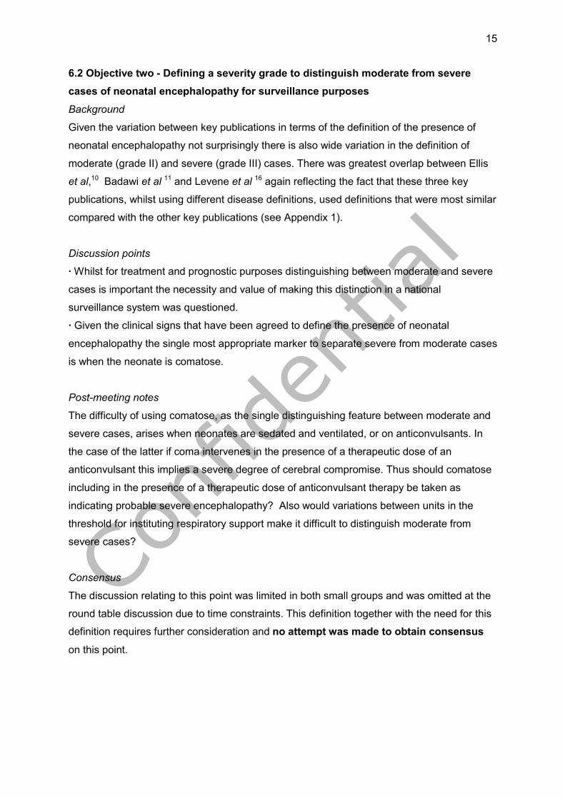
15
6.2 Objective two - Defining a severity grade to distinguish moderate from severe cases of neonatal encephalopathy for surveillance purposes Background
Given the variation between key publications in terms of the definition of the presence of
neonatal encephalopathy not surprisingly there is also wide variation in the definition of
moderate (grade II) and severe (grade III) cases. There was greatest overlap between Ellis
et al,10 Badawi et al 11 and Levene et al 16 again reflecting the fact that these three key
publications, whilst using different disease definitions, used definitions that were most similar
compared with the other key publications (see Appendix 1).
Discussion points
· Whilst for treatment and prognostic purposes distinguishing between moderate and severe
cases is important the necessity and value of making this distinction in a national
surveillance system was questioned.
· Given the clinical signs that have been agreed to define the presence of neonatal
encephalopathy the single most appropriate marker to separate severe from moderate cases
is when the neonate is comatose.
Post-meeting notes
The difficulty of using comatose, as the single distinguishing feature between moderate and
severe cases, arises when neonates are sedated and ventilated, or on anticonvulsants. In
the case of the latter if coma intervenes in the presence of a therapeutic dose of an
anticonvulsant this implies a severe degree of cerebral compromise. Thus should comatose
including in the presence of a therapeutic dose of anticonvulsant therapy be taken as
indicating probable severe encephalopathy? Also would variations between units in the
threshold for instituting respiratory support make it difficult to distinguish moderate from
severe cases?
Consensus
The discussion relating to this point was limited in both small groups and was omitted at the
round table discussion due to time constraints. This definition together with the need for this
definition requires further consideration and no attempt was made to obtain consensus
on this point.
16
6.3 Objective three - Defining the neonatal encephalopathy cases which have possibly suffered acute intrapartum or peripartum cerebral compromise for surveillance purposes
Background
Two recent authoritative task force publications have made recommendations about how to
define the presence of intrapartum hypoxic events in the context of cerebral palsy causality.
In 1999 MacLennan published, on behalf of the International Cerebral Palsy Task Force,7 “A
template for defining a causal relation between acute intrapartum events and cerebral palsy:
international consensus statement.” More recently the American College of Obstetricians
and Gynecologists (ACOG) and the American Academy of Pediatrics (AAP) published a
report defining the pathogenesis and pathophysiology of neonatal encephalopathy and
cerebral palsy.7 Both of these reports have limitations in terms of the definition the
consensus process reported here is attempting to address. Both were essentially aimed at
defining a history of acute intrapartum compromise as the basis for establishing the probable
intrapartum or peripartum cause of cases of cerebral palsy. As a consequence the essential
criteria defined by both groups include the presence of cerebral palsy. The ACOG/AAP
criteria are a more refined and stringent version of the MacLennan criteria. Both sets of
essential criteria include the requirement to have measured the acid base status of the
neonate at or soon after delivery (Appendix 1). In addition to the essential criteria both list
five additional criteria that collectively suggest intrapartum timing but are non-specific to
asphyxial insult. These criteria include early imaging study results and imply the necessity
for continuous intrapartum fetal heart rate monitoring. If evidence for some of the five
additional criteria is missing or contradictory the intrapartum timing of the encephalopathy
onset becomes increasingly in doubt.
Discussion points
· The ideal would be to use the ACOG/AAP or MacLennan criteria, however, it seems
unlikely that all the necessary information would be reliably collected, for example,
scepticism about the general quality of blood samples was expressed. The number of data
items required is also prohibitive for a routine data collection system.
· Various combinations of proxy markers were discussed but concern about the difficulty of
attributing even a possible intrapartum cause was expressed. Concern was also expressed
about the possible medico-legal implications of doing so, in particular if apgar scores were
included.
· Various sentinel events were considered (eg. intrapartum uterine rupture) and an attempt to
list these was made, following which, the view that a larger group of obstetricians would be

17
needed to define an appropriate a priori list. However, even if a list were constructed it would
potentially be very long and is likely to omit all possible events. Such a list is unlikely to be
integrated into a surveillance system.
Consensus
The final combination of proxy markers of possible intrapartum compromise proposed were:
the need for respiratory support or resuscitation at birth (including bag and mask) and
bradycardia for more than five minutes OR a history of an intrapartum sentinel event.
Concerns about all the aspects of this definition were evident and a final vote was not taken
– consensus was not reached on this point. Further discussion about this item is needed
including the utility of attempting to make this distinction in a national surveillance system.
7. Summary of the recommended definitions 7.1 Objective one - Defining neonatal encephalopathy for surveillance purposes It was unanimously agreed that the following criteria should be recommended for use to
define the presence of neonatal encephalopathy for surveillance purposes:
The presence of abnormal tone and abnormal consciousness within the first 72 hours
after birth
AND/OR
The presence of clinically diagnosed seizures within the first 72 hours after birth
Where:
Abnormal tone is defined as: hypertonia or hypotonia and if the clinician is at all uncertain
about whether there is an abnormality of tone present then for these purposes the tone
should not be regarded as abnormal.
Abnormal consciousness is defined as lethargy or coma where these are defined as:
Lethargy: not unconscious but having decreased alertness and decreased responsiveness
such that a noxious stimulus response can still be provoked eg. responsive to light, sound
and a leg squeeze.
Comatose: unresponsive to all stimuli including noxious stimuli.
Seizures are defined as clonic, repetitive, rhythmic movements which do not cease when
the infant is restrained.

18
7.2 Objective two - Defining a severity grade to distinguish moderate from severe cases of neonatal encephalopathy for surveillance purposes The discussion relating to this point was limited in both small groups and was omitted at the
round table discussion due to time constraints. This definition together with the need for this
definition requires further consideration and no attempt was made to obtain consensus
on this point.
7.3 Objective three - Defining the neonatal encephalopathy cases which have possibly suffered acute intrapartum or peripartum cerebral compromise for surveillance purposes The final combination of proxy markers of possible intrapartum compromise proposed were:
the need for respiratory support or resuscitation at birth (including bag and mask) and
bradycardia for more than five minutes OR a history of an intrapartum sentinel event.
Concerns about all the aspects of this definition were evident and a final vote was not taken
– consensus was not reached on this point. Further discussion about this item is needed
including the utility of attempting to make this distinction as part of a surveillance system.
ACKNOWLEDGEMENTS We are very grateful to the members of panel who attended and contributed so
enthusiastically to the discussions at the Stage 1 meeting. We are particularly grateful to the
experts who presented the four different perspectives about neonatal encephalopathy at the
start of the meeting. Lynne Roberts made the arrangements for the meeting and she and
Ann Kennedy kindly attended to make notes. We are very grateful for their written
contributions which were very helpful in the drafting of this discussion document.
REFERENCES 1 Patient Safety Research Programme (Policy Research Programme of the Department of Health). Call for proposal (Single tender action) – Neonatal encephalopathy. March 2004. 2 Department of Health. An organisation with a memory. Report of an expert group on learning from adverse events in the NHS. Chaired by the Chief Medical Officer. London: The Stationery Office. 2000.

19
3 http://www.rcpch.ac.uk/publications/recent_publications/ExecSummarry%20.pdf (accessed 21st November 2004) 4 Murphy MK, Black NA, Lamping DL et al. Consensus development methods, and their uses in clinical guideline development. Health Technology Assessment 1998; 2: i-iv, 1-88. 5 Susskind L, McKearnans S, Thomas-Larmer J. The consensus building handbook. London: Sage Publications. 1999. 6 Fink A, Kosecoff J, Chassin M, Brook RH. Consensus methods: characteristics and guidelines for use. Am J Public Health 1984; 74: 979-983. 7 MacLennan A for the International Cerebral Palsy Task Force. A template for defining a causal relation between acute intrapartum events and cerebral palsy: international consensus statement. Br Med J 1999; 319: 1054-9. 8 Cowan F, Rutherford M, Groenendaal F et al. Origin and timing of brain lesions in term infants with neonatal encephalopathy. Lancet 2003; 361: 736-42. 9 American College of Obstetricians and Gynecologist and American Academy of Pediatrics. Neonatal Encephalopathy and Cerebral Palsy: Defining the Pathogenesis and Pathophysiology. Washington DC: American College of Obstetricians and Gynecologist. 2002. 10 Ellis M, Manandhar N Manandhar DS, Costello AM. Risk factors for neonatal encephalopathy in Kathmandu, Nepal, a developing country: unmatched case control study. Br Med J 2000; 320:1299-36. 11 Badawi N, Kurinczuk JJ, Keogh M et al. Antepartum risk factors for newborn encephalopathy: the Western Australian case control study. Br Med J 1998; 317: 1549-53. 12 Badawi N, Kurinczuk JJ, Keogh M et al. Intrapartum risk factors for newborn encephalopathy: the Western Australian case control study. Br Med J 1998; 317: 1554-58. 13 Low JA. Intrapartum fetal asphyxia: definition, diagnosis, and classification. Am J Obstet & Gynecol 1997; 176: 957-9. 14 Nelson KB, Ellenberg JH. The asymptomatic newborn and risk of cerebral palsy. Am J Dis Child 1987: 141: 13335. 15 Amiel-Tison C, Ellison P. Birth asphyxia in the full term newborn: early assessment and outcome. Dev Med Child Neurol 1986: 28: 671-82. 16 Levene ML, Kornberg J, Williams TH. The incidence and severity of post-asphyxial encephalopathy in full-term infants. Early Hum Dev 1985; 11:21-6. 17 Fenichel GM. Hypoxic-ischemic encephalopathy in the newborn. Arch Neurol 1983; 40: 261-6. 18 Sarnat HB, Sarnat MS. Neonatal encephalopathy following fetal distress. A clinical and electroencephalographic study. Arch Neurol 1976; 33: 696-705. 19 Cresswell T, Colver A, Fenton A et al. RMSO Annual report 2001, 2002 (provisional). Newcastle upon Tyne: RMSO/North East Public Health Observatory. 2003. 20 http://www.perinatal.nhs.uk/manners/index_manners.htm (accessed 9th December 2004) 21 Trent Neonatal Survey Report 2002. Leicester: Department of Health Sciences. 2003.

APPENDIX C

Table C.1. Rate of ‘HIEII & HIEIII’ in Trent Region (1991 to 2002) and rate of ‘encephalopathy’ in Northern Region (1996 to 2003) term* births per 1,000 total live births
Year
Trent Region
Northern Region
Total live
births
Cases of
‘HIE’
Rate of
‘HIE’ per 1,000 total live births
95% Confidence
Interval**
Total live
births
Cases of
‘enceph’***
Rate of
‘enceph’*** per 1,000 total live
births
95% Confidence
Interval*
1991
63,129
86
1.36
1.09 – 1.68
-
-
-
-
1992 62,142 99 1.59 1.29 – 1.94 - - - - 1993 60,809 64 1.05 0.81 – 1.34 - - - - 1994 59,582 53 0.89 0.67 – 1.16 - - - - 1995 57,727 40 0.69 0.50 – 0.94 - - - - 1996 60,975 37 0.61 0.43 – 0.84 33,666 52 1.54 1.15 – 2.03 1997 59,561 75 1.26 0.99 – 1.58 32,869 69 2.10 1.63 – 2.66 1998 59,186 71 1.20 0.94 – 1.51 32,008 66 2.06 1.60 – 2.62 1999 57,161 78 1.36 1.08 – 1.70 30,938 60 1.94 1.48 – 2.50 2000 55,248 62 1.12 0.86 – 1.43 29,624 51 1.72 1.28 – 2.26 2001 54,009 70 1.30 1.01 – 1.64 29,006 56 1.93 1.46 – 2.51 2002 54,601 73 1.34 1.05 – 1.68 29,211 38 1.30 0.92 – 1.79 2003 - - - - 30,158 58 1.92 1.46 – 2.49
Total 704,130 808 1.15 1.07 – 1.23 247,480 450 1.82 1.65 – 1.99
*Term - ≥37 weeks gestation ** 95% Confidence intervals based on the exact binomial distribution *** ‘Encephalopathy’
1

Table C.2. 1998 standardised rates of ‘HIE’ in Trent Region (1994 – 2001) per 1,000 total live births, unadjusted and having adjusted for the effects of maternal age
Year
Unadjusted 1998 standardised rate of ‘HIE’ (per 1,000 total live births)
1998 standardised
rate of ‘HIE’ adjusted for
maternal age (per 1,000 total live
births)
95% Confidence
interval
1994
0.89
0.91
0.64 - 1.30
1995 0.69 0.71 0.48 - 1.04 1996 0.61 0.61 0.41 - 0.92 1997 1.26 1.27 0.92 - 1.76 1998 1.20* 1.20* * 1999 1.36 1.36 0.99 - 1.87 2000 1.12 1.11 0.79 - 1.57 2001
1.20 1.28 0.92 - 1.79
*All rates standardised to the 1998 maternal age distribution
2

Table C.3. Rate of ‘HIE’ in term live births per 1,000 total live births by severity indicator, Trent Region 1991 to 2002
Year
Total live
births
Cases of ‘HIE II’
(Moderate)
N (row %)
Rate of ‘HIE II’ per 1,000 total
live births
95%
Confidence Interval*
Cases of ‘HIE
III’ (Severe)
N (row %)
Rate of ‘HIE III’ per 1,000 total
live births
95%
Confidence Interval*
1991
63,129
50 (58)
0.79
0.59 – 1.04
36 (42)
0.57
0.40 – 0.79
1992 62,142 44 (44) 0.71 0.51 – 0.95 55 (56) 0.89 0.67 – 1.15 1993 60,809 31 (48) 0.51 0.35 – 0.72 33 (52) 0.54 0.37 – 0.76 1994 59,582 20 (38) 0.34 0.21 – 0.52 33 (62) 0.55 0.38 – 0.78 1995 57,727 8 (20) 0.14 0.06 – 0.27 32 (80) 0.55 0.38 – 0.78 1996 60,975 16 (43) 0.26 0.15 – 0.43 21 (57) 0.34 0.21 – 0.53 1997 59,561 24 (32) 0.40 0.26 – 0.60 51 (68) 0.86 0.64 – 1.13 1998 59,186 39 (55) 0.66 0.47 – 0.90 32 (45) 0.54 0.37 – 0.76 1999 57,161 32 (41) 0.56 0.38 – 0.79 46 (59) 0.80 0.59 – 1.07 2000 55,248 18 (29) 0.33 0.19 – 0.51 44 (71) 0.80 0.58 – 1.07 2001 54,009 30 (43) 0.56 0.37 – 0.79 40 (57) 0.74 0.53 – 1.01 2002 54,601 22 (30) 0.40 0.25 – 0.61 51 (70) 0.93 0.70 – 1.23 Total 704,130 334 (41) 0.47 0.42 – 0.53 474 (59) 0.67
0.61 – 0.74
*95% Confidence intervals based on the exact binomial distribution
3

Table C.4. Rate ratio of ‘HIE’ by maternal age group, Trent Region 1994 to 2001
Maternal age group (years)
Total live
births
Number of cases of
‘HIE’
Rate of
‘HIE’ per 1,000 total live births
Rate ratio
95%
Confidence interval**
< 20
38,711
59
1.52
1.77
1.28 – 2.43
20 – 24 95,045 88 0.93 1.08 0.81 – 1.42 25 – 29 148,974 128 0.86 1* 30 – 34 125,479 139 1.11 1.29 1.01 – 1.65 35+ 55,073 72 1.31 1.52 1.12 – 2.05
* Baseline comparison group **95% Confidence intervals based on the exact binomial distribution
4

Table C.5. Rate ratio of ‘Encephalopathy’ by maternal age group, Northern Region 1996 to 2001
Maternal age group (years)
Total live
births
Number of cases of ‘Enceph’
Rate of
‘Enceph’ per 1,000 total live
births
Rate ratio
95%
Confidence interval**
< 20
20,497
60
2.93
1.49
1.05 – 2.12
20 – 24 40,359 79 1.96 1* 25 – 29 56,544 117 2.07 1.06 0.79 – 1.42 30 – 34 48,774 121 2.48 1.27 0.95 – 1.70 35+ 21,910 51 2.33 1.18 0.82 – 1.71
* Baseline comparison group **95% Confidence intervals based on the exact binomial distribution
5
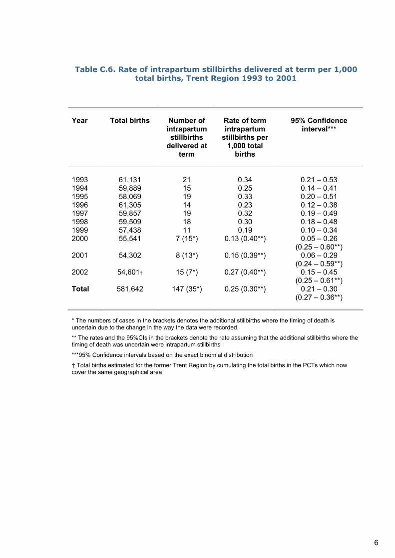
Table C.6. Rate of intrapartum stillbirths delivered at term per 1,000 total births, Trent Region 1993 to 2001
Year
Total births
Number of intrapartum stillbirths
delivered at term
Rate of term intrapartum
stillbirths per 1,000 total
births
95% Confidence
interval***
1993
61,131
21
0.34
0.21 – 0.53
1994 59,889 15 0.25 0.14 – 0.41 1995 58,069 19 0.33 0.20 – 0.51 1996 61,305 14 0.23 0.12 – 0.38 1997 59,857 19 0.32 0.19 – 0.49 1998 59,509 18 0.30 0.18 – 0.48 1999 57,438 11 0.19 0.10 – 0.34 2000 55,541 7 (15*) 0.13 (0.40**) 0.05 – 0.26
(0.25 – 0.60**) 2001 54,302 8 (13*) 0.15 (0.39**) 0.06 – 0.29
(0.24 – 0.59**) 2002 54,601† 15 (7*) 0.27 (0.40**) 0.15 – 0.45 (0.25 – 0.61**) Total 581,642 147 (35*) 0.25 (0.30**) 0.21 – 0.30
(0.27 – 0.36**)
* The numbers of cases in the brackets denotes the additional stillbirths where the timing of death is uncertain due to the change in the way the data were recorded. ** The rates and the 95%CIs in the brackets denote the rate assuming that the additional stillbirths where the timing of death was uncertain were intrapartum stillbirths ***95% Confidence intervals based on the exact binomial distribution † Total births estimated for the former Trent Region by cumulating the total births in the PCTs which now cover the same geographical area
6
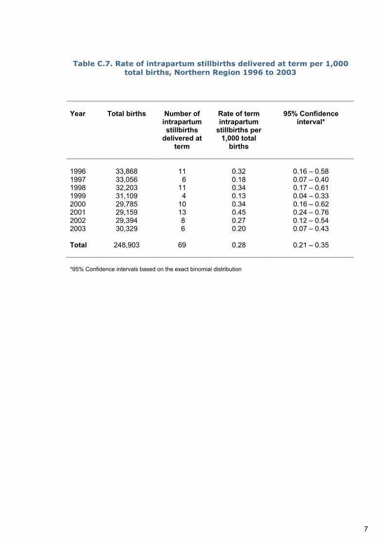
Table C.7. Rate of intrapartum stillbirths delivered at term per 1,000 total births, Northern Region 1996 to 2003
Year
Total births
Number of intrapartum stillbirths
delivered at term
Rate of term intrapartum
stillbirths per 1,000 total
births
95% Confidence
interval*
1996
33,868
11
0.32
0.16 – 0.58
1997 33,056 6 0.18 0.07 – 0.40 1998 32,203 11 0.34 0.17 – 0.61 1999 31,109 4 0.13 0.04 – 0.33 2000 29,785 10 0.34 0.16 – 0.62 2001 29,159 13 0.45 0.24 – 0.76 2002 29,394 8 0.27 0.12 – 0.54 2003 30,329 6 0.20 0.07 – 0.43 Total 248,903 69 0.28 0.21 – 0.35
*95% Confidence intervals based on the exact binomial distribution
7
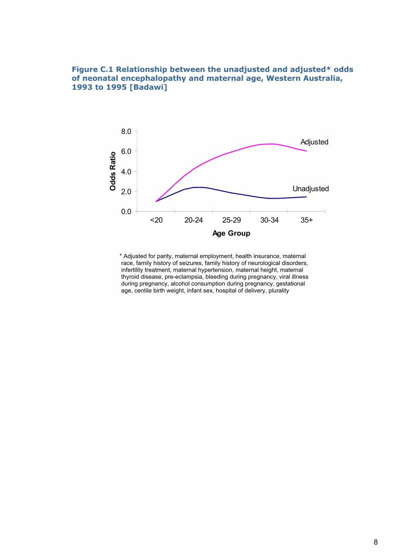
Figure C.1 Relationship between the unadjusted and adjusted* odds of neonatal encephalopathy and maternal age, Western Australia, 1993 to 1995 [Badawi]
Unadjusted
Adjusted
0.0
2.0
4.0
6.0
8.0
<20 20-24 25-29 30-34 35+
Age Group
Odd
s R
atio
* Adjusted for parity, maternal employment, health insurance, maternal race, family history of seizures, family history of neurological disorders, infertility treatment, maternal hypertension, maternal height, maternal thyroid disease, pre-eclampsia, bleeding during pregnancy, viral illness during pregnancy, alcohol consumption during pregnancy, gestational age, centile birth weight, infant sex, hospital of delivery, plurality
8

APPENDIX D

APPENDIX D
SUMMARIES OF NEONATAL ENCEPHALOPATHY PAPERS
ASSESSING THE INTRAPARTUM CONTRIBUTION
Reference Page
Becher, Bell, Keeling, McIntosh & Wyatt (2004) The Scottish perinatal neuropathology study: Clinicopathological correlation in early neonatal deaths. Archives of Disease in Childhood. Fetal and Neonatal Edition, 89, F399-407. ............................................................34
Shah, Riphagen, Beyene & Perlman (2004) Multiorgan dysfunction in infants with post-asphyxial hypoxic-ischaemic encephalopathy. Archives of Disease in Childhood Fetal Neonatal Edition, 89, F152-5. ..................................................................................................42
Bukowski, Burgett, Gei et al (2003) Impairment of fetal growth potential and neonatal encephalopathy. American Journal of Obstetrics & Gynecology, 188, 1011-5........................45
Cowan, Rutherford, Groenendaal et al (2003) Origin and timing of brain lesions in term infants with neonatal encephalopathy. Lancet, 361, 736-42....................................................46
Greenwood, Newman, Impey & Johnson (2003) Cerebral palsy and clinical negligence litigation: a cohort study. British Journal of Obstetrics & Gynaecology, 110, 6-11...................48
Khong, Lam, Tung et al (2003) MRI of neonatal encephalopathy. Clinical Radiology, 58, 833-44......................................................................................................................................50
Noren, Amer-Wahlin, Hagberg et al (2003) Fetal electrocardiography in labor and neonatal outcome: data from the Swedish randomized controlled trial on intrapartum fetal monitoring. American Journal of Obstetrics & Gynecology, 188, 183-92. ..................................................52
American College of Obstetricians and Gynecologists (2002) Neonatal Encephalopathy and Cerebral Palsy: Defining the Pathogenesis and Pathophysiology. ACOG: Washington D.C...........................................................................................................................................54
Draper, Kurinczuk, Lamming et al (2002) A confidential enquiry into cases of neonatal encephalopathy. Archives of Disease in Childhood Fetal Neonatal Edition, 87, F176-80. ......56
Jongeling, Badawi, Kurinczuk et al (2002) Cranial ultrasound as a predictor of outcome in term newborn encephalopathy. Pediatric Neurology , 26, 37-42. ............................................57
Milsom, Ladfors, Thiringer et al (2002) Influence of maternal, obstetric and fetal risk factors on the prevalence of birth asphyxia at term in a Swedish urban population. Acta Obstetricia et Gynecologica Scandinavica, 81, 909-17..................................................................................17
Evans, Rigby, Hamilton, Titchiner & Hall (2001) The relationships between neonatal encephalopathy and cerebral palsy: A cohort study. Journal of Obstetrics and Gynaecology, 21, 114-120..............................................................................................................................19
Hagberg, Hagberg, Beckung & Uvebrant (2001) Changing panorama of cerebral palsy in Sweden. VIII. Prevalence and origin in the birth year period 1991-94. Acta Paediatrica, 90, 271-7........................................................................................................................................20
Heinonen & Saarikoski (2001) Reproductive risk factors of fetal asphyxia at delivery: A population based analysis. Journal of Clinical Epidemiology, 54, 407-10. .............................21
Cowan (2000) Outcome after intrapartum asphyxia in term infants. Seminars in Neonatology, 2000, 5, 127-40........................................................................................................................66
2
Ellis, Manandhar, Manandhar & Costello (2000) Risk factors for neonatal encephalopathy in Kathmandu, Nepal, a developing country: unmatched case control study. British Medical Journal, 320, 1229-36. .............................................................................................................67
MacLennan (1999) A template for defining a causal relation between acute intrapartum events and cerebral palsy: international consensus statement. British Medical Journal, 319, 1054-9......................................................................................................................................71
Westgate, Gunn & Gunn (1999) Antecedents of neonatal encephalopathy with fetal acidaemia at term. British Journal of Obstetrics and Gynaecology, 106, 774-82. ...................73
Badawi, Kurinczuk, Keogh et al (1998) Intrapartum risk factors for newborn encephalopathy: the Western Australian case-control study. British Medical Journal, 317, 1554-8 ………….... 24
Carter, McNabb & Merenstein (1998) Prospective validation of a scoring system for predicting neonatal morbidity after acute perinatal asphyxia. Journal of Pediatrics, 132, 619-23. ............................................................................................................................................74
Ekert, Perlman, Steinlin & Hao (1997) Predicting the outcome of postasphyxial hypoxic-ischemic encephalopathy within 4 hours of birth. Journal of Pediatrics , 131, 613-7...............78
Low (1997) Intrapartum fetal asphyxia: definition, diagnosis, and classification. American Journal of Obstetrics and Gynecology, 176, 957-9..................................................................80
Adamson, Alessandri, Badawi et al (1995) Predictors of neonatal encephalopathy in full-term infants. British Medical Journal, 311, 598-602.................................................................26
Goodlin (1995) Do concepts of causes and prevention of cerebral palsy require revision? American Journal of Obstetrics and Gynecology, 172, 1830-6................................................85
Low, Simpson, Tonni & Chamberlain (1995) Limitations in the clinical prediction of intrapartum fetal asphyxia. American Journal of Obstetrics and Gynecology, 172, 801-4. .....86
Gaffney, Flavell, Johnson et al (1994) Cerebral palsy and neonatal encephalopathy. Archives of Disease in Childhood Fetal Neonatal Edition, 70, F195-200. ...............................88
Yudkin, Johnson, Clover & Murphy (1994) Clustering of perinatal markers of birth asphyxia and outcome at age five years. British Journal of Obstetrics and Gynaecology, 101, 774-81.92
Airede (1991) Birth asphyxia and hypoxic-ischaemic encephalopathy: incidence and severity. Annals of Tropical Paediatrics, 11, 331-5. ...............................................................................97
Winkler, Hauth, Tucker et al (1991) Neonatal complications at term as related to the degree of umbilical artery acidemia. American Journal of Obstetrics and Gynecology, 164, 637-41..99
Blair & Stanley (1988) Intrapartum asphyxia: a rare cause of cerebral palsy. Journal of Pediatrics, 112, 515-9. ...........................................................................................................105
Low, Galbraith, Muir et al (1988) Motor and cognitive deficits after intrapartum asphyxia in the mature fetus. American Journal of Obstetrics and Gynecology,158, 356-61...................109
Levene, Sands, Grindulis & Moore (1986) Comparison of two methods of predicting outcome in perinatal asphyxia. Lancet, 1, 67-9. ...................................................................113
Derham, Matthews & Clarke (1985) Early seizures indicate quality of perinatal care. Archives of Disease in Childhood, 60, 809-13. ......................................................................115
Low, Galbraith, Muir et al (1985) The relationship between perinatal hypoxia and newborn encephalopathy. American Journal of Obstetrics and Gynecology, 152, 256-60. .................117

3
Cyr, Usher & McLean (1984) Changing patterns of birth asphyxia and trauma over 20 years. American Journal of Obstetrics and Gynecology, 148, 490-8................................................120
POPULATION-BASED STUDIES
Wu, Backstrand, Zhao et al (2004) Declining diagnosis of birth asphyxia in California: 1991-2000. Pediatrics, 114, 1584-90. ...............................................................................................11
Caravale, Allemand & Libenson (2003) Factors predictive of seizures and neurologic outcome in perinatal depression. Pediatric Neurology, 29, 18-25. ..........................................12
Itoo, Al-Hawsawi & Khan (2003) Hypoxic ischemic encephalopathy: Incidence and risk factors in North Western Saudi Arabia. Saudi Medical Journal, 24, 147-53. ...........................13
Bailit, Garrett, Miller et al (2002) Hospital primary cesarean delivery rates and the risk of poor neonatal outcomes. American Journal of Obstetrics & Gynecology, 187, 721-7............14
Bekedam, Engelsbel, Mol et al (2002) Male predominance in fetal distress during labor. American Journal of Obstetrics and Gynecology, 187, 1605-7................................................15
Dixon, Badawi, Kurinczuk et al (2002) Early developmental outcomes after newborn encephalopathy. Pediatrics, 109, 26-33...................................................................................16
Milsom, Ladfors, Thiringer et al (2002) Influence of maternal, obstetric and fetal risk factors on the prevalence of birth asphyxia at term in a Swedish urban population. Acta Obstetricia et Gynecologica Scandinavica, 81, 909-17..................................................................................17
Moster, Lie & Markestad (2002) Joint association of Apgar scores and early neonatal symptoms with minor disabilities at school age. Archives of Disease in Childhood Fetal Neonatal Edition, 86, F16-21. ..................................................................................................18
Evans, Rigby, Hamilton, Titchiner & Hall (2001) The relationships between neonatal encephalopathy and cerebral palsy: a cohort study. Journal of Obstetrics and Gynaecology, 21, 114-120..............................................................................................................................19
Hagberg, Hagberg, Beckung & Uvebrant (2001) Changing panorama of cerebral palsy in Sweden. VIII. Prevalence and origin in the birth year period 1991-94. Acta Paediatrica, 90, 271-7........................................................................................................................................20
Heinonen & Saarikoski (2001) Reproductive risk factors of fetal asphyxia at delivery: a population based analysis. Journal of Clinical Epidemiology, 54, 407-10. .............................21
Oswyn, Vince & Friesen (2000) Perinatal asphyxia at Port Moresby General Hospital: a study of incidence, risk factors and outcome. P N G Medical Journal, 43, 110-20..................22
Ellis, Manandhar, Shrestha, et al (1999) Outcome at one year of neonatal encephalopathy in Kathmandu, Nepal. Developmental Medicine & Child Neurology, 41, 689-95....................23
Badawi, Kurinczuk, Keogh et al (1998) Antepartum risk factors for newborn encephalopathy: the Western Australian case-control study. British Medical Journal, 317, 1549-53....................................................................................................................................24
Badawi, Kurinczuk, Keogh et al (1998) Intrapartum risk factors for newborn encephalopathy: the Western Australian case-control study. British Medical Journal, 317, 1554-8......................................................................................................................................24
Hall, Smith & Smith (1996) Maternal factors contributing to asphyxia neonatorum. Journal of Tropical Pediatrics, 42, 192-5. .................................................................................................25

4
Adamson, Alessandri, Badawi et al (1995) Predictors of neonatal encephalopathy in full-term infants. British Medical Journal, 311, 598-602.................................................................26
Thornberg, Thiringer, Odeback & Milsom (1995) Birth asphyxia: incidence, clinical course and outcome in a Swedish population. Acta Paediatrica, 84, 927-32......................................27
Yudkin, Johnson, Clover & Murphy (1995) Assessing the contribution of birth asphyxia to cerebral palsy in term singletons. Paediatric and Perinatal Epidemiology, 9, 156-70. ............28
Becerra, Fry & Rowley (1991) Morbidity estimates of conditions originating in the perinatal period: United States, 1986 through 1987. Pediatrics, 88, 553-9. ...........................................29
Mir, Faquih & Legnain (1989) Perinatal risk factors in birth asphyxia: relationship of obstetric and neonatal complications to neonatal mortality in 16,365 consecutive live births. Asia-Oceania Journal of Obstetrics and Gynaecology, 15, 351-7. ..................................................30
Nelson & Ellenberg (1987) The asymptomatic newborn and risk of cerebral palsy. American Journal of Diseases of Children, 141, 1333-5..........................................................................31
MacDonald, Mulligan, Allen & Taylor (1980) Neonatal asphyxia. I. Relationship of obstetric and neonatal complications to neonatal mortality in 38,405 consecutive deliveries. Journal of Pediatrics, 96, 898-902. ...........................................................................................................32
OTHER STUDIES
Barnett, Guzzetta, Mercuri et al (2004) Can the Griffiths scales predict neuromotor and perceptual-motor impairment in term infants with neonatal encephalopathy? Archives of Disease in Childhood, 89, 637-43............................................................................................33
Becher, Bell, Keeling, McIntosh & Wyatt (2004) The Scottish perinatal neuropathology study: clinicopathological correlation in early neonatal deaths. Archives of Disease in Childhood. Fetal and Neonatal Edition, 89, F399-407. ............................................................34
Carli, Reiger & Evans (2004) One-year neurodevelopmental outcome after moderate newborn hypoxic ischaemic encephalopathy. Journal of Paediatrics and Child Health, 40, 217-20......................................................................................................................................35
Celtik, Acunas, Oner & Pala (2004) Neuron-specific enolase as a marker of the severity and outcome of hypoxic ischemic encephalopathy. Brain and Development, 26, 398-402............36
Low (2004) Determining the contribution of asphyxia to brain damage in the neonate. Journal of Obstetrics and Gynaecology Research, 30, 276-86. ...........................................................37
McDonald, Kelehan, McMenamin et al (2004) Placental fetal thrombotic vasculopathy is associated with neonatal encephalopathy. Human Pathology, 35, 875-80. ............................38
Mercuri, Anker, Guzzetta et al (2004) Visual function at school age in children with neonatal encephalopathy and low Apgar scores. Archives of Disease in Childhood Fetal Neonatal Edition, 89, F258-62.................................................................................................................38
Miller, Latal, Clark et al (2004) Clinical signs predict 30-month neurodevelopmental outcome after neonatal encephalopathy. American Journal of Obstetrics & Gynecology, 190, 93-9.....39
Ramaswamy, Miller, Barkovich et al (2004) Perinatal stroke in term infants with neonatal encephalopathy. Neurology, 62, 2088-91. ...............................................................................40
Salhab, Wyckoff, Laptook & Perlman (2004) Initial hypoglycemia and neonatal brain injury in term infants with severe fetal acidemia. Pediatrics, 114, 361-6. ..........................................40

5
Satar, Ozcan, Yapicioglu & Narli (2004) Serum insulin-like growth factor 1 and growth hormone levels of hypoxic-ischemic newborns. Biology of the Neonate, 85, 15-20. ..............41
Shah, Riphagen, Beyene & Perlman (2004) Multiorgan dysfunction in infants with post-asphyxial hypoxic-ischaemic encephalopathy. Archives of Disease in Childhood Fetal Neonatal Edition, 89, F152-5. ..................................................................................................42
Stelmach, Kallas, Pisarev & Talvik (2004) Antenatal risk factors associated with unfavorable neurologic status in newborns and at 2 years of age. Journal of Child Neurology, 19, 116-22.43
Williams & Singh (2004) Umbilical artery blood gas parameters in neonates with early onset seizures who die. British Journal of Obstetrics and Gynaecology, 111, 1042-5. .....................44
Bukowski, Burgett, Gei et al (2003) Impairment of fetal growth potential and neonatal encephalopathy. American Journal of Obstetrics & Gynecology, 188, 1011-5........................45
Cowan, Rutherford, Groenendaal et al (2003) Origin and timing of brain lesions in term infants with neonatal encephalopathy. Lancet, 361, 736-42....................................................46
Debillon, Daoud, Durand et al (2003) Whole-body cooling after perinatal asphyxia: a pilot study in term neonates. Developmental Medicine & Child Neurology, 45, 17-23. ...................47
Greenwood, Newman, Impey & Johnson (2003) Cerebral palsy and clinical negligence litigation: a cohort study. British Journal of Obstetrics & Gynaecology, 110, 6-11...................48
Kaufman, Miller, Ferriero et al (2003) Encephalopathy as a predictor of magnetic resonance imaging abnormalities in asphyxiated newborns. Pediatric Neurology, 28, 342-6...................49
Khong, Lam, Tung et al (2003) MRI of neonatal encephalopathy. Clinical Radiology, 58, 833-44......................................................................................................................................50
Leung, Lam, Lam et al (2003) Unexpected reduction in the incidence of birth trauma and birth asphyxia related to instrumental deliveries during the study period: was this the Hawthorne effect? British Journal of Obstetrics & Gynaecology, 110, 319-22. ......................51
Noren, Amer-Wahlin, Hagberg et al (2003) Fetal electrocardiography in labor and neonatal outcome: data from the Swedish randomized controlled trial on intrapartum fetal monitoring. American Journal of Obstetrics & Gynecology, 188, 183-92. ..................................................52
Sato, Okumura, Kato et al (2003) Hypoxic ischemic encephalopathy associated with neonatal seizures without other neurological abnormalities. Brain and Development, 25, 215-9........................................................................................................................................53
Yu, Kui & Ming (2003) Effect of asphyxia on non-protein-bound iron and lipid peroxidation in newborn infants. Developmental Medicine & Child Neurology, 45, 24-7. ................................53
American College of Obstetricians and Gynecologists (2002) Neonatal Encephalopathy and Cerebral Palsy: Defining the Pathogenesis and Pathophysiology. ACOG: Washington D.C...........................................................................................................................................54
Barnett, Mercuri, Rutherford et al (2002) Neurological and perceptual-motor outcome at 5 - 6 years of age in children with neonatal encephalopathy: relationship with neonatal brain MRI. Neuropediatrics, 33, 242-8. .............................................................................................55
Draper, Kurinczuk, Lamming et al (2002) A confidential enquiry into cases of neonatal encephalopathy. Archives of Disease in Childhood Fetal Neonatal Edition, 87, F176-80. ......56
Jongeling, Badawi, Kurinczuk et al (2002) Cranial ultrasound as a predictor of outcome in term newborn encephalopathy. Pediatric Neurology, 26, 37-42. .............................................57

6
Robertson, Cowan, Cox & Edwards (2002) Brain alkaline intracellular pH after neonatal encephalopathy. Annals of Neurology, 52, 732-42. .................................................................58
Scher, Steppe, Beggarly et al (2002) Neonatal EEG-sleep disruption mimicking hypoxic-ischemic encephalopathy after intrapartum asphyxia. Sleep Medicine, 3, 411-5. ...................58
Shankaran, Laptook, Wright et al (2002) Whole-body hypothermia for neonatal encephalopathy: animal observations as a basis for a randomized, controlled pilot study in term infants. Pediatrics, 110, 377-85. .....................................................................................59
Dalman, Thomas, David et al (2001) Signs of asphyxia at birth and risk of schizophrenia. Population-based case-control study. British Journal of Psychiatry, 179, 403-8. ....................60
Ellis, Shrestha, Shrestha et al (2001) Clinical predictors of outcome following mild and moderate neonatal encephalopathy in term newborns in Kathmandu, Nepal. Acta Paediatrica, 90, 316-22................................................................................................................................61
Impey, Greenwood, Sheil et al (2001) The relation between pre-eclampsia at term and neonatal encephalopathy. Archives of Disease in Childhood Fetal Neonatal Edition, 85, F170-2......................................................................................................................................63
Impey, Greenwood, MacQuillan, Reynolds & Sheil (2001) Fever in labour and neonatal encephalopathy: a prospective cohort study. British Journal of Obstetrics and Gynaecology, 108, 594-7................................................................................................................................63
Robertson, Lewis, Cowan et al (2001) Early increases in brain myo-inositol measured by proton magnetic resonance spectroscopy in term infants with neonatal encephalopathy. Pediatric Research, 50, 692-700. ............................................................................................64
Asakura, Ichikawa, Nakabayashi et al (2000) Perinatal risk factors related to neurologic outcomes of term newborns with asphyxia at birth: a prospective study. Journal of Obstetrics and Gynaecology Research, 26, 313-24. ................................................................................65
Azzopardi, Robertson, Cowan et al (2000) Pilot study of treatment with whole body hypothermia for neonatal encephalopathy. Pediatrics, 106, 684-94........................................66
Cowan (2000) Outcome after intrapartum asphyxia in term infants. Seminars in Neonatology, 2000, 5, 127-40........................................................................................................................66
Ellis, Manandhar, Manandhar & Costello (2000) Risk factors for neonatal encephalopathy in Kathmandu, Nepal, a developing country: unmatched case control study. British Medical Journal, 320, 1229-36. .............................................................................................................67
Smith, Wells & Dodd (2000) The continuing fall in incidence of hypoxic-ischaemic encephalopathy in term infants. British Journal of Obstetrics and Gynaecology, 107, 461-6..68
Al Naqeeb, Edwards, Cowan & Azzopardi (1999) Assessment of neonatal encephalopathy by amplitude-integrated electroencephalography. Pediatrics, 103, 1263-71. ..........................69
Huang, Wang, Chang, Lin & Wu (1999) Measurement of the urinary lactate: Creatinine ratio for the early identification of newborn infants at risk for hypoxic-ischemic encephalopathy. New England Journal of Medicine, 341, 328-35. .....................................................................70
MacLennan (1999) A template for defining a causal relation between acute intrapartum events and cerebral palsy: International consensus statement. British Medical Journal, 319, 1054-9......................................................................................................................................71
Pavlakis, Kingsley, Harper et al (1999) Correlation of basal ganglia magnetic resonance spectroscopy with Apgar score in perinatal asphyxia. Archives of Neurology, 56, 1476-81....71

7
Toh & Rajadurai (1999) Term infants with hypoxic ischaemic encephalopathy: poor neurodevelopmental outcome despite standard neonatal intensive care. Journal of Tropical Pediatrics, 45, 229-32. .............................................................................................................72
Westgate, Gunn & Gunn (1999) Antecedents of neonatal encephalopathy with fetal acidaemia at term. British Journal of Obstetrics and Gynaecology, 106, 774-82. ...................73
Carter, McNabb & Merenstein (1998) Prospective validation of a scoring system for predicting neonatal morbidity after acute perinatal asphyxia. Journal of Pediatrics, 132, 619-23......................................................................................................................................74
Ellis, Manandhar, Manandhar & deL Costello (1998) An Apgar score of three or less at one minute is not diagnostic of birth asphyxia but is a useful screening test for neonatal encephalopathy. Indian Pediatrics, 35, 415-21........................................................................75
Gunn, Gluckman & Gunn (1998) Selective head cooling in newborn infants after perinatal asphyxia: A safety study. Pediatrics, 102, 885-92. ..................................................................76
Pasternak & Gorey (1998) The syndrome of acute near-total intrauterine asphyxia in the term infant. Pediatric Neurology, 18, 391-8......................................................................................77
Sonntag, Wagner, Strauss & Obladen (1998) Complement and contact activation in term neonates after fetal acidosis. Archives of Disease in Childhood Fetal Neonatal Edition, 78, F125-8......................................................................................................................................77
Ekert, Perlman, Steinlin & Hao (1997) Predicting the outcome of postasphyxial hypoxic-ischemic encephalopathy within 4 hours of birth. Journal of Pediatrics, 131, 613-7................78
Leth, Toft, Herning, Peitersen & Lou (1997) Neonatal seizures associated with cerebral lesions shown by magnetic resonance imaging. Archives of Disease in Childhood Fetal Neonatal Edition, 77, F105-10. ................................................................................................79
Low (1997) Intrapartum fetal asphyxia: definition, diagnosis, and classification. American Journal of Obstetrics and Gynecology, 176, 957-9..................................................................80
Low, Lindsay & Derrick (1997) Threshold of metabolic acidosis associated with newborn complications. American Journal of Obstetrics and Gynecology, 177, 1391-4........................81
Thompson, Puterman, Linley et al (1997) The value of a scoring system for hypoxic ischaemic encephalopathy in predicting neurodevelopmental outcome. Acta Paediatrica, 86, 757-61......................................................................................................................................82
Cioni, Fazzi, Ipata et al (1996) Correlation between cerebral visual impairment and magnetic resonance imaging in children with neonatal encephalopathy. Developmental Medicine and Child Neurology, 38, 120-32. ...................................................................................................83
Rutherford, Pennock, Schwieso, Cowan & Dubowitz (1996) Hypoxic-ischaemic encephalopathy: Early and late magnetic resonance imaging findings in relation to outcome. Archives of Disease in Childhood. Fetal and Neonatal Edition, 75, F145-51. .........................83
Alcorn & Shean (1995) The origins of cerebral palsy: A consensus statement. Medical Journal of Australia, 162, 384. .................................................................................................84
Australian and New Zealand Perinatal Societies (1995) The origins of cerebral palsy--a consensus statement. The Australian and New Zealand Perinatal Societies. Medical Journal of Australia, 162, 85-90............................................................................................................84
Goodlin (1995) Do concepts of causes and prevention of cerebral palsy require revision? American Journal of Obstetrics and Gynecology, 172, 1830-6................................................85

8
Low, Simpson, Tonni & Chamberlain (1995) Limitations in the clinical prediction of intrapartum fetal asphyxia. American Journal of Obstetrics and Gynecology, 172, 801-4. .....86
Phelan, Ahn, Korst & Martin (1995) Nucleated red blood cells: A marker for fetal asphyxia? American Journal of Obstetrics and Gynecology, 173, 1380-4................................................87
Gaffney, Flavell, Johnson et al (1994) Cerebral palsy and neonatal encephalopathy. Archives of Disease in Childhood Fetal Neonatal Edition, 70, F195-200. ...............................88
Gaffney, Sellers, Flavell, Squier & Johnson (1994) Case-control study of intrapartum care, cerebral palsy, and perinatal death. British Medical Journal, 308, 743-50. .............................89
Low, Panagiotopoulos & Derrick (1994) Newborn complications after intrapartum asphyxia with metabolic acidosis in the term fetus. American Journal of Obstetrics and Gynecology, 170, 1081-7..............................................................................................................................90
Rosenbloom (1994) Dyskinetic cerebral palsy and birth asphyxia. Developmental Medicine and Child Neurology, 36, 285-9. ..............................................................................................90
Wayenberg, Vermeylen, Bormans et al (1994) Diagnosis of severe birth asphyxia and early prediction of neonatal neurological outcome in term asphyxiated newborns. Journal of Perinatal Medicine, 22, 129-36. ...............................................................................................91
Yudkin, Johnson, Clover & Murphy (1994) Clustering of perinatal markers of birth asphyxia and outcome at age five years. British Journal of Obstetrics and Gynaecology, 101, 774-81.92
Blair (1993) A research definition for 'birth asphyxia'? Developmental Medicine and Child Neurology, 35, 449-52. ............................................................................................................93
Gray, Tudehope, Masel et al (1993) Perinatal hypoxic-ischaemic brain injury: prediction of outcome. Developmental Medicine and Child Neurology, 35, 965-73. ....................................93
Goodwin, Belai, Hernandez et al (1992) Asphyxial complications in the term newborn with severe umbilical acidemia. American Journal of Obstetrics and Gynecology, 167, 1506-12. .94
Hull & Dodd (1992) Falling incidence of hypoxic-ischaemic encephalopathy in term infants. British Journal of Obstetrics and Gynaecology, 99, 386-91.....................................................95
Low, Simpson & Ramsey (1992) The clinical diagnosis of asphyxia responsible for brain damage in the human fetus. American Journal of Obstetrics and Gynecology, 167, 11-5. ....96
Airede (1991) Birth asphyxia and hypoxic-ischaemic encephalopathy: Incidence and severity. Annals of Tropical Paediatrics, 11, 331-5. ...............................................................................97
Winkler, Hauth, Tucker et al (1991) Neonatal complications at term as related to the degree of umbilical artery acidemia. American Journal of Obstetrics and Gynecology, 164, 637-41. .99
Byrne, Welch, Johnson, Darrah & Piper (1990) Serial magnetic resonance imaging in neonatal hypoxic-ischemic encephalopathy. Journal of Pediatrics, 117, 694-700.................100
Portman, Carter, Gaylord et al (1990) Predicting neonatal morbidity after perinatal asphyxia: A scoring system. American Journal of Obstetrics and Gynecology, 162, 174-82. ..............101
Torfs, van den Berg, Oechsli & Cummins (1990) Prenatal and perinatal factors in the etiology of cerebral palsy. Journal of Pediatrics, 116, 615-9. ................................................102
Gilstrap, Leveno, Burris, Williams & Little (1989) Diagnosis of birth asphyxia on the basis of fetal pH, Apgar score, and newborn cerebral dysfunction. American Journal of Obstetrics and Gynecology, 161, 825-30................................................................................................102

9
Levene, Fenton, Evans et al (1989) Severe birth asphyxia and abnormal cerebral blood-flow velocity. Developmental Medicine and Child Neurology, 31, 427-34.....................................103
Robertson, Finer & Grace (1989) School performance of survivors of neonatal encephalopathy associated with birth asphyxia at term. Journal of Pediatrics, 114, 753-60. 104
Blair & Stanley (1988) Intrapartum asphyxia: A rare cause of cerebral palsy. Journal of Pediatrics, 112, 515-9. ...........................................................................................................105
Ellenberg & Nelson (1988) Cluster of perinatal events identifying infants at high risk for death or disability. Journal of Pediatrics, 113, 546-52. ....................................................................107
Freeman & Nelson (1988) Intrapartum asphyxia and cerebral palsy. Pediatrics, 82, 240-9.108
Low, Galbraith, Muir et al (1988) Motor and cognitive deficits after intrapartum asphyxia in the mature fetus. American Journal of Obstetrics and Gynecology, 158, 356-61..................109
Lupton, Hill, Roland et al (1988) Brain swelling in the asphyxiated term newborn: Pathogenesis and outcome. Pediatrics, 82, 139-46. .............................................................110
Amiel-Tison & Ellison (1986) Birth asphyxia in the full term newborn: early assessment and outcome. Developmental Medicine & Child Neurology, 28, 671-82......................................111
Bell, Johansson, McLean & Usher (1986) Birth asphyxia, trauma, and mortality in twins: Has caesarean section improved outcome? American Journal of Obstetrics and Gynecology, 154, 235-9..............................................................................................................................112
Levene, Sands, Grindulis & Moore (1986) Comparison of two methods of predicting outcome in perinatal asphyxia. Lancet, 1, 67-9. ...................................................................113
Lipper, Voorhies, Ross, Vannucci & Auld (1986) Early predictors of one-year outcome for infants asphyxiated at birth. Developmental Medicine and Child Neurology, 28, 303-9. ......114
Derham, Matthews & Clarke (1985) Early seizures indicate quality of perinatal care. Archives of Disease in Childhood, 60, 809-13. ......................................................................115
Levene, Kornberg & Williams (1985) The incidence and severity of post-asphyxial encephalopathy in full-term infants. Early Human Development, 11, 21-6. ...........................116
Low, Galbraith, Muir et al (1985) The relationship between perinatal hypoxia and newborn encephalopathy. American Journal of Obstetrics and Gynecology, 152, 256-60. .................117
Low, Galbraith, Muir et al (1985) The contribution of fetal-newborn complications to motor and cognitive deficits. Developmental Medicine and Child Neurology, 27, 578-87. .............118
Robertson & Finer (1985) Term infants with hypoxic-ischemic encephalopathy: Outcome at 3.5 years. Developmental Medicine and Child Neurology, 27, 473-84. .................................119
Cyr, Usher & McLean (1984) Changing patterns of birth asphyxia and trauma over 20 years. American Journal of Obstetrics and Gynecology, 148, 490-8................................................120
Low, Galbraith, Muir et al (1984) Factors associated with motor and cognitive deficits in children after intrapartum fetal hypoxia. American Journal of Obstetrics and Gynecology, 148, 533-9......................................................................................................................................121
Fenichel (1983) Hypoxic-ischemic encephalopathy in the newborn. Archives of Neurology, 40, 261-6................................................................................................................................122
Finer, Robertson, Peters & Coward (1983) Factors affecting outcome in hypoxic-ischemic encephalopathy in term infants. American Journal of Diseases of Children, 137, 21-5........124
10
Green, McLean, Smith & Usher (1982) Has an increased cesarean section rate for term breech delivery reduced in incidence of birth asphyxia, trauma, and death? American Journal of Obstetrics and Gynecology, 142, 643-8.............................................................................125
D'Souza, McCartney, Nolan & Taylor (1981) Hearing, speech, and language in survivors of severe perinatal asphyxia. Archives of Disease in Childhood, 56, 245-52. ...........................125
D'Souza & Richards (1978) Neurological sequelae in newborn babies after perinatal asphyxia. Archives of Disease in Childhood, 53, 564-9........................................................126
Sarnat & Sarnat (1976) Neonatal encephalopathy following fetal distress: A clinical and electroencephalographic study. Archives of Neurology, 33, 696-705....................................127
Ziegler, Calame, Marchand et al (1976) Cerebral distress in full-term newborns and its prognostic value. A follow-up study of 90 infants. Helvetica Paediatrica Acta, 31, 299-317. 128
Amiel-Tison C (1969) Cerebral damage in full-term newborn aetiological factors, neonatal status and long-term follow-up. Biology of the Neonate, 14, 234-250. ..................................129

Neonatal Encephalopathy: Study Outlines and Case Definitions
POPULATION-BASED STUDIES
Wu, Backstrand, Zhao et al (2004) Declining diagnosis of birth asphyxia in California: 1991-2000. Pediatrics, 114, 1584-90.
Study design: Cohort study (population-based retrospective)
Objective: To determine the incidence of birth asphyxia diagnoses made over a 10 year period in California
Setting: California, USA
Population: 5,364,663 live births from 1991 to 2000. All newborns were resident in California.
Participants: 24,330 infants diagnosed with birth asphyxia (or a diagnosis that overlaps with birth asphyxia)
Birth asphyxia: (ICD codes 9th edition 768.5, 768.6, 768.9)
768.5 Severe birth asphyxia Pulse less than 100 per minute at birth and falling or steady, respiration absent or gasping, colour poor, tone absent, 1 minute Apgar score 0-3, “white asphyxia”.
768.6 Mild or moderate birth asphyxia Normal respiration not established within one minute, but heart rate 100 or above, some muscle tone present, some response to stimulation, 1-minute Apgar score 4-7, “blue asphyxia”.
768.9 Unspecified birth asphyxia in a live born infant Anoxia, asphyxia, hypoxia (NOS in live born infant) Further evaluation of other diagnoses was completed: (1) fetal distress in a live born infant, associated with newborn morbidity (768.2, 768.3, 768.4), (2) birth trauma unspecified (767.9), (3) subdural and cerebral haemorrhage as a result of birth trauma or of intrapartum anoxia or hypoxia (767.0), (4) other specified birth trauma such as encephalopathy as a result of birth injury (767.8), (5) cerebral irritability, depression, coma, and other abnormal cerebral signs, including central nervous system dysfunction in newborn not otherwise specified (779.1, 779.2), (6) convulsions in newborn (779.0), (7) unspecified anomaly of nervous system, including congenital encephalopathy (742.9), and (8) other specified and unspecified conditions originating in the perinatal period, including hypotonia (779.8, 779.9).
Incidence: 4.5 per 1000 live births
Outcome: Black ethnicity, male gender and low socioeconomic status were associated with increased risk. The diagnosis of birth asphyxia decreased by 91% during the study years. The diagnosis of birth asphyxia has declined dramatically in recent years, the factors responsible for this decline are unclear and deserve additional investigation.
11

Caravale, Allemand & Libenson (2003) Factors predictive of seizures and neurologic outcome in perinatal depression. Pediatric Neurology, 29, 18-25.
Study design: Cohort study Objective: To identify clinical variables predictive of outcome in newborns with perinatal depression.
Setting: Rome, Italy Population: All term infants (37 to 42 weeks), 19,934 consecutive deliveries in the Dept of Obstetrics & Gynecology, Policlinico Umberto I between Jan 1988 and Dec 1994.
Participants: 84 term newborns with signs of ‘perinatal depression’. 78 infants followed-up at one year. ‘confident that the large majority of their cohort suffered perinatal depression as a result of perinatal asphyxia.’ Terms perinatal depression, HIE and perinatal asphyxia used. Note: there was no comparison group in this study.
Hypoxic-ischemic encephalopathy: A persistently abnormal neurologic examination (significant abnormality of tone, alertness, spontaneous activity and/or primitive reflexes) during the first 48 hours of life and one or more of the following: • An initial arterial pH of ≤ 7.15 and/or base deficit of ≥ 12mEq/L during the first hour of life; • 5-minute Apgar score of ≤ 5; • Need for resuscitation in the delivery room (including bag
and mask ventilation for 5 minutes or more or the requirement for intubation and positive pressure ventilation); or
• Fetal heart rate monitoring abnormalities (late decelerations, bradycardia, tachycardia, or decreased beat-to-beat variability).
Severity: Modified Sarnat and Sarnat (1976) criteria. Mild – hyperalertness and had normal muscle tone, a weak suck reflex and a Moro reflex that was unusually easy to elicit. Moderate – lethargic or obtunded, had mild hypotonia, a weak or absent suck reflex, and a weak or incomplete Moro reflex. Severe – stuporous and had flaccid tone and absent Moro and suck reflexes.
Non-neurologic hypoxic-ischemic encephalopathy syndrome: An acute and significant abnormality of renal, hepatic, and/or cardiac function.
Seizures: These were clinical and/or electrographic and classified as either sporadic seizures or status epilepticus (SE). SE was defined as unremitting seizure activity over a period of 30 minutes or more. Development of late seizures (onset after the first month of life was also recorded).
Exclusion criteria: Infants with evidence of congenital malformations of brain, congenital CNS infection, inborn errors of metabolism and those with evidence of prenatal lesions on neuroimaging.
Outcome: The authors propose a scoring system based on EEG and ultrasound results at 48 hours to predict developmental outcome.
At 48 hours, number of infants: Mild = 54, Moderate = 20, Severe = 10
At 7 days of age, number of infants: Mild = 33 Moderate = 14 Severe = 7 Normal = 29 (no longer met grading criteria) Deaths = 1
72% had normal developmental outcome at one year, 15% mildly affected, 8% severely affected, 5% died. Abnormal developmental outcome at one year in mild = 14%, moderate =33%, severe = 90%.
The following correlated significantly with developmental outcome: • 5 minute Apgar score • Arterial pH and base excess • Neonatal encephalopathy grade at 48
hours and 7 days • EEG at 48 hours & 7 days • Cranial ultrasound at 48 hours & 7
days and • Occurrence of neonatal seizures EEG was the strongest factor predictive of developmental outcome at one year. Infants with sporadic seizures had a statistically significant better outcome than infants with SE.
12

Itoo, Al-Hawsawi & Khan (2003) Hypoxic ischemic encephalopathy: Incidence and risk factors in North Western Saudi Arabia. Saudi Medical Journal, 24, 147-53. Study design: Case control study (1:1)
Objective: To find the incidence, early outcome and the associated risk factors of hypoxic ischemic encephalopathy in Madina Al-Munawara, Kingdom of Saudi Arabia and compare it with other centres. To find out if any of these risk factors are preventable.
Setting: Madina Al-Munawara, Saudi Arabia
Population: 12,730 inborn term (≥ 37 weeks) babies without congenital malformations that developed HIE from June 1996 to May 1996.
Participants: 70 babies in this population had HIE
Cases: 70 inborn term babies that developed HIE Controls: 70 term single babies without major malformations born next to the index case.
Hypoxic ischemic encephalopathy: Assessed and followed closely to assign a stage of HIE according to the criteria of Sarnat & Sarnat without encephalogram.
“…clinical picture of HIE at birth on the basis of changes in the level of consciousness, muscle tone, neonatal and deep tendon reflexes and development of seizures.”
Exclusion criteria: Babies with major congenital malformations and those born outside of the hospital were excluded.
Incidence: 5.5 per 1000 term births Outcome: Mild = 20 (29%) Moderate = 18 (26%) Severe = 32 (45%)
Being a primigravida, having hypertension and having no antenatal care are high risk factors for HIE. Similarly antepartum haemorrhage, prolonged 2nd stage of labour, meconium stained amniotic fluid, abnormal fetal heart rate pattern, delivery by emergency CS, and instrumental deliveries have a high association with HIE and babies with intrauterine growth retardation are more likely to suffer HIE. The incidence of HIE can be decreased by improving the antenatal care and monitoring of mothers in labour. Carrying out early CS on mothers with fetal distress may decrease the incidence of HIE. There is a need for research into new medications and treatment modalities to decrease the morbidity and mortality from HIE.
The use of different criteria makes comparison between studies difficult. “There is a definite need to follow uniform criteria for diagnosis and identical terminology that can make comparison between different studies possible.”
13

Bailit, Garrett, Miller et al (2002) Hospital primary cesarean delivery rates and the risk of poor neonatal outcomes. American Journal of Obstetrics & Gynecology, 187, 721-7.
Objective: To determine whether risk-adjusted hospital primary caesarean delivery rates are associated with poor neonatal outcomes.
Setting: Washington, USA
Population: Infants recorded in the Washington State birth events records for 1995 and 1996
Participants: 138,682 births. 531 infants diagnosed with asphyxia.
Asphyxia (a lack of oxygen that leads to organ damage) Severe: not defined. Mild/moderate: not defined. Not otherwise specified: not defined.
Exclusion criteria: Major anomalies, weight < 500g or < 24 weeks gestation, women who delivered in hospitals with < 50 deliveries per year and deliveries outside a hospital, mother had a previous uterine scar.
Rate: 0.4% diagnosed with asphyxia
Outcome: Infants born to women who delivered at hospitals that had more than or fewer than the predicted number of primary caesarean deliveries experienced a greater risk of neonatal asphyxia.
Mothers with multiple gestations, complications of pregnancy, medical conditions or preterm deliveries were more likely to experience neonatal asphyxia. Asphyxiated infants were more likely than non-asphyxiated infants to be delivered by caesarean delivery.
14

Bekedam, Engelsbel, Mol et al (2002) Male predominance in fetal distress during labor. American Journal of Obstetrics and Gynecology, 187, 1605-7.
Objectives: To evaluate whether perinatal death and the risk of fetal distress during labour were associated with fetal sex.
Setting: Netherlands
Population: 423,033 singleton pregnancies from the national perinatal database for secondary obstetric care in the Netherlands from Jan 1990 to Dec 1994.
Participants: 417,277 registered births Fetal distress = 34,250 infants Signs of Asphyxia = 8042 infants Perinatal death = 8581 pregnancies
Signs of asphyxia during birth: 5 minute Apgar score 0-3.
Fetal distress during labour: Fetal distress as suspected by the attending obstetrician, mostly from cardiotocography and/or fetal scalp sampling, to a degree at which operative delivery (ie. emergency caesarean delivery, vacuum extraction, or forceps extraction) was performed.
Low Apgar score: A 5-minute Apgar score ≤ 3.
Perinatal death: Stillbirth, death during delivery, or death during the first 7 days after delivery.
Exclusion criteria: Multiple pregnancies and congenital malformations. 5,756 excluded due to missing data.
Outcome: Male fetuses are at increased risk of fetal distress during labour, for low Apgar scores and perinatal death. Male fetuses are at increased risk during labour and delivery. The observed differences cannot be explained by possible confounding factors (Note: the confounding factors considered are not outlined).
Fetal distress = 8.1% Signs of asphyxia = 1.9% Perinatal death = 2.1% Fetal distress in labour occurred in 9.3% of pregnancies that resulted in operative delivery of a male infant and 7% female.
15

Dixon, Badawi, Kurinczuk et al (2002) Early developmental outcomes after newborn encephalopathy. Pediatrics, 109, 26-33.
Study design: Case control study (population-based) Objective: To ascertain the early developmental status of children who have a history of newborn encephalopathy Note: This is a follow-up study from the Badawi et al (1998) papers
Setting: Western Australia Population: Children born in Western Australia between June 1993 and Dec 1996.
Cases: 276 term infants with newborn encephalopathy born in Western Australia in the study time period. Controls: 564 randomly selected term live births born during the study period in the Perth metropolitan area. Follow-up completed for 190 cases and 445 controls.
Moderate or severe newborn encephalopathy: Present in the first week of life:
A. Either seizures alone
OR
Any of 2 of the following lasting for longer than 24 hours:
B. Abnormal consciousness
B. Difficulty maintaining respiration (of presumed central origin)
B. Difficulty feeding (of presumed central origin)
B. Abnormal tone or reflexes
Further graded as moderate or severe (modified criteria from Sarnat & Sarnat):
Severe newborn encephalopathy: Fulfilled one or more of the following criteria: • Ventilation for > 24 hours • Two or more anticonvulsant treatments required • Comatose or stuporous • Died in the neonatal period.
Moderate newborn encephalopathy: The remaining infants
Outcome: Assessment primarily using the Griffiths Mental Development Scales. A number of scales used. Exclusion Criteria: None listed in the paper. However referred to the Badawi paper for methodology, where exclusions included infants with Down Syndrome or open neural tube defects.
Outcome: Moderate NE = 139 cases (18.7%) Severe NE = 50 cases (36.0%) Without CP: Moderate = 139 cases Severe = 40 cases
Newborn encephalopathy places children at significant risk of developmental delay by their second year of age. Comprehensive clinical and educational assessments are recommended for these infants.
Death occurred in 24 cases. 39% of cases and 2.7% of controls had poor developmental outcome (death, cerebral palsy, significant developmental delay). Poor outcome occurred in 25% with moderate encephalopathy and 62% with severe encephalopathy. Even after excluding neonatal deaths, twice as many infants with severe encephalopathy had a poor outcome compared to patients with moderate encephalopathy. Infants with a history of seizures in the neonatal period had a significantly higher rate of cerebral palsy than those without.
The largest developmental deficits were in speech and hearing. The impact these delays will have on future learning and educational attainment is a concern. There is a need for continued individual clinical follow-up so that appropriate early interventions and educational provisions can be made.
16

Milsom, Ladfors, Thiringer et al (2002) Influence of maternal, obstetric and fetal risk factors on the prevalence of birth asphyxia at term in a Swedish urban population. Acta Obstetricia et Gynecologica Scandinavica, 81, 909-17. Study design: Case control study (matched)
Objective: To investigate risk factors for Apgar score defined birth asphyxia, birth asphyxia with hypoxic-ischemic encephalopathy and birth asphyxia related death/disability.
Setting: Göteborg, Sweden
Population: 42,203 live born infants to mothers living in Göteborg.
Cases: 225 infants with birth asphyxia Controls (matched): 225 infants, the next infant born in the delivery unit of the same gender, gestational age = 37 weeks and with Apgar score = 7 at 5 min. Asphyxia group = 225
Asphyxia-HIE group = 75
Asphyxia-death disability group = 22
Pure birth asphyxia: Apgar score < 7 at 5 minutes Hypoxic-Ischemic Encephalopathy: ‘According to the criteria of Sarnat & Sarnat, modified by Fenichel.’ Mild: Hyperalertness and irritability, normal muscle tone, normal or hyperactive reflexes, ankle clonus and no seizures.
Moderate: lethargy, decreased spontaneous movements, proximal muscular weakness, depressed primitive reflexes and seizures.
Severe: stupor or coma, markedly reduced muscle tone or flaccidity and absent primitive reflexes. Seizures often frequent and may be difficult to control but may be absent. Exclusion Criteria: Infants with a low Apgar score for reasons other than asphyxia – opioid/anesthesia-related low Apgar score, congenital malformations and chromosomal disorders, congenital infections, congenital neuromuscular disorders and subarachnoid haemorrhage.
Incidence (per 1000 live born infants): Low Apgar score (<7 at 5 mins) = 6.9 ‘Pure birth asphyxia’ = 5.4 Birth asphyxia with HIE = 1.8 Asphyxia related neonatal mortality = 0.26 Neurological impairment at 18 months = 0.2
Outcome: There was a low incidence of birth asphyxia, HIE and asphyxia-related death/disability.
An association between neonatal asphyxia and cardiotocography parameters, intrauterine meconium release, operative delivery, breech delivery, single civil status, oxytocin augmentation, cord complication, external compression to assist delivery and neonatal leanness was found. Abnormal fetal heart rate variability, repeated late decelerations irrespective of amplitude or repeated variable decelerations, occasional late or variable decelerations and no accelerations were associated with asphyxia. Operative delivery was more common in the asphyxia group.
17

Moster, Lie & Markestad (2002) Joint association of Apgar scores and early neonatal symptoms with minor disabilities at school age. Archives of Disease in Childhood Fetal Neonatal Edition, 86, F16-21. Study design: Cohort (population-based)
Objective: To evaluate the joint association of low Apgar scores and subsequent signs of cerebral depression in the first week of life, with minor impairments at school age among children without major neurological abnormalities. Follow-up completed at 8-13 years of age. Associated paper Moster et al (2001)
Setting: Norway
Population: 235,642 babies, live born infants in Norway from 1983-1987 weighing at least 2500g identified through the Medical Birth Registry.
Participants: Cohort of 727 children with minor impairment included in the study. Initially grouped by 5 minute Apgar score (n=1018): 0-3 = 214 4-6 = 400 (random sample) 7-10 = 404 Postal questionnaire completed for follow-up in 770 infants.
Neonatal Encephalopathy: Not clearly defined. Parents were asked about the presence of 3 different symptoms that may infer neonatal encephalopathy: • Neonatal seizures • Feeding difficulties with or without the need for
intravenous or tube feeding, and • Need for ventilator treatment Exclusion criteria: Infants with birth defects or major neurological impairment. Children with major neurological impairments (including cerebral palsy, mental retardation, spinal cord injury).
Outcome: Children with a 5 minute Apgar score of ≤ 3 and signs consistent of neonatal encephalopathy had a significantly increased risk of developing minor motor impairments, epilepsy, need of extra resources in kindergarten/school and reduced performance in reading and maths. Note: This study focused on minor impairment only.
18

Evans, Rigby, Hamilton, Titchiner & Hall (2001) The relationships between neonatal encephalopathy and cerebral palsy: a cohort study. Journal of Obstetrics and Gynaecology, 21, 114-120. Study design: Cohort study (population-based) Objective: To assess the evidence for and against hypoxic ischemic injury as the cause of neonatal encephalopathy associated cerebral palsy in term infants. Study question: When CP follows neonatal encephalopathy, how often is there evidence of acute intrapartum hypoxic ischemic injury?
Setting: South West Thames, United Kingdom Population: South West Thames region, involving 15 hospitals and 57,159 births. The study was completed from 1993 to 1995.
Cases: 143 babies with gestational age ≥ 37 weeks (or weight ≥ 2500g if gestation unknown) who had neonatal encephalopathy. Later follow-up was completed for 128 survivors. Controls: 154 babies. The full-term births immediately before the case which did not result in a baby with encephalopathy.
Neonatal encephalopathy: An illness occurring at any time in the first 4 days (96 hours) of life and consisting of one or more of the following: • Definite or probable fits (whatever the clinical manifestation) and
including apnoeic attacks and cyanotic spells • Increased muscle tone • Decreased muscle tone • Abnormal neurological behaviour such as reduced responsiveness,
coma, hyper-alertness, jitteriness • Unable to feed orally for a period of at least 24 hours (unless caused
by respiratory problems, structural lesions, eg. cleft palate or oesophageal atresia, or by the need for neonatal surgery).
Exclusion Criteria: Babies with Down syndrome unless they had one or more of these findings that could not be attributed to Down Syndrome alone. Babies who initially met these criteria were included even if another diagnosis was apparent in the neonatal period or subsequently. Cases fulfilling the case definition but with unequivocal evidence of a specific cause unrelated to asphyxia were included only for calculation of neonatal encephalopathy incidence rates.
Severity: No staging system was used due to disagreement on classification, therefore cases were subdivided according to whether or not they had neonatal fits. Sentinel obstetric event: An event which is uncommon and, is likely to lead to severe impairment of placental bed transfer, and offers on its own a plausible mechanism by which an infant might suffer hypoxic damage. Types of sentinel events included undiagnosed cephalopelvic disproportion with impaction of head in pelvis, cord prolapse, shoulder dystocia, ruptured uterus, antepartum haemorrhage, intrapartum haemorrhage and anaesthetic catastrophe. (This was recorded to assess the significance of antenatal events).
“Previous studies have often relied on evidence of possible fetal distress in their case definition of HIE, but this negates any estimate of the role of obstetric events” p.118
Incidence: All neonatal encephalopathy = 2.62 per 1000 births
Neonatal encephalopathy with fits = 1.61 per 1000 births.
Of the babies with neonatal encephalopathy that subsequently had cerebral palsy there was a rate of one in 3,572 births.
Outcome: The findings suggested that when an acute, early-onset evolving encephalopathy in a term infant is followed by four limb CP, the neonatal illness usually has characteristics supporting a diagnosis of HIE. However it was acknowledged that babies with neonatal encephalopathy are a heterogenous group with many possible causes.
A gradient of severity was observed for Apgar scores, acidosis, renal dysfunction and need for ventilation.
There was good inter-rater agreement in the diagnosis of HIE among babies who had fits.
19

Hagberg, Hagberg, Beckung & Uvebrant (2001) Changing panorama of cerebral palsy in Sweden. VIII. Prevalence and origin in the birth year period 1991-94. Acta Paediatrica, 90, 271-7.
Study design: Population-based
Objective: To report on the monitoring of CP in Sweden. To supplement the report from 1996 that covered the years 1954-1990.
Setting: Sweden
Population: 113,724 live births from 1991-1994 in the western health region of Sweden. Children with CP were included if born in Sweden and lived in the study area on 31 Dec 1998. All at least 4 years of age at diagnosis.
Term and pre-term infants included.
Participants: 241 children with cerebral palsy.
Hypoxic-ischaemic encephalopathy: The presence of at least 2 of the following symptoms/signs: • Apgar score <5 at 1 or 5 min • Resuscitation/ventilation • Convulsions before day 3
Birth asphyxia (severe enough to cause CP): When a series of the following four events were present: a) intrauterine hypoxia (miscoloured amniotic fluid, fetal heart rate during labour <100 or >160 beats/min, silent pattern or dip 2 pattern on cardiotocography, cord prolapse or placental ablation; b) Apgar score <5 at 1 or 5 min; c) assisted ventilation or convulsions before day 3; and when performed, d) normal findings at early neuroimaging or evidence of acute cerebral abnormality.
Intrapartum birth asphyxia (birth asphyxia that commenced intrapartum): Apgar scores of 0-6 documented for longer than 5 min. Exclusion criteria: Obvious postnatally acquired CP. MacLennan (1999) referenced for intrapartum. They did not include fetal acidosis as they did not have the data. “Birth asphyxia cannot be regarded as a rare cause of CP in term births, particularly in dyskinetic types with severe acute compromise at delivery.”
Birth prevalence of CP: 2.12 per 1000 live births extremely preterm = 86 per 1000 very preterm = 60 per 1000 moderately preterm = 6 per 1000 Term = 1.3 per 1000
Outcome: Birth asphyxia was the likely cause of CP in 28% of term children with predisposing factors for CP in 30%. The live birth prevalence of CP from 1991-1994 decreased slightly. Gestational age-specific prevalences increased marginally in extremely and very preterm births, continued to decrease in moderately preterm births and decreased slightly in term births.
In term children HIE was the single most common peri/neonatal aetiology present in 79% (37/47 children), in all 37 (24% of term births) asphyxia considered serious enough to have caused CP was recorded. Predisposing prenatal factors had occurred in one third. The birth asphyxia was considered to have started intrapartum in 86% (32/37). The most frequent clinical subtype was dyskinetic CP with or without additional but non-dominant lower limb spasticity (59%, 19 children).
The requirement that only dyskinetic and quadriplegic types of CP should be considered to have been caused by intrapartum hypoxic events is too conservative as mild diplegic and hemiplegic subtypes occurred in 41%. The increasing proportion of multiple births was probably due to increased use of assisted fertilisation. Stable prevalence rates for CP from 1991-94 were reported. Timing of the occurrence of brain damage could be established in 73% of preterm and 86% of term births. The frequency of multiple births doubled and assisted fertilisation had occurred in 29% of multiple pregnancies.
20

Heinonen & Saarikoski (2001) Reproductive risk factors of fetal asphyxia at delivery: a population based analysis. Journal of Clinical Epidemiology, 54, 407-10. Study design: Cohort study Objective: To evaluate maternal and obstetric risk factors of intrapartum asphyxia in a Finnish population.
Setting: Kuopio, Finland Population: All births from Jan 1990 to Dec 1998 at Kuopio University hospital (a tertiary referral centre).
Participants: Singleton structurally normal pregnancies going beyond 24 weeks
Cases: 556 singleton pregnancies complicated by intrapartum fetal asphyxia
Controls: 21,746 women whose umbilical vein & artery blood gas and acid base analysis were carried out. Only singleton, structurally normal pregnancies > 24 weeks.
Intrapartum fetal asphyxia: An umbilical artery base deficit > 12 mmol/L Exclusion criteria: None listed, although only ‘structurally normal’ pregnancies included
Outcome: The incidence of intrapartum fetal asphyxia was 2.5%. Independent risk factors (by logistic regression) for intrapartum asphyxia included placental abruption (strongest association), primiparity, alcohol consumption, low birth weight, pre-eclampsia, fetal gender (males) and intrauterine growth restriction. The authors concluded that reproductive risk factors are of little value in predicting intrapartum hypoxia as asphyxia often occurs in appropriately developed newborns of healthy low risk mothers.
21

Oswyn, Vince & Friesen (2000) Perinatal asphyxia at Port Moresby General Hospital: a study of incidence, risk factors and outcome. P N G Medical Journal, 43, 110-20. Study design: Population-based case control study. Retrospective chart review and prospective data collection over a 2½ year period (Jan 1995-Feb 1997).
Aims: To determine the incidence of perinatal asphyxia and identify its risk factors. To examine the treatment of affected babies and to assess outcome. To enable the introduction of new policies or the refinement and reinforcement of current management policies to reduce the incidence of perinatal asphyxia.
Setting: Port Moresby, Papua New Guinea
Population: 22,700 live born babies.
Participants: 125 babies >2000g at birth with a gestation ≥34 weeks and no obvious congenital abnormalities diagnosed with perinatal asphyxia Cases: 114 ‘affected babies’ Controls: 115 babies, the next baby born by normal delivery with no features of PA, weight of ≥2000g and gestation >34 weeks
Perinatal Asphyxia (PA): Live born babies included as cases on the basis of the following:
1. Death in the first 24 hours of life in the absence of obvious congenital abnormality, OR
2. Abnormal neurological manifestations within the first week of life with hyperirritability, alteration in muscle tone or conscious level, convulsions, or abnormal primitive reflexes, AND
3. Prolonged resuscitation (defined as more than 5 minutes to spontaneous respiration), or a 5-minute Apgar score of 6 or less.
Uses the same inclusion criteria as Hall et al (1995). Hypoxic ischaemic encephalopathy (HIE grade): Not included in methods, but likely used the Sarnat & Sarnat classification modified by Fenichel. Includes all grades (1-3). Exclusion criteria: Infants that weighed < 2000g or were estimated to be <34 weeks gestation. As above – obvious congenital abnormality in infants that died in the first 24 hours.
Incidence: Perinatal asphyxia = 5.5 per 1000 live births.
Overall case mortality = 31%
Outcome: Grade I = 38.6% (44/114) Grade II = 31.6% (36/114) Grade III = 29.8% (34/114) Previous history of stillbirth or neonatal death was significantly associated with perinatal asphyxia (8/106 vs 0/115). Other risk factors included fetal heart rate abnormalities, membrane rupture of > 12 hours prior to delivery, meconium staining, antepartum haemorrhage, maternal fever, prolonged 1st and 2nd stages of labour, pre-term or post-term delivery and operative delivery. All babies with grade III either died or had serious neurological impairment. Note: The number of ‘cases’ changes in the paper due to problems accessing records. Use of death in the case definition.
22

Ellis, Manandhar, Shrestha, et al (1999) Outcome at one year of neonatal encephalopathy in Kathmandu, Nepal. Developmental Medicine & Child Neurology, 41, 689-95. Study design: Population based cohort study (prospective) Objectives: To determine the outcome at one year of neonatal encephalopathy (NE) and to estimate the possible contribution of birth asphyxia to childhood disability in a low-income South Asian country. Note: Moderate and severe
Setting: Kathmandu, Nepal Population: 21,609 live births. 131 term infants with neonatal encephalopathy, 208 control term infants.
Participants (those followed up to 1 year of age)
Cases: 102 term infants with NE. Infants with gestational age >37 weeks with evidence of NE when examined at 6 to 24 hours after birth. Controls: 106 term infants, the next term infant born 24 hours or more after a case who met the inclusion criteria and who did not require special care. Note: There was successful follow-up for 78% of cases and 51% of controls.
Neonatal Encephalopathy (NE): “an abnormal neurobehavioural state in a term infant which commenced within the first 24 hours of life and consisted of an altered level of consciousness with abnormalities of tone and/or sucking behaviour. Seizures and abnormalities of respiratory control, and of primitive and brain-stem reflexes may also occur.”
The Amiel-Tison (1995) method of neurological examination was used. The criteria used for grading encephalopathy were as per Ellis and Costello (1997). This was as mild, moderate or severe and this method was tested against Levene’s modification of the Fenichel grading system developed for record review.
Possible intrapartum hypoxia: Criteria ‘following’ Badawi et al (1998), see below • Presence of an abnormal fetal heart rate on
auscultation (<120 or >160 beats per minute), OR • Thick meconium in labour, OR • BOTH together with a 1 minute Apgar score of <3 and
a 5 minute Apgar score of < 7.
Outcomes: Death or neurodevelopmental impairment graded as major, minor and none. Used the Denver Developmental Screening test and Bayley II assessment tool.
Exclusion criteria: Infants were excluded of they were severely dysmorphic or had at least one major congenital abnormality including early cranial ultrasound evidence of major cerebral malformation or cyst; hepatosplenomegaly, haemorrhagic rash, and cataracts indicative of intrauterine infection; microbiological evidence of infection either blood or cerebrospinal fluid collected within 24 hours of birth; had a neurological condition which normalised when hypoglycaemia was corrected. There were 20 exclusions.
Prevalence: Major neuroimpairment at one year estimated at 1.1 per 1000 live births after NE associated with possible birth asphyxia in term infants Infant mortality Controls= 37 per 1000 live births Cases= 441 per 1000 live births Outcome: Mild = 48 (37%) Moderate = 53 (40%) Severe = 30 (23%) Moderate or severe = 83/131(63%)
Possible intrapartum hypoxia: 56 infants (43%) with NE and 11 (5%) control infants. 22 other infants had evidence of a significant intrapartum event, therefore 78 (60%) of infants with encephalopathy had evidence of possible intrapartum hypoxia. 17% of infants with mild encephalopathy died. 71% of moderate and 97% severe either died or had severe abnormalities by 1 year of age. RR infant death severe (grade III) = 25.6, RR of any impairment for grade II = 32.1. Seizures occurred in 44% of cases, usually in the first day of life. Moderate encephalopathy carries a high risk of major impairment for those infants who survive. It is estimated that NE after perinatal compromise is associated with 25% of physical disability in Kathmandu. The search for cost-effective interventions to reduce neurodevelopmental impairment in low-income countries should focus on known preventable causes of postnatal brain injury.
23

Badawi, Kurinczuk, Keogh et al (1998) Antepartum risk factors for newborn encephalopathy: the Western Australian case-control study. British Medical Journal, 317, 1549-53. Badawi, Kurinczuk, Keogh et al (1998) Intrapartum risk factors for newborn encephalopathy: the Western Australian case-control study. British Medical Journal, 317, 1554-8. Study design: Case control study population-based unmatched study Objective: To identify intrapartum and antepartum predictors of newborn encephalopathy in term infants
Setting: Perth, Western Australia Population: Term infants born in metropolitan Perth, Western Australia from 1993-1995
Participants Cases: 164 Term infants (≥ 37 weeks gestation) with moderate or severe newborn encephalopathy in the first week of life. Controls: 400 randomly selected infants from term births delivered in metropolitan Perth during the same period.
Moderate or severe neonatal encephalopathy (modified from the Sarnat criteria): Either seizures alone or any two of the following, lasting for longer than 24 hours: • Abnormal consciousness • Difficulty maintaining respiration (of presumed central origin) • Difficulty feeding (of presumed central origin) • Abnormal tone and reflexes Severe encephalopathy: Infants that fulfilled one or more of the following criteria • Ventilation for > 24 hours • Two or more anticonvulsant treatments • Comatose or stuporous • Died in the neonatal period Possible intrapartum hypoxia: Presence of an abnormal intrapartum cardiotogram OR Abnormal fetal heart rate on auscultation OR Fresh meconium in labour OR Both together with a 1 minute Apgar score < 3 and a 5 minute Apgar score < 7. Infants that didn’t strictly fill this definition but had evidence that they might have experienced a significant intrapartum event which may have been associated with intrapartum hypoxia included breech presentation, birth before arrival at hospital, head stuck, Apgar scores not measured. Exclusion criteria: Infants with Down syndrome or open neural tube defects. Note: Using birth weight rather than gestational age can lead to underestimation of the significance of growth restriction to encephalopathy
Birth prevalence: Birth prevalence of moderate or severe newborn encephalopathy = 3.8 per 1000 term live births. Possible intrapartum hypoxia = 19% of cases, 0.5% controls Possible intrapartum event = 29% (including possible intrapartum hypoxia) Possible intrapartum hypoxia in the absence of preconceptional or antepartum abnormalities = 4% Neonatal fatality = 9.1%
Outcomes: The causes of newborn encephalopathy are heterogenous and many of the causal pathways start before birth. Intrapartum hypoxia accounts for only a small proportion of neonatal encephalopathy. The antepartum period is of prime aetiological importance in most cases of newborn encephalopathy. Cases were more than 20 times less likely to be delivered by elective section than controls. It seems likely that many babies already have encephalopathy before labour and others whose reserve is diminished at the onset of labour, may have less capacity to cope with hypoxia when it occurs during labour. Risk of newborn encephalopathy increased with increasing maternal age and decreased with increasing parity. Increased risk associated with unemployed mother, unskilled manual worker or housewife, not having private health insurance, family history of seizures, family history of neurological disease & infertility treatment. Risk factors during pregnancy included maternal thyroid disease, severe pre-eclampsia, moderate or severe bleeding, clinically diagnosed viral illness, not having drunk alcohol, and abnormal placenta at delivery. Infants at 38-39 weeks had the lowest risk. An association was found between newborn encephalopathy and intrauterine growth restriction.
24

Hall, Smith & Smith (1996) Maternal factors contributing to asphyxia neonatorum. Journal of Tropical Pediatrics, 42, 192-5.
Objective: To identify maternal risk factors for birth asphyxia. Three year retrospective study.
Setting: Cape Town, South Africa
Population: 15,964 births in a tertiary hospital from 1989 to 1991. Babies ≥ 2000g or born at ≥ 34 completed weeks gestation.
Participants: 74 babies (68 mothers) with Apgar scores < 6 at 5 minutes. Note: there was no comparison group.
Asphyxia: 5 minute Apgar score of < 6 Exclusion criteria: Babies <34 weeks or <2000g at birth were excluded. “There is no general consensus or agreement on what constitutes asphyxia at birth and various definitions have been used. The incidence of birth asphyxia varies according to the definition of asphyxia and the population studied”
Incidence: Asphyxia neonatorum = 4.6 per 1000 (likely all births).
Asphyxia neonatorum (using Apgar <5 at 10 mins) = 1.2 per 1000
Mortality = 10% (90% survived)
Outcome: Babies 37 weeks/2500g: Grade I = 8.6% Grade II = 13.8% Grade III = 8.7% Convulsions = 43% (of whole group) Survival = 90% ICU care = 84% PPV = 53% Deaths = 7 early neonatal There was a disproportionately large number of multiparas (10%). Labour characterised by prolonged first and second stages with a high incidence of meconium in amniotic fluid (meconium aspiration was common). 53% of deliveries were by caesarean section of which 44% were performed for fetal distress. The authors concluded that high risk signs in labour can be recognised.
25
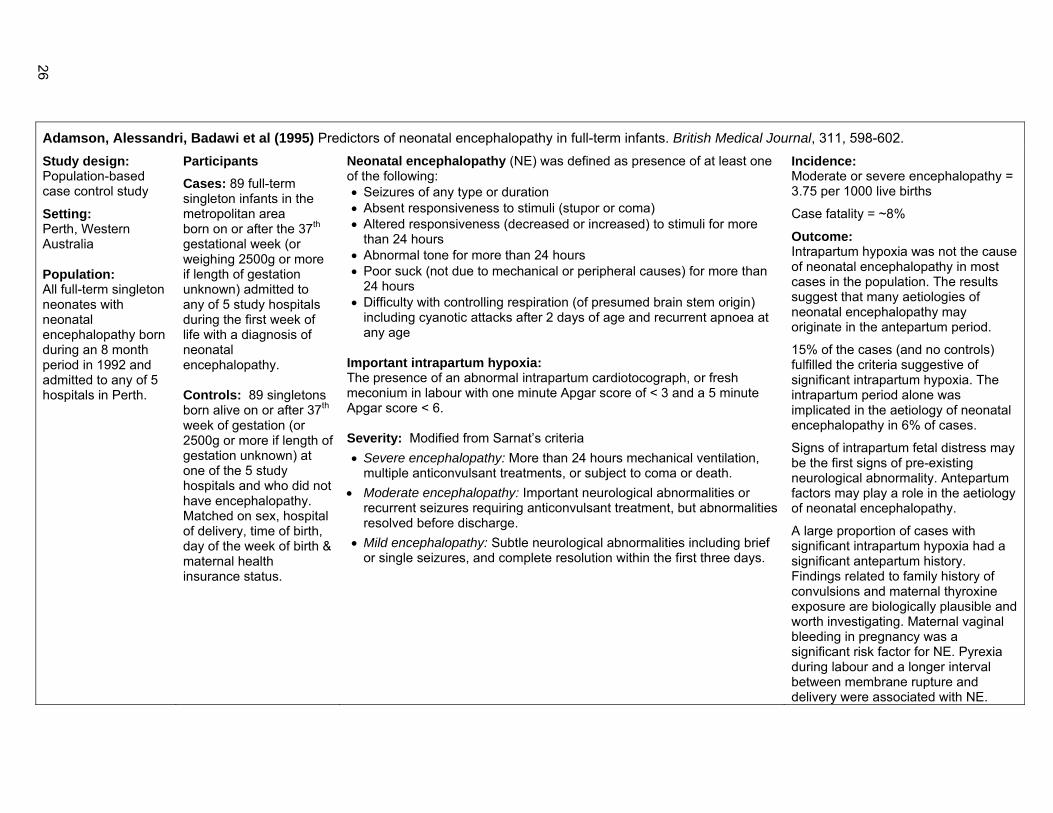
Adamson, Alessandri, Badawi et al (1995) Predictors of neonatal encephalopathy in full-term infants. British Medical Journal, 311, 598-602. Study design: Population-based case control study
Setting: Perth, Western Australia Population: All full-term singleton neonates with neonatal encephalopathy born during an 8 month period in 1992 and admitted to any of 5 hospitals in Perth.
Participants Cases: 89 full-term singleton infants in the metropolitan area born on or after the 37th gestational week (or weighing 2500g or more if length of gestation unknown) admitted to any of 5 study hospitals during the first week of life with a diagnosis of neonatal encephalopathy. Controls: 89 singletons born alive on or after 37th week of gestation (or 2500g or more if length of gestation unknown) at one of the 5 study hospitals and who did not have encephalopathy. Matched on sex, hospital of delivery, time of birth, day of the week of birth & maternal health insurance status.
Neonatal encephalopathy (NE) was defined as presence of at least one of the following: • Seizures of any type or duration • Absent responsiveness to stimuli (stupor or coma) • Altered responsiveness (decreased or increased) to stimuli for more
than 24 hours • Abnormal tone for more than 24 hours • Poor suck (not due to mechanical or peripheral causes) for more than
24 hours • Difficulty with controlling respiration (of presumed brain stem origin)
including cyanotic attacks after 2 days of age and recurrent apnoea at any age
Important intrapartum hypoxia: The presence of an abnormal intrapartum cardiotocograph, or fresh meconium in labour with one minute Apgar score of < 3 and a 5 minute Apgar score < 6. Severity: Modified from Sarnat’s criteria • Severe encephalopathy: More than 24 hours mechanical ventilation,
multiple anticonvulsant treatments, or subject to coma or death. • Moderate encephalopathy: Important neurological abnormalities or
recurrent seizures requiring anticonvulsant treatment, but abnormalities resolved before discharge.
• Mild encephalopathy: Subtle neurological abnormalities including brief or single seizures, and complete resolution within the first three days.
Incidence: Moderate or severe encephalopathy = 3.75 per 1000 live births
Case fatality = ~8%
Outcome: Intrapartum hypoxia was not the cause of neonatal encephalopathy in most cases in the population. The results suggest that many aetiologies of neonatal encephalopathy may originate in the antepartum period.
15% of the cases (and no controls) fulfilled the criteria suggestive of significant intrapartum hypoxia. The intrapartum period alone was implicated in the aetiology of neonatal encephalopathy in 6% of cases.
Signs of intrapartum fetal distress may be the first signs of pre-existing neurological abnormality. Antepartum factors may play a role in the aetiology of neonatal encephalopathy.
A large proportion of cases with significant intrapartum hypoxia had a significant antepartum history. Findings related to family history of convulsions and maternal thyroxine exposure are biologically plausible and worth investigating. Maternal vaginal bleeding in pregnancy was a significant risk factor for NE. Pyrexia during labour and a longer interval between membrane rupture and delivery were associated with NE.
26

Thornberg, Thiringer, Odeback & Milsom (1995) Birth asphyxia: incidence, clinical course and outcome in a Swedish population. Acta Paediatrica, 84, 927-32. Objective: To determine the incidence of birth asphyxia in term neonates in a Swedish population using Apgar score, HIE grading and exclusion of clinical entities not related to asphyxia; to investigate the resuscitation methods, level of care and presence of seizures related to outcome; to identify the proportion of small-for-gestational age (SGA) infants in birth asphyxia; and to assess mortality and incidence and type of neurological sequelae.
Setting: Göteborg, Sweden Population: 42,203 live births to mothers residing in Göteborg over a 7 year period from 1985-1991. This encompasses around 5% of births in Sweden. 292 infants that were full-term (≥ 37 weeks) and had Apgar scores <7 at 5 mins were identified.
Participants: 292 infants with low Apgar scores
Cases: 227 infants with ‘birth asphyxia’
75 infants categorised as birth asphyxia-HIE group
Controls: 225 infants, the next term infant born at the respective delivery hospital of the same gender and with an Apgar score ≥ 7 at 5 mins.
Pure birth asphyxia: Group of infants with asphyxia when the exclusion criteria was applied ie. “low Apgar score group excluding cases not related to birth asphyxia.”
Birth asphyxia-HIE group: Classified as mild, moderate or severe (from notes in the medical records), according to the criteria of Sarnat & Sarnat, modified by Fenichel.
Mild HIE: Characterised by hyperalertness and irritability, normal muscle tone, normal or hyperactive reflexes, ankle clonus, no seizures.
Moderate HIE: Includes lethargy, decreased spontaneous movements, proximal muscular weakness, depressed primitive reflexes and seizures.
Severe HIE: include stupor or coma, markedly reduced muscle tone or flaccidity, and absent primitive reflexes. Seizures are often frequent and may be difficult to control, but may also be totally absent.
Exclusion Criteria: Infants considered to have causes of their low Apgar score other than asphyxia were excluded: opioid/anesthesia-related low Apgar score, congenital malformations and chromosomal disorders, congenital infections, congenital neuromuscular disorders, and subarachnoid haemorrhage.
Incidence: (per 1000 live born infants) Low Apgar score = 6.9 Pure birth asphyxia = 5.4 Birth asphyxia with HIE = 1.8 Yearly incidence for each of these is also presented Mild HIE = 0.85 per 1000 Moderate HIE = 0.4 per 1000 Severe HIE = 0.3 per 1000 Neonatal mortality = 0.26 per 1000 Neurological impairment = 0.2 per 1000
Outcome: Mild = 36 (48%) Moderate = 17 (22.7%) Severe = 12 (16%) Unable to categorise = 10 (13.3%) due to ventilation and sedation but probable mild or moderate cases
Mortality among children with seizures was 41%. The number of infants with mild encephalopathy may be underestimated due to absence in the clinical records of mild clinical signs.
Infants small for gestational age were overrepresented in the birth asphyxia group but not the birth asphyxia-HIE group. All infants with severe HIE died or developed neurological damage. Half of the infants with moderate and all of the infants with mild were normal at 18 months of age. Disabilities included dyskinetic, tetraplegic, spastic diplegic, cerebral palsy and mild neuromotor dysfunction.
27

Yudkin, Johnson, Clover & Murphy (1995) Assessing the contribution of birth asphyxia to cerebral palsy in term singletons. Paediatric and Perinatal Epidemiology, 9, 156-70.
Study design: Cohort study Research questions: How likely is birth asphyxia to result in cerebral palsy? How much cerebral palsy is caused by birth asphyxia? Aims: To investigate the risk of cerebral palsy following intrapartum asphyxia at term and the contribution of intrapartum asphyxia at term to the overall rate of cerebral palsy.
Setting: Oxfordshire, UK
Population: 8,863 term (≥ 37 weeks or birth weight ≥ 2500g) singleton infants born in the John Radcliffe Hospital from Jan 1984 to Sept 1985. A retrospective examination of perinatal records of children born with CP was also completed. 11,682 singleton live births, 11,110 born at term, 30 cases of cerebral palsy (not postnatally acquired)
Participants: 166 term infants. 148 infants followed-up at 5 years of age.
Birth Asphyxia: Signs of presumed asphyxia occurring intrapartum. Based on the ACOG (1991) criteria. However unlike the ACOG, no rigid cut-off levels set for measurable items such as Apgar score and did not demand that all of the signs be present.
Potentially damaging intrapartum asphyxia: • Umbilical artery pH < 7.00 • 5 minute Apgar score ≤ 3 • Moderate or severe neonatal encephalopathy • Signs of multi-organ system dysfunction such as
cardiovascular, haematological, pulmonary or renal
Mild encephalopathy: jitteriness, excessive response to stimulation and possible mild feeding difficulties, lasting up to 48 hours.
Moderate encephalopathy: Characterised by lethargy for at least 12 hours after birth, hypotonia, impaired primitive reflexes and the likely occurrence of seizures.
Severe encephalopathy: extreme hypotonia, with a need for mechanical ventilation, weak or absent primitive reflexes, and prolonged or repetitive seizures.
Also considered fetal heart rate pattern in labour and the result of cerebral scans carried out in the early neonatal period. Cerebral oedema and widespread white matter damage were taken to support the diagnosis of birth asphyxia.
Follow-up study: Term singleton infants with 1 minute Apgar score ≤ 3. Used the Griffiths Developmental Scales and the Movement Assessment Battery for Children.
Rates: Cerebral palsy = 2.6 per 1000 singleton live births Neonatal deaths = 3.6 per 1000 singleton live births Neonatal deaths following asphyxia = 0.36 per 1000 term births (one in 2,800) Incidence of CP following birth asphyxia was one in 3,700 singleton live births.
Outcome: Birth asphyxia at term was associated with 10% of all cases of cerebral palsy and with 20% of the 15 cases of cerebral palsy in children born at term.
6/166 infants exhibited probable birth asphyxia although none completely satisfied the ACOG criteria. 2 died neonatally, 3 had spastic quadriparesis with profound developmental delay and one had a normal outcome at 5 years of age.
Mild = 4 infants Moderate = 3 infants Severe = 3 infants CP probably attributed to birth asphyxia = 3 infants ‘Damaging birth asphyxia entails serious depression at delivery.’
28

Becerra, Fry & Rowley (1991) Morbidity estimates of conditions originating in the perinatal period: United States, 1986 through 1987. Pediatrics, 88, 553-9.
Study design: Population-based survey
Objective: To provide nationally representative estimates of infant morbidity originating in the perinatal period
Setting: USA
Population: Data from the National Hospital Discharge Survey. A stratified cluster sample of patients discharged each year from short-stay, non-federal hospitals located in the 50 states and districts of Columbia from 1986-1987. This provides a weighted nationally representative sample of ~18,000 newborns per year in the USA.
Participants: 35,995 infants discharged from US hospitals.
International Classifications of Diseases, 9th Revision, Clinical Modification was utilised. Birth asphyxia: Not specified Fetal distress (intrauterine hypoxia): Not specified Birth trauma: Not specified Exclusion criteria: Neonates whose only discharge diagnosis consisted of birth defects were excluded. Neonates with birth defects and other perinatal conditions were included.
Rates: Overall morbidity = 37.6%
Morbidity for conditions originating in the perinatal period = 33.7%
Birth defects = 4.8%
Birth defects and nonteratogenic conditions = 2.5%
Perinatal morbidity related to maternal causes = 3.1%
Birth trauma = 2.5%
Fetal distress = 2.3%
Birth asphyxia = 2.1%
Infection specific to perinatal period = 2%
Complications of labour and delivery = 1.9%
Complications of caesarean delivery = 1.2%
Outcome: Morbidity from perinatal conditions was greater in males than females. See the paper for further data.
29

Mir, Faquih & Legnain (1989) Perinatal risk factors in birth asphyxia: relationship of obstetric and neonatal complications to neonatal mortality in 16,365 consecutive live births. Asia-Oceania Journal of Obstetrics and Gynaecology, 15, 351-7.
Study design: Case control study (population-based, prospective). Objective: To determine the relationship between selected perinatal risk factors in easily identifiable cases of birth asphyxia and to evaluate the usefulness of the definition of asphyxia in the prediction of neonatal mortality and morbidity.
Setting: Benghazi, Libya Population: All 16,365 consecutive live births at Jamahiria Maternity Hospital from July 1983-June 1984.
Participants
Cases: 465 infants with birth asphyxia
Controls: 15,900 infants born during the study period who did not have evidence of birth asphyxia
Asphyxia: Need for positive pressure ventilation for > 1 min before sustained respiration occurred or Apgar < 5 at 1 min. Severe birth asphyxia: Need for IPPV for > 3 min. Neonatal seizure: Single or recurrent tonic or clonic movements and/or subtle seizures as defined by Volpe (1987) Exclusion criteria: None listed
Incidence: Asphyxia = 2.8%
Outcome: There was an increasing incidence with decreasing gestational age. Impact of asphyxia greatest among more mature infants, mortality increased 3 fold in infants <34 weeks and greater than 27 fold for infants >38 weeks gestation. Intrauterine growth retardation, fetal macrosomia, hypothermia, hyaline membrane disease, seizures, hypoglycaemia and hyponatremia significantly associated with increased risk of death. No conclusions made on the usefulness of the definition.
30

Nelson & Ellenberg (1987) The asymptomatic newborn and risk of cerebral palsy. American Journal of Diseases of Children, 141, 1333-5.
Research question: Does the term infant whose birth was complicated but whose neonatal course was neurologically smooth face a higher risk of cerebral palsy than a baby with an uncomplicated birth?
Setting: Not specified Population: This paper presents results from children followed-up in the Collaborative Perinatal Project of the National Institute of Neurological & Communicative Disorders & stroke (NCPP). Pregnant women entered the study in 1959-1966 following registration for prenatal care.
Participants: 51,285 singleton live born infants
Hypoxic Ischaemic Encephalopathy: • Decreased activity after the first day of life • Need for incubator care for 3 or more days • Feeding problems • Poor suck • Respiratory difficulty and/or • Neonatal seizure
Outcome: The answer to the research question was NO. There was an increased risk of CP only in the presence of abnormal neonatal signs and this risk increased with the number of abnormal signs.
31

MacDonald, Mulligan, Allen & Taylor (1980) Neonatal asphyxia. I. Relationship of obstetric and neonatal complications to neonatal mortality in 38,405 consecutive deliveries. Journal of Pediatrics, 96, 898-902. Study design: Population-based cohort study
Objective: To assess the perinatal risk factors that might be reliable predictors of neonatal asphyxia and the contribution of these factors to the risk of an asphyxiated infant dying in the neonatal period.
Setting: Pittsburgh, USA
Population: 38,405 consecutive live born births at Magee Women’s Hospital from Jan 1970 to Dec 1975.
Participants Cases: 447 infants with neonatal asphyxia
462 infants = ‘severe’ asphyxia (not defined) Controls: 37,958 infants who did not have asphyxia but had the same supervised chart review at discharge.
Separate analyses completed for term (>36 weeks) and preterm infants (≤36 weeks).
Neonatal asphyxia (NA): Diagnosed in infants who required > 1 minute of positive pressure ventilation before sustained respiration occurred. Neonatal seizure: Diagnosed when single or recurrent tonic or clonic movements were witnessed by a physician. High risk caesarean section: Primary indication was fetal distress, prolapsed cord, abruption, placenta previa, fetal growth retardation, prolonged rupture of membranes, isoimmunisation, maternal toxaemia, or maternal diabetes.
Incidence of neonatal asphyxia: Overall = 1.2%, Pre-term = 9%, Term = 0.5%
Outcome: Prematurity was the most significant predictor of asphyxia. Asphyxia was associated with significant increase in neonatal mortality in infants >27 weeks. Growth retardation, hypothermia, hyaline membrane disease, and seizures were significantly associated with an increased risk of death.
For term infants NA occurred more significantly in black infants, infants of unmarried mothers, infants of diabetic and toxemic mothers. Breech delivery and growth retardation were associated with a higher incidence of birth asphyxia for both term and preterm. Although there was a slight increase in incidence of asphyxia among infants that underwent cesarean section only the high risk category was associated with significantly increased incidence of asphyxia, which indicates that the section itself was not causative in producing the asphyxia.
The presence of NA increased the risk of dying from a twofold increase in risk at 27-28 weeks to a hundredfold increase at >36 weeks. The overall mortality with asphyxia present was 46.1% an 88-fold increase over the 0.52% mortality when asphyxia was absent. There was an overall mortality of 46.3% among infants with severe asphyxia. Socioeconomic status was significantly associated with higher mortality in asphyxiated infants.
Efforts aimed at preventing prematurity should contribute effectively to decreasing the overall incidence of and mortality associated with perinatal asphyxia. Concerted effort to provide optimal obstetric care to women whose pregnancies have reached 36 weeks and whose gestations are identified as high risk should significantly reduce the incidence of asphyxia and neonatal mortality.
32

OTHER STUDIES
Barnett, Guzzetta, Mercuri et al (2004) Can the Griffiths scales predict neuromotor and perceptual-motor impairment in term infants with neonatal encephalopathy? Archives of Disease in Childhood, 89, 637-43.
Study design: Longitudinal cohort study Objective: To examine the predictive value of early developmental testing for identifying neuromotor & perceptual motor impairment at school age in children born at term with neonatal encephalopathy.
Setting: London, United Kingdom
Population: Term infants born at or referred to the Hammersmith hospital
Participants: 80 full-term infants diagnosed with neonatal encephalopathy
Neonatal encephalopathy: Infants who had convulsions and/or showed other abnormal neurological signs during the first 48 hours after delivery. Neurological abnormalities included abnormal tone, poor feeding and altered level of consciousness.
Exclusion criteria: Children subsequently diagnosed with genetic or metabolic syndromes or presented with other neonatal complications such as septicaemia or neonatal meningitis. Infants with dysmorphic features or other clinical or brain magnetic resonance imaging findings suggesting major congenital malformations.
The authors consider neonatal encephalopathy as incorporating infants who were in the past labelled with HIE and those presenting with neonatal convulsions.
Outcome: 40% of infants had cerebral palsy, 25% had severe spastic quadriplegia. 14% without cerebral palsy had perceptual motor impairment.
Although early neurodevelopmental assessment (using the Griffiths Scale) can to some extent predict outcome at school age, a normal neurodevelopmental assessment at 1 & 2 years does not exclude cognitive, neurological or perceptual motor abnormalities at school age. In particular false negatives often occurred among children with hemiplegia.
33

Becher, Bell, Keeling, McIntosh & Wyatt (2004) The Scottish perinatal neuropathology study: clinicopathological correlation in early neonatal deaths. Archives of Disease in Childhood. Fetal and Neonatal Edition, 89, F399-407.
Study design: Cohort study
Objectives: (a) To compare the epidemiology of infants displaying signs of birth asphyxia with those not showing signs (b) to examine the neuropathology and determine if possible the timing of brain insult comparing asphyxiated with non-asphyxiated infants (c) to compare the clinical features of those born with birth asphyxia with and without pre-labour damage.
Setting: Scotland
All 22 delivery units within Scotland were involved.
Population: The base study considered all perinatal deaths of infants who were ≥ 24 weeks gestation at birth and ≤ 7 days at time of death delivered in Scotland over the 2 year period from Jan 1996-Jan 1999. This paper concerns the live born subset of the study cohort.
Participants: 70 infants from 174 early neonatal deaths.
Cases: 53 infants with clinical evidence of birth asphyxia
Controls: 17 infants who met none of the criteria for birth asphyxia
Note: Parental consent was required for an autopsy. Includes both term and pre-term infants.
Birth Asphyxia: Infants displayed at least one of the features: • An Apgar score at 5 mins of ≤ 5.0 • Cord or initial blood pH < 7.1 • Presence of grade 2/3 neonatal encephalopathy
(as per Sarnat & Sarnat) Exclusion criteria: Infants with major chromosomal abnormalities, abnormalities of the cardiovascular and central nervous systems or central nervous system infection.
Outcome: In a large proportion of neonatal deaths brain injury predates the onset of labour. The mature infants who died after displaying neonatal encephalopathy were most likely to show neuropathological changes. Abnormal CTG & meconium staining of liquor were the only predictive factors for birth asphyxia or prenatal brain damage. Abnormal CTG was the clinical factor that differentiated the asphyxiated infants who displayed encephalopathy and neuropathological abnormality from those who do not. The neuropathological findings reported represent the more severe end of the spectrum of perinatal brain damage; infants surviving perinatal asphyxia may exhibit lesser degrees of similar pathology. The findings support the notion that the birth of a compromised ‘asphyxiated’ encephalopathic infant is not necessarily the result of mismanaged labour nor the lack of vigilance in pregnancy.
34

Carli, Reiger & Evans (2004) One-year neurodevelopmental outcome after moderate newborn hypoxic ischaemic encephalopathy. Journal of Paediatrics and Child Health, 40, 217-20. Study design: Cohort study
Objective: To define the 1-year neurodevelopmental outcome for survivors of moderate (Sarnat stage 2) neonatal hypoxic-ischemic encephalopathy to facilitate appropriate parental counselling. Babies were assessed at 4, 7 and 12 months of age.
Setting: Sydney, Australia
Population: Full-term neonates ≥ 37 weeks gestation born between 1988 and 2000 and admitted to the John Spence nursery (inborn or outborn). All babies admitted with seizures and/or low Apgar score (< 8 at 5 minutes).
Participants: 53 babies who ‘survived HIE’
42 babies were followed-up at one year of age. Infants were excluded if their low Apgar score and/or fits were not considered to be due to asphyxia or if they had mild or severe HIE.
Hypoxic-Ischemic Encephalopathy/Perinatal Asphyxia: 1. It was considered likely that there had been perinatal asphyxia, using, as far as possible, criteria as defined by the consensus statement on perinatal asphyxia and cerebral palsy (referenced to MacLennan, 1999). 2. They had abnormal neurological behaviour compatible with moderate hypoxic-ischemic encephalopathy. The Sarnat classification was used and based on clinical criteria as EEG information was not available on all babies. Exclusion criteria: • Babies for whom it was unlikely that seizures were due to perinatal
asphyxia, this included idiopathic hypoglycaemia, infection, opiate withdrawal, congenital malformations, cerebral haemorrhage or venous thrombosis, familial seizures, metabolic diseases, jaundice and hyponatremia.
• Infants with grade 1 HIE • One infant was excluded as there was no suggestion of
intrapartum compromise.
Outcome: Grade I = 13 infants (excluded) Grade II = 53 infants Grade III = 34 infants (32 neonatal deaths)
There was a 1-year mortality and major disability rate following moderate HIE of 36% with 9.5% showing developmental delay between one and two SD below the mean. These findings are in the range of other studies (20-50%).
The authors recommend systematic long-term follow-up of these infants.
35

Celtik, Acunas, Oner & Pala (2004) Neuron-specific enolase as a marker of the severity and outcome of hypoxic ischemic encephalopathy. Brain and Development, 26, 398-402. Study design: Case control study
Objectives: To evaluate serum concentrations of neuron-specific enolase (NSE) as a marker of the severity of hypoxic ischemic encephalopathy (HIE) and to elucidate the relation among the concentrations of NSE, grade of HIE and short-term outcome.
Setting: Turkey
Population: Infants treated at the neonatal ICU of Trakya University Hospital.
Participants Cases: 43 asphyxiated full-term (infants were >38 weeks) newborn infants who developed signs and symptoms of HIE AND 29 full-term newborn infants with meconium-stained amniotic fluid and normal physical examination.
Blood samples taken between 4 to 48h and 5 to 7 days after birth.
Controls: 30 healthy infants born after spontaneous vaginal delivery with normal physical findings.
Hypoxic ischemic encephalopathy: According to Sarnat & Sarnat.
Asphyxia: Fulfilled the following criteria:
1. Intrapartum distress indicated by the cardiotocograph pattern (late decelerations, absence of variability, persistent bradycardia, etc.) and /or abnormal blood flow pattern (loss or reversal of end-diastolic velocity) and/or early passage of thick meconium,
2. Requirement for resuscitation with positive pressure ventilation and laryngeal intubation,
3. Low Apgar score (1st min ≤ 3, 5th min < 6) or umbilical arterial/first postnatal (pH <7.1).
Neurological examination completed daily during hospitalisation and 1 week, 3, 6 and 12 months after discharge.
Exclusion criteria: Infants with severe congenital abnormalities or profound anaemia; history of multiple pregnancies.
Outcome: Stage I = 14 infants Stage II = 19 infants Stage III = 10 infants
Serum NSE concentrations of infants with stage III HIE were significantly higher than those in stage I & II. The authors conclude that the predictive capacity of serum NSE concentrations for poor outcome seems to be better than predicting HIE of moderate or severe degree. However, earlier and/or CSF samples are required to establish serum NSE as an early predictor for the application of neuroprotective strategies.
36

Low (2004) Determining the contribution of asphyxia to brain damage in the neonate. Journal of Obstetrics and Gynaecology Research, 30, 276-86.
Review article Setting: Canada
Intrapartum fetal asphyxia: The clinical classification of asphyxia as mild, moderate or severe is based on the presence of metabolic acidosis to confirm the occurrence of asphyxia with measures of neonatal encephalopathy and other organ system complications to express the severity of the asphyxia.
Encephalopathy Cardiovascular, respiratory and renal complications
Asphyxia
Metabolic acidosis at delivery*
Minor Moderate Severe Minor Moderate/severe
Mild + +/- +/- Moderate + + +/- Severe + + +
* Umbilical artery base deficit ≥ 12 mmol/L
37

McDonald, Kelehan, McMenamin et al (2004) Placental fetal thrombotic vasculopathy is associated with neonatal encephalopathy. Human Pathology, 35, 875-80. Study design: Case control study
Objective: To determine whether histopathological examination of the placenta in cases of neonatal encephalopathy could identify significant antenatal processes that are not recognised by clinical assessment alone.
Setting: Dublin, Ireland
Population: All live born singleton term (≥ 37 weeks gestation) infants, selected from placental tissue samples collected from 1000 consecutive deliveries, from a tertiary referral maternity centre.
Participants Cases: 93 infants, singleton term deliveries diagnosed with neonatal encephalopathy
Controls: 387 term infants with no evidence of neonatal encephalopathy
Neonatal Encephalopathy: A modification of the grading system by Sarnat and Sarnat (1976) was used. Not further specified.
Exclusion criteria: Infants with a recognised congenital anomaly.
Outcome: Placental features of infection, thrombosis and disturbed uteroplacental flow were significant independent factors in the etiology of neonatal encephalopathy. The absence of significant clinical antenatal factors supports the value of placental examination in the investigation of infants with neonatal encephalopathy.
Mercuri, Anker, Guzzetta et al (2004) Visual function at school age in children with neonatal encephalopathy and low Apgar scores. Archives of Disease in Childhood Fetal Neonatal Edition, 89, F258-62.
Objective: To assess different aspects of visual function in school age in children who suffered from neonatal encephalopathy. To evaluate the correlation between (a) various aspects of visual function, such as multiple optotypes (crowding acuity), visual fields, and stereopsis and the type and extent of the lesion, when present, as observed by neonatal MRI, (b) the results of visual assessments performed in the first year after birth and at school age.
Setting: London, UK
Population: Infants born at or referred to the Hammersmith hospital from Oct 1991 to Oct 1996
Participants: 39 full-term (≥ 38 weeks gestation) infants with neonatal encephalopathy
Neonatal Encephalopathy: Infants with low Apgar scores (< 5 at one minute and < 8 at 5 minutes) who had convulsions in the first 48 hours and/or showed other neurological abnormalities during the first 48 hours after delivery.
Neurological abnormalities: Abnormal tone, poor feeding, and altered level of consciousness.
Severity: Encephalopathy graded ‘according to the classification suggested by Sarnat & Sarnat.’
Exclusion criteria: Infants subsequently diagnosed as having genetic or metabolic syndromes or presented with other neonatal complications, such as septicaemia or neonatal meningitis. Infants with dysmorphic features or other clinical or brain MRI finding suggesting major congenital malformation.
Outcome: The presence and severity of visual impairment was related to severity of brain lesions. Moderate or severe basal ganglia lesions and severe white matter changes were always associated with abnormal visual function. Assessment of visual function in the first year was predictive of visual outcome at school age.
The authors have previously found that lesions in the basal ganglia are more likely to be associated with impaired visual function in the first year of life than lesions in the occipital cortex.
38

Miller, Latal, Clark et al (2004) Clinical signs predict 30-month neurodevelopmental outcome after neonatal encephalopathy. American Journal of Obstetrics & Gynecology, 190, 93-9. Study design: Cohort study
Objective: To determine the value of a neonatal encephalopathy score (ES) and the presence of seizures for predicting 30-month neurodevelop-mental outcome. This included cognitive development and neuromotor outcome.
Setting: San Francisco, USA
Population: 5,389 term neonates born in or transferred to the intensive care nursery from 1994 to 2000.
Participants: 68 term newborn infants with encephalopathy. Infants were excluded if gestational age <35 weeks or there was suspected or confirmed congenital malformations, congenital metabolic disease or congenital infections.
Neonatal Encephalopathy: (1) Umbilical artery pH < 7.1 (2) Umbilical artery base deficit > 10 (3) 5-minute Apgar score ≤ 5
Encephalopathy Score (ES): Encephalopathy sign Score= 0 Score= 1 Feeding Normal Gavage feeds, gastrostomy tube or not
tolerating oral feeds Alertness Alert Irritable, poorly responsive or comatose Tone Normal Hypotonia or hypertonia Respiratory status Normal Respiratory distress (need for CPAP or
mechanical ventilation) Reflexes Normal Hyperreflexia, hyporeflexia or absent reflexes
Seizures None Suspected or confirmed clinical seizure Total 0-6
Newborn infants scored daily for the first 3 days of life and the maximum score utilised. The ES only used on days the infants was not sedated or paralysed.
This is based on the Sarnat staging with the addition of feeding (as recognised by Nelson & Ellenberg 1987). Seizures were defined as per Volpe and were distinguished from ‘jitteriness’.
Seizures: Paroxymal alteration in motor, and occasionally autonomic function, including clonic, tonic and ‘subtle’ seizure manifestations.
Exclusion criteria: Gestational age <35 weeks or suspected or confirmed congenital malformations, congenital metabolic disease or congenital infections.
Outcome measurement: The Bayley Scales of Infant Development II and neurological examination using a validated neuromotor score were used.
Outcome: Together the ES and presence of seizures on the first day of life were similarly predictive of abnormal outcome with a sensitivity of 72% and specificity of 94%, positive predictive value of 84% and negative predictive value of 88%. There were 3 false positive and 6 false negative classifications. The authors concluded that the severity of neonatal encephalopathy and the presence of seizures are valuable predictors of 30-month neurodevelopmental outcome as early as the first day of life.
39
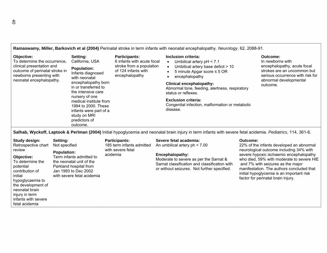
Ramaswamy, Miller, Barkovich et al (2004) Perinatal stroke in term infants with neonatal encephalopathy. Neurology, 62, 2088-91.
Objective: To determine the occurrence, clinical presentation and outcome of perinatal stroke in newborns presenting with neonatal encephalopathy.
Setting: California, USA
Population: Infants diagnosed with neonatal encephalopathy born in or transferred to the intensive care nursery of one medical institute from 1994 to 2000. These infants were part of a study on MRI predictors of outcome.
Participants: 6 infants with acute focal stroke from a population of 124 infants with encephalopathy
Inclusion criteria: • Umbilical artery pH < 7.1 • Umbilical artery base deficit > 10 • 5 minute Apgar score ≤ 5 OR • encephalopathy
Clinical encephalopathy: Abnormal tone, feeding, alertness, respiratory status or reflexes.
Exclusion criteria: Congenital infection, malformation or metabolic disease.
Outcome: In newborns with encephalopathy, acute focal strokes are an uncommon but serious occurrence with risk for abnormal developmental outcome.
Salhab, Wyckoff, Laptook & Perlman (2004) Initial hypoglycemia and neonatal brain injury in term infants with severe fetal acidemia. Pediatrics, 114, 361-6.
Study design: Retrospective chart review
Objective: To determine the potential contribution of initial hypoglycaemia to the development of neonatal brain injury in term infants with severe fetal acidemia
Setting: Not specified
Population: Term infants admitted to the neonatal unit of the Parkland hospital from Jan 1993 to Dec 2002 with severe fetal acidemia
Participants: 185 term infants admitted with severe fetal acidemia
Severe fetal academia: An umbilical artery ph < 7.00 Encephalopathy: Moderate to severe as per the Sarnat & Sarnat classification and classification with or without seizures. Not further specified.
Outcome: 22% of the infants developed an abnormal neurological outcome including 34% with severe hypoxic ischaemic encephalopathy who died, 59% with moderate to severe HIE and 7% with seizures as the major manifestation. The authors concluded that initial hypoglycemia is an important risk factor for perinatal brain injury.
40

Satar, Ozcan, Yapicioglu & Narli (2004) Serum insulin-like growth factor 1 and growth hormone levels of hypoxic-ischemic newborns. Biology of the Neonate, 85, 15-20.
Aims: To determine the levels of insulin-like growth factor 1 (IGF-1), growth hormone (GH), and cortisol in HI babies and identifying whether they differ from the levels of control infants. IGF measurements taken after the first 12-24 hours of life and repeated at 5 and 10 days.
Setting: Adana, Turkey
Population: Newborn babies admitted to the Neonatal ICU of the Cukurova University from Oct 2000 to Sept 2001. This included inborn and outborn.
Participants
Cases: 18 infants with hypoxic-ischemic encephalopathy
Controls: 19 control infants, healthy full-term infants ‘being at the same time in the Gynecology Dept in rooming with their mothers’
Hypoxic-Ischemic Encephalopathy: Abnormal neurological examination results and had at least one of the following criteria:
1. Intrapartum fetal distress (fetal heart rate <120/min or >160/min)
2. Apgar score <5 after 5 mins 3. Arterial blood pH value during the first hour <7.1 4. Need of mask-bag ventilation or intubation, and 5. Elevated aspartate aminotransferase, alanine
aminotransferase, blood urea nitrogen, and serum creatinine levels.
Abnormal neurological examination findings included 2 or more of the following:
1. encephalopathy 2. an abnormal muscle tone (hypotonia, hypertonia) and 3. seizures
Neurological examinations performed every day for the first week and patients regrouped into 3 subgroups according to Sarnat & Sarnat: stage I (mild), stage II (moderate) and stage III (severe) HIE groups. Not further specified. Exclusion criteria: Congenital malformations, metabolic and endocrine disorders, intrauterine infections, chromosomal abnormalities, and mother drug addiction.
Outcome: Mild to moderate = 11 babies (61.1%) Severe = 7 babies (38.9%) There was no statistically significant difference between severity groups in IGF-1 levels. There was no statistically significant difference in serum cortisol levels between cases and controls. At 12-24 hours cases had significantly lower levels of IGF-1 than controls, GH levels were significantly higher in this time period than controls. This study showed that decreased levels of serum IGF-1 and increased levels of GH which may be secondary to serum IGF-1 influx from the circulation of the brain as a protective mechanism or may be due to some cytokines which alter the GH/IGF axis.
41

Shah, Riphagen, Beyene & Perlman (2004) Multiorgan dysfunction in infants with post-asphyxial hypoxic-ischaemic encephalopathy. Archives of Disease in Childhood Fetal Neonatal Edition, 89, F152-5.
Study design: Cohort
Objective: To assess the patterns of involvement of each major organ/system and combinations of involvement in infants with post-asphyxial hypoxic-ischemic encephalopathy, and to describe this in relation to long term outcome.
Setting: Toronto, Canada
Population: Term (≥ 37 weeks) neonates born in and around Toronto, admitted to the neonatal ICU at the Hospital for Sick Children. Infants were admitted between 1985 and 1995. There were around 600,000 live births in the study period.
Participants: 144 full-term neonates with post-intrapartal asphyxial HIE assessed for kidney, cardiovascular system, lung and liver function. 244 infants met the original study criteria.
Post-intrapartum asphyxial HIE: (Taken from the ACOG statement in 1998 and Society of Obstetrics and Gynecologists of Canada): (1) One or more of the following:
(a) 5 minute Apgar score of < 5 (b) Metabolic acidosis (cord arterial blood or blood gas analysis
within first hour after birth) indicated by a base deficit ≥ 16 mmol/L
(c) Delayed onset of respiration for ≥ 5 minutes (2) Need for mechanical ventilation at birth (3) Evidence of encephalopathy including altered state of consciousness and/or seizures (seizures defined retrospectively from the description provided in the health records using Volpe’s criteria) (4) Infants who had complete clinical and/or investigational assessments of the function of all four organs Infants with missing data for criterion 1 were included if they were born by emergency caesarean section and had features typical of criteria 2 and 3 and other causes of neonatal encephalopathy could be excluded with confidence. Criteria for organ/system dysfunction: Further criteria are outlined in the paper (see p.F153) Exclusion criteria: Preterm (<37 weeks), congenital abnormalities including subtle dysmorphism or unknown significance or a major anomaly of a single organ, inborn errors of metabolism, congenital viral infections, haemorrhagic shock without evidence of intrapartum asphyxia, septic shock, cranial birth trauma, meconium aspiration syndrome, or evidence of antepartum asphyxia. Antepartum asphyxia: One of more of the following: a history of an antepartum episode of loss of fetal movements lasting for 24 hours or more, severe intrauterine growth retardation, oligohydramnios or a lack of fetal heart rate variability on admission of the mother to hospital.
Outcome: Multiorgan dysfunction was present in all infants with post-asphyxial HIE. There was no association between multiorgan dysfunction and outcome. By 24 months of age 55% of the infants had severe adverse outcomes (death or cerebral palsy).
42

Stelmach, Kallas, Pisarev & Talvik (2004) Antenatal risk factors associated with unfavorable neurologic status in newborns and at 2 years of age. Journal of Child Neurology, 19, 116-22. Study design: Nested case control study
Aim: To evaluate the influence of different antenatal factors on neurological signs in the first days of life and neurodevelopmental outcome at 2 years of age. The authors aim to follow-up children again at 4-5 years of age.
Setting: Tartu, Estonia
Population: 828 consecutive live births from Oct 1998 to March 1999 at the Women’s clinic Tartu University clinics. This included term and preterm infants (range 25 to 42 weeks)
Participants: 390 children (cohort of 403 children, 13 with different neurological signs excluded).
Cases: 49 infants with developmental disabilities at 2 years of age
Controls: 341 developmentally normal children at the same age as controls
Hypoxic-ischemic encephalopathy: Two classifications were used and infants had to meet both criteria.
(1) Severity classified as mild to severe (stages 1 to 3) as per Sarnat & Sarnat.
(2) Criteria as used by Hagberg: The presence of at least 2 of the following symptoms: • Apgar score <5 at 1 or 5 minutes • Resuscitation/ventilation • Convulsions before day 3
Neuroimaging investigations (cranial ultrasonography, MRI or Doppler ultrasonography) supported the clinical symptoms.
Acute intrauterine hypoxia: (using Hagberg criteria) The presence of the following signs: • Miscoloured amniotic fluid • Fetal heart rate during labour <100 or >160 beats/min • Silent pattern or dip 2 pattern on cardiotocography • Cord prolapse or placental abruption • In combination with Apgar scores ≤7 at the 5th minute of age* * This is taken from Russian studies (referenced). This was treated as a potential predisposing factor.
Exclusion criteria: Recognised congenital malformations and chromosomal anomalies. Actual exclusions included infants that died in the first month of life due to various congenital malformations, disabling conditions (Down syndrome, multiple congenital malformations, fetal alcohol syndrome), metabolic diseases detected within the first year of life (hypothyroidism, mucopolysaccharidosis), posttraumatic brain injury.
Outcome: Classified as normal (normal neuromotor development) or abnormal (mild cerebral palsy, moderate cerebral palsy and other developmental disabilities).
Note: Investigators were not blinded on follow-up. Around half of the children were involved in early rehabilitation interventions.
Outcome: At 2 years of age 49 of the 309 (15.9%) children exhibited adverse neurodevelopmental outcome. 41 had cerebral palsy (37 mild, 4 moderate). The strongest ‘correlation’ with development of HIE during the first days of life were trichomoniasis during pregnancy and acute respiratory disease (temp ≥38ºC) in the second half of pregnancy. Of the antenatal predisposing factors only bacterial vaginosis combined with imminent abortion or bleeding in the first half of pregnancy was significantly ‘responsible’ for unfavourable neurodevelopmental outcome at 2 years of age. The presence of at least one complication at delivery placed the child at risk for adverse outcome at 2 years. The most frequent perinatal complications independently associated with adverse neurological outcome were acute hypoxic event during delivery and premature rupture of membranes before onset of labour. Nearly half of the children with CP had 2 or more signs suggestive of an acute hypoxic event. The findings of this study support the possible impact of maternal infection and/or inflammation on cerebral palsy and other adverse outcome. Children who survive HIE should be regarded as at risk and followed up to at least school entry.
43

Williams & Singh (2004) Umbilical artery blood gas parameters in neonates with early onset seizures who die. British Journal of Obstetrics and Gynaecology, 111, 1042-5.
Study design: Cohort study
Objective: To relate umbilical artery blood gas parameters to mortality among neonates with hypoxic-ischaemic encephalopathy related to early onset seizures.
Setting: Vancouver, Canada
Population: Infants ≥ 32 weeks gestation with a documented seizure in the first 24 to 48 hours after admission secondary to HIE. 170 charts reviewed for infants admitted to the special care nursery with a neonatal diagnosis of HIE at BC Women’s hospital over a 10 year period from Jan 1990 to Dec 1999.
Participants: 47 infants in two groups:
1. 33 infants with neonatal seizures who survived
2. 14 infants with neonatal seizures who died related to HIE complications
Hypoxic-Ischaemic encephalopathy (HIE): Using the ‘Sarnat & Sarnat guidelines’. Ancillary documentation that the seizures were related to HIE was sought through reviewing available CT findings. Exclusion criteria: Seizures due to trauma, febrile illness or intracranial mass. Infants that were outborn, <32 weeks gestation, no cord gas done at delivery, infants with HIE but no seizures.
Outcome: The PO2 was significantly higher in the infants who died. Neither the umbilical artery pH nor base deficit is predictive of neonatal death in infants with HIE with seizures. The high PO2 in infants that died may be indicative of an inability of these infants to extract oxygen from the blood.
44
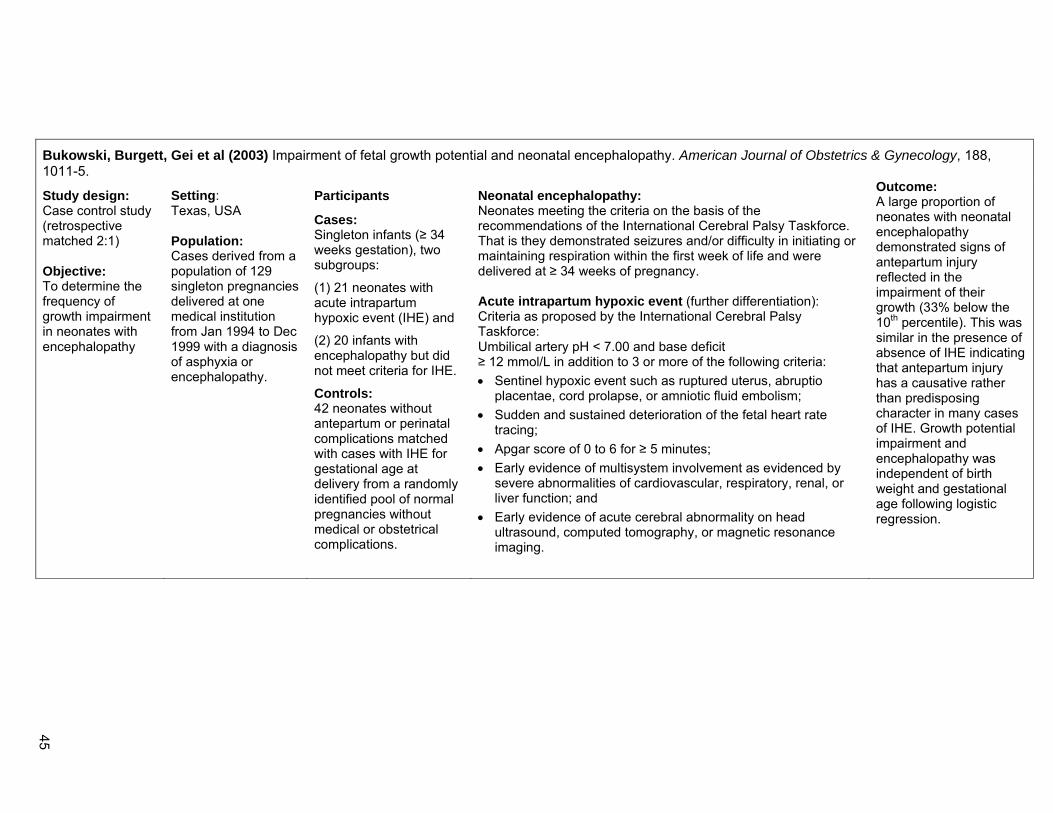
Bukowski, Burgett, Gei et al (2003) Impairment of fetal growth potential and neonatal encephalopathy. American Journal of Obstetrics & Gynecology, 188, 1011-5.
Study design: Case control study (retrospective matched 2:1) Objective: To determine the frequency of growth impairment in neonates with encephalopathy
Setting: Texas, USA Population: Cases derived from a population of 129 singleton pregnancies delivered at one medical institution from Jan 1994 to Dec 1999 with a diagnosis of asphyxia or encephalopathy.
Participants
Cases: Singleton infants (≥ 34 weeks gestation), two subgroups:
(1) 21 neonates with acute intrapartum hypoxic event (IHE) and
(2) 20 infants with encephalopathy but did not meet criteria for IHE.
Controls: 42 neonates without antepartum or perinatal complications matched with cases with IHE for gestational age at delivery from a randomly identified pool of normal pregnancies without medical or obstetrical complications.
Neonatal encephalopathy: Neonates meeting the criteria on the basis of the recommendations of the International Cerebral Palsy Taskforce. That is they demonstrated seizures and/or difficulty in initiating or maintaining respiration within the first week of life and were delivered at ≥ 34 weeks of pregnancy. Acute intrapartum hypoxic event (further differentiation): Criteria as proposed by the International Cerebral Palsy Taskforce: Umbilical artery pH < 7.00 and base deficit ≥ 12 mmol/L in addition to 3 or more of the following criteria: • Sentinel hypoxic event such as ruptured uterus, abruptio
placentae, cord prolapse, or amniotic fluid embolism; • Sudden and sustained deterioration of the fetal heart rate
tracing; • Apgar score of 0 to 6 for ≥ 5 minutes; • Early evidence of multisystem involvement as evidenced by
severe abnormalities of cardiovascular, respiratory, renal, or liver function; and
• Early evidence of acute cerebral abnormality on head ultrasound, computed tomography, or magnetic resonance imaging.
Outcome: A large proportion of neonates with neonatal encephalopathy demonstrated signs of antepartum injury reflected in the impairment of their growth (33% below the 10th percentile). This was similar in the presence of absence of IHE indicating that antepartum injury has a causative rather than predisposing character in many cases of IHE. Growth potential impairment and encephalopathy was independent of birth weight and gestational age following logistic regression.
45

Cowan, Rutherford, Groenendaal et al (2003) Origin and timing of brain lesions in term infants with neonatal encephalopathy. Lancet, 361, 736-42.
Objective: To test the hypothesis that neonatal encephalopathy, early neonatal seizures, or both in the term infant are the result of early antenatal insult.
Setting: Two tertiary referral intensive care units in Utrecht, Netherlands and London, UK Population: Full-term infants born in or referred to the Wilhemina Children’s hospital (Netherlands) and Hammersmith and Queen Charlotte’s hospitals (London) from Jan 1992 to Dec 1998. (This study was not population-based)
Participants: 351 full-term infants >36 weeks gestation who presented with neonatal encephalopathy, seizures or both within 72 hours of birth. 261 infants with neonatal encephalopathy, 90 had seizures within 72 hours of birth.
Neonatal encephalopathy: Abnormal tone pattern, feeding difficulties, altered alertness, and at least 3 of the following criteria: • Late decelerations on fetal monitoring or meconium
staining • Delayed onset of respiration • Arterial cord blood pH < 7.1 • Apgar scores less than 7 at 5 minutes, and • Multi organ failure Exclusion criteria: Infants with neural tube defects, alcohol and drug embryopathies, serious cardiac abnormality, hydrops, important gastrointestinal malformations, immediately diagnosable chromosome abnormalities, treated with hypothermia, minor congenital anomalies or those not part of a recognised syndrome.
Outcome: More than 90% of term infants with neonatal encephalopathy, seizures or both, but without specific syndromes or major congenital defects had evidence of perinatally acquired insults. The authors concluded that events in the immediate perinatal period are most important in neonatal brain injury. However, they acknowledged that their data do not exclude the possibility that antenatal factors could initiate a causal pathway for perinatal brain injury and possibly these with genetic disposition to HIE might make some infants more susceptible than others to stresses of labour and delivery. Note: These authors did not use a healthy comparison population.
46

Debillon, Daoud, Durand et al (2003) Whole-body cooling after perinatal asphyxia: a pilot study in term neonates. Developmental Medicine & Child Neurology, 45, 17-23. Study design: Pilot study
Objective: To test the practicability and safety of whole-body cooling (for 72 hours) in term neonates with moderate-to-severe hypoxic-ischemic encephalopathy and to report outcomes.
Setting: France
Population: Term neonates admitted to one of 7 paediatric or intensive care units in France from Jan 2000 to Jan 2001.
Participants: 25 term infants with moderate to severe HIE and a postnatal age <6 hours
Moderate to Severe Hypoxic-Ischemic Encephalopathy: (1) Sudden deterioration in foetal heart rhythm or meconium-stained amniotic fluid
(2) Apgar score ≤ 5 at 5 minutes or a base deficit ≥ 12 mmoles.L-1, AND
(3) Early neurological abnormalities with abnormal EEG findings during the first 24 postnatal hours
Exclusion criteria: Obvious major congenital anomalies, evidence of head trauma causing major intracranial haemorrhage, severe respiratory distress syndrome, severe hypotension, thrombocytopenia or coagulation factor V < 30%.
Outcome: Whole body cooling is feasible in term neonates with no life-threatening adverse events. Infants were cooled to a core temp of 33ºC to 34ºC, maintained for 72 hrs.
47

Greenwood, Newman, Impey & Johnson (2003) Cerebral palsy and clinical negligence litigation: a cohort study. British Journal of Obstetrics & Gynaecology, 110, 6-11. Study design: Nested cohort study
Objective: To compare the prevalence of criteria suggesting acute intrapartum hypoxia in children with CP who have and have not been the subjects of clinical negligence legal claims.
Setting: Oxfordshire, UK
Population: Singleton children with cerebral palsy born between 1984 and 1993. Identified from the Oxford Register of Early Childhood Impairment (ORECI), which has information on all children with cerebral palsy born to mothers resident within the Oxfordshire Health District. Parents of children with cerebral palsy who sought legal advice were identified from the Oxford Radcliffe Hospital NHS Legal Service Dept database.
Participants: 138 singleton children born with cerebral palsy.
Intrapartum hypoxia (as defined by the International Cerebral Palsy Task Force): Essential consensus criteria:
1. Evidence of a metabolic acidosis in intrapartum fetal umbilical arterial cord, or very early neonatal blood samples (pH < 7.00 and base deficit ≤ 12 mmol/L)
2. Early onset of severe or moderate neonatal encephalopathy in infants ≥ 34 weeks gestation
3. Cerebral palsy of spastic quadriplegic or dyskinetic type
Additional ‘non-specific’ criteria: 4. A sentinel (signal) hypoxia event occurring
immediately before or during labour 5. A sudden rapid and sustained deterioration of the
fetal heart rate pattern usually after the hypoxic sentinel event where the pattern was previously normal
6. Apgar scores of 0-6 for longer than 5 minutes. 7. Early evidence of multisystem involvement 8. Early imaging evidence of acute cerebral
abnormality Neonatal Encephalopathy: Not defined. Exclusion criteria: Infants with a well-defined postnatal cause for cerebral palsy
Outcome: One fifth of all singleton children with cerebral palsy were the subject of a legal claim. There was a high prevalence of the essential criteria in all cases of cerebral palsy. Although all three essential criteria were more likely in the claims group this did not appear to influence the outcome of a claim. Information about the essential criteria is frequently missing from medical records, in particular cord blood arterial gases were only available for 57% of the children in the study. There remains a need for better markers of acute intrapartum injury. Even if cerebral palsy is of intrapartum origin, this need not necessarily imply negligence, nor does an antenatal etiology exclude negligence. It remains to be seen whether these guidelines lead to a different approach to decision making in the legal system.
48
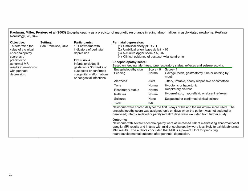
Kaufman, Miller, Ferriero et al (2003) Encephalopathy as a predictor of magnetic resonance imaging abnormalities in asphyxiated newborns. Pediatric Neurology, 28, 342-6.
Objective: To determine the value of a clinical encephalopathy score as a predictor of abnormal MRI results in newborns with perinatal depression.
Setting: San Francisco, USA
Participants: 101 newborns with indicators of perinatal depression Exclusions: Infants excluded if gestation < 36 weeks or suspected or confirmed congenital malformations or congenital infections.
Perinatal depression: (1) Umbilical artery pH < 7.1 (2) Umbilical artery base deficit > 10 (3) 5-minute Apgar score ≤ 5, OR (4) Clinical evidence of postasphyxial syndrome
Encephalopathy score: Based on feeding, alertness, tone respiratory status, reflexes and seizure activity. Encephalopathy sign Score= 0 Score= 1 Feeding Normal Gavage feeds, gastrostomy tube or nothing by
mouth Alertness Alert Jittery, irritable, poorly responsive or comatose Tone Normal Hypotonic or hypertonic Respiratory status Normal Respiratory distress
Reflexes Normal Hyperreflexic, hyporeflexic or absent reflexes
Seizures None Suspected or confirmed clinical seizure Total 0-6
Newborns were scored daily for the first 3 days of life and the maximum score used. The encephalopathy score was assigned only on days when the patient was not sedated or paralysed; infants sedated or paralysed all 3 days were excluded from further study.
Outcome: Newborns with severe encephalopathy were at increased risk of manifesting abnormal basal ganglia MRI results and infants with mild encephalopathy were less likely to exhibit abnormal MRI results. The authors concluded that MRI is a powerful tool for predicting neurodevelopmental outcome after perinatal depression.
49

Khong, Lam, Tung et al (2003) MRI of neonatal encephalopathy. Clinical Radiology, 58, 833-44.
Objective: To present a pictorial review of MRI in neonatal encephalopathy, with emphasis on advanced MRI techniques, such as diffusion-weighted imaging
Setting: Hong Kong
Diagnostic checklist for perinatal hypoxic ischaemic encephalopathy (HIE) related to intrapartum events: The diagnosis of HIE requires the presence of all three of the following criteria:
1. The presence of a clinically recognised encephalopathy within 72 hours of birth 2. Three or more supporting findings from the following list: (a) Arterial cord blood pH < 7.00 (b) Apgar score at 5 minutes of 5 or less (c) Evidence of multiorgan system dysfunction involving one or more of the following systems within 72 hours of birth: • Renal: oliguria or acute renal failure • Gastrointestinal: necrotising enterocolitis, hepatic dysfunction • Haematological: thrombocytopenia, disseminated intravascular coagulopathy • Endocrine: hypoglycaemia, hypocalcaemia, syndrome of inappropriate ADH secretion (SIADH) • Pulmonary: persistent pulmonary hypertension • Cardiac: myocardial dysfunction, tricuspid insufficiency • Evidence of foetal distress eg. persistent late decelerations, reversals of end-diastolic flow on Doppler flow studies of the umbilical artery
or a biophysical profile of 2 or less • Evidence on CT, MRI, Technetium or ultrasound brain scan (preferably performed within 7 days of birth) of diffuse or multifocal ischaemia
or of cerebral oedema. • Abnormal EEG: low amplitude and frequency, periodic, paroxysmal or isoelectric
3. The absence of an infectious cause, a congenital malformation of the brain or an inborn error of metabolism which could explain the encephalopathy
These criteria may not apply to premature newborns ≤ 34 weeks gestation.
Encephalopathy: The presence of at least three of the following findings:
• Abnormal level of consciousness: hyperalertness, lethargy, stupor or coma • Abnormal muscle tone: hypertonia, hypotonia or flaccidity • Abnormal deep tendon reflexes: increased, depressed or absent • Seizures: subtle, multifocal or focal clonic • Abnormal Moro reflex: exaggerated, incomplete or absent • Abnormal suck: weak or absent • Abnormal respiratory pattern: periodic, ataxic or apnoeic • Oculomotor or pupillary abnormalities: skew deviation, absent or reduced, doll’s eyes or fixed unreactive pupils
Outcomes: MRI can provide critical information on presence and extent of brain injury and is sometimes specific in the diagnosis of aetiology in neonatal encephalopathy.
50

Leung, Lam, Lam et al (2003) Unexpected reduction in the incidence of birth trauma and birth asphyxia related to instrumental deliveries during the study period: was this the Hawthorne effect? British Journal of Obstetrics & Gynaecology, 110, 319-22.
Objective: To identify the risk factors that could predict those difficult instrumental deliveries resulting in birth trauma and birth asphyxia.
Setting: China
Population: All singleton deliveries in cephalic presentation with an attempt of instrumental deliveries in a local teaching hospital from Mar 2000 to Mar 2001. There are around 4,000 births per year.
Participants: 670 deliveries
Severe birth asphyxia: ‘Cases with an Apgar score on the first minute of ≤ 3 requiring admission to neonatal intensive care unit.’
Outcome: There were only 4 cases of severe birth asphyxia or birth trauma in the study period. The authors concluded that the Hawthorne effect was a major contributing factor to the reduction in incidence of birth trauma and birth asphyxia related to instrumental deliveries in this study.
51

Noren, Amer-Wahlin, Hagberg et al (2003) Fetal electrocardiography in labor and neonatal outcome: data from the Swedish randomized controlled trial on intrapartum fetal monitoring. American Journal of Obstetrics & Gynecology, 188, 183-92.
Study design: Randomised controlled trial (RCT)
Objective: To evaluate the neonatal outcome in the Swedish RCT with a focus on newborn infants with complicated/ adverse neonatal outcome. Mothers were randomly allocated to monitoring with cardiotogram only or cardiotogram and ST analysis.
Setting: Southern Sweden Population: 4,966 term infants included in the RCT. Mother were in active labour with singleton fetuses in cephalic presentation at 36 weeks gestation. 2,447 infants had cardiotocogram, 2,519 infants had CTG+ST
Participants: 29 newborn infants with adverse/ complicated outcome (perinatal death, neonatal encephalopathy, metabolic acidosis)
Neonatal Encephalopathy (NE): Minor: Irritability and jitteriness Moderate: Profound lethargy or abnormal tone Severe: Coma or abnormal tone and seizures Low et al (1997) referenced for this definition. Moderate neonatal encephalopathy: Increased neuromuscular tone
Severe neonatal encephalopathy: Neonatal seizures/ death
Probable intrapartum asphyxia: • Perinatal death • Treatment in the special care baby unit for
encephalopathy AND/OR • Metabolic acidosis Cord artery metabolic acidosis: A pH of < 7.05 in combination with a base deficit in the extracellular fluid compartment of > 12.0 mmol/L, with the Siggaard-Andersen Acid Base Chart algorithm.
Incidence of moderate or severe encephalopathy: 3.3 per 1000 cases in the cardiotocogram group 0.4 per 1000 cases in the CTG+T group
Outcome: 11 infants had NE, 7 infants classified as moderate or severe.
Combined monitoring with cardiotogram and analysis of the ST waveform of the fetal electrocardiogram reduced the risk of fetuses being exposed to significant hypoxia that leads to metabolic acidosis at birth. That is, cardiotocography plus ST analysis provides accurate information about intrapartum hypoxia and may prevent intrapartum asphyxia and NE by giving a clear alert to staff members who are in charge. Combined cardiotocogram and ST changes are a more specific sign of hypoxia than cardiotocogram changes alone.
52

Sato, Okumura, Kato et al (2003) Hypoxic ischemic encephalopathy associated with neonatal seizures without other neurological abnormalities. Brain and Development, 25, 215-9.
Study design: Case reports
Objective: This paper described three case reports of infants with HIE
Setting: Japan Population: Infants admitted to one hospital from 1992 to 1999
Participants: 3 infants who had seizures and no other neurological abnormalities
Asphyxia: • 1 minute Apgar score < 6 AND/OR • the need for resuscitation in the delivery room Hypoxic ischemic encephalopathy: Inferred through: • Blood gas analysis indicating metabolic acidosis • Low Apgar score • Need for resuscitation immediately after birth • Meconium-stained amniotic fluid • EEG on the first day of life demonstrating moderate or severe
depression suggesting a suppressed brain function • Renal failure • Marked elevation of creatine kinase
Outcome: This report described three cases of infants with HIE suggesting that neonatal seizures can occur in infants with mild HIE without apparent neurological abnormalities and EEG is useful for the diagnosis of mild HIE.
Yu, Kui & Ming (2003) Effect of asphyxia on non-protein-bound iron and lipid peroxidation in newborn infants. Developmental Medicine & Child Neurology, 45, 24-7. Study design: Case control study
Objective: To investigate the effect of asphyxia on iron metabolism and lipid peroxidation in newborn infants.
Setting: China
Population: 45 term newborn infants born in three hospitals in China between Oct 1999 and Jan 2000.
Cases: 30 newborn infants with birth asphyxia (moderate and severe)
Controls: 15 healthy newborn infants
Birth asphyxia: Defined according to the diagnosis standard of asphyxiated newborn infants with hypoxic-ischemic encephalopathy of the Chinese Association of Pediatrics (1997). Not further specified. Included moderately asphyxiated infants without neurological impairments and severely asphyxiated newborn infants with neurological abnormalities.
Outcome: Asphyxia can affect iron metabolism leading to a significant increase in the detectable rate of non-protein-bound iron in plasma as well as in the concentration of lipid peroxidation in newborn infants with HIE.
53

American College of Obstetricians and Gynecologists (2002) Neonatal Encephalopathy and Cerebral Palsy: Defining the Pathogenesis and Pathophysiology. ACOG: Washington D.C.
Objective: The aim of this report was to collate and review the best scientific data available on Neonatal Encephalopathy and Cerebral Palsy. However, only a clear definition of acute hypoxic ischemic events was proposed (ie. They did not clearly define neonatal encephalopathy).
Setting: Report collated by the American College of Obstetrician and Gynaecologists
Neonatal encephalopathy: (references Sarnat and Volpe) A clinically defined syndrome of disturbed neurological function in the earliest days of life in the near-term and term infant. If an intrapartum insult is severe enough to result in ischemic cerebral injury, abnormalities will be noted in the neurological examination within 24 hours after birth. The examination is characterised by abnormalities in: (1) cortical function (lethargy, stupor, coma with or without seizures), (2) brainstem function (ie. Pupillary and cranial nerve abnormalities), (3) tone (hypotonia) and (4) reflexes (absent, hyporeflexia). Outcome is related to the maximum grade of severity. Separate criteria to define an acute intrapartum hypoxic event as sufficient to cause cerebral palsy. “Encephalopathy in the term or near-term newborn infant is manifested by serious neurologic depression in the delivery room and nursery. Clinical signs observable in the delivery room include low Apgar score and its components and correlates, such as hypotonia; depressed reflexes including cry, suck, absent Moro’s reflex; decreased consciousness; difficulty in initiating and maintaining respiration; poor colour; and bradycardia.” P. 53 Criteria to define an acute intrapartum hypoxic event as sufficient to cause cerebral palsy: Essential criteria (must meet all four): 1. Evidence of a metabolic acidosis in fetal umbilical cord arterial blood obtained at delivery (pH < 7 and base deficit ≥ 12 mmol/L) 2. Early onset of severe or moderate neonatal encephalopathy in infants born at 34 or more weeks of gestation 3. Cerebral palsy of the spastic quadriplegic or dyskinetic type* 4. Exclusion of other identifiable etiologies, such as trauma, coagulation disorders, infectious conditions, or genetic
disorders. Criteria that collectively suggest an intrapartum timing (within close proximity to labour and delivery, eg. 0-48 hours) but are non-specific to asphyxial insults: 1. A sentinel (signal) hypoxic event occurring immediately before or during labour 2. A sudden and sustained fetal bradycardia or the absence of fetal heart rate variability in the presence of persistent, late, or variable decelerations, usually after a hypoxic sentinel event when the pattern was previously normal 3. Apgar score of 0-3 beyond 5 minutes 4. Onset of multisystem involvement within 72 hours of birth 5. Early imaging study showing evidence of acute nonfocal cerebral abnormality. * Spastic quadriplegia, and less commonly, dyskinetic cerebral palsy are the only types of cerebral palsy associated with acute hypoxic intrapartum events. Spastic quadriplegia is not specific to intrapartum hypoxia. Hemiparetic cerebral palsy, hemiplegic cerebral palsy, spastic diplegia, and ataxia are unlikely to result from acute intrapartum hypoxia.
54

Barnett, Mercuri, Rutherford et al (2002) Neurological and perceptual-motor outcome at 5 - 6 years of age in children with neonatal encephalopathy: relationship with neonatal brain MRI. Neuropediatrics, 33, 242-8.
Study design: Cohort study
Objective: 1. To determine the incidence of neurological dysfunction and perceptual-motor difficulties in children aged 5½ -6½ who had been born full-term but presented with neonatal encephalopathy and low Apgar scores and 2. to examine relationships between the presence/absence of these difficulties with neonatal brain MRI.
Setting: London, UK
Population: Infants born at or referred to the Hammersmith Hospital for MRI between May 1991 and Jan 1996.
Participants: 68 full-term infants (38 weeks or more) with NE and who had at least one brain MRI performed in the neonatal period between 1 to 4 weeks from delivery and detailed neurodevelopmental follow-up when at least 2 years of age.
Neonatal Encephalopathy: Infants who had convulsions in the first 48 hours and/or showed other signs of neurological abnormalities during the first 48 hours after delivery. Neurological abnormalities included abnormal tone, poor feeding, and altered level of consciousness.
Severity: Classified during the first week of life as mild, moderate or severe (stages, I, II or III) according to Sarnat & Sarnat.
Only Apgar scores at one minute were used as a proportion of infants with NE are often intubated before 5 minutes making interpretation of the 5 minute Apgar score difficult.
Outcome: Surviving infants assessed using the Touwen Examination, the Movement ABC and the WPPSI-R.
Exclusion criteria: Infants subsequently diagnosed as suffering from genetic or metabolic syndromes or who presented with other neonatal complications, such as septicaemia or neonatal meningitis; infants with dysmorphic features or other clinical or brain MRI findings suggesting major congenital malformation.
Outcome: Neonatal deaths = 22% (15 infants) Cerebral palsy = 36% (19/53) Normal at 2 years = 34 infants but when assessed at school age 15% had minor neurological dysfunction and/or perceptual-motor difficulties, 2% had cognitive impairment 47% were normal. 80% of with minor neurological dysfunction or perceptual-motor difficulties had mild or moderate basal ganglia or more marked white matter lesions.
55

Draper, Kurinczuk, Lamming et al (2002) A confidential enquiry into cases of neonatal encephalopathy. Archives of Disease in Childhood Fetal Neonatal Edition, 87, F176-80.
Study design: Multidisciplinary case reviews (reviewing quality of care) Objective: To assess the quality of care and timing of possible asphyxial events for infants with neonatal encephalopathy, to compare the quality of care findings with those relating to the deaths from the Confidential Enquiry into Stillbirths & Deaths in Infancy (CESDI), and to assess whether the confidential enquiry method is a useful clinical governance tool for investigating morbidity.
Setting: Trent, United Kingdom Population: All infants in Trent presenting with grade II or grade III neonatal encephalopathy in 1997 who were admitted for neonatal care and were included in the Trent Neonatal Survey
Participants: 49 infants (survivors) with grade II or grade III neonatal encephalopathy
Grades II and III neonatal encephalopathy (modified from Sarnat and Sarnat): Infants that fulfilled criteria 1 and 2 plus 3 or 4:
(1) ≥ 35 weeks gestation at delivery and had no major congenital anomaly, infection or inborn error of metabolism evident during the period of neonatal care; AND
(2) survival for more than 28 days; PLUS presentation in the first 48 hours after birth with: (3) Neurological disturbance causing as a minimum
treated “fits” (grade II); OR (4) Neurological disturbance so severe that the baby
required ventilatory support (grade III) Exclusion criteria: Cases with congenital malformation, inborn error of metabolism, or infection. The timing of any apparent asphyxial episode was ascribed to the appropriate period using the answers to the following questions:
(i) Is there evidence of a peripartum insult? (ii) Does the peripartum insult alone account for the
clinical condition of the baby? (iii) Is there evidence of an antepartum insult?
Birth Prevalence: There was a birth prevalence (of grades II and III neonatal encephalopathy) of 0.82 per 1000 total live births.
Outcome: Grade II = 34 (69%) Grade III = 15 (31%)
Significant or major episodes of suboptimal care were identified for 64% of the encephalopathy cases and 75% of the deaths. Over 90% of the episodes involved the care provided by health professionals. ~90% of encephalopathy cases had evidence of a peripartum insult although the insult alone was thought likely to account for the clinical condition in only 45% of the cases. Therefore ‘better’ care may reduce the incidence of neonatal encephalopathy by half.
56

Jongeling, Badawi, Kurinczuk et al (2002) Cranial ultrasound as a predictor of outcome in term newborn encephalopathy. Pediatric Neurology, 26, 37-42.
Study design: Case control study Aim: To determine the prognostic value of ultrasound and Doppler performed in the first 72 hours of life in predicting poor outcome in a cohort of term infants with moderate or severe newborn encephalopathy
Note: This is a subanalysis from the Badawi et al (1998) study
Setting: Perth, Western Australia Population: Term infants born in metropolitan Perth, Western Australia from June 1993-Dec 1995. 276 infants born with moderate or severe newborn encephalopathy.
Participants: 212 term infants ≥ 37 weeks gestation with moderate or severe neonatal encephalopathy. Infants were followed-up to 3 years of age.
Moderate or severe newborn encephalopathy: Present in the first week of life: Either seizures alone OR Any of two of the following lasting longer than 24 hours: • Abnormal consciousness • Difficulty maintaining respiration (of presumed central
origin) • Difficulty feeding (of presumed central origin) • Abnormal tone and reflexes
Severity graded according to modified Sarnat & Sarnat criteria.
Severe encephalopathy: Fulfilled one or more of the following criteria: • Ventilated for more than 24 hours • Treated with 2 or more antiepileptic drugs • Comatose or stuporous OR • Died in the neonatal period The remaining patients were defined as moderate.
Intrapartum hypoxia: Presence of an abnormal intrapartum cardiotocograph or abnormal fetal heart rate on auscultation or fresh meconium in labour together with a 1 minute Apgar score of < 3 and a 5 minute Apgar score of < 7. Other patients who did not strictly fulfil the definition but manifested evidence of a significant intrapartum event were assumed to have manifested intrapartum hypoxia. Note: cord pH measurements were not included as they were performed infrequently.
Adverse outcome: Defined as cerebral palsy or death.
Outcome: 22% of infants had an adverse outcome, 10% were diagnosed with cerebral palsy and 14% died. 29% had evidence of possible intrapartum hypoxia. Infants with newborn encephalopathy who have an abnormally low resistive index identified in normal clinical practice are more likely to die or develop cerebral palsy than infants with a normal value. The presence of low resistive index, severe cerebral edema and severe encephalopathy had a positive predictive value of 80%. Resistive index results cannot be used in isolation (positive predictive value of 71%) although they may have a place in combination with other factors in the counselling of parents and clinical management of patients.
57

Robertson, Cowan, Cox & Edwards (2002) Brain alkaline intracellular pH after neonatal encephalopathy. Annals of Neurology, 52, 732-42.
Objective: To assess: 1. Brain pH during the first 2 weeks after birth in infants categorised according to MRI during the first 2 weeks after birth and at > 3 months of age and neurodevelopmental outcome at one year 2. The relationship between brain pH and lactate/creatine 3. Duration of alkaline brain pH
Setting: London, United Kingdom
Population: Hammersmith hospital, population not specified.
Participants: 78 term infants (≥ 36 weeks gestation) with neonatal encephalopathy
Neonatal encephalopathy: One or more of the following: • Altered conscious state • Abnormal tone and reflexes • Seizures • Poor feeding Severity graded using Sarnat & Sarnat (stage I to III). Not further specified. Exclusion criteria: Major congenital malformation or known infective diseases.
Outcome: Brain pH manipulation may offer an opportunity for neuroprotective intervention over a prolonged period after perinatal hypoxia-ischemia.
Scher, Steppe, Beggarly et al (2002) Neonatal EEG-sleep disruption mimicking hypoxic-ischemic encephalopathy after intrapartum asphyxia. Sleep Medicine, 3, 411-5.
Study design: Case control
Objective: To compare EEG-sleep organisation of asphyxiated and non-asphyxiated full-term neonates during the first 3 days after birth. To demonstrate that alterations in EEG-sleep organisation can occur after birth in neonates with moderate to severe intrapartum asphyxia, despite the absence of classical signs of hypoxic-ischemic encephalopathy and age-appropriate neurodevelopment.
Setting: Pittsburg, USA
Population: Full-term appropriate for gestational age neonates diagnosed with moderate to severe asphyxia and clinical depression at delivery at the Magee-Women’s hospital and full-term neonates without asphyxia.
Participants Cases: 10 full-term infants diagnosed with moderate to severe asphyxia and clinical depression.
Controls: 23 full-term neonates without asphyxia randomly selected from a prospective study of sleep organisation in asymptomatic full-term neonates.
Asphyxia: • Initial arterial blood gas documented a pH
< 7.2, with a base excess ≤ 10 AND • 5 minute Apgar score of ≤ 5.
Outcome: EEG sleep studies can assist in a more accurate classification of newborn encephalopathy that does not satisfy criteria for hypoxic-ischemic encephalopathy.
58

Shankaran, Laptook, Wright et al (2002) Whole-body hypothermia for neonatal encephalopathy: animal observations as a basis for a randomized, controlled pilot study in term infants. Pediatrics, 110, 377-85.
Study design: Pilot study, randomised trial
Objective: (1) To evaluate in newborn animals a commercially available cooling system to control brain temperature during whole-body hypothermia and (2) To use the results of the animal experiments to perform a pilot study evaluating the feasibility of whole-body hypothermia as a neuroprotective therapy for newborns with encephalopathy at birth.
Setting: Not specified.
Population: All term infants ≥ 36 weeks gestation, admitted to the neonatal ICU at ≤ 6 hours of age with a diagnosis of neonatal depression, acute perinatal asphyxia, or encephalopathy, from Nov 1999 to June 2000.
Participants: 78 infants screened, 20 eligible
Hypothermia group: 9 infants underwent whole body cooling for 72 hours using a blanket to 34.5ºC.
Normothermia group: 10 infants cared for under an overhead radiant warmer.
Acute perinatal event: Late or variable decelerations resulting in an emergent caesarean delivery, cord prolapse, placental abruption, uterine rupture, maternal trauma, or maternal respiratory arrest.
Moderate or severe encephalopathy: One or more signs were present in 3 of the following 6 categories: 1. Level of consciousness: lethargy (moderate), stupor or coma
(severe) 2. Spontaneous activity: decreased (moderate), absent (severe) 3. Posture: distal flexion (moderate), decerebrate (severe) 4. Tone: hypotonia (moderate), flaccid (severe) 5. Primitive reflexes: Suck weak (moderate), absent (severe) or
Moro, incomplete (moderate), absent (severe) 6. Autonomic nervous system: pupils constricted (moderate),
skew deviation or nonreactive to light (severe), heart rate, bradycardia (moderate), variable heart rate (severe), or respiration, periodic breathing (moderate), apnea (severe). Variable heart rate was defined as rate fluctuating from ≤ 80 to ≤ 100 beats per minute and ventilatory support was categorised as apnea.
Exclusion criteria: Inability to perform random assignment by 6 hours of age, chromosomal abnormality, major congenital anomaly, severe growth restriction (≤ 1800g birth weight), infant unlikely to survive, refusal of consent.
Outcome: This study indicated that this method of whole body hypothermia is feasible at <6 hours of age and provided no evidence that hypothermia involved greater hazard than benefit.
59

Dalman, Thomas, David et al (2001) Signs of asphyxia at birth and risk of schizophrenia. Population-based case-control study. British Journal of Psychiatry, 179, 403-8.
Aim: To assess the role of different complications, in particular to distinguish between disordered fetal development and hypoxia at birth.
Setting: Stockholm, Sweden
Population: Cases and controls identified from the Stockholm county in-patient register and community registers. 1,567 birth records were examined.
Participants Cases aged 9-24 years
Cases: 524 cases of schizophrenia
Controls: 1,043 matched for age, gender, hospital and parish of birth. These were the next 2 births in time of the same gender and born in the same hospital as the case. 50 cases had an Apgar score < 7.
Signs of asphyxia: (referenced Sykes 1982 and Silverman et al 1985)
Apgar score < 7 at one, five or ten minutes.
Infants without an Apgar score: ‘Assessed by the midwife by a protocol in which the 5 Apgar items heart rate, breathing, colour, tone and excitability of the infant were defined.’ Note: only 20.5% of records in the study had an Apgar score.
Outcome: 3.1% (50/1567) infants had an Apgar score <7. There was a strong association between signs of asphyxia at birth and schizophrenia (OR 4.4, 95% CI 1.9-10.3) The authors concluded that signs of asphyxia at birth are associated with an increased risk of schizophrenia in adults.
60

Ellis, Shrestha, Shrestha et al (2001) Clinical predictors of outcome following mild and moderate neonatal encephalopathy in term newborns in Kathmandu, Nepal. Acta Paediatrica, 90, 316-22. Study design: Population-based cohort study
Objectives: To describe a clinical grading system for assessment of neonatal encephalopathy developed for a large prospective study in Kathmandu
Setting: Kathmandu, Nepal
Population: 21,609 live births in Kathmandu. This included 131 term infants with encephalopathy.
Participants: 57 infants with encephalopathy
Neonatal encephalopathy: “An abnormal neurobehavioural state commencing within the first 24h of life consisting of an altered conscious level with abnormalities of tone and/or sucking behaviour.”
Operational definitions for the clinical grading of neonatal encephalopathy: Clinical variable Grade 1 (mild) Grade 2 (moderate) Grade 3 (severe) Conscious level Irritable/hyperalert Lethargic Comatose Tone Either a mildly abnormal
(hypo/hyper) Moderately abnormal (hypotonic or dissociated)
Severely abnormal (hypotonia)
Suck or b abnormal Poor Absent Primitive reflexes1 Exaggerated Depressed Absent Brainstem reflexes2 Normal Normal Impaired Seizures Absent Present Present Respiration Tachypnoea Occasional apnoeas Severe apnoea
The infant is examined daily and graded according to the clinical features described. The features in bold type must be as described to meet the minimum requirements for each grade. a,b abnormality of tone or suck should accompany altered conscious level to assign grade 1. Features not in bold may be present but are not required to make the grade assignation. The overall description of the infant refers to the most severe grade observed during the neonatal period. 1 Primitive reflexes: moro, grasp, 2 Brainstem reflexes: gag, corneal
Modified Cape Town encephalopathy scoring criteria (total possible score 22, high risk >15): proposed modifications (total possible score 18, high risk >6). Sign Score 0 Score 1 Score 2 Score 3 Proposed modifications Tone Normal Hyper Hypo Flaccid Dissociated tone (eg axial
hypotonia with limb hypertonia) = 2
LOC Normal Hyperalert, staring Lethargic Comatose Irritable = 1 Fits None Infrequent <3/d Frequent >2/d Score fits as absent = 0,
present = 1 Posture Normal Fisting, cycling Strong, distal flexion Decerebrate Omit posture variable Moro Normal Partial Absent Grasp Normal Poor Absent Suck Normal Poor Absent/bites Respiration Normal Hyperventilation Apnoeas IPPV In the absence of IPPV all
respiratory depression = 2 Fontanelle Normal Full Tense
LOC level of consciousness; IPPV intermittent positive pressure ventilation
61

The grading system was compared with Levene’s modification of the system developed by Fenichel.
Outcome: Mild encephalopathy = 28 infants. Of these 2 (7%) developed major impairment. Moderate/severe encephalopathy = 29/57 infants. Of these 55% developed major impairment. Only one infant with severe encephalopathy survived. Major impairment = 18 infants (14 (78%) spastic tetraplegic cerebral palsy, 8 (44%) multiply impaired). It seems to make little difference in practical or predictive terms whether one describes the neurological condition of the neonates using a descriptive or scoring system. “There is as yet no widely agreed definition of neonatal encephalopathy” (page 320).
62

Impey, Greenwood, Sheil et al (2001) The relation between pre-eclampsia at term and neonatal encephalopathy. Archives of Disease in Childhood Fetal Neonatal Edition, 85, F170-2.
Study design: Cohort study
Objective: To determine whether pre-eclampsia, hypothesised to be an inflammatory condition, is associated with fever in term labour, confirm and examine the reported association of pre-eclampsia at term with neonatal encephalopathy.
Setting: Dublin, Ireland
Population: 6,163 women in labour with singleton pregnancies at term at low risk for intrapartum hypoxia recruited to a randomised controlled trial examining the effect of admission cardiotocography on neonatal outcome, from Aug 1997 to April 2000.
Participants: 6,163 women with singleton pregnancies
Neonatal Encephalopathy: As described by Sarnat and Sarnat (grade 2-3 only). Not further specified. Eligibility: Women were eligible for participation if clear liquor was detected on early amniotomy and the fetus was not considered at risk for intrapartum fetal distress eg. known intrauterine growth restriction, antepartum haemorrhage. Exclusion criteria: Breech presentation, delivery before 37 weeks or after 42 weeks gestation, anomalous or inborn errors of metabolism.
Outcome: 18 (0.3%) of the infants had neonatal encephalopathy. The authors concluded that pre-eclampsia is an independent risk factor for maternal fever in labour.
Impey, Greenwood, MacQuillan, Reynolds & Sheil (2001) Fever in labour and neonatal encephalopathy: a prospective cohort study. British Journal of Obstetrics and Gynaecology, 108, 594-7.
Study design: Cohort study
Aim: To determine whether the reported association of maternal fever with neonatal encephalopathy is independent of other associated intrapartum risk factors.
Setting: Dublin, Ireland
Population: 4,915 low risk women in labour at 36-41 weeks of gestation from Aug 1997 to Aug 1999.
Participants: 4,915 women (‘all afebrile at diagnosis of labour’), including 336 that developed intrapartum fever and 16 newborn infants that developed encephalopathy
Encephalopathy: • Presence of focal or generalised seizures in the
first week of life • Apgar score of < 7 at 5 minutes • Admission to the neonatal unit
AND • Umbilical cord arterial measurement of pH and
base excess > 2 SDs below the mean (arterial pH < 7.05 and base deficit > 14.4 mmol/L).
Exclusion criteria: Gestation < 36 weeks or ≥ 42 weeks, breech presentation.
Outcome: The authors concluded that maternal fever in labour is strongly (and independently) associated with neonatal encephalopathy (OR 4.72, CI 1.28-17.4) and is a better indicator of a fetus at risk than an abnormal cardiotograph.
63

Robertson, Lewis, Cowan et al (2001) Early increases in brain myo-inositol measured by proton magnetic resonance spectroscopy in term infants with neonatal encephalopathy. Pediatric Research, 50, 692-700.
Aim: To assess brain myo-inositol/ creatine plus phosphocreatine (Cr) in the first week in term infants with neonatal encephalopathy using localised short echo time proton MRI and to relate these to measures of brain injury. Outcome was also recorded at one year of age.
Setting: London, UK
Population: Infants included in a pilot study of treatment with moderate whole-body hypothermia for neonatal encephalopathy.
Participants: 14 term infants with neonatal encephalopathy, up to one week of age.
Inclusion criteria: At least one of the following: • History of fetal distress • Early abnormal postnatal neurological signs
consistent with neonatal encephalopathy
Fetal distress: • Late decelerations on cardiotocography • Meconium-stained liquor • Acidemia (pH < 7.1 or base deficit > 12mM) in umbilical
cord blood or arterial blood within 30 mins of birth)
Neonatal Encephalopathy: Not specified
Outcome: Myo-inositol/creatine increased in the basal ganglia in the first week of birth in infants with neonatal encephalopathy who sustained a severe hypoxic ischemic brain injury and these infants had a poor neurodevelopmental outcome at one year of age.
64

Asakura, Ichikawa, Nakabayashi et al (2000) Perinatal risk factors related to neurologic outcomes of term newborns with asphyxia at birth: a prospective study. Journal of Obstetrics and Gynaecology Research, 26, 313-24.
Objective: To study the incidence of poor neurological outcomes in term newborns who suffered severe asphyxia at birth. A 1 year prospective follow-up of neurological outcomes was completed.
3 year survey
Setting: Japan
Population: 68,327 newborns admitted to 270 participating institutions. Infants included in the study had low Apgar scores at 1 and 5 minutes. A one-year survey of outcomes concluded between April 1996 to March 1998
Participants: 152 term (gestational age not specified) newborns with neonatal asphyxia whose Apgar scores were ≤ 4 at 1 minute or ≤ 6 at 5 mins and whose condition required admission to the neonatal intensive care unit.
This study was completed via questionnaires to participating institutions. The authors only made a judgement of fetal distress based on records of fetal heart rate pattern.
Fetal distress: Findings indicating fetal distress:
• Repeat severe variable decelerations • Late decelerations • Persistent loss of baseline FHR variability, & • Prolonged deceleration
Neonatal asphyxia: not defined
Exclusion criteria: Congenital abnormalities including chromosomal abnormalities. Neonatal deaths were included.
Not only newborns with CP but those with epilepsy, disturbance of consciousness, dysmyotonia (hypertonic or hypotonic), or abnormal nervous reflex were defined as newborns with neurological sequelae.
Incidence of poor outcome of surveyed cases: 0.3 per 1000 deliveries
Outcome: Poor outcome was reported in 13.8% of newborns (22 cases). The authors state that the incidence of poor neurological outcome was ‘very high’ among term infants with low Apgar scores. These infants were 10 to 20 times more likely to die or to survive with permanent disabilities than infants with higher Apgar scores.
65

Azzopardi, Robertson, Cowan et al (2000) Pilot study of treatment with whole body hypothermia for neonatal encephalopathy. Pediatrics, 106, 684-94.
Study design: Pilot study of the treatment method - whole body hypothermia Objective: To determine the feasibility of hypothermic therapy for infants with a bad neurological prognosis within 6 hours of birth, and the effect of this therapy on physiological variables.
Setting: London, United Kingdom Population: Infants suspected of having birth asphyxia (inborn and outborn), admitted to the neonatal units at the Hammersmith Hospital & Queen Charlotte and Chelsea Hospital. Infants enrolled over an 18 month period.
Participants: 16 newborn infants with clinical features of birth asphyxia.
Birth asphyxia: Evidence of fetal distress from fetal heart rate monitoring and/or the infant had a metabolic acidosis from birth with a blood pH < 7.0 and/or a base excess >-14 mmoL and artificial ventilation was needed from birth. Infants who had not apparently recovered after resuscitation and who displayed excessive irritability or neurological depression were then assessed by aEEG. Hypothermia was initiated within 6 hours of birth. Parental consent was required for participation.
Outcome: The therapy was found to be feasible and physiological changes attributable to hypothermia were ‘generally mild.’
Randomised controlled studies on the safety and efficacy of mild hypothermia were recommended.
Cowan (2000) Outcome after intrapartum asphyxia in term infants. Seminars in Neonatology, 2000, 5, 127-40. Study design: Review article Objective: Not specified
Intrapartum asphyxia (based on Yudkin et al 1994, and MacLennan 1999): • Metabolic acidosis in cord blood or very early arterial blood samples, pH < 7.00, base deficit >12mmol/L • Meconium stained liquor – old or fresh especially before term • Sudden change in fetal heart rate from a previously normal pattern to any of the following: reduced heart rate variability, increased
tachycardia, persistent or late decelerations, bradycardia • Apgar scores of ≤ 6 at 5 minutes • An acute event (placental abruption, cord prolapse, uterine rupture, shoulder dystocia, amniotic fluid embolus, maternal eclampsia) Evidence for early encephalopathy due to acute asphyxia: • Early onset of seizures • Brain swelling • Abnormal consciousness • Respiratory difficulty of apparent central origin • Poor feeding • Abnormal tone and reflexes
Multisystem involvement eg. renal impairment, liver dysfunction, coagulopathy cardiac failure/hypotension, persistent fetal circulation.
66

Ellis, Manandhar, Manandhar & Costello (2000) Risk factors for neonatal encephalopathy in Kathmandu, Nepal, a developing country: unmatched case control study. British Medical Journal, 320, 1229-36. Study design: Case control study (unmatched) Objective: To determine risk factors for neonatal encephalopathy among term infants in a developing country.
Setting: Kathmandu, Nepal Population: Infants > 37 weeks gestation with evidence of neonatal encephalopathy at 6 to 24 hours after birth. From a population of 21,609 infants born from Jan 1995 to July 1996.
Cases: 131 infants with neonatal encephalopathy Controls: 635 infants without encephalopathy. Every 25th infant born in the hospital was examined.
Neonatal encephalopathy: An abnormal neurobehavioural state commencing within the first 24 hours of life, which consists of an altered conscious level with abnormalities of neuromuscular tone or sucking behaviour. In addition there may be seizures or abnormalities of respiratory control, primitive reflexes and brainstem reflexes.
Severity: Severe (Grade III):
• comatose • severely abnormal tone • absent suck • brain stem malfunction including impaired respiratory
drive Moderate (Grade II):
• lethargic WITH
• moderately abnormal tone • poor suck • depressed Moro and grasp reflexes • seizures often clinically evident
Mild (Grade I): • Irritable or hyperalert
WITH • either mildly abnormal tone (hypo/hyper)
OR • abnormal suck
Exclusion criteria (for the infant): Preterm infants (< 37 weeks), severely dysmorphic, at least one congenital abnormality, hepatosplenomegaly, cataracts, thrombocytopenia indicative of intrauterine infection, microbiological evidence of early neonatal infection and normalised neurological condition when hypoglycaemia was corrected.
Birth prevalence: Neonatal encephalopathy = 6.1 per 1000 live births. Outcome: Moderate or severe encephalopathy in 63%, seizures in 44%. Case fatality 30.5%. Persisting clinical abnormalities of tone, suck or consciousness in 56% of survivors. 60% of encephalopathic infants had evidence of possible intrapartum hypoxia. Both antepartum and intrapartum factors are important in causation of neonatal encephalopathy in developing countries and there are likely to be continuing gains from improvements to intrapartum care.
67

Smith, Wells & Dodd (2000) The continuing fall in incidence of hypoxic-ischaemic encephalopathy in term infants. British Journal of Obstetrics and Gynaecology, 107, 461-6
Objective: To examine trends in incidence of moderate and severe hypoxic-ischaemic encephalopathy in term infants over a 20 year period in a neonatal unit. A comparison of data over 3 five-year periods was completed (1976-1980, 1984-1988, 1992-1996). This is associated with Hull (1998) when a 10 year study was completed.
Setting: Derby, UK Population: Term infants admitted to the same neonatal unit at the Derby City General Hospital from 1992-1996. Number of births: 1992-96 = 24,804 1984-88 = 24,265 1976-80 = 24,824
Participants: All term infants (> 37 weeks gestation) with hypoxic-ischaemic encephalopathy Total number affected infants: 1992-96 = 48 1984-88 = 112 1976-80 = 189 Moderate to severe HIE: 1992-96 = 29 infants 1984-88 = 44 infants 1976-80= 63 infants
Hypoxic-ischaemic encephalopathy: As defined by Levene et al (1985)
Grading: Grade 1 (Mild): • Irritability • Hyperalert • Mild hypotonia • Poor sucking
Grade 2 (Moderate): • Lethargic • Seizures • Marked abnormality of tone • Required tube feeding
Grade 3 (Severe): • Comatose • Prolonged seizures • Severe hypotonia • Failure to maintain spontaneous
respiration
Incidence: HIE in term infants: 1992-96: 1.9 per 1000 live births 1984-88: 4.6 per 1000 live births 1976-80: 7.7 per 1000 live births
Stillbirths: 1992-96 = 4.2 per 1000 19804-88 = 5.7 per 1000 1976-80 = 11.3 per 1000
Outcome: The incidence of stillbirths had declined between study periods. Rates of caesarean section had almost doubled, however infants with moderate or severe encephalopathy were less likely to have been born by emergency caesarean section or instrumental delivery. The study indicated a continuing fall in incidence of HIE in term infants. This fall could be due to improved socioeconomic factors, better antenatal care and other demographic factors together with changes in the organisation of services and in obstetric, paediatric and anaesthetic practice.
68

Al Naqeeb, Edwards, Cowan & Azzopardi (1999) Assessment of neonatal encephalopathy by amplitude-integrated electroencephalography. Pediatrics, 103, 1263-71.
Study design: Case control study Objective: To define normal and abnormal patterns, test inter-observer variability, and the prognostic accuracy of amplitude-integrated electroencephalography (aEEG) in nearly full-term and term infants soon after the onset of neonatal encephalopathy.
Setting: London, United Kingdom Population: All infants with encephalopathy admitted to neonatal units at the Hammersmith, Queen Charlotte’s and Chelsea Hospitals from Jan 1995 to Dec 1996 and a control group of newborn infants. The authors included full or near-full term infants from 35 weeks. One infant was 35 weeks and one was 36 weeks, the remainder were 37 weeks+
Participants Cases: 56 term infants (gestation 35-42 weeks) who presented with acute encephalopathy Controls: 14 healthy term newborn infants. The population from which these controls were sourced was not specified.
Encephalopathy (neonatal): Rapid appearance of abnormal neurologic signs, defined as seizures or lethargy or irritability, together with abnormal tone or reflexes.
Birth Asphyxia (this subset was further specified): Encephalopathy developed within 24 hours of birth, evidence of fetal distress (meconium staining of liquor or abnormal cardiotocograph) and at least one of the following: • Apgar score <6 at 5 minutes, or • pH <7 or base deficit greater than -15 mmol/L on cord
blood or a blood sample obtained within 60 minutes of birth.
‘Sarnat encephalopathy’ grades I, II and III (this subset was further specified but the case definition was not outlined)
aEEG traces obtained within 12 hours of birth.
Neurologic outcome: Assessed at 18 and 34 months by neurologic examination and estimation of the Griffith’s General Quotients and optimality score. Classified as ‘normal’ or ‘abnormal’ (further described in article).
Exclusion criteria: Infants without defined characteristics of intrapartum hypoxia-ischemia.
Outcome: The aEEG is a simple but accurate and reproducible clinical tool that could be useful in the assessment of infants with encephalopathy. Sarnat grading (number of infants): Grade I = 14 Grade II = 17 Grade III = 7 Not assessable in 2 infants.
69

Huang, Wang, Chang, Lin & Wu (1999) Measurement of the urinary lactate: Creatinine ratio for the early identification of newborn infants at risk for hypoxic-ischemic encephalopathy. New England Journal of Medicine, 341, 328-35.
Objective: To determine the sensitivity and specificity of the ratio of urinary lactate to creatinine for the early identification of infants in whom hypoxic-ischemic encephalopathy is likely to develop.
Setting: Tainan, Taiwan
Population: Infants were at least 36 weeks gestation, and born in the participating hospitals from June 1996 to Oct 1997.
Participants: Infants examined daily during the first week after birth
Cases: 40 newborn infants with perinatal asphyxia
Controls: 58 normal full-term newborns with no maternal illness, normal results on fetal monitoring, Apgar score of at least 8 at one and 5 minutes, and a normal course during the first week of life.
Perinatal Asphyxia (multiple references): The presence of at least 3 of the following conditions: • Intrapartum distress, as indicated by fetal bradycardia with a
heart rate of < 100 beats/minute, late decelerations, or an absence of heart-rate variability
• Thick meconium-stained amniotic fluid • Apgar score < 6 at 5 minutes • Need for resuscitation for > 1 minute with positive-pressure
ventilation and oxygen immediately after birth • Arterial-blood pH value of 7.20 or less or a base deficit of at
least 14 mmol/l within the first hour after birth. Hypoxic Ischemic Encephalopathy: Graded as mild, moderate or severe on the basis of the staging system by Sarnat & Sarnat.
Mild: Hyperexcitability or hypotonia persisted without seizures for at least 72 hours after birth
Moderate: Lethargic and hypotonia, weak primitive reflexes, and seizures
Severe: Frequent seizures, apnea, flaccid weakness, or coma Exclusion criteria: Maternal drug addiction, congenital infections, or perinatal infections, including chorioamniotitis. Outcome: Neurodevelopmental examination completed at one year for ‘surviving’ cases and 52 controls. Neuromotor assessment (based on Amiel-Tison and Grenier’s neurological examination) and the Bayley Scales of Infant Development II were used. Adverse outcome defined as impairment resulting in death, severe cerebral palsy (hemiplegia, quadriplegia, diplegia or severely impaired function associated with hypertonicity), developmental delay, blindness or deafness.
Outcome: The authors concluded that measurement of the urinary lactate: creatinine ratio soon after birth may help identify infants at high risk of hypoxic-ischaemic encephalopathy and may be useful in identifying infants most likely to benefit from intervention.
75% (30/40) of newborns with perinatal asphyxia did not have adverse neurological outcome at one year. Apgar scores, arterial blood pH and base deficits could not be used to predict development of HIE.
Urinary lactate:creatinine ratio within 6 hours of birth in infants with perinatal asphyxia can be used to identify infants who will develop HIE.
70

MacLennan (1999) A template for defining a causal relation between acute intrapartum events and cerebral palsy: International consensus statement. British Medical Journal, 319, 1054-9.
Consensus statement for the International Cerebral Palsy Task Force
Acute Intrapartum Hypoxic Event: Essential Criteria:
1. Evidence of a metabolic acidosis in intrapartum fetal, umbilical arterial cord, or very early neonatal blood samples (pH < 7.00 and base deficit ≥ 12 mmol/l)
2. Early onset of severe or moderate neonatal encephalopathy in infants of ≥ 34 weeks’ gestation 3. Cerebral palsy of the spastic quadriplegic or dyskinetic type
Criteria that together suggest an intrapartum timing but by themselves are non-specific:
4. A sentinel (signal) hypoxic event occurring immediately before or during labour 5. A sudden, rapid, and sustained deterioration of the fetal heart rate pattern usually after the hypoxic sentinel event where the
pattern was previously normal 6. Apgar scores of 0-6 for longer than 5 minutes 7. Early evidence of multisystem involvement 8. Early imaging evidence of acute cerebral abnormality
Pavlakis, Kingsley, Harper et al (1999) Correlation of basal ganglia magnetic resonance spectroscopy with Apgar score in perinatal asphyxia. Archives of Neurology, 56, 1476-81. Study design: Case series?
Objective: To determine which brain metabolite ratios have the strongest correlation with the Apgar scores in infants with possible asphyxia; whether the correlation is stronger with basal ganglia or anterior border-zone metabolites, and whether a combination approach using routine MRI, diffusion-weighted MRI and MRS can be used to evaluate the severity of neonatal asphyxia.
Setting: New York, USA
Population: Not specified
Participants: 20 infants (37 to 42 weeks gestation) with perinatal asphyxia, ie. 1 minute Apgar scores ≤ 6. Note: No comparison group.
Perinatal asphyxia: 1 minute Apgar score ≤ 6. Exclusion criteria: Known metabolic disease or brain structure abnormalities.
Outcome: 10 of the infants had acidosis (pH <7.2) and/or a perinatal event such as abruption placentae, pre-eclampsia, or fetal bradycardia. If the Apgar score indicates the severity of asphyxia or proximity of asphyxia to the delivery the results suggest that there is a correlation between brain metabolite levels and asphyxia. The 1 minute Apgar score may represent the best degree of asphyxia. The brain metabolite levels show no correlation with 10-minute Apgar scores.
71

Toh & Rajadurai (1999) Term infants with hypoxic ischaemic encephalopathy: poor neurodevelopmental outcome despite standard neonatal intensive care. Journal of Tropical Pediatrics, 45, 229-32.
Objective: To evaluate the aetiology, severity and outcome of term babies who sustained HIE following perinatal insult and to assess the factors which predict immediate and long-term neurological outcome.
Setting: Singapore
Population: Term infants (>37 weeks) with HIE admitted to the Kandang Kerbau Women’s and Children’s Hospital.
Participants: 23 term infants with HIE Note: There was no control group
Inclusion criteria: • Infants with a gestational age >37 weeks • Depressed 5 min Apgar score ≤ 5 • Those needing immediate resuscitation including
intubation or bag and mask ventilation • Those with evidence of cerebral dysfunction like
alterations of the level of consciousness or muscle tone and the presence of abnormal primitive reflexes.
Hypoxic ischaemic encephalopathy: Severity classification based on the Sarnat & Sarnat classification. This was the most severe stage seen in the first week of life.
Outcomes: Death or major neurological disability (cerebral palsy, developmental retardation, convulsive disorder, deafness or blindness) between 12 to 18 months.
Exclusion criteria: Infants who had congenital malformations and CNS or chromosomal abnormality.
Outcome: Favourable prognostic markers included a 5 min Apgar score >5, first pH >7.1 and stage I HIE. Despite standard neonatal intensive care management the mortality and morbidity for these infants remained high.
HIE Stage I: 8.7% Stage II: 52.2% Stage III: 39.1%
39.1% died (9), 55.5% (5) within one week and 22.2% (2) between 7 to 28 days and 22.2% (2) beyond the 28th postnatal day. Eight of the infants that died were stage III and one was stage II.
42.8% of the 14 survivors (6 infants) had neurological disability; spastic quadriplegia with athetosis in one, convulsive disorder and persistent hypotonia in one.
Decreasing 5 minute Apgar scores, decreasing first arterial pH values and increasing stage of HIE were predictors of death and disability.
72

Westgate, Gunn & Gunn (1999) Antecedents of neonatal encephalopathy with fetal acidaemia at term. British Journal of Obstetrics and Gynaecology, 106, 774-82.
Objective: To identify the relative contribution of antenatal hypoxia, obstetric catastrophe during labour and fetal monitoring practice to the occurrence of neonatal encephalopathy associated with academia at term. This was part of a study to assess the safety of head cooling as a means of neuroprotection following asphyxial brain injury.
Setting: Auckland, New Zealand Population: Term babies (≥ 37 weeks) born at the National Women’s Hospital from Jan 1996 and Oct 1997
Participants: 22 term babies with moderate to severe neonatal encephalopathy attributed to perinatal hypoxic events.
Compared with data from the National Women’s Hospital 1996; incidence from 9,157 births.
Encephalopathy associated with academia: Umbilical arterial pH ≤ 7.09
or
Apgar score ≤ 6 at 5 minutes
AND
Moderate or severe encephalopathy when assessed within 5 hours of birth (Gunn 1998 article referenced).
Incidence: The incidence all possible antenatal risk factors in the study group was 68%.
Outcome: The incidence was lower than expected. Although the authors state they probably excluded the majority of cases of encephalopathy of antenatal origin.
23% of infants with acidaemia and encephalopathy had evidence of antenatal hypoxia, 23% exposed to catastrophic events during labour, 54% had evidence of suboptimal fetal monitoring practice during induction or labour. The authors concluded that 46% of cases were associated with antenatal hypoxia or a catastrophic event in labour. None of the cases were associated with ‘high risk’ pregnancies (proteinuric hypertension, breech, small for date (note: exclusion for children <37 weeks), or gestational age beyond 42 weeks). A significant proportion of babies with encephalopathy associated with academia at term experience either antenatal hypoxia or catastrophic events beyond the clinician.
73

Carter, McNabb & Merenstein (1998) Prospective validation of a scoring system for predicting neonatal morbidity after acute perinatal asphyxia. Journal of Pediatrics, 132, 619-23.
Study design: Observational
Objective: To prospectively validate a previously reported scoring system (Portman et al 1990) for identifying the near-term infant at risk for the multiple organ system sequelae of acute perinatal asphyxia. Hypothesis: In the previously described scoring system, a score of ≥ 6 points would prospectively identify newborns at risk for multiple organ system morbidity in the immediate neonatal period with better accuracy than a 5 minute Apgar score or umbilical artery pH <7.00.
Setting: Denver, USA
Population: 3,238 newborn infants ≥ 36 weeks gestation admitted to three Denver teaching hospitals from Oct 1988 to Oct 1990.
Participants: 366 infants from the described population who required neonatal ICU care.
Acute perinatal asphyxia: Not defined neither is the scoring system. See Portman (1990).
Outcome: The authors state they demonstrated the validity of a clinical scoring system, encompassing graded abnormalities of intrapartum FHR monitoring, umbilical artery BD, and the 5-minute Apgar score for identifying newborns ≤ 36 weeks gestation at risk for the multiple organ system sequelae of acute perinatal asphyxia.
Listed predictive indicators of asphyxia and multiple organ system morbidity: • Asphyxia score ≥ 6 • 5 minute Apgar score ≤ 3 • Base deficit ≥ 10mEq/L • Umbilical arterial pH < 7.20 • Umbilical arterial pH < 7.10 • Umbilical arterial pH < 7.00 • FHR with persistent bradycardia
74

Ellis, Manandhar, Manandhar & deL Costello (1998) An Apgar score of three or less at one minute is not diagnostic of birth asphyxia but is a useful screening test for neonatal encephalopathy. Indian Pediatrics, 35, 415-21.
Objectives: To evaluate the relationship between an Apgar score of ≤ 3 at one minute of life and subsequent risk of developing neonatal encephalopathy.
Setting: Kathmandu, Nepal Population: All infants (14,371 live births) born in a 12 month period in 1995 at the Prasuti Griha Maternity Hospital. 734 infants had an Apgar score ≤ 3 at one minute and 91 had NE.
Participants: 91 infants with NE. Infants with a gestational age of more than 37 completed weeks with neurobehavioural evidence of encephalopathy at examination 6 to 24 hours after birth were designated as infants with neonatal encephalopathy.
Neonatal Encephalopathy (NE): Modified and derived from Fenichel (1983) and Levene et al (1985). Grade 1 (mild) Grade 2 (moderate) Grade 3 (severe) Conscious level
Irritable/hyperalert Lethargic Comatose
Tone Eithera Mildly abnormal (hypo/hyper)
Moderately abnormal (hypotonic or dissociated)
Severely abnormal (hypotonia)
Suck Orb Abnormal Poor Absent Primitive reflexes
Exaggerated Depressed Absent
Seizures Absent Present Present Brain stem reflexes
Normal Normal Impaired
Respiration Tachypneic Occasional apneas Severe apnea
The features in bold must be as described to meet the minimum requirements for each grade. Features not in bold may be present but are not required to make the syndrome assignation. a/b: Either abnormal tone or abnormal suck should accompany altered conscious level to assign grade 1. Duration was not included in the case definition as this ‘renders the scheme contradictory’. Grading was completed ‘daily throughout the study period’. The final grading was of the most severe encephalopathic state of the infant.
Significant birth asphyxia (The WHO ICD classification 10th revision referenced): Apgar score ≤ 3 at one minute of age.
Exclusion criteria: Severely dysmorphic infants with at least one major congenital abnormality; infants with hepatosplenomegaly and cataracts indicative of intrauterine infection; infants with positive blood/CSF cultures; infants whose neurological condition normalized when hypoglycemia was corrected and preterm infants (<37 weeks).
Outcome: Mild = 30 infants (32.9%) Moderate = 36 infants (39.6%) Severe = 25 infants (27.5%) The authors concluded that an Apgar score ≤ 3 at one minute is a useful screening test for clinically significant ‘birth asphyxia (NE)’. However it overestimated by 8 fold the scale of the ‘birth asphyxia problem’, but identifies a high risk group requiring further observation of their neurological condition. Note: the authors appear to use the terms neonatal encephalopathy and birth asphyxia to refer to the same condition. They state they are using NE as the ‘gold standard measure of birth asphyxia’.
75

Gunn, Gluckman & Gunn (1998) Selective head cooling in newborn infants after perinatal asphyxia: A safety study. Pediatrics, 102, 885-92.
Aims: To determine the practicality and safety of head cooling with mild or minimal systemic hypothermia in term neonates with moderate to severe hypoxic-ischemic encephalopathy.
Setting: Auckland, New Zealand
Population: Term infants ≥ 37 weeks gestation considered to be at high risk after perinatal asphyxia and admitted to the participating neonatal ICUs from Jan 1996 to Oct 1997.
Participants 22 term infants
Cases: 6 infants in the minimal systemic hypothermia group, rectal temperatures 36.5ºC to 36.0ºC.
6 infants in the mild hypothermic group with rectal temperatures 35.9ºC to 35.5ºC.
Controls: 10 infants, rectal temperature maintained from 37.2ºC to 36.8ºC.
Inclusion criteria: Umbilical artery pH ≤ 7.09 or Apgar score ≤6 at 5 minutes plus encephalopathy consisting of lethargy/stupor, hypotonia, abnormal reflexes including an absent or weak suck. Infants evaluated 2 to 5 hours after birth.
Exclusion criteria: Obvious major congenital abnormalities or metabolic diseases.
Cooling: Cooled for 72 hours but this was discontinued between 48 and 72 hours if the infant recovered neurologically. Cooled using a cooling cap.
Outcome: The authors concluded that mild selective head cooling combined with mild systemic hypothermia in term newborns after perinatal asphyxia is a safe and convenient method of quickly reducing cerebral temperature with an increased gradient between the surface of the scalp and core temperature and this justifies a multicentre trial of selective head cooling for neonatal encephalopathy.
76

Pasternak & Gorey (1998) The syndrome of acute near-total intrauterine asphyxia in the term infant. Pediatric Neurology, 18, 391-8.
Study design: Case series
Objective: To better define the spectrum of clinical and radiological features for infants following acute near-total hypoxic-ischemic insult.
Setting: Chicago, USA Population: Term and preterm infants hospitalised in the Infant Special Care Unit of the Evanston hospital from June 1989 to Aug 1995 who developed neonatal seizures.
Participants: 11 infants that sustained an acute, near total hypoxic-ischemic insult that was of sufficient severity to damage the brain and who had experienced seizures, gestation > 37 weeks and documented persistent terminal bradycardia without recovery at the end of labour.
Neonatal encephalopathy: Not clearly defined but separated into moderate and severe.
Moderate: obtundation or lethargy
Severe: coma or stupor
All infants had seizures. A list was provided of clinical presentation. Seizures: Determined as per Volpe.
Outcome: Moderate = 7 infants Severe = 4 infants Imaging studies documented a consistent pattern of injury in subcortical brain nuclei, including thalamus, basal ganglia, and brainstem; in contrast the cerebral cortex and white matter were completely or relatively spared. This pattern of injury correlated with the acute and long-term neurologic syndromes in these patients.
Sonntag, Wagner, Strauss & Obladen (1998) Complement and contact activation in term neonates after fetal acidosis. Archives of Disease in Childhood Fetal Neonatal Edition, 78, F125-8.
Aims: To evaluate complement and contact activation after fetal acidosis in term neonates with hypoxic-ischemic encephalopathy
Setting: Berlin, Germany
Population: Neonates admitted to a neonatal ICU from July 1995 to March 1996
Participants Cases: 15 term neonates with HIE with umbilical artery pH <7.10
Controls: 15 healthy neonates with umbilical artery pH > 7.20
Hypoxic-Ischemic Encephalopathy (HIE): HIE 20 hours after birth. Not further defined. Sarnat & Sarnat is referenced.
Outcome: Complement and contact activation occurred in the newborns with HIE. Activation of these systems generates mediators which can trigger inflammation and tissue injury.
77
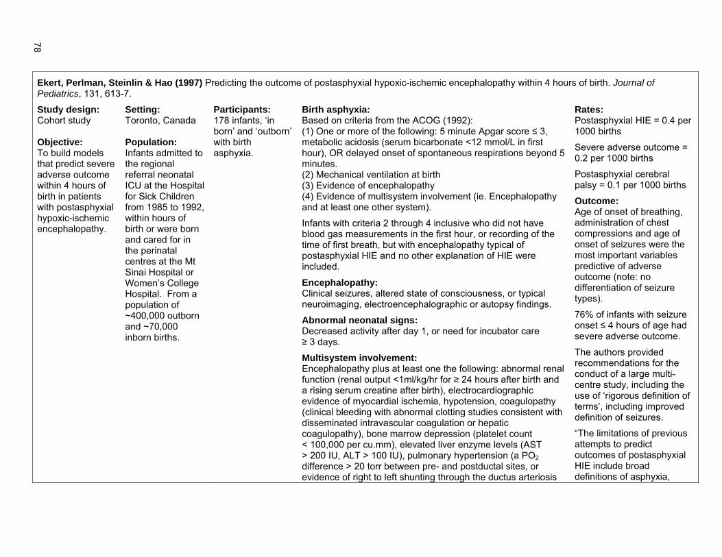
Ekert, Perlman, Steinlin & Hao (1997) Predicting the outcome of postasphyxial hypoxic-ischemic encephalopathy within 4 hours of birth. Journal of Pediatrics, 131, 613-7.
Study design: Cohort study Objective: To build models that predict severe adverse outcome within 4 hours of birth in patients with postasphyxial hypoxic-ischemic encephalopathy.
Setting: Toronto, Canada Population: Infants admitted to the regional referral neonatal ICU at the Hospital for Sick Children from 1985 to 1992, within hours of birth or were born and cared for in the perinatal centres at the Mt Sinai Hospital or Women’s College Hospital. From a population of ~400,000 outborn and ~70,000 inborn births.
Participants: 178 infants, ‘in born’ and ‘outborn’ with birth asphyxia.
Birth asphyxia: Based on criteria from the ACOG (1992): (1) One or more of the following: 5 minute Apgar score ≤ 3, metabolic acidosis (serum bicarbonate <12 mmol/L in first hour), OR delayed onset of spontaneous respirations beyond 5 minutes. (2) Mechanical ventilation at birth (3) Evidence of encephalopathy (4) Evidence of multisystem involvement (ie. Encephalopathy and at least one other system).
Infants with criteria 2 through 4 inclusive who did not have blood gas measurements in the first hour, or recording of the time of first breath, but with encephalopathy typical of postasphyxial HIE and no other explanation of HIE were included.
Encephalopathy: Clinical seizures, altered state of consciousness, or typical neuroimaging, electroencephalographic or autopsy findings.
Abnormal neonatal signs: Decreased activity after day 1, or need for incubator care ≥ 3 days.
Multisystem involvement: Encephalopathy plus at least one the following: abnormal renal function (renal output <1ml/kg/hr for ≥ 24 hours after birth and a rising serum creatine after birth), electrocardiographic evidence of myocardial ischemia, hypotension, coagulopathy (clinical bleeding with abnormal clotting studies consistent with disseminated intravascular coagulation or hepatic coagulopathy), bone marrow depression (platelet count < 100,000 per cu.mm), elevated liver enzyme levels (AST > 200 IU, ALT > 100 IU), pulmonary hypertension (a PO2 difference > 20 torr between pre- and postductal sites, or evidence of right to left shunting through the ductus arteriosis
Rates: Postasphyxial HIE = 0.4 per 1000 births
Severe adverse outcome = 0.2 per 1000 births
Postasphyxial cerebral palsy = 0.1 per 1000 births
Outcome: Age of onset of breathing, administration of chest compressions and age of onset of seizures were the most important variables predictive of adverse outcome (note: no differentiation of seizure types).
76% of infants with seizure onset ≤ 4 hours of age had severe adverse outcome.
The authors provided recommendations for the conduct of a large multi-centre study, including the use of ‘rigorous definition of terms’, including improved definition of seizures.
“The limitations of previous attempts to predict outcomes of postasphyxial HIE include broad definitions of asphyxia,
78

or foramen ovale on echocardiography).
Exclusion criteria: Congenital anomalies including minor dysmorphism and inborn metabolic errors, congenital infections, evidence of head trauma (skull fracture or intracranial haemorrhages) and haemorrhagic and septic shock. Some infants also excluded for mild HIE. Outcome: Severe adverse outcome: death or severe disability at a minimum age of 1 year. Severe disability: severe cerebral palsy (severe impairment of daily activities associated with hypertonia and hyperreflexia) with moderate or severe developmental delay (moderate delay determined by a Bayley test), blindness or deafness.
raising questions about its severity and timing, use of single arbitrarily selected risk factors, imprecise or ambiguous timing of predictions, and small numbers of subjects.”
Leth, Toft, Herning, Peitersen & Lou (1997) Neonatal seizures associated with cerebral lesions shown by magnetic resonance imaging. Archives of Disease in Childhood Fetal Neonatal Edition, 77, F105-10.
Objective: To evaluate the role of MRI in describing the brain lesions associated with neonatal seizures and to correlate the lesions with clinical data to suggest the aetiology and possible timing of the brain lesions. Neurological examinations completed at discharge, 5 months, 1 & 2 years of age.
Setting: Denmark
Population: All inborn term neonates admitted to the neonatal ICU from June 1992 to March 1995 (hospital not specified) presenting with clinical seizures.
Participants: 31 term neonates admitted for seizures.
Hypoxic-ischemic encephalopathy: Three or more of the following: • Bradycardia (heart rate < 80/minute) or late
decelerations on cardiotocography, or meconium stained amniotic fluid
• Umbilical pH < 7.10 • Apgar score of < 5 at 5 minutes • Delayed onset of spontaneous respiration • Multi-organ failure
Severity: Neonatal encephalopathy was graded by clinical severity according to Sarnat & Sarnat (1976).
Neonatal seizures: Diagnosed clinically and classified according to Volpe (1989) as (1) subtle, (2) clonic, (3) tonic and (4) myoclonic.
Exclusion criteria: As far as possible routine investigation excluded infection, haematological and metabolic-toxic factors as possible causes.
Outcome: The authors concluded that MRI detected a high incidence of brain lesions in neonatal seizures. Almost half were of prenatal origin and pathogenesis may be attributed to hypoxic and/or haemodynamic causes.
61% of brain lesions were attributed to hypoxic and or haemodynamic causes. Brain abnormalities were detected in 68% of the infants. Seizure aetiology was considered to be hypoxic-ischaemic in 35%.
79

Low (1997) Intrapartum fetal asphyxia: definition, diagnosis, and classification. American Journal of Obstetrics and Gynecology, 176, 957-9.
Clinical opinion article
Objective: To propose a classification to improve prediction of outcome after an exposure of the fetus to asphyxia during the intrapartum period.
Setting: Canada
Fetal asphyxia: A condition of impaired blood gas exchange leading to progressive hypoxemia and hypercapnia with a significant metabolic acidosis.
Encephalopathy Cardiovascular, respiratory & renal complications
Asphyxia
Metabolic acidosis at delivery* Minor Moderate Severe Minor Mod/Severe
Mild + ± ± Moderate + + ± Severe + + +
* Umbilical artery base deficits ≥ 12 mmol/L The clinical signs of newborn encephalopathy associated with intrapartum fetal asphyxia occur more often on day 1 after delivery with decreasing frequency on days 2 and 3.
Newborn encephalopathy: Mild: jitteriness and irritability Moderate: lethargy or abnormal tone Severe: coma or abnormal tone and multiple seizures.
Cardiovascular complications: Minor: Bradycardia or tachycardia (determined by the 95% confidence limits for heart rate for term and preterm newborns) Moderate: Hypertension or hypotension (defined by the 95% confidence limits for blood pressure for term and preterm newborns) Severe: Abnormal electrocardiographic or echocardiographic findings
Respiratory complications: Minor: Required supplementary oxygen Moderate: Required continuous positive airway pressure or transient ventilation (<24 hours) Severe: Required mechanical ventilation for > 24 hours
Renal complications: Minor: Hematuria observed Moderate: Elevation of serum creatinine (>100 µmol/L) Severe: clinical evidence of oliguria (<1ml/kg/hr) or anuria
The author states “…the determination of the short-term outcome as expressed by newborn encephalopathy and organ system complications provides an indicator of the severity of an asphyxial insult and criteria for the prediction of long-term outcome.”
80

Low, Lindsay & Derrick (1997) Threshold of metabolic acidosis associated with newborn complications. American Journal of Obstetrics and Gynecology, 177, 1391-4. Study design: Case control study (matched)
Objective: To determine the threshold of metabolic acidosis at delivery associated with newborn complications.
Setting: Canada
Population: Term neonates delivered between May 1995 and April 1996.
Infants matched with 2 controls on gestational age (±1 week) and birth weight (±250gm).
Participants: 164 term newborn infants. 3 groups defined by arterial base deficit at birth.
Cases: 58 = 12 to 16 mmol/L Controls: Matched with one infant from each of the two groups with the following base deficits: 58 = 4 to 8 mmol/L 58 = 8 to 12 mmol/L
Newborn encephalopathy: Minor: irritability or jitteriness Moderate: profound lethargy or abnormal tone Severe: coma or abnormal tone and seizures Cardiovascular complications: Minor: bradycardia (≤100 beats/min) Moderate: hypotension or hypertension (defined by the 95% confidence limits for blood pressure in term neonates) Severe: abnormal electrocardiographic or echocardiographic findings Respiratory complications: Minor: requiring supplementary oxygen Moderate: requiring continuous positive airway pressure or ventilation < 24 hours Severe: requiring mechanical ventilation > 24 hours Abnormalities of renal function: Minor: hematuria observed Moderate: elevation of serum creatinine (>100 µmol/L) Severe: anuria or oliguria <1 ml/kg/hr) A scoring system expressing the magnitude of the complications in each neonate was calculated, for each complication this was: Minor = 1, Moderate = 2, Severe = 4 The maximum complication score was 16. Antepartum risk factors: 1. Prior stillbirth or neonatal death 2. Maternal medical complications including hypertension, renal disease, collagen
disease, respiratory disease, endocrinopathies and severe acute infections during pregnancy
3. Obstetric complications including pregnancy-induced hypertension or pre-eclampsia and antepartum haemorrhage
4. Fetal complications including major congenital anomalies, fetal growth restriction, and oligohydramnios or polyhydramnios
Intrapartum risk factors: 1. post-term labour 2. meconium in the amniotic fluid, and 3. abnormal labour including prolonged labour, unfavourable progress in labour, or a
major malpresentation
Outcome: The threshold of fetal metabolic acidosis at delivery when moderate or severe newborn complications may occur is an umbilical artery base deficit of 12 mmol/L. Thereafter, increasing metabolic acidosis is associated with a progression in severity of newborn complications.
81

Thompson, Puterman, Linley et al (1997) The value of a scoring system for hypoxic ischaemic encephalopathy in predicting neurodevelopmental outcome. Acta Paediatrica, 86, 757-61.
Objective: To evaluate the predictive value of a numeric scoring system for the assessment of hypoxic ischaemic encephalopathy, for neurodevelopmental outcome at one year of age.
Setting: Cape Town, South Africa
Population: Term infants ≥ 37 weeks who developed clinical signs of HIE after birth at the Groote Schuur Hospital.
Participants: 45 infants, 40 infants followed-up to 1 year.
The HIE Score: Score Day Sign 0 1 2 3 1 2 3 Tone Normal Hyper Hypo Flaccid LOC Normal Hyper alert,
stare Lethargic Comatose
Fits None Infreq <3d-1 Frequent >2/day
Posture Normal Fisting, cycling
Strong, distal flexion
Decerebrate
Moro Normal Partial Absent Grasp Normal Poor Absent Suck Normal Poor Absent ± bites Resp. Normal Hypervent Brief apnoea IPPV
(apnoea)
Font’l Normal Full, not tense
Tense
Total score per day Font’l = fontanelle tension, Resp = respiratory pattern, LOC = level of consciousness Infants scored daily until the score was 0 or until hospital discharge. Based on the Sarnat & Sarnat descriptive gradings.
Outcome: The scoring system has high predictive value, predictive values of 92% and 100% and sensitivities and specificities of 100% and 93%. The scoring system is easy to use. A parent can be assured of a normal outcome if the infant has a score of 10 or less and is normal by 7 days. Infants with a score >15 and who remain abnormal after 7days must have a guarded prognosis.
82

Cioni, Fazzi, Ipata et al (1996) Correlation between cerebral visual impairment and magnetic resonance imaging in children with neonatal encephalopathy. Developmental Medicine and Child Neurology, 38, 120-32.
Aim: To study the correlation between lesions in the main visual areas (as shown by MRI) and visual impairment in small children who had neonatal encephalopathy. Median age at follow-up was 13 months (range 7-86 months).
Setting: Not specified Population: All children admitted to a child neurology department for a developmental check-up between 1990-1993 and who underwent brain MRI and a complete visual assessment.
Participants: 80 children with neonatal encephalopathy due to hypoxia and/or cerebral haemorrhage.
Neonatal Encephalopathy due to hypoxia and/or cerebral haemorrhage as confirmed by medical history, neurological examination, neuroimaging and EEG performed in the neonatal period and confirmed later by MRI. Not further specified.
Exclusion criteria: Infants with evidence of congenital diseases and those with severe opthalmological abnormalities.
Outcome: The authors conclude that children with neonatal encephalopathy are at ‘high risk’ for CVI. However, please note this was a select population of children attending for follow-up assessment. 60% of the children had visual impairment, 56% had lesions of the optic radiations and 24% damage to the visual cortex. MRI findings correlated with the degree of visual impairment. MRI can play a role in the early detection of visual deficits in these infants.
Rutherford, Pennock, Schwieso, Cowan & Dubowitz (1996) Hypoxic-ischaemic encephalopathy: Early and late magnetic resonance imaging findings in relation to outcome. Archives of Disease in Childhood. Fetal and Neonatal Edition, 75, F145-51.
Objective: To document the MRI findings in infants with hypoxic ischaemic encephalopathy between 12 and 24 months of age and relate these to MRI in the neonatal period and the clinical outcome at the time of the scan.
Setting: Not specified
Population: Not specified
Participants: 16 term or post-term infants, 37-43 weeks gestation with evidence of fetal distress, neurologically abnormal in the first 48 hours of life, with abnormalities of tone with or without convulsions and altered consciousness. Infants followed-up to 2 years of age.
Fetal Distress: The presence of cardiotocographic abnormalities of bradycardia (< 100/minute or late decelerations (type II dips) with or without meconium stained liquor and with low Apgar scores and the necessity for resuscitation. Hypoxic-ischaemic encephalopathy: Classified as mild, moderate or severe (I,II or III) according to Sarnat & Sarnat.
Outcome: The authors used the Griffiths developmental assessment.
Outcome: The authors concluded that MRI permits detection of exact patterns of early and late damage in infants with hypoxic ischaemic encephalopathy. However, specific areas of damage have not yet been correlated with specific clinical sequelae, in part as few areas are damaged in isolation in these infants.
83

Alcorn & Shean (1995) The origins of cerebral palsy: A consensus statement. Medical Journal of Australia, 162, 384.
Consensus statement of the Australian and New Zealand Perinatal Societies
Perinatal asphyxia: An event or condition during the perinatal period that is likely to severely reduce oxygen delivery and lead to acidosis eg. major antenatal haemorrhage or cord prolapse
AND
A failure of function of at least two organs (usually the brain and kidneys) consistent with the effects of asphyxia. Neonatal Encephalopathy: A clinical syndrome of signs suggesting neonatal neurological abnormality. It has many causes and intrapartum hypoxia cannot be considered a possible cause unless encephalopathy is evidence within 24 hours of delivery. The extent of encephalopathy is a reasonable predictor of neurological outcome. Levene et al (1986) cited for criteria: Mild: minor disturbances of tone, hyperalertness, slight feeding difficulties recovering by 48 hours. Moderate: lethargy, more pronounced abnormalities of tone, poor feeding, convulsions recovering by 7 days. Severe: coma, failure to maintain adequate ventilation, profound hypotonia, convulsions.
Australian and New Zealand Perinatal Societies (1995) The origins of cerebral palsy--a consensus statement. The Australian and New Zealand Perinatal Societies. Medical Journal of Australia, 162, 85-90.
Birth Asphyxia: Birth asphyxia is not a well-defined term. It implies some sort of dysfunction resulting from a lack of oxygen supply to the baby's tissues during the birth process. The term should not be used clinically because of the difficulty in ascribing clinical signs and symptoms in the neonate to an event during birth. Low pH and/or low Apgar scores at birth are supportive evidence of asphyxia but should not be used alone to make the diagnosis. Until more information is available, perinatal asphyxia is the preferred term to describe a neonate in whom there is:
• An event or condition during the perinatal period that is likely to severely reduce oxygen delivery and lead to acidosis (e.g. major antenatal haemorrhage or cord prolapse); and
• A failure of function of at least two organs (usually the brain and kidneys) consistent with the effects of asphyxia.
However, even these criteria are not specific for recent hypoxia and can be the result of any of the antenatal causes of cerebral palsy. They are minimum requirements to suggest the possibility of asphyxia but do not prove its intrapartum origin.
Neonatal encephalopathy: Neonatal encephalopathy is a clinical syndrome of signs suggesting neonatal neurological abnormality. It has many causes and intrapartum hypoxia cannot be considered as a possible cause unless encephalopathy is evident within 24 hours of delivery. The extent of encephalopathy is a reasonable predictor of neurological outcome. Levene et al graded the signs and recorded the varied outcomes.
84

Goodlin (1995) Do concepts of causes and prevention of cerebral palsy require revision? American Journal of Obstetrics and Gynecology, 172, 1830-6. Objective: To explore (validity of) the criteria of the ACOG for perinatal asphyxia to be linked to subsequent Cerebral Palsy (CP).
Four case studies of infants with intrapartum fetal insults with subsequent CP used in the evaluation of the criteria.
Setting: California, USA
Perinatal Asphyxia (ACOG 1992 criteria): The following criteria must be present:
1. An umbilical artery pH <7.00 2. Apgar score of 0 to 3 for longer than 5 minutes 3. Neonatal neurological sequelae, such as seizures, coma or hypotonia; and 4. Multiorgan dysfunction, involving the cardiovascular, gastrointestinal, haematological,
pulmonary or renal system.
Outcome: There are no well-done laboratory or clinical studies that unequivocally support the ‘criteria’ that umbilical artery pH must be <7.00 or the requirements of Apgar score <3, hypoxic-ischemic encephalopathy, and multiple organ dysfunction. The ACOG criteria for CP linkage and the role of parturition in CP should be re-evaluated.
85

Low, Simpson, Tonni & Chamberlain (1995) Limitations in the clinical prediction of intrapartum fetal asphyxia. American Journal of Obstetrics and Gynecology, 172, 801-4.
Study design: Cohort study Objective: To examine the limitations of risk scoring and periodic fetal assessment in the prediction of pregnancy at risk of intrapartum fetal asphyxia determined biochemically at delivery.
Setting: Canada (not defined) Population: 1,909 consecutive pregnancies in which an umbilical vein and artery blood gas and acid-base analysis was obtained at delivery.
Participants: 44 newborns with intrapartum fetal asphyxia
Intrapartum fetal asphyxia: An umbilical artery buffer base < 34 mmol/L (equivalent to a base deficit > 12 mmol/L). Intrapartum risk factors: • Preterm or post term labour • Meconium in the amniotic fluid • Abnormal labour including prolonged labour,
unfavourable progress in labour or a major malpresentation.
Antepartum risk factors: • Prior stillbirth or neonatal death • Maternal medical complications including
hypertension, renal disease, diabetes, collagen disease, respiratory disease, endocrinopathies and severe acute infections during pregnancy;
• Obstetric complications, including pregnancy-induced hypertension or pre-eclampsia and antepartum haemorrhage;
• Fetal complications, including major congenital anomalies, fetal growth retardation, and oligohydramnios or polyhydramnios.
Incidence: The incidence of intrapartum fetal asphyxia was 2.3%.
Outcome: The authors found a significant proportion of intrapartum fetal asphyxia occurred in pregnancies with no risk factors. Risk factors were present in ~50% of pregnancies at the onset of labour and in ⅔ before delivery. No individual risk factors demonstrated a strong association with intrapartum fetal asphyxia. There was a low positive predictive value of 3% and a large number of false positives. Screening methods need to be developed to identify asphyxial events occurring in pregnancies with no clinical risk factors. At present, the only definitive method to assure the diagnosis of intrapartum fetal asphyxia is routine umbilical cord blood gas analysis at birth.
86

Phelan, Ahn, Korst & Martin (1995) Nucleated red blood cells: A marker for fetal asphyxia? American Journal of Obstetrics and Gynecology, 173, 1380-4.
Study design: Case control study Objective: To determine whether a relationship exists between the presence of nucleated red blood cells, hypoxic ischemic encephalopathy, and long-term neonatal neurological impairment.
Setting: Not specified
Population: Not specified
Participants Cases: 46 singleton term neurologically impaired neonates with hypoxic ischemic encephalopathy
Controls: 83 term non-asphyxiated newborns, appropriate for gestational age at ≥ 37 weeks gestation, birth weight > 2800g, Apgar score ≥ 7 at both 1 and 5 minutes, normal intrapartum fetal heart rate pattern, clear amniotic fluid, normal neurologic evaluation at discharge and hematocrit ≥ 45%.
Hypoxic Ischemic Encephalopathy: Not defined. “All infants had evidence of hypoxic ischemic encephalopathy with long-term neurologic impairment confirmed by a pediatric neurologist.”
Exclusion criteria: Pregnancies known to be associated with elevated nucleated red blood cell count, such as Rh sensitisation, fetal anaemia, diabetes mellitus, ABO incompatibility, twins, preterm births, or evidence of intrauterine growth retardation were excluded.
Outcome: Nucleated red blood cells appeared to aid in identifying the presence of fetal asphyxia. The closer the birth was to the asphyxial event, the lower the number of nucleated red blood cells. Therefore cord blood nucleated red blood cells could assist in the timing of fetal neurologic injury.
87

Gaffney, Flavell, Johnson et al (1994) Cerebral palsy and neonatal encephalopathy. Archives of Disease in Childhood Fetal Neonatal Edition, 70, F195-200. Study design: Cohort study Hypothesis: Children born at term with cerebral palsy with signs of neurological dysfunction preceded by depression at birth (termed neonatal encephalopathy) differ from those without such signs in the frequency of antenatal and perinatal factors and the severity and characteristics of their features.
Setting: Oxford, United Kingdom Population: Children with cerebral palsy born between 1984-1987, to mothers resident in the Oxford region, identified from the Oxford regional register of early childhood impairment.
Participants 37 completed weeks gestation
Cases: 41 children from singleton deliveries after 37 weeks gestation without congenital anomaly and who had cerebral palsy and neonatal encephalopathy. Controls: 100 children with cerebral palsy born of a singleton pregnancy after 37 weeks gestation without congenital anomaly who did not have neonatal encephalopathy.
Neonatal encephalopathy: Signs of neonatal encephalopathy including: Depression at birth, based on a one minute Apgar score ≤ 6 followed by evidence of neonatal neurological abnormality such as lethargy, coma, impaired respiration, seizures and/or tone changes.
Note: seizures alone was not sufficient to be included.
Severity: No grading was used.
Exclusion criteria: Major congenital anomaly, definite postnatal cause for cerebral palsy such as neonatal meningitis or trauma, infants with transient jitteriness.
Outcome: Children with cerebral palsy who were born at term and have neonatal encephalopathy are more likely to have had signs of intrapartum asphyxia and are more likely to have a more severe form of cerebral palsy than those without a history of neonatal encephalopathy. Although this group represents only one in 10 of all cases of cerebral palsy, some of these may be obstetrically preventable.
The mothers of children with neonatal encephalopathy were more likely to be primigravidae and have a pregnancy > 41 weeks gestation. Intrapartum complications were more frequent in the neonatal encephalopathy group. At 5 years of age infants with neonatal encephalopathy were more likely to have developed spastic quadriplegia, to be non-walking and to have visual impairment.
88

Gaffney, Sellers, Flavell, Squier & Johnson (1994) Case-control study of intrapartum care, cerebral palsy, and perinatal death. British Medical Journal, 308, 743-50.
Study design: Case control study (1:2)
Objective: To investigate the relation between suboptimal intrapartum obstetric care and cerebral palsy and death.
Setting: Oxford, UK
Population: Infants from the Oxford Region Register of Early Childhood Impairments, children of mothers resident within the Oxford health region at time of delivery. There were 132,743 live births in the region with 339 included on the CP register.
Participants Singletons, ≥ 37 weeks with no congenital anomaly or postnatal cause Cases: Children with CP born in 1984-87 Controls: The 2 babies born immediately before the case, singleton infants, ≥ 37 weeks gestation with no major congenital anomaly.
Neonatal encephalopathy (NE): Depression at birth, based on Apgar score at one minute ≤ 6, followed by evidence of neonatal neurological abnormality such as lethargy, coma, impaired respiration, and seizures or change in tone, or both. (The Sarnat paper was referenced). Exclusion criteria: Babies with transient jitteriness and babies who seemed neurologically normal after birth and developed seizures after the first three days of life were not considered to have NE. Congenital anomalies or postnatal causes for CP. Cerebral Palsy: A permanent impairment of voluntary movement or posture presumed to be due to permanent damage to the immature brain.
Prevalence: CP = 2.4 per 1000 live births (postnatal causes excluded)
Outcome: There is an association between quality of intrapartum care and death. The findings suggest an association between suboptimal care and CP, but only in a small proportion of cases. The contribution of adverse antenatal factors in the origin of CP needs further study.
Only 7% of children with CP have a history of suboptimal response to distress followed by signs of NE. Most CP in children born at term is likely to have antenatal origin.
89

Low, Panagiotopoulos & Derrick (1994) Newborn complications after intrapartum asphyxia with metabolic acidosis in the term fetus. American Journal of Obstetrics and Gynecology, 170, 1081-7.
Study design: Case control study
Objective: To determine the newborn complications after respiratory or metabolic acidosis at delivery and to demonstrate the characteristics of an asphyxial insult predictive of these complications.
Setting: Ontario, Canada
Population: 14,453 pregnancies between 1984 and 1992 where a fetal blood gas and acid-base assessment was performed at delivery.
Participants Cases: 59 term fetuses with metabolic acidosis.
Controls: 59 fetuses with normal blood gas measures at delivery.
51 fetuses with respiratory acidosis.
Controls were matched by gestational age (± 1 week) and birth weight (± 100g) and were born within 12 months of the index case (average interval 4 months).
Newborn encephalopathy: Minor: abnormal behaviour (irritability, jitteriness, lethargy)
Moderate: abnormal tone
Severe: abnormal tone and seizure Complications: A newborn complication score was used incorporating encephalopathy, assessments (EEG, CT ultrasonography), and cardiovascular, respiratory and renal complications. Further described in the paper.
Outcome: A severe respiratory acidosis at delivery does not lead to newborn complications, however intrapartum fetal asphyxia with severe metabolic acidosis at delivery accounts for complications in the CNS, respiratory system and kidney. The magnitude of the complications is dictated in part by the severity and duration of the metabolic acidosis at delivery. In the fetus with severe metabolic acidosis at delivery the duration of the metabolic acidosis and the fetal response to the asphyxia, expressed by the Apgar score at 1 minute, are valuable index values in the prediction of newborn complications.
Rosenbloom (1994) Dyskinetic cerebral palsy and birth asphyxia. Developmental Medicine and Child Neurology, 36, 285-9.
Objective: To examine the connections between birth asphyxia and cerebral palsy (CP).
Setting: Merseyside, UK
Population: Children included on the Cerebral Palsy Register, born between 1967 and 1984.
Participants: 115 children with dyskinetic cerebral palsy, gestation > 37 weeks and birth weight > 10th centile for gestational age.
Birth asphyxia: Not defined.
Hypoxic-ischaemic encephalopathy: Not defined.
Degree of encephalopathy: Graded using Sarnat & Sarnat (1976).
Outcome: It would appear that acute and severe fetal distress and a severe degree of asphyxia immediately after birth can be followed by mild or moderate, rather than severe hypoxic-ischaemic encephalopathy. This clinical picture should be distinguished from the pattern of birth asphyxia which precedes development of spastic quadriplegic CP.
90

Wayenberg, Vermeylen, Bormans et al (1994) Diagnosis of severe birth asphyxia and early prediction of neonatal neurological outcome in term asphyxiated newborns. Journal of Perinatal Medicine, 22, 129-36.
Objective: To determine the value of several postnatal criteria available during the first 2 hours of life for the diagnosis and the short term neurological prognosis of birth asphyxia.
Setting: Not specified Population: Full term neonates born at the Hospital Français and neonatal ICU of the Erasmus Hospital from July 1989 to December 1991.
Participants: 60 full term neonates who experienced ‘significant birth asphyxia.’
Significant birth asphyxia: Two or more of the following criteria: • Intrapartum foetal distress by the recognition of
abnormal heart rate patterns or fresh meconial amniotic fluid
• Presence of immediate neonatal distress as shown by a low (< 7) one or five minute Apgar score, or a delay in establishing spontaneous regular respiration of > 1 minute
• Presence of a hypoperfusion syndrome, defined as the persistence at 30 minutes of life of pallor, increased capillary time and/or respiratory distress accompanied by significant metabolic acidosis (defined as a base deficit exceeding 10mEq/l on radial artery puncture performed at 30 minutes of life).
Severe birth asphyxia (as described by Finer et al 1981): Significant birth asphyxia followed by symptoms of moderate or severe postasphyxial encephalopathy as described by Finer et al (1981).
Encephalopathy (as Finer, 1981): Mild: hyperalertness, hyperreflexia and hyperexcitability
Moderate: Lethargy, weak Moro and sucking responses, seizures, and hypotonia
Severe: Stupor, flaccidity and absent primitive reflexes.
Exclusions criteria (reasons for actual exclusion): Causes of neurological abnormalities other than asphyxia. Infants excluded for neonatal intoxication or withdrawal syndrome due to maternal use of anxiolytic or sedative drugs or other neurological disorders (massive subarachnoid haemorrhage, congenital rubella encephalopathy, Marfan syndrome or metabolic disease).
Outcome: Encephalopathy: Normal = 29 (48.3%) Mild = 18 (30%) Moderate or severe = 13 (21.7%) Normal delay in establishing regular respiration and normal Apgar scores do not exclude severe birth asphyxia. Arterial pH and base deficit at 30 minutes was found to be the best criteria for the diagnosis of severe birth asphyxia, but lacked positive predictive value. The best predictive tool for the short-term neurological prognosis of birth asphyxia was a single score established at 30 minutes of life and based on the evaluation of consciousness, respiration and neonatal reflexes.
91
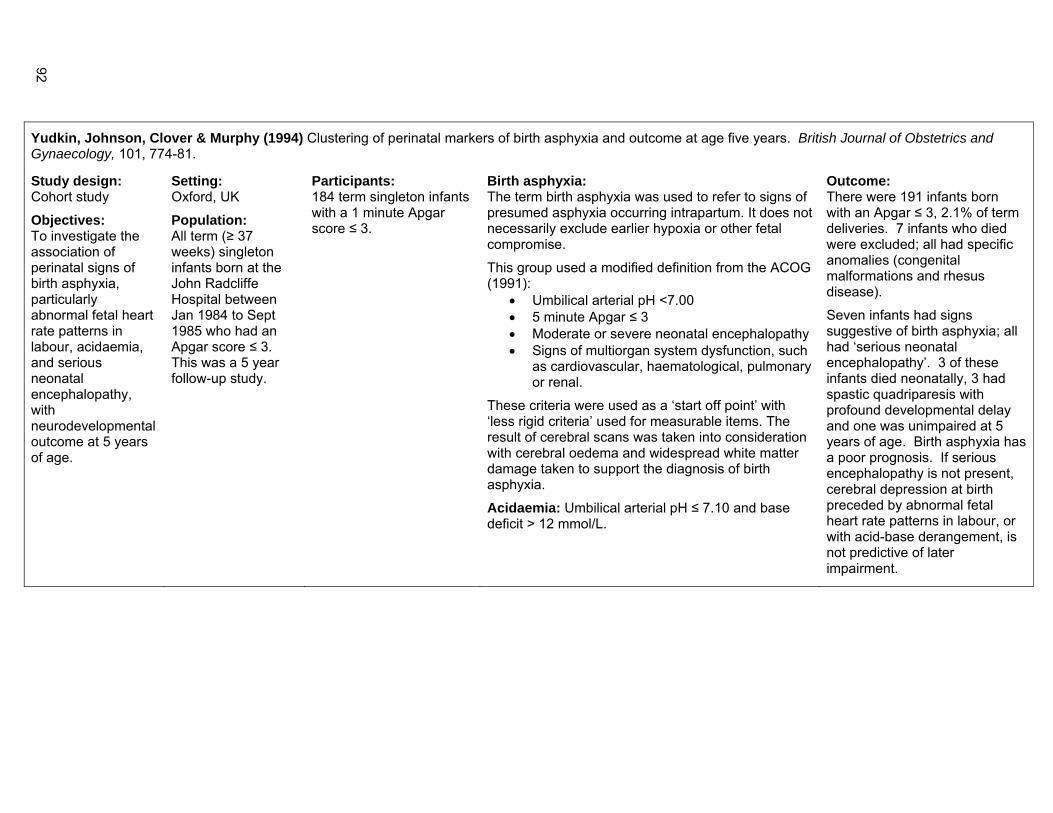
Yudkin, Johnson, Clover & Murphy (1994) Clustering of perinatal markers of birth asphyxia and outcome at age five years. British Journal of Obstetrics and Gynaecology, 101, 774-81.
Study design: Cohort study
Objectives: To investigate the association of perinatal signs of birth asphyxia, particularly abnormal fetal heart rate patterns in labour, acidaemia, and serious neonatal encephalopathy, with neurodevelopmental outcome at 5 years of age.
Setting: Oxford, UK
Population: All term (≥ 37 weeks) singleton infants born at the John Radcliffe Hospital between Jan 1984 to Sept 1985 who had an Apgar score ≤ 3. This was a 5 year follow-up study.
Participants: 184 term singleton infants with a 1 minute Apgar score ≤ 3.
Birth asphyxia: The term birth asphyxia was used to refer to signs of presumed asphyxia occurring intrapartum. It does not necessarily exclude earlier hypoxia or other fetal compromise.
This group used a modified definition from the ACOG (1991):
• Umbilical arterial pH <7.00 • 5 minute Apgar ≤ 3 • Moderate or severe neonatal encephalopathy • Signs of multiorgan system dysfunction, such
as cardiovascular, haematological, pulmonary or renal.
These criteria were used as a ‘start off point’ with ‘less rigid criteria’ used for measurable items. The result of cerebral scans was taken into consideration with cerebral oedema and widespread white matter damage taken to support the diagnosis of birth asphyxia.
Acidaemia: Umbilical arterial pH ≤ 7.10 and base deficit > 12 mmol/L.
Outcome: There were 191 infants born with an Apgar ≤ 3, 2.1% of term deliveries. 7 infants who died were excluded; all had specific anomalies (congenital malformations and rhesus disease).
Seven infants had signs suggestive of birth asphyxia; all had ‘serious neonatal encephalopathy’. 3 of these infants died neonatally, 3 had spastic quadriparesis with profound developmental delay and one was unimpaired at 5 years of age. Birth asphyxia has a poor prognosis. If serious encephalopathy is not present, cerebral depression at birth preceded by abnormal fetal heart rate patterns in labour, or with acid-base derangement, is not predictive of later impairment.
92

Blair (1993) A research definition for 'birth asphyxia'? Developmental Medicine and Child Neurology, 35, 449-52.
Annotation article Birth asphyxia: Sequence of cellular events:
Impaired gas exchange → 1°energy failure → cytotoxic mechanisms → 2° energy failure → neuronal death Birth asphyxia: Clinically observable correlates: Reduced fetal FHR Acidosis Low Apgar Abnormal Altered Seizures Growth abnormality scores conscious state muscle tone
Gray, Tudehope, Masel et al (1993) Perinatal hypoxic-ischaemic brain injury: prediction of outcome. Developmental Medicine and Child Neurology, 35, 965-73.
Objective: To investigate the prognosis for infants with early-onset neonatal encephalopathy who had evidence suggestive of perinatal asphyxia, and relate the results of investigatory techniques performed during the neonatal period to subsequent neurodevelopmental outcome. Follow-up was completed to at least one year of age.
Setting: Brisbane, Australia
Population: Term infants admitted to the neonatal unit at the Mater Mother’s Hospital from Oct 1988 to July 1991.
Participants: 26 term infants with features consistent with perinatal asphyxia and an abnormal neurological examination compatible with hypoxic-ischemic encephalopathy.
19 surviving infants were followed-up at one year of age.
Asphyxia: The presence of at least 2 of the following: (1) documentation of intrapartum fetal
distress on fetal heart rate monitoring, with or without the presence of meconium staining of the amniotic fluid
(2) the presence of an Apgar score < 6 at 5 minutes, and/or cord blood pH or an arterial blood pH taken shortly after the delivery below 7.15
(3) the need for immediate neonatal resuscitation with ventilation by bag and mask or via an endotracheal tube.
Severity classification: Infants graded as moderate or severe, not defined but Fenichel (1983) paper referenced.
Outcome: Developmental testing using the Griffiths Mental Development Scale at 8 or 12 months of age and 2 years. Adverse outcome defined as cerebral palsy, developmental delay or death.
Exclusion criteria: Infants with congenital anomalies of the nervous system, chromosomal abnormalities or transitory drug depression were excluded.
Outcome: 10 of the 19 infants followed-up had multiple disabilities, 9 with spastic CP and one with athetoid CP. All infants with severe encephalopathy died or had severe disability, just under one third of infants with moderate encephalopathy died or had a severe disability.
Generalised decreased tissue density on CT scan was associated with adverse outcome. Abnormal mean CBFV in the anterior cerebral artery and a low resistance index in both arteries were significantly associated with adverse outcome.
93

Goodwin, Belai, Hernandez et al (1992) Asphyxial complications in the term newborn with severe umbilical acidemia. American Journal of Obstetrics and Gynecology, 167, 1506-12.
Objective: To determine the relationship of umbilical acid-base status and Apgar score to neonatal asphyxial sequelae in infants with severe academia
Setting: Los Angeles, USA Population: 69,340 term deliveries at the University of Southern California Women’s hospital Jan 1986 to June 1990.
Participants: 129 term, nonanomalous singleton infants, with umbilical pH <7.00 (severe academia).
8,100 infants from the population of 69,340 term deliveries had umbilical samples taken. 202 cases had severe academia.
Cerebral dysfunction or hypoxic ischemic encephalopathy: Seizures occurring within the first 48 hours of life or hypotonia persisting beyond the first 24 hours of life. This was further defined as hypoxic ischemic encephalopathy, seizures and hypotonia, seizures only, hypotonia only and no hypoxic ischemic encephalopathy. Exclusion criteria: Infants were excluded if < 37 weeks gestation, birth weight < 2500g unless gestational age > 37 weeks, major anomalies, implausible result and absence of records. Follow-up: Gessell Developmental evaluation or Stanford-Binet test.
Outcome: 31% of these infants had hypoxic ischemic encephalopathy. The authors stated that in the pH range <7.00 a 5 minute Apgar score ≤ 3 has only moderate predictive value for hypoxic ischemic encephalopathy. The Apgar score was not highly predictive of asphyxial complications.
94

Hull & Dodd (1992) Falling incidence of hypoxic-ischaemic encephalopathy in term infants. British Journal of Obstetrics and Gynaecology, 99, 386-91. Study design: Retrospective examination of records
Objective: To examine trends in the incidence of hypoxic-ischaemic encephalopathy over a 13-year period.
Setting: Derby, UK
Population: All births in maternity services served by neonatal units in two 5 year periods 1976-1980 (24,824) & 1984-1988 (24,265).
Participants: 301 infants over 37 weeks gestation diagnosed with HIE, compared with data from the Cardiff Births Survey.
Hypoxic-ischaemic encephalopathy: Graded according to Levene (1985).
Grade I (mild): irritability, ‘hyperalert’, mild hypotonia, poor sucking. A progression of symptoms over the first 48 hours with complete clinical recovery by 5 days was required.
Grade II (moderate): lethargic, seizures, marked abnormalities of tone, required tube feeding. Seizures, marked abnormalities in tone and poor feed were required.
Grade III (severe): comatose, prolonged seizures, severe hypotonia, failure to maintain spontaneous respiration. Many seizures and ventilatory support were required.
Not all features of each category were present in every infant.
Infants with hypoglycaemia and irritability were included if the neurological abnormalities persisted after the blood glucose had been corrected.
Rates: 1976 to 1980 Stillbirths = 11.3 per 1000 births HIE incidence = 7.7 per 1000 live births Grades II and III = 2.6 per 1000 live births
1984 to 1988 Stillbirths = 5.7 per 1000 births HIE incidence = 4.6 per 1000 live births Grades I and II = 1.8 per 1000 live births
Outcome: 1976 to 1980 HIE (189 infants): Grade I: 126 infants (66.7%) Grade II: 39 infants (20.6%) Grade III: 24 infants (12.7%) 1984 to 1988 HIE (112 infants): Grade I: 68 infants (60.7%) Grade II: 29 infants (25.9%) Grade III: 15 infants (13.4%)
There was a significant fall in incidence of HIE between the two 5 year periods. This needs to be considered against the background of falling stillbirth and perinatal mortality rates over this period. The majority of infants with grade II encephalopathy died and around 20% of infants with moderate encephalopathy had serious handicap. A significant number of infants had major brain problems in terms of severe encephalopathy that was not anticipated by signs before birth nor from their response to birth, and which later resulted in death or major permanent handicap.
95

Low, Simpson & Ramsey (1992) The clinical diagnosis of asphyxia responsible for brain damage in the human fetus. American Journal of Obstetrics and Gynecology, 167, 11-5.
Objective: To review the clinical findings in infants who died in the perinatal period with brain damage attributable to asphyxia.
Setting: Ontario, Canada
Population: 208 perinatal deaths between 1976 and 1989 in whom a satisfactory neuropathological examination was performed.
Participants: 22 cases of fetal asphyxia
Asphyxia: Not defined.
Criteria for defining intervals between asphyxial insult and neuropathologic condition observed at death
Time Pathologic criteria
18 to 48 hr Neuronal necrosis
Immature neurons: Nuclear pyknosis and karyorrhexis
< 72 hr Appearance of white matter macrophages or microglia
> 72 hr Appearance of reactive astrocytes in white matter
> 4 hr Macroscopic cavitation
Outcome: Antepartum asphyxia may occur at any time in the last half of pregnancy. Eight cases of antepartum asphyxia occurred when there was no apparent indicators that the fetus was at risk, ie. 50% of the antepartum asphyxia occurred when the pregnancy had no risk factors.
96

Airede (1991) Birth asphyxia and hypoxic-ischaemic encephalopathy: Incidence and severity. Annals of Tropical Paediatrics, 11, 331-5.
Objective: To report the incidence and severity of post-asphyxial encephalopathy in full-term infants over a 3 year period.
Setting: Nigeria
Population: Full-term (37-42 completed weeks gestation) infants delivered at the Jos University Teaching hospital over 3 years from Jan 1987 to Dec 1989 who required admission, observation or treatment for birth asphyxia. Infants identified through chart review.
Participants: 166 infants born at term with birth asphyxia
Birth asphyxia: Birth asphyxia or symptoms and signs possibly related to asphyxia such as irritability, hypotonia, convulsions, cerebral cry or poor feeding. Post-asphyxial hypoxic-ischaemic encephalopathy (PAHIE): Graded according to a modification of that suggested by Fenichel: Grade I Mild
Grade II Moderate
Grade III Severe
Irritability, ‘hyperalert’
Lethargic
Seizures
Comatose
Prolonged and persistent seizures
Mild hypotonia
Marked abnormalities of tone
Severe hypotonia
Poor sucking Required tube feeding
Failure to maintain spontaneous respiration
The gradings referred to the most severe abnormalities shown by the infant. Not all features of each grade were needed for diagnosis.
Abnormalities on the intrapartum monitoring trace, when done, were used to support intrapartum asphyxia as a cause of the clinical signs.
Grade I: Increasing irritability with some degree of hypotonia along with poor sucking, which recovered completely by 3 days of age. The often appeared ‘hyperalert’ – wanting to feed avidly, but feeding poorly. Any baby who showed persistent neurological abnormalities was not included in grade I.
Rates: Rates per 1000 live births:
1987: 27.1
1988: 24.6
1989: 27.4
Grade I: 14.4
Grade II: 5.6
Grade III: 6.5
Grade II or III: 12.1
Mortality: 18.7% (mostly grade III)
Outcome: The overall incidence of birth asphyxia was 26.5 per 1000 live births.
The incidence of post-asphyxial hypoxic-ischemic encephalopathy remains exceedingly high in the middle-belt region of Nigeria. There was no marked difference between years.
To define asphyxia only on the basis of an Apgar score at 1 or 5 mins is potentially misleading, 21.1% of infants with PAHIE have unremarkable scores shortly after delivery.
There was marked involvement of infants with growth retardation (30.7%) and few
97

Grade II: Seizures usually beginning within 12-24 hours of birth, accompanied by abnormalities of tone and marked lethargy were necessary as was improvement of these symptoms within 7 days of birth. These infants frequently needed to be tube fed.
Grade III: Infants who required some ventilatory support, had persistent or prolonged seizures and were comatose. Recovery was very slow, taking longer than 7 days, or usually not seen in those who succumbed. Exclusion criteria: Metabolic screening was used to exclude other possible causes of irritability such as hypoglycaemia.
infants who were large for gestational age (3%).
98

Winkler, Hauth, Tucker et al (1991) Neonatal complications at term as related to the degree of umbilical artery acidemia. American Journal of Obstetrics and Gynecology, 164, 637-41.
Study design: Case control study (unmatched)
Objective: To determine the correlation between the degree of umbilical artery academia, type of academia, and Apgar scores and the immediate neonatal complications that have commonly been correlated with adverse neurological outcomes.
Setting: Not specified
Population: 2,764 term deliveries from Oct 1987 to April 1989.
Participants Cases: 358 mother-infant pairs, fetuses whose gestational age was ≥ 37 weeks who had paired umbilical and vein cord gas samples and whose umbilical artery pH was < 7.20. Controls: 358 mother-infant pairs with delivery occurring in the same time period meeting the same criteria except umbilical artery pH ≥ 7.20.
Intrapartum asphyxia: This was assessed by detection of complications known to be associated with intrapartum asphyxia including:
• neonatal seizures • persistent hypotonia • evidence of end-organ dysfunction such
as acute cardiac or renal failure Not further specified. Immediate neonatal complications known to be associated with intrapartum asphyxia: Neonatal seizures, persistent hypotonia, and evidence of end-organ dysfunction such as acute cardiac or renal failure.
Outcome: 2/23 infants (9%) with severe academia had newborn complications indicative of intrapartum asphyxia.
The authors could not confirm that infants born with an umbilical artery pH < 7.20 are at greater risk for short term complications attributable to intrapartum asphyxia than infants with a pH ≥ 7.20. Newborns with severe acidemia (umbilical artery pH <7.00) as well as low 1 and 5 minute Apgar scores may be at increased risk for neonatal complications associated with neurological sequelae. The positive predictive value of umbilical cord gas analysis for immediate newborn complications is poor but is useful for excluding intrapartum asphyxia.
99
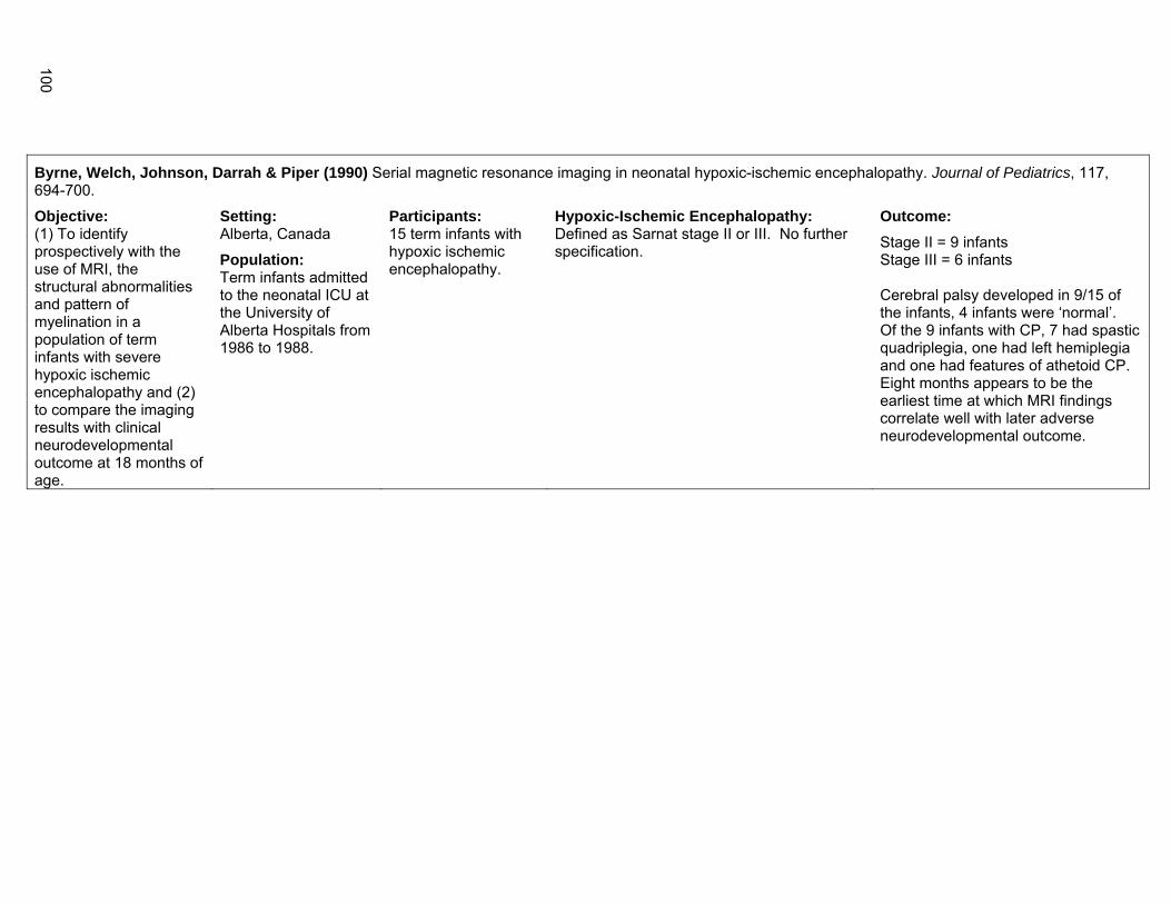
Byrne, Welch, Johnson, Darrah & Piper (1990) Serial magnetic resonance imaging in neonatal hypoxic-ischemic encephalopathy. Journal of Pediatrics, 117, 694-700.
Objective: (1) To identify prospectively with the use of MRI, the structural abnormalities and pattern of myelination in a population of term infants with severe hypoxic ischemic encephalopathy and (2) to compare the imaging results with clinical neurodevelopmental outcome at 18 months of age.
Setting: Alberta, Canada
Population: Term infants admitted to the neonatal ICU at the University of Alberta Hospitals from 1986 to 1988.
Participants: 15 term infants with hypoxic ischemic encephalopathy.
Hypoxic-Ischemic Encephalopathy: Defined as Sarnat stage II or III. No further specification.
Outcome: Stage II = 9 infants Stage III = 6 infants Cerebral palsy developed in 9/15 of the infants, 4 infants were ‘normal’. Of the 9 infants with CP, 7 had spastic quadriplegia, one had left hemiplegia and one had features of athetoid CP. Eight months appears to be the earliest time at which MRI findings correlate well with later adverse neurodevelopmental outcome.
100

Portman, Carter, Gaylord et al (1990) Predicting neonatal morbidity after perinatal asphyxia: A scoring system. American Journal of Obstetrics and Gynecology, 162, 174-82.
Objective: To devise a scoring system that would rapidly predict organ dysfunction observed in the immediate neonatal period. These criteria would rapidly assess the presence and severity of asphyxia to define the group at greatest risk for multi-organ system dysfunction.
Setting: Denver, USA
Population: Neonates ≥ 36 weeks gestation admitted to the Children’s Hospital Newborn ICU in 1983 (an outborn population was used). The reliability of the scoring system was tested in 1984. See Carter (1998) for the follow-up study.
Participants: 48 infants meeting the possible scoring criteria.
Birth asphyxia: Apgar score ≤ 3 at one minute or ≤ 6 at 5 minutes.
In the classification system infants with severe asphyxia score ≥ 6 points and infants with moderate ≤ 5 points. How these points are allocated was not defined.
Severe: Three or more organ systems affected.
Moderate: one or two organ systems affected.
Definitions of organ system dysfunction: Pulmonary Ventilator-dependent respiratory distress or hood oxygen requirement for > 24 hours (to include meconium aspiration syndrome). Cardiovascular Shock or hypotension (systolic blood pressure <50 mmHg, the need for pressor agents or colloid infusion to maintain BP), congestive heart failure not associated with structural heart disease or abnormal fluid status. Neurologic Seizures, abnormally low or increased neuromuscular tonus, intracranial haemorrhage. Gastrointestinal Necrotising enterocolitis Hepatic Elevated transaminases, prolonged prothrombin time or partial thromboplastin time with no evidence of disseminated intravascular coagulopathy. Hematologic Disseminated intravascular coagulopathy Renal Acute tubular necrosis, oliguria-anuria beyond the first 24 hours, hematuria-proteinuria >1+ quantitation on successive samples, renal tubular acidosis, hypertension. Mortality
Outcome: Moderate = 32 (1983), 26 (1984) infants Severe = 16 (1983), 24 (1984) infants
Indicators of asphyxia significantly associated with organ dysfunction: • 5 minute Apgar score ≤ 6 • Abnormal fetal heart rate
monitoring • Base deficit >10 mEq/L
The authors state that this system allows for the rapid recognition of asphyxia and assessment of its severity or potential for severe short-term morbidity as manifested by multi-organ system dysfunction or death.
101

Torfs, van den Berg, Oechsli & Cummins (1990) Prenatal and perinatal factors in the etiology of cerebral palsy. Journal of Pediatrics, 116, 615-9.
Study design: Cohort study Objective: To evaluate the gestational and perinatal risk factors for CP in a cohort of children followed for more than 5 years and to make a comparison with Nelson & Ellenberg (1985, 1986). Children were followed-up for a minimum of 5 years.
Setting: California, USA Population: 19,044 live born infants from the California Child Health and Development Studies, all mothers who belonged to a pre-paid medical plan and registered their first prenatal medical visit in selected hospitals in California from 1959 to 1966.
Participants Cases: 55 children with CP
Controls: 561 live born children who did not have CP (from congenital or known causes).
Birth asphyxia: Time to cry longer than 5 minutes
Exclusions: Neuromotor dysfunction resulting from known postnatal causes, such as infection or injury, or from recognised progressive diseases; neural tube defects.
Outcome: Children who had seizures within 48 hours of birth were at high risk of developing CP. 78% of children with CP did not have birth asphyxia and the 22% who did had other prenatal risk factors that may have compromised their recovery. 16 control infants (2.9%) had birth asphyxia but recovered without neurological damage. The strongest predictors of CP were the presence of a major congenital anomaly, low birth weight, low placental weight, and an abnormal fetal position, antecedents of the birthing process and strong indicators of fetal compromise before labour or delivery. New etiological studies must focus on prenatal, gestational and neonatal risk factors such as genetic and environmental factors.
Gilstrap, Leveno, Burris, Williams & Little (1989) Diagnosis of birth asphyxia on the basis of fetal pH, Apgar score, and newborn cerebral dysfunction. American Journal of Obstetrics and Gynecology, 161, 825-30.
Objective: To more precisely define birth asphyxia based on fetal condition as measured by umbilical artery pH, Apgar scores and neurological condition of newborns.
Setting: Texas, USA
Population: Complicated term (≥ 2500g) pregnancies from Aug 1987 to Feb 1988 delivered at the Parkland Memorial Hospital. 8,678 infants were born at the institute during the study period.
Participants: 2,738 singleton live born term infants with cephalic presentations.
Birth asphyxia: Not clearly defined.
Mild asphyxia: Apgar score ≤ 6 at 1 and 5 minutes
Severe asphyxia: Apgar score ≤ 3 at 1 and 5 minutes Complicated pregnancies: Caesarean section, forceps delivery, meconium in the amniotic fluid, oxytocin stimulation of labour and abnormal fetal heart rate.
Outcome: Infants must be severely depressed at delivery before birth asphyxia can be reliably diagnosed, including Apgar score ≤ 3 at 1 and 5 minutes plus umbilical artery pH values < 7.00.
102

Levene, Fenton, Evans et al (1989) Severe birth asphyxia and abnormal cerebral blood-flow velocity. Developmental Medicine and Child Neurology, 31, 427-34. Purpose: Assessment of cerebral blood flow velocity (CBFV) to determine its positive predictive value in determining death or severe impairment among infants with moderate or severe PAE.
Setting: Not specified
Population: Full-term infants (37 to 42 weeks) born from March 1985 to June 1988 with moderate or severe PAE.
Participants Cases: 34 full-term infants with moderate or severe PAE Grade II = 7 Grade III = 27
Controls: 126 healthy infants without clinical evidence of asphyxia and normal ultrasound scans.
Moderate or severe postasphyxial encephalopathy (PAE): According to the clinical criteria of Levene (1985). The infants all had multiple convulsions and those with severe PAE required mechanical support for respiratory failure. Note: Infants with mild PAE were not included.
Outcomes: Abnormal CBFV (Doppler ultrasound method) for predicting adverse outcome (death or severe handicap): Sensitivity = 57% Specificity = 88% Positive predictive value = 94%
CBFV was found to be elevated in the majority of infants with severe PAE, and this was more predictive of adverse outcome than low Pourcelot’s Resistance Index (PRI). The reason for the high CBFV and low PRI is probably vasoparalysis, a form of irreversible cerebral vascular injury.
103

Robertson, Finer & Grace (1989) School performance of survivors of neonatal encephalopathy associated with birth asphyxia at term. Journal of Pediatrics, 114, 753-60.
Study design: Case control study
Objective: To describe the school performance for reading, spelling and arithmetic at 8 years of age of previously reported survivors of neonatal encephalopathy associated with birth asphyxia at term compared to a peer group. Previous papers have reported on outcomes at 2 and 3½ years.
Setting: Alberta, Canada
Population: Infants admitted to two tertiary intensive care units (University of Alberta and Royal Alexandra hospitals) in the perinatal program from 1974-1979.
Participants Cases: 145 children with neonatal encephalopathy associated with birth asphyxia.
Controls: 155 school children 8 years of age (± 4 months) at the time of testing following completion of a cognitive abilities test.
Encephalopathy (neonatal encephalopathy associated with birth asphyxia): Mild: hyperalertness and hyperexcitability. Moderate: lethargy, hypotonia and suppressed primitive reflexes. Including infants with transient hypotonia. Severe: stupor, flaccidity and absence of primitive reflexes.
Exclusion criteria: Diagnosed syndromes or malformations of the central nervous system known to be associated with developmental delay.
Outcomes: In hospital and post-discharge deaths occurred most frequently among infants with severe neonatal encephalopathy. Children with mild neonatal encephalopathy continued to be free of neurological impairment and had performance scores similar to their peers. Children with moderate or severe neonatal encephalopathy associated with birth asphyxia at term are at risk of physical and mental impairment and reduced school performance. Using the clinical categories of moderate and severe neonatal encephalopathy associated with birth asphyxia provides an opportunity for early identification of children at risk of developmental disabilities.
104

Blair & Stanley (1988) Intrapartum asphyxia: A rare cause of cerebral palsy. Journal of Pediatrics, 112, 515-9.
Study design: Case control study (matched) Objective: To investigate the relationship between birth asphyxia and spastic cerebral palsy.
Setting: Western Australia Population: Infants included in the WA cerebral palsy register and infants from a birth notifications system born in Western Australia between 1975 and 1980.
Participants Cases: 183 children born in WA from 1975-1980 with a diagnosis of spastic cerebral palsy by 5 years of age. Cases selected from the WA Cerebral Palsy Register.
Controls: 549 live born children (3 controls per case) selected from the WA midwives birth notification system. Controls matched by year of birth, birth weight, maternal race, infant sex and plurality.
Birth asphyxia: • Any fetal distress with an Apgar score at 1 minute of < 7
AND/OR
• A known time to spontaneous respiration of > 2 minutes. Fetal distress: • Any one or more of the following: • Meconium • Fetal heart rate > 160 or <120 beats per minute • Abnormal fetal heart rate tracing on electronic recordings OR • ‘Fetal distress’ not otherwise specified Abnormal neonatal neurologic signs: A record of any one of the following: • Change in conscious state (jitteriness,
excitability-irritability, or hypotonia-lethargy) • Poor suck • Seizures Criteria indicating likelihood of brain damage from birth asphyxia 1. Definitely not birth asphyxial damage a. no abnormal neonatal neurological signs OR b. definitive evidence that damage had occurred at times other than during labour or delivery (eg. evidence of periventricular leukomalacia on ultrasonography at birth)
2. Birth asphyxial damage very unlikely a. some abnormal neurological signs present but
they did not conform to required type, severity, onset or duration OR b. Neurologic signs present, but with no perinatal indication of asphyxia (eg. uncomplicated delivery, without evidence of fetal distress and good condition at birth)
Outcome: An association between clinically observed perinatal signs of birth asphyxia and spastic cerebral palsy was found. However, intrapartum asphyxia was the possible cause of brain damage in about 8% of children with spastic CP. It was concluded that the contribution of intrapartum events and obstetric management to overall CP rates is probably less than previously thought.
105

3. Possible birth asphyxial damage a. abnormal neurologic signs present, but with insufficient or conflicting evidence of birth asphyxia or trauma OR b. Both antenatal and perinatal factors present 4. Birth asphyxial damage very likely
a. abnormal neurologic signs of required type, severity, onset and duration, AND b. No or few adverse antenatal factors AND/OR c. Moderate or severe “birth asphyxia” AND/OR
5. Traumatic events preceding delivery a. possibly existed that ‘birth asphyxia’ could have been missed or not recorded (eg. unattended or unmonitored birth) b. Traumatic events preceding delivery so immediately that insufficient time had elapsed for condition to deteriorate and for ‘birth asphyxia’ to have been diagnosed. Moderate or severe birth asphyxia: • Apgar score < 4 at 5 minutes • Time to spontaneous respiration > 5 minutes • Fetal distress particularly if > 1500g birth weight
106

Ellenberg & Nelson (1988) Cluster of perinatal events identifying infants at high risk for death or disability. Journal of Pediatrics, 113, 546-52.
Objective: To determine the prognostic import of neonatal seizures according to the presence or absence of certain other postnatal characteristics.
Setting: USA
Population: 39,000 infants with a birth weight >2500g, these infants were part of the NCPP Collaborative Perinatal Project of the National Institute of Neurological and Communicative Disorders and Stroke, between 1959 and 1966.
Participants: 39,000 infants for whom neurological status was known.
Hypoxic-Ischemic Encephalopathy: • Decreased activity after the first day of life • Need for incubator care for 3 or more days • Feeding problems • Poor suck, or • Respiratory difficulty
Analyses were performed with and without children with major malformations outside the central nervous system.
Outcome: The results were ‘similar’ including and excluding children with major malformations.
Children with clinically recognized neonatal seizures and 5-minute Apgar scores less than or equal to 5 and who had at least one of five signs compatible with neonatal encephalopathy had a risk for first-year death of 33%. Survivors of this cluster of events (low Apgar score-abnormal signs-seizures) had a risk for motor disability of 55%. In contrast, survivors of neonatal seizures who did not have poor Apgar scores or other abnormal signs had a risk for motor disability of only 0.13%. Thus, among infants with neonatal seizures the risk for cerebral palsy was 420 times greater if there had been a low 5-minute Apgar score and other neonatal signs. Low Apgar score-abnormal signs-seizures constituted a cluster of events that served to identify, within the first days of life, a tiny subgroup of term newborn infants in whom risk for chronic motor disability was 55%, and for death or disability was 70%.
107
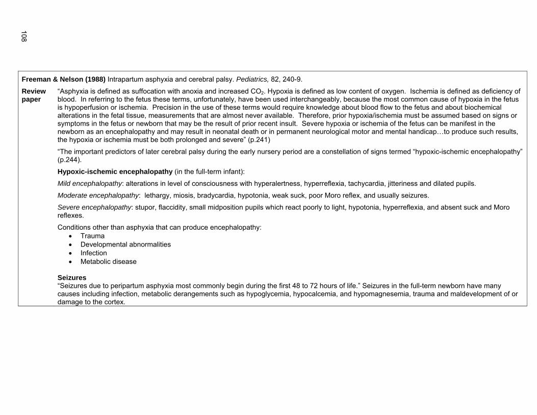
Freeman & Nelson (1988) Intrapartum asphyxia and cerebral palsy. Pediatrics, 82, 240-9.
Review paper
“Asphyxia is defined as suffocation with anoxia and increased CO2. Hypoxia is defined as low content of oxygen. Ischemia is defined as deficiency of blood. In referring to the fetus these terms, unfortunately, have been used interchangeably, because the most common cause of hypoxia in the fetus is hypoperfusion or ischemia. Precision in the use of these terms would require knowledge about blood flow to the fetus and about biochemical alterations in the fetal tissue, measurements that are almost never available. Therefore, prior hypoxia/ischemia must be assumed based on signs or symptoms in the fetus or newborn that may be the result of prior recent insult. Severe hypoxia or ischemia of the fetus can be manifest in the newborn as an encephalopathy and may result in neonatal death or in permanent neurological motor and mental handicap…to produce such results, the hypoxia or ischemia must be both prolonged and severe” (p.241)
“The important predictors of later cerebral palsy during the early nursery period are a constellation of signs termed “hypoxic-ischemic encephalopathy” (p.244).
Hypoxic-ischemic encephalopathy (in the full-term infant): Mild encephalopathy: alterations in level of consciousness with hyperalertness, hyperreflexia, tachycardia, jitteriness and dilated pupils.
Moderate encephalopathy: lethargy, miosis, bradycardia, hypotonia, weak suck, poor Moro reflex, and usually seizures.
Severe encephalopathy: stupor, flaccidity, small midposition pupils which react poorly to light, hypotonia, hyperreflexia, and absent suck and Moro reflexes.
Conditions other than asphyxia that can produce encephalopathy: • Trauma • Developmental abnormalities • Infection • Metabolic disease
Seizures “Seizures due to peripartum asphyxia most commonly begin during the first 48 to 72 hours of life.” Seizures in the full-term newborn have many causes including infection, metabolic derangements such as hypoglycemia, hypocalcemia, and hypomagnesemia, trauma and maldevelopment of or damage to the cortex.
108

Low, Galbraith, Muir et al (1988) Motor and cognitive deficits after intrapartum asphyxia in the mature fetus. American Journal of Obstetrics and Gynecology, 158, 356-61.
Study design: Case control study
Objective: To examine the range of motor and cognitive deficits associated with intrapartum fetal asphyxia at one year of age
Setting: Canada
Population: Not specified
Participants Cases: 37 term infants with evidence of intrapartum fetal asphyxia at birth. Controls: 76 term infants, ‘normally grown’ with no evidence of fetal asphyxia at delivery. The mothers had no medical, obstetric, labour or delivery complications.
Newborn encephalopathy (also referred to as neonatal encephalopathy): Based on observation of behaviour changes, abnormalities of tone, presence of seizures and recurrent apnea.
Mild: Behaviour: Hyperalertness, irritability, jitteriness Tone: Transient, hypertonia or hypotonia
Moderate: Behaviour: Lethargy Tone: Severe hypotonia Seizures: Occasional
Severe: Behaviour: Coma Seizures: Multiple Respiration: Recurrent apnea Seizures: subtle, tonic or clonic. Recurrent apnea: a repetitive respiratory pause > 20 seconds, requiring active resuscitation, that was not related to associated respiratory complications.
Intrapartum fetal asphyxia: Evidence of a metabolic acidosis at delivery as expressed by an umbilical artery buffer base < 34 mmol/L.
Major motor deficits were characteristic of cerebral palsy, accompanied with ‘mental retardation’ in some cases.
Minor deficits were characteristic of motor developmental delays with ‘apparently satisfactory cognitive development.’
Outcome: Infants with ‘fetal asphyxia’ were significantly more likely to have (minor or major) motor and cognitive deficits (41% of infants with ‘fetal asphyxia’ had evidence of motor and cognitive deficits, as opposed to 8% of controls).
Note: the table that documents the results totals to 102%. Less than 50% of the infants with asphyxia had evidence of newborn encephalopathy. This paper classified infants initially by presence of fetal asphyxia and then presence of newborn encephalopathy. Eight infants were documented as having ‘asphyxia’ but no evidence of newborn encephalopathy.
109

Lupton, Hill, Roland et al (1988) Brain swelling in the asphyxiated term newborn: Pathogenesis and outcome. Pediatrics, 82, 139-46.
Objective: To determine the incidence, temporal profile and significance of clinically recognisable brain swelling in the asphyxiated term newborn by means of intracranial pressure measurements and CT during the acute phase of hypoxic-ischemic encephalopathy and to correlate these measurements with neurologic outcome.
Setting: Vancouver, Canada
Population: Asphyxiated infants admitted to the neonatal ICU at British Columbia’s Children’s Hospital from July 1985 to Sept 1986. These were term infants ≥ 37 weeks gestation, appropriate birth weight for gestational age and had clinical features consistent with acute hypoxic ischemic encephalopathy.
Participants: 32 asphyxiated term newborns during the first week of life.
Perinatal asphyxia: One or more of the following:
(1) Fetal bradycardia (heart rate less than 80 beats per minute for at least 60 seconds) or evidence of late decelerations during labour as seen by fetal monitoring
(2) Apgar score < 5 at five minutes (3) Requirement of positive pressure
ventilation for at least 2 minutes following delivery
(4) Acidosis (pH < 7.1 within the first hour of life).
Degree of encephalopathy: As per Sarnat and Sarnat (1976). Not further specified.
Exclusion criteria: Congenital anomalies, infections or traumatic injuries that may mimic hypoxic-ischemic encephalopathy.
Outcome: The data suggest that clinically recognisable brain swelling is a relatively uncommon feature of hypoxic ischemic encephalopathy in the term newborn.
110

Amiel-Tison & Ellison (1986) Birth asphyxia in the full term newborn: early assessment and outcome. Developmental Medicine & Child Neurology, 28, 671-82.
Review article
“There are several reasons for pursuing better definitions and methodologies in the study of asphyxia: (1) to improve research and to make possible comparisons among studies, (2) to improve clinical treatment of the acutely asphyxiated neonate, (3) to permit more precise prediction of outcome and, (4) to contribute to the prevention of severe asphyxia, insofar as that is possible.” In the immediate newborn period asphyxia is indicated by low Apgar scores, especially 5 mins or more and the need for mask or intubation resuscitation. The use of an Apgar score of ≤ 6 at 5 mins for moderate asphyxia and ≤ 3 for severe asphyxia is generally accepted. Variables in the neonatal period should include neurological signs and symptoms, including seizures, and, where appropriate, electroencephalogram, cerebral imaging (ultrasound, computed tomography or nuclear magnetic resonance) and brain auditory evoked response studies.
The Amiel-Tison figure on clinical changes in hypoxic-ischemic encephalopathy is outlined in this article.
111

Bell, Johansson, McLean & Usher (1986) Birth asphyxia, trauma, and mortality in twins: Has caesarean section improved outcome? American Journal of Obstetrics and Gynecology, 154, 235-9.
Study design: Cohort study Objective: To determine to what extent a liberal policy for cesarean section in twin pregnancies, especially those with malpresentations, has affected the incidence of birth asphyxia and trauma.
Setting: Quebec, Canada Population: Consecutive twins delivered in the Royal Victoria hospital before and after the caesarean section rate increased.
Participants: Twins delivered at the same hospital after 28 weeks gestation. 554 delivered from 1963-1972, AND 614 twins delivered from 1978-1984. Comparisons were made between term (≥ 37 weeks) and pre-term (29-36 weeks) infants.
Birth asphyxia: Depression at birth necessitating positive-pressure manual ventilation. Clinical depression at birth: Infant respiratory depression sufficient to require positive-pressure ventilation to establish regular sustained respirations after birth. Severe depression: Infant required more than 3 minutes of ventilation. The authors stated that none of the infants developed ‘encephalopathy’. However ‘encephalopathy’ was not defined.
Outcome: There was no reduction in the overall incidence of moderate or severe depression at birth. The authors concluded that there was no justification for the marked increase in the rate of caesarean delivery of twins. That is, the study did not indicate that this increase had improved the condition of twin infants at birth.
112

Levene, Sands, Grindulis & Moore (1986) Comparison of two methods of predicting outcome in perinatal asphyxia. Lancet, 1, 67-9.
Objective: To compare two methods of diagnosing intrapartum asphyxia (Apgar score and postasphyxial encephalopathy) for their ability to identify infants who are likely to have a poor prognosis or a good prognosis.
Setting: Leicestershire, UK
Population: 20,975 full-term live born infants born in a teaching hospital maternity unit over 4 years from 1980. 126 infants had postasphyxial encephalopathy.
Participants: 122 full-term infants with post-asphyxial encephalopathy.
Encephalopathy (based on Fenichel): Mild: Minor disturbances of tone, hyperalertness and slight feeding difficulties, recovering by 48 hours after birth
Moderate: lethargy, more pronounced abnormalities of tone, poor feeding, and convulsions, with signs of recovery by 7 days
Severe: coma, failure to maintain adequate ventilation, profound hypotonia and seizures.
Incidence: Postasphyxial encephalopathy = 6.0 per 1000.
Outcome: A 10 minute Apgar score of ≤ 5 was the most sensitive of 6 different Apgar ratings in predicting adverse outcome (sensitivity 43%, specificity 95%) but this was less sensitive than the presence of moderate or severe encephalopathy in predicting death or severe handicap (sensitivity 96%). The incidence of death or severe handicap was 1 in 1000 deliveries.
113

Lipper, Voorhies, Ross, Vannucci & Auld (1986) Early predictors of one-year outcome for infants asphyxiated at birth. Developmental Medicine and Child Neurology, 28, 303-9.
Study design: Hospital-based outcome assessment
Objective: To determine the predictive value (at one year) of scoring systems to evaluate neurological status and CT scans following hypoxic-ischemic insult. The study attempted to identify within the first 48 hours of life those infants sustaining hypoxic-ischemic insults and destined to have neurodevelopmental abnormalities at one year of age.
Setting: New York, USA
Population: Infants inborn & outborn), appropriate weight for gestational age, admitted to the Neonatal Intensive Care Unit of the New York Hospital from Nov 1981 to Oct 1982.
Participants: 45 full-term asphyxiated infants. 28 followed-up (this was all survivors) at one year.
Asphyxia: One and/or five minute Apgar scores below 6 in association with one or more of the following conditions:
• Fetal distress • Cord pH < 7.2 • Need for resuscitation at birth and • Meconium aspiration.
A post-asphyxia score was assigned within the first 24 hours of life derived from summing scores from the 17-item neurological assessment.
Fetal distress: Detected by auscultation or electronic fetal monitoring defined by:
• Fetal bradycardia • Variable or late decelerations • Loss of beat-to-beat variability and/or • Fetal scalp pH < 7.2.
A post-asphyxial score was calculated for each infant within 24 hours of birth derived from summing scores for 17 items from a 17 item neurological assessment. A low density index was calculated from CT scans.
Exclusion Criteria: Congenital anomalies, traumatic cerebral injury, hydrocephalus, infection or drug depression.
Outcome: 6 infants died, 4 in the neonatal period, 2 within the first 4 months of life.
The post-asphyxia score and CT low-density index appear to be valuable, complementary tools in the early assessment of neurobehavioural outcome for full term asphyxiated infants and in determining their ultimate prognosis. The severity of perinatal hypoxia is difficult to quantify at birth, but these measurements offer a sufficiently good indication of later neurobehavioural function. The false positive rate for the PAS was less than 9%.
114
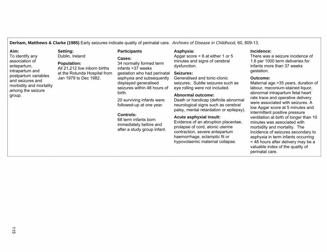
Derham, Matthews & Clarke (1985) Early seizures indicate quality of perinatal care. Archives of Disease in Childhood, 60, 809-13.
Aim: To identify any association of antepartum, intrapartum and postpartum variables and seizures and morbidity and mortality among the seizure group.
Setting: Dublin, Ireland
Population: All 21,212 live inborn births at the Rotunda Hospital from Jan 1979 to Dec 1982.
Participants Cases: 34 normally formed term infants >37 weeks gestation who had perinatal asphyxia and subsequently displayed generalised seizures within 48 hours of birth.
20 surviving infants were followed-up at one year. Controls: 68 term infants born immediately before and after a study group infant.
Asphyxia: Apgar score < 6 at either 1 or 5 minutes and signs of cerebral dysfunction.
Seizures: Generalised and tonic-clonic seizures. Subtle seizures such as eye rolling were not included.
Abnormal outcome: Death or handicap (definite abnormal neurological signs such as cerebral palsy, mental retardation or epilepsy).
Acute asphyxial insult: Evidence of an abruption placentae, prolapse of cord, atonic uterine contraction, severe antepartum haemorrhage, eclamptic fit or hypovolaemic maternal collapse.
Incidence: There was a seizure incidence of 1.6 per 1000 term deliveries for infants more than 37 weeks gestation. Outcome: Maternal age >35 years, duration of labour, meconium-stained liquor, abnormal intrapartum fetal heart rate trace and operative delivery were associated with seizures. A low Apgar score at 5 minutes and intermittent positive pressure ventilation at birth of longer than 10 minutes was associated with morbidity and mortality. The incidence of seizures secondary to asphyxia in term infants occurring < 48 hours after delivery may be a valuable index of the quality of perinatal care.
115

Levene, Kornberg & Williams (1985) The incidence and severity of post-asphyxial encephalopathy in full-term infants. Early Human Development, 11, 21-6.
Aim: To describe the frequency and severity of post-asphyxic encephalopathy occurring in full-term infants born in the 1980s.
Setting: Leicester, UK
Population: Full-term infants born between Jan 1980 and Dec 1983 in the Leicester Royal Infirmary Hospital.
Participants: 126 full-term infants with post-asphyxial encephalopathy.
Post-asphyxial encephalopathy (modified from Fenichel):
Grade I (mild)
Grade II (moderate)
Grade III (severe)
Irritability ‘hyperalert’
Lethargic Comatose
Mild hypotonia
Seizures, marked abnormalities of tone
Prolonged seizures, severe hypotonia
Poor sucking
Requires tube feeding
Failure to maintain spontaneous respiration
‘Symptoms possibly related to asphyxia’: Irritability, hypotonia, convulsions, poor feeding.
Outcome: Incidence of post-asphyxial encephalopathy (per 1000 live births): Grade I: 3.9 Grade II: 1.1 Grade III: 1.0 Overall incidence: 6.0
Incidence by year (per 1000 live births): 1980: 5.3 1981: 6.1 1982: 6.3 1983: 6.2
Overall mortality was 8.7%
A seasonal distribution of cases was identified with more cases in winter months.
116

Low, Galbraith, Muir et al (1985) The relationship between perinatal hypoxia and newborn encephalopathy. American Journal of Obstetrics and Gynecology, 152, 256-60.
Objective: To provide further evidence of the relationship between fetal and newborn hypoxia and the spectrum of newborn encephalopathy as observed in selected high-risk preterm and term newborn infants. Follow-up of infants was completed at 6 & 12 months of age to evaluate motor and cognitive development.
Setting: Ontario, Canada
Population: High-risk pre-term and term newborn infants.
Participants: 303 selected ‘high-risk’ pre-term and term newborn infants.
152 premature 151 mature at birth High-risk: One or more of the following fetal-newborn complications – very low birth weight (<1500g), fetal growth retardation, a diabetic mother, moderate or severe newborn respiratory complications, major newborn infection or newborn encephalopathy.
Encephalopathy (Newborn): • Behavioural changes
Abnormalities of behaviour, irritability, jitteriness, lethargy and coma identified through observations of spontaneous movement of the newborn infant
• Abnormalities of tone Increased and decreased tone identified during observations of posture and active or passive movement
• Seizures Subtle, tonic and clonic as described by Volpe
• Recurrent apnoea Repetitive respiratory pause greater than 20 seconds requiring active resuscitation when out of keeping with associated respiratory complications.
Severity: Severe: Coma or multiple seizures and/or recurrent apnoea Moderate: Lethargy, severe hypotonia, occasional seizures Mild: Behavioural changes (hyperalertness, irritability, jitteriness) and/or abnormalities of tone (transient hypertonia and hypotonia).
Intrapartum fetal hypoxia: An umbilical artery buffer base < 34.0 mmol/L.
Exclusion criteria: Major anomalies including genetically determined and central nervous system anomalies.
Outcome: Neonatal encephalopathy was observed in 30% of the high risk infants. There was an increased incidence of motor and cognitive deficits in children with newborn encephalopathy, particularly when severe, observed in both term and preterm children. Fetal hypoxia occurred in 12% of infants with mild-moderate and 22% with severe encephalopathy. The incidence in term and preterm infants at risk because of fetal complications was 30% (including 21% mild-moderate encephalopathy and 9% severe encephalopathy).
Significant relationships to newborn encephalopathy were demonstrated by fetal hypoxia when the umbilical artery buffer base was <30mmol/L, by newborn respiratory complications when moderate or severe, and by major newborn infections.
117

Low, Galbraith, Muir et al (1985) The contribution of fetal-newborn complications to motor and cognitive deficits. Developmental Medicine and Child Neurology, 27, 578-87. Objective: To examine the relationship between prospectively documented fetal-newborn complications and deficits occurring in children at one year of age.
Setting: Not specified Population: Infants born in the Queen’s Kingston Health Science Complex.
Participants: 364 preterm and term children delivered from April 1978 to March 1982 who were ‘at risk because of fetal newborn complications.’
Criteria for inclusion: • Immaturity measured by gestational age and birth
weight • Fetal growth retardation • Major anomalies (excluding genetically determined
and CNS anomalies) • Infants of diabetic mothers (IDM) • Fetal hypoxia • Newborn respiratory complications (graded as
moderate if requiring CPAP and severe if requiring mechanically assisted ventilation)
• Major infection • Newborn hypoglycaemia • Newborn encephalopathy
Fetal hypoxia: Biochemically defined by an umbilical artery buffer base determination < 34 mmol/l
Newborn encephalopathy: Defined as moderate in the presence of lethargy and hypotonia, severe in the presence of seizures and/or recurrent apnoea, associated with abnormalities of behaviour or tone.
Exclusion criteria: ‘Major anomalies’ (genetically determined and CNS anomalies).
Follow-up assessment: Using neurological examination, the Bayley Scales of Infant Development and the Uzgiris and Hunt scale of cognitive development. Infants classified as normal or having major or minor deficit of motor and/or cognitive development.
Outcome: Motor and/or cognitive deficits were identified at one year in 24% of the children (10% major, 14% minor). Fetal newborn complications associated with deficits included gestational age, birth weight, fetal hypoxia, respiratory complications, infection and newborn encephalopathy (significant p < 0.01 for severe as opposed to none). Gestational age, birth weight and anomalies did not have a significant relationship with deficits at one year.
“Newborn encephalopathy is a valuable marker of central nervous system injury, and an important predictor of subsequent deficits.” p.585.
Severe encephalopathy = 45% Mild or moderate encephalopathy= 33% (in children with fetal hypoxia, respiratory complications or infection). 53% had no evidence of fetal hypoxia, respiratory complications or infection. The results suggest that in a population with fetal-newborn complications, severe intrapartum fetal hypoxia, moderate and severe newborn respiratory complications and major newborn infections may contribute to 47% of the motor and cognitive deficits at one year and that 45% of the children with these fetal newborn complications and subsequent deficits will have had severe newborn encephalopathy during the newborn period.
118
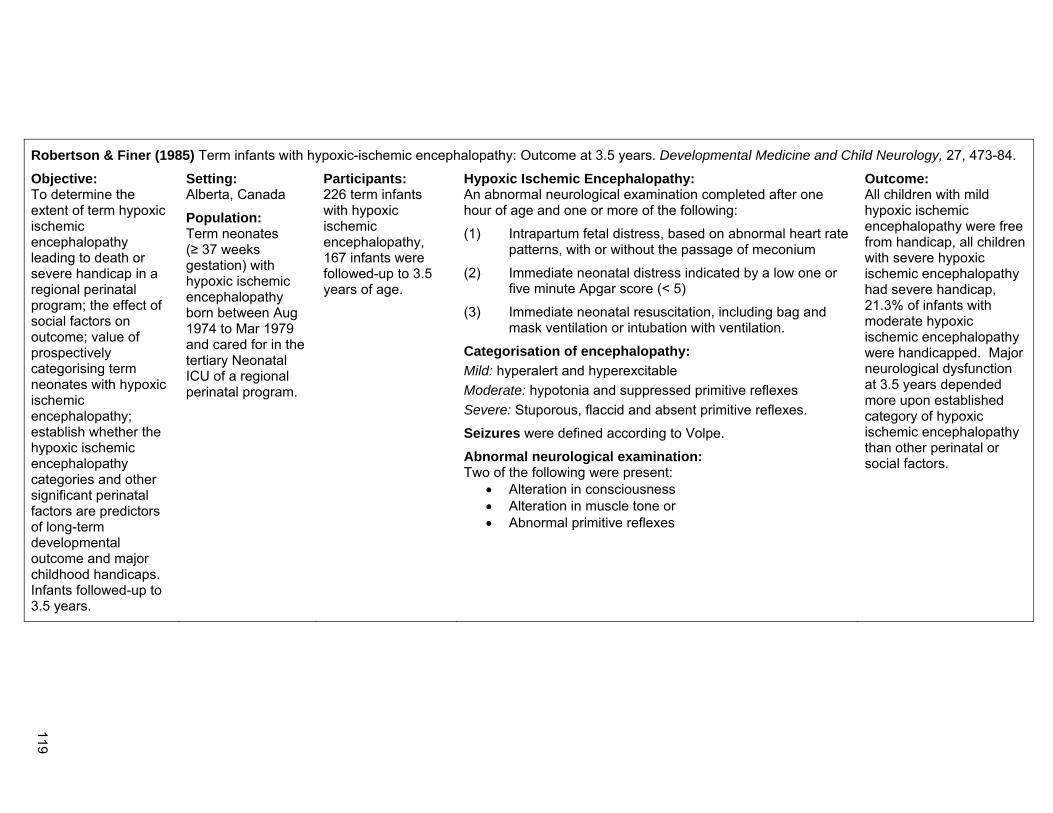
Robertson & Finer (1985) Term infants with hypoxic-ischemic encephalopathy: Outcome at 3.5 years. Developmental Medicine and Child Neurology, 27, 473-84.
Objective: To determine the extent of term hypoxic ischemic encephalopathy leading to death or severe handicap in a regional perinatal program; the effect of social factors on outcome; value of prospectively categorising term neonates with hypoxic ischemic encephalopathy; establish whether the hypoxic ischemic encephalopathy categories and other significant perinatal factors are predictors of long-term developmental outcome and major childhood handicaps. Infants followed-up to 3.5 years.
Setting: Alberta, Canada
Population: Term neonates (≥ 37 weeks gestation) with hypoxic ischemic encephalopathy born between Aug 1974 to Mar 1979 and cared for in the tertiary Neonatal ICU of a regional perinatal program.
Participants: 226 term infants with hypoxic ischemic encephalopathy, 167 infants were followed-up to 3.5 years of age.
Hypoxic Ischemic Encephalopathy: An abnormal neurological examination completed after one hour of age and one or more of the following:
(1) Intrapartum fetal distress, based on abnormal heart rate patterns, with or without the passage of meconium
(2) Immediate neonatal distress indicated by a low one or five minute Apgar score (< 5)
(3) Immediate neonatal resuscitation, including bag and mask ventilation or intubation with ventilation.
Categorisation of encephalopathy: Mild: hyperalert and hyperexcitable Moderate: hypotonia and suppressed primitive reflexes Severe: Stuporous, flaccid and absent primitive reflexes.
Seizures were defined according to Volpe.
Abnormal neurological examination: Two of the following were present:
• Alteration in consciousness • Alteration in muscle tone or • Abnormal primitive reflexes
Outcome: All children with mild hypoxic ischemic encephalopathy were free from handicap, all children with severe hypoxic ischemic encephalopathy had severe handicap, 21.3% of infants with moderate hypoxic ischemic encephalopathy were handicapped. Major neurological dysfunction at 3.5 years depended more upon established category of hypoxic ischemic encephalopathy than other perinatal or social factors.
119

Cyr, Usher & McLean (1984) Changing patterns of birth asphyxia and trauma over 20 years. American Journal of Obstetrics and Gynecology, 148, 490-8.
Study design: Cohort study Objective: To determine whether deleterious outcomes seen in 1960 to 1962 (a prior study) have been avoided and whether any new hazards have developed with increased obstetric interventions. To review the incidence and causes of postasphyxia/post trauma perinatal death in both periods and report on the outcome of follow-up of the most severely asphyxiated infants in the 1978-1980 period.
Setting: Canada Population: Data from a previous study (1960-1962) was compared with births from 1978-1980 at the Royal Victoria Hospital.
Participants: Infants born at the Royal Victoria Hospital in 1978 to 1980 who manifested postasphyxia/ posttrauma encephalopathy. Compared to a prior study in 1966. High-risk referrals from other hospitals were excluded.
Severe birth asphyxia and trauma Severe asphyxia: Requiring more than 3 minutes of resuscitation by positive-pressure ventilation prior to onset of sustained regular breathing.
Postasphyxial/posttrauma encephalopathy: • Neonatal convulsions (tonic/clonic, usually
repetitive, after perinatal asphyxia or trauma)
OR • Abnormal cerebral signs (cerebral irritation,
that is, hypotonia, opisthotonus, tremor, irritability, bicycling movements)
AND/OR • Depression (hypotonia, lethargy, poor
sucking) after perinatal asphyxia or trauma.
The authors also refer to ‘neonatal encephalopathy due to birth asphyxia’ and ‘encephalopathy’ but do not provide definitions for these conditions. It may be assumed they are referring to postasphyxial/posttrauma encephalopathy as neonatal encephalopathy and birth asphyxia for its subset hypoxic-ischemic encephalopathy.
Note: At least 10 different terms were used in this paper including – neonatal encephalopathy, birth asphyxia, birth asphyxia and trauma, severe birth asphyxia, postasphyxia/post trauma encephalopathy, peripartum asphyxia and trauma, neonatal postasphyxia encephalopathy, severe asphyxia, encephalopathy and severe asphyxial trauma.
Incidence: The incidence of severe asphyxia was (per 1000 live births for infants 1001-2499g): 1978-80 = 47.1 1960-62 = 55.8
Outcome: The authors stated that there was a dramatic reduction in perinatal death and neonatal encephalopathy due to birth asphyxia and trauma and only rarely did infants develop permanent cerebral injury. However, the frequency of severe birth asphyxia remained unchanged. They recommended using forceps less often and more carefully, to reduce birth asphyxia. Duration of ventilation required was closely correlated with the frequency of encephalopathy and encephalopathy (especially convulsions) with brain damage. Fetal heart rate patterns were of little predictive value. .
120

Low, Galbraith, Muir et al (1984) Factors associated with motor and cognitive deficits in children after intrapartum fetal hypoxia. American Journal of Obstetrics and Gynecology, 148, 533-9.
Objective: To define the factors which distinguish children with deficits from those without deficits of motor and cognitive development.
Setting: Canada
Population: Not specified
Participants: 60 infants with biochemical evidence of intrapartum fetal hypoxia.
Intrapartum fetal hypoxia: A significant metabolic acidosis expressed by an umbilical artery buffer base < 34.0mEq/L at delivery.
Outcome: Children with deficits had an episode of hypoxia that was more severe and prolonged and subsequent to delivery a greater incidence of severe respiratory complications, apnea and newborn encephalopathy. An episode of hypoxia < 1 hour may occur without subsequent deficits. An episode of hypoxia > 1 hour resulting in a metabolic acidosis (of 25 mEq/L) will result in motor and cognitive deficits in ~50% of children.
The authors claim that the most definitive measure of fetal hypoxia (as used in this study) is blood gas and acid base assessment with evidence of a severe metabolic acidosis (no references to support this are provided).
Note: This paper is similar to the Low et al (1988) paper and may be an early presentation of data from the same study.
121

Fenichel (1983) Hypoxic-ischemic encephalopathy in the newborn. Archives of Neurology, 40, 261-6.
Review article The newborns at risk for major neurologic handicaps have evidence of derangements in many organs, have depressed cerebral function at birth that continues for days or weeks, and in many cases, convulsions soon after birth. In the term newborn, HIE can be divided into three grades of severity according to the clinical symptoms.
Mild encephalopathy: In newborns with mild encephalopathy, the symptoms are maximal during the first 24 hours after birth and then progressively diminish. Consciousness in not materially impaired, except for a brief interval of lethargy immediately after birth. The characteristic feature is jitteriness - a hyperalert state in which there are prolonged periods of wakefulness, irritability, and excessive responsiveness to stimulation. The typical response to stimulation is a low-frequency, high-amplitude shaking of the limbs and jaw. Jitteriness is commonly associated with a low threshold for the Moro reflex, but can occur in the absence of any apparent external manipulation and may be mistaken for a convulsion. Muscle tone is normal when the newborn is at rest or suspended vertically or horizontally. Mild head lag during the traction response is the only demonstrable disturbance in tone. Spontaneous movement and strength are normal in the limbs. Muscle stretch reflexes are normal or slightly hyperactive, and ankle clonus is present and frequently sustained. The anterior fontanel is soft, cranial nerve function is normal, and convulsions do not occur. The EEG is usually normal, but may show lack of background variability. Voltage suppression is not present. The clinical syndrome indicates that the newborn has experienced cerebral distress but does not have cerebral necrosis or increased intracranial pressure. Newborns who have mild, transient HIE recover completely and are not at risk for neurological handicaps. There is no evidence that jitteriness evolves into hyperactivity or that mild encephalopathy is a prologue to learning disabilities.
Moderate encephalopathy: Newborns with a moderate encephalopathy are lethargic or obtunded for at least the first 12 hours after birth. Efforts at arousal produce jitteriness. Hypotonia is present at rest, and spontaneous movement of the limbs is decreased. Proximal weakness, in which the muscles about the shoulder are weaker than the muscles about the pelvis, has been described and attributed to edema occurring in the parasaggital region of the cortex and involving those portions of the motor strip that represent limb-girdle muscles. However, this disparity in strength between the shoulder and pelvic muscles is difficult to appreciate in a newborn who is generally hypotonic. In the usual course of events, the period between 48 and 72 hours after birth is a critical interval during which the encephalopathy either worsens or improves. In those newborns whose condition improves spontaneously, tone increases and arousal is more readily accomplished and associated with jitteriness. In others, there is either lack of improvement or progressive obtundation caused by some combination of convulsions, generalised cerebral edema, hyponatremia secondary to the inappropriate secretion of antidiuretic hormone, and hypoammonemia due to hypoxic liver damage. Convulsions, prolongation of the obtunded state, and progression to stupor are associated with a worsening prognosis. The EEG is always abnormal and may demonstrate lack of background variability, epileptiform activity, or voltage suppression. Lack of background variability has no prognostic implications, but epileptiform activity and voltage suppression are predictive of a bad outcome. Sensory evoked responses can serve as an additional tool for determining the degree of brain damage and the eventual outcome in asphyxiated newborns. Visual evoked responses may prove to be most useful in preterm newborns (the visual radiations are consistently involved in the lesions of periventricular leukomalacia, and brain-stem auditory responses in term newborns. All asphyxiated term newborns in whom the amplitude of wave 1 was more than twice the amplitude of wave 5 either died or were demonstrated to have severe neurological impairment.
122

Severe encephalopathy: Newborns with severe encephalopathy are stuporous or comatose immediately after birth. Respirations are irregular or periodic, and mechanical ventilation is necessary to sustain life. Apnea and convulsions begin during the first 12 hours after birth and progress to tonic and multifocal clonic patterns before the end of the first day. Hypotonia is severe. The newborn lies motionless with legs extended and fully abducted, and the arms remain in any position in which they fall. When traction is tested, there is no grasp reflex, no flexion movements of the head, and no resistance of the limbs. The Moro reflex, tonic neck reflex, and muscle stretch reflexes are usually absent. Pupillary and doll’s eye reflexes are usually normal, but oculomotor palsies may be present. Normal sucking and swallowing are depressed or absent, but intermittent sucking and chewing movements may be present as a convulsive manifestation. Between 12 and 24 hours after birth, some improvement in responsiveness may be noted; stimulation at this time provokes a jittery response. Most children remain stuporous. Convulsions increase in frequency and severity, sometimes progressing to status epilepticus. The EEG is either markedly suppressed or shows a burst-suppression pattern. Overall, deterioration in the newborn’s condition, with brain-stem dysfunction as a prominent feature, occurs between 24 and 72 hours after birth and includes coma, loss of pupillary and vestibulo-ocular reflexes, and respiratory arrest. The fontanel is bulging, and postmortem examination discloses massive cerebral edema and transtentorial herniation. Survivors may remain in a stuporous state for weeks, although convulsions become progressively less frequent and usually cease by the end of the first week. Jitteriness is common as the level of consciousness slowly increases. Some newborns who survive the acute encephalopathy will die later during infancy. Those who live have severe neurologic handicaps.
123

Finer, Robertson, Peters & Coward (1983) Factors affecting outcome in hypoxic-ischemic encephalopathy in term infants. American Journal of Diseases of Children, 137, 21-5.
Objectives: To review the neonatal course and neurodevelopmental outcome in infants with hypoxic ischemic encephalopathy to at least one year of age.
Setting: Edmonton, Alberta
Population: Infants with hypoxic ischemic encephalopathy admitted to the Royal Alexandra Hospital newborn ICU from Sept 1978 to Dec 1979.
Participants: 49 term infants diagnosed with hypoxic-ischemic encephalopathy.
Hypoxic-Ischemic Encephalopathy: The infant initially had abnormal findings from a neurological examination and a history of perinatal distress as shown by one or more of the following: 1. Demonstration of intrapartum fetal distress with the recognition of
abnormal fetal heart rate patterns, with or without the passage of meconium
2. Presence of immediate neonatal distress as shown by a low (≤ 5) one minute or 5 minute Apgar score
AND 3. Need for immediate neonatal resuscitation including bag-and-mask
ventilation, intubation or the need for prolonged mechanical ventilation of greater than 5 minutes.
Abnormal neurological examination: Presence of 2 or more of the following: 1. Alterations of the level of consciousness assessed after vigorous
stimulation to rouse the infant 2. Alterations of muscle tone categorised as global hypotonia or
hypertonia, as well as localised tone disturbances, AND 3. Abnormal primitive reflexes, including Moro’s grasp and sucking
responses.
Sarnat classification: (without EEG) Stage I: Hyperalertness, hyperreflexia, dilated pupils, tachycardia, and absence of seizures. Stage II: Lethargy, hyperreflexia, miosis, bradycardia, seizures, hypotonia, and weak sucking and Moro responses. Stage III: stupor, flaccidity, small to mid-position pupils that react poorly to light, decreased stretch reflexes, hypothermia, and absent Moro’s and sucking responses.
Seizures were observed as described by Volpe.
Outcome: The authors reported an improved outcome from their previous study (Finer et al 1981). The Sarnat staging was concluded to be highly predictive for the term infant with hypoxic ischemic encephalopathy. Administration of Dexamethasone did not appear to be of significant value, although not randomly assigned. Infants with frequent tonic-clonic seizures unresponsive to Phenobarbital therapy and requiring additional anticonvulsant therapy had a greater frequency of significant handicap or death.
124
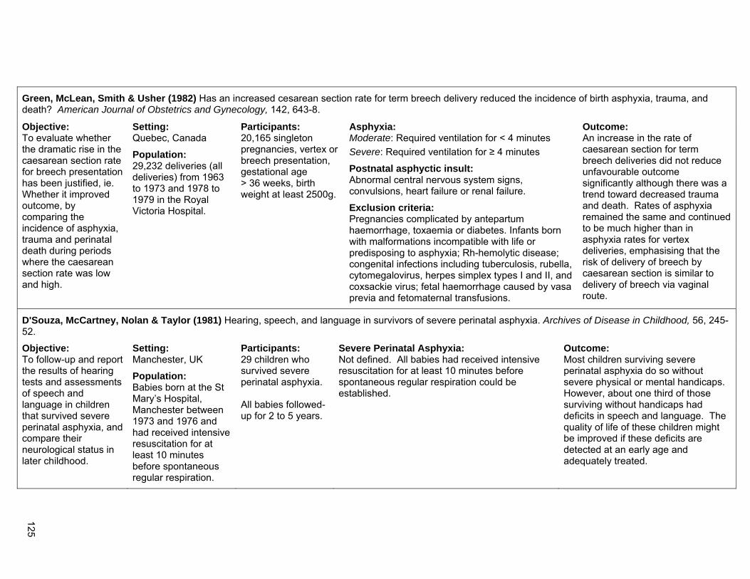
Green, McLean, Smith & Usher (1982) Has an increased cesarean section rate for term breech delivery reduced the incidence of birth asphyxia, trauma, and death? American Journal of Obstetrics and Gynecology, 142, 643-8.
Objective: To evaluate whether the dramatic rise in the caesarean section rate for breech presentation has been justified, ie. Whether it improved outcome, by comparing the incidence of asphyxia, trauma and perinatal death during periods where the caesarean section rate was low and high.
Setting: Quebec, Canada
Population: 29,232 deliveries (all deliveries) from 1963 to 1973 and 1978 to 1979 in the Royal Victoria Hospital.
Participants: 20,165 singleton pregnancies, vertex or breech presentation, gestational age > 36 weeks, birth weight at least 2500g.
Asphyxia: Moderate: Required ventilation for < 4 minutes Severe: Required ventilation for ≥ 4 minutes
Postnatal asphyctic insult: Abnormal central nervous system signs, convulsions, heart failure or renal failure.
Exclusion criteria: Pregnancies complicated by antepartum haemorrhage, toxaemia or diabetes. Infants born with malformations incompatible with life or predisposing to asphyxia; Rh-hemolytic disease; congenital infections including tuberculosis, rubella, cytomegalovirus, herpes simplex types I and II, and coxsackie virus; fetal haemorrhage caused by vasa previa and fetomaternal transfusions.
Outcome: An increase in the rate of caesarean section for term breech deliveries did not reduce unfavourable outcome significantly although there was a trend toward decreased trauma and death. Rates of asphyxia remained the same and continued to be much higher than in asphyxia rates for vertex deliveries, emphasising that the risk of delivery of breech by caesarean section is similar to delivery of breech via vaginal route.
D'Souza, McCartney, Nolan & Taylor (1981) Hearing, speech, and language in survivors of severe perinatal asphyxia. Archives of Disease in Childhood, 56, 245-52.
Objective: To follow-up and report the results of hearing tests and assessments of speech and language in children that survived severe perinatal asphyxia, and compare their neurological status in later childhood.
Setting: Manchester, UK
Population: Babies born at the St Mary’s Hospital, Manchester between 1973 and 1976 and had received intensive resuscitation for at least 10 minutes before spontaneous regular respiration.
Participants: 29 children who survived severe perinatal asphyxia. All babies followed-up for 2 to 5 years.
Severe Perinatal Asphyxia: Not defined. All babies had received intensive resuscitation for at least 10 minutes before spontaneous regular respiration could be established.
Outcome: Most children surviving severe perinatal asphyxia do so without severe physical or mental handicaps. However, about one third of those surviving without handicaps had deficits in speech and language. The quality of life of these children might be improved if these deficits are detected at an early age and adequately treated.
125

D'Souza & Richards (1978) Neurological sequelae in newborn babies after perinatal asphyxia. Archives of Disease in Childhood, 53, 564-9.
Objective: To find out whether the types of neurological signs exhibited by babies (with neurological abnormalities and a history of fetal distress in labour) has any bearing on their outcome in later infancy and childhood. Infants were followed-up for between 2 and 5 years.
Setting: Manchester, UK
Population: Singleton babies born between 1971 and 1975 at St Mary’s hospital.
Participants: 53 babies 37 to 44 weeks gestation with a history of fetal distress in labour diagnosed as severely neurologically abnormal.
Fetal distress and diagnosed as severely neurologically abnormal:
• Unequivocal presence of abnormal tone (hypertonia or hypotonia),
• Irritability on handling (marked increase in spontaneous activity with alteration in respiration, or crying),
• A ‘cerebral’ (high-pitched) cry, or • Convulsions
Outcome: A higher proportion of babies with fetal distress were born by an abnormal delivery. A considerable improvement in function was found in most of the apparently brain-injured babies. The authors suggested that such babies exhibiting apathy initially but subsequently hyperexcitability and extensor hypertonia carry the worst prognosis.
126

Sarnat & Sarnat (1976) Neonatal encephalopathy following fetal distress: A clinical and electroencephalographic study. Archives of Neurology, 33, 696-705. Objective: To provide a systematic clinical approach to the identification of transitory neurologic signs that appear sequentially following asphyxia near or at the time of birth and to suggest a relation of the duration of these signs to prognosis.
Setting: Not specified Population: Not specified
Participants: 21 infants >36 weeks gestation at birth. All had a ‘well defined episode of fetal distress’ or an Apgar score of ≤ 5 at one or 5 minutes after delivery.
Note: A definition was not provided for ‘fetal distress’. Exclusions: Evidence of congenital heart disease, traumatic cerebral injuries, hydrocephalus or infection. Infants with meconium aspiration, respiratory distress syndrome or other illness resulting in chronic, continuing hypoxia.
Stage 1 Stage 2 Stage 3 Level of consciousness Hyperalert Lethargic or obtunded Stuporous Neuromuscular control Muscle tone Normal Mild hypotonia Flaccid Posture Mild distal flexion Strong distal flexion Intermittent
decerebration Stretch reflexes Overactive Overactive Decreased or absent Segmental myoclonus Present Present Absent
Complex reflexes
Suck Weak Weak or absent Absent Moro Strong, low threshold Weak, incomplete, high
threshold Absent
Oculovestibular Normal Overactive Weak or absent Tonic neck Slight Strong Absent
Autonomic function Generalised sympathetic
Generalised parasympathetic
Both systems depressed
Pupils Mydiasis Miosis Variable, often unequal, poor light reflex
Heart rate Tachycardia Bradycardia Variable Bronchial and salivary secretions
Sparse Profuse Variable
Gastrointestinal motility
Normal or decreased Increased, diarrhoea Variable
Seizures None Common, focal or multifocal Uncommon (excluding decerebration)
Electroencephalograph findings
Normal (awake) Early: low voltage continuous delta and theta. Later: periodic pattern (awake). Seizures: focal 1-1½-Hz spike-and-wave
Early: periodic pattern with isopotential phases. Later: Totally isopotential.
Duration Less than 24 hours Two to 14 days Hours to weeks
127
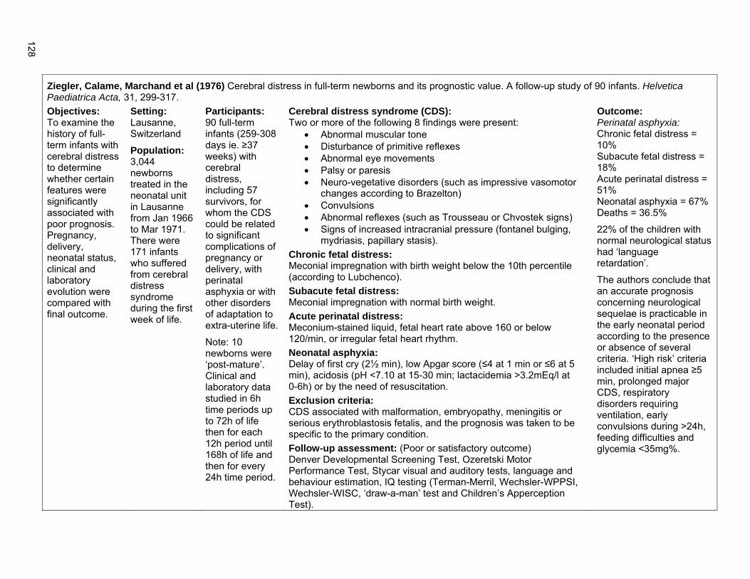
Ziegler, Calame, Marchand et al (1976) Cerebral distress in full-term newborns and its prognostic value. A follow-up study of 90 infants. Helvetica Paediatrica Acta, 31, 299-317. Objectives: To examine the history of full-term infants with cerebral distress to determine whether certain features were significantly associated with poor prognosis. Pregnancy, delivery, neonatal status, clinical and laboratory evolution were compared with final outcome.
Setting: Lausanne, Switzerland
Population: 3,044 newborns treated in the neonatal unit in Lausanne from Jan 1966 to Mar 1971. There were 171 infants who suffered from cerebral distress syndrome during the first week of life.
Participants: 90 full-term infants (259-308 days ie. ≥37 weeks) with cerebral distress, including 57 survivors, for whom the CDS could be related to significant complications of pregnancy or delivery, with perinatal asphyxia or with other disorders of adaptation to extra-uterine life.
Note: 10 newborns were ‘post-mature’. Clinical and laboratory data studied in 6h time periods up to 72h of life then for each 12h period until 168h of life and then for every 24h time period.
Cerebral distress syndrome (CDS): Two or more of the following 8 findings were present:
• Abnormal muscular tone • Disturbance of primitive reflexes • Abnormal eye movements • Palsy or paresis • Neuro-vegetative disorders (such as impressive vasomotor
changes according to Brazelton) • Convulsions • Abnormal reflexes (such as Trousseau or Chvostek signs) • Signs of increased intracranial pressure (fontanel bulging,
mydriasis, papillary stasis). Chronic fetal distress: Meconial impregnation with birth weight below the 10th percentile (according to Lubchenco). Subacute fetal distress: Meconial impregnation with normal birth weight. Acute perinatal distress: Meconium-stained liquid, fetal heart rate above 160 or below 120/min, or irregular fetal heart rhythm. Neonatal asphyxia: Delay of first cry (2½ min), low Apgar score (≤4 at 1 min or ≤6 at 5 min), acidosis (pH <7.10 at 15-30 min; lactacidemia >3.2mEq/l at 0-6h) or by the need of resuscitation. Exclusion criteria: CDS associated with malformation, embryopathy, meningitis or serious erythroblastosis fetalis, and the prognosis was taken to be specific to the primary condition. Follow-up assessment: (Poor or satisfactory outcome) Denver Developmental Screening Test, Ozeretski Motor Performance Test, Stycar visual and auditory tests, language and behaviour estimation, IQ testing (Terman-Merril, Wechsler-WPPSI, Wechsler-WISC, ‘draw-a-man’ test and Children’s Apperception Test).
Outcome: Perinatal asphyxia: Chronic fetal distress = 10% Subacute fetal distress = 18% Acute perinatal distress = 51% Neonatal asphyxia = 67% Deaths = 36.5%
22% of the children with normal neurological status had ‘language retardation’.
The authors conclude that an accurate prognosis concerning neurological sequelae is practicable in the early neonatal period according to the presence or absence of several criteria. ‘High risk’ criteria included initial apnea ≥5 min, prolonged major CDS, respiratory disorders requiring ventilation, early convulsions during >24h, feeding difficulties and glycemia <35mg%.
128

Amiel-Tison C (1969) Cerebral damage in full-term newborn aetiological factors, neonatal status and long-term follow-up. Biology of the Neonate, 14, 234-250.
Purpose: To establish the neuro-psychological prognosis of babies born at term who presented after birth with signs of severe brain damage.
Setting: Paris
Population: Babies born from 1962 to 1964 at term or near term (at least 38 weeks) at the Baudelcoque Maternity Hospital. Follow-up was completed up to 2 to 5 years.
Participants: 41 babies with neurological signs
Signs of cerebral damage/severe cerebral symptoms: Separately defined conditions including status epilepticus, irritability syndrome, disturbances of muscle tone, absence of primary reflexes, ocular signs, disturbances of consciousness, respiratory signs and signs of intra-cranial hypertension. Further described in the paper.
Exclusions: Meningitis or kernicterus, any obvious trisomic syndromes or immediately obvious congenital malformations.
Outcome: Long standing fetal distress (whatever the cause) leads to severe permanent damage. Obstetrical trauma causing an acute but brief distress may lead to brain damage from which complete recovery is possible provided that the baby does not have status epilepticus. No cerebral palsy was seen in the 25 children reviewed between 2 and 5 years of age, motor sequelae appear rare after birth trauma in full-term newborns. Conclusions could not be made about intellectual deficit. A very active attitude towards treatment of these babies can be justified except in the case of status epilepticus. Nearly two thirds of these babies will become normal children.
129