research article acoustic cavitation enhances focused
TRANSCRIPT

Research ArticleAcoustic Cavitation Enhances Focused Ultrasound Ablationwith Phase-Shift Inorganic Perfluorohexane Nanoemulsions:An In Vitro Study Using a Clinical Device
Lu-Yan Zhao,1 Jian-Zhong Zou,1 Zong-Gui Chen,1 Shan Liu,1 Jiao Jiao,1 and Feng Wu1,2
1State Key Laboratory of Ultrasound Engineering in Medicine, College of Biomedical Engineering, Chongqing Medical University,Chongqing, China2HIFU Unit, The Churchill Hospital, Oxford University Hospitals, Headington, Oxford OX3 7LE, UK
Correspondence should be addressed to Feng Wu; [email protected]
Received 18 February 2016; Accepted 15 May 2016
Academic Editor: Enzo Terreno
Copyright © 2016 Lu-Yan Zhao et al. This is an open access article distributed under the Creative Commons Attribution License,which permits unrestricted use, distribution, and reproduction in any medium, provided the original work is properly cited.
Purpose.To investigatewhether acoustic cavitation could increase the evaporation of a phase-shift inorganic perfluorohexane (PFH)nanoemulsion and enhance high intensity focused ultrasound (HIFU) ablation. Materials and Methods. PFH was encapsulatedby mesoporous silica nanocapsule (MSNC) to form a nanometer-sized droplet (MSNC-PFH). It was added to a tissue-mimickingphantom, whereas phosphate buffered saline (PBS) was added as a control (PBS-control). HIFU (𝑃ac = 150W, 𝑡 = 5/10 s) exposureswere performed in both phantoms with various duty cycles (DC). US images, temperature, and cavitation emissions were recordedduring HIFU exposure. HIFU-induced lesions were measured and calculated. Results. Compared to PBS-control, MSNC-PFHnanoemulsion could significantly increase the volume of HIFU-induced lesion (𝑃 < 0.01). Peak temperatures were 78.16 ± 5.64∘Cat a DC of 100%, 70.17 ± 6.43∘C at 10%, 53.17 ± 4.54∘C at 5%, and 42.00 ± 5.55∘C at 2%, respectively. Inertial cavitation was muchstronger in the pulsed-HIFU than that in the continuous-waveHIFU exposure. Compared to 100%-DC exposure, themean volumeof lesion induced by 5 s exposure at 10%-DCwas significantly larger, but smaller at 2%-DC.Conclusions.MSNC-PFHnanoemulsioncan significantly enhance HIFU ablation. Appropriate pulsed-HIFU exposure could significantly increase the volume of lesion andreduce total US energy required for HIFU ablation.
1. Introduction
As one of the most promising noninvasive treatment modali-ties, high intensity focused ultrasound (HIFU) has been suc-cessfully used in the clinical management of cancer patients[1–3]. However, it needs long treatment duration to ablatethe volume of a clinically relevant tumor. In addition, due torapid energy attenuation along the focused ultrasound (US)pathway, acoustic intensities at the focus are not enough toablate a deep-seated tumor efficiently and completely. Thesepotentially limit the use of HIFU as a routine treatment in theclinical applications.
To address this clinical need, microbubble ultrasoundcontrast agents have been already investigated to enhanceHIFU thermal ablation in experimental studies. They areusually used for diagnostic ultrasound imaging. In HIFU
treatment regime they serve to nucleate cavitation andincrease ultrasonic absorption, resulting in a larger volumeof ablation in a shorter amount of time [4–6]. However,circulating microbubbles have a very short half-life in vivo(minutes) and rapidly disappear from the circulation [7].They are also too large to extravasate from the vascularspace to tissue, leading to enhanced heat that occurs only inand around blood vessels [8]. In addition, they can enhanceHIFU-mediated heating at multiple points along the beampath, leading to unwanted damage to the tissues proximal tothe transducer focus [9].
An alternative to the microbubbles is phase-shift per-fluorocarbon (PFC) nanoparticles, which serve as in situcavitation nucleation agents. Under HIFU exposure, thePFC phase is expected to change from liquid to gas form,and a large amount of microbubbles from evaporation can
Hindawi Publishing CorporationBioMed Research InternationalVolume 2016, Article ID 7936902, 9 pageshttp://dx.doi.org/10.1155/2016/7936902

2 BioMed Research International
Table 1: Total ultrasound energy delivered for HIFU exposures at varied duty cycles.
Duty cycles Acoustic power (watts) Exposure duration (second) Total ultrasound energy (joules)2% exposure 150 5 10 15 (5 s) 30 (10 s)5% exposure 150 5 10 37.5 (5 s) 75 (10 s)10% exposure 150 5 10 75 (5 s) 150 (10 s)100% exposure 150 5 10 750 (5 s) 1500 (10 s)
subsequently enhance HIFU thermal effect on the targetedtissues [10–14]. In addition, they have a significantly longerhalf-life than gas-filled microbubbles in the circulation [15].As one of PFC compounds, perfluorohexane (PFH) is atemperature-sensitive biocompatible liquid with a boilingpoint of about 56∘C. It can be encapsulated by mesoporoussilica nanocapsule (MSNC) to form a nanometer-sized inor-ganic agent (MSNC-PFH). Wang et al. [12] found that, dueto the evaporation from local temperature rise, MSNC-PFH could significantly enhance HIFU thermal ablation.However, it was still unknown whether acoustic cavitationcould significantly facilitate the phase transformation of theMSNC-PFH nanoemulsion, leading to the enhancement ofHIFU ablation. Using a clinical HIFU device the aim of thisstudy was to investigate whether acoustic cavitation couldincrease the evaporation of MSNC-PFH and subsequentlyenhance HIFU ablation in a tissue-mimicking phantom.
2. Material and Methods
2.1. Phase-Shift InorganicMSNC-PFHNanoemulsion. MSNC-PFH nanoemulsion was kindly provided by Professor Han-grong Chen at State Key Laboratory of High PerformanceCeramic and Superfine Microstructures, Shanghai Instituteof Ceramics, Chinese Academy of Science (Shanghai, China).The preparation and characteristics of MSNC-PFH werepreviously described in detail [12]. Briefly, it consisted ofmesoporous silica nanocapsule as a carrier and perfluorohex-ane liquid as the inner core. Under electron microscopy theaverage diameter of the preparedMSNC-PFH nanoemulsionwas around 300 nm with mesoporous shell thickness of50 nm. Dynamic light scattering showed that it had a narrowsize distribution with an overall hydrodynamic diameter of346 nm in water. It was stably and uniformly dispersed inwater with the vaporization temperature of around 56∘C.
2.2. Tissue-Mimicking Phantom. Based on previously de-scribed methods [16], an egg white-based, heat-responsivephantomwas fabricated in the study. It was nearly transparentat room temperature. When heated up to 60∘C, the phantombecame a visibly opaque lesion because of the denaturationand coagulation of egg white protein.
The phantom consisted of 15% acrylamide solution(Sigma-Aldrich, St. Louis, MO), 40% egg white, 44.5%degassed deionized water, and 0.5% ammonium persulfatesolution (Sigma-Aldrich). After the mixed solution wasdegassed for 10min, 0.2mL MSNC-PFH nanoemulsion wasadded and then stirred gently to achieve a uniform distribu-tion. The concentration of droplets was 107 droplets/mL in
the phantom. In comparison, the same amount of phosphatebuffered saline (PBS) was added as a control (PBS-control)without MSNC-PFH nanoemulsion. Finally, 0.15mL 1,2-bis(dimethylamino)ethane (Sigma-Aldrich) was added to theentire solution to initiate polymerization. The phantom waskept in a 12∘C water bath during polymerization period. Thedimensions of each phantom used in the experiments werearound 6 × 5 × 3.3 cm.
2.3. High Intensity Focused Ultrasound System. Experimentswere carried out using a clinical CE-approved ultrasound-guided HIFU system (Model JC200, Chongqing Haifu Med-ical Technology Co., Ltd., Chongqing, China). A diagnos-tic probe (Esaote, Genoa, Italy) was located in the centerof a concave HIFU transducer operating at 0.9MHz. Theintegrated transducer can be automatically moved in sixdirections.The diameter of theHIFU transducer was 220mmwith the focal length of 145mm. The focal region wasellipsoid, with dimensions of 8mm along the longitudinalbeam axis and 3mm in the transverse direction.
All exposures were performed at varied duty cycles (DC).The acoustic power delivered for all the experiments was150 watts, and focal intensity (𝐼SPTA, spatial-peak temporal-average intensity) was 9750W/cm2. Exposure duration wasset to either 5 s or 10 s.TheDC ofHIFU exposure was 2%, 5%,10%, and 100%, respectively, at a pulse repetition frequencyof 100Hz.The total energy delivered for each HIFU exposurewas shown in Table 1.
2.4. Experimental Setup. Schematic diagram of the in vitroexperimental setup was shown in Figure 1. The phantomwas immersed in a large tank filled with degassed water andplaced above the integrated HIFU transducer and diagnosticprobe with real-time monitoring of US imaging. A 0.7mmneedle thermocouple (Omega Engineering Inc., Stamford,Connecticut, USA) was used for temperature measurementsduring HIFU exposure. It was inserted into the phantomat the focal plane, paralleled to the HIFU beam axis but0.1mm off-axis laterally in order to reduce the artifact effectof US vibration on the thermocouple tip. By moving thephantom, the HIFU focus was correctly positioned aroundthe thermocouple tip under US imaging guidance. A low-power HIFU exposure (𝑃ac = 30W, 𝑡 = 1 s) was testedto confirm the exact position of the thermocouple tip untilthe maximal temperature rise was observed in 𝑥, 𝑦, and 𝑧axes, respectively. During HIFU exposure the temperaturechange in the phantom was recorded by a temperature datalogger (Model FLE5008, Hangzhou Fenle Electronics Co.Ltd., Zhejiang, China).

BioMed Research International 3
Central console
Thermocouple
Egg white phantom
Passive cavitation detector
HIFU power generator
US imaging probe
HIFU transducer
Ultrasound imaging system
0Digital signal analyzer
Temperature acquisition system
(3.5MHz)
(0.9MHz)
Acquisition (cycle 100ms)
(5MHz)
Figure 1: Schematic diagram of the setup for ultrasound-guidedhigh intensity focused ultrasound experiments. The phantom isplaced above a 0.9MHzHIFU transducerwith real-timemonitoringof ultrasound imaging. Both passive cavitation detector and thermalcouple are separately placed around the phantom.
A passive cavitation detection (PCD) system was usedto detect the acoustic emission at the focus of the HIFUtransducer. It consisted of a 5MHz focused transducer (V309,Panametrics,Waltham,MA, USA) and a high-speed digitizer(PXIe-5122, National Instruments, Austin, TX, USA). Theaperture of the PCD transducer was 13mm and focal lengthwas 40mmwith a bandwidth of 3.3–7MHz at the−6 dB level.In order to detect acoustic emissions from the focus, thistransducerwas placed perpendicular to and confocal with theHIFU beams. The acoustic emission signals were sampled at20MHz rate by the digitizer and finally saved by the com-puter. LabVIEW software (National Instruments) graphicalprogramming language was used to create the displacementcalculation algorithms used in the spectral analysis. Using fastFourier transform routines, all sampled waveforms were firsttransformed to the frequency domain. The level of inertialcavitation was then determined by calculating the root meansquare (RMS) amplitude of the broadband noise for eachspectrum using a method similar to Chen et al. [17]. Thecalculated RMS amplitude of the broadband noise producedby HIFU exposure was superimposed on the backgroundnoise, which could be subtracted as a baseline.
2.5. Ultrasound Image Analysis. Real-time US images ob-tained before and after each HIFU exposure were immedi-ately compared to determine whether a hyperechoic regionappeared at the HIFU focus. The hyperechoic region wasdefined as a region with a distinct increase in the grayscaleintensity that was easily observable by aHIFUoperator. It wasa clinically useful sign indicating the extent of coagulationnecrosis. UsingHIFU device software, the extent of the brighthyperechoic region at the HIFU focus was determined bythe operator, and then the area of the hyperechogenicity wasautomatically measured. In addition, real-time US imagingvideos were recorded during pulsed-HIFU exposure.
2.6. Lesion Volume Assessment. Ablation lesions were visibleas opaque regions in the transparent phantom. After HIFUablation, the phantomswere sliced into 1-2mm along the lon-gitude beam axis, and each ablation lesion was determined bydirect visualization andmeasuredwith aVernier caliper.They
included themaximal length of the lesion along the longitudebeam axis and the maximal width along the perpendicularaxis. The volume of lesion was calculated using the formula:Volume = 𝜋 ×Maximum Length ×Maximum Width2/6.
2.7. Statistical Analysis. SPSS version 17.0 software (SPSS,Chicago, IL, USA) was used for statistical analysis in thisstudy. Data sets were evaluated using one-way analysis ofvariance (ANOVA), Student’s 𝑡-test, and the least significantdifference 𝑡-test, respectively. All measurement data areexpressed as mean values ± standard deviations. At least6 HIFU exposures were performed for each experimentalcondition and 𝑃 values less than 0.05 were considered to bestatistically significant.
3. Results
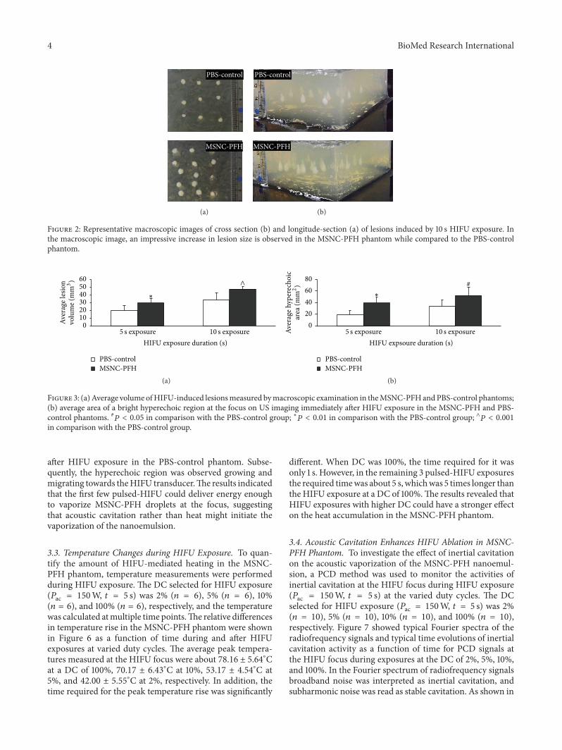
3.1. MSNC-PFH Nanoemulsion Increases the Volume HIFU-Induced Lesion. To investigate the effect of MSNC-PFHnanoemulsion on HIFU ablation, HIFU exposures (𝑃ac =150W, 𝑡 = 5 s or 10 s, DC = 100%) were performed in theMSNC-PFH (𝑛 = 10) and PBS-control phantoms (𝑛 = 10),respectively. Representative images of macroscopic lesionsinduced by 10 s HIFU exposure were shown in Figure 2,including the cross section and longitude-section of theHIFU lesions between the MSNC-PFH and PBS-controlphantoms. Compared to the PBS-control, the mean volumeof lesions induced by either 5 s or 10 s exposures was signifi-cantly larger in theMSNC-PFHphantom (Figure 3(a)).Therewere statistical differences between theMSNC-PFHandPBS-control phantoms in 5 s exposure (𝑃 < 0.001) and 10 s expo-sure (𝑃 < 0.01). These results demonstrated that MSNC-PFH nanoemulsion could enhance HIFU thermal ablation,resulting in a larger volume of lesions in the phantom.
Real-time B-mode US images were also collected beforeand immediately after HIFU exposure to determine a hyper-echoic area at the HIFU focus between the MSNC-PFHand PBS-control phantoms. Representative images beforeand immediately after HIFU in both MSNC-PFH and PBSphantoms were shown in Figure 4. A bright hyperechoicregion was obviously seen at the HIFU focus on US imagingwhile compared to the imaging before HIFU. The mean areaof the hyperechogenicity in the MSNC-PFH phantom wassignificantly larger than that in the PBS-control phantom.There were statistical differences between them in 5 s expo-sure (𝑃 < 0.01) and in 10 s exposure (𝑃 < 0.05), as shown inFigure 3(b).
3.2. Real-Time US Imaging during HIFU Exposure. In orderto reduce the interference of HIFU with the imaging system,pulsed-HIFU exposure was used to help the recording ofreal-time US imaging videos. HIFU exposures with 2% DC(𝑃ac = 150W, 𝑡 = 10 s) were performed to determine whenthe hyperechogenicity occurred at the HIFU focus in bothMSNC-PFH (𝑛 = 6) and PBS phantoms (𝑛 = 6). As shownin Figure 5, a bright hyperechoic region occurred on theUS imaging at 0.1 s after HIFU exposure in the MSNC-PFHphantom, whereas the hyperechogenicity occurred at 0.9 s
4 BioMed Research International
PBS-control
MSNC-PFH
(a)
PBS-control
MSNC-PFH
(b)
Figure 2: Representative macroscopic images of cross section (b) and longitude-section (a) of lesions induced by 10 s HIFU exposure. Inthe macroscopic image, an impressive increase in lesion size is observed in the MSNC-PFH phantom while compared to the PBS-controlphantom.
Aver
age l
esio
n
PBS-controlMSNC-PFH
HIFU exposure duration (s)10 s exposure5 s exposure
∗
0102030405060
volu
me (
mm
3) ∧
(a)
Aver
age h
yper
echo
ic
PBS-controlMSNC-PFH
HIFU expsoure duration (s)10 s exposure5 s exposure
∗
#
020406080
area
(mm
2)
(b)
Figure 3: (a)Average volumeofHIFU-induced lesionsmeasured bymacroscopic examination in theMSNC-PFHandPBS-control phantoms;(b) average area of a bright hyperechoic region at the focus on US imaging immediately after HIFU exposure in the MSNC-PFH and PBS-control phantoms. #𝑃 < 0.05 in comparison with the PBS-control group; ∗𝑃 < 0.01 in comparison with the PBS-control group; ∧𝑃 < 0.001in comparison with the PBS-control group.
after HIFU exposure in the PBS-control phantom. Subse-quently, the hyperechoic region was observed growing andmigrating towards theHIFU transducer.The results indicatedthat the first few pulsed-HIFU could deliver energy enoughto vaporize MSNC-PFH droplets at the focus, suggestingthat acoustic cavitation rather than heat might initiate thevaporization of the nanoemulsion.
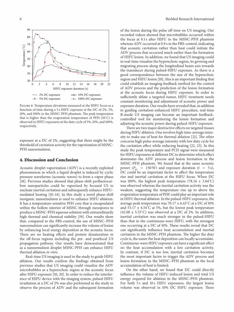
3.3. Temperature Changes during HIFU Exposure. To quan-tify the amount of HIFU-mediated heating in the MSNC-PFH phantom, temperature measurements were performedduring HIFU exposure. The DC selected for HIFU exposure(𝑃ac = 150W, 𝑡 = 5 s) was 2% (𝑛 = 6), 5% (𝑛 = 6), 10%(𝑛 = 6), and 100% (𝑛 = 6), respectively, and the temperaturewas calculated atmultiple time points.The relative differencesin temperature rise in the MSNC-PFH phantom were shownin Figure 6 as a function of time during and after HIFUexposures at varied duty cycles. The average peak tempera-tures measured at the HIFU focus were about 78.16 ± 5.64∘Cat a DC of 100%, 70.17 ± 6.43∘C at 10%, 53.17 ± 4.54∘C at5%, and 42.00 ± 5.55∘C at 2%, respectively. In addition, thetime required for the peak temperature rise was significantly
different. When DC was 100%, the time required for it wasonly 1 s. However, in the remaining 3 pulsed-HIFU exposuresthe required timewas about 5 s, whichwas 5 times longer thanthe HIFU exposure at a DC of 100%.The results revealed thatHIFU exposures with higher DC could have a stronger effecton the heat accumulation in the MSNC-PFH phantom.
3.4. Acoustic Cavitation Enhances HIFU Ablation in MSNC-PFH Phantom. To investigate the effect of inertial cavitationon the acoustic vaporization of the MSNC-PFH nanoemul-sion, a PCD method was used to monitor the activities ofinertial cavitation at the HIFU focus during HIFU exposure(𝑃ac = 150W, 𝑡 = 5 s) at the varied duty cycles. The DCselected for HIFU exposure (𝑃ac = 150W, 𝑡 = 5 s) was 2%(𝑛 = 10), 5% (𝑛 = 10), 10% (𝑛 = 10), and 100% (𝑛 = 10),respectively. Figure 7 showed typical Fourier spectra of theradiofrequency signals and typical time evolutions of inertialcavitation activity as a function of time for PCD signals atthe HIFU focus during exposures at the DC of 2%, 5%, 10%,and 100%. In the Fourier spectrum of radiofrequency signalsbroadband noise was interpreted as inertial cavitation, andsubharmonic noise was read as stable cavitation. As shown in

BioMed Research International 5
HIF
U b
eam
dire
ctio
n
10
s exp
osur
e5
s exp
osur
e
(a) PBS-control (left: pre-HIFU, right: post-HIFU)
10
s exp
osur
e5
s exp
osur
e
HIF
U b
eam
dire
ctio
n
(b) MSNC-PFH (left: pre-HIFU, right: post-HIFU)
Figure 4: Representative real-time ultrasound images of theMSNC-PFH and PBS-control phantoms before and immediately after 5 s and 10 sHIFU exposure at a duty cycle of 100%. A bright hyperechoic region (arrowhead) is observed immediately after exposure in bothMSNC-PFHand PBS-control phantoms.
HIF
U b
eam
dire
ctio
n
7 s 9 s 10 s
5 s4 s2 s 3 s
8 s
1 s0.9 s0.7 s0.5 s
0.3 s0.2 s0.1 s0 s (start)
(a) PBS-control
HIF
U b
eam
dire
ctio
n
7 s 9 s 10 s
5 s4 s2 s 3 s
8 s
1 s0.9 s0.7 s0.5 s
0.3 s0.2 s0.1 s0 s (start)
(b) MSNC-PFH
Figure 5: Representative real-time ultrasound images of time evolution of a hyperechoic region (arrowhead) with 10 s HIFU exposure at aduty cycle of 2% in the MSNC-PFH and PBS-control phantoms. (a) Hyperechoic changes at the HIFU focus in the PBS-control phantom: abright hyperechoic region occurs on the US imaging at 0.9 s after HIFU exposure, with expanded views of the region of the HIFU lesion (from1 s to 10 s). (b) Hyperechoic changes at the HIFU focus in the MSNC-PFH phantom: a bright hyperechoic region occurs on the US imagingat 0.1 s after HIFU exposure, with expanded views of the region of the HIFU lesion (from 0.2 s to 10 s). During pulsed-HIFU exposure, thehyperechoic region is observed growing and migrating towards the HIFU transducer in both MSNC-PFH and PBS-control phantoms.
Figure 7(a), significant increases were observed in the levelof both broadband and subharmonic noises during HIFUexposures. Erratic changes with respect to rise and fall inamplitude of inertial cavitation level were also seen, with anoverall increase in level of cavitation during HIFU exposuresat the various DC (Figure 7(b)). However, our results showedthat inertial cavitation was much stronger in the pulsed-HIFU than that in the continuous-wave (100% DC) HIFUexposure. Among them, the strongest cavitation activity wasobserved at a DC of 10%.
Subsequently, the mean volume of lesions was measuredby macroscopic examination in the MSNC-PFH phantomafter HIFU exposures (𝑃ac = 150W, 𝑡 = 5 s or 10 s) at thevariousDC.As shown in Figure 8, themean volumes of lesionwere directly related to theDCofHIFU exposure. For both 5 sand 10 s HIFU exposures, the mean lesion volumes induced
by HIFU exposure at the DC of 100%, 10%, 5%, and 2% were29.55 ± 5.51 and 47.48 ± 11.69, 49.76 ± 6.12 and 50.98 ± 7.61,35.36±8.28 and 41.22±4.67, and 20.38±4.77 and 28.65±4.10,respectively. Compared to the exposure at a DC of 100%,the mean volume of lesion at a DC of 2% was significantlysmaller in 5 s exposure (𝑃 < 0.005) and 10 s exposure (𝑃 <0.001). However, the mean volume of lesion induced by 5 sexposure at a DC of 10% was significantly larger than that ata DC of 100% (𝑃 < 0.005). No significant difference of thelesion volume in 5 s and 10 s exposure was observed betweenthe exposures at the DC of 100% and 5% (𝑃 > 0.05). Theresults revealed that acoustic cavitation delivered by HIFUexposure at a DC of 10% could significantly increase thevaporization of MSNC-PFH droplets, resulting in strongercavitation-enhanced HIFU ablation. However, our study alsoshowed that this effect was significantly limited in HIFU
6 BioMed Research International
90
80
70
60
50
40
30
20
Tem
pera
ture
(∘C)
2220181614121086420
∗
∗
∗
∗
∗
∗∗
∗∗
∗∗
#
# #
#
##
# # #
∧
∧
∧
∧
∧
∧
∧
∧∧
∧ ∧
#2% DC exposure5% DC exposure ∗
∧ 10% DC exposure100% DC exposure
HIFU exposure duration (s)
Figure 6: Temperature elevations measured at the HIFU focus as afunction of time during a 5 s HIFU exposure at the DC of 2%, 5%,10%, and 100% in the MSNC-PFH phantom. The peak temperaturethat is higher than the evaporation temperature of PFH (56∘C) isobserved in HIFU exposures at the duty cycle of 5%, 10%, and 100%,respectively.
exposure at a DC of 2%, suggesting that there might be thethreshold of cavitation activity for the vaporization ofMSNC-PFH nanoemulsion.
4. Discussion and Conclusion
Acoustic droplet vaporization (ADV) is a recently exploitedphenomenon in which a liquid droplet is induced by cyclicpressure waveforms (acoustic waves) to form a vapor phase[18]. Previous studies showed that phase-shift perfluorocar-bon nanoparticles could be vaporized by focused US tonucleate inertial cavitation and subsequently enhance HIFU-mediated heating [10–14]. In this study a novel phase-shiftinorganic nanoemulsion is used to enhance HIFU ablation.It has a temperature-sensitive PFH core that is encapsulatedwithin the hollow interior of MSNC through mesopores toproduce aMSNC-PFH aqueous solution with extraordinarilyhigh thermal and chemical stability [19]. Our results showthat, compared to the PBS-control, the use of MSNC–PFHnanoemulsion can significantly increase the volume of lesionby enhancing local energy deposition at the acoustic focus.There are no heating effects and protein denaturation inthe off-focus regions including the pre- and postfocal USpropagation pathway. Our results have demonstrated thatas a nanoemulsion droplet MSNC-PFH can enhance HIFUthermal ablation in vitro.
Real-time US imaging is used in the study to guide HIFUablation. Our results confirm the findings obtained fromprevious studies that US imaging could visualize the ADVmicrobubbles as a hyperechoic region at the acoustic focusafter HIFU exposure [10, 20]. In order to reduce the interfer-ence of HIFU device with the imaging system, pulsed-HIFUirradiation at a DC of 2% was also performed in the study toobserve the process of ADV and the subsequent formation
of the lesion during the pulse off-time on US imaging. Ourrecorded videos showed that microbubbles occurred withinthe focus at 0.1 s after HIFU in the MSNC-PFH phantomwhereas ADV occurred at 0.9 s in the PBS-control, indicatingthat acoustic cavitation rather than heat could initiate theADV process that occurred much earlier than the formationof HIFU lesion. In addition, we found that US imaging couldin real-time visualize the hyperechoic region, its growing andmigrating process along the longitudinal beam axis towardsthe transducer during pulsed-HIFU exposure. As there is agood correspondence between the size of the hyperechoicregion and HIFU lesion [10], this is an important finding thatcould establish an imaging feedback method for the controlof ADV process and the prediction of the lesion formationat the acoustic focus during HIFU exposure. In order tosufficiently ablate a targeted tumor, HIFU treatment needsconstant monitoring and adjustment of acoustic power andexposure duration. Our results have revealed that, in additionto guiding cavitation-enhanced HIFU procedure, real-timeB-mode US imaging can become an important feedback-controlled tool for monitoring the lesion formation andadjusting the acoustic power during pulsed-HIFU exposure.
There are twomajor destructive effects on targeted tissuesduringHIFU ablation. One involves high time-average inten-sity to make use of heat for thermal ablation [21]. The otherconcerns high pulse-average intensity with low duty cycle forthe cavitation effect while reducing heating [22, 23]. In thisstudy the peak temperature and PCD signal were measuredforHIFU exposures at different DC to determine which effectdominates the ADV process and lesion formation in theMSNC-PFH phantom. We found that at the same acousticpower (𝑃ac = 150W) and exposure duration (𝑡 = 5 s),DC could be an important factor to affect the temperaturerise and inertial cavitation at the HIFU focus. When DCwas 100%, the highest peak temperature (78.16 ± 5.64∘C)was observed whereas the inertial cavitation activity was theweakest, suggesting the temperature rise up to above theevaporation temperature of PHF could play an important roleinHIFU thermal ablation. In the pulsed-HIFU exposures, theaverage peak temperature was 70.17 ± 6.43∘C at a DC of 10%and 53.17 ± 4.54∘C at 5%, but the lowest peak temperature(42.00 ± 5.55∘C) was observed at a DC of 2%. In addition,inertial cavitation was much stronger in the pulsed-HIFUthan that in the continuous-wave HIFU, with the strongestone occurring at a DC of 10%. These results reveal that DCcan significantly influence heat accumulation and inertialcavitation in the MSNC-PFH phantom. The higher the dutycycle is, the easier the heat deposition can locally accumulate.Continuous-waveHIFUexposure canhave a significant effecton the heat accumulation with a low cavitation activity.In contrast, if DC is too low, inertial cavitation becomesthe most important factor to trigger the ADV process andlesion formation in the MSNC–PFH phantom as the localaccumulation of heat is limited.
On the other hand, we found that DC could directlyinfluence the volume of HIFU-induced lesion and total USenergy required for ablation in the MSNC-PFH phantom.For both 5 s and 10 s HIFU exposures, the largest lesionvolume was observed in 10% DC HIFU exposure. There

BioMed Research International 7
100% DC exposure
Frequency (Hz)
Am
plitu
de (d
B)−20
−40
−60
−80
−100
−120
−140
−160
0 1E + 71 × 106
3 × 10⁶
5 × 10⁶
7 × 10⁶
Am
plitu
de (d
B)
10% DC exposure−20
−40
−60
−80
−100
−120
−140
−160
Frequency (Hz)0 1E + 71 × 10
63 × 10
65 × 10
67 × 10
6
Am
plitu
de (d
B)
5% DC exposure−20
−40
−60
−80
−100
−120
−140
−160
Frequency (Hz)0 1E + 71 × 10
63 × 10
65 × 10
67 × 10
6
Am
plitu
de (d
B)
2% DC exposure−20
−40
−60
−80
−100
−120
−140
−160
Frequency (Hz)0 1E + 71 × 10
63 × 10
65 × 10
67 × 10
6
(a) Fourier spectrum of radiofrequency signals
5% DC exposure
0.0010.00120.00140.00160.0018
0.002
0 0.5 1 1.5 2 2.5 3 3.5 4 4.5 5HIFU exposure duration (s)
Sign
al am
plitu
de(m
axVp
p)
2% DC exposure
0.0010.00120.00140.00160.0018
0.002
0 0.5 1 1.5 2 2.5 3 3.5 4 4.5 5HIFU exposure duration (s)
Sign
al am
plitu
de(m
axVp
p)
10% DC exposure
0.0010.0015
0.0020.0025
0.0030.0035
0 0.5 1 1.5 2 2.5 3 3.5 4 4.5 5HIFU exposure duration (s)
Sign
al am
plitu
de(m
axVp
p)
100% DC exposure
0.0010.00120.00140.00160.0018
0.002
0 0.5 1 1.5 2 2.5 3 3.5 4 4.5 5HIFU exposure duration (s)
Sign
al am
plitu
de(m
axVp
p)
(b) Inertial cavitation signals
Figure 7: Representative images of Fourier spectra of the radiofrequency signals (a) and typical time evolutions of inertial cavitation activityas a function of time for PCD signals (b) at theHIFU focus during exposures at theDC of 2%, 5%, 10%, and 100% in theMSNC-PFHphantom.
was significant difference of the lesion volume between 5 scontinuous-wave (100% DC) and 10% DC HIFU exposures.As the peak temperature at a DC of 10% is higher thanthe vaporization temperature of PFH, these results haveindicated that both inertial cavitation and heat could signif-icantly increase the ADV of MSNC-PFH droplets, leadingto stronger cavitation-enhanced HIFU ablation. In addition,compared to continuous-wave HIFU, 5 s HIFU exposure at aDC of 10% can reduce total US energy required for ablationfrom 750 J to 75 J, as shown in Table 1. These demonstratethat pulsed-HIFU exposure at a DC of 10% can significantly
reduce total US energy required for MSNC-PFH enhancedablation and treatment time, as well as increasing the volumeof lesion.However, when theDCdecreases to 2%, the thermaleffect is limited and only cavitation-enhanced HIFU ablationoccurs, resulting in the smaller lesion volume in the MSNC-PFH phantom.
The long-term goal of using nanodroplets is to reduce USenergy and treatment time required to ablate solid tumors,as well as improve the safety of HIFU in clinical applications.Our results demonstrate that using a clinical HIFU device itis possible to vaporize MSNC-PFH nanoemulsions in vitro

8 BioMed Research International
0102030405060
Aver
age l
esio
n
HIFU exposure duration (s)10 s exposure5 s exposure
volu
me (
mm
3)
100% DC exposure10% DC exposure
5% DC exposure2% DC exposure
∗
∗
∧
Figure 8: Average volume of lesions measured by macroscopicexamination after HIFU exposures at the DC of 2%, 5%, 10%, and100% in the MSNC-PFH phantom. ∗𝑃 < 0.005 in comparison with100% DCHIFU exposure; ∧𝑃 < 0.001 in comparison with 100% DCHIFU exposure.
at low duty cycle. However, there are some limitations in thestudy. The gel phantom is not as attenuate as solid tumorsandwith no bloodperfusion; thesewill certainly influence theamount ofUS energy required forHIFUablation. In addition,the peak temperature measured by a single thermocouplecannot represent the spatial distribution of heating at thefocus.
In conclusion, acoustic cavitation can significantlyincrease the vaporization of MSNC-PFH nanoemulsions andsubsequently enhance HIFU thermal ablation in the ther-mosensitive phantom. Appropriate pulsed-HIFU exposurecan not only significantly increase the volume of lesion butalso reduce total US energy required for MSNC-PFH nanoe-mulsion-mediated HIFU thermal ablation. However, furtherstudies are needed to investigate the enhanced effects ofMSNC-PFH nanoemulsion on HIFU thermal ablation inanimal tumor models.
Competing Interests
This work was supported by the Ministry of Science &Technology of China (National Key Basic Research Program,Grant 2011 CB707900). No potential conflict of interests wasdisclosed. The authors alone are responsible for the contentand writing of the paper.
Authors’ Contributions
Jian-Zhong Zou and Feng Wu contributed equally to thisstudy.
Acknowledgments
The authors thank Professor Hangrong Chen and Dr. MingMa at State Key Laboratory of High Performance Ceramicand Superfine Microstructures, Shanghai Institute of Ceram-ics, Chinese Academy of Science, for providing the MSNC-PFH nanoemulsion and assisting in the phantoms prepara-tion.
References
[1] J. E. Kennedy, “High-intensity focused ultrasound in the treat-ment of solid tumours,”Nature Reviews Cancer, vol. 5, no. 4, pp.321–327, 2005.
[2] J. F. Ward, “High-intensity focused ultrasound for therapeutictissue ablation in surgical oncology,” Surgical Oncology Clinicsof North America, vol. 20, no. 2, pp. 389–407, 2011.
[3] C. M. C. Tempany, N. J. McDannold, K. Hynynen, and F. A.Jolesz, “Focused ultrasound surgery in oncology: overview andprinciples,” Radiology, vol. 259, no. 1, pp. 39–56, 2011.
[4] D. Elbes, Q. Denost, C. Laurent, H. Trillaud, A. Rullier, andB. Quesson, “Pre-clinical study of in vivo magnetic resonance-guided bubble-enhanced heating in pig liver,” Ultrasound inMedicine and Biology, vol. 39, no. 8, pp. 1388–1397, 2013.
[5] D. J. Chung, S. H. Cho, J. M. Lee, and S.-T. Hahn, “Effectof microbubble contrast agent during high intensity focusedultrasound ablation on rabbit liver in vivo,” European Journalof Radiology, vol. 81, no. 4, pp. e519–e523, 2012.
[6] K. Okita, K. Sugiyama, S. Takagi, and Y. Matsumto, “Microbub-ble behavior in an ultrasound field for high intensity focusedultrasound therapy enhancement,” Journal of the AcousticalSociety of America, vol. 134, no. 2, pp. 1576–1585, 2013.
[7] L. Mullin, R. Gessner, J. Kwan, M. Kaya, M. A. Borden, andP. A. Dayton, “Effect of anesthesia carrier gas on in vivocirculation times of ultrasound microbubble contrast agents inrats,” Contrast Media and Molecular Imaging, vol. 6, no. 3, pp.126–131, 2011.
[8] K. Ferrara, R. Pollard, and M. Borden, “Ultrasound microbub-ble contrast agents: fundamentals and application to gene anddrug delivery,” Annual Review of Biomedical Engineering, vol. 9,pp. 415–447, 2007.
[9] N. J. McDannold, N. I. Vykhodtseva, and K. Hynynen,“Microbubble contrast agent with focused ultrasound to createbrain lesions at low power levels: MR imaging and histologicstudy in rabbits,” Radiology, vol. 241, no. 1, pp. 95–106, 2006.
[10] M. Zhang, M. L. Fabiilli, K. J. Haworth et al., “Acoustic dropletvaporization for enhancement of thermal ablation by highintensity focused ultrasound,” Academic Radiology, vol. 18, no.9, pp. 1123–1132, 2011.
[11] J. A. Kopechek, E.-J. Park, Y.-Z. Zhang, N. I. Vykhodtseva, N.J. McDannold, and T. M. Porter, “Cavitation-enhanced MR-guided focused ultrasound ablation of rabbit tumors in vivousing phase shift nanoemulsions,” Physics in Medicine andBiology, vol. 59, no. 13, pp. 3465–3481, 2014.
[12] X. Wang, H. Chen, Y. Chen et al., “Perfluorohexane-encapsulated mesoporous silica nanocapsules as enhancementagents for highly efficient high intensity focused ultrasound(HIFU),” Advanced Materials, vol. 24, no. 6, pp. 785–791, 2012.
[13] L. C. Phillips, C. Puett, P. S. Sheeran, G. Wilson Miller, T.O. Matsunaga, and P. A. Dayton, “Phase-shift perfluorocarbonagents enhance high intensity focused ultrasound thermaldelivery with reduced near-field heating,” The Journal of theAcoustical Society of America, vol. 134, no. 2, pp. 1473–1482, 2013.
[14] Y. Zhou, Z. Wang, Y. Chen et al., “Microbubbles fromgas-generating perfluorohexane nanoemulsions for targetedtemperature-sensitive ultrasonography and synergistic HIFUablation of tumors,” Advanced Materials, vol. 25, no. 30, pp.4123–4130, 2013.
[15] N. Rapoport, K.-H.Nam, R.Gupta et al., “Ultrasound-mediatedtumor imaging and nanotherapy using drug loaded, block

BioMed Research International 9
copolymer stabilized perfluorocarbon nanoemulsions,” Journalof Controlled Release, vol. 153, no. 1, pp. 4–15, 2011.
[16] K. Takegami, Y. Kaneko, T. Watanabe, T. Maruyama, Y. Mat-sumoto, and H. Nagawa, “Polyacrylamide gel containing eggwhite as new model for irradiation experiments using focusedultrasound,”Ultrasound in Medicine and Biology, vol. 30, no. 10,pp. 1419–1422, 2004.
[17] W.-S. Chen, A. A. Brayman, T. J. Matula, L. A. Crum, and M.W. Miller, “The pulse length-dependence of inertial cavitationdose and hemolysis,” Ultrasound in Medicine and Biology, vol.29, no. 5, pp. 739–748, 2003.
[18] C.-Y. Lin and W. G. Pitt, “Acoustic droplet vaporization inbiology andmedicine,”BioMedResearch International, vol. 2013,Article ID 404361, 13 pages, 2013.
[19] Y. Chen, H. Chen, and J. Shi, “Nanobiotechnology promotesnoninvasive high-intensity focused ultrasound cancer surgery,”Advanced Healthcare Materials, vol. 4, no. 1, pp. 158–165, 2015.
[20] M. Zhang, M. L. Fabiilli, K. J. Haworth et al., “Initial investi-gation of acoustic droplet vaporization for occlusion in caninekidney,” Ultrasound in Medicine and Biology, vol. 36, no. 10, pp.1691–1703, 2010.
[21] A. Chapman and G. ter Haar, “Thermal ablation of uterinefibroids using MR-guided focused ultrasound-a truly non-invasive treatmentmodality,” European Radiology, vol. 17, no. 10,pp. 2505–2511, 2007.
[22] A. Brotchie, F. Grieser, and M. Ashokkumar, “Effect of powerand frequency on bubble-size distributions in acoustic cavita-tion,” Physical Review Letters, vol. 102, no. 8, Article ID 084302,2009.
[23] J. Xu, T. A. Bigelow, and H. Lee, “Effect of pulse repetitionfrequency and scan step size on the dimensions of the lesionsformed in agar by HIFU histotripsy,” Ultrasonics, vol. 53, no. 4,pp. 889–896, 2013.

Submit your manuscripts athttp://www.hindawi.com
Stem CellsInternational
Hindawi Publishing Corporationhttp://www.hindawi.com Volume 2014
Hindawi Publishing Corporationhttp://www.hindawi.com Volume 2014
MEDIATORSINFLAMMATION
of
Hindawi Publishing Corporationhttp://www.hindawi.com Volume 2014
Behavioural Neurology
EndocrinologyInternational Journal of
Hindawi Publishing Corporationhttp://www.hindawi.com Volume 2014
Hindawi Publishing Corporationhttp://www.hindawi.com Volume 2014
Disease Markers
Hindawi Publishing Corporationhttp://www.hindawi.com Volume 2014
BioMed Research International
OncologyJournal of
Hindawi Publishing Corporationhttp://www.hindawi.com Volume 2014
Hindawi Publishing Corporationhttp://www.hindawi.com Volume 2014
Oxidative Medicine and Cellular Longevity
Hindawi Publishing Corporationhttp://www.hindawi.com Volume 2014
PPAR Research
The Scientific World JournalHindawi Publishing Corporation http://www.hindawi.com Volume 2014
Immunology ResearchHindawi Publishing Corporationhttp://www.hindawi.com Volume 2014
Journal of
ObesityJournal of
Hindawi Publishing Corporationhttp://www.hindawi.com Volume 2014
Hindawi Publishing Corporationhttp://www.hindawi.com Volume 2014
Computational and Mathematical Methods in Medicine
OphthalmologyJournal of
Hindawi Publishing Corporationhttp://www.hindawi.com Volume 2014
Diabetes ResearchJournal of
Hindawi Publishing Corporationhttp://www.hindawi.com Volume 2014
Hindawi Publishing Corporationhttp://www.hindawi.com Volume 2014
Research and TreatmentAIDS
Hindawi Publishing Corporationhttp://www.hindawi.com Volume 2014
Gastroenterology Research and Practice
Hindawi Publishing Corporationhttp://www.hindawi.com Volume 2014
Parkinson’s Disease
Evidence-Based Complementary and Alternative Medicine
Volume 2014Hindawi Publishing Corporationhttp://www.hindawi.com