research article anti-ctgf single-chain variable … · ctgf and decrease the differentiation of...
TRANSCRIPT

RESEARCH ARTICLE
Anti-CTGF Single-Chain Variable FragmentDimers Inhibit Human Airway SmoothMuscle (ASM) Cell Proliferation by Down-Regulating p-Akt and p-mTOR LevelsWei Gao1., Liting Cai1., Xudong Xu2, Juxiang Fan1, Xiulei Xue1, Xuejiao Yan1,Qinrong Qu1, Xihua Wang3, Chen Zhang4, Guoqiu Wu4*
1. Medical School, Southeast University, Nanjing 210009, China, 2. Department of Biological engineering,Southeast University, Nanjing 210009, China, 3. Department of Respiration, Zhongda Hospital, SoutheastUniversity, Nanjing 210009, China, 4. Center of Clinical Laboratory Medicine, Zhongda Hospital, SoutheastUniversity, Nanjing 210009, China
. These authors contributed equally to this work.
Abstract
Connective tissue growth factor (CTGF) contributes to airway smooth muscle
(ASM) cell hyperplasia in asthma. Humanized single-chain variable fragment
antibody (scFv) was well characterized as a CTGF antagonist in the differentiation
of fibroblast into myofibroblast and pulmonary fibrosis in our previous studies. To
further improve the bioactivity of scFv, we constructed a plasmid to express scFv-
linker-matrilin-66His fusion proteins that could self-assemble into the scFv dimers
by disulfide bonds in matrilin under non-reducing conditions. An immunoreactivity
assay demonstrated that the scFv dimer could highly bind to CTGF in a
concentration-dependent manner. The MTT and EdU assay results revealed that
CTGF (>10 ng/mL) promoted the proliferation of ASM cells, and this effect was
inhibited when the cells were treated with anti-CTGF scFv dimer. The western blot
analysis results showed that increased phosphorylation of Akt and mTOR induced
by CTGF could be suppressed by this scFv dimer. Based on these findings, anti-
CTGF scFv dimer may be a potential agent for the prevention of airway remodeling
in asthma.
OPEN ACCESS
Citation: Gao W, Cai L, Xu X, Fan J, Xue X, etal. (2014) Anti-CTGF Single-Chain VariableFragment Dimers Inhibit Human Airway SmoothMuscle (ASM) Cell Proliferation by Down-Regulating p-Akt and p-mTOR Levels. PLoSONE 9(12): e113980. doi:10.1371/journal.pone.0113980
Editor: Oliver Eickelberg, Helmholtz ZentrumMunchen/Ludwig-Maximilians-University Munich,Germany
Received: July 7, 2014
Accepted: November 2, 2014
Published: December 5, 2014
Copyright: � 2014 Gao et al. This is an open-access article distributed under the terms of theCreative Commons Attribution License, whichpermits unrestricted use, distribution, and repro-duction in any medium, provided the original authorand source are credited.
Data Availability: The authors confirm that all dataunderlying the findings are fully available withoutrestriction. All relevant data are within the paper.
Funding: The authors thank the National NaturalScience Foundation of China (No. 30970809,81271636), the Natural Science Foundation ofJiangsu Province (No. BK2009274) and theSpecial Fund of Clinical Medicine, JiangsuProvince, China (No. BL2012063) for the financialsupport to this work.
Competing Interests: The authors have declaredthat no competing interests exist.
PLOS ONE | DOI:10.1371/journal.pone.0113980 December 5, 2014 1 / 17
Introduction
Asthma is considered an inflammatory disease of the airway that responds poorly
to current therapies and affects more than 300 million individuals worldwide [1].
Severe persistent asthma can result in structural changes in the airway, such as
thickening of the basement membrane, airway smooth muscle (ASM) hyperplasia
and/or hypertrophy, changes in the extracellular matrix (ECM) composition and
an increase in the number of blood vessels, which are collectively referred to as
airway remodeling [2–5].
Transforming growth factor-b (TGF-b) is commonly associated with various
disorders involving inflammation and repair, such as asthma and chronic
obstructive airway disease. Previous studies have shown that TGF-b expression is
up-regulated in bronchial biopsies from patients with asthma and stimulates
human ASM cell growth [6, 7]. Connective tissue growth factor (CTGF), a
downstream mediator of TGF-b, plays various roles in promoting broad cellular
responses, such as proliferation, chemotaxis, adhesion, migration and ECM
production, as well as in regulating diverse processes in vivo, such as angiogenesis,
differentiation and wound healing [8, 9]. The role of CTGF in ASM cell
proliferation and airway remodeling, however, remains unclear [6, 10].
In our previous study, the humanized single-chain variable fragment (scFv)
antibody against the CTGF C-terminal domain was obtained from a phage display
human antibody library [11], and it was shown that it may play a potential role in
attenuating pulmonary fibrosis in mice [12]. It has been shown that the
recombinant anti-CTGF scFv antibody can neutralize the biological activity of
CTGF and decrease the differentiation of fibroblast into myofibroblast by
inhibiting Akt phosphorylation [13].
Matrilin-1, previously referred to as cartilage matrix protein (CMP), is
composed of three functional domains and a C-terminal module that consists of
42 amino acids, which forms a coiled-coil structure that allows the subunits to
assemble [14]. Matrilin-1 may play a role in stabilizing the extracellular matrix
structure because it has been shown that it can self-associate into supramolecular
structures, which results in the formation of filamentous networks [14–16]. If the
matrilin-1 module is incorporated into the scFv antibody C-terminal and a scFv
antibody dimer forms through the covalent linking of two scFv monomers, the
biological activity of the anti-CTGF scFv antibody may be enhanced. In this study,
we designed a plasmid named pET28a-scFv-matrilin and an anti-CTGF scFv
antibody dimer was expressed and purified. Next, we investigated the effects of
this dimer on the ASM cell proliferation and the expression of phosphorylated Akt
(protein kinase B) (p-AKt) and phosphorylated mTOR (mammalian target of
rapamycin) (p-mTOR) induced by CTGF in human ASM cells.
Role of scFV in the Inhibition of ASM Cells Proliferation in Asthma
PLOS ONE | DOI:10.1371/journal.pone.0113980 December 5, 2014 2 / 17

Materials and Methods
Materials and reagents
Human ASM cells were purchased from ScienCell (Corte Del Cedro Carlsbad, CA,
USA). E. coli BL21(DE3) and the pET28(a)+ vector were purchased from Novagen
(Germany). The recombinant human CTGF/CCN2 C-terminal domain, Cyr61/
CCN1 and NOV/CCN3 were purchased from PROSPEC (USA). The monoclonal
mouse anti-human CTGF/CCN2 C-terminus, Cyr61/CCN1 and NOV/CCN3
monoclonal antibody were purchased from R&D system (USA). The anti-6xHis
monoclonal antibody was from abcam (UK). The Cell_Light EdU DNA Cell
Proliferation Kit was from Ribobio (China). LY294002 (PI3K inhibitor), rabbit
antibodies against Akt, mTOR, b-actin, phosphorylated-Akt (Ser473), and
phosphorylated-mTOR, and HRP-goat anti-rabbit IgG were purchased from Cell
Signaling (USA). NcoI, XhoI, T4 DNA ligase and Easy Taq DNA polymerase were
purchased from TaKaRa (Japan). The BCA protein assay kit was provided by
Tiangen (China). Cell culture media were purchased from Gibco BRL (USA). The
newborn calf serum was obtained from Shiqing (China). Isopropyl-1-thio-b-D-
galactoside (IPTG) was purchased from Sigma-Aldrich. The His-bind resin
column and total protein extraction kit were purchased from KeyGEN (China).
Vector construction
To construct a pET28a-scFv-matrilin plasmid, the nucleotide sequence encoding
C-terminal domain of matrilin-1 was inserted into 39-terminal of scFv antibody
sequence, and two sequences was linked by nucleotide sequence encoding
GGGSGGGSGS. Based on the amino acid sequence in GenBank, access no.
GQ390339 (anti-CTGF scFv), and a previous report (C-terminal domain of
matrilin-1) [14], the nucleotide sequence encoding scFv-GGGSGGGSGS-Matrilin
was chemically synthesized by Genscript Biotech (Nanjing, China). The resulting
product was used as the template in a PCR to incorporate the NcoI and XhoI
restriction sites at the 59 and 39 ends of the sequence. The amplified product and
pET28(a)+ vector were digested with the NcoI and XhoI enzymes for 2 h, gel
purified and ligated using T4 DNA ligase to obtain the pET28a-scFv-matrilin
plasmid. Finally, the plasmid was verified by DNA sequencing (BioAsia Biology,
Shanghai, China).
Expression and purification of fusion protein
The protein was expressed as previously described [11]. Briefly, a single colony of
E. coli BL21 containing the pET28a-scFv-matrilin plasmid was inoculated in 5 mL
LB medium supplemented with kanamycin (50 mg/mL) at 37 C. When the optical
density (OD600) reached 0.6–0.7, the cells were collected and resuspended in
500 mL LB medium containing kanamycin. The temperature was initially
maintained at 37 C until the culture OD600 reached 0.6–0.7, and then, the
temperature was adjusted to 17 C, IPTG was added to a final concentration of
0.3 mM and the cells were incubated overnight (12 h) for protein expression.
Role of scFV in the Inhibition of ASM Cells Proliferation in Asthma
PLOS ONE | DOI:10.1371/journal.pone.0113980 December 5, 2014 3 / 17

After this incubation with shaking, the harvested cells were resuspended in 20 mL
of bacterial lysis buffer (50 mM Tris-HCl, 100 mM NaCl, 1 mM EDTA, pH 8.0)
and lysed using sonication at 300 W for 50 cycles (5 s on, 5 s off) in an ice-water
bath. The supernatant was collected after centrifugation at 10,0006g for 10 min
and subjected to a Nickel-affinity chromatography. The purified protein was
subjected to 12% sodium dodecyl sulfate-polyacrylamide gel electrophoresis
(SDS-PAGE) and 12% native-PAGE, and the results were analyzed using Quantity
One software (Bio-Rad, USA). In addition, Western bolt was performed using an
anti-6xHis monoclonal antibody as described below.
Enzyme-linked immuno sorbent assay (ELISA)
The immunoreactivity of the scFv dimer against human CTGF was evaluated
using ELISA according to a previously described method [13]. Briefly, 96-well flat-
bottom Costar microplates were coated with purified scFv dimer or scFv
monomer (100 mL per well, the scFv monomer was produced earlier by our
laboratory [13]) at concentrations ranging from 0.0312 to 2 mg/mL and 0.05 M
sodium bicarbonate (pH 9.6) and were incubated overnight at 4 C. Next, the wells
were blocked in 0.1 M phosphate-buffered saline (PBS, pH 7.4) plus 2% bovine
serum albumin and 0.1% Tween-20. After washing with PBS, the wells were
incubated with 100 mL of CTGF, Cyr61 or NOV (1 mg/mL) for 2 h at 37 C. Then,
100 mL of anti-CTGF, anti-Cyr61 or anti-NOV monoclonal antibody (1 mg/mL)
was added respectively, and the wells incubated for 1 h. After washing, the
secondary antibody (HRP-conjugated goat anti-rabbit IgG) in blocking buffer
(1:1,000) was added, and the wells incubated for 1 h. Finally, the tetramethyl-
benzidine substrate (100 mL/well) was added, and the wells incubated for 10 min.
The plates were analyzed using an automated ELISA reader at 450 nm (Bio-Rad,
USA).
Cell culture and MTT assay
Human ASM cells were cultured in six-well plates with Dulbecco’s modified Eagle
medium (DMEM), supplemented with L-glutamine, 10% newborn calf serum,
100 IU/mL penicillin, and 100 mg/mL streptomycin in a 5% CO2 atmosphere at
37 C for 24 h as previously described [17, 18]. Next, the cells were trypsinized and
diluted to a concentration of 16103 cells/100 mL in serum free medium. A total
of 100 mL human ASM cells were added to each well of 96-well plates. After
incubating overnight, the cells were treated with recombinant human CTGF at a
final concentration of 0–30 ng/mL and cultured for 1–5 days. For the proliferation
inhibition assay, the cells were incubated in 10 ng/mL CTGF and 100 ng/mL anti-
CTGF scFv monomer, 100 ng/mL anti-CTGF scFv dimer, or 1 mg/mL LY294002
for 1–5 days. A total of 20 mL MTT reagent [3-(4,5-Dimethylthiazol-2-yl)-2,5-
diphenyltetrazolium bromide, 5 mg/mL] was added to each well, and the cells
were incubated at 37 C for 4 h. The medium in each well was then replaced with
150 mL dimethylsulfoxide (DMSO). The plates were incubated on a shaker for
Role of scFV in the Inhibition of ASM Cells Proliferation in Asthma
PLOS ONE | DOI:10.1371/journal.pone.0113980 December 5, 2014 4 / 17

15 min at room temperature, and then the absorbance was measured using an
automated ELISA reader (Bio-Rad, USA) at 490 nm.
5-ethynyl-29-deoxyuridine (EdU) assay
DNA synthesis was examined by way of EdU assay. ASM cells were resuspended in
fresh complete medium, counted and plated on 0.01% poly-L-lysine-coated 96-
well plates at a density of 56104 cells/mL. After treatment with CTGF in the
presence or absence of inhibitors 50 mM EdU was added to allow culture for
additional 2 h. ASM cells were fixed with 4% formaldehyde in PBS for 30 min,
followed by assay using the EdU kit according to the manufacturer’s protocol. The
result was expressed as a percentage of maximum absorbance based on three
replicates in three independent experiments.
Western blot
To determine whether CTGF promotes human ASM cell growth through the PI3K
signaling pathway, the expression levels of p-Akt/Akt and p-mTOR/mTOR were
determined using Western blot analysis. Briefly, ASM cells were cultured in
DMEM and exposed to starvation medium for 24 hours before treatment. The
cells were divided into the following five treatment groups: the control (saline
water) group, CTGF (10 ng/mL) group, CTGF (10 ng/mL)+scFv dimer (100 ng/
mL) group, CTGF (10 ng/mL)+LY294002 (1 mg/mL) group, and CTGF (10 ng/
mL)+mAb (100 ng/mL) group. For the groups with pharmacological inhibitors,
the cells were cultured for 1 h with inhibitor before CTGF was added, and then
the cells were cultured for 3 days in the presence of both the inhibitor and CTGF.
Cells were lysed using a total protein extraction kit. After centrifugation at
13,0006g for 15 min at 4 C, the supernatant was collected.
Besides, the purified recombinant protein was obtained in the expression and
purification of fusion protein section as above and the protein levels were
determined using a BCA protein assay kit. Additionally, a 50 mg sample of
supernatant was subjected to 10% SDS-PAGE in a Mini-Protean II
Electrophoresis Cell (Bio-Rad, USA). The separated proteins were transferred
onto a polyvinylidene fluoride (PVDF) membrane (Millipore) in a Mini Trans-
Blot chamber set at 200 mA for 1 h with transfer buffer (25 mM Tris-HCl,
192 mM glycine, and 20% methanol). The PVDF membrane was blocked for 1 h
in a 5% instant skim milk Tris-buffered saline solution and then incubated in
rabbit anti-66His tag antibody, Akt, mTOR, phosphorylated Akt or phos-
phorylated mTOR antibody (1:1,000) overnight at 4 C, and then horseradish
peroxidase-linked goat anti-rabbit immunoglobulin G (1:5,000) was added.
Finally, the membrane was washed, the results were visualized using BU-Western
Blot Detection System (Biouniquer, China), and the image was obtained using X-
ray film exposure (Kodak, Japan). Anti-b-actin monoclonal antibody was used to
normalize the protein expression levels. The Quantity One analysis software was
used to quantify each band. All the experiments were performed in triplicate.
Role of scFV in the Inhibition of ASM Cells Proliferation in Asthma
PLOS ONE | DOI:10.1371/journal.pone.0113980 December 5, 2014 5 / 17

Reverse transcription-polymerase chain reaction (RT-PCR)
RT-PCR was used to determine the intracellular level of mTOR mRNA. Total
RNA was extracted from cells treating for three days described as above using the
TRizol total RNA kit according to the manufacturer’s instructions. The total RNA
concentration was determined by measuring the absorbance at 260 nm in a
photometer (Eppendorf Biophotometer). The ratios of absorption (260/280 nm)
for all of the preparations were between 1.8 and 2.0. Aliquots of the RNA samples
were subjected to electrophoresis through a 1.0% agarose formaldehyde gel to
verify their integrity.
The reverse transcription (RT) reaction mixture for first strand cDNA synthesis
consisted of 5 mL 56RT buffer, 1.25 mL 10 mM dNTPs, 1 mL 200 mM-MLV
reverse transcriptase, 5 mL 2.5 mM random hexamer primers, and 2 mg of total
RNA in a final volume of 25 mL. RNA samples were denatured at 70 C for 5 min
and placed on ice for 5 min with random hexamer primers and dNTP before
reverse transcription. The samples were incubated for 1 h at 42 C and 10 min at
95 C, and they were then chilled to 4 C.
For polymerase chain reaction analysis, 2 mL of the RT reaction mixture was
used in a 25 mL total reaction volume containing 1.5 U Easy Taq DNA
polymerase, 0.25 mM of each dNTP, 2.0 mM MgCl2, 1.0 mM each of the forward
and reverse primers (59-CTGGGACTCAAATGTGTGCAGTTC-39 and 59-
GAACAATAGGGTGAATGATCCGGG-39) and 10 mL SYBRGreen I dye. The
primers used for b-actin were 59-CACTCTTCCAGCCTTCCTTCC-39 and 59-
TGCCAGGGTACATGGTGGT-39. Amplification was performed using 35 cycles
of 95 C for 30 s, 50.5 C for 30 s, and 72 C for 45 s in a CFX-96 real time system
(BIO-RAD, USA). The cycle threshold (CT) values were measured and calculated
using computer software. The relative transcript levels were calculated as
x522DDCT, in which DDCT5DCT(experiment group) – DCT(control group) and
DCT5CT(mTOR)- CT(b-actin).
Statistical analysis
Data are expressed as the means ¡ SD, and the statistical analysis was conducted
using an analysis of variance (ANOVA) followed by a Bonferroni post hoc test. All
of the data were analyzed using the SPSS 18.0 statistical software package, and
probability values less than 0.05 were considered statistically significant.
Results
Construction of the pET28a-scFv-matrilin vector
The pET28a-scFv-matrilin plasmid was successfully constructed by ligating the
gene encoding 296-amino acid residues with the pET-28a(+) plasmid (Figure 1A),
and. the final sequence was scFv-linker-matrilin-66His (Figure 1B). The
sequencing result for the recombinant plasmid was consistent with the predicted
Role of scFV in the Inhibition of ASM Cells Proliferation in Asthma
PLOS ONE | DOI:10.1371/journal.pone.0113980 December 5, 2014 6 / 17

sequence. Schematic representation of the scFv dimer structure was shown in
Figure 1C.
Expression and purification of scFv dimer
A soluble scFv fusion protein was effectively expressed under optimal
condition. The amount of the recombinant protein was more than 40% of the
total protein in the supernatant of the cell lysate (Figure 2B, lane 3 and lane
4). After affinity chromatography, the purity of scFv-matrilin was over 95%
according to the SDS-PAGE analysis results (Figure 2A, lane 1 and lane 2).
The native-PAGE results revealed a main band that was approximate 62 kDa (
Figure 3A), which is consistent with calculated size of the scFv-matrilin
Figure 1. Schematic representations of the expression vector pET28a-scFv-matrilin (A), the scFv-matrilin fusion protein sequence (B) and the scFv dimer structure (C) are shown.
doi:10.1371/journal.pone.0113980.g001
Role of scFV in the Inhibition of ASM Cells Proliferation in Asthma
PLOS ONE | DOI:10.1371/journal.pone.0113980 December 5, 2014 7 / 17

dimer. Western blot result demonstrated that the recombinant protein
specifically combined with the anti- 6xHis monoclonal anbody (Figure 2C,
lane 6 and lane 7; Figure 3B).
Figure 2. Expression and purification of scFv-matrilin were evaluated using SDS-PAGE and Westernblot. M marker, lanes 1, 2 show the fusion protein purified by nickel affinity chromatography, lanes 3, 4 showthe supernatant of the cell lysate after IPTG (0.3 mM) induction at 37˚C for 12 h, lane 5 shows the supernatantof the cell lysate before IPTG induction, lanes 6, 7 show immunodotting bands with.anti-6xHis tag antibody(C).
doi:10.1371/journal.pone.0113980.g002
Figure 3. ScFv-matrilin analyzed using native-PAGE and Western blot. M marker, lanes 1, 2 show thefusion protein purified by nickel affinity chromatography, lanes 3, 4 show immunodotting bands with.anti-6xHistag antibody.
doi:10.1371/journal.pone.0113980.g003
Role of scFV in the Inhibition of ASM Cells Proliferation in Asthma
PLOS ONE | DOI:10.1371/journal.pone.0113980 December 5, 2014 8 / 17
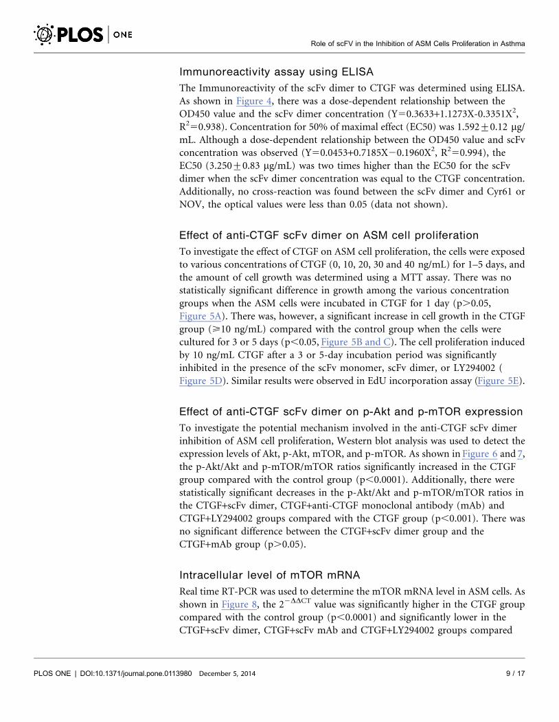
Immunoreactivity assay using ELISA
The Immunoreactivity of the scFv dimer to CTGF was determined using ELISA.
As shown in Figure 4, there was a dose-dependent relationship between the
OD450 value and the scFv dimer concentration (Y50.3633+1.1273X-0.3351X2,
R250.938). Concentration for 50% of maximal effect (EC50) was 1.592¡0.12 mg/
mL. Although a dose-dependent relationship between the OD450 value and scFv
concentration was observed (Y50.0453+0.7185X20.1960X2, R250.994), the
EC50 (3.250¡0.83 mg/mL) was two times higher than the EC50 for the scFv
dimer when the scFv dimer concentration was equal to the CTGF concentration.
Additionally, no cross-reaction was found between the scFv dimer and Cyr61 or
NOV, the optical values were less than 0.05 (data not shown).
Effect of anti-CTGF scFv dimer on ASM cell proliferation
To investigate the effect of CTGF on ASM cell proliferation, the cells were exposed
to various concentrations of CTGF (0, 10, 20, 30 and 40 ng/mL) for 1–5 days, and
the amount of cell growth was determined using a MTT assay. There was no
statistically significant difference in growth among the various concentration
groups when the ASM cells were incubated in CTGF for 1 day (p.0.05,
Figure 5A). There was, however, a significant increase in cell growth in the CTGF
group (>10 ng/mL) compared with the control group when the cells were
cultured for 3 or 5 days (p,0.05, Figure 5B and C). The cell proliferation induced
by 10 ng/mL CTGF after a 3 or 5-day incubation period was significantly
inhibited in the presence of the scFv monomer, scFv dimer, or LY294002 (
Figure 5D). Similar results were observed in EdU incorporation assay (Figure 5E).
Effect of anti-CTGF scFv dimer on p-Akt and p-mTOR expression
To investigate the potential mechanism involved in the anti-CTGF scFv dimer
inhibition of ASM cell proliferation, Western blot analysis was used to detect the
expression levels of Akt, p-Akt, mTOR, and p-mTOR. As shown in Figure 6 and 7,
the p-Akt/Akt and p-mTOR/mTOR ratios significantly increased in the CTGF
group compared with the control group (p,0.0001). Additionally, there were
statistically significant decreases in the p-Akt/Akt and p-mTOR/mTOR ratios in
the CTGF+scFv dimer, CTGF+anti-CTGF monoclonal antibody (mAb) and
CTGF+LY294002 groups compared with the CTGF group (p,0.001). There was
no significant difference between the CTGF+scFv dimer group and the
CTGF+mAb group (p.0.05).
Intracellular level of mTOR mRNA
Real time RT-PCR was used to determine the mTOR mRNA level in ASM cells. As
shown in Figure 8, the 22DDCT value was significantly higher in the CTGF group
compared with the control group (p,0.0001) and significantly lower in the
CTGF+scFv dimer, CTGF+scFv mAb and CTGF+LY294002 groups compared
Role of scFV in the Inhibition of ASM Cells Proliferation in Asthma
PLOS ONE | DOI:10.1371/journal.pone.0113980 December 5, 2014 9 / 17

with the CTGF group (p,0.0001). No significant difference was observed among
the CTGF+scFv dimer group, CTGF+scFv mAb group and CTGF+LY294002
group (p.0.05). These results suggest that the scFv dimer, mAb and LY294002
inhibit the up-regulation in mTOR mRNA expression induced by CTGF in
human ASM cells.
Discussion
CTGF/CCN2, a member of the CCN family, is a small cysteine-rich secreted
multi-modular protein that regulates various physiological and pathological
processes [19, 20]. It contains four domains: an insulin-like growth factorbinding
domain (module I), a Von Willebrand factor type C repeat domain (module II), a
thrombospondin type1 repeat domain (module III), and a C-terminal cysteine
knot (Cys-knot) (module IV). The N-terminal domain (modules I and II) has
been shown to mediate differentiation and collagen synthesis, and the C-terminal
domain (modules III and IV) has been shown to regulate cell proliferation [21–
23]. Recently, it has been shown that ASM cells produce and release CTGF [6, 24],
which plays a role in increasing angiogenesis and the accumulation of ECM in the
asthmatic airway [25]. The increase in the amount of CTGF released from
asthmatic ASM cells may have an autocrine feedback effect that increases the
amount of ECM protein released from ASM cells, which would further stimulate
the remodeling occurring in the asthmatic airway [25].
ScFv, which contains a complete antigen-binding site (a variable heavy and a
variable light domain), is considered a promising antibody in specific medical
applications and has certain advantageous qualities: it is a smaller molecule and
has less immunogenicity compared with full-length monoclonal antibodies [26].
It has been shown that anti-CTGF scFv significantly attenuates bleomycin (BL)-
induced pulmonary fibrosis in mice by inhibiting the CTGF bioactivity [12]. To
further improve the avidity of the scFv antibody, we constructed the expression
Figure 4. Immunoreactivity assay determined by ELISA. Both the scFv dimer and scFv monomerspecifically reacted with CTGF. The regression equations are Y50.3633+1.1273X-0.3351X2 (R250.938), andY50.0453+0.7185X20.1960X2 (R250.994) for the scFv dimer and scFv monomer, respectively. The OD450value for the scFv dimer was two times higher than the value for scFv monomer when the concentrations wereequal to the CTGF concentration.
doi:10.1371/journal.pone.0113980.g004
Role of scFV in the Inhibition of ASM Cells Proliferation in Asthma
PLOS ONE | DOI:10.1371/journal.pone.0113980 December 5, 2014 10 / 17

plasmid pET28a-scFv-matrilin, which includes the matrilin-1 C-terminal domain
that allows for scFv dimer formation because of the two disulfide bonds that form
in the coiled-coil domains (Figure 1). A soluble recombinant scFv dimer with
higher purity was obtained by transformation, induction, expression and
Figure 5. Human ASM cell proliferation after stimulated by CTGF. A. Results from the MTTassay after ASM cells were incubated in CTGF (0–30 ng/mL)for 1 day. B. Results from the MTTassay after ASM cells were incubated in CTGF (0–30 ng/mL) for 3 days. C. Results from the MTTassay after ASM cellswere incubated in CTGF (0–30 ng/mL) for 5 days. D. The results from the MTTassay after ASM cells were incubated for 1–5 days in CTGF (10 ng/mL) withor without the scFv monomer, scFv dimer, or LY294002. E. The results from EdU incorporation assay after ASM cells were incubated for 1–5 days in CTGF(10 ng/mL) with or without the scFv monomer, scFv dimer, or LY294002. * p,0.05, ** p,0.01, *** p,0.005, **** p,0.001.
doi:10.1371/journal.pone.0113980.g005
Role of scFV in the Inhibition of ASM Cells Proliferation in Asthma
PLOS ONE | DOI:10.1371/journal.pone.0113980 December 5, 2014 11 / 17

purification as expected (Figure 2 and 3). The scFv dimer exhibited an excellent
immunoreactivity to CTGF by ELISA assay, and the EC50 value was two times
lower that of the scFv monomer (Figure 4).
In this study, we found that >10 ng/mL CTGF increased ASM cell
proliferation, and this effect lasted at least 3–5 days (Figure 5B and C).
Furthermore, this proliferation was also inhibited when the cells were treated with
anti-CTGF scFv dimer or mAb. It has been shown that TGF-b induces hyperplasia
and hypertrophy in ASM cells, and the protein and mRNA expression levels of
CTGF are significantly higher in response to TGF-b in asthmatic ASM cells
[6, 27]. The combination with these results and our previous result, which
demonstrate that the anti-CTGF scFv antibody suppresses the TGF-b-induced
differentiation of fibroblast into myofibroblast by inhibiting AKT phosphoryla-
tion [13], suggest that TGF-b contributes to the hyperplasia and hypertrophy of
ASM cells through a mechanism in which CTGF acts as a downstream factor of
TGF-b.
Figure 6. Expression levels of p-Akt/Akt in human ASM cells determined by Western blot analysis. Theratio was higher in cells co-cultured in CTGF (p,0.0001). The increase in the p-Akt level induced by CTGFwas significantly inhibited when the cells were treated with scFv dimer, LY294002, or mAb (p,0.001). Therewas no significant difference between the scFv group and mAb group (p.0.05).
doi:10.1371/journal.pone.0113980.g006
Role of scFV in the Inhibition of ASM Cells Proliferation in Asthma
PLOS ONE | DOI:10.1371/journal.pone.0113980 December 5, 2014 12 / 17

Recent studies investigating the mechanism involved in TGF-b signaling
dependent ASM proliferation have demonstrated the importance of several
pathways, including the mitogen-activated protein kinase (MAPK) and phos-
phatidylinositol 3-kinase (PI3K) pathways [28, 29]. PI3K/Akt is a classic signal
pathway, and its activation induces the G1/S cell cycle transition [30, 31]. MTOR
is a highly conserved serine/threonine kinase that belongs to the PI3K family and
is one of the downstream targets of Akt in regulating cell proliferation and protein
translation. The evidence suggests that the mTOR pathway is vital for airway and
vascular remodeling, which is a key characteristic of severe asthma [32]. We firstly
find that CTGF can enhance ASM proliferation in this study, however, the
potential mechanism remains to be investigated. Our study shows that the p-Akt/
Akt and p-mTOR/mTOR ratios were dramatically higher after ASM cells were
cultured in CTGF for 3 days and were significantly lower when the cells were also
treated with anti-scFv dimer, anti-CTGF mAb and the specific PI3K inhibitor
LY294002 (Figure 6 and 7). The result was similar to that of real-time RT-PCR for
mTOR mRNA (Figure 8). The inhibiting effect of the scFv dimer was no different
Figure 7. Expression levels of p-mTOR/mTOR in human ASM cells determined by Western blotanalysis. The ratio was higher in cells co-cultured in CTGF (p,0.0001). The increase in the p-mTOR inducedby CTGF was significantly inhibited when the cells were treated with scFv dimer, LY294002, or mAb(p,0.001). There was no significant difference between the scFv group and mAb group (p.0.05).
doi:10.1371/journal.pone.0113980.g007
Role of scFV in the Inhibition of ASM Cells Proliferation in Asthma
PLOS ONE | DOI:10.1371/journal.pone.0113980 December 5, 2014 13 / 17

from those of inhibitors LY294002 and mAb (p.0.05). This finding proves the
effectiveness of the scFV dimer as a new CTGF antagonist.
Because of culturing cells for three days, we can not distinguish between direct
effects and indirect effects, but it seems to be an indirect effect from the data
analysis and experimental design. In addition, we found that the increased mTOR
mRNA was not parallel to the western blot data in total protein. In fact, the
increased total mRNA does not always result in protein high-expression. The
mRNA translation is affected by various regulatory factors or inference, such as
transcription factors, miRNAs, and external chemical or biological agents [33, 34].
Moreover, many signal molecules such as AKT, ERK, and mTOR are short-life
proteins, not structural protein [35], and these signal proteins may be accelerately
degraded by the ubiquitin-proteasome system under the regulation or influence of
certain factors [36, 37].
Altogether, our results clearly indicate that CTGF may participate in ASM cell
proliferation through the PI3K/Akt/mTOR pathway and that the anti-CTGF scFv
dimer may inhibit the increased p-Akt and p-mTOR levels induced by CTGF in
ASM cells. CTGF is potentially a key factor in developing and maintaining the
structural changes that are associated with severe persistent asthma. Based on
these findings, it is possible that neutralizing the CTGF bioactivity using a
Figure 8. MTOR mRNA level in human ASM cells determined by RT-PCR. All 22DDCT values werecalculated, and the value was assumed to be 1 for the control group. The increase in the mTOR mRNA levelin ASM cells was significantly inhibited when the cells were treated with scFv dimer, LY294002, or mAb(p,0.0001). There was no statistically significant difference between the scFv group and mAb group(p.0.05).
doi:10.1371/journal.pone.0113980.g008
Role of scFV in the Inhibition of ASM Cells Proliferation in Asthma
PLOS ONE | DOI:10.1371/journal.pone.0113980 December 5, 2014 14 / 17

monoclonal antibody, such as the anti-CTGF scFv dimer, may be an effective
treatment for minimizing the amount of airway remodeling in asthma patients.
Acknowledgments
We would like to thank the National Natural Science Foundation of China
(No. 30970809, 81271636), the Natural Science Foundation of Jiangsu Province
(No. BK2009274) and the Special Fund of Clinical Medicine, Jiangsu Province,
China (No. BL2012063) for the financial support in this work.
Author ContributionsConceived and designed the experiments: GQW XHW WG LTC. Performed the
experiments: WG LTC XDX XLX. Analyzed the data: WG LTC JXF XJY.
Contributed reagents/materials/analysis tools: WG LTC GQW CZ QRQ. Wrote
the paper: WG LTC GQW.
References
1. Swathy P, Octavian CI (2014) Asthma and Obstructive Sleep Apnea: Clinical and PathogenicInteractions. Journal of Investigative Medicine 62: 665–675.
2. James A, Carroll N (2000) Airway smooth muscle in health and disease; methods of measurement andrelation to function. European Respiratory Journal 15: 782–789.
3. James A, Mauad T, Abramson M, Green F (2012) Airway Smooth Muscle Hypertrophy andHyperplasia in Asthma. American Journal of Respiratory and Critical Care Medicine 186: 568–568.
4. Hackett TL (2012) Epithelial-mesenchymal transition in the pathophysiology of airway remodelling inasthma. Current Opinion in Allergy and Clinical Immunology 12: 53–59.
5. Kudo M, Ishigatsubo Y, Aoki I (2013) Pathology of asthma. Frontiers in Microbiology 4.
6. Burgess JK, Johnson PRA, Ge Q, Au WW, Poniris MH, et al. (2003) Expression of connective tissuegrowth factor in asthmatic airway smooth muscle cells. American Journal of Respiratory and CriticalCare Medicine 167: 71–77.
7. Cohen P, Rajah R, Rosenbloom J, Herrick DJ (2000) IGFBP-3 mediates TGF-beta1-induced cellgrowth in human airway smooth muscle cells. Am J Physiol Lung Cell Mol Physiol 278: L545–551.
8. Duncan MR, Frazier KS, Abramson S, Williams S, Klapper H, et al. (1999) Connective tissue growthfactor mediates transforming growth factor beta-induced collagen synthesis: down- regulation by cAMP.FASEB J 13: 1774–1786.
9. Leask A, Abraham DJ (2006) All in the CCN family: essential matricellular signaling modulators emergefrom the bunker. Journal of Cell Science 119: 4803–4810.
10. Johnson PRA, Burgess JK, Ge Q, Poniris M, Boustany S, et al. (2006) Connective tissue growthfactor induces extracellular matrix in asthmatic airway smooth muscle. American Journal of Respiratoryand Critical Care Medicine 173: 32–41.
11. Wu GQ, Fan XB, Wu HB, Liu NF, Li XF, et al. (2010) Bioscreening of phage display antibody library andexpression of a humanized single-chain variable fragment antibody against human connective tissuegrowth factor (CTGF/CCN2). Biotechnology and Applied Biochemistry 56: 95–102.
12. Wang XH, Wu GQ, Gou LX, Liu ZZ, Wang XY, et al. (2011) A novel single-chain-Fv antibody againstconnective tissue growth factor attenuates bleomycin-induced pulmonary fibrosis in mice. Respirology16: 500–507.
Role of scFV in the Inhibition of ASM Cells Proliferation in Asthma
PLOS ONE | DOI:10.1371/journal.pone.0113980 December 5, 2014 15 / 17

13. Wu GQ, Wang XY, Deng XP, Wu PP, Xue XL, et al. (2012) A novel single-chain Fv antibody forconnective tissue growth factor against the differentiation of fibroblast into myofibroblast. AppliedMicrobiology and Biotechnology 93: 2475–2482.
14. Frank S, Schulthess T, Landwehr R, Lustig A, Mini T, et al. (2002) Characterization of the matrilincoiled-coil domains reveals seven novel isoforms. Journal of Biological Chemistry 277: 19071–19079.
15. Deak F, Wagener R, Kiss I, Paulsson M (1999) The matrilins: a novel family of oligomeric extracellularmatrix proteins. Matrix Biology 18: 55–64.
16. Zhang Y, Chen Q (2000) Changes of matrilin forms during endochondral ossification - Molecular basis ofoligomeric assembly. Journal of Biological Chemistry 275: 32628–32634.
17. Prakash YS, Thompson MA, Pabelick CM (2009) Brain-Derived Neurotrophic Factor in TNF- alphaModulation of Ca2+ in Human Airway Smooth Muscle. American Journal of Respiratory Cell andMolecular Biology 41: 603–611.
18. Xie SP, Sukkar MB, Issa R, Khorasani NM, Chung KF (2007) Mechanisms of induction of airwaysmooth muscle hyperplasia by transforming growth factor-beta. American Journal of Physiology- LungCellular and Molecular Physiology 293: L245–L253.
19. Blalock TD, Gibson DJ, Duncan MR, Tuli SS, Grotendorst GR, et al. (2012) A Connective TissueGrowth Factor Signaling Receptor in Corneal Fibroblasts. Investigative Ophthalmology & Visual Science53: 3387–3394.
20. Bradham DM, Igarashi A, Potter RL, Grotendorst GR (1991) Connective-Tissue Growth-Factor - aCysteine-Rich Mitogen Secreted by Human Vascular Endothelial-Cells Is Related to the Src- InducedImmediate Early Gene-Product Cef-10. Journal of Cell Biology 114: 1285–1294.
21. Holbourn KP, Acharya KR, Perbal B (2008) The CCN family of proteins: structure-functionrelationships. Trends in Biochemical Sciences 33: 461–473.
22. Moussad EE, Brigstock DR (2000) Connective tissue growth factor: what’s in a name? Mol GenetMetab 71: 276–292.
23. Grotendorst GR, Duncan MR (2005) Individual domains of connective tissue growth factor regulatefibroblast proliferation and myofibroblast differentiation. FASEB J 19: 729–738.
24. Xie S, Sukkar MB, Issa R, Oltmanns U, Nicholson AG, et al. (2005) Regulation of TGF-beta 1-induced connective tissue growth factor expression in airway smooth muscle cells. Am J Physiol LungCell Mol Physiol 288: L68–76.
25. Burgess JK (2005) Connective tissue growth factor: a role in airway remodelling in asthma? Clin ExpPharmacol Physiol 32: 988–994.
26. Weisser NE, Hall JC (2009) Applications of single-chain variable fragment antibodies in therapeuticsand diagnostics. Biotechnol Adv 27: 502–520.
27. Bosse Y, Thompson C, Stankova J, Rola-Pleszczynski M (2006) Fibroblast growth factor 2 andtransforming growth factor beta 1 synergism in human bronchial smooth muscle cell proliferation.American Journal of Respiratory Cell and Molecular Biology 34: 746–753.
28. Shi Y, Massague J (2003) Mechanisms of TGF-beta signaling from cell membrane to the nucleus. Cell113: 685–700.
29. Halwani R, Al-Muhsen S, Al-Jahdali H, Hamid Q (2011) Role of Transforming Growth Factor- beta inAirway Remodeling in Asthma. American Journal of Respiratory Cell and Molecular Biology 44: 127–133.
30. Ma B, Sen T, Asnaghi L, Valapala M, Yang F, et al. (2011) beta A3/A1-Crystallin controls anoikis-mediated cell death in astrocytes by modulating PI3K/AKT/mTOR and ERK survival pathways throughthe PKD/Bit1-signaling axis. Cell Death & Disease 2.
31. Wang GH, Wang F, Ding WF, Wang JC, Jing RR, et al. (2013) APRIL Induces Tumorigenesis andMetastasis of Colorectal Cancer Cells via Activation of the PI3K/Akt Pathway. Plos One 8.
32. Thomson AW, Turnquist HR, Raimondi G (2009) Immunoregulatory functions of mTOR inhibition.Nature Reviews Immunology 9: 324–337.
33. Yewdell JW (2007) Plumbing the sources of endogenous MHC class I peptide ligands. Current Opinionin Immunology 19: 79–86.
Role of scFV in the Inhibition of ASM Cells Proliferation in Asthma
PLOS ONE | DOI:10.1371/journal.pone.0113980 December 5, 2014 16 / 17

34. Yewdell JW (2003) Hide and seek in the peptidome. Science 301: 1334–5.
35. Qian S, Princiotta MF, Bennink JR, Yewdell JW (2006) Characterization of rapidly degradedpolypeptides in mammalian cells reveals a novel layer of nascent protein quality control. The Journal ofbiological chemistry 281: 392–400.
36. Komatsu M, Waguri S, Chiba T, et al. (2006) Loss of autophagy in the central nervous system causesneurodegeneration in mice. Nature 441: 880–4.
37. Yewdell JW, Nicchitta CV (2006) The DRiP hypothesis decennial: support, controversy, refinement andextension. Trends Immunol. 27: 368–73.
Role of scFV in the Inhibition of ASM Cells Proliferation in Asthma
PLOS ONE | DOI:10.1371/journal.pone.0113980 December 5, 2014 17 / 17