research article contribution to the determination of in
TRANSCRIPT
Hindawi Publishing CorporationComputational and Mathematical Methods in MedicineVolume 2013 Article ID 814025 11 pageshttpdxdoiorg1011552013814025
Research ArticleContribution to the Determination of In Vivo MechanicalCharacteristics of Human Skin by Indentation Test
Marie-Angegravele Abellan Hassan Zahouani and Jean-Michel Bergheau
Universite de Lyon ENISE LTDS UMR 5513 CNRS 58 rue Jean Parot 42023 Saint-Etienne France
Correspondence should be addressed to Marie-Angele Abellan marie-angeleabellanenisefr
Received 18 July 2013 Revised 9 September 2013 Accepted 9 September 2013
Academic Editor Eddie Ng
Copyright copy 2013 Marie-Angele Abellan et al This is an open access article distributed under the Creative Commons AttributionLicense which permits unrestricted use distribution and reproduction in any medium provided the original work is properlycited
This paper proposes a triphasic model of intact skin in vivo based on a general phenomenological thermohydromechanical andphysicochemical (THMPC) approach of heterogeneous media The skin is seen here as a deforming stratified medium composedof four layers and made out of different fluid-saturated materials which contain also an ionic component All the layers are treatedas linear isotropic materials described by their own behaviour lawThe numerical simulations of in vivo indentation test performedon human skin are given The numerical results correlate reasonably well with the typical observations of indented human skinThe discussion shows the versatility of this approach to obtain a better understanding on the mechanical behaviour of human skinlayers separately
1 Introduction
Human skin is the largest organ of the human bodyThe skinprotects the body against external influences by preventingfluid loss when exposed to sun the penetration of undesirablesubstances in case of pollution and the development of dis-eases due to the direct application of external chemical ormechanical loads linked to clinical problems surgery or aes-thetic treatments The answers of this barrier to these chemi-cal biological mechanical and thermal loads depend on theperson the site on the body the age the health its nutritionalstatus its properties its state (intact or damaged) and itsevolutions [1] Numerous studies have shown that humanskin has a stratified structure consisting from the skin outersurface inward of threemain layers the epidermis (composedof the stratumcorneumand the viable epidermis) the dermisand the hypodermis [2ndash6] Dryness microcracks and lossof elasticity are thought to be influenced by fluid flow andthe associated changes in ion concentration as a direct resultof mechanical stress states However these phenomena arecomplex to understand and to model due to the strongcouplings that exist between them and due to the complexbehaviour of the different layers of skin soft tissues
Studies of the mechanical behaviour of human skinhave observed that the skin is a stratified nonhomogeneous
anisotropic nonlinear viscoelastic material which is sub-jected to a prestress in vivo [7ndash9] In addition its propertiesvary with age throughout the body and per person Diffi-culties arise when trying to obtain quantitative descriptionsof mechanical properties of the skin Numerous mechanicalexperiments have been performed on the skin tensile testingsuction methods torsion tests and indentation experiments[10ndash16] For in vivo performed mechanical experiments themeasured behaviour is generally ascribed to the dermis due tothe relative height of the viable epidermis and of the stratumcorneum However the various skin layers are tied togetherand it is hard to isolate the contribution of each of themIn vitro experiments give the opportunity to separate theskin layers But in vitro experimental procedures change themechanical properties of the individual skin layersMoreoverit is hard to compare results obtained with different measure-ment conditionsThis illustrates the need for an experimentasystem to measure the mechanical behaviour of the differentskin layers in a noninvasive and objective manner and alsoindependently of the experimental setup The LTDS-inden-tation device is able to give a better understanding of themechanical behaviour of the skin by characterizing themechanical behaviour of several distinct skin layers in vivowithout disturbing its natural stress state before the experi-ment [15]
2 Computational and Mathematical Methods in Medicine
To achieve this several experimental setups are developedto load the skin mechanically theoretical models are derivedto describe the experiments and numerical models areimplemented to characterize themechanical behaviour of theskin layers [17ndash25]
Within the framework of a general phenomenologi-cal thermohydromechanical and physicochemical (THMPC)approach of heterogeneousmedia [22] a triphasic skinmodelis proposed in [18 26] which incorporates a solid phase withthree solid materials a fluid phase and an ionic componentunder ambient constant conditions Although not negligibleelectrical effects are not taken into account in this modelThedriving forces for transport are the gradients of the chemicalpotentials of the fluid and of the ions coupled with thegradients of the displacements of the different solids In thismodel skin is considered as a stratified material with threelayers modelling the three outer layers of skin the stratumcorneum the viable epidermis and the dermis All layers ofthe skin model are supposed to be made of fluid-saturatedmaterials Furthermore each layer is seen as a different solidmaterial within the solid phase and it is described by its ownbehaviour law In [18 26] the solid materials are seen asisotropic linear elastic materials each of them with its ownelastic coefficients In [27] the solid materials are modelled asnonlinear isotropic Mooney-Rivlin materials with one mate-rial constant leading to the determination of three materialconstants in total In [19] the solid materials are describedas nonlinear isotropic Mooney-Rivlin materials with twomaterial constants being able to face large deformations Eachsolid material has its own strain energy density function ofMooney-Rivlin leading to the need to identify six materialconstants in total and which allow analysing the decoupledbehaviour of each skin layer These analyses have shown thecapability of this model to describe the transient water flowand ion transport through damaged or undamaged skin afterapplication of a saline solution and to gain insight on themechanical behaviour of human skin layers separately
The ionic componentmodels the chemical load applied atthe skin outer surface
This paper proposes to extend this theoretical-experi-mental-numerical setup of human skin to the numerical sim-ulation of in vivo indentation test Skin is seen here as a tri-phasic medium with four solid materials in the solid phasea fluid phase and an ionic component Furthermore skin isconsidered as a stratified material with four layers modelingthe four layers of skin the stratum corneum the viable epi-dermis the dermis and the hypodermis All the layers aresupposed to be made of fluid-saturated materials and aretreated as isotropic linear materials described by their ownbehaviour law The governing partial differential equationsthat arise from the equilibrium the kinematic and the con-stitutive equations are solved under varying physically admis-sible initial and boundary conditions for the ion concentra-tions the fluid and the solids proposed for describing in vivoindentation test available in the laboratory A finite differenceanalysis is carried out which provides a quantitative under-standing of water and ion movements through undamagedskinThe numerical simulation allows quantifying the in vivomechanical properties of the different skin layers of soft tissueseparately
Stratumcorneum
Epidermis
Dermis
Hypodermis
Figure 1 Schematic view of the cross-section of human skinshowing the distinct layers
Wewill first present the context of this paper linked to thehistology of human skin in vivo Then to provide a propersetting we will recapitulate the governing equations for adeforming porous medium including ions The noninvasiveexperimental device is presentedThen example calculationsfor a specimen of in vivo undamaged skin are given andthe obtained numerical results are discussed Finally someconcluding remarks are made
2 Histology of Skin
From the skin surface inwards skin is composed of stra-tum corneum viable epidermis dermis and hypodermis(Figure 1) A detailed look to these different layers shows upthe following points which are of particular interest in ourstudy
21 Hypodermis The hypodermis is an adipose tissue com-posed of loose fatty connective tissue found between the der-mis and themuscles It acts as an insulating layer and a protec-tive cushion Its thickness varies some mm with anatomicalsite age sex race and health of the individual
22 Dermis The dermis can be from 1 to 4mm thick It islargely composed of a very dense fibre network of collagenelastin and minute quantities of reticulin and a support-ing matrix of amorphous ground substance all bathed inphysiological fluid Physiological fluid provides the cells withnutrients and consists of a water solvent containing mineraland organic solutes as well as waste products from the cellsThe dermis contains also microstructures like blood vesselslymph vessels nerve endings sweat glands sebaceous glands
Computational and Mathematical Methods in Medicine 3
and hair follicles The amorphous ground substance com-bines with the water of the physiological fluid to form a gelwhich does not leak out the dermis even under high pressure
23 Viable Epidermis The viable epidermis is a thin (10ndash100 120583m) stratified squamous epithelial of soft keratinized liv-ing cells with nuclei migrating to the outer skin surface thekeratinocytes The viable epidermis is a nonvascular struc-ture Cells are surrounded nourished and bathed by a physi-ological fluid originating in the dermis and transportedacross the epidermal-dermal junction
24 Stratum Corneum The stratum corneum is a 10ndash25 120583mthick dense coating of hard keratinized dead hexagonal flatcells the corneocytes held together by lipid bridges and cor-neosomes in what is commonly referred to as a brick-and-mortar structure The corneocytes are the keratinocytes thatwere migrating to the skin outer surface and that have losttheir nuclei Although the corneocytes are nonviable cells thestratum corneum is considered to be fully functional partic-ularly in terms of barrier properties and retains metabolicfunctions Because of its structure and composition the cellsof the stratum corneum have less capacity to bind water thanthe living cells of the viable epidermis or of the dermis
The stratum corneum and the viable epidermis are con-tinuously renewed by desquamationwithin 6 to 30 days Cellsare shed from the outside and replaced by new ones
25 Consequence on the Skin Model Skinrsquos histology hasshown that skin soft tissues are heterogeneous materials con-sisting of several components If we combine this statementwith the commonly admitted hypothesis that skin is a non-linear anisotropic hyperelastic and viscoelastic material thechoice ismade here to derive the theoreticalmodel for humanskin soft tissues seen as a stratified triphasicmaterial with fourlayers four solids in the solid phase one fluid in the fluidphase and an ionic component Figure 2 displays the studiedspecimen of skin where it is considered that
(i) the four layers are as follows layer 1 simulates the stra-tum corneum layer 2 for the viable epidermis layer 3for the dermis and layer 4 for the hypodermis
(ii) the four solids for the solid phase are as follows solid 1(s1) simulates the corneocites and the lipid mortar
present in the stratum corneum solid 2 (s2) simulates
the evolving cells of the viable epidermis solid 3 (s3)
simulates the different cells of the dermis includingthe lymph and blood vessels and solid 4 (s
4) simulates
the fatty connective tissue of the hypodermis(iii) the fluid (f ) in the fluid phase simulates the 10
bound water in the lipid mortar of the stratum cor-neum plus the physiological fluid in the viable epider-mis plus the physiological fluid in the dermis plus thephysiological fluid in the hypodermis
(iv) the ionic component (i) simulates some cream depos-ited at the outer surface of the skin either for aestheticor medical purposes and of which it is relevant to fol-low the penetration
Dermis
Hypodermis
Stratum corneumi
s1
s2
s3
s4
f
f
f
f
0
1516mm
Viable epidermis
Figure 2 Schematic view of the cross-section of the skin specimenshowing the distinct layers and components
The study presented in the following paragraphs is basedon this skin specimen
3 Theoretical Model
Under the phenomenological hypothesis [28] the generalTHMPC approach of heterogeneousmedia [22] describes theoverall material behaviour of the heterogeneous medium as acombination of the behaviour of each individual componentIt is based on the principle of interaction of the componentswith the following assumptions
(i) in each infinitesimal volume of a heterogeneousmedium a finite number of components are present
(ii) each component contributes to the total materialbehaviour in the same proportion as its volumetricparticipation given by its volumic ratio
(iii) all the components are extended to the total studiedunit volume of heterogeneous medium
This approach is applied here to model the human skinspecimen introduced in Section 25 (Figure 2)
As it was said skin is seen here as a triphasicmaterial withfour solids for the solid phase (120587 = s
1 s2 s3 s4) one for each
layer a fluid (120587= f ) and an ionic component (120587= i) subjectedto the restriction of small displacement gradients no masstransfer and no chemical reactions between the constituentsthe electrical effects are not taken into account and the com-ponents will be considered as intrinsically incompressibleThe studied processes occur isothermally In addition the ini-tial configuration of the skin solid skeleton is chosen here as areference domain for deriving the field equations Hereafterall physical quantities are assumed to be functions of the Eulervariables (x 119905) where t is the time and x is the spatial vectordefining the position of the material particle in the current
4 Computational and Mathematical Methods in Medicine
configuration at time t Hence the implicit arguments of allvectors or tensors or scalars are the Euler variables (x 119905)With these assumptions the source of mass of constituent 120587
coming from the other constituents present in the medium isequal to zero and the balance of mass for each constituent 120587
(for 120587 = s1 s2 s3 s4 f i) reads120597
120597119905120588120587
+ nabla sdot [120588120587V120587] = 0 (1)
with V120587(x 119905) being the absolute velocity of constituent 120587 in
msdotsminus1 and 120588120587(x 119905) being the relative mass density of constitu-
ent 120587 in kgsdotmminus3 defined for 120587 = s1 s2 s3 s4 f i by
120588120587
= 1198991205871205881015840
120587 (2)
where 1205881015840
120587(x 119905) is the absolute mass density of constituent 120587 in
kgsdotmminus3 and 119899120587(x 119905) is the volumic ratio of constituent120587 As in
the remainder of this paper the subscripts s1 s2 s3 s4 f and
i denote the solids the fluid and the ions respectively Thedifferent constituents are supposed to be intrinsically incom-pressibleTherefore their absolute mass density are kept con-stant in this paper Further the sum of volumic ratio over theconstituents present in the medium should equal one
1198991199041
+ 1198991199042
+ 1198991199043
+ 1198991199044
+ 119899119891
+ 119899119894= 1 (3)
Neglecting inertia forces convective terms and the grav-ity acceleration the balance of linear momentum for eachconstituent 120587 (for 120587 = s
1 s2 s3 s4 f i) reduces to
nabla sdot 120590120587
+ 1199011015840
120587= 0 (4)
with 120590120587(x 119905) being the Cauchy stress tensor of constituent 120587
in Pa and 1199011015840
120587(x 119905) being the source of momentum for con-
stituent 120587 coming from the other constituents in kgsdotmminus2sdotsminus2which takes into account the possible local drag interactionsbetween the solids the fluid and the ions and which satisfiesthe momentum production constraint
1199011015840
1199041
+ 1199011015840
1199042
+ 1199011015840
1199043
+ 1199011015840
1199044
+ 1199011015840
119891+ 1199011015840
119894= 0 (5)
Under the assumption of chemically inert fluid and ionsand with solid matrix materials the material state relationexpressing the chemical potential of the fluid 120583
119891(x 119905) encom-
passes the interactions between solids fluid and ions For thefluid it reads
120583119891
= 119901 minus Π + 120595 (6)
where 119901(x 119905) is the fluid pressure in Pa 120595(x 119905) is the matrixpotential accounting for fluid-solid interactions (capillaryand adsorptive effects) in Pa and Π(x 119905) is the osmotic pres-sure accounting for fluid-ion interactions in PaThe chemicalpotential of the fluid is in Jsdotmminus3 These quantities need to beexperimentally determined in order to write a close mathe-matical problem but they are difficult to obtain Reference[23] considered relations between chemical potential andporosity from different references and proposes the followingexperimental fit
119901 + 120595 = (11989097119864minus5
minus 119890(97119864minus5)119899
119891) (984119864 + 10) (7)
Π = 2119877119879119888119894 (8)
with 119877 being the universal gas constant in J sdot Kminus1 sdot molminus1119879 being the absolute temperature in K and 119888
119894(x 119905) being the
concentration of the ions per unit fluid volume in molsdotmminus3For the ionic component the chemical potential 120583
119894(x 119905)
is defined as
120583119894= 1205831198940
+ 119877119879 ln (119888119894) (9)
with 1205831198940
being the chemical potential of the ions in a referencestate in Jsdotmminus3 Neglecting couplings between velocity and heatflux fluid flow through a saturated porous medium with anionic component is expressed by a generalized Darcyrsquos lawhereunder defined in terms of the gradient of the chemicalpotential 120583
119891(x 119905) of the fluid the gradient of the chemical
potential 120583119894(x 119905) of the ions and a second-order permeability
tensor 119870(x 119905)
119899119891
(V119891
minus V119904) = minus119870 sdot [nabla120583
119891+
119899119894
119899119891
nabla120583119894] (10)
In a matrix-vector notation the permeability tensor appear-ing in (10) is a position and time dependent function definedas
119870 (x 119905) = [
[
119870119909119909
(x 119905) 119870119909119910
(x 119905) 119870119909119911
(x 119905)
119870119910119909
(x 119905) 119870119910119910
(x 119905) 119870119910119911
(x 119905)
119870119911119909
(x 119905) 119870119911119910
(x 119905) 119870119911119911
(x 119905)
]
]
(11)
where each component119870119894119895(x 119905) for 119894 119895 isin 119909 119910 119911 is a function
of position and time and it has the unit m4 sdot Nminus1 sdot sminus1The diffusion of ions through the fluid phase of a porous
medium is taken into account through a Fickrsquos law-type rela-tion by means of a second-order diffusion tensor of the ions119863119894
119899119894(V119894minus V119891) = minus119863
119894nabla120583119894 (12)
In a matrix-vector notation the diffusion tensor of the ionsappearing in (12) is a position and time dependent functiondefined as
119863119894(x 119905) = [
[
119863119894119909119909
(x 119905) 119863119894119909119910
(x 119905) 119863119894119909119911
(x 119905)
119863119894119910119909
(x 119905) 119863119894119910119910
(x 119905) 119863119894119910119911
(x 119905)
119863119894119911119909
(x 119905) 119863119894119911119910
(x 119905) 119863119894119911119911
(x 119905)
]
]
(13)
where each component 119863119894119895119896
(x 119905) for 119895 119896 isin 119909 119910 119911 is a func-tion of position and time and it has the unit m2 sdot sminus1
The stress-strain relations for the solids are elaboratedunder the classical assumption for heterogeneous media thatthe Cauchy stress tensor of the total heterogeneous mediumis composed of a solid and a fluid part
120590 = 1205901199041
+ 1205901199042
+ 1205901199043
+ 1205901199044
minus 119901119868 (14)
with 120590 being the Cauchy stress tensor of the total heteroge-neous medium in Pa and 119868 being the second-order identitytensor
Under the assumption of small displacements and smallstrains skin is considered as a linear isotropic elastic mate-rial and a Hookersquos stress-strain relation is taken for eachsolid skeleton (120587 = s
1 s2 s3 s4)
120590120587
= 119863119890
120587 120576120587
for 120587 = 1199041 1199042 1199043 1199044 (15)
Computational and Mathematical Methods in Medicine 5
where 119863119890
120587(x 119905) is the elasticity tensor of the solid material 120587
in Pa and 120576120587(x 119905) is the strain tensor of solid 120587 defined by
120576120587
= nabla119904119906120587 (16)
with 119906120587(x 119905) being the displacement field of solid 120587 in m It
should be noticed that the Cauchy stress tensor the straintensor the elasticity tensor and the displacement field arefunctions of the space and time variables The superscript 119904
denotes the symmetric part of the gradient operatorFor later use in Section 5 (15) can be rewritten more
conveniently in a matrix-vector notation
120576120587
= 119864120587
120590120587
for 120587 = 1199041 1199042 1199043 1199044 (17)
with
119864120587
= (119863119890
120587)minus1 for 120587 = 119904
1 1199042 1199043 1199044 (18)
where 119864120587(x 119905) is the matrix of the elasticity compliances
defined as the inverse of the matrix 119863119890
120587of the elasticity coeffi-
cients Each component119864120587119894119895
(x 119905) for 119894 119895 isin 119909 119910 119911 of the elas-ticity compliance matrix 119864
120587(x 119905) is space and time depen-
dent with unit Paminus1TheCauchy stress and the strain in vectornotation are given by
120590120587
(x 119905) =
[[[[[[[
[
120590120587119909119909
(x 119905)
120590120587119910119910
(x 119905)
120590120587119911119911
(x 119905)
120590120587119909119910
(x 119905)
120590120587119910119911
(x 119905)
120590120587119911119909
(x 119905)
]]]]]]]
]
120576120587
(x 119905) =
[[[[[[[
[
120576120587119909119909
(x 119905)
120576120587119910119910
(x 119905)
120576120587119911119911
(x 119905)
120576120587119909119910
(x 119905)
120576120587119910119911
(x 119905)
120576120587119911119909
(x 119905)
]]]]]]]
]
(19)
with the elasticity compliance matrix
119864120587
(x 119905)
= [
[
119864120587119909119909
(x 119905) 119864120587119909119910
(x 119905) 119864120587119909119911
(x 119905)
119864120587119910119909
(x 119905) 119864120587119910119910
(x 119905) 119864120587119910119911
(x 119905)
119864120587119911119909
(x 119905) 119864120587119911119910
(x 119905) 119864120587119911119911
(x 119905)
]
]
=
[[[[[[[[[[[[[[[[[[[[[
[
1
119864120587
minus]120587
119864120587
minus]120587
119864120587
0 0 0
minus]120587
119864120587
1
119864120587
minus]120587
119864120587
0 0 0
minus]120587
119864120587
minus]120587
119864120587
1
119864120587
0 0 0
0 0 01 + ]120587
119864120587
0 0
0 0 0 01 + ]120587
119864120587
0
0 0 0 0 01 + ]120587
119864120587
]]]]]]]]]]]]]]]]]]]]]
]
(20)
Sphericalindenter
Acquisition
Figure 3 Experimental setup of the LTDS-indentation device
where 119864120587is the Youngmodulus of the solid material 120587 and ]
120587
is the Poissonrsquos ratio of the solid material 120587The field equations that is the balance of mass for the
fluid the balance of mass for the ions and the balance ofmomentum for the solids are complemented by the bound-ary conditions which hold on complementary parts of theboundary in terms of prescribed external traction prescribedvelocity prescribed outflow of pore fluid and prescribedpressure prescribed outflow of ions and prescribed chemicalpotential respectively The initial conditions which specifythe displacements 119906
120587for the solid grains 120587 = s
1 s2 s3 s4 the
velocities V120587for the solid grains 120587 = s
1 s2 s3 s4 and the
chemical potentials 120583120587for the fluid and the ionic component
120587 = f i at t = 0 close the initial value problem
4 Experimental Device
The original LTDS-indentation device developed by the teamof Professor Zahouani permits studying of the mechanicalresponse of the human skin in vivo This indentation deviceloads the skin mechanically by applying a controlled normalforce onto the surface of the skin The experimental setup ispresented Figure 3
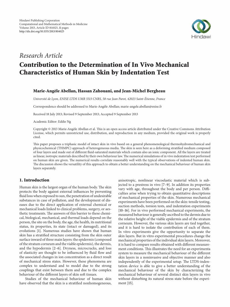
The penetration depth of the rigid spherical indenter(diameter 6mm) is recorded as a function of the normalapplied force Fload during a loading-unloading experimentThe recorded curve for an indentation test performed on thevolar forearm zone of a volunteer healthy adult is given inFigure 4This location is chosen because it is easily accessiblerelatively flat and less disturbed by the natural movementof the body It makes the indentation tests less tiring for thevolunteer because of the position of the arm during the testTherefore it disturbs in the least possible way the skinrsquos natu-ral state of stressThe indentation test is realized for a constantindentation speed of 5000120583ms at ambient temperature andwithout surface treatment on skin before the test
This loading-unloading curve is reversible with a very lowhysteresis due to the dissipated energy No plastic behaviouris observed in the sense that there is no residual print ontothe surface of the skin allowing the measurement of a plasticdepth Therefore in this load range it can be considered thathuman skin soft tissues can be modeled as elastic materials
6 Computational and Mathematical Methods in Medicine
70
60
50
40
30
20
10
00 05 1 15 2 25
Floa
d (m
N)
Penetration depth (120583m)minus10
Figure 4 Recorded curve for a loading-unloading indentation teston the volar forearm of an adult
Time (s)
Floa
d (N
)
6E minus 2
5E minus 2
4E minus 2
3E minus 2
2E minus 2
1E minus 2
0E + 00E + 0 2E minus 3 4E minus 3 6E minus 3 8E minus 3 1E minus 2
Figure 5 Applied mechanical load Fload versus time for the load-ing-unloading steps of the numerical simulations
Moreover this recorded experimental curve (Figure 4) isreworked and gives a curve of the applied mechanical loadFload versus time for the loading-unloading steps of thenumerical simulations (Figure 5)
These experimental data are used in the numerical sim-ulation hereafter to define physically admissible boundaryand initial conditions and to help characterizing in vivoequivalent mechanical parameters of human soft tissues
As it is not possible tomeasure the thickness of the skin onthe inner forearm we will assume in the following that thethickness of the skin at this part (stratum corneum + viableepidermis + dermis + hypodermis) is approximately 1516 120583m(12 120583m for the stratum corneum + 102 120583m for the viable epi-dermis + 1002 120583m for the dermis + 400 120583m for the hypoder-mis) which are mean values typical for biological soft tissuesthat can be found already in the literature [29] Hence thetotal response of the skin is the composite response of theindividual contributions of the stratum corneum the viableepidermis the dermis and the hypodermis
5 Numerical Simulation
51 Finite Difference Model A finite difference analysis hasbeen carried out that allows for a quantitative understandingof fluid flow and ion transport through intact skin and alsoof the deformations of the skin layers The spatial derivativesappearing in the field equations that is the balance ofmomentum (2) for constituents 120587 = s
1 s2 s3 s4 f i the
balance of mass (1) for the fluid 120587 = f and the balance of mass
Fload p = p(nf)
1516mm
u = 0m p = p(nf)
Dermis
Hypodermis
Stratum corneum s1
s2
s3
s4
f
f
f
f
0
Viable epidermis
C = 015 [M] NaCl
C = 0 [M] NaCl
Figure 6 Finite difference mesh and boundary conditions
(1) for the ions 120587 = i are approximated with a second-orderaccurate finite difference scheme Explicit forward finitedifferences are used to approximate the temporal derivativeswhich are first-order accurate As implied in the field equa-tions the velocities of the solids the fluid and the ions aretaken as fundamental unknowns and the displacements areobtained by integration when needed
The constitutive relations for the solids the fluid and theions are incremental relations giving directly the increment oftheir associated variable All calculations are carried out for aspecimen of intact skin with a depth of 1516 120583m at an ambienttemperature of 21∘CThe thickness is assumed to be uniformwithin each layer The tissue composite consists of 380 ele-ments (Figure 6)
For the simulation the model needs to be complementedby a set of material parameters This set is also taken as meanvalues for human skin available in the literature except asregards of themechanical parameters for which amechanicalparameters estimation procedure is carried out in Section 53
52 Governing Equations For this 1-Dim numerical sim-ulation of undamaged skin some equations and relationsof Section 3 can be rewritten more simply They are summa-rized below with variables that are functions of the verticalEulerian component 119911 of the reference frame
(i) Balance of mass for 120587 = 1199041 1199042 1199043 1199044 f i Equation (1)
reduces to
120597
120597119905120588120587
+120597
120597119911[120588120587V120587119911
] = 0 (21)
with V120587119911
(119911 119905) being the vertical component of theabsolute velocity of constituent 120587
Computational and Mathematical Methods in Medicine 7
(ii) Balance of momentum for 120587 = 1199041 1199042 1199043 1199044 f i Equa-
tion (2) can be rewritten as follows
120597
120597119911120590120587119911119911
+ 1199011015840
120587119911= 0 (22)
with 120590120587119911119911
(119911 119905) being the component of the Cauchystress tensor of constituent 120587 in the z direction and1199011015840
120587119911(119911 119905) being the vertical component of the source
of momentum for constituent 120587(iii) Generalized Darcyrsquos law for the fluid 120587 = f Assuming
isotropic material relation (10) leads to
119899119891
(V119891119911
minus V119904119911
) = minus119870119911119911
[120597120583119891
120597119911+
119899119894
119899119891
120597120583119894
120597119911] (23)
with 119870119911119911being the permeability coefficient
(iv) Fickrsquos law for the ions120587= i For isotropicmaterial rela-tion (12) reduces to
119899119894(V119894119911
minus V119891119911
) = minus119863119894119911119911
120597120583119894
120597119911 (24)
where 119863119894119911119911
is the diffusion coefficient for the ions(v) Stress-strain relations for the solids 120587 = 119904
1 1199042 1199043 1199044
Taking into account relations (19) and (20) intorelations (17) and (16) leads to
1205901199041119911119911
= 1198641199041
1205761199041119911119911
= 1198641199041
1205971199061199041119911
120597119911
1205901199042119911119911
= 1198641199042
1205761199042119911119911
= 1198641199042
1205971199061199042119911
120597119911
1205901199043119911119911
= 1198641199043
1205761199043119911119911
= 1198641199043
1205971199061199043119911
120597119911
1205901199044119911119911
= 1198641199044
1205761199044119911119911
= 1198641199044
1205971199061199044119911
120597119911
(25)
where 120590120587119911119911
(119911 119905) is the component of the Cauchy stresstensor of solid 120587 on the z direction 120576
120587119911119911
(119911 119905) is thecomponent of the strain tensor of solid 120587 on the zdirection 119906
120587119911
(119911 119905) is the vertical component of thedisplacement of solid 120587 and 119864
1199041
1198641199042
1198641199043
and 1198641199044
are the Young moduli of respectively the stratumcorneum the viable epidermis the dermis and thehypodermis The other relations of the theoreticalmodel are used directly without being reworked
53 Mechanical Parameter Estimation An iterative proce-dure is used to adjust the mechanical parameters of the mate-rial model 119864
1199041
1198641199042
1198641199043
and 1198641199044
for respectively the stratumcorneum the viable epidermis the dermis and the hypo-dermis Material parameters are set in the input file of thefinite difference model The applied load (Figure 5) is alsoused as an input in the finite difference model then thesimulation runs and the numerical results are examined Thecomputed skin surface displacements during the loading-unloading steps are extracted and compared with the exper-imentally obtained axial displacement of the skin surface
0 002 004 006
Load (N)
Neg
ativ
e disp
lace
men
ts (m
)
Recorded displacementsSC120000 VE1000 DE4000 HY2000SC125000 VE1000 DE4000 HY2000SC130000 VE1000 DE4000 HY2000
00E + 0
50E minus 7
10E minus 6
15E minus 6
20E minus 6
25E minus 6
30E minus 6
35E minus 6
Figure 7 Comparison between numerical displacements curvesand the experimental displacement curve for various combinationsof material parameters
under the indenter Figure 7 displays the comparison betweenthe experimental displacement curve and the numericallyobtained surface displacements curves for various combina-tions of discrete values of thematerial parameters of both lay-ers The curve ldquoSC120000 VE1000 DE4000 HY2000rdquo corre-sponds to a simulation carried out with elasticmoduli of119864
1199041
=120E3 Pa for the stratum corneum (SC) 119864
1199042
= 1E3 Pa for theviable epidermis (VE) 119864
1199043
= 4E3 Pa for the dermis (DE) and1198641199044
= 2E3 Pa for the hypodermis (HY) The curve ldquoSC125000VE1000 DE4000 HY2000rdquo corresponds to a simulation car-ried out with elastic moduli of 119864
1199041
= 125E3 Pa for the stratumcorneum (SC) 119864
1199042
= 1E3 Pa for the viable epidermis (VE)and 119864
1199043
= 4E3 Pa for the dermis (DE) and 1198641199044
= 2E3 Pa forthe hypodermis (HY)The curve ldquoSC130000 VE1000DE4000HY2000rdquo corresponds to a simulation carried out with elasticmoduli of 119864
1199041
= 130E3 Pa for the stratum corneum (SC) 1198641199042
=1E3 Pa for the viable epidermis (VE) 119864
1199043
= 4E3 Pa for thedermis (DE) and 119864
1199044
= 2E3 Pa for the hypodermis (HY)The comparison shows a minimum difference at approx-
imately the curve ldquoSC130000 VE1000 DE4000 HY2000rdquo
54 Numerical Model Therefore the skin soft tissues aremodelled as isotropic linear elastic materials with an elasticmodulus of 119864
1199041
= 130E3 Pa for the stratum corneum-simulat-ing solid 119904
1 1198641199042
= 1E3 Pa for the viable epidermis-simulatingsolid 119904
2 1198641199043
= 4E3 Pa for the dermis-simulating solid 1199043
and 1198641199044
= 2E3 Pa for the hypodermis-simulating solid 1199044 An
absolute mass density 1205881015840
120587= 1330 kgsdotmminus3 is assumed for each
solid 120587 =s1 s2 s3 s4 For the fluid an absolute mass density
1205881015840
119891= 1000 kgsdotmminus3 is adopted For the ions an absolute mass
8 Computational and Mathematical Methods in MedicineVo
lum
ic ra
tio o
f the
ions
Depth (m)148E minus 3 149E minus 3 150E minus 3 151E minus 3 152E minus 3
0E + 0
1E minus 7
2E minus 7
3E minus 7
4E minus 7
5E minus 7
6E minus 7
7E minus 7
8E minus 7
32E minus 4 s24E minus 3 s48E minus 3 s
Figure 8 Volumic ratio of the ions
density 1205881015840
119894= 1549 kgsdotmminus3 is taken In the calculations the
permeability 119870 = 198Eminus21 m4 sdot Nminus1 sdot sminus1 and the diffusioncoefficient 119863
119894= 33Eminus11m2 sdot sminus1 are adopted
Environmental conditions are assumed according to thefollowing pattern
(i) Upper skin surface a force is applied at the outerupper skin surface node equivalent to the imposedindentation load obtained from the experimentalcurve (Figure 5) atmospheric pressure for the fluidthe skin surface is in contact with a 015 [M] NaClsolution
(ii) Inward skin surface the inward lower surface node issubjected to a displacement condition of 0mm dis-placement a zero flux of fluid and a zero flux of ions
Figure 6 gives the initial mesh and the boundary conditionstaken in the simulations
In the initial state all layers are considered made out offully saturated material with no ionic component The calcu-lus starts with an initial state based on experimental data orfit given in [23] Moreover with respect to the volumic ratioof the ions it is set equal to zero for all the layers in the initialstate
6 Results and Discussion
For the above set of parameters the computed numericalresults give the evolutions of all the state variables for all theconstituents with respect to space and time They are givenand discussed hereafter for some variables in terms of profilesalong the specimen of intact skin in vivo
The volumic ratios of the ions are given in Figure 8 for 3steps of calculations t = 32Eminus4 s 24Eminus3 s and 48Eminus3 s forthe upper layers of skin soft tissues They show a downwardmovement of the ions penetrating the stratum corneum andalso the first layers of the viable epidermis
The numerical results show that the ions penetrate tillthe layer with coordinate 1392Eminus4m of the viable epidermiswith associated decreasingmagnitude of their volumic ratios369Eminus41 for the layer-simulating node with coordinate
Velo
city
of t
he io
ns (m
s)
Depth (m)
minus68E minus 4
minus58E minus 4
minus48E minus 4
minus38E minus 4
minus28E minus 4
minus18E minus 4
20E minus 5
00E + 0 50E minus 4 10E minus 3 15E minus 3
32E minus 4 s24E minus 3 s48E minus 3 s
minus80E minus 5
Figure 9 Velocity of the ions
1392Eminus4m and 727Eminus7 for the upper surface-simulatingnode with coordinate 1516Eminus4m
This penetration occurs with a velocity of the ions shownin Figure 9 for the same 3 steps of calculations t = 32Eminus4 s24Eminus3 s and 48Eminus3 s for the upper layers of skin soft tissuesIn spite of the low diffusion coefficient of the skin soft tissues119863119894= 33Eminus11m2 sdotsminus1 the order of magnitude for the velocities
of the ions are quite high minus592Eminus4msdotsminus1 for node with coor-dinate 1392Eminus4m and minus645Eminus4msdotsminus1 for the upper nodewith coordinate 1516Eminus4m for time 48Eminus3 s Also the veloc-ities tend to increase with respect to time
Figure 10 displays the linear profiles of the volumic ratioof the fluid for 5 steps of calculations t = 32Eminus4 s 24Eminus3 s48Eminus3 s 72Eminus3 s and 96Eminus3 sThe fluid volumic ratio of theskin surface (stratum-corneum-simulating nodes) changesalmost immediately after application of the chemical andmechanical external loads The volumic ratio of deeper lay-ers of the skin (viable-epidermis-simulating nodes and thedermis-simulating nodes) reacts more slowlyThemagnitudeof these evolutions is low as shown in Figure 10 with theoverlapping curves
Moreover these evolutions exhibit two different types ofmovement for the fluid as given by the velocities of the fluid(Figure 11) First the velocity map reveals negative velocitiesfor all nodes of themesh during the chemical andmechanicalloading steps (from0 s to 48Eminus3 s)Thefluid is flowing slowlyfrom the upper surface layers inward Then the velocitiesbecome positive for all nodes of the mesh during the unload-ing steps (from 48Eminus3 s to 96Eminus3 s) The fluid tends to flowupward in search of a new equilibrium
The negative displacements of the solids are presented inFigure 12 for 5 steps of calculations t = 32Eminus4 s 24Eminus3 s48Eminus3 s 72Eminus3 s and 96Eminus3 s During the chemical andmechanical loading steps (from 0 s to 48Eminus3 s) the solidsdisplay a consolidation-type behaviour all along the specimenof skin soft tissues-simulating nodes
On the contrary during the unloading steps (from48Eminus3 s to 96Eminus3 s) the magnitude of the relative displace-ments becomes higher and the volume of the specimen
Computational and Mathematical Methods in Medicine 9
012
017
022
027
032
037
Velo
city
ratio
of t
he fl
uid
Depth (m)00E + 0 50E minus 4 10E minus 3 15E minus 3
32E minus 4 s24E minus 3 s
72E minus 3 s96E minus 3 s
48E minus 3 s
Figure 10 Volumic ratio of the fluid
Velo
city
of t
he fl
uid
(ms
)
Depth (m)00E + 0 50E minus 4 10E minus 3 15E minus 3
10E minus 3
50E minus 4
00E + 0
minus50E minus 4
minus10E minus 3
minus15E minus 3
32E minus 4 s24E minus 3 s
72E minus 3 s96E minus 3 s
48E minus 3 s
Figure 11 Velocity of the fluid
increases In spite of the choice of an isotropic linear elasticbehaviour for all the skin tissues-simulating solids it can beobserved some kind of viscosity behaviour because the stateat the end of the unloading steps has not yet come back tothe initial one (curve 32Eminus4 s and curve 96Eminus3 s are notoverlapping each other)
The initial state taken as an input for the specimen ofhuman skin is based on experiments Therefore it includesphysiological influences and couplings which are not yettaken into account in our model but which are impossible todecouple An example is relations (7) and (8) used for thecalculus of the chemical potential of the fluid Nowadays it isaccepted that fluid pressure is generated by the heart Itpushes water out of the capillaries On the other hand waterpotential is created due to the ability of small solutes to passthrough membranes This movement induces osmosis thatis water passing from a high concentration (of water) outsideto a low concentration inside in an attempt to reach an equi-librium There osmotic pressure drives water back inside
32E minus 4 s24E minus 3 s
72E minus 3 s96E minus 3 s
48E minus 3 s
Disp
lace
men
ts (m
)
Depth (m)00E + 0 50E minus 4 10E minus 3 15E minus 3
minus35E minus 6
minus30E minus 6
minus25E minus 6
minus20E minus 6
minus15E minus 6
minus10E minus 6
minus50E minus 7
00E + 0
Figure 12 Displacements of the solids
Because of the heart the physiological fluid is constantlyflowing and equilibrium is never reached Because of thedifferences between fluid pressure and osmotic pressure theflow of water pushes water outside again and influences theimbalance in solutes favoring the net movement of physi-ological fluid This influence on our model can be seen inFigure 7 at the beginning of the loading steps and at the endof the unloading stepsThe studied specimen of skin is not inequilibrium at the beginning of the loading steps It does notreach an equilibrium at the end of the unloading steps A zeroapplied mechanical load Fload does not lead to a zero com-puted numerical penetration depth because the couplingsbetween solids-fluid-ions are still imposing nonzero fluidand ions internal loads resulting in these nonzero negativedisplacements
The applied chemical and mechanical external loads atthe upper outer surface of the skin specimen lead to gradientsof concentration of ions and to gradients of deformations forthe different solids This leads to nonzero velocities for ionsinitiating the penetration of the ions in the upper layers of thestratum corneum It gives also nonzero velocities for the fluidwhich starts flowing downward from the upper surface of thestratum corneum to the deepest layers of the hypodermis Insearch of equilibrium new values of the fluid pressure influ-ence the magnitude of the chemical potential of the fluid andof the chemical potential of the ions Through the hypothesismade with relation (13) on the definition of the Cauchy stresstensor of the totalmedium the evolutions of each solid is thentriggered by these coupled phenomena
Comparison with results from the literature is not easy incase of nonlinear material behaviour like in vivo skin How-ever making an attempt of comparison it can be said that thenumerical results obtained with the analysis lead to qualita-tively equivalent evolutions when compared with the typicalobservation of indented human skin in vivo [20 21] Thedense coating of hard keratinized dead cells of the stratumcorneum is rich in insoluble keratin proteins It gives the stra-tum corneum its very strong tensile strength The stratumcorneum transmits almost entirely the received externalmechanical load
10 Computational and Mathematical Methods in Medicine
The viable epidermis is a stratified epithelial of soft livingcells It is very souple and answers the transmitted part of theexternal mechanical load by a consolidation-type behaviourThe viable epidermis takes a big part of this transmitted load
The dermis is made up of collagen and elastin fibersembedded in a gel The interwoven collagen fibers providestrength while the rubber-like elastin fibers account for theskinrsquos elastic behaviour The gel protects the soft living cellsfrom the transmitted external mechanical load till a yieldpressure is reachedThe coupled behaviour of the solidmatrixof the dermis and the gel leads to a consolidation-typebehaviour of the dermis with a magnitude lower than the oneof the viable epidermis
The loose fatty cells of the hypodermis reacts almost likea rubber mattress with a reversible behaviour in an answerto the transmitted external mechanical load (as shown inFigure 12 with the overlapping beginning of curve 32Eminus4 sand curve 96Eminus3 s for the hypodermis-simulating nodes)
7 Conclusion
This paper has presented a triphasic skin model where skinis composed of four solids a fluid and an ionic componentDespite the large number of simplifications and assumptionsit is shown that this model is able to give information onthe transient fluid flow and ion transport through skin layerswhen the outside surface of the skin is in contact with a salinesolution Based on this model the numerical simulation per-forms reasonably well in describing indentation experimentThe estimation procedure for the Young modulus of eachlayer resulted in 119864
1199041
= 130E3 Pa for the stratum corneum1198641199042
= 1E3 Pa for the viable epidermis 1198641199043
= 4E3 Pa for thedermis and 119864
1199044
= 2E3 Pa for the hypodermis To the authorrsquosknowledge the obtained values can be said to be realisticfor the stratum corneum the dermis and the hypodermis Aseparate Young modulus for the viable epidermis has not yetbeen reported However it is thought to be physically reason-able because of the qualitatively good agreement between theobtained numerical descriptions of the overall response of theskin specimen and the experimental observations available inthe literature
In conclusion the theoretical and numerical model pre-sented here enable capturing deformations of the differentlayers of the skin composite separately It offers perspectivesfor the in vivo determination of the mechanical properties ofskin soft tissues
References
[1] G L Wilkes I A Brown and R H Wildnauer ldquoThe biome-chanical properties of skinrdquo Critical Reviews in Bioengineeringvol 1 no 4 pp 453ndash495 1973
[2] G Odland Structure of the Skin Physiology Biochemistry andMolecular Biology of the Skin Oxford University Press OxfordUK 1991 Edited by LA Goldsmith
[3] C Sheppard and D Shotton Confocal Laser Scanning Micro-scopy Royal Microscopical Society Microscopic Handbooks BiosScientific Publishers Oxford UK 1997
[4] M Rajadhyaksha S Gonzalez J M Zavislan R R Andersonand R H Webb ldquoIn vivo confocal scanning laser microscopyof human skin II advances in instrumentation and comparisonwith histologyrdquo Journal of Investigative Dermatology vol 113 no3 pp 293ndash303 1999
[5] Y Pan E Lankenau JWelzel R Birngruber and R EngelhardtldquoOptical coherencemdashgated imaging of biological tissuesrdquo IEEEJournal on Selected Topics in Quantum Electronics vol 2 no 4pp 1029ndash1034 1996
[6] J Ginefri I Darasse and P Crozat ldquoHigh-temperature super-conducting surface coil for in vivo microimaging of the humanskinrdquo Magnetic Resonance in Medicine vol 45 pp 376ndash3822001
[7] J De Rigal and J L Leveque ldquoIn vivo measurement of the stra-tum corneum elasticityrdquo Bioengineering and the Skin vol 1 no1 pp 13ndash23 1985
[8] J L Leveque J De Rigal P G Agache and C MonneurldquoInfluence of ageing on the in vivo extensibility of human skinat a low stressrdquoArchives of Dermatological Research vol 269 no2 pp 127ndash135 1980
[9] A Rochefort Proprietes Biomecaniques Du Stratum CorneumModelisation RheologiquemdashApplication A La CosmetologyThese de Doctorat Universite de Franche-Comte BesanconFrance 1986
[10] P G Agache C Monneur J L Leveque and J De RigalldquoMechanical properties and Youngrsquos modulus of human skin invivordquo Archives of Dermatological Research vol 269 no 3 pp221ndash232 1980
[11] C H Daly ldquoBiomechanical properties of dermisrdquo Journal ofInvestigative Dermatology vol 79 pp 17ndash20 1982
[12] C Escoffier J De Rigal A Rochefort R Vasselet J-L Levequeand P G Agache ldquoAge-related mechanical properties of humanskin an in vivo studyrdquo Journal of Investigative Dermatology vol93 no 3 pp 353ndash357 1989
[13] J F M Manschot and A J M Brakkee ldquoThe measurement andmodelling of the mechanical properties of human skin in vivo-I The measurementrdquo Journal of Biomechanics vol 19 no 7 pp511ndash515 1986
[14] C Pailler-Mattei andH Zahouani ldquoAnalysis of adhesive behav-iour of human skin in vivo by an indentation testrdquo TribologyInternational vol 39 no 1 pp 12ndash21 2006
[15] C Pailler-Mattei S Bec and H Zahouani ldquoIn vivo measure-ments of the elastic mechanical properties of human skin byindentation testsrdquoMedical Engineering and Physics vol 30 no5 pp 599ndash606 2008
[16] A Barel W Courage and P Clarys Suction Method For Mea-surement of Skin Mechanical Properties the Cutometer Hand-book of Non-Invasive Methods and the skinCRC Press BocaRaton Fla USA 1995 Edited by J Serup and G B E Jemec
[17] J M Huyghe and J D Janssen ldquoThermo-chemo-electro-mechanical formulation of saturated charged porous solidsrdquoTransport in Porous Media vol 34 no 1ndash3 pp 129ndash141 1999
[18] M-A Abellan H Zahouani E Feulvarch and J-M BergheauldquoModelling water and ion transport through damaged skinrdquo inProceedings of the 2nd Euro-Mediterranean Conference on Bio-engineering and Biomaterials (EMCBB rsquo12) Fez Morocco July2012
[19] M-A Abellan E Feulvarch H Zahouani and J-M BergheauldquoNumerical simulation of in vivo indentation tests determina-tion of themechanical properties of human skinrdquo inProceedingsof the 21e Congres Francais de Mecanique (CFM rsquo13) pp 26ndash30Aout Bordeaux France 2013
Computational and Mathematical Methods in Medicine 11
[20] FMHendriks D Brokken CW J Oomens D L Bader and FP T Baaijens ldquoThe relative contributions of different skin layersto the mechanical behavior of human skin in vivo using suctionexperimentsrdquoMedical Engineering and Physics vol 28 no 3 pp259ndash266 2006
[21] A Hung K Mithraratne M Sagar and P Hunter ldquoMultilayersoft tissue continuum model towards realistic simulation offacial expressions World Academy of Sciencerdquo Engineering andTechnology vol 54 pp 134ndash138 2009
[22] P Jouanna and M-A Abellan ldquoA generalized approach to het-erogeneousmediardquoTransport in PorousMedia vol 25 no 3 pp351ndash374 1996
[23] P M Van Kemenade Water and Ion Transport Through IntactandDamaged Skin DissertatIon EindhovenUniversity of Tech-nology 1998
[24] J Yoon S Cai Z Suo and R C Hayward ldquoPoroelastic swellingkinetics of thin hydrogel layers comparison of theory andexperimentrdquo Soft Matter vol 6 no 23 pp 6004ndash6012 2010
[25] Y Hu X Chen G M Whitesides J J Vlassak and Z SuoldquoIndentation of polydimethylsiloxane submerged in organicsolventsrdquo Journal of Materials Research vol 26 no 6 pp 785ndash795 2011
[26] M-A Abellan E Feulvarch J-M Bergheau and H ZahouanildquoCoupled fluid flow and ion transport through intact skinrdquo inProceedings of the 38e Congres de la Societe de Biomecanique (SBrsquo13) Marseille-Luminy France 2013
[27] M-A Abellan E Feulvarch H Zahouani and J-M BergheauldquoContribution a la caracterisation experimentale et simulationnumerique du comportement de la peau humaine in vivoindentation sans contactrdquo in Proceedings of the 11e ColloqueNational en Calculs des Structures (CSMA rsquo13) pp 13ndash17 maiGiens France 2013
[28] C Truesdell and R ToupinThe Classical Field Theories Hand-buch Der Physik IIII Springer Berlin Germany 1960
[29] FMHendriksMechanical Behaviour ofHumanEpiDermal andDermal Layers in Vivo Dissertation Eindhoven University ofTechnology 2005
Submit your manuscripts athttpwwwhindawicom
Stem CellsInternational
Hindawi Publishing Corporationhttpwwwhindawicom Volume 2014
Hindawi Publishing Corporationhttpwwwhindawicom Volume 2014
MEDIATORSINFLAMMATION
of
Hindawi Publishing Corporationhttpwwwhindawicom Volume 2014
Behavioural Neurology
EndocrinologyInternational Journal of
Hindawi Publishing Corporationhttpwwwhindawicom Volume 2014
Hindawi Publishing Corporationhttpwwwhindawicom Volume 2014
Disease Markers
Hindawi Publishing Corporationhttpwwwhindawicom Volume 2014
BioMed Research International
OncologyJournal of
Hindawi Publishing Corporationhttpwwwhindawicom Volume 2014
Hindawi Publishing Corporationhttpwwwhindawicom Volume 2014
Oxidative Medicine and Cellular Longevity
Hindawi Publishing Corporationhttpwwwhindawicom Volume 2014
PPAR Research
The Scientific World JournalHindawi Publishing Corporation httpwwwhindawicom Volume 2014
Immunology ResearchHindawi Publishing Corporationhttpwwwhindawicom Volume 2014
Journal of
ObesityJournal of
Hindawi Publishing Corporationhttpwwwhindawicom Volume 2014
Hindawi Publishing Corporationhttpwwwhindawicom Volume 2014
Computational and Mathematical Methods in Medicine
OphthalmologyJournal of
Hindawi Publishing Corporationhttpwwwhindawicom Volume 2014
Diabetes ResearchJournal of
Hindawi Publishing Corporationhttpwwwhindawicom Volume 2014
Hindawi Publishing Corporationhttpwwwhindawicom Volume 2014
Research and TreatmentAIDS
Hindawi Publishing Corporationhttpwwwhindawicom Volume 2014
Gastroenterology Research and Practice
Hindawi Publishing Corporationhttpwwwhindawicom Volume 2014
Parkinsonrsquos Disease
Evidence-Based Complementary and Alternative Medicine
Volume 2014Hindawi Publishing Corporationhttpwwwhindawicom

2 Computational and Mathematical Methods in Medicine
To achieve this several experimental setups are developedto load the skin mechanically theoretical models are derivedto describe the experiments and numerical models areimplemented to characterize themechanical behaviour of theskin layers [17ndash25]
Within the framework of a general phenomenologi-cal thermohydromechanical and physicochemical (THMPC)approach of heterogeneousmedia [22] a triphasic skinmodelis proposed in [18 26] which incorporates a solid phase withthree solid materials a fluid phase and an ionic componentunder ambient constant conditions Although not negligibleelectrical effects are not taken into account in this modelThedriving forces for transport are the gradients of the chemicalpotentials of the fluid and of the ions coupled with thegradients of the displacements of the different solids In thismodel skin is considered as a stratified material with threelayers modelling the three outer layers of skin the stratumcorneum the viable epidermis and the dermis All layers ofthe skin model are supposed to be made of fluid-saturatedmaterials Furthermore each layer is seen as a different solidmaterial within the solid phase and it is described by its ownbehaviour law In [18 26] the solid materials are seen asisotropic linear elastic materials each of them with its ownelastic coefficients In [27] the solid materials are modelled asnonlinear isotropic Mooney-Rivlin materials with one mate-rial constant leading to the determination of three materialconstants in total In [19] the solid materials are describedas nonlinear isotropic Mooney-Rivlin materials with twomaterial constants being able to face large deformations Eachsolid material has its own strain energy density function ofMooney-Rivlin leading to the need to identify six materialconstants in total and which allow analysing the decoupledbehaviour of each skin layer These analyses have shown thecapability of this model to describe the transient water flowand ion transport through damaged or undamaged skin afterapplication of a saline solution and to gain insight on themechanical behaviour of human skin layers separately
The ionic componentmodels the chemical load applied atthe skin outer surface
This paper proposes to extend this theoretical-experi-mental-numerical setup of human skin to the numerical sim-ulation of in vivo indentation test Skin is seen here as a tri-phasic medium with four solid materials in the solid phasea fluid phase and an ionic component Furthermore skin isconsidered as a stratified material with four layers modelingthe four layers of skin the stratum corneum the viable epi-dermis the dermis and the hypodermis All the layers aresupposed to be made of fluid-saturated materials and aretreated as isotropic linear materials described by their ownbehaviour law The governing partial differential equationsthat arise from the equilibrium the kinematic and the con-stitutive equations are solved under varying physically admis-sible initial and boundary conditions for the ion concentra-tions the fluid and the solids proposed for describing in vivoindentation test available in the laboratory A finite differenceanalysis is carried out which provides a quantitative under-standing of water and ion movements through undamagedskinThe numerical simulation allows quantifying the in vivomechanical properties of the different skin layers of soft tissueseparately
Stratumcorneum
Epidermis
Dermis
Hypodermis
Figure 1 Schematic view of the cross-section of human skinshowing the distinct layers
Wewill first present the context of this paper linked to thehistology of human skin in vivo Then to provide a propersetting we will recapitulate the governing equations for adeforming porous medium including ions The noninvasiveexperimental device is presentedThen example calculationsfor a specimen of in vivo undamaged skin are given andthe obtained numerical results are discussed Finally someconcluding remarks are made
2 Histology of Skin
From the skin surface inwards skin is composed of stra-tum corneum viable epidermis dermis and hypodermis(Figure 1) A detailed look to these different layers shows upthe following points which are of particular interest in ourstudy
21 Hypodermis The hypodermis is an adipose tissue com-posed of loose fatty connective tissue found between the der-mis and themuscles It acts as an insulating layer and a protec-tive cushion Its thickness varies some mm with anatomicalsite age sex race and health of the individual
22 Dermis The dermis can be from 1 to 4mm thick It islargely composed of a very dense fibre network of collagenelastin and minute quantities of reticulin and a support-ing matrix of amorphous ground substance all bathed inphysiological fluid Physiological fluid provides the cells withnutrients and consists of a water solvent containing mineraland organic solutes as well as waste products from the cellsThe dermis contains also microstructures like blood vesselslymph vessels nerve endings sweat glands sebaceous glands
Computational and Mathematical Methods in Medicine 3
and hair follicles The amorphous ground substance com-bines with the water of the physiological fluid to form a gelwhich does not leak out the dermis even under high pressure
23 Viable Epidermis The viable epidermis is a thin (10ndash100 120583m) stratified squamous epithelial of soft keratinized liv-ing cells with nuclei migrating to the outer skin surface thekeratinocytes The viable epidermis is a nonvascular struc-ture Cells are surrounded nourished and bathed by a physi-ological fluid originating in the dermis and transportedacross the epidermal-dermal junction
24 Stratum Corneum The stratum corneum is a 10ndash25 120583mthick dense coating of hard keratinized dead hexagonal flatcells the corneocytes held together by lipid bridges and cor-neosomes in what is commonly referred to as a brick-and-mortar structure The corneocytes are the keratinocytes thatwere migrating to the skin outer surface and that have losttheir nuclei Although the corneocytes are nonviable cells thestratum corneum is considered to be fully functional partic-ularly in terms of barrier properties and retains metabolicfunctions Because of its structure and composition the cellsof the stratum corneum have less capacity to bind water thanthe living cells of the viable epidermis or of the dermis
The stratum corneum and the viable epidermis are con-tinuously renewed by desquamationwithin 6 to 30 days Cellsare shed from the outside and replaced by new ones
25 Consequence on the Skin Model Skinrsquos histology hasshown that skin soft tissues are heterogeneous materials con-sisting of several components If we combine this statementwith the commonly admitted hypothesis that skin is a non-linear anisotropic hyperelastic and viscoelastic material thechoice ismade here to derive the theoreticalmodel for humanskin soft tissues seen as a stratified triphasicmaterial with fourlayers four solids in the solid phase one fluid in the fluidphase and an ionic component Figure 2 displays the studiedspecimen of skin where it is considered that
(i) the four layers are as follows layer 1 simulates the stra-tum corneum layer 2 for the viable epidermis layer 3for the dermis and layer 4 for the hypodermis
(ii) the four solids for the solid phase are as follows solid 1(s1) simulates the corneocites and the lipid mortar
present in the stratum corneum solid 2 (s2) simulates
the evolving cells of the viable epidermis solid 3 (s3)
simulates the different cells of the dermis includingthe lymph and blood vessels and solid 4 (s
4) simulates
the fatty connective tissue of the hypodermis(iii) the fluid (f ) in the fluid phase simulates the 10
bound water in the lipid mortar of the stratum cor-neum plus the physiological fluid in the viable epider-mis plus the physiological fluid in the dermis plus thephysiological fluid in the hypodermis
(iv) the ionic component (i) simulates some cream depos-ited at the outer surface of the skin either for aestheticor medical purposes and of which it is relevant to fol-low the penetration
Dermis
Hypodermis
Stratum corneumi
s1
s2
s3
s4
f
f
f
f
0
1516mm
Viable epidermis
Figure 2 Schematic view of the cross-section of the skin specimenshowing the distinct layers and components
The study presented in the following paragraphs is basedon this skin specimen
3 Theoretical Model
Under the phenomenological hypothesis [28] the generalTHMPC approach of heterogeneousmedia [22] describes theoverall material behaviour of the heterogeneous medium as acombination of the behaviour of each individual componentIt is based on the principle of interaction of the componentswith the following assumptions
(i) in each infinitesimal volume of a heterogeneousmedium a finite number of components are present
(ii) each component contributes to the total materialbehaviour in the same proportion as its volumetricparticipation given by its volumic ratio
(iii) all the components are extended to the total studiedunit volume of heterogeneous medium
This approach is applied here to model the human skinspecimen introduced in Section 25 (Figure 2)
As it was said skin is seen here as a triphasicmaterial withfour solids for the solid phase (120587 = s
1 s2 s3 s4) one for each
layer a fluid (120587= f ) and an ionic component (120587= i) subjectedto the restriction of small displacement gradients no masstransfer and no chemical reactions between the constituentsthe electrical effects are not taken into account and the com-ponents will be considered as intrinsically incompressibleThe studied processes occur isothermally In addition the ini-tial configuration of the skin solid skeleton is chosen here as areference domain for deriving the field equations Hereafterall physical quantities are assumed to be functions of the Eulervariables (x 119905) where t is the time and x is the spatial vectordefining the position of the material particle in the current
4 Computational and Mathematical Methods in Medicine
configuration at time t Hence the implicit arguments of allvectors or tensors or scalars are the Euler variables (x 119905)With these assumptions the source of mass of constituent 120587
coming from the other constituents present in the medium isequal to zero and the balance of mass for each constituent 120587
(for 120587 = s1 s2 s3 s4 f i) reads120597
120597119905120588120587
+ nabla sdot [120588120587V120587] = 0 (1)
with V120587(x 119905) being the absolute velocity of constituent 120587 in
msdotsminus1 and 120588120587(x 119905) being the relative mass density of constitu-
ent 120587 in kgsdotmminus3 defined for 120587 = s1 s2 s3 s4 f i by
120588120587
= 1198991205871205881015840
120587 (2)
where 1205881015840
120587(x 119905) is the absolute mass density of constituent 120587 in
kgsdotmminus3 and 119899120587(x 119905) is the volumic ratio of constituent120587 As in
the remainder of this paper the subscripts s1 s2 s3 s4 f and
i denote the solids the fluid and the ions respectively Thedifferent constituents are supposed to be intrinsically incom-pressibleTherefore their absolute mass density are kept con-stant in this paper Further the sum of volumic ratio over theconstituents present in the medium should equal one
1198991199041
+ 1198991199042
+ 1198991199043
+ 1198991199044
+ 119899119891
+ 119899119894= 1 (3)
Neglecting inertia forces convective terms and the grav-ity acceleration the balance of linear momentum for eachconstituent 120587 (for 120587 = s
1 s2 s3 s4 f i) reduces to
nabla sdot 120590120587
+ 1199011015840
120587= 0 (4)
with 120590120587(x 119905) being the Cauchy stress tensor of constituent 120587
in Pa and 1199011015840
120587(x 119905) being the source of momentum for con-
stituent 120587 coming from the other constituents in kgsdotmminus2sdotsminus2which takes into account the possible local drag interactionsbetween the solids the fluid and the ions and which satisfiesthe momentum production constraint
1199011015840
1199041
+ 1199011015840
1199042
+ 1199011015840
1199043
+ 1199011015840
1199044
+ 1199011015840
119891+ 1199011015840
119894= 0 (5)
Under the assumption of chemically inert fluid and ionsand with solid matrix materials the material state relationexpressing the chemical potential of the fluid 120583
119891(x 119905) encom-
passes the interactions between solids fluid and ions For thefluid it reads
120583119891
= 119901 minus Π + 120595 (6)
where 119901(x 119905) is the fluid pressure in Pa 120595(x 119905) is the matrixpotential accounting for fluid-solid interactions (capillaryand adsorptive effects) in Pa and Π(x 119905) is the osmotic pres-sure accounting for fluid-ion interactions in PaThe chemicalpotential of the fluid is in Jsdotmminus3 These quantities need to beexperimentally determined in order to write a close mathe-matical problem but they are difficult to obtain Reference[23] considered relations between chemical potential andporosity from different references and proposes the followingexperimental fit
119901 + 120595 = (11989097119864minus5
minus 119890(97119864minus5)119899
119891) (984119864 + 10) (7)
Π = 2119877119879119888119894 (8)
with 119877 being the universal gas constant in J sdot Kminus1 sdot molminus1119879 being the absolute temperature in K and 119888
119894(x 119905) being the
concentration of the ions per unit fluid volume in molsdotmminus3For the ionic component the chemical potential 120583
119894(x 119905)
is defined as
120583119894= 1205831198940
+ 119877119879 ln (119888119894) (9)
with 1205831198940
being the chemical potential of the ions in a referencestate in Jsdotmminus3 Neglecting couplings between velocity and heatflux fluid flow through a saturated porous medium with anionic component is expressed by a generalized Darcyrsquos lawhereunder defined in terms of the gradient of the chemicalpotential 120583
119891(x 119905) of the fluid the gradient of the chemical
potential 120583119894(x 119905) of the ions and a second-order permeability
tensor 119870(x 119905)
119899119891
(V119891
minus V119904) = minus119870 sdot [nabla120583
119891+
119899119894
119899119891
nabla120583119894] (10)
In a matrix-vector notation the permeability tensor appear-ing in (10) is a position and time dependent function definedas
119870 (x 119905) = [
[
119870119909119909
(x 119905) 119870119909119910
(x 119905) 119870119909119911
(x 119905)
119870119910119909
(x 119905) 119870119910119910
(x 119905) 119870119910119911
(x 119905)
119870119911119909
(x 119905) 119870119911119910
(x 119905) 119870119911119911
(x 119905)
]
]
(11)
where each component119870119894119895(x 119905) for 119894 119895 isin 119909 119910 119911 is a function
of position and time and it has the unit m4 sdot Nminus1 sdot sminus1The diffusion of ions through the fluid phase of a porous
medium is taken into account through a Fickrsquos law-type rela-tion by means of a second-order diffusion tensor of the ions119863119894
119899119894(V119894minus V119891) = minus119863
119894nabla120583119894 (12)
In a matrix-vector notation the diffusion tensor of the ionsappearing in (12) is a position and time dependent functiondefined as
119863119894(x 119905) = [
[
119863119894119909119909
(x 119905) 119863119894119909119910
(x 119905) 119863119894119909119911
(x 119905)
119863119894119910119909
(x 119905) 119863119894119910119910
(x 119905) 119863119894119910119911
(x 119905)
119863119894119911119909
(x 119905) 119863119894119911119910
(x 119905) 119863119894119911119911
(x 119905)
]
]
(13)
where each component 119863119894119895119896
(x 119905) for 119895 119896 isin 119909 119910 119911 is a func-tion of position and time and it has the unit m2 sdot sminus1
The stress-strain relations for the solids are elaboratedunder the classical assumption for heterogeneous media thatthe Cauchy stress tensor of the total heterogeneous mediumis composed of a solid and a fluid part
120590 = 1205901199041
+ 1205901199042
+ 1205901199043
+ 1205901199044
minus 119901119868 (14)
with 120590 being the Cauchy stress tensor of the total heteroge-neous medium in Pa and 119868 being the second-order identitytensor
Under the assumption of small displacements and smallstrains skin is considered as a linear isotropic elastic mate-rial and a Hookersquos stress-strain relation is taken for eachsolid skeleton (120587 = s
1 s2 s3 s4)
120590120587
= 119863119890
120587 120576120587
for 120587 = 1199041 1199042 1199043 1199044 (15)
Computational and Mathematical Methods in Medicine 5
where 119863119890
120587(x 119905) is the elasticity tensor of the solid material 120587
in Pa and 120576120587(x 119905) is the strain tensor of solid 120587 defined by
120576120587
= nabla119904119906120587 (16)
with 119906120587(x 119905) being the displacement field of solid 120587 in m It
should be noticed that the Cauchy stress tensor the straintensor the elasticity tensor and the displacement field arefunctions of the space and time variables The superscript 119904
denotes the symmetric part of the gradient operatorFor later use in Section 5 (15) can be rewritten more
conveniently in a matrix-vector notation
120576120587
= 119864120587
120590120587
for 120587 = 1199041 1199042 1199043 1199044 (17)
with
119864120587
= (119863119890
120587)minus1 for 120587 = 119904
1 1199042 1199043 1199044 (18)
where 119864120587(x 119905) is the matrix of the elasticity compliances
defined as the inverse of the matrix 119863119890
120587of the elasticity coeffi-
cients Each component119864120587119894119895
(x 119905) for 119894 119895 isin 119909 119910 119911 of the elas-ticity compliance matrix 119864
120587(x 119905) is space and time depen-
dent with unit Paminus1TheCauchy stress and the strain in vectornotation are given by
120590120587
(x 119905) =
[[[[[[[
[
120590120587119909119909
(x 119905)
120590120587119910119910
(x 119905)
120590120587119911119911
(x 119905)
120590120587119909119910
(x 119905)
120590120587119910119911
(x 119905)
120590120587119911119909
(x 119905)
]]]]]]]
]
120576120587
(x 119905) =
[[[[[[[
[
120576120587119909119909
(x 119905)
120576120587119910119910
(x 119905)
120576120587119911119911
(x 119905)
120576120587119909119910
(x 119905)
120576120587119910119911
(x 119905)
120576120587119911119909
(x 119905)
]]]]]]]
]
(19)
with the elasticity compliance matrix
119864120587
(x 119905)
= [
[
119864120587119909119909
(x 119905) 119864120587119909119910
(x 119905) 119864120587119909119911
(x 119905)
119864120587119910119909
(x 119905) 119864120587119910119910
(x 119905) 119864120587119910119911
(x 119905)
119864120587119911119909
(x 119905) 119864120587119911119910
(x 119905) 119864120587119911119911
(x 119905)
]
]
=
[[[[[[[[[[[[[[[[[[[[[
[
1
119864120587
minus]120587
119864120587
minus]120587
119864120587
0 0 0
minus]120587
119864120587
1
119864120587
minus]120587
119864120587
0 0 0
minus]120587
119864120587
minus]120587
119864120587
1
119864120587
0 0 0
0 0 01 + ]120587
119864120587
0 0
0 0 0 01 + ]120587
119864120587
0
0 0 0 0 01 + ]120587
119864120587
]]]]]]]]]]]]]]]]]]]]]
]
(20)
Sphericalindenter
Acquisition
Figure 3 Experimental setup of the LTDS-indentation device
where 119864120587is the Youngmodulus of the solid material 120587 and ]
120587
is the Poissonrsquos ratio of the solid material 120587The field equations that is the balance of mass for the
fluid the balance of mass for the ions and the balance ofmomentum for the solids are complemented by the bound-ary conditions which hold on complementary parts of theboundary in terms of prescribed external traction prescribedvelocity prescribed outflow of pore fluid and prescribedpressure prescribed outflow of ions and prescribed chemicalpotential respectively The initial conditions which specifythe displacements 119906
120587for the solid grains 120587 = s
1 s2 s3 s4 the
velocities V120587for the solid grains 120587 = s
1 s2 s3 s4 and the
chemical potentials 120583120587for the fluid and the ionic component
120587 = f i at t = 0 close the initial value problem
4 Experimental Device
The original LTDS-indentation device developed by the teamof Professor Zahouani permits studying of the mechanicalresponse of the human skin in vivo This indentation deviceloads the skin mechanically by applying a controlled normalforce onto the surface of the skin The experimental setup ispresented Figure 3
The penetration depth of the rigid spherical indenter(diameter 6mm) is recorded as a function of the normalapplied force Fload during a loading-unloading experimentThe recorded curve for an indentation test performed on thevolar forearm zone of a volunteer healthy adult is given inFigure 4This location is chosen because it is easily accessiblerelatively flat and less disturbed by the natural movementof the body It makes the indentation tests less tiring for thevolunteer because of the position of the arm during the testTherefore it disturbs in the least possible way the skinrsquos natu-ral state of stressThe indentation test is realized for a constantindentation speed of 5000120583ms at ambient temperature andwithout surface treatment on skin before the test
This loading-unloading curve is reversible with a very lowhysteresis due to the dissipated energy No plastic behaviouris observed in the sense that there is no residual print ontothe surface of the skin allowing the measurement of a plasticdepth Therefore in this load range it can be considered thathuman skin soft tissues can be modeled as elastic materials
6 Computational and Mathematical Methods in Medicine
70
60
50
40
30
20
10
00 05 1 15 2 25
Floa
d (m
N)
Penetration depth (120583m)minus10
Figure 4 Recorded curve for a loading-unloading indentation teston the volar forearm of an adult
Time (s)
Floa
d (N
)
6E minus 2
5E minus 2
4E minus 2
3E minus 2
2E minus 2
1E minus 2
0E + 00E + 0 2E minus 3 4E minus 3 6E minus 3 8E minus 3 1E minus 2
Figure 5 Applied mechanical load Fload versus time for the load-ing-unloading steps of the numerical simulations
Moreover this recorded experimental curve (Figure 4) isreworked and gives a curve of the applied mechanical loadFload versus time for the loading-unloading steps of thenumerical simulations (Figure 5)
These experimental data are used in the numerical sim-ulation hereafter to define physically admissible boundaryand initial conditions and to help characterizing in vivoequivalent mechanical parameters of human soft tissues
As it is not possible tomeasure the thickness of the skin onthe inner forearm we will assume in the following that thethickness of the skin at this part (stratum corneum + viableepidermis + dermis + hypodermis) is approximately 1516 120583m(12 120583m for the stratum corneum + 102 120583m for the viable epi-dermis + 1002 120583m for the dermis + 400 120583m for the hypoder-mis) which are mean values typical for biological soft tissuesthat can be found already in the literature [29] Hence thetotal response of the skin is the composite response of theindividual contributions of the stratum corneum the viableepidermis the dermis and the hypodermis
5 Numerical Simulation
51 Finite Difference Model A finite difference analysis hasbeen carried out that allows for a quantitative understandingof fluid flow and ion transport through intact skin and alsoof the deformations of the skin layers The spatial derivativesappearing in the field equations that is the balance ofmomentum (2) for constituents 120587 = s
1 s2 s3 s4 f i the
balance of mass (1) for the fluid 120587 = f and the balance of mass
Fload p = p(nf)
1516mm
u = 0m p = p(nf)
Dermis
Hypodermis
Stratum corneum s1
s2
s3
s4
f
f
f
f
0
Viable epidermis
C = 015 [M] NaCl
C = 0 [M] NaCl
Figure 6 Finite difference mesh and boundary conditions
(1) for the ions 120587 = i are approximated with a second-orderaccurate finite difference scheme Explicit forward finitedifferences are used to approximate the temporal derivativeswhich are first-order accurate As implied in the field equa-tions the velocities of the solids the fluid and the ions aretaken as fundamental unknowns and the displacements areobtained by integration when needed
The constitutive relations for the solids the fluid and theions are incremental relations giving directly the increment oftheir associated variable All calculations are carried out for aspecimen of intact skin with a depth of 1516 120583m at an ambienttemperature of 21∘CThe thickness is assumed to be uniformwithin each layer The tissue composite consists of 380 ele-ments (Figure 6)
For the simulation the model needs to be complementedby a set of material parameters This set is also taken as meanvalues for human skin available in the literature except asregards of themechanical parameters for which amechanicalparameters estimation procedure is carried out in Section 53
52 Governing Equations For this 1-Dim numerical sim-ulation of undamaged skin some equations and relationsof Section 3 can be rewritten more simply They are summa-rized below with variables that are functions of the verticalEulerian component 119911 of the reference frame
(i) Balance of mass for 120587 = 1199041 1199042 1199043 1199044 f i Equation (1)
reduces to
120597
120597119905120588120587
+120597
120597119911[120588120587V120587119911
] = 0 (21)
with V120587119911
(119911 119905) being the vertical component of theabsolute velocity of constituent 120587
Computational and Mathematical Methods in Medicine 7
(ii) Balance of momentum for 120587 = 1199041 1199042 1199043 1199044 f i Equa-
tion (2) can be rewritten as follows
120597
120597119911120590120587119911119911
+ 1199011015840
120587119911= 0 (22)
with 120590120587119911119911
(119911 119905) being the component of the Cauchystress tensor of constituent 120587 in the z direction and1199011015840
120587119911(119911 119905) being the vertical component of the source
of momentum for constituent 120587(iii) Generalized Darcyrsquos law for the fluid 120587 = f Assuming
isotropic material relation (10) leads to
119899119891
(V119891119911
minus V119904119911
) = minus119870119911119911
[120597120583119891
120597119911+
119899119894
119899119891
120597120583119894
120597119911] (23)
with 119870119911119911being the permeability coefficient
(iv) Fickrsquos law for the ions120587= i For isotropicmaterial rela-tion (12) reduces to
119899119894(V119894119911
minus V119891119911
) = minus119863119894119911119911
120597120583119894
120597119911 (24)
where 119863119894119911119911
is the diffusion coefficient for the ions(v) Stress-strain relations for the solids 120587 = 119904
1 1199042 1199043 1199044
Taking into account relations (19) and (20) intorelations (17) and (16) leads to
1205901199041119911119911
= 1198641199041
1205761199041119911119911
= 1198641199041
1205971199061199041119911
120597119911
1205901199042119911119911
= 1198641199042
1205761199042119911119911
= 1198641199042
1205971199061199042119911
120597119911
1205901199043119911119911
= 1198641199043
1205761199043119911119911
= 1198641199043
1205971199061199043119911
120597119911
1205901199044119911119911
= 1198641199044
1205761199044119911119911
= 1198641199044
1205971199061199044119911
120597119911
(25)
where 120590120587119911119911
(119911 119905) is the component of the Cauchy stresstensor of solid 120587 on the z direction 120576
120587119911119911
(119911 119905) is thecomponent of the strain tensor of solid 120587 on the zdirection 119906
120587119911
(119911 119905) is the vertical component of thedisplacement of solid 120587 and 119864
1199041
1198641199042
1198641199043
and 1198641199044
are the Young moduli of respectively the stratumcorneum the viable epidermis the dermis and thehypodermis The other relations of the theoreticalmodel are used directly without being reworked
53 Mechanical Parameter Estimation An iterative proce-dure is used to adjust the mechanical parameters of the mate-rial model 119864
1199041
1198641199042
1198641199043
and 1198641199044
for respectively the stratumcorneum the viable epidermis the dermis and the hypo-dermis Material parameters are set in the input file of thefinite difference model The applied load (Figure 5) is alsoused as an input in the finite difference model then thesimulation runs and the numerical results are examined Thecomputed skin surface displacements during the loading-unloading steps are extracted and compared with the exper-imentally obtained axial displacement of the skin surface
0 002 004 006
Load (N)
Neg
ativ
e disp
lace
men
ts (m
)
Recorded displacementsSC120000 VE1000 DE4000 HY2000SC125000 VE1000 DE4000 HY2000SC130000 VE1000 DE4000 HY2000
00E + 0
50E minus 7
10E minus 6
15E minus 6
20E minus 6
25E minus 6
30E minus 6
35E minus 6
Figure 7 Comparison between numerical displacements curvesand the experimental displacement curve for various combinationsof material parameters
under the indenter Figure 7 displays the comparison betweenthe experimental displacement curve and the numericallyobtained surface displacements curves for various combina-tions of discrete values of thematerial parameters of both lay-ers The curve ldquoSC120000 VE1000 DE4000 HY2000rdquo corre-sponds to a simulation carried out with elasticmoduli of119864
1199041
=120E3 Pa for the stratum corneum (SC) 119864
1199042
= 1E3 Pa for theviable epidermis (VE) 119864
1199043
= 4E3 Pa for the dermis (DE) and1198641199044
= 2E3 Pa for the hypodermis (HY) The curve ldquoSC125000VE1000 DE4000 HY2000rdquo corresponds to a simulation car-ried out with elastic moduli of 119864
1199041
= 125E3 Pa for the stratumcorneum (SC) 119864
1199042
= 1E3 Pa for the viable epidermis (VE)and 119864
1199043
= 4E3 Pa for the dermis (DE) and 1198641199044
= 2E3 Pa forthe hypodermis (HY)The curve ldquoSC130000 VE1000DE4000HY2000rdquo corresponds to a simulation carried out with elasticmoduli of 119864
1199041
= 130E3 Pa for the stratum corneum (SC) 1198641199042
=1E3 Pa for the viable epidermis (VE) 119864
1199043
= 4E3 Pa for thedermis (DE) and 119864
1199044
= 2E3 Pa for the hypodermis (HY)The comparison shows a minimum difference at approx-
imately the curve ldquoSC130000 VE1000 DE4000 HY2000rdquo
54 Numerical Model Therefore the skin soft tissues aremodelled as isotropic linear elastic materials with an elasticmodulus of 119864
1199041
= 130E3 Pa for the stratum corneum-simulat-ing solid 119904
1 1198641199042
= 1E3 Pa for the viable epidermis-simulatingsolid 119904
2 1198641199043
= 4E3 Pa for the dermis-simulating solid 1199043
and 1198641199044
= 2E3 Pa for the hypodermis-simulating solid 1199044 An
absolute mass density 1205881015840
120587= 1330 kgsdotmminus3 is assumed for each
solid 120587 =s1 s2 s3 s4 For the fluid an absolute mass density
1205881015840
119891= 1000 kgsdotmminus3 is adopted For the ions an absolute mass
8 Computational and Mathematical Methods in MedicineVo
lum
ic ra
tio o
f the
ions
Depth (m)148E minus 3 149E minus 3 150E minus 3 151E minus 3 152E minus 3
0E + 0
1E minus 7
2E minus 7
3E minus 7
4E minus 7
5E minus 7
6E minus 7
7E minus 7
8E minus 7
32E minus 4 s24E minus 3 s48E minus 3 s
Figure 8 Volumic ratio of the ions
density 1205881015840
119894= 1549 kgsdotmminus3 is taken In the calculations the
permeability 119870 = 198Eminus21 m4 sdot Nminus1 sdot sminus1 and the diffusioncoefficient 119863
119894= 33Eminus11m2 sdot sminus1 are adopted
Environmental conditions are assumed according to thefollowing pattern
(i) Upper skin surface a force is applied at the outerupper skin surface node equivalent to the imposedindentation load obtained from the experimentalcurve (Figure 5) atmospheric pressure for the fluidthe skin surface is in contact with a 015 [M] NaClsolution
(ii) Inward skin surface the inward lower surface node issubjected to a displacement condition of 0mm dis-placement a zero flux of fluid and a zero flux of ions
Figure 6 gives the initial mesh and the boundary conditionstaken in the simulations
In the initial state all layers are considered made out offully saturated material with no ionic component The calcu-lus starts with an initial state based on experimental data orfit given in [23] Moreover with respect to the volumic ratioof the ions it is set equal to zero for all the layers in the initialstate
6 Results and Discussion
For the above set of parameters the computed numericalresults give the evolutions of all the state variables for all theconstituents with respect to space and time They are givenand discussed hereafter for some variables in terms of profilesalong the specimen of intact skin in vivo
The volumic ratios of the ions are given in Figure 8 for 3steps of calculations t = 32Eminus4 s 24Eminus3 s and 48Eminus3 s forthe upper layers of skin soft tissues They show a downwardmovement of the ions penetrating the stratum corneum andalso the first layers of the viable epidermis
The numerical results show that the ions penetrate tillthe layer with coordinate 1392Eminus4m of the viable epidermiswith associated decreasingmagnitude of their volumic ratios369Eminus41 for the layer-simulating node with coordinate
Velo
city
of t
he io
ns (m
s)
Depth (m)
minus68E minus 4
minus58E minus 4
minus48E minus 4
minus38E minus 4
minus28E minus 4
minus18E minus 4
20E minus 5
00E + 0 50E minus 4 10E minus 3 15E minus 3
32E minus 4 s24E minus 3 s48E minus 3 s
minus80E minus 5
Figure 9 Velocity of the ions
1392Eminus4m and 727Eminus7 for the upper surface-simulatingnode with coordinate 1516Eminus4m
This penetration occurs with a velocity of the ions shownin Figure 9 for the same 3 steps of calculations t = 32Eminus4 s24Eminus3 s and 48Eminus3 s for the upper layers of skin soft tissuesIn spite of the low diffusion coefficient of the skin soft tissues119863119894= 33Eminus11m2 sdotsminus1 the order of magnitude for the velocities
of the ions are quite high minus592Eminus4msdotsminus1 for node with coor-dinate 1392Eminus4m and minus645Eminus4msdotsminus1 for the upper nodewith coordinate 1516Eminus4m for time 48Eminus3 s Also the veloc-ities tend to increase with respect to time
Figure 10 displays the linear profiles of the volumic ratioof the fluid for 5 steps of calculations t = 32Eminus4 s 24Eminus3 s48Eminus3 s 72Eminus3 s and 96Eminus3 sThe fluid volumic ratio of theskin surface (stratum-corneum-simulating nodes) changesalmost immediately after application of the chemical andmechanical external loads The volumic ratio of deeper lay-ers of the skin (viable-epidermis-simulating nodes and thedermis-simulating nodes) reacts more slowlyThemagnitudeof these evolutions is low as shown in Figure 10 with theoverlapping curves
Moreover these evolutions exhibit two different types ofmovement for the fluid as given by the velocities of the fluid(Figure 11) First the velocity map reveals negative velocitiesfor all nodes of themesh during the chemical andmechanicalloading steps (from0 s to 48Eminus3 s)Thefluid is flowing slowlyfrom the upper surface layers inward Then the velocitiesbecome positive for all nodes of the mesh during the unload-ing steps (from 48Eminus3 s to 96Eminus3 s) The fluid tends to flowupward in search of a new equilibrium
The negative displacements of the solids are presented inFigure 12 for 5 steps of calculations t = 32Eminus4 s 24Eminus3 s48Eminus3 s 72Eminus3 s and 96Eminus3 s During the chemical andmechanical loading steps (from 0 s to 48Eminus3 s) the solidsdisplay a consolidation-type behaviour all along the specimenof skin soft tissues-simulating nodes
On the contrary during the unloading steps (from48Eminus3 s to 96Eminus3 s) the magnitude of the relative displace-ments becomes higher and the volume of the specimen
Computational and Mathematical Methods in Medicine 9
012
017
022
027
032
037
Velo
city
ratio
of t
he fl
uid
Depth (m)00E + 0 50E minus 4 10E minus 3 15E minus 3
32E minus 4 s24E minus 3 s
72E minus 3 s96E minus 3 s
48E minus 3 s
Figure 10 Volumic ratio of the fluid
Velo
city
of t
he fl
uid
(ms
)
Depth (m)00E + 0 50E minus 4 10E minus 3 15E minus 3
10E minus 3
50E minus 4
00E + 0
minus50E minus 4
minus10E minus 3
minus15E minus 3
32E minus 4 s24E minus 3 s
72E minus 3 s96E minus 3 s
48E minus 3 s
Figure 11 Velocity of the fluid
increases In spite of the choice of an isotropic linear elasticbehaviour for all the skin tissues-simulating solids it can beobserved some kind of viscosity behaviour because the stateat the end of the unloading steps has not yet come back tothe initial one (curve 32Eminus4 s and curve 96Eminus3 s are notoverlapping each other)
The initial state taken as an input for the specimen ofhuman skin is based on experiments Therefore it includesphysiological influences and couplings which are not yettaken into account in our model but which are impossible todecouple An example is relations (7) and (8) used for thecalculus of the chemical potential of the fluid Nowadays it isaccepted that fluid pressure is generated by the heart Itpushes water out of the capillaries On the other hand waterpotential is created due to the ability of small solutes to passthrough membranes This movement induces osmosis thatis water passing from a high concentration (of water) outsideto a low concentration inside in an attempt to reach an equi-librium There osmotic pressure drives water back inside
32E minus 4 s24E minus 3 s
72E minus 3 s96E minus 3 s
48E minus 3 s
Disp
lace
men
ts (m
)
Depth (m)00E + 0 50E minus 4 10E minus 3 15E minus 3
minus35E minus 6
minus30E minus 6
minus25E minus 6
minus20E minus 6
minus15E minus 6
minus10E minus 6
minus50E minus 7
00E + 0
Figure 12 Displacements of the solids
Because of the heart the physiological fluid is constantlyflowing and equilibrium is never reached Because of thedifferences between fluid pressure and osmotic pressure theflow of water pushes water outside again and influences theimbalance in solutes favoring the net movement of physi-ological fluid This influence on our model can be seen inFigure 7 at the beginning of the loading steps and at the endof the unloading stepsThe studied specimen of skin is not inequilibrium at the beginning of the loading steps It does notreach an equilibrium at the end of the unloading steps A zeroapplied mechanical load Fload does not lead to a zero com-puted numerical penetration depth because the couplingsbetween solids-fluid-ions are still imposing nonzero fluidand ions internal loads resulting in these nonzero negativedisplacements
The applied chemical and mechanical external loads atthe upper outer surface of the skin specimen lead to gradientsof concentration of ions and to gradients of deformations forthe different solids This leads to nonzero velocities for ionsinitiating the penetration of the ions in the upper layers of thestratum corneum It gives also nonzero velocities for the fluidwhich starts flowing downward from the upper surface of thestratum corneum to the deepest layers of the hypodermis Insearch of equilibrium new values of the fluid pressure influ-ence the magnitude of the chemical potential of the fluid andof the chemical potential of the ions Through the hypothesismade with relation (13) on the definition of the Cauchy stresstensor of the totalmedium the evolutions of each solid is thentriggered by these coupled phenomena
Comparison with results from the literature is not easy incase of nonlinear material behaviour like in vivo skin How-ever making an attempt of comparison it can be said that thenumerical results obtained with the analysis lead to qualita-tively equivalent evolutions when compared with the typicalobservation of indented human skin in vivo [20 21] Thedense coating of hard keratinized dead cells of the stratumcorneum is rich in insoluble keratin proteins It gives the stra-tum corneum its very strong tensile strength The stratumcorneum transmits almost entirely the received externalmechanical load
10 Computational and Mathematical Methods in Medicine
The viable epidermis is a stratified epithelial of soft livingcells It is very souple and answers the transmitted part of theexternal mechanical load by a consolidation-type behaviourThe viable epidermis takes a big part of this transmitted load
The dermis is made up of collagen and elastin fibersembedded in a gel The interwoven collagen fibers providestrength while the rubber-like elastin fibers account for theskinrsquos elastic behaviour The gel protects the soft living cellsfrom the transmitted external mechanical load till a yieldpressure is reachedThe coupled behaviour of the solidmatrixof the dermis and the gel leads to a consolidation-typebehaviour of the dermis with a magnitude lower than the oneof the viable epidermis
The loose fatty cells of the hypodermis reacts almost likea rubber mattress with a reversible behaviour in an answerto the transmitted external mechanical load (as shown inFigure 12 with the overlapping beginning of curve 32Eminus4 sand curve 96Eminus3 s for the hypodermis-simulating nodes)
7 Conclusion
This paper has presented a triphasic skin model where skinis composed of four solids a fluid and an ionic componentDespite the large number of simplifications and assumptionsit is shown that this model is able to give information onthe transient fluid flow and ion transport through skin layerswhen the outside surface of the skin is in contact with a salinesolution Based on this model the numerical simulation per-forms reasonably well in describing indentation experimentThe estimation procedure for the Young modulus of eachlayer resulted in 119864
1199041
= 130E3 Pa for the stratum corneum1198641199042
= 1E3 Pa for the viable epidermis 1198641199043
= 4E3 Pa for thedermis and 119864
1199044
= 2E3 Pa for the hypodermis To the authorrsquosknowledge the obtained values can be said to be realisticfor the stratum corneum the dermis and the hypodermis Aseparate Young modulus for the viable epidermis has not yetbeen reported However it is thought to be physically reason-able because of the qualitatively good agreement between theobtained numerical descriptions of the overall response of theskin specimen and the experimental observations available inthe literature
In conclusion the theoretical and numerical model pre-sented here enable capturing deformations of the differentlayers of the skin composite separately It offers perspectivesfor the in vivo determination of the mechanical properties ofskin soft tissues
References
[1] G L Wilkes I A Brown and R H Wildnauer ldquoThe biome-chanical properties of skinrdquo Critical Reviews in Bioengineeringvol 1 no 4 pp 453ndash495 1973
[2] G Odland Structure of the Skin Physiology Biochemistry andMolecular Biology of the Skin Oxford University Press OxfordUK 1991 Edited by LA Goldsmith
[3] C Sheppard and D Shotton Confocal Laser Scanning Micro-scopy Royal Microscopical Society Microscopic Handbooks BiosScientific Publishers Oxford UK 1997
[4] M Rajadhyaksha S Gonzalez J M Zavislan R R Andersonand R H Webb ldquoIn vivo confocal scanning laser microscopyof human skin II advances in instrumentation and comparisonwith histologyrdquo Journal of Investigative Dermatology vol 113 no3 pp 293ndash303 1999
[5] Y Pan E Lankenau JWelzel R Birngruber and R EngelhardtldquoOptical coherencemdashgated imaging of biological tissuesrdquo IEEEJournal on Selected Topics in Quantum Electronics vol 2 no 4pp 1029ndash1034 1996
[6] J Ginefri I Darasse and P Crozat ldquoHigh-temperature super-conducting surface coil for in vivo microimaging of the humanskinrdquo Magnetic Resonance in Medicine vol 45 pp 376ndash3822001
[7] J De Rigal and J L Leveque ldquoIn vivo measurement of the stra-tum corneum elasticityrdquo Bioengineering and the Skin vol 1 no1 pp 13ndash23 1985
[8] J L Leveque J De Rigal P G Agache and C MonneurldquoInfluence of ageing on the in vivo extensibility of human skinat a low stressrdquoArchives of Dermatological Research vol 269 no2 pp 127ndash135 1980
[9] A Rochefort Proprietes Biomecaniques Du Stratum CorneumModelisation RheologiquemdashApplication A La CosmetologyThese de Doctorat Universite de Franche-Comte BesanconFrance 1986
[10] P G Agache C Monneur J L Leveque and J De RigalldquoMechanical properties and Youngrsquos modulus of human skin invivordquo Archives of Dermatological Research vol 269 no 3 pp221ndash232 1980
[11] C H Daly ldquoBiomechanical properties of dermisrdquo Journal ofInvestigative Dermatology vol 79 pp 17ndash20 1982
[12] C Escoffier J De Rigal A Rochefort R Vasselet J-L Levequeand P G Agache ldquoAge-related mechanical properties of humanskin an in vivo studyrdquo Journal of Investigative Dermatology vol93 no 3 pp 353ndash357 1989
[13] J F M Manschot and A J M Brakkee ldquoThe measurement andmodelling of the mechanical properties of human skin in vivo-I The measurementrdquo Journal of Biomechanics vol 19 no 7 pp511ndash515 1986
[14] C Pailler-Mattei andH Zahouani ldquoAnalysis of adhesive behav-iour of human skin in vivo by an indentation testrdquo TribologyInternational vol 39 no 1 pp 12ndash21 2006
[15] C Pailler-Mattei S Bec and H Zahouani ldquoIn vivo measure-ments of the elastic mechanical properties of human skin byindentation testsrdquoMedical Engineering and Physics vol 30 no5 pp 599ndash606 2008
[16] A Barel W Courage and P Clarys Suction Method For Mea-surement of Skin Mechanical Properties the Cutometer Hand-book of Non-Invasive Methods and the skinCRC Press BocaRaton Fla USA 1995 Edited by J Serup and G B E Jemec
[17] J M Huyghe and J D Janssen ldquoThermo-chemo-electro-mechanical formulation of saturated charged porous solidsrdquoTransport in Porous Media vol 34 no 1ndash3 pp 129ndash141 1999
[18] M-A Abellan H Zahouani E Feulvarch and J-M BergheauldquoModelling water and ion transport through damaged skinrdquo inProceedings of the 2nd Euro-Mediterranean Conference on Bio-engineering and Biomaterials (EMCBB rsquo12) Fez Morocco July2012
[19] M-A Abellan E Feulvarch H Zahouani and J-M BergheauldquoNumerical simulation of in vivo indentation tests determina-tion of themechanical properties of human skinrdquo inProceedingsof the 21e Congres Francais de Mecanique (CFM rsquo13) pp 26ndash30Aout Bordeaux France 2013
Computational and Mathematical Methods in Medicine 11
[20] FMHendriks D Brokken CW J Oomens D L Bader and FP T Baaijens ldquoThe relative contributions of different skin layersto the mechanical behavior of human skin in vivo using suctionexperimentsrdquoMedical Engineering and Physics vol 28 no 3 pp259ndash266 2006
[21] A Hung K Mithraratne M Sagar and P Hunter ldquoMultilayersoft tissue continuum model towards realistic simulation offacial expressions World Academy of Sciencerdquo Engineering andTechnology vol 54 pp 134ndash138 2009
[22] P Jouanna and M-A Abellan ldquoA generalized approach to het-erogeneousmediardquoTransport in PorousMedia vol 25 no 3 pp351ndash374 1996
[23] P M Van Kemenade Water and Ion Transport Through IntactandDamaged Skin DissertatIon EindhovenUniversity of Tech-nology 1998
[24] J Yoon S Cai Z Suo and R C Hayward ldquoPoroelastic swellingkinetics of thin hydrogel layers comparison of theory andexperimentrdquo Soft Matter vol 6 no 23 pp 6004ndash6012 2010
[25] Y Hu X Chen G M Whitesides J J Vlassak and Z SuoldquoIndentation of polydimethylsiloxane submerged in organicsolventsrdquo Journal of Materials Research vol 26 no 6 pp 785ndash795 2011
[26] M-A Abellan E Feulvarch J-M Bergheau and H ZahouanildquoCoupled fluid flow and ion transport through intact skinrdquo inProceedings of the 38e Congres de la Societe de Biomecanique (SBrsquo13) Marseille-Luminy France 2013
[27] M-A Abellan E Feulvarch H Zahouani and J-M BergheauldquoContribution a la caracterisation experimentale et simulationnumerique du comportement de la peau humaine in vivoindentation sans contactrdquo in Proceedings of the 11e ColloqueNational en Calculs des Structures (CSMA rsquo13) pp 13ndash17 maiGiens France 2013
[28] C Truesdell and R ToupinThe Classical Field Theories Hand-buch Der Physik IIII Springer Berlin Germany 1960
[29] FMHendriksMechanical Behaviour ofHumanEpiDermal andDermal Layers in Vivo Dissertation Eindhoven University ofTechnology 2005
Submit your manuscripts athttpwwwhindawicom
Stem CellsInternational
Hindawi Publishing Corporationhttpwwwhindawicom Volume 2014
Hindawi Publishing Corporationhttpwwwhindawicom Volume 2014
MEDIATORSINFLAMMATION
of
Hindawi Publishing Corporationhttpwwwhindawicom Volume 2014
Behavioural Neurology
EndocrinologyInternational Journal of
Hindawi Publishing Corporationhttpwwwhindawicom Volume 2014
Hindawi Publishing Corporationhttpwwwhindawicom Volume 2014
Disease Markers
Hindawi Publishing Corporationhttpwwwhindawicom Volume 2014
BioMed Research International
OncologyJournal of
Hindawi Publishing Corporationhttpwwwhindawicom Volume 2014
Hindawi Publishing Corporationhttpwwwhindawicom Volume 2014
Oxidative Medicine and Cellular Longevity
Hindawi Publishing Corporationhttpwwwhindawicom Volume 2014
PPAR Research
The Scientific World JournalHindawi Publishing Corporation httpwwwhindawicom Volume 2014
Immunology ResearchHindawi Publishing Corporationhttpwwwhindawicom Volume 2014
Journal of
ObesityJournal of
Hindawi Publishing Corporationhttpwwwhindawicom Volume 2014
Hindawi Publishing Corporationhttpwwwhindawicom Volume 2014
Computational and Mathematical Methods in Medicine
OphthalmologyJournal of
Hindawi Publishing Corporationhttpwwwhindawicom Volume 2014
Diabetes ResearchJournal of
Hindawi Publishing Corporationhttpwwwhindawicom Volume 2014
Hindawi Publishing Corporationhttpwwwhindawicom Volume 2014
Research and TreatmentAIDS
Hindawi Publishing Corporationhttpwwwhindawicom Volume 2014
Gastroenterology Research and Practice
Hindawi Publishing Corporationhttpwwwhindawicom Volume 2014
Parkinsonrsquos Disease
Evidence-Based Complementary and Alternative Medicine
Volume 2014Hindawi Publishing Corporationhttpwwwhindawicom
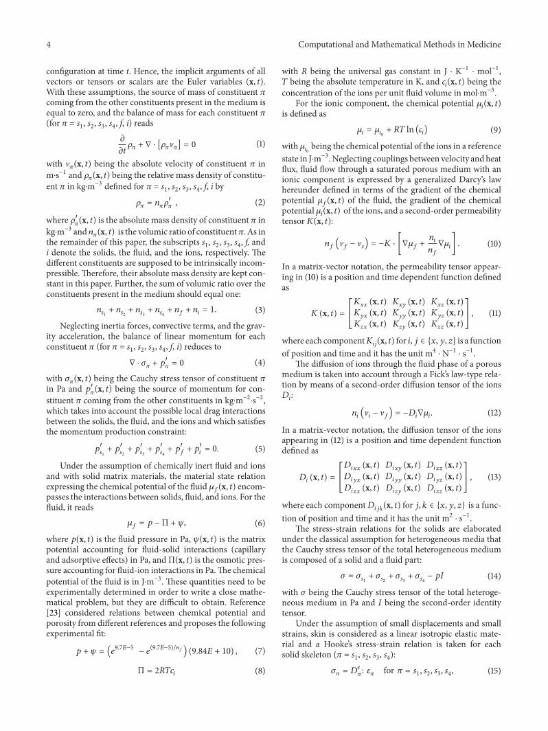
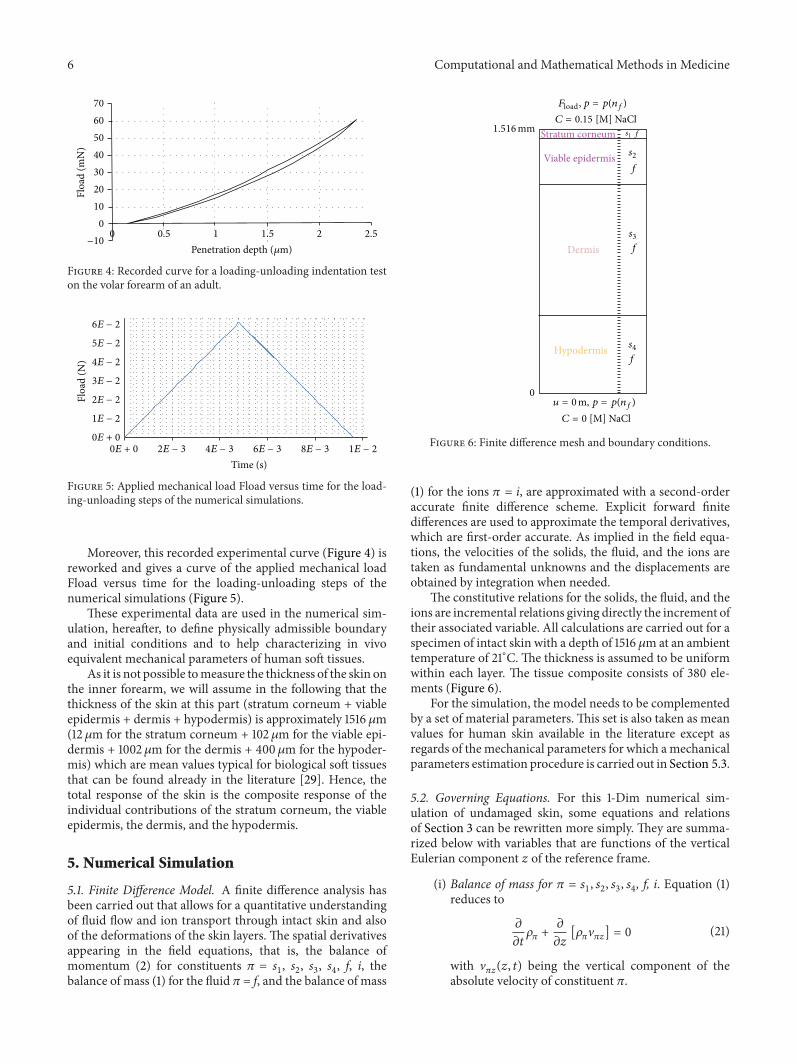
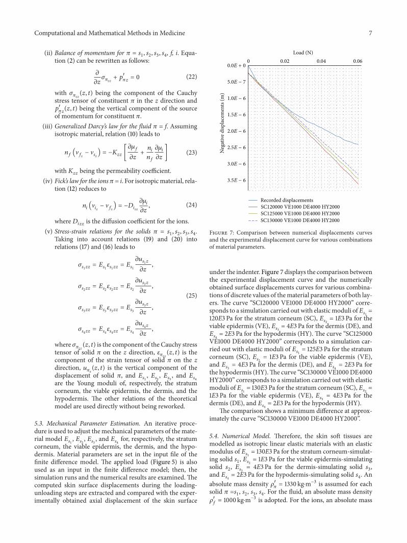
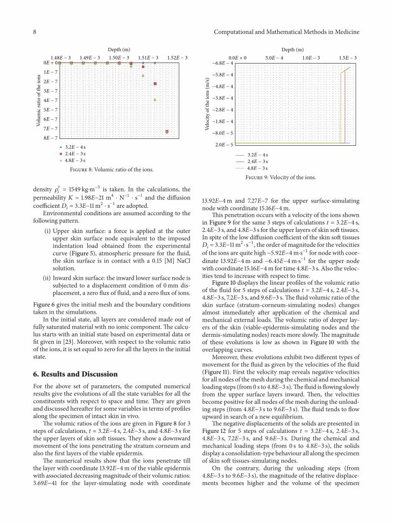
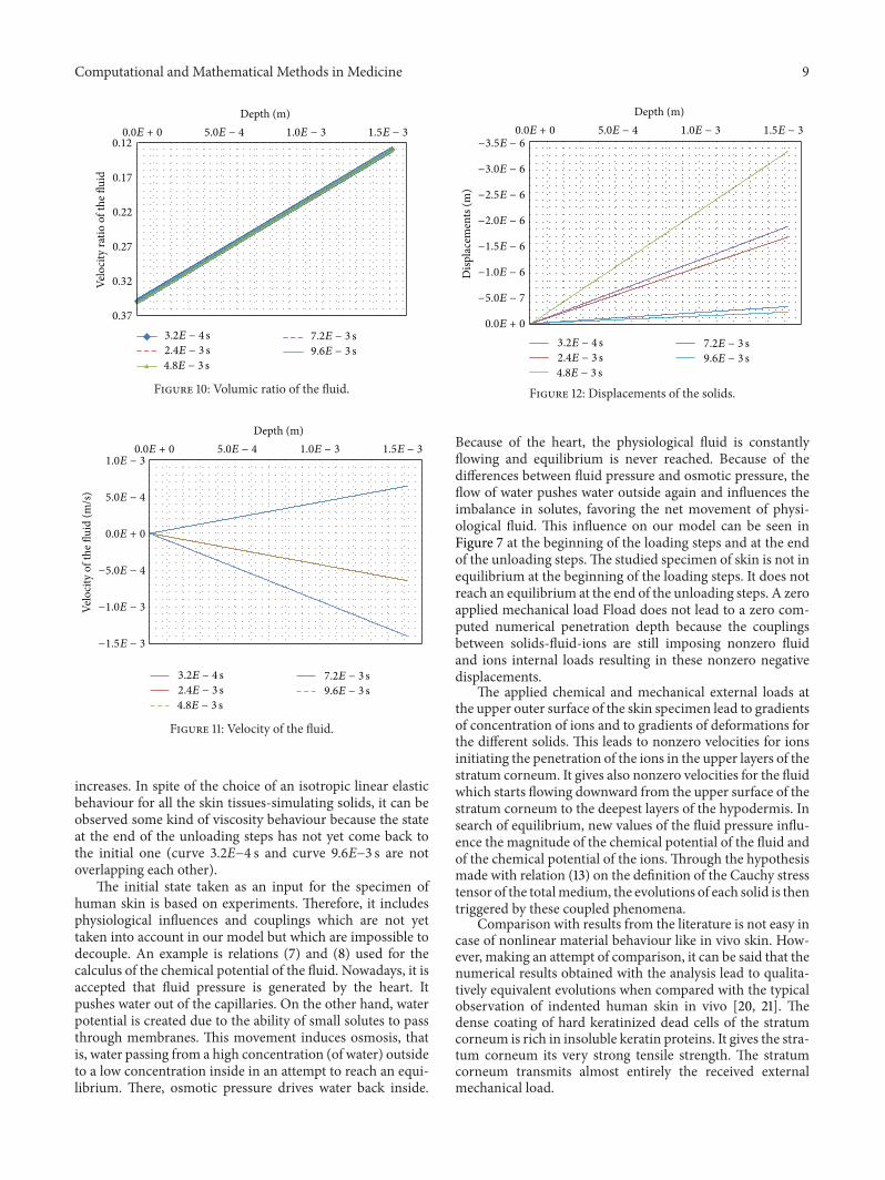