research article cytokine patterns in brain tumour progression
TRANSCRIPT

Hindawi Publishing CorporationMediators of InflammationVolume 2013, Article ID 979748, 7 pageshttp://dx.doi.org/10.1155/2013/979748
Research ArticleCytokine Patterns in Brain Tumour Progression
Radu Albulescu,1,2 Elena Codrici,1 Ionela Daniela Popescu,1 Simona Mihai,1 LauraGeorgiana Necula,1,3 Daniel Petrescu,4 Mihaela Teodoru,5 and Cristiana Pistol Tanase1
1 Victor Babes National Institute of Pathology, 99-101 Splaiul Independentei, 050096 Bucharest, Romania2National Institute for Chemical Pharmaceutical R&D, 112 Calea Vitan, 031299 Bucharest, Romania3 Stefan S Nicolau Institute of Virology, 285 Soseaua Mihai Bravu, 030304 Bucharest, Romania4Neurology and Neurovascular Diseases National Institute, 10-12 Soseaua Berceni, 041914 Bucharest, Romania5 Elias Emergency University Hospital, 19 Bulevardul Marasti, 011462 Bucharest, Romania
Correspondence should be addressed to Cristiana Pistol Tanase; [email protected]
Received 4 March 2013; Accepted 4 June 2013
Academic Editor: Gila Moalem-Taylor
Copyright © 2013 Radu Albulescu et al.This is an open access article distributed under the Creative CommonsAttribution License,which permits unrestricted use, distribution, and reproduction in any medium, provided the original work is properly cited.
Inflammation represents the immune system response to external or internal aggressors such as injury or infection in certain tissues.The body’s response to cancer has many parallels with inflammation and repair; the inflammatory cells and cytokines present intumours are more likely to contribute to tumour growth, progression, and immunosuppression, rather than in building an effectiveantitumour defence. Using new proteomic technology, we have investigated serum profile of pro- (IL-1𝛽, IL-6, IL-8, IL-12, GM-CSF, and TNF-𝛼) and anti-inflammatory cytokines (IL-4, IL-10), along with angiogenic factors (VEGF, bFGF) in order to assesstumoural aggressiveness. Our results indicate significant dysregulation in serum levels of cytokines and angiogenic factors, withover threefold upregulation of IL-6, IL-1𝛽, TNF-𝛼, and IL-10 and up to twofold upregulation of VEGF, FGF-2, IL-8, IL-2, andGM-CSF. These molecules are involved in tumour progression and aggressiveness, and are also involved in a generation of diseaseassociated pain.
1. Introduction
Glioblastomas are the most aggressive type of intracranialtumours, highly resistant to combined treatment, in patientsdisplaying a median survival time of 15 months [1]. Themolecular mechanisms underlying these clinical features arethe existence of specific genetic and molecular profiles ofthese tumour cells. Recent reports show genomic instability(especially in tumours from short-term survival patients),chromosomal alterations, somatic mutations, and polymor-phisms [2]. Knowing this particular brain tumour cell, onecan wonder if, besides the intrinsic cellular features, theinflammatory milieu triggered by the development of such atumour cannot influence the particular clinical developmentin glioblastomas as well.
The relationship between inflammation and cancer hasfirst been suggested in modern time, by Virchow in 1863,who found “lymphoreticular infiltrates” in neoplastic tissues,
consequently suggesting that these reflect the origin of can-cer of sites of chronic inflammation. Massive experimentalproofs appeared in the recent years to support Virchow’sconcept [3].
In a synthetic formulation, inflammation is defined as“the seventh hallmark of cancer”, by Colotta et al. [4]. Thebody’s response to cancer hasmany analogies with inflamma-tion and repair; the inflammatory cells and cytokines presentin tumours are more likely to contribute to tumour growth,progression, and immunosuppression, rather than in build-ing an effective antitumour defence. Cancer susceptibility andseverity are often associated with functional polymorphismsin cytokine genes. As plastically described by Balkwill andMantovani, if genetic damage is the “match that lights the fire”of cancer, some types of inflammation may provide the “fuelthat feeds the flames” [3].
Tumour initiation and progression is a complex pro-cess involving genomic mutations, micro environmental

2 Mediators of Inflammation
factors, and inflammatory mediators. Within the tumourenvironment inflammatory markers are responsible for cellproliferation, tumour invasion, marked angiogenesis, andsuppression of certain immune functions [5].
Inflammation represents the immune system response toexternal or internal aggressors, such as injury or infectionin certain tissues. Typical signs of inflammation includeswelling, redness, pain, temperature rise, and subsequentlyloss of function. Numerous studies have shown that themajority of tumour tissues are associated with inflammatorysigns. However, a clear connection between inflammationand cancer has yet to be demonstrated.
Glioblastoma represents the most common and lethalprimary brain tumour. The prognosis is poor, especially forhigher grade glioma—the most common primary neoplasmof the central nervous system, composing over 40%of all suchtumours, with an incidence ranging from 8% to 27% [6].
A broad array of cytokines displays modified expressionin cancers, including glioblastoma multiforme [7, 8].
The changes arise from the interaction of tumour cells andnontumour cells, like macrophages, lymphocytes, or stromalcells, and provide regulatory support for tumour growth,angiogenesis, invasion, and metastasis [8–10].
The vascular system of brain cancers inappropriatelyexpresses membrane proteins, resulting in blood extravasa-tion. The production of inflammatory mediators (such ascytokines and nitric oxide), and tumour hypoxia have beeninvolved in these effects [11].
Pain belongs to the “classical” markers of inflammation,described over 2000 years ago by Aulus Celsus (calor, rubor,tumour et dolor). Various molecular actors of inflammation,including mediators of pain have been described in therecent years; a key role appears to be played by cytokines,which sometimes appear to conduct the orchestra of smallmolecule mediators, such as nitric oxide and prostaglandins.Many studies show that inflammation may be involved indifferent stages of tumour development. Several cancer riskfactors like cigarette smoke, alcohol and growth factors canactivate signaling pathways related with inflammation (suchas NF𝜅B and STAT3 signaling). Some chronic infections leadto inflammatory conditions and are associated with carcino-genesis (e.g., hepatitis B virus). Chemotherapeutic agentsand gamma irradiation can also interfere in the regulationof expression of some genes implicated in inflammation,survival, proliferation, invasion, angiogenesis, and cancermetastasis [12]. Inflammationmay be involved in carcinogen-esis through mutations, genomic instability, and epigeneticmodifications [13]. Also, inflammation can participate inpremalignant cells proliferation, stimulate angiogenesis, andpromote metastatic spread (Figure 1).
Among the cytokines, often found to be over expressed attumour level, IL1-beta, TNF-alpha, IL-6, IL-10, IFN-gamma,CX3CL1, to name just a few, have been closely related to painfor a long time [14–16].
In the case of glioblastoma, headache is one of the mostfrequently claimed signs by the patients, but also, very oftenthe diagnostic is set too late for a successful therapeuticalapproach.
Environmental exposureDietary lifestyleTherapy induced
Microbial pathogensChronic inflammation
Proinflammatorymediators
(cytokines, chemokines,free radicals, growthfactors, MMPs, etc.)
CancerInitiation
PromotionMetastasis
Figure 1: Implication of chronic inflammation in different stagesof tumour development. Mediators of inflammation, triggered bydifferent processes, may stimulate premalignant cell proliferation,angiogenesis, and metastasis. Reversely, tumour cells have theability to stimulate other cells or to produce by themselves proinflammatory and pro-angiogenic factors.
Acknowledging the worldwide research effort in the fieldof glioblastoma, we have embarked in the study of circulatorycytokines to pinpoint the serum inflammatory pattern thatcan characterize the glioblastoma patient’s evolution. There-fore, our study investigated the serum levels of several pro-and anti-inflammatory cytokines and of angiogenic factorsin brain tumour patients diagnosed in stages III and IV(glioblastoma), in order to establish their roles and behaviourin tumour progression.
2. Material and Method
2.1. Patients and Samples. Samples (serum) were collectedfrom 55 patients with glioblastoma (28 men and 27 women;mean ages: 58 and 62 years, resp., range: 37–79 years) fromNeurology and Neurovascular Diseases National Institute,Elias Hospital Neurosurgery Department and 20 controls(healthy individuals with no known history of inflammatoryor neoplastic diseases, 12 men and 8 women; mean age:57 years, range: 25–70 years). Written informed consenthas been obtained upon sample prelevation according toHelsinki IIDeclaration andEthicsCommittee ofVictor BabesNational Institute of Pathology that has approved the study.The collection of total peripheral blood from patients andcontrols has been achieved in vacutainers (Systems, BectonDickinson) without anticoagulant. Serum was aliquoted andstored at −80∘C until analysis.
2.2. xMAP Analysis and ELISA. The xMAP assay was per-formed according to the manufacturers’ protocols, and theplates were analysed using Luminex 200 system. Cytokineslevels and angiogenic factors were determined using theHuman cytokine 12-plex Kit, with 12 analyte-specific bead

Mediators of Inflammation 3
sets (simultaneous quantification)—pro-inflammatory IL-1𝛽, IL-2, IL-6, IL-8, TNF𝛼, GM-CSF, and INF𝛾, anti-inflammatory IL-4, IL-10, and IL-12, and angiogenic factorsVEGF and FGF-2. Multiplex data acquisition and analysiswere performed using STarStation 2.3. Triplicate sampleswere used for all specimens. Values for individual proteinsmeasured by this multiplexed protein array technology havebeen shown to correlate with single ELISA measurements.
Immunoenzymatic ELISA analysis was performed withQuantikine (R&D Systems). Serum level of growth factorswas determined according to the manufacturer’s protocol.All samples were assayed in triplicate, and the mean valuesof cytokines were taken into account. Optical density wasmeasured at 450 nm on an Anthos Zenith 3100 multimodemicro plate reader. Minimum detectable concentrations werefound to be less than 9.0 pg/mL for VEGF and less than3.0 pg/mL for bFGF.
2.3. Statistical Analysis. Data were collected and expressedas the mean ± standard error of three independent repeats.Differences between groups were analysed by One WayAnova; 𝑃 values less than 0.05 were considered statisticallysignificant; Pearson correlation (r, p) was used to explore theassociation between cytokine expressions. Statistical analysiswas performed using SPSS 19.0 software.
3. Results and Discussion
Frommultiplex assay (Luminex 200) a strong overexpressionwas detected for IL-6, IL-1𝛽, TNF-𝛼, and IL-10 (over 3-fold stimulation in glioblastoma patients). Significant up-regulation (up to 2-fold) was found for VEGF, FGF-2, IL-8, IL-2, and GM-CSF. Cytokines expression was significantlyhigher and strongly correlated with tumour grade, prolifer-ation markers, and clinical aggressiveness in glioblastomas.Comparing the patient groups and control for growth factors,the obtained values by xMAP array were comparable to theoutline obtained by the ELISA analysis.
Based on xMAP analysis, the changes in average serumlevels (compared to the controls) are presented in Figure 2.
Several molecules display a modification in plasma levelsof more than 2-fold, which is, as a general practice, a criterionof acceptance as potential marker. However, it also appearsevident that, for several cytokines, the intervals of variationsin patients were broad. Further details on expression areprovided in Figures 3, 4, and 5, where the distributions canbe better examined and also cover the behaviour of controls.
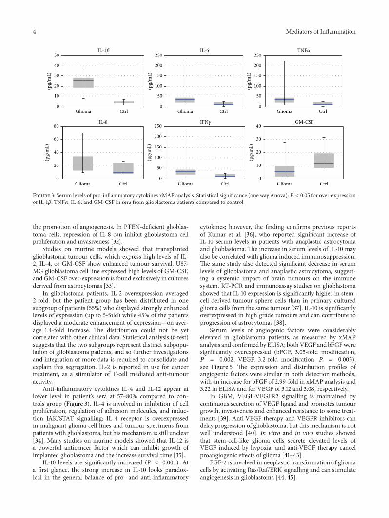
The enhanced expression of IL-1𝛽 appears to directlycorrelate with IL-6 and IL-8 levels and inversely correlatewith IL-4. In brain, IL-1𝛽 regulates survival and invasivenessof glioblastoma cells, and anti-IL-1𝛽 antibodies inhibit boththe growth and invasion of glioblastoma cells [17]. Wang etal. showed that in LN-229 glioma cell line, IL-1𝛽 and TGF-𝛽 can induce glioma stem cells phenotype and contributeto carcinogenesis [18]. Enhanced secretion of IL-1𝛽, IL-6,and IL-8 by glioma cells was reported by Yeung et al. [19],and these cytokines are related with the expansion of GBM(glioblastoma multiforme). In other types of cancers (gastric
02468
10121416
Modulation of serum cytokine levels in glioblastoma patients
IL-2
IL-8
GM
-CSF
IL-4
IL-10
IL-12
VEG
F
Fold
mod
ifica
tion
vers
us co
ntro
l
bFG
F
IL1
-𝛽
IL-6
TNF𝛼
IFN𝛾
Figure 2: Modulation of serum cytokine levels in glioblastomapatients. The data represent group averages of fold modificationversus controls + standard deviations. Statistical significance (oneway ANOVA): pro-inflammatory cytokines, 𝑃 < 0.05 for IL-1𝛽, IL-6, and TNF𝛼, GM-CSF; anti-inflammatory cytokines, 𝑃 < 0.05 forIL-4, and IL-10; angiogenic factors, 𝑃 < 0.05 for bFGF and VEGF.Expression levels of IL-2, IL-8, IFN-𝛾, and IL-12 were modified, butwith low statistical significance.
and oesophageal), IL-1𝛽 was involved in carcinogenesis andproliferation and played a crucial role in the development ofchemical carcinogen-induced tumours [20, 21].
IL-6 appeared overexpressed (average 4-fold) in glioblas-toma patients. The determined serum levels are consistentwith the ability of tumour cell to secrete pro-inflammatorycytokines, as well as with IL-6 role in stimulation of angio-genesis. According to our data, IL-6 expression correlateswith IL-1𝛽, IL-8, and IFN-𝛾. Ancrile et al. showed IL-6is involved in carcinogenesis by angiogenesis and tumourgrowth and may be a potential anti-invasion target [22].In U251, T98G and U87 MG glioblastoma cell lines, IL-6promotes vascular endothelial cell migration and facilitatestumour angiogenesis and invasion [23, 24]. Amplification ofthe IL-6 gene in patients with glioblastoma multiforme iscorrelated with decreased survival [25].
Serum levels of TNF𝛼 appeared significantly enhanced(𝑃 = 2.5𝐸 − 8), suggesting a strong correlation with the dis-ease; however, the correlation with other molecules is not sostrong, suggesting its implication in distinct/complementaryregulatory cascades. Hagemann et al. suggested that TNFis involved in tumour cell invasion through upregula-tion of migration-inhibitory factor (MIF) and throughenhanced MMPs production in tumour cells via NF-𝜅𝛽-and JNK-signalling [26]. In ovarian cancer, TNF𝛼 stimu-lated other cytokines (IL-6), angiogenic factors (VEGF), andchemokines (CCL2 and CXCL12) that promoted tumourgrowth and metastases [27]. Other studies have showedthat TNF over-expression enhancesmigration andmetastasisthrough induction of CXCR4, MCP-1, and IL-8 and matrixmetalloproteinase [28, 29]. Recent studies on U373MG andC6 human glioma cell lines showed that TNF-𝛼 inducesIL-6 synthesis through the JAK/STAT3 pathway and TNFinhibitors can reduce tumour cell invasion [30, 31].
IL-8 has been found to be up-regulated (fold stimulation1.9) in patient sera, compared to controls. Many studiesshowed that IL8 is upregulated in gliomas and is involved in
4 Mediators of Inflammation
0
10
20
30
40
50
Glioma Ctrl
Glioma Ctrl Glioma Ctrl Glioma Ctrl
Glioma Ctrl Glioma Ctrl
(pg/
mL)
(pg/
mL)
(pg/
mL)
IL-1𝛽
0
50
100
150
200
250
(pg/
mL)
(pg/
mL)
0
50
100
150
200
250
(pg/
mL)
0
50
100
150
200
250IL-6 TNF𝛼
0
20
40
60
80IL-8 IFN𝛾
0
10
20
30
40GM-CSF
Figure 3: Serum levels of pro-inflammatory cytokines xMAP analysis. Statistical significance (one way Anova): 𝑃 < 0.05 for over-expressionof IL-1𝛽, TNF𝛼, IL-6, and GM-CSF in sera from glioblastoma patients compared to control.
the promotion of angiogenesis. In PTEN-deficient glioblas-toma cells, repression of IL-8 can inhibit glioblastoma cellproliferation and invasiveness [32].
Studies on murine models showed that transplantedglioblastoma tumour cells, which express high levels of IL-2, IL-4, or GM-CSF show enhanced tumour survival. U87-MG glioblastoma cell line expressed high levels of GM-CSF,and GM-CSF over-expression is found exclusively in culturesderived from astrocytomas [33].
In glioblastoma patients, IL-2 overexpression averaged2-fold, but the patient group has been distributed in onesubgroup of patients (55%) who displayed strongly enhancedlevels of expression (up to 5-fold) while 45% of the patientsdisplayed a moderate enhancement of expression—on aver-age 1.4-fold increase. The distribution could not be yetcorrelated with other clinical data. Statistical analysis (𝑡-test)suggests that the two subgroups represent distinct subpopu-lation of glioblastoma patients, and so further investigationsand integration of more data is required to consolidate andexplain this segregation. IL-2 is reported in use for cancertreatment, as a stimulator of T-cell mediated anti-tumouractivity.
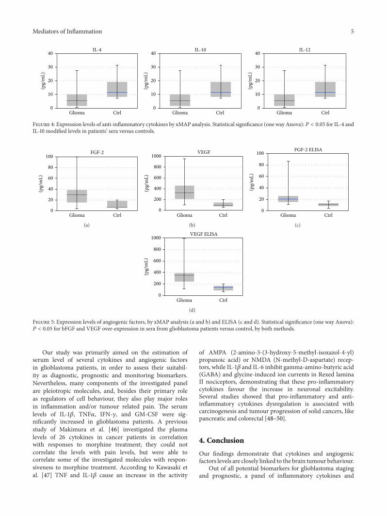
Anti-inflammatory cytokines IL-4 and IL-12 appear atlower level in patient’s sera at 57–80% compared to con-trols group (Figure 3). IL-4 is involved in inhibition of cellproliferation, regulation of adhesion molecules, and induc-tion JAK/STAT signalling; IL-4 receptor is overexpressedin malignant glioma cell lines and tumour specimens frompatients with glioblastoma, but his mechanism is still unclear[34]. Many studies on murine models showed that IL-12 isa powerful anticancer factor which can inhibit growth ofimplanted glioblastoma and the increase survival time [35].
IL-10 levels are significantly increased (𝑃 < 0.001). Ata first glance, the strong increase in IL-10 looks paradox-ical in the general balance of pro- and anti-inflammatory
cytokines; however, the finding confirms previous reportsof Kumar et al. [36], who reported significant increase ofIL-10 serum levels in patients with anaplastic astrocytomaand glioblastoma. The increase in serum levels of IL-10 mayalso be correlated with glioma induced immunosuppression.The same study also detected significant decrease in serumlevels of glioblastoma and anaplastic astrocytoma, suggest-ing a systemic impact of brain tumours on the immunesystem. RT-PCR and immunoassay studies on glioblastomashowed that IL-10 expression is significantly higher in stem-cell-derived tumour sphere cells than in primary culturedglioma cells from the same tumour [37]. IL-10 is significantlyoverexpressed in high grade tumours and can contribute toprogression of astrocytomas [38].
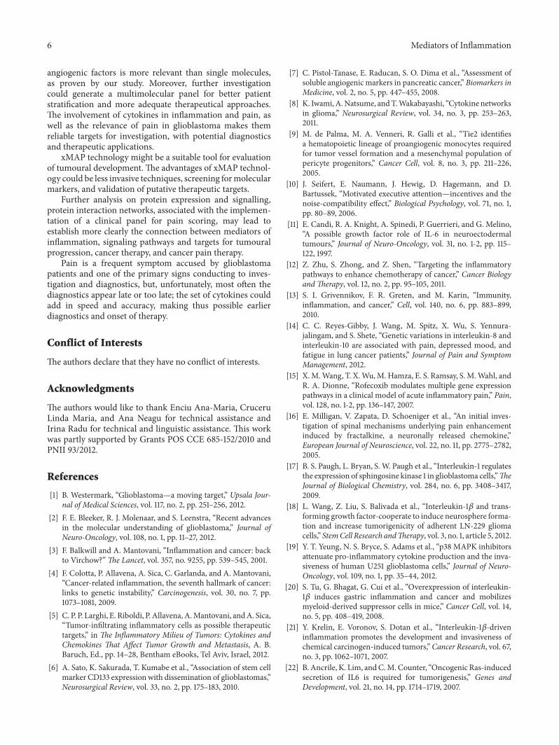
Serum levels of angiogenic factors were considerablyelevated in glioblastoma patients, as measured by xMAPanalysis and confirmed by ELISA; bothVEGF and bFGFweresignificantly overexpressed (bFGF, 3.05-fold modification,𝑃 = 0.002, VEGF, 3.2-fold modification, 𝑃 = 0.005),see Figure 5. The expression and distribution profiles ofangiogenic factors were similar in both detection methods,with an increase for bFGF of 2.99-fold in xMAP analysis and3.22 in ELISA and for VEGF of 3.12 and 3.08, respectively.
In GBM, VEGF-VEGFR2 signalling is maintained bycontinuous secretion of VEGF ligand and promotes tumourgrowth, invasiveness and enhanced resistance to some treat-ments [39]. Anti-VEGF therapy and VEGFR inhibitors candelay progression of glioblastoma, but this mechanism is notwell understood [40]. In vitro and in vivo studies showedthat stem-cell-like glioma cells secrete elevated levels ofVEGF induced by hypoxia, and anti-VEGF therapy cancelproangiogenic effects of glioma [41–43].
FGF-2 is involved in neoplastic transformation of gliomacells by activating Ras/Raf/ERK signalling and can stimulateangiogenesis in glioblastoma [44, 45].
Mediators of Inflammation 5
0
10
20
30
40
Glioma Ctrl
(pg/
mL)
0
10
20
30
40
Glioma Ctrl
(pg/
mL)
0
10
20
30
40
Glioma Ctrl
(pg/
mL)
IL-4 IL-10 IL-12
Figure 4: Expression levels of anti-inflammatory cytokines by xMAP analysis. Statistical significance (one way Anova): 𝑃 < 0.05 for IL-4 andIL-10 modified levels in patients’ sera versus controls.
0
20
40
60
80
100
Glioma Ctrl
(pg/
mL)
FGF-2
(a)
Glioma Ctrl
(pg/
mL)
0
200
400
600
800
1000VEGF
(b)
0
20
40
60
80
100
Glioma Ctrl
(pg/
mL)
FGF-2 ELISA
(c)
Glioma Ctrl
(pg/
mL)
0
200
400
600
800
1000VEGF ELISA
(d)
Figure 5: Expression levels of angiogenic factors, by xMAP analysis (a and b) and ELISA (c and d). Statistical significance (one way Anova):𝑃 < 0.05 for bFGF and VEGF over-expression in sera from glioblastoma patients versus control, by both methods.
Our study was primarily aimed on the estimation ofserum level of several cytokines and angiogenic factorsin glioblastoma patients, in order to assess their suitabil-ity as diagnostic, prognostic and monitoring biomarkers.Nevertheless, many components of the investigated panelare pleiotropic molecules, and, besides their primary roleas regulators of cell behaviour, they also play major rolesin inflammation and/or tumour related pain. The serumlevels of IL-1𝛽, TNF𝛼, IFN-𝛾, and GM-CSF were sig-nificantly increased in glioblastoma patients. A previousstudy of Makimura et al. [46] investigated the plasmalevels of 26 cytokines in cancer patients in correlationwith responses to morphine treatment; they could notcorrelate the levels with pain levels, but were able tocorrelate some of the investigated molecules with respon-siveness to morphine treatment. According to Kawasaki etal. [47] TNF and IL-1𝛽 cause an increase in the activity
of AMPA (2-amino-3-(3-hydroxy-5-methyl-isoxazol-4-yl)propanoic acid) or NMDA (N-methyl-D-aspartate) recep-tors, while IL-1𝛽 and IL-6 inhibit gamma-amino-butyric acid(GABA) and glycine-induced ion currents in Rexed laminaII nociceptors, demonstrating that these pro-inflammatorycytokines favour the increase in neuronal excitability.Several studies showed that pro-inflammatory and anti-inflammatory cytokines dysregulation is associated withcarcinogenesis and tumour progression of solid cancers, likepancreatic and colorectal [48–50].
4. Conclusion
Our findings demonstrate that cytokines and angiogenicfactors levels are closely linked to the brain tumour behaviour.
Out of all potential biomarkers for glioblastoma stagingand prognostic, a panel of inflammatory cytokines and
6 Mediators of Inflammation
angiogenic factors is more relevant than single molecules,as proven by our study. Moreover, further investigationcould generate a multimolecular panel for better patientstratification and more adequate therapeutical approaches.The involvement of cytokines in inflammation and pain, aswell as the relevance of pain in glioblastoma makes themreliable targets for investigation, with potential diagnosticsand therapeutic applications.
xMAP technology might be a suitable tool for evaluationof tumoural development. The advantages of xMAP technol-ogy could be less invasive techniques, screening formolecularmarkers, and validation of putative therapeutic targets.
Further analysis on protein expression and signalling,protein interaction networks, associated with the implemen-tation of a clinical panel for pain scoring, may lead toestablish more clearly the connection between mediators ofinflammation, signaling pathways and targets for tumouralprogression, cancer therapy, and cancer pain therapy.
Pain is a frequent symptom accused by glioblastomapatients and one of the primary signs conducting to inves-tigation and diagnostics, but, unfortunately, most often thediagnostics appear late or too late; the set of cytokines couldadd in speed and accuracy, making thus possible earlierdiagnostics and onset of therapy.
Conflict of Interests
The authors declare that they have no conflict of interests.
Acknowledgments
The authors would like to thank Enciu Ana-Maria, CruceruLinda Maria, and Ana Neagu for technical assistance andIrina Radu for technical and linguistic assistance. This workwas partly supported by Grants POS CCE 685-152/2010 andPNII 93/2012.
References
[1] B. Westermark, “Glioblastoma—a moving target,” Upsala Jour-nal of Medical Sciences, vol. 117, no. 2, pp. 251–256, 2012.
[2] F. E. Bleeker, R. J. Molenaar, and S. Leenstra, “Recent advancesin the molecular understanding of glioblastoma,” Journal ofNeuro-Oncology, vol. 108, no. 1, pp. 11–27, 2012.
[3] F. Balkwill and A. Mantovani, “Inflammation and cancer: backto Virchow?”The Lancet, vol. 357, no. 9255, pp. 539–545, 2001.
[4] F. Colotta, P. Allavena, A. Sica, C. Garlanda, and A. Mantovani,“Cancer-related inflammation, the seventh hallmark of cancer:links to genetic instability,” Carcinogenesis, vol. 30, no. 7, pp.1073–1081, 2009.
[5] C. P. P. Larghi, E. Riboldi, P. Allavena, A.Mantovani, andA. Sica,“Tumor-infiltrating inflammatory cells as possible therapeutictargets,” in The Inflammatory Milieu of Tumors: Cytokines andChemokines That Affect Tumor Growth and Metastasis, A. B.Baruch, Ed., pp. 14–28, Bentham eBooks, Tel Aviv, Israel, 2012.
[6] A. Sato, K. Sakurada, T. Kumabe et al., “Association of stem cellmarker CD133 expression with dissemination of glioblastomas,”Neurosurgical Review, vol. 33, no. 2, pp. 175–183, 2010.
[7] C. Pistol-Tanase, E. Raducan, S. O. Dima et al., “Assessment ofsoluble angiogenic markers in pancreatic cancer,” Biomarkers inMedicine, vol. 2, no. 5, pp. 447–455, 2008.
[8] K. Iwami, A.Natsume, andT.Wakabayashi, “Cytokine networksin glioma,” Neurosurgical Review, vol. 34, no. 3, pp. 253–263,2011.
[9] M. de Palma, M. A. Venneri, R. Galli et al., “Tie2 identifiesa hematopoietic lineage of proangiogenic monocytes requiredfor tumor vessel formation and a mesenchymal population ofpericyte progenitors,” Cancer Cell, vol. 8, no. 3, pp. 211–226,2005.
[10] J. Seifert, E. Naumann, J. Hewig, D. Hagemann, and D.Bartussek, “Motivated executive attention—incentives and thenoise-compatibility effect,” Biological Psychology, vol. 71, no. 1,pp. 80–89, 2006.
[11] E. Candi, R. A. Knight, A. Spinedi, P. Guerrieri, and G. Melino,“A possible growth factor role of IL-6 in neuroectodermaltumours,” Journal of Neuro-Oncology, vol. 31, no. 1-2, pp. 115–122, 1997.
[12] Z. Zhu, S. Zhong, and Z. Shen, “Targeting the inflammatorypathways to enhance chemotherapy of cancer,” Cancer BiologyandTherapy, vol. 12, no. 2, pp. 95–105, 2011.
[13] S. I. Grivennikov, F. R. Greten, and M. Karin, “Immunity,inflammation, and cancer,” Cell, vol. 140, no. 6, pp. 883–899,2010.
[14] C. C. Reyes-Gibby, J. Wang, M. Spitz, X. Wu, S. Yennura-jalingam, and S. Shete, “Genetic variations in interleukin-8 andinterleukin-10 are associated with pain, depressed mood, andfatigue in lung cancer patients,” Journal of Pain and SymptomManagement, 2012.
[15] X.M.Wang, T. X.Wu,M.Hamza, E. S. Ramsay, S.M.Wahl, andR. A. Dionne, “Rofecoxib modulates multiple gene expressionpathways in a clinical model of acute inflammatory pain,” Pain,vol. 128, no. 1-2, pp. 136–147, 2007.
[16] E. Milligan, V. Zapata, D. Schoeniger et al., “An initial inves-tigation of spinal mechanisms underlying pain enhancementinduced by fractalkine, a neuronally released chemokine,”European Journal of Neuroscience, vol. 22, no. 11, pp. 2775–2782,2005.
[17] B. S. Paugh, L. Bryan, S.W. Paugh et al., “Interleukin-1 regulatesthe expression of sphingosine kinase 1 in glioblastoma cells,”TheJournal of Biological Chemistry, vol. 284, no. 6, pp. 3408–3417,2009.
[18] L. Wang, Z. Liu, S. Balivada et al., “Interleukin-1𝛽 and trans-forming growth factor-cooperate to induce neurosphere forma-tion and increase tumorigenicity of adherent LN-229 gliomacells,” StemCell Research andTherapy, vol. 3, no. 1, article 5, 2012.
[19] Y. T. Yeung, N. S. Bryce, S. Adams et al., “p38 MAPK inhibitorsattenuate pro-inflammatory cytokine production and the inva-siveness of human U251 glioblastoma cells,” Journal of Neuro-Oncology, vol. 109, no. 1, pp. 35–44, 2012.
[20] S. Tu, G. Bhagat, G. Cui et al., “Overexpression of interleukin-1𝛽 induces gastric inflammation and cancer and mobilizesmyeloid-derived suppressor cells in mice,” Cancer Cell, vol. 14,no. 5, pp. 408–419, 2008.
[21] Y. Krelin, E. Voronov, S. Dotan et al., “Interleukin-1𝛽-driveninflammation promotes the development and invasiveness ofchemical carcinogen-induced tumors,”Cancer Research, vol. 67,no. 3, pp. 1062–1071, 2007.
[22] B. Ancrile, K. Lim, andC.M.Counter, “Oncogenic Ras-inducedsecretion of IL6 is required for tumorigenesis,” Genes andDevelopment, vol. 21, no. 14, pp. 1714–1719, 2007.
Mediators of Inflammation 7
[23] Q. Liu, G. Li, R. Li et al., “IL-6 promotion of glioblastoma cellinvasion and angiogenesis in U251 and T98G cell lines,” Journalof Neuro-Oncology, vol. 100, no. 2, pp. 165–176, 2010.
[24] R. Li, G. Li, L. Deng et al., “IL-6 augments the invasivenessof U87MG human glioblastoma multiforme cells via up-regulation of MMP-2 and fascin-1,” Oncology Reports, vol. 23,no. 6, pp. 1553–1559, 2010.
[25] A. Tchirkov, T. Khalil, E. Chautard et al., “Interleukin-6 geneamplification and shortened survival in glioblastoma patients,”The British Journal of Cancer, vol. 96, no. 3, pp. 474–476, 2007.
[26] T. Hagemann, J. Wilson, H. Kulbe et al., “Macrophages induceinvasiveness of epithelial cancer cells via NF-𝜅B and JNK,”Journal of Immunology, vol. 175, no. 2, pp. 1197–1205, 2005.
[27] H. Kulbe, R. Thompson, J. L. Wilson et al., “The inflammatorycytokine tumor necrosis factor-𝛼 generates an autocrine tumor-promoting network in epithelial ovarian cancer cells,” CancerResearch, vol. 67, no. 2, pp. 585–592, 2007.
[28] H. Kulbe, T. Hagemann, P. W. Szlosarek, F. R. Balkwill, and J.L. Wilson, “The inflammatory cytokine tumor necrosis factor-𝛼 regulates chemokine receptor expression on ovarian cancercells,” Cancer Research, vol. 65, no. 22, pp. 10355–10362, 2005.
[29] J.W. Pollard, “Tumour-educatedmacrophages promote tumourprogression and metastasis,” Nature Reviews Cancer, vol. 4, no.1, pp. 71–78, 2004.
[30] J. Ryu, B. M. Ku, Y. K. Lee et al., “Resveratrol reduces TNF-𝛼-induced U373MG human glioma cell invasion through regu-lating NF-𝜅B activation and uPA/uPAR expression,” AnticancerResearch, vol. 31, no. 12, pp. 4223–4230, 2011.
[31] K. Tanabe, R. Matsushima-Nishiwaki, S. Yamaguchi, H. Iida, S.Dohi, and O. Kozawa, “Mechanisms of tumor necrosis factor-𝛼-induced interleukin-6 synthesis in glioma cells,” Journal ofNeuroinflammation, vol. 7, article 16, 2010.
[32] N. de la Iglesia, G. Konopka, K. Lim et al., “Deregulationof a STAT3-interleukin 8 signaling pathway promotes humanglioblastoma cell proliferation and invasiveness,” Journal ofNeuroscience, vol. 28, no. 23, pp. 5870–5878, 2008.
[33] C. S. Curran, M. D. Evans, and P. J. Bertics, “GM-CSF produc-tion by glioblastoma cells has a functional role in eosinophilsurvival, activation, and growth factor production for enhancedtumor cell proliferation,” Journal of Immunology, vol. 187, no. 3,pp. 1254–1263, 2011.
[34] M. Kawakami, K. Kawakami, and R. K. Puri, “Interleukin-4-Pseudomonas exotoxin chimeric fusion protein for malignantglioma therapy,” Journal of Neuro-Oncology, vol. 65, no. 1, pp.15–25, 2003.
[35] T. L. Chiu, M. J. Wang, and C. C. Su, “The treatment ofglioblastoma multiforme through activation of microglia andTRAIL induced by rAAV2-mediated IL-12 in a syngeneic ratmodel,” Journal of Biomedical Science, vol. 19, article 45, 2012.
[36] R. Kumar, D. Kamdar, L. Madden et al., “Th1/Th2 cytokineimbalance inmeningioma, anaplastic astrocytoma and glioblas-toma multiforme patients,” Oncology Reports, vol. 15, no. 6, pp.1513–1516, 2006.
[37] B. Qiu, D. Zhang, C. Wang et al., “IL-10 and TGF-𝛽2 areoverexpressed in tumor spheres cultured from human gliomas,”Molecular Biology Reports, vol. 38, no. 5, pp. 3585–3591, 2011.
[38] C. Huettner, W. Paulus, and W. Roggendorf, “Increasedamounts of IL-10 mRNA in anaplastic astrocytomas andglioblastoma multiforme,” Verhandlungen der DeutschenGesellschaft fur Pathologie, vol. 78, pp. 418–422, 1994.
[39] P. Hamerlik, J. D. Lathia, R. Rasmussen et al., “Autocrine VEGF-VEGFR2-Neuropilin-1 signaling promotes glioma stem-like cellviability and tumor growth,” Journal of Experimental Medicine,vol. 209, no. 3, pp. 507–520, 2012.
[40] Y. Piao, J. Liang, L. Holmes et al., “Glioblastoma resistance toanti-VEGF therapy is associated with myeloid cell infiltration,stem cell accumulation, and amesenchymal phenotype,”Neuro-Oncology, vol. 14, no. 11, pp. 1379–1392, 2012.
[41] S. Bao, Q. Wu, S. Sathornsumetee et al., “Stem cell-like gliomacells promote tumor angiogenesis through vascular endothelialgrowth factor,” Cancer Research, vol. 66, no. 16, pp. 7843–7848,2006.
[42] M. T. Chiao, Y. C. Yang, W. Y. Cheng, C. C. Shen, and J. L. Ko,“CD133+ glioblastoma stem-like cells induce vascular mimicryin vivo,” Current Neurovascular Research, vol. 8, no. 3, pp. 210–219, 2011.
[43] C. Xu, X. Wu, and J. Zhu, “VEGF promotes proliferation ofhuman glioblastoma multiforme stem-like cells through VEGFreceptor 2,” The Scientific World Journal, vol. 2013, Article ID417413, 8 pages, 2013.
[44] W. Loilome,A.D. Joshi, C.M. J. apRhys et al., “Glioblastoma cellgrowth is suppressed by disruption of fibroblast growth factorpathway signaling,” Journal of Neuro-Oncology, vol. 94, no. 3, pp.359–366, 2009.
[45] G. P. Dunn, M. L. Rinne, J. Wykosky et al., “Emerging insightsinto themolecular and cellular basis of glioblastoma,”Genes andDevelopment, vol. 26, no. 8, pp. 756–784, 2012.
[46] C. Makimura, T. Arao, H. Matsuoka et al., “Prospective studyevaluating the plasma concentrations of twenty-six cytokinesand response to morphine treatment in cancer patients,” Anti-cancer Research, vol. 31, no. 12, pp. 4561–4568, 2011.
[47] Y. Kawasaki, L. Zhang, J. Cheng, and R. Ji, “Cytokine mech-anisms of central sensitization: distinct and overlapping roleof interleukin-1𝛽, interleukin-6, and tumor necrosis factor-𝛼in regulating synaptic and neuronal activity in the superficialspinal cord,” Journal of Neuroscience, vol. 28, no. 20, pp. 5189–5194, 2008.
[48] N. Momi, S. Kaur, S. R. Krishn, and S. K. Batra, “Discoveringthe route from inflammation to pancreatic cancer,” MinervaGastroenterologica eDietologica, vol. 58, no. 4, pp. 283–297, 2012.
[49] T. A. Ullman and S. H. Itzkowitz, “Intestinal inflammation andcancer,” Gastroenterology, vol. 140, no. 6, pp. 1807–1816, 2011.
[50] S. O. Dima, C. Tanase, R. Albulescu et al., “An exploratory studyof inflammatory cytokines as prognostic biomarkers in patientswith ductal pancreatic adenocarcinoma,” Pancreas, vol. 41, no.7, pp. 1001–1007, 2012.

Submit your manuscripts athttp://www.hindawi.com
Stem CellsInternational
Hindawi Publishing Corporationhttp://www.hindawi.com Volume 2014
Hindawi Publishing Corporationhttp://www.hindawi.com Volume 2014
MEDIATORSINFLAMMATION
of
Hindawi Publishing Corporationhttp://www.hindawi.com Volume 2014
Behavioural Neurology
EndocrinologyInternational Journal of
Hindawi Publishing Corporationhttp://www.hindawi.com Volume 2014
Hindawi Publishing Corporationhttp://www.hindawi.com Volume 2014
Disease Markers
Hindawi Publishing Corporationhttp://www.hindawi.com Volume 2014
BioMed Research International
OncologyJournal of
Hindawi Publishing Corporationhttp://www.hindawi.com Volume 2014
Hindawi Publishing Corporationhttp://www.hindawi.com Volume 2014
Oxidative Medicine and Cellular Longevity
Hindawi Publishing Corporationhttp://www.hindawi.com Volume 2014
PPAR Research
The Scientific World JournalHindawi Publishing Corporation http://www.hindawi.com Volume 2014
Immunology ResearchHindawi Publishing Corporationhttp://www.hindawi.com Volume 2014
Journal of
ObesityJournal of
Hindawi Publishing Corporationhttp://www.hindawi.com Volume 2014
Hindawi Publishing Corporationhttp://www.hindawi.com Volume 2014
Computational and Mathematical Methods in Medicine
OphthalmologyJournal of
Hindawi Publishing Corporationhttp://www.hindawi.com Volume 2014
Diabetes ResearchJournal of
Hindawi Publishing Corporationhttp://www.hindawi.com Volume 2014
Hindawi Publishing Corporationhttp://www.hindawi.com Volume 2014
Research and TreatmentAIDS
Hindawi Publishing Corporationhttp://www.hindawi.com Volume 2014
Gastroenterology Research and Practice
Hindawi Publishing Corporationhttp://www.hindawi.com Volume 2014
Parkinson’s Disease
Evidence-Based Complementary and Alternative Medicine
Volume 2014Hindawi Publishing Corporationhttp://www.hindawi.com