research article expression profiling of genes related to...
TRANSCRIPT
Research ArticleExpression Profiling of Genes Related toEndothelial Cells Biology in Patients with Type 2Diabetes and Patients with Prediabetes
Sara Moradipoor1 Patimah Ismail1 Ali Etemad1 Wan Aliaa Wan Sulaiman2
and Salma Ahmadloo1
1Department of Biomedical Sciences Faculty of Medicine and Health Sciences Universiti PutraMalaysia Serdang Selangor Malaysia2Department of Medicine Faculty of Medicine and Health Sciences Universiti Putra Malaysia Serdang Selangor Malaysia
Correspondence should be addressed to Patimah Ismail patimahismailgmailcom and Ali Etemad etal1900gmailcom
Received 10 June 2016 Revised 18 August 2016 Accepted 30 August 2016
Academic Editor Gelin Xu
Copyright copy 2016 Sara Moradipoor et al This is an open access article distributed under the Creative Commons AttributionLicense which permits unrestricted use distribution and reproduction in any medium provided the original work is properlycited
Endothelial dysfunction appears to be an early sign indicating vascular damage and predicts the progression of atherosclerosisand cardiovascular disorders Extensive clinical and experimental evidence suggests that endothelial dysfunction occurs in Type2 Diabetes Mellitus (T2DM) and prediabetes patients This study was carried out with an aim to appraise the expression levelsin the peripheral blood of 84 genes related to endothelial cells biology in patients with diagnosed T2DM or prediabetes tryingto identify new genes whose expression might be changed under these pathological conditions The study covered a total of 45participants The participants were divided into three groups group 1 patients with T2DM group 2 patients with prediabetesgroup 3 control group The gene expression analysis was performed using the Endothelial Cell Biology RT2 Profiler PCR ArrayIn the case of T2DM 59 genes were found to be upregulated and four genes were observed to be downregulated In prediabetespatients increased expressionwas observed for 49 genes with two downregulated genes observed Our results indicate that diabeticand prediabetic conditions change the expression levels of genes related to endothelial cells biology and consequently may increasethe risk for occurrence of endothelial dysfunction
1 Introduction
The endothelium lines the blood vessels and controls a widearray of vascular functions along with maintaining vascularhomeostasis [1] Some of the major functions of the endothe-lial cells include regulating the vessel integrity vascular tonevascular growth and remodelling immune responses celladhesion angiogenesis inflammatory responses coagulationand platelet activation haemostasis and vascular perme-ability [2] Sometimes due to various factors the endothe-lium is not able to maintain vascular homeostasis leadingto ldquoendothelial dysfunctionrdquo [3] Substantial clinical andexperimental evidence suggests that endothelial dysfunctiongenerally occurs in patients diagnosed with Type 2 DiabetesMellitus (T2DM) in both the resistance and conduit vesselsof the peripheral blood circulation along with the coronarycirculation [4 5] and it is one of the major factors that can
contribute to the pathogenesis of micro- and macrovasculardiseases in these patients [1] In fact previous researchindicates that as diabetes progresses in patients there is anincrease in the progression of endothelial dysfunction ulti-mately leading to atherosclerosis [6] Despite many proposedmechanisms for this relationship the definitive pathogenesisremains unclear possibly because diabetes patients usuallydisplay multiple homeostatic imbalances alongside the typ-ically described hyperglycemia Hyperglycemia and otherrisk factors such as insulin resistance oxidative stress andproinflammatory factors interact with each other to impairendothelial function in patientswithT2DM and the resultingimpairments are irreversible in some circumstances In addi-tion to impaired vasodilator function diabetes-associatedendothelial dysfunction also includes reduction in anticoagu-lant properties increase in platelet aggregation and elevationof adhesion molecules chemokines and cytokines expression
Hindawi Publishing CorporationBioMed Research InternationalVolume 2016 Article ID 1845638 12 pageshttpdxdoiorg10115520161845638
2 BioMed Research International
Table 1 Clinical characteristics of the participants in the different groups
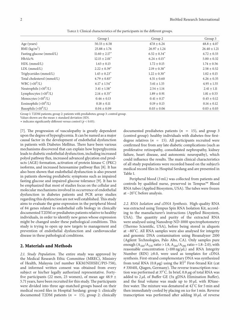
Group 1 Group 2 Group 3Age (years) 5033 plusmn 658 478 plusmn 624 488 plusmn 407BMI (kgm2) 2588 plusmn 176 2697 plusmn 124 2640 plusmn 121Fasting glucose (mmolL) 1160 plusmn 217lowast 652 plusmn 034lowast 472 plusmn 033HbA1c 1215 plusmn 201lowast 624 plusmn 015lowast 500 plusmn 032HDL (mmolL) 163 plusmn 013 172 plusmn 015 174 plusmn 056LDL (mmolL) 222 plusmn 039lowast 219 plusmn 038lowast 258 plusmn 052Triglycerides (mmolL) 145 plusmn 023lowast 122 plusmn 030lowast 102 plusmn 015Total cholesterol (mmolL) 479 plusmn 083lowast 451 plusmn 060 426 plusmn 035WBC (times109L) 617 plusmn 154lowast 561 plusmn 133 495 plusmn 155Neutrophils (times109L) 341 plusmn 158lowast 254 plusmn 114 241 plusmn 111Lymphocytes (times109L) 214 plusmn 035lowast 189 plusmn 091 181 plusmn 053Monocytes (times109L) 046 plusmn 013 041 plusmn 017 045 plusmn 012Eosinophils (times109L) 018 plusmn 011 019 plusmn 013 014 plusmn 012Basophils (times109L) 004 plusmn 009 003 plusmn 006 003 plusmn 003Group 1 T2DM patients group 2 patients with prediabetes group 3 control groupValues shown are the mean plusmn standard deviation (SD)lowast indicates significantly different versus control (119901 lt 005)
[7] The progression of vasculopathy is greatly dependentupon the degree of hyperglycemia It can be named as amajorcausal factor in the development of endothelial dysfunctionin patients with Diabetes Mellitus There have been variousmechanisms discovered that can explain how hyperglycemialeads to diabetic endothelial dysfunction including increasedpolyol pathway flux increased advanced glycation end prod-ucts (AGE) formation activation of protein kinase C (PKC)isoforms and increased hexosamine pathway flux [8] It hasalso been shown that endothelial dysfunction is also presentin patients showing prediabetic symptoms such as impairedfasting glucose and impaired glucose tolerance [9] It has tobe emphasized that most of studies focus on the cellular andmolecular mechanisms involved in occurrence of endothelialdysfunction in diabetes patients and PCR array studiesregarding this dysfunction are not well establishedThis studyaims to evaluate the gene expression in the peripheral bloodof 84 genes related to endothelial cells biology in clinicallydocumented T2DMor prediabetes patients relative to healthyindividuals in order to identify new genes whose expressionmight be changed under these pathological conditions Thisstudy is trying to open up new targets to management andprevention of endothelial dysfunction and cardiovasculardisease in these pathological conditions
2 Materials and Methods
21 Study Population The entire study was approved bythe Medical Research Ethic Committee (MREC) Ministryof Health Malaysia (ref number KKMNIHSECP15-758)and informed written consent was obtained from everysubject or hisher legally authorized representative Forty-five participants (22 men 23 women) of mean age 489 plusmn571 years have been recruited for this studyThe participantswere divided into three age-matched groups based on theirmedical record files in Hospital Serdang group 1 clinicallydocumented T2DM patients (119899 = 15) group 2 clinically
documented prediabetes patients (119899 = 15) and group 3(control group) healthy individuals with diabetes-free first-degree relatives (119899 = 15) All participants recruited wereconfirmed free from any late diabetic complications (such asproliferative retinopathy consolidated nephropathy kidneyfailure heart disease and autonomic neuropathy) whichcould influence the results The main clinical characteristicsof all study populations were recorded based on the subjectrsquosmedical record files in Hospital Serdang and are presented inTable 1
Peripheral blood (3mL) was collected from patients andcontrols by qualified nurse preserved in Tempus BloodRNA tubes (Applied Biosystem USA)The tubes were frozenat minus20∘C before analysis
22 RNA Isolation and cDNA Synthesis High-quality RNAwas extracted using Tempus Spin RNA Isolation Kit accord-ing to the manufacturerrsquos instructions (Applied BiosystemUSA) The quantity and purity of the extracted RNAwere analysed using Nanodrop ND-1000 spectrophotometry(Thermo Scientific USA) before being stored in aliquotsat minus80∘C All RNA samples were also analysed for integrityand genomic DNA contamination using Bioanalyzer 2100(Agilent Technologies Palo Alto CA) Only samples pureenough (A
260A230
ratio gt 18 A260
A280
ratio = 18ndash20) withreasonable concentration (gt100 ng120583L) and RNA IntegrityNumber (RIN) ge80 were used as templates for cDNAsynthesis First-strand complementary DNAwas synthesizedfrom total RNA (08120583g) using the RT2 First-Strand Kit (cat 330401 Qiagen Germany) The reverse transcription reac-tion was performed at 37∘C In brief 08 120583g of total RNA wasadded to 2 120583L of Buffer GE (5x gDNA Elimination Buffer)and the final volume was made up to 10 120583L with RNase-free water The mixture was denatured at 42∘C for 5min andthen immediately cooled by placing on ice for 1min Reversetranscription was performed after adding 10 120583L of reverse
BioMed Research International 3
transcription mix to the solution The reaction mixture wasincubated at 42∘C for 15min after which it was terminatedby heating at 95∘C for 5min The cDNA samples generated(20120583L) were then diluted with 91 120583L RNase-free water andstored at minus20∘C until further analysis
23 Gene Expression Profiling Real-time PCR was carriedout by using a Rotor-Gene 6000 Real-Time PCR detectionsystem (Qiagen Germany) Gene expression was examinedusing the Human Endothelial Cell Biology RT2 ProfilerPCR Array (cat 330231 Qiagen Germany) Expression of84 different genes involved in permeability and vascular toneangiogenesis endothelial cell activation and endothelial cellinjury was targeted for detection by real-time PCR TheRT2Profiler PCR Array contains built-in primers for 84tested and 5 housekeeping genes and positive control ele-ments to determine the efficiency of the reverse transcriptionreaction performance of the PCR reaction and detectionof genomic DNA contamination The PCR mixture for 100reactions contained 1150 120583L of SYBR Green ROX FASTMastermix (Qiagen Germany) 102 120583L cDNA template and1048 120583L RNase-free water The PCR reaction mix was addedto the wells of the PCR plate in equal amounts (20120583L) andthen the real-timePCRcycling programwas runThe thermalcycling program recommended by plates manufacturer forRotor-Gene 6000 was as follows 10min at 95∘C followed by40 cycles denaturation at 95∘C for 15 s with 30 s annealingand elongation at 60∘C followed by melting curve analysisThe software version 210 (Qiagen Germany) was used foranalysis
24 Data Analysis To determine the significant differencesin the clinical characteristics between the three groups theWilcoxon test was carried out with a significant differenceof 119901 lt 005 Data is expressed as the mean plusmn standarddeviation
The values of CycleThreshold (CT) obtained by real-timePCR experiments were used to calculate the relative changesin gene expression accordingly to 2minusΔΔCT method B2M andRPLP0 were chosen from the group of five House KeepingGenes (HKG) as the best and least varying reference gene tonormalize the gene expression data in order to increase thereliability of comparative CT method-based gene expressionquantification Changes in the gene expression level forevaluated genes were assessed for case groups in relation tothe control group with gene expression level set up arbitrarilyas 1 The differentially expressed genes with fold regulationgreater than plusmn3 with 119901 lt 005 are emphasized in thisstudy Data analyses were performed using the web-basedPCR Array data analysis software version 35 available at(httppcrdataanalysissabiosciencescompcrarrayanalysisphp)
25 Pathway Analysis To determine if any regulatory mech-anisms exist between dysregulated genes we performedin silico analysis using the GNC Pro online analysis tool(httpgncprosabiosciencescomgncprogncprophp)
3 Results
Our results demonstrated significantly higher fasting glucoseHbA1c LDL triglyceride and cholesterol in T2DM patientsas compared to the control group The significant differencewas also detected in the level of fasting glucose HbA1cLDL and triglyceride between prediabetes and control Wealso compared various haematological indices between casegroups and control There was a significantly higher totalwhite blood cell count neutrophils and lymphocytes inT2DM patients compared to control (Table 1)
In this study we have used the Human Endothelial CellBiology RT2 Profiler PCR Array to examine the expressionin the peripheral blood of 84 genes related to biologyof endothelial cells in patients with T2DM and in thosewith prediabetes relative to healthy individuals This PCRArray includes representative genes from various biologicalpathways angiogenesis vasoconstriction and vasodilationinflammatory response apoptosis cell adhesion coagulationand platelet activation In case of T2DM 59 genes showed asignificant upregulation Decreased expression was observedfor 4 genes The significant differences observed in the geneexpression profiles in patients with T2DM are presented inTable 2 A majority of these differentially expressed geneswere dysregulated less than 3-fold in their expression GenesALOX5 APOE CDH5 CX3CL1 FN1 IL3 ITGB3 MMP1MMP9 PLAU PLG SERPINE1 andTHBDwere upregulatedby greater than 3-fold in their expression (Figure 1) IL3 andFN1 were the most upregulated genes with more than 40-foldchanges in their expression (Figure 1(a)) The overall changesobserved in the gene expression patterns for T2DM patientsare represented in the form of a ldquovolcano plotrdquo wherein Log2-transformed fold changes in gene expression are plottedagainst the Student 119905-test 119901 values (Figure 2(a)) The genesthat are plotted further away from the central axes havegreater fold changes and 119901 values
We were able to identify 51 differentially expressed genesin the prediabetic patients (Table 2) PECAM-1 (minus42076)and SOD1 (minus18987) were seen to be significantly down-regulated and 49 genes were significantly upregulated inthese individuals as compared to the control Genes withdysregulation more than 3-fold in their expression are shownin Figure 3 The overall changes that were observed inthe gene expression patterns for prediabetes patients arerepresented in Figure 2(b) in the form of a volcano plot
Pathway mapping in Figure 4 is showing the regulatorymechanisms between genes that were dysregulatedmore than3-fold in T2DM and prediabetes patients
The gene expression levels between diabetic and predi-abetic groups were also compared 17 genes showed signif-icantly greater expression in T2DM relative to prediabetespatients whereas three genes with a higher expression in pre-diabetes patients were observed (Table 3) Genes with alteredexpression more than 3-fold are represented in Figure 5
4 Discussion
The macro- and microvascular pathologies associated withdiabetic condition are all characterized by endothelial
4 BioMed Research International
Table 2 Genes with altered expression in group 1 and group 2 comparing to group 3
Symbol DescriptionT2DM Prediabetes
Fold changes 95 CIlowast Fold changes 95 CIlowast
ACE Angiotensin I converting enzyme 169 (095 243) mdash mdashADAM17 ADAMmetallopeptidase domain 17 1788 (000001 393) 15804 (101 215)
AGT Angiotensinogen (serpin peptidase inhibitorclade A member 8)
mdash mdash 18188 (116 247)
ALOX5 Arachidonate 5-lipoxygenase 36158 (193 530) 21048 (133 288)ANGPT1 Angiopoietin 1 23495 (129 341) mdash mdashANXA5 Annexin A5 25639 (179 334) 18502 (116 254)APOE Apolipoprotein E 32588 (176 476) mdash mdashBAX BCL2-associated X protein 25497 (169 341) 1883 (121 256)BCL2 B-cell CLLlymphoma 2 20567 (129 282) 16294 (103 223)BCL2L1 BCL2-like 1 18493 (067 303) mdash mdashCALCA Calcitonin-related polypeptide alpha 26938 (151 388) 18562 (117 255)CASP1 Caspase 1 apoptosis-related cysteine peptidase 23823 (162 314) 22207 (141 304)CASP3 Caspase 3 apoptosis-related cysteine peptidase 29032 (000001 608) 28474 (000001 598)CCL2 Chemokine (C-C motif) ligand 2 17255 (068 277) mdash mdashCCL5 Chemokine (C-C motif) ligand 5 06962 (052 087) mdash mdashCDH5 Cadherin 5 type 2 (vascular endothelium) 34734 (153 542) mdash mdashCFLAR CASP8 and FADD-like apoptosis regulator 23999 (133 347) 15695 (074 240)COL18A1 Collagen type XVIII alpha 1 24144 (085 398) 2171 (113 321)CX3CL1 Chemokine (C-X3-C motif) ligand 1 42901 (208 650) 2816 (123 441)EDN1 Endothelin 1 18622 (111 262) 15294 (078 228)EDN2 Endothelin 2 2456 (041 450) 13964 (096 184)EDNRA Endothelin receptor type A 19702 (081 313) mdashENG Endoglin mdash mdash 19575 (125 266)F2R Coagulation factor II (thrombin) receptor mdash mdash 21302 (116 310)
F3 Coagulation factor III (thromboplastin tissuefactor)
27747 (153 402) 19368 (094 293)
FAS Fas (TNF receptor superfamily member 6) 24166 (117 366) mdash mdashFASLG Fas ligand (TNF superfamily member 6) mdash mdash 16629 (103 230)FGF1 Fibroblast growth factor 1 (acidic) 07285 (059 087) mdash mdashFLT1 Fms-related tyrosine kinase 1 2177 (154 281) 14185 (099 184)FN1 Fibronectin 1 484245 (000001 10564) 30987 (100 520)ICAM1 Intercellular adhesion molecule 1 mdash mdash 18786 (117 258)IL11 Interleukin 11 21411 (131 297) mdash mdashIL1B Interleukin 1 beta 29739 (187 408) 18899 (124 254)
IL3 Interleukin 3 (colony-stimulating factormultiple)
176599 (000001 47401) 24413 (144 344)
IL6 Interleukin 6 (interferon beta 2) 16806 (097 239) mdash mdashIL7 Interleukin 7 13835 (071 206) mdash mdash
ITGA5 Integrin alpha 5 (fibronectin receptor alphapolypeptide)
26864 (161 376) 18891 (119 259)
ITGAV Integrin alpha V (vitronectin receptor alphapolypeptide antigen CD51)
17913 (117 241) 16077 (100 221)
ITGB1Integrin beta 1 (fibronectin receptor beta
polypeptide antigen CD29 including MDF2MSK12)
19693 (144 249) 16144 (098 224)
ITGB3 Integrin beta 3 (platelet glycoprotein IIIaantigen CD61)
49899 (290 708) 25427 (065 443)
BioMed Research International 5
Table 2 Continued
Symbol Description T2DM PrediabetesFold changes 95 CIlowast Fold changes 95 CIlowast
KDR Kinase insert domain receptor (a type IIIreceptor tyrosine kinase) 16884 (078 259) mdash mdash
KIT V-kit Hardy-Zuckerman 4 feline sarcoma viraloncogene homolog 16025 (089 231) 16211 (107 218)
MMP1 Matrix metallopeptidase 1 128082 (318 2243)MMP2 Matrix metallopeptidase 2 26715 (087 447) 17691 (113 241)MMP9 Matrix metallopeptidase 9 44005 (221 660) mdash mdashNOS3 Nitric oxide synthase 3 (endothelial cell) 25532 (162 348) mdash mdashPECAM1 Plateletendothelial cell adhesion molecule 05887 (035 082) 02377 (014 033)PF4 Platelet factor 4 mdash mdash 1397 (062 217)PGF Placental growth factor 29547 (187 404) 22258 (121 324)PLAT Plasminogen activator tissue 1788 (103 255) 20088 (103 299)PLAU Plasminogen activator urokinase 78263 (273 1293) 36782 (153 583)PLG Plasminogen 38718 (136 638) 218 (052 384)PROCR Protein C receptor endothelial mdash mdash 17096 (108 234)PTGIS Prostaglandin I2 (prostacyclin) synthase mdash mdash 218 (122 314)
PTGS2Prostaglandin-endoperoxide synthase 2
(prostaglandin GH synthase andcyclooxygenase)
25639 (108 405) 20312 (125 281)
PTK2 PTK2 protein tyrosine kinase 2 17263 (084 262) mdash mdashSELE Selectin E 18021 (090 270) 23075 (117 344)SELL Selectin L 24044 (158 322) 16476 (116 213)SELPLG Selectin P ligand 242 (180 304) 20023 (137 264)
SERPINE1Serpin peptidase inhibitor clade E (nexinplasminogen activator inhibitor type 1)
member 132035 (187 453) 20463 (112 297)
SOD1 Superoxide dismutase 1 soluble 06517 (046 084) 05267 (033 073)SPHK1 Sphingosine kinase 1 22258 (135 310) 16339 (065 262)TEK TEK tyrosine kinase endothelial mdash mdash 22454 (129 320)
TFPI Tissue factor pathway inhibitor(lipoprotein-associated coagulation inhibitor) 14959 (095 204) mdash mdash
TGFB1 Transforming growth factor beta 1 28056 (188 373) 3142 (170 458)THBD Thrombomodulin 3685 (200 537) 20097 (120 282)THBS1 Thrombospondin 1 18588 (118 254) 1433 (075 212)TIMP1 TIMP metallopeptidase inhibitor 1 26814 (182 354) 3283 (182 474)TNF Tumor necrosis factor 15922 (111 207) mdash mdash
TNFSF10 Tumor necrosis factor (ligand) superfamilymember 10 15084 (107 194) mdash mdash
TYMP Thymidine phosphorylase mdash mdash 29739 (101 493)VEGFA Vascular endothelial growth factor A 20406 (144 264) mdash mdashVWF VonWillebrand factor 2035 (141 266) 16226 (092 232)Group 1 T2DM patients group 2 patients with prediabetes group 3 control grouplowast95 confidence interval
dysfunction [1] Most studies focus on the cellular andmolecular mechanisms involved in occurrence of endothelialdysfunction in diabetes patients and gene expression studiesregarding this dysfunction in diabetic condition are not wellestablished This is a first study of its kind performed inpatients with diagnosed T2DM or prediabetes to determine
the changes in the blood expression levels of genes relatedto the biology of endothelial cells This study is trying toopen up new targets in the management and prevention ofendothelial dysfunction and cardiovascular disease in thesepathological conditions The expression levels of 84 geneswere assessed in both patient groups and the data was
6 BioMed Research International
ControlT2DM
020406080
100120140160180
Fold
regu
latio
n
FN1 IL3
(a)
ControlT2DM
0
2
4
6
8
10
12
14
Fold
regu
latio
n
SERP
INE1
CX3C
L1
ALO
X5
ITG
B3
PLAU
MM
P9
MM
P1
APO
E
THBD
CDH
5
PLG
(b)
Figure 1 Fold regulation in gene expression in T2DM compared to healthy individuals Genes with altered expression more than 40-fold inT2DM patients (a) Genes with altered expression more than 3-fold but less than 40-fold in T2DM patients (b)
minus118 minus018 082 482182 682382282 582 782FC of group 1control group)
072
172
272
372
472
572
(p value)
log2(
minuslo
g 10
(a)
minus132 minus032minus232 068 168 268FC of group 2control group)
046
096
146
196
246
296
346
396(p
value)
log2(
minuslo
g 10
(b)
Figure 2 Relative expression comparison for 84 endothelial cells-related gene between cases groups and control group Volcano plot analysisapplied to the PCR Array data revealed 63 genes significantly expressed (119901 lt 005 with FC ge 1 (up or down)) in diabetic patients (a) and 51genes significantly expressed (119901 lt 005with FC ge 1 (up or down)) in prediabetic patients (b) compared to healthy individualsThe plot showsa log 2-fold change in gene expression between the two groups on the 119909-axis and the negative log of 119905-test 119901 values on the 119910-axis Each geneis represented by a single point
compared to their expression levels in the control groupMajority of differentially expressed genes were common fortwo or more biological processes (Table 4) The alterationsin the expression levels of these genes might be associatedwith the occurrence of endothelial dysfunction in T2DM andprediabetes patients
Our results appear to provide the first reported data onincreased expression levels of IL-3 in patients with T2DM in awhole blood gene expression profiling study IL-3 is a productof mature T cells and mast cells after activation Elevated
lymphocyte (T cells and B cells) counts were detected inT2DM patients as you can see in Table 1 Moreover elementsof diabetes can directly or indirectly activate T cells [10]This can be an explanation for overexpression of IL-3 indiabetes patients The expression of IL-3 was also elevated24413-fold in prediabetes patients which is less than itsoverexpression in T2DM patients (176599-fold) Though wedo not have any convincing reason for this lower expressionof IL-3 in prediabetes patients IL-3 is classically describedas a hematopoietic growth factor However it is also known
BioMed Research International 7
FN1
PECA
M1
PLAU
TGFB
1
TIM
P1
ControlPrediabetes
minus5
minus4
minus3
minus2
minus1
01234
Fold
regu
latio
n
Figure 3 Genes with altered expression more than 3-fold in prediabetic patients compared to healthy individual
PLAU
MMP1
SERPINE1ALOX5
THBD
FN1
ITGB3
IL3
PLG
CX3CL1
APOE
CDH5
MMP9
(a)
FN1
PECAM1TIMP1
TGFB1
PLAU
(b)
Figure 4 Pathway analysis of genes with altered expression more than 3-fold in T2DM (a) and prediabetes patients (b) The interactionsamong these genes are represented graphically in this figure The red line represents downregulation the green line represents upregulationthe yellow line represents physical interaction the blue line represents posttranslationalmodification the blue dotted line represents predictedprotein interaction and the purple dotted line represents predicted transcription factor regulation
as an endothelial cell activator [11] and can play a role ininflammation [12] The involvement of IL-3 in inflammatorydiseases is supported by its capability to induce expression ofadhesion molecules such as E- and P-selectin proliferationof endothelial cell and production of IL-8 [11ndash13] IncreasedIL-3 level in plasma serum vitreous and bone marrowsupernatant has been reported in diabetic condition [14ndash17]IL-3 is a ldquogoodrdquo IL in terms of glucosemetabolism presentingdefensive effects in experimental diabetes Administration ofIL-3 twice weekly starting at 2ndash4 weeks of age delayed theonset and reduced the overall incidence of diabetes in miceBonemarrow cells obtained from IL-3-treatedmice protectedother mice from cyclophosphamide-induced diabetes [18]The uptake of glucose into the cell represents a key pointin the regulation of its metabolism and is known to bestimulated by IL-3 [19] IL-3 regulates glucose uptake by
modulating the intrinsic transporting ability of glucose trans-porters [20] IL-3 transcriptionally upregulates GLUT1 butalso has posttranslational effects on trafficking that are likelyto be mediated by Akt and mTOR [21] On the other handIL-3 is believed to play a role in advanced lesions by smoothmuscle cell accumulation increasing macrophage activationand neovascularization of the plaque and in the early stagesof atherogenesis by facilitating leukocyte extravasation [22]In spontaneously contracting cultured cardiac myocytesperfusion with IL-3 induced arrhythmias resulting in acomplete cessation of spontaneous contractions and a severeloss of myocyte inotropy the effects were concentration-dependent and reversible [23] Thus overexpression of IL-3in T2DM patients can have both protective and damagingeffects protective for diabetes and yet damaging for heart andvascular The major potential for IL-3 in clinical applications
8 BioMed Research International
Table 3 Direct comparison of gene expression levels between groups 1 and 2
Gene Description Fold changes 95 CIlowast
ADAM17 ADAMmetallopeptidase domain 17 11313 (000001 249)ALOX5 Arachidonate 5-lipoxygenase 17179 (085 259)APOE Apolipoprotein E 22932 (115 344)EDN2 Endothelin 2 17589 (024 328)EDNRA Endothelin receptor type A 18643 (097 276)FGF1 Fibroblast growth factor 1 (acidic) 06051 (044 077)FLT1 Fms-related tyrosine kinase 1 15347 (104 203)FN1 Fibronectin 1 156273 (000001 3483)IL11 Interleukin 11 24116 (137 345)IL1B Interleukin 1 beta 15735 (093 222)IL3 Interleukin 3 723372 (000001 19469)MMP2 Matrix metallopeptidase 2 15101 (052 250)MMP9 Matrix metallopeptidase 9 32641 (158 495)NOS3 Nitric oxide synthase 3 (endothelial cell) 21347 (137 290)PECAM1 Plateletendothelial cell adhesion molecule 24771 (167 328)PLAU Plasminogen activator urokinase 21278 (114 311)PTGIS Prostaglandin I2 (prostacyclin) synthase 04289 (023 063)SELL Selectin L 14594 (090 202)THBD Thrombomodulin 18336 (119 248)TYMP Thymidine phosphorylase 04013 (013 067)Group 1 patients with type 2 diabetes Group 2 patients with prediabeteslowast95 confidence interval
Table 4 Functional gene grouping
AngiogenesisANGPT1 CCL2 CCL5 CX3CL1 EDN1 EDNRA ENG F3 FASLG FGF1 FLT1 FN1 IL1B IL6ITGA5 ITGAV ITGB1 ITGB3 KDR KIT MMP2 MMP9 NOS3 PF4 PGF PLAU PTGS2
SERPINE1 SPHK1 TEK THBS1 TYMP VEGFA
Vasoconstriction amp vasodilation ACE AGT ALOX5 APOE CALCA CX3CL1 EDN1 EDN2 EDNRA F2R ICAM1 NOS3 PTGISPTGS2 SOD1
Inflammatory response ACE AGT ALOX5 APOE CALCA CCL2 CCL5 CX3CL1 EDNRA F2R F3 FN1 IL1B IL6PTGS2 SELE SPHK1 TGFB1 THBS1 TNF
Apoptosis ANXA5 BAX BCL2 BCL2L1 CASP1 CASP3 CCL2 CCL5 CFLAR CX3CL1 EDN1 EDNRAFAS FASLG IL1B IL3 IL6 IL7 PF4 PTK2 SPHK1 TEK THBS1 TNF TNFSF10
Cell adhesionADAM17 AGT BCL2 CALCA CDH5 COL18A1 CX3CL1 ENG FGF1 FN1 ICAM1 IL1BITGA5 ITGAV ITGB1 ITGB3 KDR PECAM1 PLAU PLG PTK2 SELE SELL SELPLG
SERPINE1 TGFB1 THBS1 TNF VEGFA VWF
Coagulation ANXA5 EDN1 F2R F3 FN1 MMP1 PECAM1 PF4 PLAT PLAU PLG PROCR PTK2 SELLSELPLG SERPINE1 TEK TFPI THBD THBS1 TIMP1 VWF
Platelet activation APOE CX3CL1 F2R FN1 IL11 IL6 ITGB3 NOS3 PECAM1 PF4 PLG SERPINE1 SOD1TGFB1 THBD THBS1 TIMP1 VEGFA VWF
is dependent upon its capability to promote the survivalproliferation and maintenance of hematopoietic progenitorcells [24]The IL-3 has been used in culture to produce bloodcells of various lineages [25] Moreover administration of IL-3 to human and primate subjects has enhanced multilineage-hematopoiesis [26 27] It increases the number of leukocytes(primarily neutrophils lymphocytes and eosinophils) aswell as reticulocytes and platelets [28] However IL-3 has aninhibitory effect on the expansion of long-term-repopulatinghematopoietic progenitor cells and often it was reported that
this cytokine can play a negative role in primitive cell expan-sion [29] Increased FN1 expression is one of themain featuresof diabetic angiopathy Diabetes causes FN1 upregulation inthe retina kidney heart and plasma It can reflect endothelialextracellular matrix changes and consequently vessel walldamage in these patients [30 31] Diabetes leads to the upreg-ulation of FN1 via an endothelin- (ET-) dependent pathwayinvolving activation of NF-120581B and AP-1 transcription factors[31] According to a study completed by Kanters et al ele-vated plasma levels of FN1 may reflect a common pathway of
BioMed Research International 9
PrediabetesT2DM
0
10
20
30
40
50
60
70
80
Fold
regu
latio
n
IL3 MMP9FN1
Figure 5 Genes with altered expression more than 3-fold compar-ing prediabetic group and T2DM group
endothelial cell activation in patients with diabetes which arenot found in atherosclerosis without diabetes [30] Yaghoubiet al approved the positive association between FN1 levelin serum and atherosclerosis progression [32] Accordingto the results obtained by Rohwedder et al FN1 can playa dichotomous role in atherosclerosis while FN worsensthe course of atherosclerosis by increasing the atherogenicplaque area it stabilizes the plaques with fibrous caps andprotects from secondary damage and vascular occlusion[33] The increased MMP-1 MMP-2 and MMP-9 activitiesand expression induced by high glucose exposure can ele-vate matrix degradation thereby accelerating atherogenesisand potentially decreasing plaque stability in diabetes [34]MMP-1 is a biomarker for venous disease and has a rolein modulating endothelial permeability by regulation ofjunctional integrity [35 36] T2DM is associated with theelevated blood level of MMP-1 which is positively correlatedwith coronary heart disease occurrence in these patients [37]The increase inMMP-9 level and activitymay have importantconsequences for the development of vascular complicationsassociated with diabetes For instance the elevated levelof MMP-9 in plasma and retinas of diabetic patients cancontribute to the development of diabetic retinopathy byaltering vascular permeability and capillary cell apoptosis[38 39] The proapoptotic role of MMP-9 in hyperglycaemicconditions occurs through the activation of caspase-3 [39]ALOX5 expression is greater in diabetic compared withnondiabetic plaques and is associated with increased MMP-2 and MMP-9 expression It has been stated that localizedincrease in ALOX5 has the potential to cause the acuteplaque disruption that precedes the onset of symptoms inboth the coronary and cerebral circulations in diabetes [40]The involvement of ALOX5 in atherosclerosis is not onlyduring the development of atherosclerotic plaques but alsoduring the progression of atherosclerotic plaques towardinstability [41] In this study we were able to demonstratethat the expression level of the PLAU gene was significantlyelevated in the T2DM patients The same result was reportedby Kenichi et al where they observed increased values for
mRNA and protein expressions of PLAU among diabetic ratsas compared to control rats [42] The PLAU is produced bythe renal epithelial and the endothelial cells stimulated by theinflammatory cytokines [43] Hence chronic inflammationassociated with diabetes could be one of the reasons foroverexpression of PLAU in diabetes patients The increasedexpression of PLAU in the endothelial cells alters thearterial elastic laminae causes vascular constriction witha narrowing of the lumen and also enhances the growthof early atherosclerotic lesions [44] A number of reportshave established a direct association between the PLG levelor plasmin activity and the occurrence of CAD To elabo-rate two cohort studies with separate perspectives that isthe FINRISK rsquo92 Haemostasis Study [45] and the Atheroscle-rosis Risk in Communities Study (ARIC) [46] demonstratethat the PLG level is an independent risk factor for CADIntegrins have distinct roles in inflammatory cell recruitmentto the damaged vessel wall in atherosclerosis [47] Overex-pression of integrins under high glucose concentration canlead to an altered interaction of vascular endothelial cellswith their basement membranes further causing a firmercell-matrix adhesion [48] In patients with diabetes inte-grins induce proangiogenic signalling resulting in aberrantsignalling under diabetes that is characteristic of diabeticretinopathy nephropathy and macrosomia [7] According tothe results from integrin beta-3 knockout this integrin is notlikely to be a crucial player during development however itdoes mediate pathological neovascularization in adults [49]ITGB3 plays a prominent role in the angiogenic responsein proliferative diabetic retinopathy [50] According to theresults observed in our study the expression of the ITGA5gene was found to be significantly upregulated in T2DMpatients A similar result was observed by Stoynev et alwho reported increased levels of ITGA5 in T2DM patients ascompared to healthy individuals [51] An optimal expressionof APOE is crucial for maintaining normal metabolism oflipoproteins Decreased levels of APOE impair the clear-ance of triglyceride-rich lipoproteins On the other handupregulation of APOE may lead to hypertriglyceridemiathrough stimulating the production of VLDL triglyceride inthe liver and impairing the LPL-mediated lipolysis [52] It ispossible that the nephropathy in T2DM may be associatedwith the polymorphism of the APOE gene [53] Vascularendothelial-cadherin is a crucial factor for plaque neovascu-larization and a subsequent development of plaque instabilityHigher expression of CDH5 was observed in complicatedplaques and high-grade stenotic lesions [54] CX3CL1 isa very important factor of atherogenesis and increasedstaining of CX3CL1 has been reported in human atheroscle-rotic coronaries and diabetic vessels [55] In addition toits role as a chemokine and adhesion molecule CX3CL1induces vascular dysfunction by increasing NADPH oxidase-dependent superoxide formation and reduced NO bioavail-ability [56] The hyperglycaemia formation of advancedglycation end products and cytokine activation in thediabetic condition can induce overproduction of CX3CL1in the kidneys and aggravate diabetic nephropathy [57]It has been stated that CX3CL1 can play a prominentrole in diabetic renal injury through overproduction of
10 BioMed Research International
ECM [58] Zumbach et al stated that THBD levels weresignificantly higher in diabetic patients with microvascularcomplications [59] These elevations in plasma THBD indiabetic patients are inversely related to protein C activityand positively related to increased markers of thrombin gen-eration hence constituting a marker of a procoagulant statereflecting proteolytic injury to the vascular endothelium [60]In a study conducted by Ewing et al ANXA5 therapy reducesthe vascular inflammation and remodelling also improvingendothelial function inmice [61] thereby indicating its role asa therapeutic potential against atherosclerotic cardiovasculardiseases To the best of our knowledge no reports in theliterature relate the overexpression of ANXA5 to endothelialdysfunction or atherosclerosis It could be possible thatoverexpression of this gene protects and limits damage tothe target organs in this disease condition In our studywe have demonstrated that people with T2DM expressedhigher levels of TIMP1 Higher levels of TIMP1 expressionin the venous endothelial cell and in plasma are an earlysign of endothelial dysfunction [62] The same results wereobserved by Derosa et al where the authors noticed that theplasma concentration of TIMP1was significantly increased inT2DM patients which may cause an abnormal extracellularmatrix (ECM) metabolism [63] The PECAM-1 which isessential for the survival migration and functional organi-zation of endothelial cells during vascular development andangiogenesis [64] has been reported to be degraded in theplatelets of T2DM patients [65] The absence of endothelialPECAM-1 resulted in a decreased angiogenesis [66] andmight lead to endothelial dysfunction [67]
5 Conclusions
Taken together our findings suggest that the diabetic andprediabetic condition can disrupt the expression of genesinvolved in the regulation of endothelial cells function andhomeostasis Dysregulation in the expression of these genescan be associated with increased risk for occurrence ofendothelial dysfunction in these patients As endothelialdysfunction appears to be an early indicator of vascular dam-age therefore further research on the expression of geneswhich can affect endothelial cells function could providenew targets in the management and prevention of macro-and microvascular complications in these pathological con-ditions However all of these data need to be confirmed basedon a higher number of patients
Competing Interests
The authors declare that they have no competing interests
Acknowledgments
The authors would like to extend their gratitude to all thevolunteers who have participated in this research
References
[1] C G Schalkwijk and C D A Stehouwer ldquoVascular complica-tions in diabetes mellitus the role of endothelial dysfunctionrdquoClinical Science vol 109 no 2 pp 143ndash159 2005
[2] M Feletou The Endothelium Part 1 Multiple Functions ofthe Endothelial CellsmdashFocus onEndothelium-DerivedVasoactiveMediators Morgan amp Claypool Life Sciences 2011
[3] J Xu and M-H Zou ldquoMolecular insights and therapeutictargets for diabetic endothelial dysfunctionrdquo Circulation vol120 no 13 pp 1266ndash1286 2009
[4] S B Williams J A Cusco M-A Roddy M T Johnstone andM A Creager ldquoImpaired nitric oxide-mediated vasodilation inpatients with non-insulin-dependent diabetesmellitusrdquo Journalof the American College of Cardiology vol 27 no 3 pp 567ndash5741996
[5] H H Ting F K Timimi K S Boles S J Creager P Ganz andM A Creager ldquoVitamin C improves endothelium-dependentvasodilation in patients with non-insulin-dependent diabetesmellitusrdquoThe Journal of Clinical Investigation vol 97 no 1 pp22ndash28 1996
[6] L Zhang D Gong S Li and X Zhou ldquoMeta-analysis of theeffects of statin therapy on endothelial function in patients withdiabetesmellitusrdquoAtherosclerosis vol 223 no 1 pp 78ndash85 2012
[7] G K Kolluru S C Bir and C G Kevil ldquoEndothelial dysfunc-tion and diabetes effects on angiogenesis vascular remodelingand wound healingrdquo International Journal of VascularMedicinevol 2012 Article ID 918267 30 pages 2012
[8] C V Diogo J M Suski M Lebiedzinska et al ldquoCardiacmitochondrial dysfunction during hyperglycemiamdashthe role ofoxidative stress and p66Shc signalingrdquo International Journal ofBiochemistry and Cell Biology vol 45 no 1 pp 114ndash122 2013
[9] Y Su X-M Liu Y-M Sun Y-Y Wang Y Luan and YWu ldquoEndothelial dysfunction in impaired fasting glycemiaimpaired glucose tolerance and type 2 diabetes mellitusrdquo TheAmerican Journal of Cardiology vol 102 no 4 pp 497ndash4982008
[10] C-C Wu H-K Sytwu K-C Lu and Y-F Lin ldquoRole of Tcells in type 2 diabetic nephropathyrdquo Experimental DiabetesResearch vol 2011 Article ID 514738 9 pages 2011
[11] M F Brizzi G Garbarino P R Rossi et al ldquoInterleukin3 stimulates proliferation and triggers endothelial-leukocyteadhesionmolecule 1 gene activation of human endothelial cellsrdquoThe Journal of Clinical Investigation vol 91 no 6 pp 2887ndash28921993
[12] Y Khew-Goodall C M Butcher M S Litwin et al ldquoChronicexpression of P-selectin on endothelial cells stimulated by the T-Cell cytokine interleukin-3rdquo Blood vol 87 no 4 pp 1432ndash14381996
[13] E I Korpelainen J R Gamble W B Smith et al ldquoThe receptorfor interleukin 3 is selectively induced in human endothelialcells by tumor necrosis factor 120572 and potentiates interleukin8 secretion and neutrophil transmigrationrdquo Proceedings of theNational Academy of Sciences of the United States of Americavol 90 no 23 pp 11137ndash11141 1993
[14] J M Starkey S J Haidacher W S LeJeune et al ldquoDiabetes-induced activation of canonical and noncanonical nuclearfactor-120581B pathways in renal cortexrdquo Diabetes vol 55 no 5 pp1252ndash1259 2006
BioMed Research International 11
[15] Z Yu C Gong B Lu et al ldquoDendrobium chrysotoxum Lindlalleviates diabetic retinopathy by preventing retinal inflamma-tion and tight junction protein decreaserdquo Journal of DiabetesResearch vol 2015 Article ID 518317 10 pages 2015
[16] Y Liu L F Leo C McGregor A Grivitishvili C J Barnsta-ble and J Tombran-Tink ldquoPigment epithelium-derived factor(PEDF) peptide eye drops reduce inflammation cell death andvascular leakage in diabetic retinopathy in Ins2(Akita) micerdquoMolecular Medicine vol 18 pp 1387ndash1401 2012
[17] S Hazra Endothelial Progenitor Cell Dysfunction in DiabetesUniversity of Florida Gainesville Fla USA 2011
[18] A Ito N Aoyanagi and T Maki ldquoRegulation of autoimmunediabetes by interleukin 3-dependent bonemarrow-derived cellsin NOD micerdquo Journal of Autoimmunity vol 10 no 4 pp 331ndash338 1997
[19] O Kan S A Baldwin and A D Whetton ldquoApoptosis isregulated by the rate of glucose transport in an interleukin 3dependent cell linerdquoThe Journal of Experimental Medicine vol180 no 3 pp 917ndash923 1994
[20] K D McCoy N Ahmed A S Tan and M V Berridge ldquoThehemopoietic growth factor interleukin-3 promotes glucosetransport by increasing the specific activity andmaintaining theaffinity for glucose of plasma membrane glucose transportersrdquoThe Journal of Biological Chemistry vol 272 no 28 pp 17276ndash17282 1997
[21] H L Wieman J A Wofford and J C Rathmell ldquoCytokinestimulation promotes glucose uptake via phosphatidylinositol-3kinaseAkt regulation of Glut1 activity and traffickingrdquoMolecu-lar Biology of the Cell vol 18 no 4 pp 1437ndash1446 2007
[22] J H von der Thusen J Kuiper T J C van Berkel and E AL Biessen ldquoInterleukins in atherosclerosis molecular pathwaysand therapeutic potentialrdquo Pharmacological Reviews vol 55 no1 pp 133ndash166 2003
[23] D Weisensee J Bereiter-Hahn W Schoeppe and I Low-Friedrich ldquoEffects of cytokines on the contractility of culturedcardiac myocytesrdquo International Journal of Immunopharmacol-ogy vol 15 no 5 pp 581ndash587 1993
[24] J D Priest ADWatts J SWhittaker et al ldquoParameter selectedgm-csf il-3 il-4 il-5 and chimeras thereof for therapeutic anddiagnostic purposesrdquo Google Patents 2006
[25] S Huang Z Chen J F Yu et al ldquoCorrelation between IL-3 receptor expression and growth potential of human CD34+hematopoietic cells from different tissuesrdquo STEM CELLS vol17 no 5 pp 265ndash272 1999
[26] A Ganser A Lindemann G Seipelt et al ldquoEffects ofrecombinant human interleukin-3 in patients with normalhematopoiesis and in patients with bonemarrow failurerdquoBloodvol 76 no 4 pp 666ndash676 1990
[27] R E Donahue J Seehra M Metzger et al ldquoHuman IL-3 andGM-CSF act synergistically in stimulating hematopoiesis inprimatesrdquo Science vol 241 no 4874 pp 1820ndash1823 1988
[28] M H Mangi and A C Newland ldquoInterleukin-3 in hematologyand oncology current state of knowledge and future directionsrdquoCytokines Cellular and Molecular Therapy vol 5 no 2 pp 87ndash95 1999
[29] G Shatirishvili K Mardaleishvili and G Loladze ldquoCord bloodhematopoietic stem cell expansion preclinical studies andclinical trials Reviewrdquo CellR4 The official Journal of The CureAlliance vol 2 no 5 article e1209 2014
[30] S D J M Kanters J-D Banga A Algra R C J M Frijns J JBeutler and R Fijnheer ldquoPlasma levels of cellular fibronectin indiabetesrdquo Diabetes Care vol 24 no 2 pp 323ndash327 2001
[31] S Chen Z A Khan M Cukiernik and S Chakrabarti ldquoDif-ferential activation of NF-120581B and AP-1 in increased fibronectinsynthesis in target organs of diabetic complicationsrdquo AmericanJournal of PhysiologymdashEndocrinology and Metabolism vol 284no 6 pp E1089ndashE1097 2003
[32] A R Yaghoubi F Kargar and F Khaki-Khatibi ldquoAssociation offibronectin leptin and LDL-oxide serum levels with coronaryartery disease in non-smoker and non-diabetic patientsrdquo Cres-cent Journal of Medical and Biological Sciences vol 3 pp 23ndash272016
[33] I Rohwedder E Montanez K Beckmann et al ldquoPlasmafibronectin deficiency impedes atherosclerosis progression andfibrous cap formationrdquo EMBOMolecular Medicine vol 4 no 7pp 564ndash576 2012
[34] A K Death E J Fisher K C Y McGrath and D K Yue ldquoHighglucose alters matrix metalloproteinase expression in two keyvascular cells potential impact on atherosclerosis in diabetesrdquoAtherosclerosis vol 168 no 2 pp 263ndash269 2003
[35] T Alsaigh E S Pocock J J Bergan and G W Schmid-Schonbein ldquoAcute venous occlusion enhances matrix metal-loprotease activity implications on endothelial dysfunctionrdquoMicrovascular Research vol 81 no 1 pp 108ndash116 2011
[36] J S Alexander and JW Elrod ldquoExtracellular matrix junctionalintegrity andmatrix metalloproteinase interactions in endothe-lial permeability regulationrdquo Journal of Anatomy vol 200 no 6pp 561ndash574 2002
[37] J Drzewoski A Sliwinska K Przybyłowska et al ldquoGenepolymorphisms and antigen levels of matrixmetalloproteinase-1 in type 2 diabetes mellitus coexisting with coronary heartdiseaserdquoKardiologia Polska vol 66 no 10 pp 1042ndash1049 2008
[38] N Yildirim A Sahin N Erol S Kara S Uslu and S TopbasldquoThe relationship between plasma MMP-9 and TIMP-2 levelsand intraocular pressure elevation in diabetic patients afterintravitreal triamcinolone injectionrdquo Journal of Glaucoma vol17 no 4 pp 253ndash256 2008
[39] R A Kowluru ldquoRole of matrix metalloproteinase-9 in thedevelopment of diabetic retinopathy and its regulation by H-Rasrdquo Investigative Ophthalmology and Visual Science vol 51 no8 pp 4320ndash4326 2010
[40] Y J Zhou J H Wang L Li H W Yang D L Wen and Q CHe ldquoExpanding expression of the 5-lipoxygenaseleukotrieneB4 pathway in atherosclerotic lesions of diabetic patients pro-motes plaque instabilityrdquo Biochemical and Biophysical ResearchCommunications vol 363 no 1 pp 30ndash36 2007
[41] F CipolloneAMezzettiM L Fazia et al ldquoAssociation between5-lipoxygenase expression and plaque instability in humansrdquoArteriosclerosis Thrombosis and Vascular Biology vol 25 no8 pp 1665ndash1670 2005
[42] M Kenichi M Masanobu K Takehiko et al ldquoRenal synthesisof urokinase type-plasminogen activator its receptor andplasminogen activator inhibitor-1 in diabetic nephropathy inrats modulation by angiotensin-converting-enzyme inhibitorrdquoJournal of Laboratory and Clinical Medicine vol 144 no 2 pp69ndash77 2004
[43] J Wojta R L Hoover and T O Daniel ldquoVascular origin deter-mines plasminogen activator expression in human endothelialcells Renal endothelial cells produce large amounts of singlechain urokinase type plasminogen activatorrdquo The Journal ofBiological Chemistry vol 264 no 5 pp 2846ndash2852 1989
[44] M Falkenberg C TomM B DeYoung SWen R Linnemannand D A Dichek ldquoIncreased expression of urokinase duringatherosclerotic lesion development causes arterial constriction
12 BioMed Research International
and lumen loss and accelerates lesion growthrdquo Proceedings ofthe National Academy of Sciences of the United States of Americavol 99 no 16 pp 10665ndash10670 2002
[45] M Rajecki P Pajunen P Jousilahti V Rasi E Vahtera andV Salomaa ldquoHemostatic factors as predictors of stroke andcardiovascular diseases the FINRISK 101584092 Hemostasis StudyrdquoBlood Coagulation and Fibrinolysis vol 16 no 2 pp 119ndash1242005
[46] A R Folsom N Aleksic E Park V Salomaa H Junejaand K K Wu ldquoProspective study of fibrinolytic factors andincident coronary heart disease the Atherosclerosis Risk inCommunities (ARIC) Studyrdquo Arteriosclerosis Thrombosis andVascular Biology vol 21 no 4 pp 611ndash617 2001
[47] C Burtea S Laurent O Murariu et al ldquoMolecular imagingof 120572v1205733 integrin expression in atherosclerotic plaques with amimetic of RGD peptide grafted to Gd-DTPArdquo CardiovascularResearch vol 78 no 1 pp 148ndash157 2008
[48] T Roth F Podesta M A Stepp D Boeri and M LorenzildquoIntegrin overexpression induced by high glucose and byhuman diabetes potential pathway to cell dysfunction indiabeticmicroangiopathyrdquo Proceedings of the National Academyof Sciences of the United States of America vol 90 no 20 pp9640ndash9644 1993
[49] A R Reynolds L E Reynolds T E Nagel et al ldquoElevatedFlk1 (vascular endothelial growth factor receptor 2) signalingmediates enhanced angiogenesis in 120573
3-integrin-deficient micerdquo
Cancer Research vol 64 no 23 pp 8643ndash8650 2004[50] A Ning J Cui D Maberley P Ma and J Matsubara ldquoExpres-
sion of integrins in human proliferative diabetic retinopathymembranesrdquo Canadian Journal of Ophthalmology vol 43 no6 pp 683ndash688 2008
[51] N Stoynev I Dimova B Rukova et al ldquoGene expression inperipheral blood of patients with hypertension and patientswith type 2 diabetesrdquo Journal of Cardiovascular Medicine vol15 no 9 pp 702ndash709 2014
[52] Y Huang X Q Liu S C Rall Jr et al ldquoOverexpression andaccumulation of apolipoprotein E as a cause of hypertriglyc-eridemiardquo The Journal of Biological Chemistry vol 273 no 41pp 26388ndash26393 1998
[53] E Leiva V Mujica R Orrego M Prieto and M ArredondoldquoApolipoprotein E polymorphism in type 2 diabetic patients ofTalca ChilerdquoDiabetes Research andClinical Practice vol 68 no3 pp 244ndash249 2005
[54] F Sigala G Vourliotakis S Georgopoulos et al ldquoVascularendothelial cadherin expression in human carotid atheroscle-rotic plaque and its relationship with plaque morphology andclinical datardquo European Journal of Vascular and EndovascularSurgery vol 26 no 5 pp 523ndash528 2003
[55] B W C Wong D Wong and B M McManus ldquoCharac-terization of fractalkine (CX3CL1) and CX3CR1 in humancoronary arteries with native atherosclerosis diabetes mellitusand transplant vascular diseaserdquo Cardiovascular Pathology vol11 no 6 pp 332ndash338 2002
[56] A Schafer C Schulz D Fraccarollo et al ldquoThe CX3Cchemokine fractalkine induces vascular dysfunction by gener-ation of superoxide anionsrdquo Arteriosclerosis Thrombosis andVascular Biology vol 27 no 1 pp 55ndash62 2007
[57] Y Kikuchi T Imakiire T Hyodo et al ldquoAdvanced glycationend-product induces fractalkine gene upregulation in normalrat glomerulirdquo Nephrology Dialysis Transplantation vol 20 no12 pp 2690ndash2696 2005
[58] K H Song J Park J H Park R Natarajan and H HaldquoFractalkine and its receptor mediate extracellular matrix accu-mulation in diabetic nephropathy in micerdquo Diabetologia vol56 no 7 pp 1661ndash1669 2013
[59] M Zumbach M Hofmann V Borcea et al ldquoTissue factorantigen is elevated in patients withmicrovascular complicationsof diabetes mellitusrdquo Experimental and Clinical Endocrinologyand Diabetes vol 105 no 4 pp 206ndash212 1997
[60] Y Aso Y Fujiwara K Tayama K Takebayashi T Inukai andY Takemura ldquoRelationship between soluble thrombomodulinin plasma and coagulation or fibrinolysis in type 2 diabetesrdquoClinica Chimica Acta vol 301 no 1-2 pp 135ndash145 2000
[61] M M Ewing M R de Vries M Nordzell et al ldquoAnnexinA5 therapy attenuates vascular inflammation and remodelingand improves endothelial function in micerdquo ArteriosclerosisThrombosis and Vascular Biology vol 31 no 1 pp 95ndash101 2011
[62] R Moore A Hawley R Sigler et al ldquoTissue inhibitor of metal-loproteinase-1 is an early marker of acute endothelial dysfunc-tion in a rodent model of venous oxidative injuryrdquo Annals ofVascular Surgery vol 23 no 4 pp 498ndash505 2009
[63] G Derosa A DrsquoAngelo C Tinelli et al ldquoEvaluation of metal-loproteinase 2 and 9 levels and their inhibitors in diabetic andhealthy subjectsrdquo Diabetes and Metabolism vol 33 no 2 pp129ndash134 2007
[64] T A DiMaio S Wang Q Huang E A Scheef C M Sorensonand N Sheibani ldquoAttenuation of retinal vascular developmentand neovascularization in PECAM-1-deficient micerdquo Develop-mental Biology vol 315 no 1 pp 72ndash88 2008
[65] V Randriamboavonjy F Pistrosch B Bolck et al ldquoPlatelet sar-coplasmic endoplasmic reticulum Ca2+-ATPase and 120583-calpainactivity are altered in type 2 diabetes mellitus and restored byrosiglitazonerdquo Circulation vol 117 no 1 pp 52ndash60 2008
[66] A Solowiej P Biswas D Graesser and J A Madri ldquoLack ofplatelet endothelial cell adhesion molecule-1 attenuates foreignbody inflammation because of decreased angiogenesisrdquo TheAmerican Journal of Pathology vol 162 no 3 pp 953ndash962 2003
[67] TThum A Haverich and J Borlak ldquoCellular dedifferentiationof endothelium is linked to activation and silencing of certainnuclear transcription factors implications for endothelial dys-function and vascular biologyrdquo The FASEB Journal vol 14 no5 pp 740ndash751 2000
Submit your manuscripts athttpwwwhindawicom
Hindawi Publishing Corporationhttpwwwhindawicom Volume 2014
Anatomy Research International
PeptidesInternational Journal of
Hindawi Publishing Corporationhttpwwwhindawicom Volume 2014
Hindawi Publishing Corporation httpwwwhindawicom
International Journal of
Volume 2014
Zoology
Hindawi Publishing Corporationhttpwwwhindawicom Volume 2014
Molecular Biology International
GenomicsInternational Journal of
Hindawi Publishing Corporationhttpwwwhindawicom Volume 2014
The Scientific World JournalHindawi Publishing Corporation httpwwwhindawicom Volume 2014
Hindawi Publishing Corporationhttpwwwhindawicom Volume 2014
BioinformaticsAdvances in
Marine BiologyJournal of
Hindawi Publishing Corporationhttpwwwhindawicom Volume 2014
Hindawi Publishing Corporationhttpwwwhindawicom Volume 2014
Signal TransductionJournal of
Hindawi Publishing Corporationhttpwwwhindawicom Volume 2014
BioMed Research International
Evolutionary BiologyInternational Journal of
Hindawi Publishing Corporationhttpwwwhindawicom Volume 2014
Hindawi Publishing Corporationhttpwwwhindawicom Volume 2014
Biochemistry Research International
ArchaeaHindawi Publishing Corporationhttpwwwhindawicom Volume 2014
Hindawi Publishing Corporationhttpwwwhindawicom Volume 2014
Genetics Research International
Hindawi Publishing Corporationhttpwwwhindawicom Volume 2014
Advances in
Virolog y
Hindawi Publishing Corporationhttpwwwhindawicom
Nucleic AcidsJournal of
Volume 2014
Stem CellsInternational
Hindawi Publishing Corporationhttpwwwhindawicom Volume 2014
Hindawi Publishing Corporationhttpwwwhindawicom Volume 2014
Enzyme Research
Hindawi Publishing Corporationhttpwwwhindawicom Volume 2014
International Journal of
Microbiology
2 BioMed Research International
Table 1 Clinical characteristics of the participants in the different groups
Group 1 Group 2 Group 3Age (years) 5033 plusmn 658 478 plusmn 624 488 plusmn 407BMI (kgm2) 2588 plusmn 176 2697 plusmn 124 2640 plusmn 121Fasting glucose (mmolL) 1160 plusmn 217lowast 652 plusmn 034lowast 472 plusmn 033HbA1c 1215 plusmn 201lowast 624 plusmn 015lowast 500 plusmn 032HDL (mmolL) 163 plusmn 013 172 plusmn 015 174 plusmn 056LDL (mmolL) 222 plusmn 039lowast 219 plusmn 038lowast 258 plusmn 052Triglycerides (mmolL) 145 plusmn 023lowast 122 plusmn 030lowast 102 plusmn 015Total cholesterol (mmolL) 479 plusmn 083lowast 451 plusmn 060 426 plusmn 035WBC (times109L) 617 plusmn 154lowast 561 plusmn 133 495 plusmn 155Neutrophils (times109L) 341 plusmn 158lowast 254 plusmn 114 241 plusmn 111Lymphocytes (times109L) 214 plusmn 035lowast 189 plusmn 091 181 plusmn 053Monocytes (times109L) 046 plusmn 013 041 plusmn 017 045 plusmn 012Eosinophils (times109L) 018 plusmn 011 019 plusmn 013 014 plusmn 012Basophils (times109L) 004 plusmn 009 003 plusmn 006 003 plusmn 003Group 1 T2DM patients group 2 patients with prediabetes group 3 control groupValues shown are the mean plusmn standard deviation (SD)lowast indicates significantly different versus control (119901 lt 005)
[7] The progression of vasculopathy is greatly dependentupon the degree of hyperglycemia It can be named as amajorcausal factor in the development of endothelial dysfunctionin patients with Diabetes Mellitus There have been variousmechanisms discovered that can explain how hyperglycemialeads to diabetic endothelial dysfunction including increasedpolyol pathway flux increased advanced glycation end prod-ucts (AGE) formation activation of protein kinase C (PKC)isoforms and increased hexosamine pathway flux [8] It hasalso been shown that endothelial dysfunction is also presentin patients showing prediabetic symptoms such as impairedfasting glucose and impaired glucose tolerance [9] It has tobe emphasized that most of studies focus on the cellular andmolecular mechanisms involved in occurrence of endothelialdysfunction in diabetes patients and PCR array studiesregarding this dysfunction are not well establishedThis studyaims to evaluate the gene expression in the peripheral bloodof 84 genes related to endothelial cells biology in clinicallydocumented T2DMor prediabetes patients relative to healthyindividuals in order to identify new genes whose expressionmight be changed under these pathological conditions Thisstudy is trying to open up new targets to management andprevention of endothelial dysfunction and cardiovasculardisease in these pathological conditions
2 Materials and Methods
21 Study Population The entire study was approved bythe Medical Research Ethic Committee (MREC) Ministryof Health Malaysia (ref number KKMNIHSECP15-758)and informed written consent was obtained from everysubject or hisher legally authorized representative Forty-five participants (22 men 23 women) of mean age 489 plusmn571 years have been recruited for this studyThe participantswere divided into three age-matched groups based on theirmedical record files in Hospital Serdang group 1 clinicallydocumented T2DM patients (119899 = 15) group 2 clinically
documented prediabetes patients (119899 = 15) and group 3(control group) healthy individuals with diabetes-free first-degree relatives (119899 = 15) All participants recruited wereconfirmed free from any late diabetic complications (such asproliferative retinopathy consolidated nephropathy kidneyfailure heart disease and autonomic neuropathy) whichcould influence the results The main clinical characteristicsof all study populations were recorded based on the subjectrsquosmedical record files in Hospital Serdang and are presented inTable 1
Peripheral blood (3mL) was collected from patients andcontrols by qualified nurse preserved in Tempus BloodRNA tubes (Applied Biosystem USA)The tubes were frozenat minus20∘C before analysis
22 RNA Isolation and cDNA Synthesis High-quality RNAwas extracted using Tempus Spin RNA Isolation Kit accord-ing to the manufacturerrsquos instructions (Applied BiosystemUSA) The quantity and purity of the extracted RNAwere analysed using Nanodrop ND-1000 spectrophotometry(Thermo Scientific USA) before being stored in aliquotsat minus80∘C All RNA samples were also analysed for integrityand genomic DNA contamination using Bioanalyzer 2100(Agilent Technologies Palo Alto CA) Only samples pureenough (A
260A230
ratio gt 18 A260
A280
ratio = 18ndash20) withreasonable concentration (gt100 ng120583L) and RNA IntegrityNumber (RIN) ge80 were used as templates for cDNAsynthesis First-strand complementary DNAwas synthesizedfrom total RNA (08120583g) using the RT2 First-Strand Kit (cat 330401 Qiagen Germany) The reverse transcription reac-tion was performed at 37∘C In brief 08 120583g of total RNA wasadded to 2 120583L of Buffer GE (5x gDNA Elimination Buffer)and the final volume was made up to 10 120583L with RNase-free water The mixture was denatured at 42∘C for 5min andthen immediately cooled by placing on ice for 1min Reversetranscription was performed after adding 10 120583L of reverse
BioMed Research International 3
transcription mix to the solution The reaction mixture wasincubated at 42∘C for 15min after which it was terminatedby heating at 95∘C for 5min The cDNA samples generated(20120583L) were then diluted with 91 120583L RNase-free water andstored at minus20∘C until further analysis
23 Gene Expression Profiling Real-time PCR was carriedout by using a Rotor-Gene 6000 Real-Time PCR detectionsystem (Qiagen Germany) Gene expression was examinedusing the Human Endothelial Cell Biology RT2 ProfilerPCR Array (cat 330231 Qiagen Germany) Expression of84 different genes involved in permeability and vascular toneangiogenesis endothelial cell activation and endothelial cellinjury was targeted for detection by real-time PCR TheRT2Profiler PCR Array contains built-in primers for 84tested and 5 housekeeping genes and positive control ele-ments to determine the efficiency of the reverse transcriptionreaction performance of the PCR reaction and detectionof genomic DNA contamination The PCR mixture for 100reactions contained 1150 120583L of SYBR Green ROX FASTMastermix (Qiagen Germany) 102 120583L cDNA template and1048 120583L RNase-free water The PCR reaction mix was addedto the wells of the PCR plate in equal amounts (20120583L) andthen the real-timePCRcycling programwas runThe thermalcycling program recommended by plates manufacturer forRotor-Gene 6000 was as follows 10min at 95∘C followed by40 cycles denaturation at 95∘C for 15 s with 30 s annealingand elongation at 60∘C followed by melting curve analysisThe software version 210 (Qiagen Germany) was used foranalysis
24 Data Analysis To determine the significant differencesin the clinical characteristics between the three groups theWilcoxon test was carried out with a significant differenceof 119901 lt 005 Data is expressed as the mean plusmn standarddeviation
The values of CycleThreshold (CT) obtained by real-timePCR experiments were used to calculate the relative changesin gene expression accordingly to 2minusΔΔCT method B2M andRPLP0 were chosen from the group of five House KeepingGenes (HKG) as the best and least varying reference gene tonormalize the gene expression data in order to increase thereliability of comparative CT method-based gene expressionquantification Changes in the gene expression level forevaluated genes were assessed for case groups in relation tothe control group with gene expression level set up arbitrarilyas 1 The differentially expressed genes with fold regulationgreater than plusmn3 with 119901 lt 005 are emphasized in thisstudy Data analyses were performed using the web-basedPCR Array data analysis software version 35 available at(httppcrdataanalysissabiosciencescompcrarrayanalysisphp)
25 Pathway Analysis To determine if any regulatory mech-anisms exist between dysregulated genes we performedin silico analysis using the GNC Pro online analysis tool(httpgncprosabiosciencescomgncprogncprophp)
3 Results
Our results demonstrated significantly higher fasting glucoseHbA1c LDL triglyceride and cholesterol in T2DM patientsas compared to the control group The significant differencewas also detected in the level of fasting glucose HbA1cLDL and triglyceride between prediabetes and control Wealso compared various haematological indices between casegroups and control There was a significantly higher totalwhite blood cell count neutrophils and lymphocytes inT2DM patients compared to control (Table 1)
In this study we have used the Human Endothelial CellBiology RT2 Profiler PCR Array to examine the expressionin the peripheral blood of 84 genes related to biologyof endothelial cells in patients with T2DM and in thosewith prediabetes relative to healthy individuals This PCRArray includes representative genes from various biologicalpathways angiogenesis vasoconstriction and vasodilationinflammatory response apoptosis cell adhesion coagulationand platelet activation In case of T2DM 59 genes showed asignificant upregulation Decreased expression was observedfor 4 genes The significant differences observed in the geneexpression profiles in patients with T2DM are presented inTable 2 A majority of these differentially expressed geneswere dysregulated less than 3-fold in their expression GenesALOX5 APOE CDH5 CX3CL1 FN1 IL3 ITGB3 MMP1MMP9 PLAU PLG SERPINE1 andTHBDwere upregulatedby greater than 3-fold in their expression (Figure 1) IL3 andFN1 were the most upregulated genes with more than 40-foldchanges in their expression (Figure 1(a)) The overall changesobserved in the gene expression patterns for T2DM patientsare represented in the form of a ldquovolcano plotrdquo wherein Log2-transformed fold changes in gene expression are plottedagainst the Student 119905-test 119901 values (Figure 2(a)) The genesthat are plotted further away from the central axes havegreater fold changes and 119901 values
We were able to identify 51 differentially expressed genesin the prediabetic patients (Table 2) PECAM-1 (minus42076)and SOD1 (minus18987) were seen to be significantly down-regulated and 49 genes were significantly upregulated inthese individuals as compared to the control Genes withdysregulation more than 3-fold in their expression are shownin Figure 3 The overall changes that were observed inthe gene expression patterns for prediabetes patients arerepresented in Figure 2(b) in the form of a volcano plot
Pathway mapping in Figure 4 is showing the regulatorymechanisms between genes that were dysregulatedmore than3-fold in T2DM and prediabetes patients
The gene expression levels between diabetic and predi-abetic groups were also compared 17 genes showed signif-icantly greater expression in T2DM relative to prediabetespatients whereas three genes with a higher expression in pre-diabetes patients were observed (Table 3) Genes with alteredexpression more than 3-fold are represented in Figure 5
4 Discussion
The macro- and microvascular pathologies associated withdiabetic condition are all characterized by endothelial
4 BioMed Research International
Table 2 Genes with altered expression in group 1 and group 2 comparing to group 3
Symbol DescriptionT2DM Prediabetes
Fold changes 95 CIlowast Fold changes 95 CIlowast
ACE Angiotensin I converting enzyme 169 (095 243) mdash mdashADAM17 ADAMmetallopeptidase domain 17 1788 (000001 393) 15804 (101 215)
AGT Angiotensinogen (serpin peptidase inhibitorclade A member 8)
mdash mdash 18188 (116 247)
ALOX5 Arachidonate 5-lipoxygenase 36158 (193 530) 21048 (133 288)ANGPT1 Angiopoietin 1 23495 (129 341) mdash mdashANXA5 Annexin A5 25639 (179 334) 18502 (116 254)APOE Apolipoprotein E 32588 (176 476) mdash mdashBAX BCL2-associated X protein 25497 (169 341) 1883 (121 256)BCL2 B-cell CLLlymphoma 2 20567 (129 282) 16294 (103 223)BCL2L1 BCL2-like 1 18493 (067 303) mdash mdashCALCA Calcitonin-related polypeptide alpha 26938 (151 388) 18562 (117 255)CASP1 Caspase 1 apoptosis-related cysteine peptidase 23823 (162 314) 22207 (141 304)CASP3 Caspase 3 apoptosis-related cysteine peptidase 29032 (000001 608) 28474 (000001 598)CCL2 Chemokine (C-C motif) ligand 2 17255 (068 277) mdash mdashCCL5 Chemokine (C-C motif) ligand 5 06962 (052 087) mdash mdashCDH5 Cadherin 5 type 2 (vascular endothelium) 34734 (153 542) mdash mdashCFLAR CASP8 and FADD-like apoptosis regulator 23999 (133 347) 15695 (074 240)COL18A1 Collagen type XVIII alpha 1 24144 (085 398) 2171 (113 321)CX3CL1 Chemokine (C-X3-C motif) ligand 1 42901 (208 650) 2816 (123 441)EDN1 Endothelin 1 18622 (111 262) 15294 (078 228)EDN2 Endothelin 2 2456 (041 450) 13964 (096 184)EDNRA Endothelin receptor type A 19702 (081 313) mdashENG Endoglin mdash mdash 19575 (125 266)F2R Coagulation factor II (thrombin) receptor mdash mdash 21302 (116 310)
F3 Coagulation factor III (thromboplastin tissuefactor)
27747 (153 402) 19368 (094 293)
FAS Fas (TNF receptor superfamily member 6) 24166 (117 366) mdash mdashFASLG Fas ligand (TNF superfamily member 6) mdash mdash 16629 (103 230)FGF1 Fibroblast growth factor 1 (acidic) 07285 (059 087) mdash mdashFLT1 Fms-related tyrosine kinase 1 2177 (154 281) 14185 (099 184)FN1 Fibronectin 1 484245 (000001 10564) 30987 (100 520)ICAM1 Intercellular adhesion molecule 1 mdash mdash 18786 (117 258)IL11 Interleukin 11 21411 (131 297) mdash mdashIL1B Interleukin 1 beta 29739 (187 408) 18899 (124 254)
IL3 Interleukin 3 (colony-stimulating factormultiple)
176599 (000001 47401) 24413 (144 344)
IL6 Interleukin 6 (interferon beta 2) 16806 (097 239) mdash mdashIL7 Interleukin 7 13835 (071 206) mdash mdash
ITGA5 Integrin alpha 5 (fibronectin receptor alphapolypeptide)
26864 (161 376) 18891 (119 259)
ITGAV Integrin alpha V (vitronectin receptor alphapolypeptide antigen CD51)
17913 (117 241) 16077 (100 221)
ITGB1Integrin beta 1 (fibronectin receptor beta
polypeptide antigen CD29 including MDF2MSK12)
19693 (144 249) 16144 (098 224)
ITGB3 Integrin beta 3 (platelet glycoprotein IIIaantigen CD61)
49899 (290 708) 25427 (065 443)
BioMed Research International 5
Table 2 Continued
Symbol Description T2DM PrediabetesFold changes 95 CIlowast Fold changes 95 CIlowast
KDR Kinase insert domain receptor (a type IIIreceptor tyrosine kinase) 16884 (078 259) mdash mdash
KIT V-kit Hardy-Zuckerman 4 feline sarcoma viraloncogene homolog 16025 (089 231) 16211 (107 218)
MMP1 Matrix metallopeptidase 1 128082 (318 2243)MMP2 Matrix metallopeptidase 2 26715 (087 447) 17691 (113 241)MMP9 Matrix metallopeptidase 9 44005 (221 660) mdash mdashNOS3 Nitric oxide synthase 3 (endothelial cell) 25532 (162 348) mdash mdashPECAM1 Plateletendothelial cell adhesion molecule 05887 (035 082) 02377 (014 033)PF4 Platelet factor 4 mdash mdash 1397 (062 217)PGF Placental growth factor 29547 (187 404) 22258 (121 324)PLAT Plasminogen activator tissue 1788 (103 255) 20088 (103 299)PLAU Plasminogen activator urokinase 78263 (273 1293) 36782 (153 583)PLG Plasminogen 38718 (136 638) 218 (052 384)PROCR Protein C receptor endothelial mdash mdash 17096 (108 234)PTGIS Prostaglandin I2 (prostacyclin) synthase mdash mdash 218 (122 314)
PTGS2Prostaglandin-endoperoxide synthase 2
(prostaglandin GH synthase andcyclooxygenase)
25639 (108 405) 20312 (125 281)
PTK2 PTK2 protein tyrosine kinase 2 17263 (084 262) mdash mdashSELE Selectin E 18021 (090 270) 23075 (117 344)SELL Selectin L 24044 (158 322) 16476 (116 213)SELPLG Selectin P ligand 242 (180 304) 20023 (137 264)
SERPINE1Serpin peptidase inhibitor clade E (nexinplasminogen activator inhibitor type 1)
member 132035 (187 453) 20463 (112 297)
SOD1 Superoxide dismutase 1 soluble 06517 (046 084) 05267 (033 073)SPHK1 Sphingosine kinase 1 22258 (135 310) 16339 (065 262)TEK TEK tyrosine kinase endothelial mdash mdash 22454 (129 320)
TFPI Tissue factor pathway inhibitor(lipoprotein-associated coagulation inhibitor) 14959 (095 204) mdash mdash
TGFB1 Transforming growth factor beta 1 28056 (188 373) 3142 (170 458)THBD Thrombomodulin 3685 (200 537) 20097 (120 282)THBS1 Thrombospondin 1 18588 (118 254) 1433 (075 212)TIMP1 TIMP metallopeptidase inhibitor 1 26814 (182 354) 3283 (182 474)TNF Tumor necrosis factor 15922 (111 207) mdash mdash
TNFSF10 Tumor necrosis factor (ligand) superfamilymember 10 15084 (107 194) mdash mdash
TYMP Thymidine phosphorylase mdash mdash 29739 (101 493)VEGFA Vascular endothelial growth factor A 20406 (144 264) mdash mdashVWF VonWillebrand factor 2035 (141 266) 16226 (092 232)Group 1 T2DM patients group 2 patients with prediabetes group 3 control grouplowast95 confidence interval
dysfunction [1] Most studies focus on the cellular andmolecular mechanisms involved in occurrence of endothelialdysfunction in diabetes patients and gene expression studiesregarding this dysfunction in diabetic condition are not wellestablished This is a first study of its kind performed inpatients with diagnosed T2DM or prediabetes to determine
the changes in the blood expression levels of genes relatedto the biology of endothelial cells This study is trying toopen up new targets in the management and prevention ofendothelial dysfunction and cardiovascular disease in thesepathological conditions The expression levels of 84 geneswere assessed in both patient groups and the data was
6 BioMed Research International
ControlT2DM
020406080
100120140160180
Fold
regu
latio
n
FN1 IL3
(a)
ControlT2DM
0
2
4
6
8
10
12
14
Fold
regu
latio
n
SERP
INE1
CX3C
L1
ALO
X5
ITG
B3
PLAU
MM
P9
MM
P1
APO
E
THBD
CDH
5
PLG
(b)
Figure 1 Fold regulation in gene expression in T2DM compared to healthy individuals Genes with altered expression more than 40-fold inT2DM patients (a) Genes with altered expression more than 3-fold but less than 40-fold in T2DM patients (b)
minus118 minus018 082 482182 682382282 582 782FC of group 1control group)
072
172
272
372
472
572
(p value)
log2(
minuslo
g 10
(a)
minus132 minus032minus232 068 168 268FC of group 2control group)
046
096
146
196
246
296
346
396(p
value)
log2(
minuslo
g 10
(b)
Figure 2 Relative expression comparison for 84 endothelial cells-related gene between cases groups and control group Volcano plot analysisapplied to the PCR Array data revealed 63 genes significantly expressed (119901 lt 005 with FC ge 1 (up or down)) in diabetic patients (a) and 51genes significantly expressed (119901 lt 005with FC ge 1 (up or down)) in prediabetic patients (b) compared to healthy individualsThe plot showsa log 2-fold change in gene expression between the two groups on the 119909-axis and the negative log of 119905-test 119901 values on the 119910-axis Each geneis represented by a single point
compared to their expression levels in the control groupMajority of differentially expressed genes were common fortwo or more biological processes (Table 4) The alterationsin the expression levels of these genes might be associatedwith the occurrence of endothelial dysfunction in T2DM andprediabetes patients
Our results appear to provide the first reported data onincreased expression levels of IL-3 in patients with T2DM in awhole blood gene expression profiling study IL-3 is a productof mature T cells and mast cells after activation Elevated
lymphocyte (T cells and B cells) counts were detected inT2DM patients as you can see in Table 1 Moreover elementsof diabetes can directly or indirectly activate T cells [10]This can be an explanation for overexpression of IL-3 indiabetes patients The expression of IL-3 was also elevated24413-fold in prediabetes patients which is less than itsoverexpression in T2DM patients (176599-fold) Though wedo not have any convincing reason for this lower expressionof IL-3 in prediabetes patients IL-3 is classically describedas a hematopoietic growth factor However it is also known
BioMed Research International 7
FN1
PECA
M1
PLAU
TGFB
1
TIM
P1
ControlPrediabetes
minus5
minus4
minus3
minus2
minus1
01234
Fold
regu
latio
n
Figure 3 Genes with altered expression more than 3-fold in prediabetic patients compared to healthy individual
PLAU
MMP1
SERPINE1ALOX5
THBD
FN1
ITGB3
IL3
PLG
CX3CL1
APOE
CDH5
MMP9
(a)
FN1
PECAM1TIMP1
TGFB1
PLAU
(b)
Figure 4 Pathway analysis of genes with altered expression more than 3-fold in T2DM (a) and prediabetes patients (b) The interactionsamong these genes are represented graphically in this figure The red line represents downregulation the green line represents upregulationthe yellow line represents physical interaction the blue line represents posttranslationalmodification the blue dotted line represents predictedprotein interaction and the purple dotted line represents predicted transcription factor regulation
as an endothelial cell activator [11] and can play a role ininflammation [12] The involvement of IL-3 in inflammatorydiseases is supported by its capability to induce expression ofadhesion molecules such as E- and P-selectin proliferationof endothelial cell and production of IL-8 [11ndash13] IncreasedIL-3 level in plasma serum vitreous and bone marrowsupernatant has been reported in diabetic condition [14ndash17]IL-3 is a ldquogoodrdquo IL in terms of glucosemetabolism presentingdefensive effects in experimental diabetes Administration ofIL-3 twice weekly starting at 2ndash4 weeks of age delayed theonset and reduced the overall incidence of diabetes in miceBonemarrow cells obtained from IL-3-treatedmice protectedother mice from cyclophosphamide-induced diabetes [18]The uptake of glucose into the cell represents a key pointin the regulation of its metabolism and is known to bestimulated by IL-3 [19] IL-3 regulates glucose uptake by
modulating the intrinsic transporting ability of glucose trans-porters [20] IL-3 transcriptionally upregulates GLUT1 butalso has posttranslational effects on trafficking that are likelyto be mediated by Akt and mTOR [21] On the other handIL-3 is believed to play a role in advanced lesions by smoothmuscle cell accumulation increasing macrophage activationand neovascularization of the plaque and in the early stagesof atherogenesis by facilitating leukocyte extravasation [22]In spontaneously contracting cultured cardiac myocytesperfusion with IL-3 induced arrhythmias resulting in acomplete cessation of spontaneous contractions and a severeloss of myocyte inotropy the effects were concentration-dependent and reversible [23] Thus overexpression of IL-3in T2DM patients can have both protective and damagingeffects protective for diabetes and yet damaging for heart andvascular The major potential for IL-3 in clinical applications
8 BioMed Research International
Table 3 Direct comparison of gene expression levels between groups 1 and 2
Gene Description Fold changes 95 CIlowast
ADAM17 ADAMmetallopeptidase domain 17 11313 (000001 249)ALOX5 Arachidonate 5-lipoxygenase 17179 (085 259)APOE Apolipoprotein E 22932 (115 344)EDN2 Endothelin 2 17589 (024 328)EDNRA Endothelin receptor type A 18643 (097 276)FGF1 Fibroblast growth factor 1 (acidic) 06051 (044 077)FLT1 Fms-related tyrosine kinase 1 15347 (104 203)FN1 Fibronectin 1 156273 (000001 3483)IL11 Interleukin 11 24116 (137 345)IL1B Interleukin 1 beta 15735 (093 222)IL3 Interleukin 3 723372 (000001 19469)MMP2 Matrix metallopeptidase 2 15101 (052 250)MMP9 Matrix metallopeptidase 9 32641 (158 495)NOS3 Nitric oxide synthase 3 (endothelial cell) 21347 (137 290)PECAM1 Plateletendothelial cell adhesion molecule 24771 (167 328)PLAU Plasminogen activator urokinase 21278 (114 311)PTGIS Prostaglandin I2 (prostacyclin) synthase 04289 (023 063)SELL Selectin L 14594 (090 202)THBD Thrombomodulin 18336 (119 248)TYMP Thymidine phosphorylase 04013 (013 067)Group 1 patients with type 2 diabetes Group 2 patients with prediabeteslowast95 confidence interval
Table 4 Functional gene grouping
AngiogenesisANGPT1 CCL2 CCL5 CX3CL1 EDN1 EDNRA ENG F3 FASLG FGF1 FLT1 FN1 IL1B IL6ITGA5 ITGAV ITGB1 ITGB3 KDR KIT MMP2 MMP9 NOS3 PF4 PGF PLAU PTGS2
SERPINE1 SPHK1 TEK THBS1 TYMP VEGFA
Vasoconstriction amp vasodilation ACE AGT ALOX5 APOE CALCA CX3CL1 EDN1 EDN2 EDNRA F2R ICAM1 NOS3 PTGISPTGS2 SOD1
Inflammatory response ACE AGT ALOX5 APOE CALCA CCL2 CCL5 CX3CL1 EDNRA F2R F3 FN1 IL1B IL6PTGS2 SELE SPHK1 TGFB1 THBS1 TNF
Apoptosis ANXA5 BAX BCL2 BCL2L1 CASP1 CASP3 CCL2 CCL5 CFLAR CX3CL1 EDN1 EDNRAFAS FASLG IL1B IL3 IL6 IL7 PF4 PTK2 SPHK1 TEK THBS1 TNF TNFSF10
Cell adhesionADAM17 AGT BCL2 CALCA CDH5 COL18A1 CX3CL1 ENG FGF1 FN1 ICAM1 IL1BITGA5 ITGAV ITGB1 ITGB3 KDR PECAM1 PLAU PLG PTK2 SELE SELL SELPLG
SERPINE1 TGFB1 THBS1 TNF VEGFA VWF
Coagulation ANXA5 EDN1 F2R F3 FN1 MMP1 PECAM1 PF4 PLAT PLAU PLG PROCR PTK2 SELLSELPLG SERPINE1 TEK TFPI THBD THBS1 TIMP1 VWF
Platelet activation APOE CX3CL1 F2R FN1 IL11 IL6 ITGB3 NOS3 PECAM1 PF4 PLG SERPINE1 SOD1TGFB1 THBD THBS1 TIMP1 VEGFA VWF
is dependent upon its capability to promote the survivalproliferation and maintenance of hematopoietic progenitorcells [24]The IL-3 has been used in culture to produce bloodcells of various lineages [25] Moreover administration of IL-3 to human and primate subjects has enhanced multilineage-hematopoiesis [26 27] It increases the number of leukocytes(primarily neutrophils lymphocytes and eosinophils) aswell as reticulocytes and platelets [28] However IL-3 has aninhibitory effect on the expansion of long-term-repopulatinghematopoietic progenitor cells and often it was reported that
this cytokine can play a negative role in primitive cell expan-sion [29] Increased FN1 expression is one of themain featuresof diabetic angiopathy Diabetes causes FN1 upregulation inthe retina kidney heart and plasma It can reflect endothelialextracellular matrix changes and consequently vessel walldamage in these patients [30 31] Diabetes leads to the upreg-ulation of FN1 via an endothelin- (ET-) dependent pathwayinvolving activation of NF-120581B and AP-1 transcription factors[31] According to a study completed by Kanters et al ele-vated plasma levels of FN1 may reflect a common pathway of
BioMed Research International 9
PrediabetesT2DM
0
10
20
30
40
50
60
70
80
Fold
regu
latio
n
IL3 MMP9FN1
Figure 5 Genes with altered expression more than 3-fold compar-ing prediabetic group and T2DM group
endothelial cell activation in patients with diabetes which arenot found in atherosclerosis without diabetes [30] Yaghoubiet al approved the positive association between FN1 levelin serum and atherosclerosis progression [32] Accordingto the results obtained by Rohwedder et al FN1 can playa dichotomous role in atherosclerosis while FN worsensthe course of atherosclerosis by increasing the atherogenicplaque area it stabilizes the plaques with fibrous caps andprotects from secondary damage and vascular occlusion[33] The increased MMP-1 MMP-2 and MMP-9 activitiesand expression induced by high glucose exposure can ele-vate matrix degradation thereby accelerating atherogenesisand potentially decreasing plaque stability in diabetes [34]MMP-1 is a biomarker for venous disease and has a rolein modulating endothelial permeability by regulation ofjunctional integrity [35 36] T2DM is associated with theelevated blood level of MMP-1 which is positively correlatedwith coronary heart disease occurrence in these patients [37]The increase inMMP-9 level and activitymay have importantconsequences for the development of vascular complicationsassociated with diabetes For instance the elevated levelof MMP-9 in plasma and retinas of diabetic patients cancontribute to the development of diabetic retinopathy byaltering vascular permeability and capillary cell apoptosis[38 39] The proapoptotic role of MMP-9 in hyperglycaemicconditions occurs through the activation of caspase-3 [39]ALOX5 expression is greater in diabetic compared withnondiabetic plaques and is associated with increased MMP-2 and MMP-9 expression It has been stated that localizedincrease in ALOX5 has the potential to cause the acuteplaque disruption that precedes the onset of symptoms inboth the coronary and cerebral circulations in diabetes [40]The involvement of ALOX5 in atherosclerosis is not onlyduring the development of atherosclerotic plaques but alsoduring the progression of atherosclerotic plaques towardinstability [41] In this study we were able to demonstratethat the expression level of the PLAU gene was significantlyelevated in the T2DM patients The same result was reportedby Kenichi et al where they observed increased values for
mRNA and protein expressions of PLAU among diabetic ratsas compared to control rats [42] The PLAU is produced bythe renal epithelial and the endothelial cells stimulated by theinflammatory cytokines [43] Hence chronic inflammationassociated with diabetes could be one of the reasons foroverexpression of PLAU in diabetes patients The increasedexpression of PLAU in the endothelial cells alters thearterial elastic laminae causes vascular constriction witha narrowing of the lumen and also enhances the growthof early atherosclerotic lesions [44] A number of reportshave established a direct association between the PLG levelor plasmin activity and the occurrence of CAD To elabo-rate two cohort studies with separate perspectives that isthe FINRISK rsquo92 Haemostasis Study [45] and the Atheroscle-rosis Risk in Communities Study (ARIC) [46] demonstratethat the PLG level is an independent risk factor for CADIntegrins have distinct roles in inflammatory cell recruitmentto the damaged vessel wall in atherosclerosis [47] Overex-pression of integrins under high glucose concentration canlead to an altered interaction of vascular endothelial cellswith their basement membranes further causing a firmercell-matrix adhesion [48] In patients with diabetes inte-grins induce proangiogenic signalling resulting in aberrantsignalling under diabetes that is characteristic of diabeticretinopathy nephropathy and macrosomia [7] According tothe results from integrin beta-3 knockout this integrin is notlikely to be a crucial player during development however itdoes mediate pathological neovascularization in adults [49]ITGB3 plays a prominent role in the angiogenic responsein proliferative diabetic retinopathy [50] According to theresults observed in our study the expression of the ITGA5gene was found to be significantly upregulated in T2DMpatients A similar result was observed by Stoynev et alwho reported increased levels of ITGA5 in T2DM patients ascompared to healthy individuals [51] An optimal expressionof APOE is crucial for maintaining normal metabolism oflipoproteins Decreased levels of APOE impair the clear-ance of triglyceride-rich lipoproteins On the other handupregulation of APOE may lead to hypertriglyceridemiathrough stimulating the production of VLDL triglyceride inthe liver and impairing the LPL-mediated lipolysis [52] It ispossible that the nephropathy in T2DM may be associatedwith the polymorphism of the APOE gene [53] Vascularendothelial-cadherin is a crucial factor for plaque neovascu-larization and a subsequent development of plaque instabilityHigher expression of CDH5 was observed in complicatedplaques and high-grade stenotic lesions [54] CX3CL1 isa very important factor of atherogenesis and increasedstaining of CX3CL1 has been reported in human atheroscle-rotic coronaries and diabetic vessels [55] In addition toits role as a chemokine and adhesion molecule CX3CL1induces vascular dysfunction by increasing NADPH oxidase-dependent superoxide formation and reduced NO bioavail-ability [56] The hyperglycaemia formation of advancedglycation end products and cytokine activation in thediabetic condition can induce overproduction of CX3CL1in the kidneys and aggravate diabetic nephropathy [57]It has been stated that CX3CL1 can play a prominentrole in diabetic renal injury through overproduction of
10 BioMed Research International
ECM [58] Zumbach et al stated that THBD levels weresignificantly higher in diabetic patients with microvascularcomplications [59] These elevations in plasma THBD indiabetic patients are inversely related to protein C activityand positively related to increased markers of thrombin gen-eration hence constituting a marker of a procoagulant statereflecting proteolytic injury to the vascular endothelium [60]In a study conducted by Ewing et al ANXA5 therapy reducesthe vascular inflammation and remodelling also improvingendothelial function inmice [61] thereby indicating its role asa therapeutic potential against atherosclerotic cardiovasculardiseases To the best of our knowledge no reports in theliterature relate the overexpression of ANXA5 to endothelialdysfunction or atherosclerosis It could be possible thatoverexpression of this gene protects and limits damage tothe target organs in this disease condition In our studywe have demonstrated that people with T2DM expressedhigher levels of TIMP1 Higher levels of TIMP1 expressionin the venous endothelial cell and in plasma are an earlysign of endothelial dysfunction [62] The same results wereobserved by Derosa et al where the authors noticed that theplasma concentration of TIMP1was significantly increased inT2DM patients which may cause an abnormal extracellularmatrix (ECM) metabolism [63] The PECAM-1 which isessential for the survival migration and functional organi-zation of endothelial cells during vascular development andangiogenesis [64] has been reported to be degraded in theplatelets of T2DM patients [65] The absence of endothelialPECAM-1 resulted in a decreased angiogenesis [66] andmight lead to endothelial dysfunction [67]
5 Conclusions
Taken together our findings suggest that the diabetic andprediabetic condition can disrupt the expression of genesinvolved in the regulation of endothelial cells function andhomeostasis Dysregulation in the expression of these genescan be associated with increased risk for occurrence ofendothelial dysfunction in these patients As endothelialdysfunction appears to be an early indicator of vascular dam-age therefore further research on the expression of geneswhich can affect endothelial cells function could providenew targets in the management and prevention of macro-and microvascular complications in these pathological con-ditions However all of these data need to be confirmed basedon a higher number of patients
Competing Interests
The authors declare that they have no competing interests
Acknowledgments
The authors would like to extend their gratitude to all thevolunteers who have participated in this research
References
[1] C G Schalkwijk and C D A Stehouwer ldquoVascular complica-tions in diabetes mellitus the role of endothelial dysfunctionrdquoClinical Science vol 109 no 2 pp 143ndash159 2005
[2] M Feletou The Endothelium Part 1 Multiple Functions ofthe Endothelial CellsmdashFocus onEndothelium-DerivedVasoactiveMediators Morgan amp Claypool Life Sciences 2011
[3] J Xu and M-H Zou ldquoMolecular insights and therapeutictargets for diabetic endothelial dysfunctionrdquo Circulation vol120 no 13 pp 1266ndash1286 2009
[4] S B Williams J A Cusco M-A Roddy M T Johnstone andM A Creager ldquoImpaired nitric oxide-mediated vasodilation inpatients with non-insulin-dependent diabetesmellitusrdquo Journalof the American College of Cardiology vol 27 no 3 pp 567ndash5741996
[5] H H Ting F K Timimi K S Boles S J Creager P Ganz andM A Creager ldquoVitamin C improves endothelium-dependentvasodilation in patients with non-insulin-dependent diabetesmellitusrdquoThe Journal of Clinical Investigation vol 97 no 1 pp22ndash28 1996
[6] L Zhang D Gong S Li and X Zhou ldquoMeta-analysis of theeffects of statin therapy on endothelial function in patients withdiabetesmellitusrdquoAtherosclerosis vol 223 no 1 pp 78ndash85 2012
[7] G K Kolluru S C Bir and C G Kevil ldquoEndothelial dysfunc-tion and diabetes effects on angiogenesis vascular remodelingand wound healingrdquo International Journal of VascularMedicinevol 2012 Article ID 918267 30 pages 2012
[8] C V Diogo J M Suski M Lebiedzinska et al ldquoCardiacmitochondrial dysfunction during hyperglycemiamdashthe role ofoxidative stress and p66Shc signalingrdquo International Journal ofBiochemistry and Cell Biology vol 45 no 1 pp 114ndash122 2013
[9] Y Su X-M Liu Y-M Sun Y-Y Wang Y Luan and YWu ldquoEndothelial dysfunction in impaired fasting glycemiaimpaired glucose tolerance and type 2 diabetes mellitusrdquo TheAmerican Journal of Cardiology vol 102 no 4 pp 497ndash4982008
[10] C-C Wu H-K Sytwu K-C Lu and Y-F Lin ldquoRole of Tcells in type 2 diabetic nephropathyrdquo Experimental DiabetesResearch vol 2011 Article ID 514738 9 pages 2011
[11] M F Brizzi G Garbarino P R Rossi et al ldquoInterleukin3 stimulates proliferation and triggers endothelial-leukocyteadhesionmolecule 1 gene activation of human endothelial cellsrdquoThe Journal of Clinical Investigation vol 91 no 6 pp 2887ndash28921993
[12] Y Khew-Goodall C M Butcher M S Litwin et al ldquoChronicexpression of P-selectin on endothelial cells stimulated by the T-Cell cytokine interleukin-3rdquo Blood vol 87 no 4 pp 1432ndash14381996
[13] E I Korpelainen J R Gamble W B Smith et al ldquoThe receptorfor interleukin 3 is selectively induced in human endothelialcells by tumor necrosis factor 120572 and potentiates interleukin8 secretion and neutrophil transmigrationrdquo Proceedings of theNational Academy of Sciences of the United States of Americavol 90 no 23 pp 11137ndash11141 1993
[14] J M Starkey S J Haidacher W S LeJeune et al ldquoDiabetes-induced activation of canonical and noncanonical nuclearfactor-120581B pathways in renal cortexrdquo Diabetes vol 55 no 5 pp1252ndash1259 2006
BioMed Research International 11
[15] Z Yu C Gong B Lu et al ldquoDendrobium chrysotoxum Lindlalleviates diabetic retinopathy by preventing retinal inflamma-tion and tight junction protein decreaserdquo Journal of DiabetesResearch vol 2015 Article ID 518317 10 pages 2015
[16] Y Liu L F Leo C McGregor A Grivitishvili C J Barnsta-ble and J Tombran-Tink ldquoPigment epithelium-derived factor(PEDF) peptide eye drops reduce inflammation cell death andvascular leakage in diabetic retinopathy in Ins2(Akita) micerdquoMolecular Medicine vol 18 pp 1387ndash1401 2012
[17] S Hazra Endothelial Progenitor Cell Dysfunction in DiabetesUniversity of Florida Gainesville Fla USA 2011
[18] A Ito N Aoyanagi and T Maki ldquoRegulation of autoimmunediabetes by interleukin 3-dependent bonemarrow-derived cellsin NOD micerdquo Journal of Autoimmunity vol 10 no 4 pp 331ndash338 1997
[19] O Kan S A Baldwin and A D Whetton ldquoApoptosis isregulated by the rate of glucose transport in an interleukin 3dependent cell linerdquoThe Journal of Experimental Medicine vol180 no 3 pp 917ndash923 1994
[20] K D McCoy N Ahmed A S Tan and M V Berridge ldquoThehemopoietic growth factor interleukin-3 promotes glucosetransport by increasing the specific activity andmaintaining theaffinity for glucose of plasma membrane glucose transportersrdquoThe Journal of Biological Chemistry vol 272 no 28 pp 17276ndash17282 1997
[21] H L Wieman J A Wofford and J C Rathmell ldquoCytokinestimulation promotes glucose uptake via phosphatidylinositol-3kinaseAkt regulation of Glut1 activity and traffickingrdquoMolecu-lar Biology of the Cell vol 18 no 4 pp 1437ndash1446 2007
[22] J H von der Thusen J Kuiper T J C van Berkel and E AL Biessen ldquoInterleukins in atherosclerosis molecular pathwaysand therapeutic potentialrdquo Pharmacological Reviews vol 55 no1 pp 133ndash166 2003
[23] D Weisensee J Bereiter-Hahn W Schoeppe and I Low-Friedrich ldquoEffects of cytokines on the contractility of culturedcardiac myocytesrdquo International Journal of Immunopharmacol-ogy vol 15 no 5 pp 581ndash587 1993
[24] J D Priest ADWatts J SWhittaker et al ldquoParameter selectedgm-csf il-3 il-4 il-5 and chimeras thereof for therapeutic anddiagnostic purposesrdquo Google Patents 2006
[25] S Huang Z Chen J F Yu et al ldquoCorrelation between IL-3 receptor expression and growth potential of human CD34+hematopoietic cells from different tissuesrdquo STEM CELLS vol17 no 5 pp 265ndash272 1999
[26] A Ganser A Lindemann G Seipelt et al ldquoEffects ofrecombinant human interleukin-3 in patients with normalhematopoiesis and in patients with bonemarrow failurerdquoBloodvol 76 no 4 pp 666ndash676 1990
[27] R E Donahue J Seehra M Metzger et al ldquoHuman IL-3 andGM-CSF act synergistically in stimulating hematopoiesis inprimatesrdquo Science vol 241 no 4874 pp 1820ndash1823 1988
[28] M H Mangi and A C Newland ldquoInterleukin-3 in hematologyand oncology current state of knowledge and future directionsrdquoCytokines Cellular and Molecular Therapy vol 5 no 2 pp 87ndash95 1999
[29] G Shatirishvili K Mardaleishvili and G Loladze ldquoCord bloodhematopoietic stem cell expansion preclinical studies andclinical trials Reviewrdquo CellR4 The official Journal of The CureAlliance vol 2 no 5 article e1209 2014
[30] S D J M Kanters J-D Banga A Algra R C J M Frijns J JBeutler and R Fijnheer ldquoPlasma levels of cellular fibronectin indiabetesrdquo Diabetes Care vol 24 no 2 pp 323ndash327 2001
[31] S Chen Z A Khan M Cukiernik and S Chakrabarti ldquoDif-ferential activation of NF-120581B and AP-1 in increased fibronectinsynthesis in target organs of diabetic complicationsrdquo AmericanJournal of PhysiologymdashEndocrinology and Metabolism vol 284no 6 pp E1089ndashE1097 2003
[32] A R Yaghoubi F Kargar and F Khaki-Khatibi ldquoAssociation offibronectin leptin and LDL-oxide serum levels with coronaryartery disease in non-smoker and non-diabetic patientsrdquo Cres-cent Journal of Medical and Biological Sciences vol 3 pp 23ndash272016
[33] I Rohwedder E Montanez K Beckmann et al ldquoPlasmafibronectin deficiency impedes atherosclerosis progression andfibrous cap formationrdquo EMBOMolecular Medicine vol 4 no 7pp 564ndash576 2012
[34] A K Death E J Fisher K C Y McGrath and D K Yue ldquoHighglucose alters matrix metalloproteinase expression in two keyvascular cells potential impact on atherosclerosis in diabetesrdquoAtherosclerosis vol 168 no 2 pp 263ndash269 2003
[35] T Alsaigh E S Pocock J J Bergan and G W Schmid-Schonbein ldquoAcute venous occlusion enhances matrix metal-loprotease activity implications on endothelial dysfunctionrdquoMicrovascular Research vol 81 no 1 pp 108ndash116 2011
[36] J S Alexander and JW Elrod ldquoExtracellular matrix junctionalintegrity andmatrix metalloproteinase interactions in endothe-lial permeability regulationrdquo Journal of Anatomy vol 200 no 6pp 561ndash574 2002
[37] J Drzewoski A Sliwinska K Przybyłowska et al ldquoGenepolymorphisms and antigen levels of matrixmetalloproteinase-1 in type 2 diabetes mellitus coexisting with coronary heartdiseaserdquoKardiologia Polska vol 66 no 10 pp 1042ndash1049 2008
[38] N Yildirim A Sahin N Erol S Kara S Uslu and S TopbasldquoThe relationship between plasma MMP-9 and TIMP-2 levelsand intraocular pressure elevation in diabetic patients afterintravitreal triamcinolone injectionrdquo Journal of Glaucoma vol17 no 4 pp 253ndash256 2008
[39] R A Kowluru ldquoRole of matrix metalloproteinase-9 in thedevelopment of diabetic retinopathy and its regulation by H-Rasrdquo Investigative Ophthalmology and Visual Science vol 51 no8 pp 4320ndash4326 2010
[40] Y J Zhou J H Wang L Li H W Yang D L Wen and Q CHe ldquoExpanding expression of the 5-lipoxygenaseleukotrieneB4 pathway in atherosclerotic lesions of diabetic patients pro-motes plaque instabilityrdquo Biochemical and Biophysical ResearchCommunications vol 363 no 1 pp 30ndash36 2007
[41] F CipolloneAMezzettiM L Fazia et al ldquoAssociation between5-lipoxygenase expression and plaque instability in humansrdquoArteriosclerosis Thrombosis and Vascular Biology vol 25 no8 pp 1665ndash1670 2005
[42] M Kenichi M Masanobu K Takehiko et al ldquoRenal synthesisof urokinase type-plasminogen activator its receptor andplasminogen activator inhibitor-1 in diabetic nephropathy inrats modulation by angiotensin-converting-enzyme inhibitorrdquoJournal of Laboratory and Clinical Medicine vol 144 no 2 pp69ndash77 2004
[43] J Wojta R L Hoover and T O Daniel ldquoVascular origin deter-mines plasminogen activator expression in human endothelialcells Renal endothelial cells produce large amounts of singlechain urokinase type plasminogen activatorrdquo The Journal ofBiological Chemistry vol 264 no 5 pp 2846ndash2852 1989
[44] M Falkenberg C TomM B DeYoung SWen R Linnemannand D A Dichek ldquoIncreased expression of urokinase duringatherosclerotic lesion development causes arterial constriction
12 BioMed Research International
and lumen loss and accelerates lesion growthrdquo Proceedings ofthe National Academy of Sciences of the United States of Americavol 99 no 16 pp 10665ndash10670 2002
[45] M Rajecki P Pajunen P Jousilahti V Rasi E Vahtera andV Salomaa ldquoHemostatic factors as predictors of stroke andcardiovascular diseases the FINRISK 101584092 Hemostasis StudyrdquoBlood Coagulation and Fibrinolysis vol 16 no 2 pp 119ndash1242005
[46] A R Folsom N Aleksic E Park V Salomaa H Junejaand K K Wu ldquoProspective study of fibrinolytic factors andincident coronary heart disease the Atherosclerosis Risk inCommunities (ARIC) Studyrdquo Arteriosclerosis Thrombosis andVascular Biology vol 21 no 4 pp 611ndash617 2001
[47] C Burtea S Laurent O Murariu et al ldquoMolecular imagingof 120572v1205733 integrin expression in atherosclerotic plaques with amimetic of RGD peptide grafted to Gd-DTPArdquo CardiovascularResearch vol 78 no 1 pp 148ndash157 2008
[48] T Roth F Podesta M A Stepp D Boeri and M LorenzildquoIntegrin overexpression induced by high glucose and byhuman diabetes potential pathway to cell dysfunction indiabeticmicroangiopathyrdquo Proceedings of the National Academyof Sciences of the United States of America vol 90 no 20 pp9640ndash9644 1993
[49] A R Reynolds L E Reynolds T E Nagel et al ldquoElevatedFlk1 (vascular endothelial growth factor receptor 2) signalingmediates enhanced angiogenesis in 120573
3-integrin-deficient micerdquo
Cancer Research vol 64 no 23 pp 8643ndash8650 2004[50] A Ning J Cui D Maberley P Ma and J Matsubara ldquoExpres-
sion of integrins in human proliferative diabetic retinopathymembranesrdquo Canadian Journal of Ophthalmology vol 43 no6 pp 683ndash688 2008
[51] N Stoynev I Dimova B Rukova et al ldquoGene expression inperipheral blood of patients with hypertension and patientswith type 2 diabetesrdquo Journal of Cardiovascular Medicine vol15 no 9 pp 702ndash709 2014
[52] Y Huang X Q Liu S C Rall Jr et al ldquoOverexpression andaccumulation of apolipoprotein E as a cause of hypertriglyc-eridemiardquo The Journal of Biological Chemistry vol 273 no 41pp 26388ndash26393 1998
[53] E Leiva V Mujica R Orrego M Prieto and M ArredondoldquoApolipoprotein E polymorphism in type 2 diabetic patients ofTalca ChilerdquoDiabetes Research andClinical Practice vol 68 no3 pp 244ndash249 2005
[54] F Sigala G Vourliotakis S Georgopoulos et al ldquoVascularendothelial cadherin expression in human carotid atheroscle-rotic plaque and its relationship with plaque morphology andclinical datardquo European Journal of Vascular and EndovascularSurgery vol 26 no 5 pp 523ndash528 2003
[55] B W C Wong D Wong and B M McManus ldquoCharac-terization of fractalkine (CX3CL1) and CX3CR1 in humancoronary arteries with native atherosclerosis diabetes mellitusand transplant vascular diseaserdquo Cardiovascular Pathology vol11 no 6 pp 332ndash338 2002
[56] A Schafer C Schulz D Fraccarollo et al ldquoThe CX3Cchemokine fractalkine induces vascular dysfunction by gener-ation of superoxide anionsrdquo Arteriosclerosis Thrombosis andVascular Biology vol 27 no 1 pp 55ndash62 2007
[57] Y Kikuchi T Imakiire T Hyodo et al ldquoAdvanced glycationend-product induces fractalkine gene upregulation in normalrat glomerulirdquo Nephrology Dialysis Transplantation vol 20 no12 pp 2690ndash2696 2005
[58] K H Song J Park J H Park R Natarajan and H HaldquoFractalkine and its receptor mediate extracellular matrix accu-mulation in diabetic nephropathy in micerdquo Diabetologia vol56 no 7 pp 1661ndash1669 2013
[59] M Zumbach M Hofmann V Borcea et al ldquoTissue factorantigen is elevated in patients withmicrovascular complicationsof diabetes mellitusrdquo Experimental and Clinical Endocrinologyand Diabetes vol 105 no 4 pp 206ndash212 1997
[60] Y Aso Y Fujiwara K Tayama K Takebayashi T Inukai andY Takemura ldquoRelationship between soluble thrombomodulinin plasma and coagulation or fibrinolysis in type 2 diabetesrdquoClinica Chimica Acta vol 301 no 1-2 pp 135ndash145 2000
[61] M M Ewing M R de Vries M Nordzell et al ldquoAnnexinA5 therapy attenuates vascular inflammation and remodelingand improves endothelial function in micerdquo ArteriosclerosisThrombosis and Vascular Biology vol 31 no 1 pp 95ndash101 2011
[62] R Moore A Hawley R Sigler et al ldquoTissue inhibitor of metal-loproteinase-1 is an early marker of acute endothelial dysfunc-tion in a rodent model of venous oxidative injuryrdquo Annals ofVascular Surgery vol 23 no 4 pp 498ndash505 2009
[63] G Derosa A DrsquoAngelo C Tinelli et al ldquoEvaluation of metal-loproteinase 2 and 9 levels and their inhibitors in diabetic andhealthy subjectsrdquo Diabetes and Metabolism vol 33 no 2 pp129ndash134 2007
[64] T A DiMaio S Wang Q Huang E A Scheef C M Sorensonand N Sheibani ldquoAttenuation of retinal vascular developmentand neovascularization in PECAM-1-deficient micerdquo Develop-mental Biology vol 315 no 1 pp 72ndash88 2008
[65] V Randriamboavonjy F Pistrosch B Bolck et al ldquoPlatelet sar-coplasmic endoplasmic reticulum Ca2+-ATPase and 120583-calpainactivity are altered in type 2 diabetes mellitus and restored byrosiglitazonerdquo Circulation vol 117 no 1 pp 52ndash60 2008
[66] A Solowiej P Biswas D Graesser and J A Madri ldquoLack ofplatelet endothelial cell adhesion molecule-1 attenuates foreignbody inflammation because of decreased angiogenesisrdquo TheAmerican Journal of Pathology vol 162 no 3 pp 953ndash962 2003
[67] TThum A Haverich and J Borlak ldquoCellular dedifferentiationof endothelium is linked to activation and silencing of certainnuclear transcription factors implications for endothelial dys-function and vascular biologyrdquo The FASEB Journal vol 14 no5 pp 740ndash751 2000
Submit your manuscripts athttpwwwhindawicom
Hindawi Publishing Corporationhttpwwwhindawicom Volume 2014
Anatomy Research International
PeptidesInternational Journal of
Hindawi Publishing Corporationhttpwwwhindawicom Volume 2014
Hindawi Publishing Corporation httpwwwhindawicom
International Journal of
Volume 2014
Zoology
Hindawi Publishing Corporationhttpwwwhindawicom Volume 2014
Molecular Biology International
GenomicsInternational Journal of
Hindawi Publishing Corporationhttpwwwhindawicom Volume 2014
The Scientific World JournalHindawi Publishing Corporation httpwwwhindawicom Volume 2014
Hindawi Publishing Corporationhttpwwwhindawicom Volume 2014
BioinformaticsAdvances in
Marine BiologyJournal of
Hindawi Publishing Corporationhttpwwwhindawicom Volume 2014
Hindawi Publishing Corporationhttpwwwhindawicom Volume 2014
Signal TransductionJournal of
Hindawi Publishing Corporationhttpwwwhindawicom Volume 2014
BioMed Research International
Evolutionary BiologyInternational Journal of
Hindawi Publishing Corporationhttpwwwhindawicom Volume 2014
Hindawi Publishing Corporationhttpwwwhindawicom Volume 2014
Biochemistry Research International
ArchaeaHindawi Publishing Corporationhttpwwwhindawicom Volume 2014
Hindawi Publishing Corporationhttpwwwhindawicom Volume 2014
Genetics Research International
Hindawi Publishing Corporationhttpwwwhindawicom Volume 2014
Advances in
Virolog y
Hindawi Publishing Corporationhttpwwwhindawicom
Nucleic AcidsJournal of
Volume 2014
Stem CellsInternational
Hindawi Publishing Corporationhttpwwwhindawicom Volume 2014
Hindawi Publishing Corporationhttpwwwhindawicom Volume 2014
Enzyme Research
Hindawi Publishing Corporationhttpwwwhindawicom Volume 2014
International Journal of
Microbiology
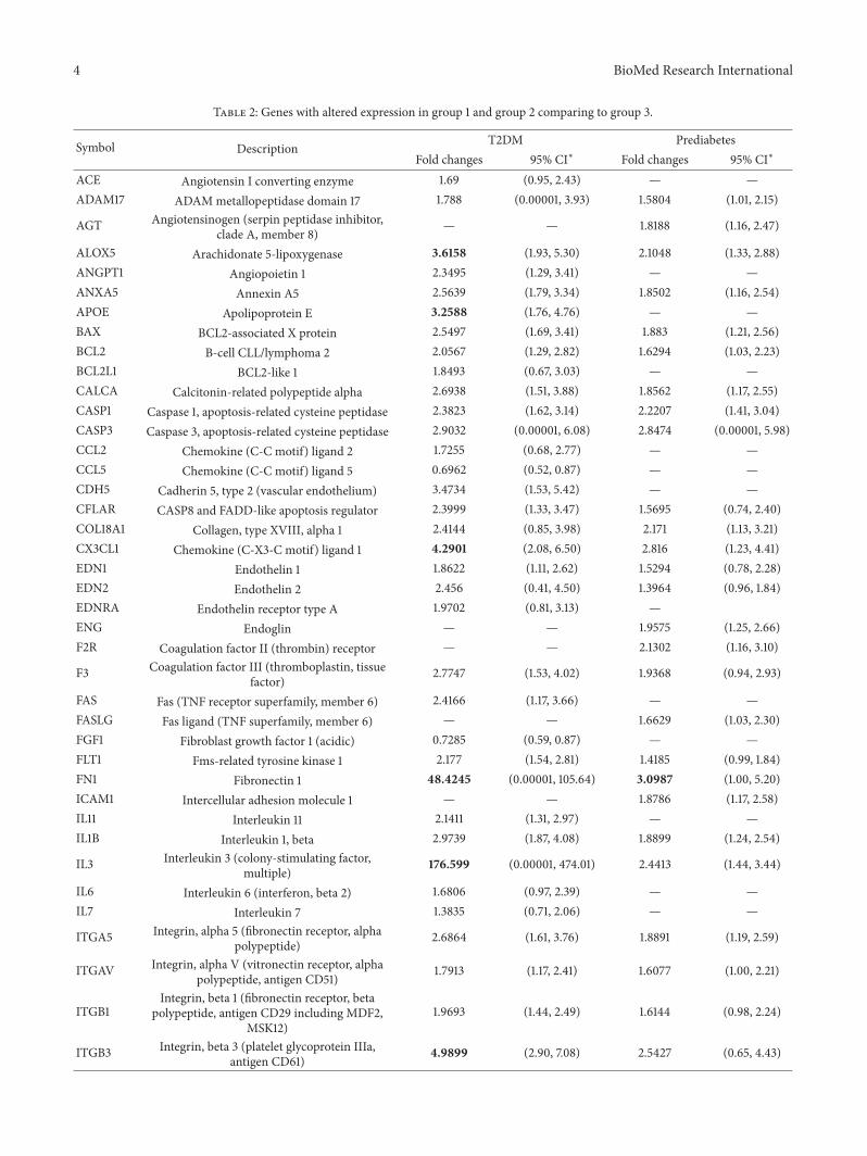