research article syzygium cumini leaf extract reverts...
TRANSCRIPT
Research ArticleSyzygium cumini Leaf Extract Reverts Hypertriglyceridemia viaDownregulation of the Hepatic XBP-1s/PDI/MTP Axis inMonosodium L-Glutamate-Induced Obese Rats
Lucas Martins França ,1 Caio Fernando Ferreira Coêlho,1 Larissa Nara Costa Freitas,1
Ivana Letícia Santos Souza,1 Vinicyus Teles Chagas,1 Victor Debbas,2
Thais Martins de Lima,3 Heraldo Possolo de Souza,3 Francisco Rafael Martins Laurindo,2
and Antonio Marcus de Andrade Paes 1
1Laboratory of Experimental Physiology (LeFisio), Department of Physiological Sciences, Federal University of Maranhão,Av. dos Portugueses, 1966–Cidade Universitária Dom Delgado, São Luís, MA 65080-805, Brazil2Laboratory of Vascular Biology (LBV), Heart Institute, School of Medicine, University of São Paulo, Av. Dr. Enéas deCarvalho Aguiar, 44–Cerqueira César, São Paulo, SP 05403-900, Brazil3Laboratory of Medical Investigation (LIM-51), Emergency Medicine Department, School of Medicine, University of São Paulo,Av. Dr. Arnaldo, 455-Cerqueira César, São Paulo, SP 01246-903, Brazil
Correspondence should be addressed to Antonio Marcus de Andrade Paes; [email protected]
Received 30 November 2018; Revised 11 January 2019; Accepted 5 February 2019; Published 20 March 2019
Guest Editor: Mohamed M. Abdel-Daim
Copyright © 2019 Lucas Martins França et al. This is an open access article distributed under the Creative Commons AttributionLicense, which permits unrestricted use, distribution, and reproduction in any medium, provided the original work isproperly cited.
Syzygium cumini is used worldwide for the treatment of metabolic syndrome-associated outcomes. Previously, we described theantihypertriglyceridemic effect of the hydroethanolic extract of S. cumini leaf (HESc) in monosodium L-glutamate- (MSG-)induced obese rats. This study sought to investigate the molecular mechanisms underlying the antihypertriglyceridemic effect ofHESc in MSG-obese rats. Newborn male Wistar rats were injected subcutaneously with MSG (4.0 g/kg/day, obese group) orsaline 1.25% (1.0mL/kg/day, lean group), from 2nd through 10th postnatal day. At 8 weeks old, obese rats started to be orallytreated with HESc (0.5 or 1.0 g/kg/day, n = 7) or saline 0.9% (1mL/kg/day, n = 7). Lean rats received saline solution(1mL/kg/day, n = 7). Upon 8-week treatment, animals were euthanized for blood and tissue collection. Another set of adultnonobese Wistar rats was used for the assessment of HESc acute effects on Triton WR1339-induced hypertriglyceridemia. HEScreduced weight gain, as well as adipose tissue fat pads, without altering food intake of obese rats. HESc restored fasting serumglucose, triglycerides, total cholesterol, and free fatty acids, as well as insulin sensitivity, to levels similar to lean rats.Additionally, HESc halved the triglyceride content into very low-density lipoprotein particles, as well as healed liver steatosis, inobese rats. Hepatic protein expression of the endoplasmic reticulum chaperone GRP94 was decreased by HESc, which alsodownregulated the hepatic triglyceride secretion pathway by reducing the splicing of X-box binding protein 1 (XBP-1s), as wellas protein disulfide isomerase (PDI) and microsomal triglyceride transfer protein (MTP) translational levels. This action wasfurther corroborated by the acute inhibitory effect of HESc on triglyceride accumulation on Triton WR1339-treated rats. Ourdata support the downregulation of the XBP-1s/PDI/MTP axis in the liver of MSG-obese rats as a novel feasible mechanism forthe antihypertriglyceridemic effect promoted by the polyphenolic phytocomplex present in S. cumini leaf.
1. Introduction
Nonalcoholic fatty liver disease (NAFLD) is considered themain hepatic manifestation of metabolic syndrome (MetS)
[1].UnderMetS,white adipose tissuehypertrophy causes localinsulin resistance that, in turn, increases adipocyte lipolyticactivity anddecreases local free fatty acid (FFA) recycling, rais-ing serum FFA levels. Increased FFA uptake by hepatocytes
HindawiOxidative Medicine and Cellular LongevityVolume 2019, Article ID 9417498, 14 pageshttps://doi.org/10.1155/2019/9417498
leads to ectopic fat accumulation and lipotoxicity due to thelimited liver capacity to oxidize and/or export excess FFA [2].Hyperinsulinemia additionally imposes de novo lipogenesisoversizing hepatic fat accumulation, an outcome partiallycompensated by increased triglyceride (TG) secretion via verylow-density lipoprotein (VLDL)particles, thatultimately leadsto hypertriglyceridemia [3]. Hypertriglyceridemia is an inde-pendent risk factor for cardiovascular diseases, which are theleading cause of morbimortality worldwide [4].
Despite the abovementioned evidences, the molecularmechanisms involved in NAFLD and hypertriglyceridemiaonset remain incompletely defined. During the last decade,the endoplasmic reticulum (ER) stress has been proposed asa key player by its role in unfolded protein response (UPR)[5]. UPR occurs when the ER becomes overwhelmed andcauses luminal misfolded protein accumulation, elicitingthe phosphorylation of three ER transmembrane sensingproteins, namely, inositol-requiring enzyme-1α (IRE-1α),protein kinase RNA-like ER kinase (PERK), and activatingtranscription factor 6 (ATF6) [5]. IRE-1α subsequentlysplices the X-box binding protein 1 (XBP-1s) mRNA, a tran-scription factor importantly involved in the reestablishmentof ER homeostasis [6] and hepatic lipogenesis regulation[7]. Studies conducted by us [8] and others [9–11] have dem-onstrated the importance of the IRE1α/XBP-1s pathway forhepatic lipid homeostasis, through either lipogenesis modu-lation or stimulation of TG secretion, a process mediatedby upregulation of microsomal triglyceride transfer protein(MTP) and protein disulfide isomerase (PDI) expression inhepatocytes [8]. Henceforth, the XBP-1s/PDI/MTP axis hasemerged as a potential therapeutic target for the treatmentof lipidmetabolism disorders, especially hypertriglyceridemia[12], despite the plethora of other regulatory targets.
Herbal medicines constitute an important source of bio-active and potentially therapeutic molecules enabled to fulfillamultiple-target strategy forMetS-related outcome treatment[13, 14]. These properties are specially related to their antiox-idant capacity, although other mechanisms might feasibly beinvolved [15]. In this context, cardiometabolic potentialitiesof Syzygium cumini (L.) Skeels (syn: S. jambolanum D.C.,Eugenia jambolana Lam.) have been highlighted [16]. S.cumini is an Indian native tree from the Myrtaceae familywidely cultivated throughout the world and popularly knownas jambolão, jambolan, java plum, or black plum [17]. It is tra-ditionally used to treat a variety of illnesses, most of themrelated to MetS and its comorbidities [16, 18–21]. Moreover,its ethnopharmacological relevance has been recognized bythe Brazilian Ministry of Health, which included S. cuminispecies in the National Index of Medicinal Plants of Interestto the Unified Public Health System, acronym RENISUS [22].
In a previous study, we showed that the hydroethanolicextract of S. cumini leaf (HESc) improved the metabolic pro-file of monosodium L-glutamate- (MSG-) induced obese rats,especially by reverting TG accumulation in both the liver andserum. These effects were associated with the improvementof peripheral insulin sensitivity and β-cell function andattributed to the polyphenolic profile—mainly composed bymyricetin derivatives, as well as other flavonoids andtannins—identified in HESc [23]. More recently, we reported
the HPLC-MS/MS phytochemical characterization of apolyphenol-rich extract (PESc) prepared from the aforemen-tioned HESc, which allowed the identification of five maincompounds as follows: gallic acid, myricetin, myriceti-n-3-α-arabinopyranoside, myricetin deoxyhexoside, andquercetin, with myricetin accounting for nearly 20% of PESctotal polyphenol content [24]. Notwithstanding, PESc exhibiteda strong antioxidant capacity against both biological and nonbi-ological oxidants, which enabled it to protect mice from an oxi-dative stress-induceddiabetic state [24].However, themolecularmechanisms responsible for the improvement of lipid metabo-lism promoted by S. cumini leaf remain poorly investigated.
Thus, taking into account our previous reports thathypertriglyceridemia in MSG-obese rats is associated withactivation of the XBP-1s/PDI/MTP axis [8] and that HEScreverted their characteristic NAFLD and hypertriglyc-eridemia [23], in the present study, we sought to investigatethe molecular mechanisms underlying the antihypertrigly-ceridemic activity of HESc in MSG-obese rats. The data pre-sented herein endorse our hypothesis by presenting a novelfeasible mechanism for the HESc antihypertriglyceridemiceffect, which corroborates S. cumini leaf as a source of com-pounds for hypolipemiant purposes.
2. Materials and Methods
2.1. Plant Material. Leaves from Syzygium cumini (L.) Skeels,popularly known as jambolão in Brazil and java plum orblack plum in English-spoken countries, were collected fromspecimens located at the Dom Delgado Campus (2°33′11.7″S44°18′22.7″W) of the Federal University of Maranhão(UFMA; São Luís, Maranhão, Brazil). A voucher specimenwas identified by Prof. Dr. Eduardo Bezerra Almeida Jr., abotanist at the Herbarium of Maranhão (MAR, Departmentof Biology, UFMA), and stored under the register number4574. Furthermore, the species’ name was confirmed inhttp://www.theplantlist.org on 08/15/2018.
2.2. Hydroethanolic Extract Preparation. After leaf collection,the hydroethanolic extract of S. cumini leaf (HESc) wasobtained as previously described [23]. Upon lyophilization,HESc powder was stored at 4°C and freshly diluted in 0.9%NaCl at proper concentrations for oral administration tothe animals. An aliquot of HESc was analyzed byHPLC-MS/MS to validate its authenticity. As shown inSupplementary Figure 1, HESc fingerprint corresponds tothe same polyphenolic profile previously reported by us[23], whose main compounds are shown in Figure 1.
2.3. MSG Obesity Induction and Experimental Design.Newborn maleWistar rats were subcutaneously injected withthe MSG solution (4.0 g/kg/day, Sigma-Aldrich, USA,Cat# G1626) or saline 1.25% (1.0mL/kg/day), in accordancewith our previous report [8]. From birth, all animals werekept under controlled conditions of temperature (23 ± 2°C)and light (12 h light/12 h dark) with filtered water and regularchow (CR-1 Nuvilab, Curitiba, Brazil) provided ad libitum.At 8 weeks of age, obesity development was assessed by cal-culating the Lee index (LI) (body weight (g)1/3/nasoanal
2 Oxidative Medicine and Cellular Longevity

length cm × 1000) [25]. MSG-obese rats and their appro-priated lean controls were randomly divided into 4 groupsand orally treated (gavage) as follows:
(1) Lean: lean rats receiving 1.0mL/kg/day saline0.9% (n = 7)
(2) Obese: MSG-obese rats receiving 1.0mL/kg/daysaline 0.9% (n = 7)
(3) Obese+HESc 0.5: MSG-obese rats receiving0.5 g/kg/day HESc (n = 7)
(4) Obese+HESc 1.0: MSG-obese rats receiving1.0 g/kg/day HESc (n = 7)
Body weight and food intake were measured twice a weekthroughout 8 weeks of treatment. At the end, the LI was againcalculated to evaluate the effects of the treatment on body
HO
HO
OH
O
O
O
HO
O
OO
OH
OH
OH
OO
HO
HO
OHHO
OH
OH
(a)
O
O
HO
HO
OH
OH
HO
O
CH3
OOH
OH
(b)
OHO
OH
OH
OH
OH
(c)
OHO
OH
O
OH
OH
O
CH3
HO
OH
OH
O
OH
(d)
OHO
OH
O
OH
OH
O
CH3
HO
OH
OH
O
O CH3
O
(e)
Figure 1: Main polyphenolic compounds identified in the hydroethanolic extract of Syzygium cumini leaves (HESc). (a) Tetragalloylglucose,(b) hexahydroxydiphenoyl-glucose, (c) quercetin, (d) myricetin deoxyhexoside, and (e) acylated myricetin deoxyhexoside.
3Oxidative Medicine and Cellular Longevity
mass. Next, upon overnight fasting, animals were anesthe-tized (10mg/kg xylazine+40mg/kg ketamine, i.p.) for bloodcollection via abdominal aorta puncture and subsequenteuthanasia by exsanguination. The liver and both retroperi-toneal and periepididymal fat pads were collected, weighed,and appropriately stored for posterior assessments. Allanimal procedures were in accordance with the NationalCouncil for the Control of Animal Experimentation(CONCEA, Brazil) and approved by the Committee forEthics and Welfare on Animal Use (CEUA) of UFMA underruling number 23115.01983/2013-41.
2.4. Serum Biochemical Analysis and Assessment of InsulinResistance. Glucose (GL), total cholesterol (TC), TG, FFA,aspartate aminotransferase (AST), and alanine aminotrans-ferase (ALT) levels were assessed in serum samples usingspectrophotometric commercial kits according to the manu-facturers’ instructions (Labtest, MG, Brazil, and Wako, VA,USA). Insulin resistance was inferred by calculating theTyG index (TyG = natural logarithm fasting TG mg/dL ×fastingGL mg/dL /2 ) [26].
2.5. Liver Histological Analysis. Liver slides were obtainedthrough 6μm thick transversal sections, stained withhematoxylin-eosin (HE), and assessed by 2 independentresearchers in a double-blind way for the determination ofthe NAFLD activity score (NAS). This score is based on asemiquantitative analysis of the three definer criteria ofNASH: steatosis (0-3), ballooning (0-3), and lobular inflam-mation (0-2). Total score is a value ranging from 0 to 8,which indicates a hepatic prognostic status. Scores > 6 indi-cate NASH; from 3 to 5, borderline; and from 0 to 2, it isnot NASH [27].
2.6. Liver Lipid Profile. The hepatic lipid profile was assessedas previously described [28]. Briefly, a chloroform :methanol(2 : 1) solution was used to extract total fat from 500mg liversamples, which were resuspended in a Triton-X100 :metha-nol (2 : 1) solution for the measurement of TG and TC levelsas described in Section 2.4.
2.7. Chromatographic Analysis of Serum Lipoproteins. Serumlipoproteins were separated by fast protein liquid chromatog-raphy (FPLC) in a Superose 6 HR 10/30 column (AmershamBiosciences, Sweden) elutedwith Tris buffer (pH7.0; 10mmolTris, 150mmol NaCl, 1M EDTA, and 0.03% NaN3) at a rateof 0.5mL/min, as previously described [29]. A total of 60 frac-tions were collected in a chronological order representing thedensity of each lipoprotein particle. Specifically, fractions 1-15were labeled as VLDL, 16-30 were labeled as low-density lipo-protein (LDL), 31-45 were labeled as high-density lipoprotein(HDL), and 45-60 were labeled as other serum proteins. TheTC and TG contents in each fraction were measured asdescribed in Section 2.4. The total protein content was deter-mined from absorbance at 280nm.
2.8. Evaluation of Protein Expression by Western Blotting.Liver samples (n = 7) were homogenized by sonication withlysis buffer containing protease inhibitors (1μg/mL aproti-nin, 1μg/mL leupeptin, and 10mM PMSF). For each sample,
30μg of total protein was diluted with sample buffer andloaded into a SDS-PAGE gel for protein separation, whichwas transferred to nitrocellulose membranes. For thedetection of the proteins of interest, membranes wereincubated with primary antibodies: anti-KDEL (Enzo LifeSciences,USA,Cat#ADI-SPA-827), anti-XBP1 (EnzoLife Sci-ences,USA,Cat#ADI-905-739), anti-PDI (EnzoLife Sciences,USA, Cat# ADI-SPA-891), and anti-MTP (Sigma-Aldrich,USA, Cat# AV43618), followed by incubation withperoxidase-conjugated secondary antibodies for chemilumi-nescent detection (peroxidase-H2O2-luminol). β-Actin(Sigma-Aldrich, USA, Cat# A5441) was used as proteinloading control.
2.9. Induction of Acute Hypertriglyceridemia with TritonWR1339. Eight-week-old male nonobese Wistar rats wererandomized and administered with a single oral dose ofsaline 0.9% (0.1mL/100 g) or either 0.5 g/kg or 1.0 g/kg HESc.After 1 hour, HESc-treated rats were intraperitoneallyinjected with 0.3 g/kg Triton WR1339 (Sigma-Aldrich,USA, Cat# T8761) and referred as the +HESc 0.5 g/kg and+HESc 1.0 g/kg groups (n = 7 per group). Saline-treated ratswere injected with either equal Triton WR1339 dose (TritonWR1339 group, n = 7) or saline 0.9% (0.1mL/100 g; controlgroup, n = 7). Fasting blood samples were collected fromthe tail vein before (0 h) as well as 24, 48, and 72h after theadministration of Triton WR1339 for the determination ofTG levels as described in Section 2.4 [30].
2.10. Statistical Analysis. Results were expressed as themean ± standard error of the mean (SEM). The Shapiro-Wilk test was applied for normality assuring and groupscompared by one-way analysis of variance (ANOVA)followed by the Newman-Keuls as posttest with Prism 7.0(GraphPad, USA). Statistically significant differences wereset at 5% with p < 0 05.
3. Results
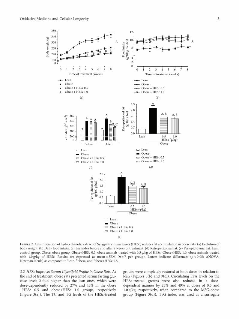
3.1. HESc Reduces Adipose Tissue Accumulation in ObeseRats. The lean group had higher mean body weight thanthe MSG-obese groups throughout the 8-week treatmentperiod (Figure 2(a)), a peculiar feature of this animal modelbecause of its shorter body length associated with deficientgrowth hormone (GH) secretion. However, MSG-obese ratspresented a LI value 11% higher than the lean group(Figure 2(c)), accompanied by a 4-fold increase of retroperi-toneal and periepididymal fat pads (Figures 2(d) and 2(e),respectively), denoting their obese condition. HESc treat-ment (0.5 and 1.0 g/kg/day) reduced the weight gain of obeserats by 15% regardless of the administered dose (Figure 2(a)),though no effect had been detected on food intake(Figure 2(b)). Besides, the LI was reduced by 7% and 10%in the obese+HESc 0.5 and obese+HESc 1.0 groups, respec-tively (Figure 2(c)). Weight loss was followed by a strongdecrease of white adipose tissue accumulation, since retro-peritoneal and periepididymal fat pads were, respectively,reduced by nearly 47% and 40% at both doses (Figures 2(d)and 2(e)).
4 Oxidative Medicine and Cellular Longevity
3.2. HESc Improves Serum Glycolipid Profile in Obese Rats. Atthe end of treatment, obese rats presented serum fasting glu-cose levels 2-fold higher than the lean ones, which weredose-dependently reduced by 27% and 43% in the obese+HESc 0.5 and obese+HESc 1.0 groups, respectively(Figure 3(a)). The TC and TG levels of the HESc-treated
groups were completely restored at both doses in relation tolean (Figures 3(b) and 3(c)). Circulating FFA levels on theHESc-treated groups were also reduced in a dose-dependent manner by 23% and 49% at doses of 0.5 and1.0 g/kg, respectively, when compared to the MSG-obesegroup (Figure 3(d)). TyG index was used as a surrogate
0
0 1 2 3
B B B B B
A
LeanObeseObese + HESc 0.5Obese + HESc 1.0
4Time of treatment (weeks)
5 6 7 8
180
220
260Bo
dy w
eigh
t (g)
300
340
380
(a)
LeanObeseObese + HESc 0.5Obese + HESc 1.0
0 1 2 3 4Time of treatment (weeks)
5 6 7 8
A
024
8
Food
inta
ke(g
/100
g bw
/day
) 10
12
(b)
A A AA
BB, C
LeanObeseObese + HESc 0.5Obese + HESc 1.0
0260
Before After
Lee i
ndex
(g1/
3 .cm-1
)
320300
340360
(c)
A
A, B A, B
ObeseHESc (g/kg)
LeanObeseObese + HESc 0.5Obese + HESc 1.0
3.5
2.8
2.1
1.4
0.7
0.0
Retro
perit
onea
l fat
(g/1
00 g
bw
)
Lean 0.5 1.0
(d)
A
A, B A, B
ObeseHESc (g/kg)
LeanObeseObese + HESc 0.5Obese + HESc 1.0
2.5
2.0
1.5
1.0
0.5
0.0Lean 0.5 1.0
Perie
pidi
dym
al fa
t(g
/100
g b
w)
(e)
Figure 2: Administration of hydroethanolic extract of Syzygium cumini leaves (HESc) reduces fat accumulation in obese rats. (a) Evolution ofbody weight. (b) Daily food intake. (c) Lee index before and after 8 weeks of treatment. (d) Retroperitoneal fat. (e) Periepididymal fat. Lean:control group. Obese: obese group. Obese+HESc 0.5: obese animals treated with 0.5 g/kg of HESc. Obese+HESc 1.0: obese animals treatedwith 1.0 g/kg of HESc. Results are expressed as mean ± SEM (n = 7 per group). Letters indicate differences (p < 0 05; ANOVA;Newman-Keuls) as compared to alean, bobese, and cobese+HESc 0.5.
5Oxidative Medicine and Cellular Longevity
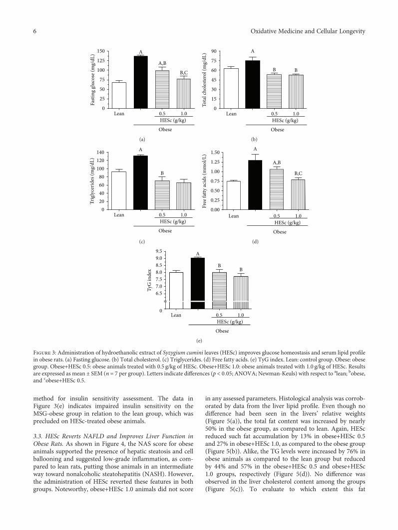
method for insulin sensitivity assessment. The data inFigure 3(e) indicates impaired insulin sensitivity on theMSG-obese group in relation to the lean group, which wasprecluded on HESc-treated obese animals.
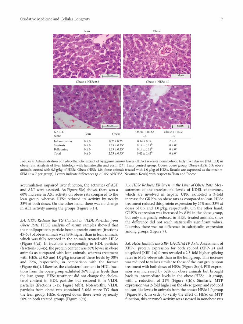
3.3. HESc Reverts NAFLD and Improves Liver Function inObese Rats. As shown in Figure 4, the NAS score for obeseanimals supported the presence of hepatic steatosis and cellballooning and suggested low-grade inflammation, as com-pared to lean rats, putting those animals in an intermediateway toward nonalcoholic steatohepatitis (NASH). However,the administration of HESc reverted these features in bothgroups. Noteworthy, obese+HESc 1.0 animals did not score
in any assessed parameters. Histological analysis was corrob-orated by data from the liver lipid profile. Even though nodifference had been seen in the livers’ relative weights(Figure 5(a)), the total fat content was increased by nearly50% in the obese group, as compared to lean. Again, HEScreduced such fat accumulation by 13% in obese+HESc 0.5and 27% in obese+HESc 1.0, as compared to the obese group(Figure 5(b)). Alike, the TG levels were increased by 76% inobese animals as compared to the lean group but reducedby 44% and 57% in the obese+HESc 0.5 and obese+HESc1.0 groups, respectively (Figure 5(d)). No difference wasobserved in the liver cholesterol content among the groups(Figure 5(c)). To evaluate to which extent this fat
Obese
HESc (g/kg)Lean
A
B,C
A,B
Fasti
ng g
luco
se (m
g/dL
) 150
125
100
75
50
25
00.5 1.0
(a)
Tota
l cho
leste
rol (
mg/
dL) 90
75
60
45
30
15
0
Obese
HESc (g/kg)Lean
A
B B
0.5 1.0
(b)
Trig
lyce
rides
(mg/
dL)
140120100
806040
020
A
B
Obese
HESc (g/kg)Lean 0.5 1.0
(c)
A
B,C
A,B
Free
fatty
acid
s (m
mol
/L) 1.50
1.25
1.00
0.75
0.50
0.25
0.00
Obese
HESc (g/kg)Lean 0.5 1.0
(d)
TyG
inde
x
9.59.08.58.07.5
6.57.0
0
Obese
HESc (g/kg)Lean 0.5 1.0
A
BB
(e)
Figure 3: Administration of hydroethanolic extract of Syzygium cumini leaves (HESc) improves glucose homeostasis and serum lipid profilein obese rats. (a) Fasting glucose. (b) Total cholesterol. (c) Triglycerides. (d) Free fatty acids. (e) TyG index. Lean: control group. Obese: obesegroup. Obese+HESc 0.5: obese animals treated with 0.5 g/kg of HESc. Obese+HESc 1.0: obese animals treated with 1.0 g/kg of HESc. Resultsare expressed asmean ± SEM (n = 7 per group). Letters indicate differences (p < 0 05; ANOVA; Newman-Keuls) with respect to alean; bobese,and cobese+HESc 0.5.
6 Oxidative Medicine and Cellular Longevity
accumulation impaired liver function, the activities of ASTand ALT were assessed. As Figure 5(e) shows, there was a60% increase in AST activity on obese rats compared to thelean group, whereas HESc reduced its activity by nearly35% at both doses. On the other hand, there was no changein ALT activity among the groups (Figure 5(f)).
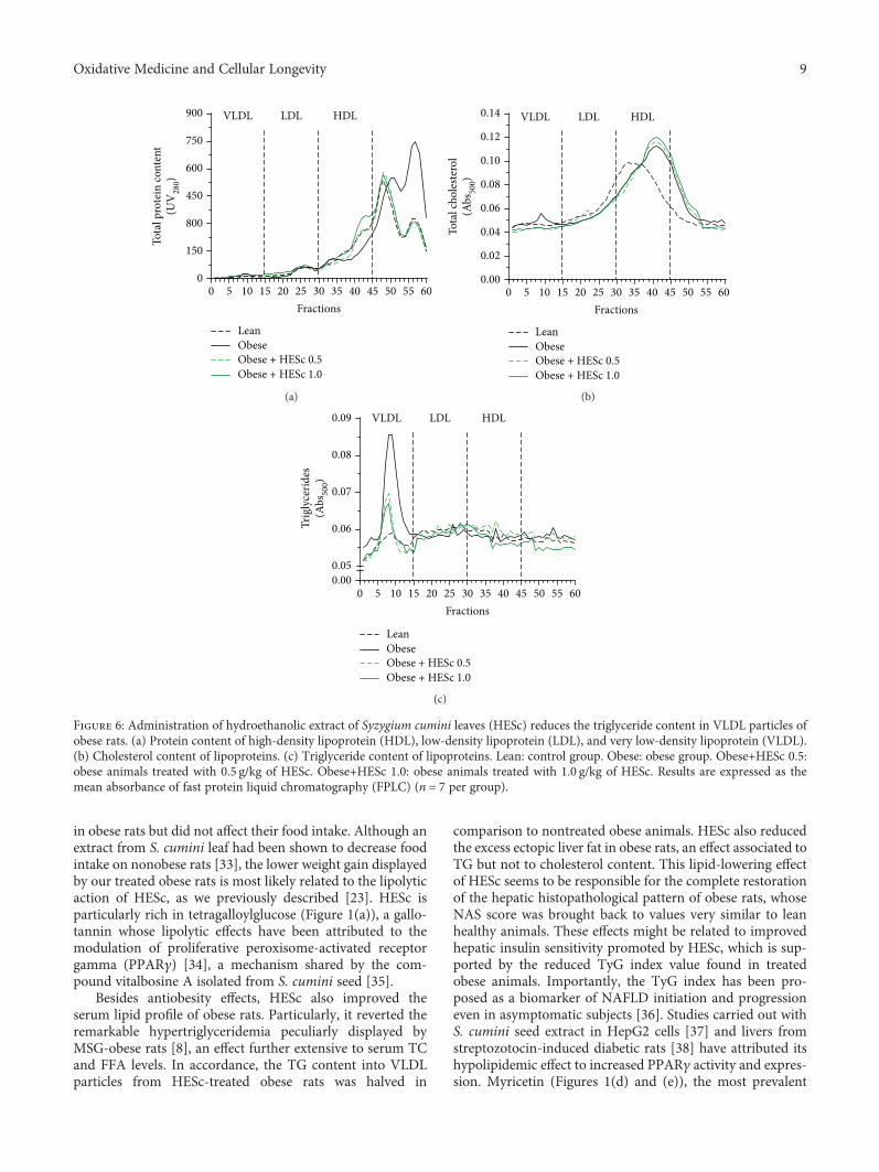
3.4. HESc Reduces the TG Content in VLDL Particles fromObese Rats. FPLC analysis of serum samples showed thatthe nonlipoprotein particle-bound protein content (fractions45-60) of obese animals was 48% higher than in lean animals,which was fully restored in the animals treated with HESc(Figure 6(a)). In fractions corresponding to HDL particles(fractions 30-45), the protein content was 30% lower in obeseanimals as compared with lean animals, whereas treatmentwith HESc at 0.5 and 1.0 g/kg increased these levels by 30%and 72%, respectively, in comparison with the former(Figure 6(a)). Likewise, the cholesterol content in HDL frac-tions from the obese group exhibited 36% higher levels thanthe lean group. HESc treatment did not change the choles-terol content in HDL particles but restored it in VLDLparticles (fractions 1-15; Figure 6(b)). Noteworthy, VLDLparticles from obese rats contained 3-fold more TG thanthe lean group. HESc dropped down these levels by nearly50% in both treated groups (Figure 6(c)).
3.5. HESc Reduces ER Stress in the Liver of Obese Rats. Mea-surement of the translational levels of KDEL chaperones,which are involved in hepatic UPR, exhibited a 3-foldincrease for GRP94 on obese rats as compared to lean. HESctreatment reduced this protein expression by 27% and 33% atdoses of 0.5 and 1.0 g/kg, respectively. On the other hand,GRP78 expression was increased by 83% in the obese group,but only marginally reduced in HESc-treated animals, sincethe difference did not reach statistically significant values.Likewise, there was no difference in calreticulin expressionamong groups (Figure 7).
3.6. HESc Inhibits the XBP-1s/PDI/MTP Axis. Assessment ofXBP-1 protein expression for both spliced (XBP-1s) andunspliced (XBP-1u) forms revealed a 2.5-fold higher splicingrates in MSG-obese rats than in the lean group. This increasewas reduced to values similar to those of the lean group upontreatment with both doses of HESc (Figure 8(a)). PDI expres-sion was increased by 52% on obese animals but broughtback to intermediate levels in the obese+HESc 1.0 group,with a reduction of 21% (Figure 8(b)). Similarly, MTPexpression was 2-fold higher on the obese group and reducedto lean-like levels in animals from the obese+HESc 1.0 group(Figure 8(c)). In order to verify the effect of HESc on MTPfunction, this enzyme’s activity was assessed in nonobese rats
NAFLDscore Lean Obese Obese + HESc
0.5Obese + HESc
1.0Inflammation 0 ± 0 0.25± 0.25 0.14 ± 0.14 0 ± 0 Steatosis 0 ± 0 1.25 ± 0.25a 0.14 ± 0.14b 0 ± 0b
Ballooning 0 ± 0 1.25 ± 0.25a 0.14 ± 0.14b 0 ± 0b
Total 0 ± 0 2.75 ± 0.75a 0.42 ± 0.42b 0 ± 0b
Lean Obese
Obese + HESc 0.5 Obese + HESc 1.0
Figure 4: Administration of hydroethanolic extract of Syzygium cumini leaves (HESc) reverses nonalcoholic fatty liver disease (NAFLD) inobese rats. Analysis of liver histology with hematoxylin and eosin [27]. Lean: control group. Obese: obese group. Obese+HESc 0.5: obeseanimals treated with 0.5 g/kg of HESc. Obese+HESc 1.0: obese animals treated with 1.0 g/kg of HESc. Results are expressed as the mean ±SEM (n = 7 per group). Letters indicate differences (p < 0 05; ANOVA; Newman-Keuls) with respect to alean and bobese.
7Oxidative Medicine and Cellular Longevity

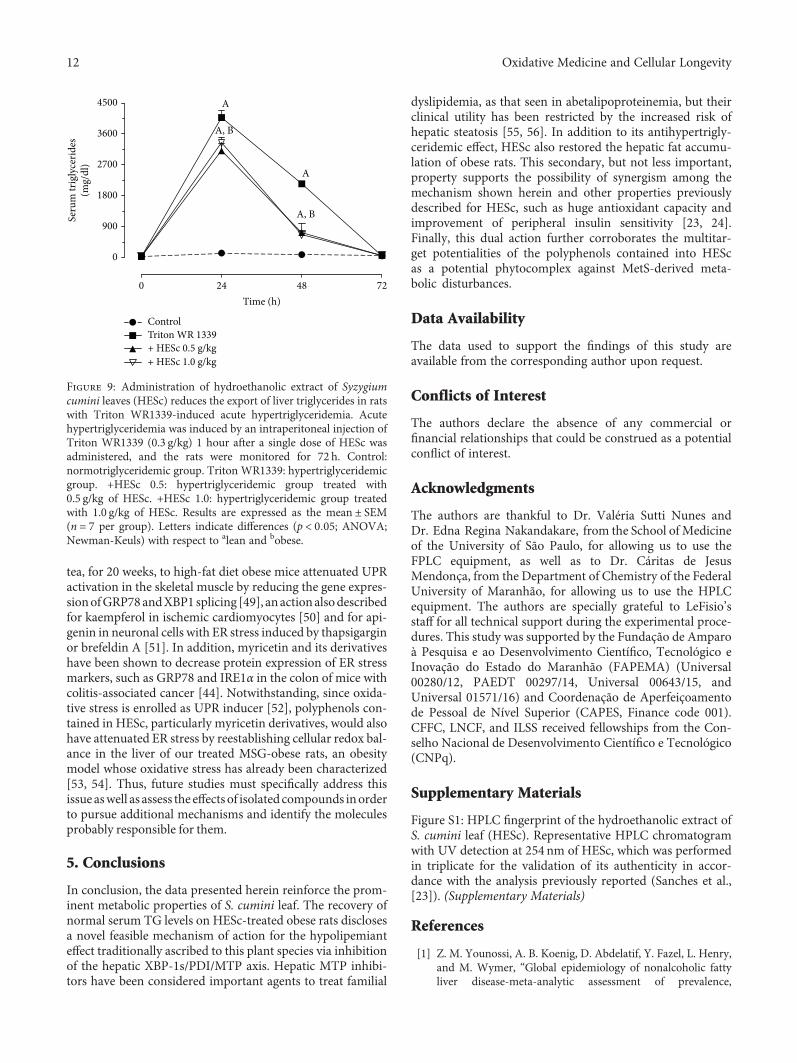
acutely injected with Triton WR1339, which is a well-knownmodel of MTP-mediated hypertriglyceridemia [30]. Oraladministration of HESc, at the same abovementioned doses,1-hour prior induction reduced serum TG accumulationwithin 24h, as well as hastened its clearance in the following48 h (Figure 9), which is in line with a lower rate of VLDLparticles assembly and secretion from the liver.
4. Discussion
This study strengthens S. cumini pharmacological potential-ities since it corroborates our previous report that HEScrestores serum TG levels in hypertriglyceridemic MSG-obese rats [23]. The data presented herein expand thesefindings by showing that oral administration of HESc toMSG-obese rats for 8 weeks detains weight gain, improves
fatty liver disease, and reverts hypertriglyceridemia, besidesother metabolic outcomes typically found in this MetS rodentmodel. Specifically, this study shows that HESc inhibitedboth expression and activity of hepatic MTP by downregula-tion of the XBP-1s/PDI/MTP axis, reducing the incorpora-tion of TG into VLDL particles and consequently loweringthe circulating TG levels.
Neonatal administration of MSG damages hypothalamicnuclei, e.g., arcuate nucleus, leading to impaired GH secre-tion; therefore, adult animals are shorter and lighter thanage-matched controls but present higher fat mass [31]. Fur-thermore, these animals also exhibit autonomic unbalancecharacterized by enhanced vagus nerve tonus, which imposesincreased insulin secretion and consequent development ofperipheral insulin resistance and elevation of fat stores [32].In this study, treatment with HESc reduced body weight gain
2.5
2.0
1.5
Live
r(g
/100
g bw
)
1.0
0.5
0.0Lean 0.5
HESc (g/kg)Obese
1.0
(a)
300 AA
B250200150
Hep
atic
tota
l fat
(mg/
g liv
er)
10050
0Lean 0.5
HESc (g/kg)Obese
1.0
(b)
4.54.03.5
Hep
atic
chol
este
rol
(mg/
g liv
er) 3.0
2.52.01.51.00.50.0
Lean 0.5HESc (g/kg)
Obese
1.0
(c)
18
15
12
9
Hep
atic
trig
lyce
rides
(mg/
g liv
er)
6
3
0
A
B
B
Lean 0.5HESc (g/kg)
Obese
1.0
(d)
100
80
60
AST
(IU
/L)
40
20
0
A
B B
Lean 0.5HESc (g/kg)
Obese
1.0
(e)
60
40
50
30
ALT
(IU
/L)
20
10
0Lean 0.5
HESc (g/kg)Obese
1.0
(f)
Figure 5: Administration of hydroethanolic extract of Syzygium cumini leaves (HESc) improves lipid profile and liver function of obese rats.(a) Liver weight. (b) Total liver fat. (c) Hepatic cholesterol. (d) Hepatic triglycerides. (e) Serum aspartate aminotransferase (AST). (f) Serumalanine aminotransferase (ALT). Lean: control group. Obese: obese group. Obese+HESc 0.5: obese animals treated with 0.5 g/kg of HESc.Obese+HESc 1.0: obese animals treated with 1.0 g/kg of HESc. Results are expressed as the mean ± SEM (n = 7 per group). Letters indicatedifferences (p < 0 05; ANOVA; Newman-Keuls) with respect to alean and bobese.
8 Oxidative Medicine and Cellular Longevity
in obese rats but did not affect their food intake. Although anextract from S. cumini leaf had been shown to decrease foodintake on nonobese rats [33], the lower weight gain displayedby our treated obese rats is most likely related to the lipolyticaction of HESc, as we previously described [23]. HESc isparticularly rich in tetragalloylglucose (Figure 1(a)), a gallo-tannin whose lipolytic effects have been attributed to themodulation of proliferative peroxisome-activated receptorgamma (PPARγ) [34], a mechanism shared by the com-pound vitalbosine A isolated from S. cumini seed [35].
Besides antiobesity effects, HESc also improved theserum lipid profile of obese rats. Particularly, it reverted theremarkable hypertriglyceridemia peculiarly displayed byMSG-obese rats [8], an effect further extensive to serum TCand FFA levels. In accordance, the TG content into VLDLparticles from HESc-treated obese rats was halved in
comparison to nontreated obese animals. HESc also reducedthe excess ectopic liver fat in obese rats, an effect associated toTG but not to cholesterol content. This lipid-lowering effectof HESc seems to be responsible for the complete restorationof the hepatic histopathological pattern of obese rats, whoseNAS score was brought back to values very similar to leanhealthy animals. These effects might be related to improvedhepatic insulin sensitivity promoted by HESc, which is sup-ported by the reduced TyG index value found in treatedobese animals. Importantly, the TyG index has been pro-posed as a biomarker of NAFLD initiation and progressioneven in asymptomatic subjects [36]. Studies carried out withS. cumini seed extract in HepG2 cells [37] and livers fromstreptozotocin-induced diabetic rats [38] have attributed itshypolipidemic effect to increased PPARγ activity and expres-sion. Myricetin (Figures 1(d) and (e)), the most prevalent
900 VLDL LDL HDL
750
600To
tal p
rote
in co
nten
t(U
V28
0)
450
800
150
00 5 10 15 20 25 30
Fractions35 40 45 50 55 60
Obese + HESc 1.0Obese + HESc 0.5ObeseLean
(a)
Obese + HESc 1.0Obese + HESc 0.5ObeseLean
0.14 VLDL LDL HDL0.12
0.10
0.08
Tota
l cho
leste
rol
(Abs
500)
0.06
0.04
0.02
0.000 5 10 15 20 25 30
Fractions35 40 45 50 55 60
(b)
0.09 VLDL
Obese + HESc 1.0Obese + HESc 0.5ObeseLean
LDL HDL
0.08
Trig
lyce
rides
(Abs
500)
0.07
0.06
0.050.00
0 5 10 15 20 25 30Fractions
35 40 45 50 55 60
(c)
Figure 6: Administration of hydroethanolic extract of Syzygium cumini leaves (HESc) reduces the triglyceride content in VLDL particles ofobese rats. (a) Protein content of high-density lipoprotein (HDL), low-density lipoprotein (LDL), and very low-density lipoprotein (VLDL).(b) Cholesterol content of lipoproteins. (c) Triglyceride content of lipoproteins. Lean: control group. Obese: obese group. Obese+HESc 0.5:obese animals treated with 0.5 g/kg of HESc. Obese+HESc 1.0: obese animals treated with 1.0 g/kg of HESc. Results are expressed as themean absorbance of fast protein liquid chromatography (FPLC) (n = 7 per group).
9Oxidative Medicine and Cellular Longevity

flavonoid in HESc [23], has been shown to improve insulinsensitivity [39] and promote hepatic lipid oxidation byincreasing PPARα expression in the liver [40].
In addition to the extensive knowledge on S. cumini effectson peripheral insulin sensitivity, particularly on the PPARα/γpathways, we hypothesize that HESc polyphenols might alsointerfere with the ER stress-sensing IRE1α/XBP-1s pathway,which has also been proposed as an important mechanismunderlying the development of NAFLD and hypertriglyc-eridemia, as demonstrated in hepatocyte-specific IRE1α-nullmice [11] and MSG-obese rats [8].
In the past decade, hepatic ER stress has been proposed asa main contributing factor for NAFLD onset and progres-sion, as well as MetS-associated dyslipidemias [5, 41]. InitialUPR is characterized by elevated gene/protein expression ofKDEL chaperones, namely, glucose-regulated protein 94
(GRP94), GRP78, and calreticulin, to mitigate protein mis-folding and reestablish ER homeostasis within a negativefeedback loop regulated by both the IRE1α and ATF6 path-ways [41, 42]. Our obese rats showed a clear increase ofhepatic GRP94 and GRP78 protein expressions, denotingactive UPR, which was partially attenuated in HESc-treatedobese rats. Many actions of HESc, such as improvement ofinsulin sensitivity and lower FFA circulating levels, mightalso be involved in this effect since it has been shown thatincreased serum FFA levels might induce hepatic ER stress[43], meanwhile polyphenols such as myricetin derivativesare able to attenuate it [44].
Studies have demonstrated that the IRE1α/XBP-1s path-way is the most conserved arm of UPR [45], which is impor-tantly involved in the control of glucose homeostasis andlipid metabolism [9, 10]. Regardless of a recent discussion
(a)
(b)
(c)
A
A
A, BA, B
0.8
0.6
0.4
GRP
94 ex
pres
sion
(rel
ativ
e to �훽
-act
in)
0.2
0.0Lean 0.5
HESc (g/kg)Obese
1.0
0.25
0.75
1.00
0.50
GRP
78 ex
pres
sion
(rel
ativ
e to �훽
-act
in)
0.25
0.00Lean 0.5
HESc (g/kg)Obese
1.0
0.25
0.75
1.00
0.50
Calre
ticul
in ex
pres
sion
(rel
ativ
e to �훽
-act
in)
0.25
0.00Lean 0.5
HESc (g/kg)Obese
1.0
Lean
ObeseObeseObeseObeseObese
HESc 0.5 HESc 1.0MW (kDa)100 - – GRP94
76 - – GRP78
– Calreticulin
– �훽 – Actin
52 -
Figure 7: Administration of hydroethanolic extract of Syzygium cumini leaves (HESc) attenuates endoplasmic reticulum stress in the livers ofobese rats. Protein expression was determined by western blotting. (a) Glucose response protein 94 (GRP94). (b) Glucose response protein 78(GRP78). (c) Calreticulin. Lean: control group. Obese: obese group. Obese+HESc 0.5: obese animals treated with 0.5 g/kg of HESc. Obese+HESc 1.0: obese animals treated with 1.0 g/kg of HESc. Densitometry results are expressed as the mean ± SEM (n = 4 per group). Lettersindicate differences (p < 0 05; ANOVA; Newman-Keuls) with respect to alean and bobese.
10 Oxidative Medicine and Cellular Longevity
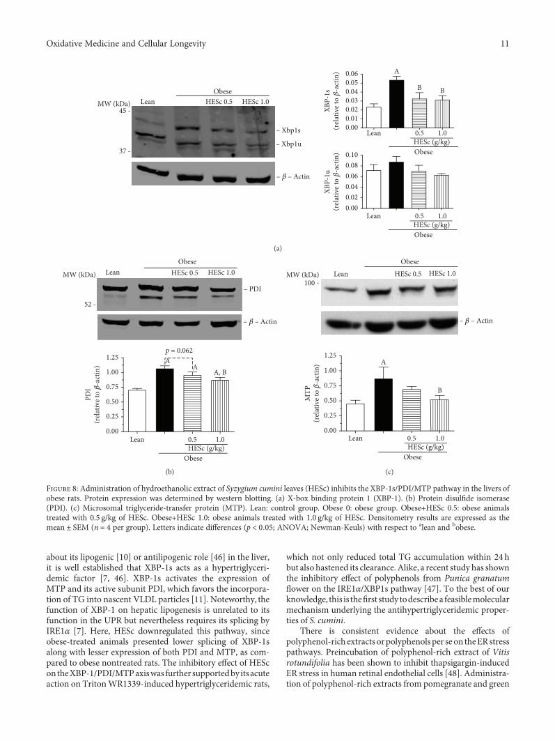
about its lipogenic [10] or antilipogenic role [46] in the liver,it is well established that XBP-1s acts as a hypertriglyceri-demic factor [7, 46]. XBP-1s activates the expression ofMTP and its active subunit PDI, which favors the incorpora-tion of TG into nascent VLDL particles [11]. Noteworthy, thefunction of XBP-1 on hepatic lipogenesis is unrelated to itsfunction in the UPR but nevertheless requires its splicing byIRE1α [7]. Here, HESc downregulated this pathway, sinceobese-treated animals presented lower splicing of XBP-1salong with lesser expression of both PDI and MTP, as com-pared to obese nontreated rats. The inhibitory effect of HEScontheXBP-1/PDI/MTPaxiswas further supportedby itsacuteaction on TritonWR1339-induced hypertriglyceridemic rats,
which not only reduced total TG accumulation within 24hbut also hastened its clearance. Alike, a recent study has shownthe inhibitory effect of polyphenols from Punica granatumflower on the IRE1α/XBP1s pathway [47]. To the best of ourknowledge, this is thefirst study todescribea feasiblemolecularmechanism underlying the antihypertriglyceridemic proper-ties of S. cumini.
There is consistent evidence about the effects ofpolyphenol-rich extracts orpolyphenolsper seon theERstresspathways. Preincubation of polyphenol-rich extract of Vitisrotundifolia has been shown to inhibit thapsigargin-inducedER stress in human retinal endothelial cells [48]. Administra-tion of polyphenol-rich extracts from pomegranate and green
– Xbp1s
– �훽 – Actin
– Xbp1u
MW (kDa) LeanObese
0.060.050.040.03
XBP-
1s(r
elat
ive t
o �훽
-act
in)
0.020.010.00
0.100.080.06
XBP-
1u(r
elat
ive t
o �훽
-act
in)
0.040.020.00
Lean 0.5 1.0
B
A
B
HESc (g/kg)Obese
Lean 0.5 1.0HESc (g/kg)Obese
HESc 0.5 HESc 1.0
37 -
45 -
(a)
1.25
1.00
0.75
PDI
(rel
ativ
e to �훽
-act
in)
0.50
0.25
0.00Lean 0.5 1.0
HESc (g/kg)Obese
MW (kDa) LeanObese
HESc 0.5 HESc 1.0
52 -
– PDI
– �훽 – Actin
Ap = 0.062
AA, B
(b)
1.25A
B
1.00
0.75
MTP
(rel
ativ
e to �훽
-act
in)
0.50
0.25
0.00Lean 0.5 1.0
HESc (g/kg)Obese
– �훽 – Actin
LeanObese
HESc 0.5 HESc 1.0100 -
MW (kDa)
(c)
Figure 8: Administration of hydroethanolic extract of Syzygium cumini leaves (HESc) inhibits the XBP-1s/PDI/MTP pathway in the livers ofobese rats. Protein expression was determined by western blotting. (a) X-box binding protein 1 (XBP-1). (b) Protein disulfide isomerase(PDI). (c) Microsomal triglyceride-transfer protein (MTP). Lean: control group. Obese 0: obese group. Obese+HESc 0.5: obese animalstreated with 0.5 g/kg of HESc. Obese+HESc 1.0: obese animals treated with 1.0 g/kg of HESc. Densitometry results are expressed as themean ± SEM (n = 4 per group). Letters indicate differences (p < 0 05; ANOVA; Newman-Keuls) with respect to alean and bobese.
11Oxidative Medicine and Cellular Longevity
tea, for 20 weeks, to high-fat diet obese mice attenuated UPRactivation in the skeletal muscle by reducing the gene expres-sionofGRP78andXBP1 splicing [49], anactionalsodescribedfor kaempferol in ischemic cardiomyocytes [50] and for api-genin in neuronal cells with ER stress induced by thapsigarginor brefeldin A [51]. In addition, myricetin and its derivativeshave been shown to decrease protein expression of ER stressmarkers, such as GRP78 and IRE1α in the colon of mice withcolitis-associated cancer [44]. Notwithstanding, since oxida-tive stress is enrolled as UPR inducer [52], polyphenols con-tained in HESc, particularly myricetin derivatives, would alsohave attenuated ER stress by reestablishing cellular redox bal-ance in the liver of our treated MSG-obese rats, an obesitymodel whose oxidative stress has already been characterized[53, 54]. Thus, future studies must specifically address thisissue aswell as assess the effectsof isolated compounds inorderto pursue additional mechanisms and identify the moleculesprobably responsible for them.
5. Conclusions
In conclusion, the data presented herein reinforce the prom-inent metabolic properties of S. cumini leaf. The recovery ofnormal serum TG levels on HESc-treated obese rats disclosesa novel feasible mechanism of action for the hypolipemianteffect traditionally ascribed to this plant species via inhibitionof the hepatic XBP-1s/PDI/MTP axis. Hepatic MTP inhibi-tors have been considered important agents to treat familial
dyslipidemia, as that seen in abetalipoproteinemia, but theirclinical utility has been restricted by the increased risk ofhepatic steatosis [55, 56]. In addition to its antihypertrigly-ceridemic effect, HESc also restored the hepatic fat accumu-lation of obese rats. This secondary, but not less important,property supports the possibility of synergism among themechanism shown herein and other properties previouslydescribed for HESc, such as huge antioxidant capacity andimprovement of peripheral insulin sensitivity [23, 24].Finally, this dual action further corroborates the multitar-get potentialities of the polyphenols contained into HEScas a potential phytocomplex against MetS-derived meta-bolic disturbances.
Data Availability
The data used to support the findings of this study areavailable from the corresponding author upon request.
Conflicts of Interest
The authors declare the absence of any commercial orfinancial relationships that could be construed as a potentialconflict of interest.
Acknowledgments
The authors are thankful to Dr. Valéria Sutti Nunes andDr. Edna Regina Nakandakare, from the School of Medicineof the University of São Paulo, for allowing us to use theFPLC equipment, as well as to Dr. Cáritas de JesusMendonça, from the Department of Chemistry of the FederalUniversity of Maranhão, for allowing us to use the HPLCequipment. The authors are specially grateful to LeFisio’sstaff for all technical support during the experimental proce-dures. This study was supported by the Fundação de Amparoà Pesquisa e ao Desenvolvimento Científico, Tecnológico eInovação do Estado do Maranhão (FAPEMA) (Universal00280/12, PAEDT 00297/14, Universal 00643/15, andUniversal 01571/16) and Coordenação de Aperfeiçoamentode Pessoal de Nível Superior (CAPES, Finance code 001).CFFC, LNCF, and ILSS received fellowships from the Con-selho Nacional de Desenvolvimento Científico e Tecnológico(CNPq).
Supplementary Materials
Figure S1: HPLC fingerprint of the hydroethanolic extract ofS. cumini leaf (HESc). Representative HPLC chromatogramwith UV detection at 254 nm of HESc, which was performedin triplicate for the validation of its authenticity in accor-dance with the analysis previously reported (Sanches et al.,[23]). (Supplementary Materials)
References
[1] Z. M. Younossi, A. B. Koenig, D. Abdelatif, Y. Fazel, L. Henry,and M. Wymer, “Global epidemiology of nonalcoholic fattyliver disease-meta-analytic assessment of prevalence,
0
0 24Time (h)
+ HESc 1.0 g/kg+ HESc 0.5 g/kgTriton WR 1339Control
48 72
900
1800
2700
3600
4500 A
A, B
A
A, BSeru
m tr
igly
cerid
es(m
g/dl
)
Figure 9: Administration of hydroethanolic extract of Syzygiumcumini leaves (HESc) reduces the export of liver triglycerides in ratswith Triton WR1339-induced acute hypertriglyceridemia. Acutehypertriglyceridemia was induced by an intraperitoneal injection ofTriton WR1339 (0.3 g/kg) 1 hour after a single dose of HESc wasadministered, and the rats were monitored for 72 h. Control:normotriglyceridemic group. Triton WR1339: hypertriglyceridemicgroup. +HESc 0.5: hypertriglyceridemic group treated with0.5 g/kg of HESc. +HESc 1.0: hypertriglyceridemic group treatedwith 1.0 g/kg of HESc. Results are expressed as the mean ± SEM(n = 7 per group). Letters indicate differences (p < 0 05; ANOVA;Newman-Keuls) with respect to alean and bobese.
12 Oxidative Medicine and Cellular Longevity
incidence, and outcomes,” Hepatology, vol. 64, no. 1, pp. 73–84, 2016.
[2] E. Fabbrini, S. Sullivan, and S. Klein, “Obesity and nonalco-holic fatty liver disease: biochemical, metabolic, and clinicalimplications,” Hepatology, vol. 51, no. 2, pp. 679–689, 2010.
[3] G. I. Shulman, “Ectopic fat in insulin resistance, dyslipidemia,and cardiometabolic disease,” The New England Journal ofMedicine, vol. 371, no. 12, pp. 1131–1141, 2014.
[4] J. Borén, N. Matikainen, M. Adiels, andM.-R. Taskinen, “Post-prandial hypertriglyceridemia as a coronary risk factor,” Clin-ica Chimica Acta, vol. 431, pp. 131–142, 2014.
[5] X.-Q. Zhang, C. F. Xu, C. H. Yu, W. X. Chen, and Y. M. Li,“Role of endoplasmic reticulum stress in the pathogenesis ofnonalcoholic fatty liver disease,”World Journal of Gastroenter-ology, vol. 20, no. 7, pp. 1768–1776, 2014.
[6] H. Yoshida, T. Matsui, A. Yamamoto, T. Okada, and K. Mori,“XBP1 mRNA is induced by ATF6 and spliced by IRE1 inresponse to ER stress to produce a highly active transcriptionfactor,” Cell, vol. 107, no. 7, pp. 881–891, 2001.
[7] A.-H. Lee, E. F. Scapa, D. E. Cohen, and L. H. Glimcher, “Reg-ulation of hepatic lipogenesis by the transcription factorXBP1,” Science, vol. 320, no. 5882, pp. 1492–1496, 2008.
[8] L. M. Franca, L. N. C. Freitas, V. T. Chagas et al., “Mechanismsunderlying hypertriglyceridemia in rats with monosodiumL-glutamate-induced obesity: evidence of XBP-1/PDI/MTPaxis activation,” Biochemical and Biophysical Research Com-munications, vol. 443, no. 2, pp. 725–730, 2014.
[9] J. Ning, T. Hong, A. Ward et al., “Constitutive role forIRE1α-XBP1 signaling pathway in the insulin-mediatedhepatic lipogenic program,” Endocrinology, vol. 152, no. 6,pp. 2247–2255, 2011.
[10] J. S. So, K. Y. Hur, M. Tarrio et al., “Silencing of lipid metabo-lism genes through IRE1α-mediated mRNA decay lowersplasma lipids in mice,” Cell Metabolism, vol. 16, no. 4,pp. 487–499, 2012.
[11] S. Wang, Z. Chen, V. Lam et al., “IRE1α-XBP1s induces PDIexpression to increase MTP activity for hepatic VLDL assem-bly and lipid homeostasis,” Cell Metabolism, vol. 16, no. 4,pp. 473–486, 2012.
[12] C. Piperi, C. Adamopoulos, and A. G. Papavassiliou, “XBP1: apivotal transcriptional regulator of glucose and lipid metabo-lism,” Trends in Endocrinology and Metabolism, vol. 27,no. 3, pp. 119–122, 2016.
[13] N. Vasudeva, N. Yadav, and S. K. Sharma, “Natural products: asafest approach for obesity,” Chinese Journal of IntegrativeMedicine, vol. 18, no. 6, pp. 473–480, 2012.
[14] P. K. Prabhakar and M. Doble, “A target based therapeuticapproach towards diabetes mellitus using medicinal plants,”Current Diabetes Reviews, vol. 4, no. 4, pp. 291–308, 2008.
[15] Y. J. Zhang, R. Y. Gan, S. Li et al., “Antioxidant phytochemicalsfor the prevention and treatment of chronic diseases,” Mole-cules, vol. 20, no. 12, pp. 21138–21156, 2015.
[16] V. T. Chagas, L. M. França, S. Malik, and A. M. . A. Paes,“Syzygium cumini (L.) skeels: a prominent source of bioactivemolecules against cardiometabolic diseases,” Frontiers in Phar-macology, vol. 6, p. 259, 2015.
[17] M. Ayyanar and P. Subash-Babu, “Syzygium cumini (L.)Skeels: a review of its phytochemical constituents and tradi-tional uses,” Asian Pacific Journal of Tropical Biomedicine,vol. 2, no. 3, pp. 240–246, 2012.
[18] A. Helmstadter, “Syzygium cumini (L.) Skeels (Myrtaceae)against diabetes – 125 years of research,” Pharmazie,vol. 63, no. 2, pp. 91–101, 2008.
[19] G. Baldissera, N. D. M. Sperotto, H. T. Rosa et al., “Effects ofcrude hydroalcoholic extract of Syzygium cumini (L.) Skeelsleaves and continuous aerobic training in rats with diabetesinduced by a high-fat diet and low doses of streptozotocin,”Journal of Ethnopharmacology, vol. 194, pp. 1012–1021, 2016.
[20] L. M. Cercato, P. A. S. White, F. K. Nampo, M. R. V. Santos,and E. A. Camargo, “A systematic review of medicinalplants used for weight loss in Brazil: is there potential forobesity treatment?,” Journal of Ethnopharmacology, vol. 176,pp. 286–296, 2015.
[21] N. Braboza Da Silva, A. C. D. Regis, M. A. Esquibel, J. do Espír-ito Santo Santos, and M. Z. de Almeida, “Uso de plantas med-icinais na comunidade quilombola da Barra II–Bahia, Brasil,”Boletin Latinoamericano y del Caribe de Plantas Medicinalesy Aromaticas, vol. 11, no. 5, pp. 435–453, 2012.
[22] R. BRASIL, Relação Nacional de Plantas Medicinais deInteresse ao SUS. Portal. Saúde, 2009, July 2019, gov.br/portal/arquivos/pdf/RENISUS.pdf.
[23] J. R. Sanches, L. M. França, V. T. Chagas et al., “Polyphenol-rich extract of Syzygium cumini leaf dually improvesperipheral insulin sensitivity and pancreatic islet function inmonosodium L-glutamate-induced obese rats,” Frontiers inPharmacology, vol. 7, p. 48, 2016.
[24] V. T. Chagas, R. M. R. . S. Coelho, R. S. Gaspar et al., “Protec-tive effects of a polyphenol-rich extract from Syzygium cumini(L.) Skeels leaf on oxidative stress-induced diabetic rats,” Oxi-dative Medicine and Cellular Longevity, vol. 2018, Article ID5386079, 13 pages, 2018.
[25] L. L. Bernardis and B. D. Patterson, “Correlation between ‘Leeindex’ and carcass fat content in weanling and adult femalerats with hypothalamic lesions,” The Journal of Endocrinology,vol. 40, no. 4, pp. 527-528, 1968.
[26] L. E. Simental-Mendia, M. Rodriguez-Moran, andF. Guerrero-Romero, “The product of fasting glucose and tri-glycerides as surrogate for identifying insulin resistance inapparently healthy subjects,”Metabolic Syndrome and RelatedDisorders, vol. 6, no. 4, pp. 299–304, 2008.
[27] D. E. Kleiner, E. M. Brunt, M. van Natta et al., “Design and val-idation of a histological scoring system for nonalcoholic fattyliver disease,” Hepatology, vol. 41, no. 6, pp. 1313–1321, 2005.
[28] B. D. Freedman, E. J. Lee, Y. Park, and J. L. Jameson, “Adominant negative peroxisome proliferator-activated recep-tor-γ knock-in mouse exhibits features of the metabolic syn-drome,” Journal of Biological Chemistry, vol. 280, no. 17,pp. 17118–17125, 2005.
[29] M. M. Jericó, F. C. De Chiquito, K. Kajihara et al., “Chromato-graphic analysis of lipid fractions in healthy dogs and dogswith obesity or hyperadrenocorticism,” Journal of VeterinaryDiagnostic Investigation, vol. 21, no. 2, pp. 203–207, 2009.
[30] R. M. Silva, F. Santos, M. Maciel, A. Pinto, and V. Rao, “Effectof trans-dehydrocrotonin, a 19-nor-clerodane diterpene fromCroton cajucara on experimental hypertriglyceridaemia andhypercholesterolaemia induced by Triton WR 1339 (tyloxa-pol) in mice,” Planta Medica, vol. 67, no. 8, pp. 763–765, 2001.
[31] W. J.Millard, J. B.Martin, J. Audet, S.M. Sagar, and J. B.Martin,“Evidence that reduced growth hormone secretion observed inmonosodium glutamate-treated rats is the result of a deficiencyin growth hormone-releasing factor,” Endocrinology, vol. 110,no. 2, pp. 540–550, 1982.
13Oxidative Medicine and Cellular Longevity

[32] S. L. Balbo, S. Grassiolli, R. A. Ribeiro et al., “Fat storage is par-tially dependent on vagal activity and insulin secretion ofhypothalamic obese rat,” Endocrine, vol. 31, no. 2, pp. 142–148, 2007.
[33] A. C. P. Oliveira, D. C. Endringer, L. A. S. Amorim, M. D. G. L.Brandão, and M. M. Coelho, “Effect of the extracts and frac-tions of Baccharis trimera and Syzygium cumini on glycaemiaof diabetic and non-diabetic mice,” Journal of Ethnopharma-cology, vol. 102, no. 3, pp. 465–469, 2005.
[34] M. H. Yang, Y. Vasquez, Z. Ali, I. A. Khan, and S. I. Khan,“Constituents from Terminalia species increase PPARα andPPARγ levels and stimulate glucose uptake without enhancingadipocyte differentiation,” Journal of Ethnopharmacology,vol. 149, no. 2, pp. 490–498, 2013.
[35] G. Thiyagarajan, P. Muthukumaran, B. Sarath Kumar, V. S.Muthusamy, and B. S. Lakshmi, “Selective inhibition of PTP1Bby Vitalboside a from Syzygium cumini enhances insulin sen-sitivity and attenuates lipid accumulation via partial agonismto PPARγ: in vitro and in silico investigation,” Chemical Biol-ogy & Drug Design, vol. 88, no. 2, pp. 302–312, 2016.
[36] L. E. Simental-Mendia, E. Simental-Mendía,H. Rodríguez-Hernández, M. Rodríguez-Morán, andF. Guerrero-Romero, “The product of triglycerides and glu-cose as biomarker for screening simple steatosis and NASHin asymptomatic women,” Annals of Hepatology, vol. 15,no. 5, pp. 715–720, 2016.
[37] B. Sharma, C. Balomajumder, and P. Roy, “Hypoglycemic andhypolipidemic effects of flavonoid rich extract from Eugeniajambolana seeds on streptozotocin induced diabetic rats,”Food and Chemical Toxicology, vol. 46, no. 7, pp. 2376–2383,2008.
[38] A. K. Sharma, S. Bharti, R. Kumar et al., “Syzygium cuminiameliorates insulin resistance and β-cell dysfunction via mod-ulation of PPARγ, dyslipidemia, oxidative stress, and TNF-αin type 2 diabetic rats,” Journal of Pharmacological Sciences,vol. 119, no. 3, pp. 205–213, 2012.
[39] H. N. Choi, M. J. Kang, S. J. Lee, and J. I. Kim, “Ameliorativeeffect of myricetin on insulin resistance in mice fed a high-fat,high-sucrose diet,” Nutrition Research and Practice, vol. 8,no. 5, pp. 544–549, 2014.
[40] C. J. Chang, T. F. Tzeng, S. S. Liou, Y. S. Chang, and I. M. Liu,“Myricetin increases hepatic peroxisome proliferator-activatedreceptor α protein expression and decreases plasma lipids andadiposity in rats,” Evidence-based Complementary and Alter-native Medicine, vol. 2012, Article ID 787152, 11 pages, 2012.
[41] M. Cnop, F. Foufelle, and L. A. Velloso, “Endoplasmic reticu-lum stress, obesity and diabetes,” Trends in Molecular Medi-cine, vol. 18, no. 1, pp. 59–68, 2012.
[42] M. D. Shoulders, L. M. Ryno, J. C. Genereux et al., “Stress-in-dependent activation of XBP1s and/or ATF6 reveals threefunctionally diverse ER proteostasis environments,” CellReports, vol. 3, no. 4, pp. 1279–1292, 2013.
[43] A. M. Nivala, L. Reese, M. Frye, C. L. Gentile, andM. J. Paglias-sotti, “Fatty acid-mediated endoplasmic reticulum stressin vivo: differential response to the infusion of soybean andlard oil in rats,” Metabolism, vol. 62, no. 5, pp. 753–760, 2013.
[44] F. Wang, Z. Y. Song, X. J. Qu et al., “M10, a novel derivative ofmyricetin, prevents ulcerative colitis and colorectal tumorthrough attenuating robust endoplasmic reticulum stress,”Carcinogenesis, vol. 39, no. 7, pp. 889–899, 2018.
[45] J. S. Cox, C. E. Shamu, and P. Walter, “Transcriptional induc-tion of genes encoding endoplasmic reticulum resident
proteins requires a transmembrane protein kinase,” Cell,vol. 73, no. 6, pp. 1197–1206, 1993.
[46] H. Herrema, Y. Zhou, D. Zhang et al., “XBP1s is ananti-lipogenic protein,” The Journal of Biological Chemistry,vol. 291, no. 33, pp. 17394–17404, 2016.
[47] D. Tang, L. Liu, D. Ajiakber et al., “Anti-diabetic effect ofPunica granatum flower polyphenols extract in type 2 diabeticrats: activation of Akt/GSK-3β and inhibition of IRE1α-XBP1pathways,” Frontiers in Endocrinology, vol. 9, p. 586, 2018.
[48] J. H. Ha, P. K. Shil, P. Zhu, L. Gu, Q. Li, and S. Chung, “Ocularinflammation and endoplasmic reticulum stress are attenuatedby supplementation with grape polyphenols in human retinalpigmented epithelium cells and in C57BL/6 mice,” The Journalof Nutrition, vol. 144, no. 6, pp. 799–806, 2014.
[49] J. Rodriguez, H. Gilson, C. Jamart et al., “Pomegranate andgreen tea extracts protect against ER stress induced by ahigh-fat diet in skeletal muscle of mice,” European Journal ofNutrition, vol. 54, no. 3, pp. 377–389, 2015.
[50] D. S. Kim, K. C. Ha, D. Y. Kwon et al., “Kaempferol protectsischemia/reperfusion-induced cardiac damage through theregulation of endoplasmic reticulum stress,” Immunopharma-cology and Immunotoxicology, vol. 30, no. 2, pp. 257–270,2008.
[51] A. Y. Choi, J. H. Choi, J. Y. Lee et al., “Apigenin protects HT22murine hippocampal neuronal cells against endoplasmic retic-ulum stress-induced apoptosis,” Neurochemistry Interna-tional, vol. 57, no. 2, pp. 143–152, 2010.
[52] P. Bozaykut, A. Sahin, B. Karademir, and N. K. Ozer, “Endo-plasmic reticulum stress related molecular mechanisms innonalcoholic steatohepatitis,” Mechanisms of Ageing andDevelopment, vol. 157, pp. 17–29, 2016.
[53] C. H. Park, M. Y. Kim, D. E. Sok, J. H. Kim, J. H. Lee, andM. R.Kim, “Butterbur (Petasites japonicus Max.) extract improveslipid profiles and antioxidant activities in monosodiumL-glutamate-challenged mice,” Journal of Medicinal Food,vol. 13, no. 5, pp. 1216–1223, 2010.
[54] F. R. Seiva, L. G. A. Chuffa, C. P. Braga, J. P. A. Amorim, andA. A. H. Fernandes, “Quercetin ameliorates glucose and lipidmetabolism and improves antioxidant status in postnatallymonosodium glutamate-induced metabolic alterations,” Foodand Chemical Toxicology, vol. 50, no. 10, pp. 3556–3561, 2012.
[55] A. S. Wierzbicki, T. Hardman, and W. T. Prince, “Future chal-lenges for microsomal transport protein inhibitors,” CurrentVascular Pharmacology, vol. 7, no. 3, pp. 277–286, 2009.
[56] M. Rizzo and A. S. Wierzbicki, “New lipid modulating drugs:the role of microsomal transport protein inhibitors,” CurrentPharmaceutical Design, vol. 17, no. 9, pp. 943–949, 2011.
14 Oxidative Medicine and Cellular Longevity
Stem Cells International
Hindawiwww.hindawi.com Volume 2018
Hindawiwww.hindawi.com Volume 2018
MEDIATORSINFLAMMATION
of
EndocrinologyInternational Journal of
Hindawiwww.hindawi.com Volume 2018
Hindawiwww.hindawi.com Volume 2018
Disease Markers
Hindawiwww.hindawi.com Volume 2018
BioMed Research International
OncologyJournal of
Hindawiwww.hindawi.com Volume 2013
Hindawiwww.hindawi.com Volume 2018
Oxidative Medicine and Cellular Longevity
Hindawiwww.hindawi.com Volume 2018
PPAR Research
Hindawi Publishing Corporation http://www.hindawi.com Volume 2013Hindawiwww.hindawi.com
The Scientific World Journal
Volume 2018
Immunology ResearchHindawiwww.hindawi.com Volume 2018
Journal of
ObesityJournal of
Hindawiwww.hindawi.com Volume 2018
Hindawiwww.hindawi.com Volume 2018
Computational and Mathematical Methods in Medicine
Hindawiwww.hindawi.com Volume 2018
Behavioural Neurology
OphthalmologyJournal of
Hindawiwww.hindawi.com Volume 2018
Diabetes ResearchJournal of
Hindawiwww.hindawi.com Volume 2018
Hindawiwww.hindawi.com Volume 2018
Research and TreatmentAIDS
Hindawiwww.hindawi.com Volume 2018
Gastroenterology Research and Practice
Hindawiwww.hindawi.com Volume 2018
Parkinson’s Disease
Evidence-Based Complementary andAlternative Medicine
Volume 2018Hindawiwww.hindawi.com
Submit your manuscripts atwww.hindawi.com