research open access microbial community of predatory bugs
TRANSCRIPT

RESEARCH Open Access
Microbial community of predatory bugs of thegenus Macrolophus (Hemiptera: Miridae)Thijs Machtelinckx1, Thomas Van Leeuwen1, Tom Van De Wiele2, Nico Boon2, Winnok H De Vos3,4,Juan-Antonio Sanchez5, Mauro Nannini6, Godelieve Gheysen7, Patrick De Clercq1*
Abstract
Background: The predatory mirids of the genus Macrolophus are key natural enemies of various economicallyimportant agricultural pests. Both M. caliginosus and M. pygmaeus are commercially available for the augmentativebiological control of arthropod pests in European greenhouses. The latter species is known to be infected withWolbachia -inducing cytoplasmic incompatibility in its host- but the presence of other endosymbionts has notbeen demonstrated. In the present study, the microbial diversity was examined in various populations of M.caliginosus and M. pygmaeus by 16S rRNA sequencing and denaturing gradient gel electrophoresis.
Results: Besides Wolbachia, a co-infection of 2 Rickettsia species was detected in all M. pygmaeus populations.Based on a concatenated alignment of the 16S rRNA gene, the gltA gene and the coxA gene, the first isphylogenetically related to Rickettsia bellii, whereas the other is closely related to Rickettsia limoniae. All M.caliginosus populations were infected with the same Wolbachia and limoniae-like Rickettsia strain as M. pygmaeus,but did not harbour the bellii-like Rickettsia strain. Interestingly, individuals with a single infection were not found. APCR assay on the ovaries of M. pygmaeus and M. caliginosus indicated that all endosymbionts are verticallytransmitted. The presence of Wolbachia and Rickettsia in oocytes was confirmed by a fluorescence in situhybridisation. A bio-assay comparing an infected and an uninfected M. pygmaeus population suggested that theendosymbionts had minor effects on nymphal development of their insect host and did not influence itsfecundity.
Conclusion: Two species of the palaearctic mirid genus Macrolophus are infected with multiple endosymbionts,including Wolbachia and Rickettsia. Independent of the origin, all tested populations of both M. pygmaeus and M.caliginosus were infected with three and two endosymbionts, respectively. There was no indication that infectionwith endosymbiotic bacteria had a fitness cost in terms of development and fecundity of the predators.
BackgroundIn recent years, an increasing number of endosymbioticbacteria have been detected in arthropods, often havingintimate associations with their host. In some cases,these bacteria are obligatory for the survival and devel-opment of their host, providing them with essentialnutrients [1,2], while other endosymbionts are faculta-tive and benefit their hosts’ fitness by protecting themfrom parasites and diseases [3]. However, some arthro-pod endosymbionts are considered as ‘reproductiveparasites’ [4]. These parasites manipulate the
reproduction of their host to promote their own propa-gation, but these alterations may affect the fitness oftheir host [5].The best studied and most widely spread arthropod
endosymbiont is Wolbachia, an obligate intracellularAlpha-proteobacterium that infects approximately 66%of all insects [6]. Wolbachia alters its host in variousways, of which cytoplasmic incompatibility (CI) is prob-ably most studied [7]. Cytoplasmic incompatibilityoccurs when an uninfected female mates with aninfected male (unidirectional CI) or when an infectedfemale mates with an infected male bearing anotherWolbachia-strain (bidirectional CI). This cross results inembryonic death, while all other crosses produce normalprogeny. Other manipulations of Wolbachia are male
* Correspondence: [email protected] of Agrozoology, Department of Crop Protection, Faculty ofBioscience Engineering, Ghent University, Ghent, BelgiumFull list of author information is available at the end of the article
Machtelinckx et al. BMC Microbiology 2012, 12(Suppl 1):S9http://www.biomedcentral.com/1471-2180/12/S1/S9
© 2012 Machtelinckx et al; licensee BioMed Central Ltd. This is an open access article distributed under the terms of the CreativeCommons Attribution License (http://creativecommons.org/licenses/by/2.0), which permits unrestricted use, distribution, andreproduction in any medium, provided the original work is properly cited.
killing, in which infected male embryos die [8], parthe-nogenesis, in which nonfertilized infected mothers onlyproduce infected female offspring [9] and feminization,in which genetic males are converted into fertile females[10]. In rare cases, Wolbachia is obligate for its insecthost: in the parasitoid wasp Asobara tabida, the bacter-ium is necessary for oogenesis completion [11].Besides Wolbachia, a wide range of other inherited
bacteria are currently being investigated. One of thesesymbionts, Cardinium, [12] does not infect as manyarthropods as Wolbachia, but can affect its host almostas strikingly by causing CI, parthenogenesis and femini-zation [13-15]. Other important endosymbionts manipu-lating the reproduction of their host includeSpiroplasma, Arsenophonus, Flavobacterium andRickettsia.Insights into the importance of Rickettsia as a repro-
ductive parasite are increasing rapidly [16]. Rickettsiabacteria are Alpha-proteobacteria closely related to Wol-bachia and are best known as arthropod-borne verte-brate pathogens. One Rickettsia is a known plantpathogen, causing papaya bunchy top disease vectoredby a leafhopper [17]. In recent years, however, anincreasing amount of non-pathogenic ‘arthropod Rickett-sia’ have been discovered, lacking a secondary host.These endosymbionts are dispersed throughout differentarthropod classes, including a wide range of insect spe-cies [18]. Although their biological role needs to be lar-gely elucidated, these ‘arthropod Rickettsia’ can act asreproductive parasites. In the ladybirds Adalia bipunc-tata and Adalia decempunctata as well as in the bupres-tid beetle Brachys tessellatus the endosymbiont has beendemonstrated to cause male embryonic lethality [19-21].Further, parthenogenesis induction is described in theparasitoid wasps Pnigalio soemius and Neochrysocharisformosa [22,23]. Perotti et al. [24] also found evidenceof an obligate Rickettsia in the booklouse Liposcelis bos-trychophila with a key role for egg production.Endosymbiotic bacteria have been described in harm-
ful as well as beneficial arthropods. The presence androle of endosymbionts are well studied in certain groupsof beneficial arthropods, including hymenopteran parasi-toids and coccinellid predators [25]. However, relativelyfew studies have focused on the endosymbiotic bacteriaof predatory Heteroptera (true bugs), despite their eco-nomic importance as biological control agents of agri-cultural pests [26]. In the present study, the microbialcommunity of Macrolophus spp. is examined. Macrolo-phus is a genus of polyphagous mirid predators com-monly used in European greenhouses for the biologicalcontrol of whiteflies, spider mites, thrips, aphids, andleaf miners [27,28]. The two major species that havebeen used in commercial biological control are M. cali-ginosus and M. pygmaeus. It has been established that
M. pygmaeus carries Wolbachia, which induces strongCI in its host and may thus have a substantial impacton the practical use of the predator in programmes ofbiological pest control [29]. However, other endosym-biotic bacteria have not been demonstrated to infectMacrolophus spp. The microbial population of M. pyg-maeus and M. caliginosus was examined by 16S rRNAgene sequencing and denaturing gradient gel electro-phoresis (PCR-DGGE). The latter technique has beenused to characterize complex bacterial compositions ofenvironmental samples [30,31], but also proved useful toexplore bacterial communities in arthropods [32-34].Furthermore, a fluorescence in situ hybridization (FISH)analysis was performed to visualize the co-localization ofdifferent endosymbionts. Improving our understandingof the composition and functions of the endosymbioticcommunity of these predatory insects may contribute tooptimizing their use as natural enemies of agriculturalpests.
MethodsInsect populationsAdults of different Macrolophus populations were col-lected from sites in Greece, Spain and Italy (Table 1) andpreserved in 70% ethanol. A laboratory strain of M. pyg-maeus originating from Koppert B.V. (Berkel en Roden-rijs, The Netherlands) and an endosymbiont-free strain(cured by tetracycline) which originated from the samestock culture [29] was maintained in the Laboratory ofAgrozoology of Ghent University since 2006 and 2008,respectively. The endosymbiont-free strain was cured byfeeding it on an artificial diet containing tetracycline for13 generations [29]. From the next generation on, thispopulation was supplied with frozen eggs of the Mediter-ranean flour moth Ephestia kuehniella (also from Kop-pert B.V). A PCR-assay using endosymbiont-specificprimers (Table 2) was performed (every 3 to 4 genera-tions) to ensure its cured status. A laboratory populationof M. caliginosus was established based on field collectedindividuals in Santa Margherita di Pula, Sardinia, Italy.Both Macrolophus spp. were reared in Plexiglas cylin-
ders (9 cm diameter, 3.5 cm high) at 23°C, 65% relativehumidity and a 16 : 8 light : dark (L : D) h photoperiod.A small bell pepper plant (Capsicum annuum L. cv.California Wonder) was used as an oviposition substrateand a source of moisture [28]. The predator was fedwith frozen E. kuehniella eggs which were replenishedevery 2 days.
DNA extractionMale and female adults were surface sterilized in 70%ethanol and rinsed with sterilized water. Individualsfrom laboratory-reared populations were starved for 24hbefore extraction to allow voiding of the gut content. A
Machtelinckx et al. BMC Microbiology 2012, 12(Suppl 1):S9http://www.biomedcentral.com/1471-2180/12/S1/S9
Page 2 of 14

DNeasy Blood and Tissue Kit (Qiagen, Venlo, TheNetherlands) was used to extract the DNA, applying themanufacturer’s instructions for gram-positive bacteria. Ano-template control and DNA from the cured strainwas also included in each DNA-extraction to preventfalse positive results in the PCR and PCR-DGGE reac-tions. DNA was eluted in 50 µl of DNeasy buffer AE (10mM Tris-Cl, 0.5 mM EDTA, pH 9.0) after which DNA-quality was checked by staining a 1% agarose gel in 0.5x TAE with ethidium bromide and visualizing with UV-illumination (Bio-Rad Gel Doc XR System, 254 nm; Bio-
Rad, Hercules, CA, USA). DNA-concentration was mea-sured with the Nanodrop ND-1000 spectrophotometer(Thermo Fisher Scientific, Wilmington, DE, USA).Ovaries and guts were dissected in a vertical laminar
flow and washed twice with sterilized water under astereomicroscope. Between 20 to 30 guts or ovarieswere used for the DNA-extraction.
PrimersAll primers used in this study are listed in Table 2.Macrolophus species determination was clarified by
Table 1 Macrolophus spp. populations used in this study.
Strain name Origin Host plant Species Accession no.
AmaDV Amaliada, Greece Dittrichia viscosa M. caliginosus HE583190
AmaSN Amaliada, Greece Solanum nigrum M. pygmaeus HE583191
Esp La Vereda, Murcia, Spain Solanum lycopersicum M. pygmaeus HE583192
Grec Thessaloniki, Greece S. nigrum M. pygmaeus HE583193
KorDV Korinthos, Greece D. viscosa M. caliginosus HE583194
KorSN Korinthos, Greece S. nigrum M. pygmaeus HE583195
Kp Laboratory strain, originating from Koppert BV Capsicum annuum M. pygmaeus HE583196
KypDV Kyparissia, Greece D. viscosa M. caliginosus HE583197
KypSN Kyparissia, Greece S. nigrum M. pygmaeus HE583198
Sard Santa Margherita di Pula, Sardinia, Italy D. viscosa M. caliginosus HE583199
Skyd Skydra, Greece S. nigrum M. pygmaeus HE583200
ThivDV Thiva, Greece D. viscosa M. pygmaeus HE583201
Table 2 Primer sequences used in this study for PCR and PCR-DGGE. The accession numbers point to the genes thatwere used to construct the gene specific primers.
Targeted gene Name Sequence Accession number/ Reference
Cytochrome b gene of Macrolophus spp. CB-1 5’- TATGTACTACCATGAGGACAAATATC -3’ [68]
CB-2 5’- ATTACACCTCCTAATTTATTAGGAAT -3’ [68]
Lau1F 5’- AATGGCTATGAGGGGGRTTCTC -3’ [35]
General primers for the bacterial 16S rRNA gene 27F 5’- AGAGTTTGATCMTGGCTCAG -3’ [43]
806R 5’- GGACTACCAGGGTATCTAAT -3’ [69]
1492R 5’- TACGGYTACCTTGTTACGACTT -3’ [43]
1525R 5’- AAAGGAGGTGWTCCARC -3’ [69]
V3 region of the bacterial 16S rRNA gene* 338FGC 5’- CGCCCGCCGCGCGCGGC [43]
GGGGCGGGGGCACGGGGGG
ACTCCTACGGGAGGCAGCAG -3’
518R 5’- ATTACCGCGGCTGCTGG -3’ [30]
wsp gene of Wolbachia wsp81F 5’- TGGTCCAATAAGTGATGAAGAAAC -3’ [70]
wsp691R 5’- AAAAATTAAACGCTACTCCA -3’ [70]
16S rRNA gene of R. limoniae and R. bellii Rick-1F 5’- ATACCGAGTGRGTGAYGAAG -3’ AF322442, L36103
16S rRNA gene of R. limoniae Ricklimoniae-F 5’- CGGTACCTGACCAAGAAAGC -3’ AF322442
16S rRNA gene of R. bellii Rickbellii-R 5’- TCCACGTCGCCGTCTTGC -3’ L36103
Citrate synthase gene (gltA) gltA133f 5’- GGTTTTATGTCTACTGCTTCKTG -3’ [17]
gltA1197r 5’- CATTTCTTTCCATTGTGCCATC- 3’ [17]
Cytochrome c oxidase gene (coxA) coxA322f 5’- GGTGCTCCTGATATGGCATT -3’ [18]
coxA1413r 5’- CATATTCCAACCGGCAAAAG -3’ [18]
p-GEMT cloning vector T7 5’- TAATACGACTCACTATAGGG -3’ Promega
SP6 5’- CTATTTAGGTGACACTATAG -3’ Promega
*The sequence of the GC-clamp is indicated in bold
Machtelinckx et al. BMC Microbiology 2012, 12(Suppl 1):S9http://www.biomedcentral.com/1471-2180/12/S1/S9
Page 3 of 14
targeting a part of the cytochrome b gene [35]. The bac-terial community was characterized in M. pygmaeus byusing universal primers 27F-806R and 27F-1525R whichamplify the bacterial 16S rRNA gene. Specific Rickettsia-primers targeting the 16S rRNA gene were constructedusing primer3 [36] as implemented in primer-BLAST[http://www.ncbi.nlm.nih.gov/]. The primer pair Rick1F-1492R amplified a part of both Rickettsia species,whereas the Wolbachia primers were based on the wspgene (Table 2).
PCR and cloningAll PCR reactions were executed using a BiometraTProfessional Standard Gradient Thermocycler (West-burg, Leusden, The Netherlands) in 50 µl containing 2mM MgCl, 0.2 mM deoxynucleotide triphosphate(dNTP) mix (Invitrogen, Carlsbad, CA, USA), 2 mMMgCl2, 5 µl 10x PCR-buffer (Invitrogen), 1 U Taq DNApolymerase (Invitrogen) and 1 µl DNA template(between 100 and 200 ng/µl). PCR for species determi-nation was executed under the following conditions[35]: 5 min at 95 °C, 36 cycles of 45 s at 95 °C, 30 s at50 °C, 30 s at 72 °C and a final extension of 10 min at72 °C. Amplification conditions for all other PCR reac-tions were 2 min at 94 °C, 35 cycles of 30 s at 94 °C, 45s at 54 °C, 1 min 30 s at 72 °C and a final elongationstep of 5 min at 72 °C. PCR products were electrophor-esed on a 1 % agarose gel in 0.5 x TAE-buffer and afterstaining with ethidium bromide visualized under UV-light (Bio-Rad Gel Doc XR System, 254 nm). PCR pro-ducts were purified using the EZNA Cycle Pure Kit(Omega Bio-Tek Inc., Norcross, GA, USA). If necessary,purified PCR products were cloned into the pGEM-TVector (Promega, Madison, WI, USA) and transformedin Escherichia coli DH5a cells. Plasmids containinginserts with expected sizes were selected and sequencedwith SP6/T7 primers (Table 2) by LGC Genomics (Ber-lin, Germany). Sequences were submitted to the EMBLNucleotide Sequence Database.
Phylogenetic analysis of the Rickettsia endosymbiontsDNA sequences of the amplified Rickettsia species werealigned with Rickettsia sequences found via BLASTN-searches against the NCBI nucleotide (nr) databank [37].Alignments were made with ClustalW as implementedin BioEdit [38]. A concatenated alignment of threegenes was constructed, using the 16S rRNA gene, thecitrate synthase gene (gltA) and the cytochrome c oxi-dase I gene (coxA). Genes used for constructing thephylogenetic tree are summarized in additional file 1.Missing data was allowed in our alignment, as not allthree genes have been sequenced for all used Rickettsiasequences [18]. Phylogenetic reconstruction was per-formed under Bayesian Maximum Likelihood Inference,
using Mr. Bayes version 3.1.2 [39]. The model of evolu-tion was chosen with MrModeltest version 2.2 [40] andthe Akaike information criterion. The general timereversible (GTR) + invariant sites (I) + gamma distribu-tion (G) model was chosen, in which 106 generationswere analyzed, sampling trees every 100 generations.The first 2500 trees were discarded as ‘burn-in’. Orientiatsutsugamushi was chosen as the outgroup. All treeswere visualized in Treeview [41].
Denaturing Gradient Gel Electrophoresis (PCR-DGGE)A PCR-DGGE was performed using the hypervariableV3-region of the 16S rRNA gene. For this purpose,genomic DNA was extracted from male and femaleadults from the collected M. pygmaeus and M. caligino-sus populations and from a tetracycline-cured strain ofM. pygmaeus. Five to ten adults were pooled for eachpopulation. First, a PCR-DGGE was carried out using anon-nested PCR approach with primer pair 318F-518R(Table 2) in 50µl reaction mixtures as described above.Amplification conditions were: 95 °C for 5 min, followedby 33 cycles of 95 °C for 30 s, 55 °C for 45 s, 72 °C for 1min 30 s and a final elongation of 65 min at 72 °C toavoid artifactual double bands [42]. However, thisapproach also amplified the 18S rRNA gene of Macrolo-phus spp. (data not shown). The high amplification ofthis gene can suppress the detection of bacteria with alow titer. Consequently, a semi-nested PCR was carriedout on all populations to avoid the Macrolophus 18SrDNA band showing up in the PCR-DGGE-profile. Thesemi-nested PCR was carried out using the 27F-primer,which is widely used for the molecular detection of bac-teria [43,44]. The first PCR-step was performed usingthe primer pair 27F-518R with amplification conditions:5 min at 95 °C, 13 cycles of 30 s at 95 °C, 45 s at 55 °C,1 min 30 s at 72 °C and a final elongation of 65 min at72 °C. Next, 1 µl of each product was used in a touch-down PCR reaction with primers 338f-518R with a pro-file of 5 min at 95 °C, 10 cycles of 30 s at 95 °C, 45 s at(60 °C – 0.5 °C), 1 min 30 s at 72 °C, 13 cycles of 30 sat 95 °C, 45 s at 55 °C, 1 min 30 s at 72 °C and a finalelongation step of 65 min at 72 °C. This PCR-DGGEprovided a similar profile as the non-nested PCR-DGGE, but the eukaryotic 18S rRNA gene was absent.The empty lane of the no-template control indicated theabsence of contamination. The Bio-Rad DCode systemwas used for the analysis. Gels with 8 % (w/v) polyacry-lamide were ran in 1 x TAE (40 mM Tris-Cl, 20 mMglacial acetic acid, 1 mM disodium EDTA.2H2O, pH7.4) with a denaturing gradient of 45 to 60 % (100 %denaturant contains 7 M urea and 40% formamide) for16 h at 38 V. Gels were stained with SYBR-Green andvisualized under UV light (Isogen ProXima 16 Phi sys-tem, Isogen Life Science, Sint-Pieters-Leeuw, Belgium).
Machtelinckx et al. BMC Microbiology 2012, 12(Suppl 1):S9http://www.biomedcentral.com/1471-2180/12/S1/S9
Page 4 of 14
To analyze the different bands of the DGGE-pattern,bands were excised from gel, and washed for threetimes in sterile water. DNA was then eluted from thegel by heating at 37 °C with 100 µl of sterile water; 1 µlwas used for reamplification. PCR-products were clonedin the pGEM-T vector, reamplified using primer pair338F-518R and run on a PCR-DGGE gel to discriminatethe different bands. Plasmids corresponding to bands ofinterest were sent to LGC genomics for sequencing.
Fluorescence in situ hybridisationThe co-localization of Rickettsia and Wolbachia in thereproductive tissues was confirmed with a fluorescent insitu hybridization (FISH). The analysis was carried outfollowing the protocol of Crotti et al. [45] for whole-mounted samples with slight modifications. Ovaries ofinfected and cured M. pygmaeus females were collectedin a drop of 1 x PBS under a stereomicroscope, fixed for1 h in 4 % paraformaldehyde in 1 x PBS and washedthree times with 1 x PBS. The ovaries were then incu-bated for 1 min in a 100 µg/ml pepsin solution andwashed again three times with 1 x PBS and one timewith the hybridization buffer without probe (2 x SSC, 50% formamide). Hybridization was carried out overnightat 46°C in hybridization buffer with 10 pmol/ml fluores-cent probe. The next day, samples were washed inhybridization buffer without probe, two times in 0.1 xSSC and two times in 1 x PBS. Subsequently, the sam-ples were whole-mounted with Vectashield MountingMedium (Vector Labs, Burlingame, CA, USA) andimages were acquired using a Nikon A1R confocalmicroscope, mounted on a Nikon Ti body, using a 60 x(NA1.4) oil objective. Probes used for the analysis werethe Rickettsia-specific probe Rb1-Cy3 [33] (5’-Cy3-TCCACGTCGCCGTCTTGC-3’), and both WOL2 [46](5’-Cy5-CTTCTGTGAGTACCGTCATTATC-3’) andWOL3 [47] (5’-Cy5-AACCGACCCTATCCCTTC-GAATA-3’), targeting Wolbachia. A no-probe experi-ment and the hybridization of an aposymbiotic ovariolewas executed as a specifity control.
Fitness effectsTo investigate the effect of the endosymbionts on the fit-ness of M. pygmaeus, nymphal development and fecund-ity of the predator were compared between the infectedlaboratory-strain of M. pygmaeus and an endosymbiont-free M. pygmaeus population. The general procedure lar-gely follows the method of Vandekerkhove et al. [48],with slight modifications. First instars (<24h) of the 39th
generation of each population were individually caged invented plastic cups (4 cm diameter and 2.5 cm high) con-taining a wax paper drenched in paraffin. A parafilmdome filled with water and E. kuehniella eggs were pro-vided as a source of water and food, respectively. Water
domes and eggs were replaced every two days. Nymphswhich died on the first or second day of the experimentwere replaced by new ones, assuming that their deathwas caused by handling. Nymphal development and sur-vival were checked daily. Nymphs that successfullyreached the adult stage were sexed and weighed at emer-gence (i.e., within 24 h after moulting). Adult pairs werethen transferred to a new plastic cup containing atobacco leaf disc placed with the upper side on a 1 %agar layer. Two crosses were tested: infected males withinfected females [I♂ x I♀] and uninfected males withuninfected females [U♂ x U♀]. Eggs of E. kuehniellawere offered as a food source for the adult predators,whereas the tobacco leaf served as a source of moistureand an oviposition substrate. After 7 days, females weredissected and oocytes were counted [28]: late vitellogenicto mature oocytes were scored 1; early to mid vitellogenicoocytes 0.5 and previtellogenic oocytes 0.25. Matureoocytes present in the oviducts were also scored as 1.The scores for all ovarioles were then summed providinga weighted sum of oocytes, which can reliably be used topredict the lifetime fecundity of M. pygmaeus [28].Furthermore, the leaf discs were immersed in safraninand screened for oviposited eggs. Effects of infection sta-tus on nymphal development, adult weight and fecunditywere statistically examined by a one-way analysis of var-iance (ANOVA) or a Mann-Whitney U Test using SPSS17.0 [49].
ResultsInsect species collection and identificationThe Macrolophus populations from Greece and Italywere collected on the wild plants Solanum nigrum andDittrichia viscosa which are considered to be conserva-tion host plants for M. pygmaeus and M. caliginosus,respectively [50,51]. Some M. pygmaeus populationswere also collected on D. viscosa, although their survivalis reported to be poor on this plant [50]. In Spain, M.pygmaeus was also collected on tomato, Solanumlycopersicum.The primer pairs CB1-CB2 and LAU1f-CB2, which
both amplify a part of the cytochrome b gene, wereused to elucidate the species identity of the collectedinsects. In accordance with Martinez-Cascales et al. [35],the primer pair CB1-CB2 yielded poor results for M.caliginosus DNA; therefore the LAU1F-CB2 primer pairwas used for species identification. The latter amplifiedall Macrolophus-DNA, although the LAU1-primer wasdesigned to specifically amplify M. caliginosus-DNA[35]. Results are summarized in Table 1.
16S rRNA gene sequencingA PCR assay was carried out on a pool of adult M. pyg-maeus males and females of the laboratory strain using
Machtelinckx et al. BMC Microbiology 2012, 12(Suppl 1):S9http://www.biomedcentral.com/1471-2180/12/S1/S9
Page 5 of 14
general primers targeting the bacterial 16S rRNA gene.A total of 23 clones were sequenced, varying in lengthdepending on the use of primer pair 27F-806R or 27F-1525R (Table 2). These sequences were compared withthe non-redundant (nr) nucleotide database at theNational Center for Biotechnology (NCBI) usingBLASTN. Three of the cloned bacteria can be consid-ered as endosymbionts, namely Wolbachia and twoRickettsia species (Table 3). The two Rickettsia specieswere identified using the primer pair 27F-806R. In orderto obtain approximately 1500 base pairs of their 16SrRNA gene, a PCR using a forward primer based on thepartially known sequences of the two Rickettsia specieswas designed and combined with the general bacterial1492R primer (Rick1F-1492R, Table 2). One of theseRickettsia species exhibited a 99% similarity to Rickettsialimoniae and the Rickettsia endosymbiont of the waterbeetle Deronectes platynotus. The second one was 99%similar to Rickettsia bellii and the Rickettsia endosym-biont of the pea aphid Acyrthosiphon pisum. Othercloned bacteria are not regarded as endosymbiotic bac-teria, but rather as environmental or gut bacteria (Table3).To investigate the presence of similar endosymbionts
in the other (wild) populations of M. pygmaeus and theclosely related species M. caliginosus, a PCR assay wasperformed using Rickettsia- (RicklimF-1492R and 27F-
RickBelR) and Wolbachia-specific primers (Table 2).This assay revealed the presence of all three endosym-bionts in all M. pygmaeus populations. In addition, Wol-bachia and a Rickettsia-species that was 100% similar tothe R. limoniae-species of M. pygmaeus were detected inall M. caliginosus populations. However, the bellii-likeRickettsia present in M. pygmaeus was not found in M.caliginosus.A diagnostic PCR using Rickettsia-specific primers and
wsp-primers on 20 adult males and 20 adult females ofthe laboratory strain of M. pygmaeus showed that alltested individuals were infected with the three endosym-bionts. The same experiment was repeated using a M.caliginosus strain found on D. viscosa in Sardinia, Italy,revealing that all adults were infected with Wolbachiaand R. limoniae. The presence of Wolbachia and Rick-ettsia in the ovaries of M. pygmaeus and M. caliginosuswas confirmed by PCR using 20 ovaries of both species.
Phylogenetic analysisA Bayesian inference (BI) phylogenetic tree based on aconcatenated alignment of the 16S rRNA, gltA and coxAgenes was constructed to check the phylogeny of thetwo Rickettsia species (Fig. 1). However, the gltA-pri-mers did not amplify the citrate synthase gene of‘Macrolophus symbiont 2’ (Fig. 1).The phylogenetic rela-tionships of the Wolbachia strain in M. pygmaeus were
Table 3 Partial 16S rDNA sequences isolated in this study by cloning and PCR-DGGE. The accession number of theclosest relative is indicated between brackets.
Closest known relative Phylogenetically related class Sequenced length (bp) Identity (%) Accession no.
16S rRNA PCR cloning of M. pygmaeus
Rickettsia limoniae strain Brugge (AF322443) Alpha-proteobacteria 1422 99 HE583202
Rickettsia bellii (L36103) Alpha-proteobacteria 1422 99 HE583203
Wolbachia endosymbiont of Culex quinquefasciatus (AM999887) Alpha-proteobacteria 1461 98 HE583204
Uncultured bacterium (GQ360069) Gamma-proteobacteria 1496 99 HE583205
Uncultured bacterium (HM812162) Firmicutes 767 100 HE583206
Uncultured bacterium (FJ512272) Firmicutes 764 99 HE583207
Uncultured bacterium (GU118480) Beta-proteobacteria 743 99 HE583208
PCR-DGGE*
1) Wolbachia endosymbiont of Polydrusus pilifer (JF304463) Alpha-proteobacteria 135 100 HE583209
2) Rickettsia bellii (L36103) Alpha-proteobacteria 135 99 HE583210
3) Uncultured bacterium (JF011887) Gamma-proteobacteria 160 100 HE583211
4) Uncultured bacterium (JF011887) Gamma-proteobacteria 160 99 HE583212
5) Rickettsia limoniae strain Brugge (AF322443) Alpha-proteobacteria 137 100 HE583213
6) Uncultured Streptococcus sp. (GU132113) Firmicutes 161 100 HE583214
7) Uncultured bacterium (FN421660) Gamma-proteobacteria 157 99 HE583215
8) Rickettsia bellii (L36103) Alpha-proteobacteria 135 99 HE583216
9) Uncultured bacterium (JF206698) Gamma-proteobacteria 160 100 HE583217
10) Serratia sp. (HQ891979) Gamma-proteobacteria 160 100 HE583218
11) Enterobacter cloacae (HQ888762) Gamma-proteobacteria 160 100 HE583219
12) Serratia sp. (HQ888762) Gamma-proteobacteria 160 100 HE583220
*the numbers correspond to the bands in Fig. 2 and Fig. 3
Machtelinckx et al. BMC Microbiology 2012, 12(Suppl 1):S9http://www.biomedcentral.com/1471-2180/12/S1/S9
Page 6 of 14
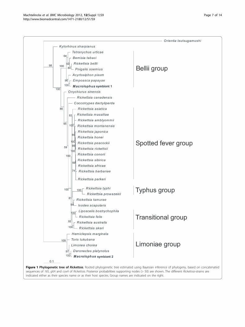
Figure 1 Phylogenetic tree of Rickettsia. Rooted phylogenetic tree estimated using Bayesian inference of phylogeny, based on concatenatedsequences of 16S, gltA and coxA of Rickettsia. Posterior probabilities supporting nodes (> 50) are shown. The different Rickettsia-strains areindicated either as their species name or as their host species. Group names are indicated on the right.
Machtelinckx et al. BMC Microbiology 2012, 12(Suppl 1):S9http://www.biomedcentral.com/1471-2180/12/S1/S9
Page 7 of 14
previously elucidated [29]. The two Rickettsia species arerelated to two different clades. The phylogenetic treeindicated that the first M. pygmaeus Rickettsia endosym-biont is associated with the ‘Bellii’ group, clustering withthe Rickettsia endosymbionts of the two-spotted spidermite Tetranychus urticae, the pea aphid A. pisum andthe tobacco whitefly Bemisia tabaci, among others. Thesecond Rickettsia endosymbiont is situated in the ances-tral ‘Limoniae’ group, clustering with the Rickettsiaendosymbiont of the water beetle Deronectes platynotusand the cranefly Limonia chorea.
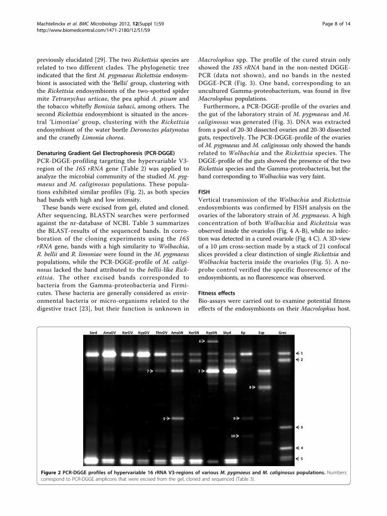
Denaturing Gradient Gel Electrophoresis (PCR-DGGE)PCR-DGGE-profiling targeting the hypervariable V3-region of the 16S rRNA gene (Table 2) was applied toanalyze the microbial community of the studied M. pyg-maeus and M. caliginosus populations. These popula-tions exhibited similar profiles (Fig. 2), as both specieshad bands with high and low intensity.These bands were excised from gel, eluted and cloned.
After sequencing, BLASTN searches were performedagainst the nr-database of NCBI. Table 3 summarizesthe BLAST-results of the sequenced bands. In corro-boration of the cloning experiments using the 16SrRNA gene, bands with a high similarity to Wolbachia,R. bellii and R. limoniae were found in the M. pygmaeuspopulations, while the PCR-DGGE-profile of M. caligi-nosus lacked the band attributed to the bellii-like Rick-ettsia. The other excised bands corresponded tobacteria from the Gamma-proteobacteria and Firmi-cutes. These bacteria are generally considered as envir-onmental bacteria or micro-organisms related to thedigestive tract [23], but their function is unknown in
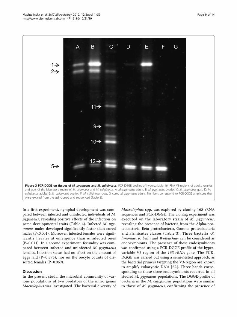
Macrolophus spp. The profile of the cured strain onlyshowed the 18S rRNA band in the non-nested DGGE-PCR (data not shown), and no bands in the nestedDGGE-PCR (Fig. 3). One band, corresponding to anuncultured Gamma-proteobacterium, was found in fiveMacrolophus populations.Furthermore, a PCR-DGGE-profile of the ovaries and
the gut of the laboratory strain of M. pygmaeus and M.caliginosus was generated (Fig. 3). DNA was extractedfrom a pool of 20-30 dissected ovaries and 20-30 dissectedguts, respectively. The PCR-DGGE-profile of the ovariesof M. pygmaeus and M. caliginosus only showed the bandsrelated to Wolbachia and the Rickettsia species. TheDGGE-profile of the guts showed the presence of the twoRickettsia species and the Gamma-proteobacteria, but theband corresponding to Wolbachia was very faint.
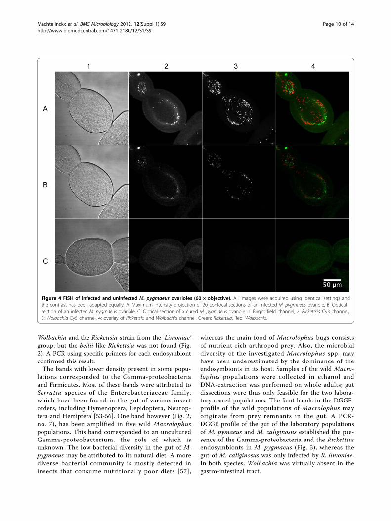
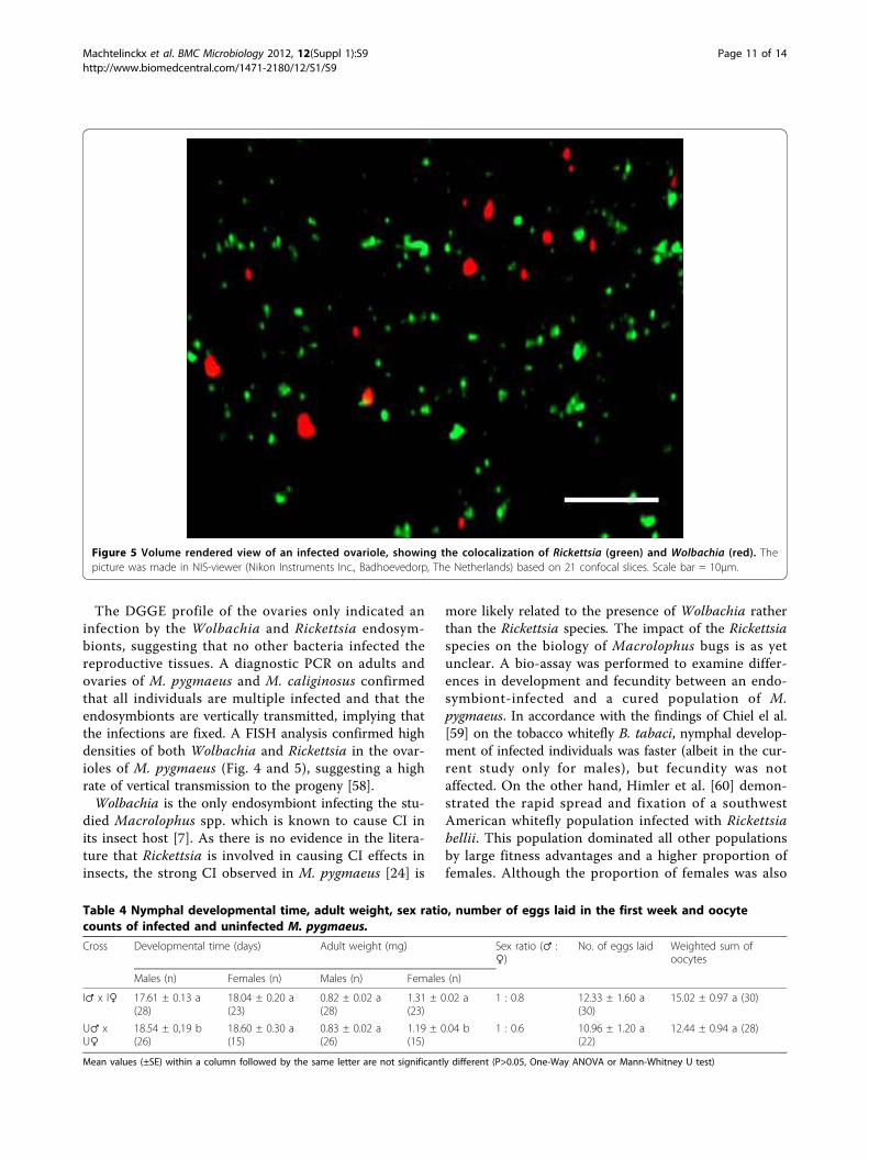
FISHVertical transmission of the Wolbachia and Rickettsiaendosymbionts was confirmed by FISH analysis on theovaries of the laboratory strain of M. pygmaeus. A highconcentration of both Wolbachia and Rickettsia wasobserved inside the ovarioles (Fig. 4 A-B), while no infec-tion was detected in a cured ovariole (Fig. 4 C). A 3D-viewof a 10 µm cross-section made by a stack of 21 confocalslices provided a clear distinction of single Rickettsia andWolbachia bacteria inside the ovarioles (Fig. 5). A no-probe control verified the specific fluorescence of theendosymbionts, as no fluorescence was observed.
Fitness effectsBio-assays were carried out to examine potential fitnesseffects of the endosymbionts on their Macrolophus host.
Figure 2 PCR-DGGE profiles of hypervariable 16 rRNA V3-regions of various M. pygmaeus and M. caliginosus populations. Numberscorrespond to PCR-DGGE amplicons that were excised from the gel, cloned and sequenced (Table 3).
Machtelinckx et al. BMC Microbiology 2012, 12(Suppl 1):S9http://www.biomedcentral.com/1471-2180/12/S1/S9
Page 8 of 14
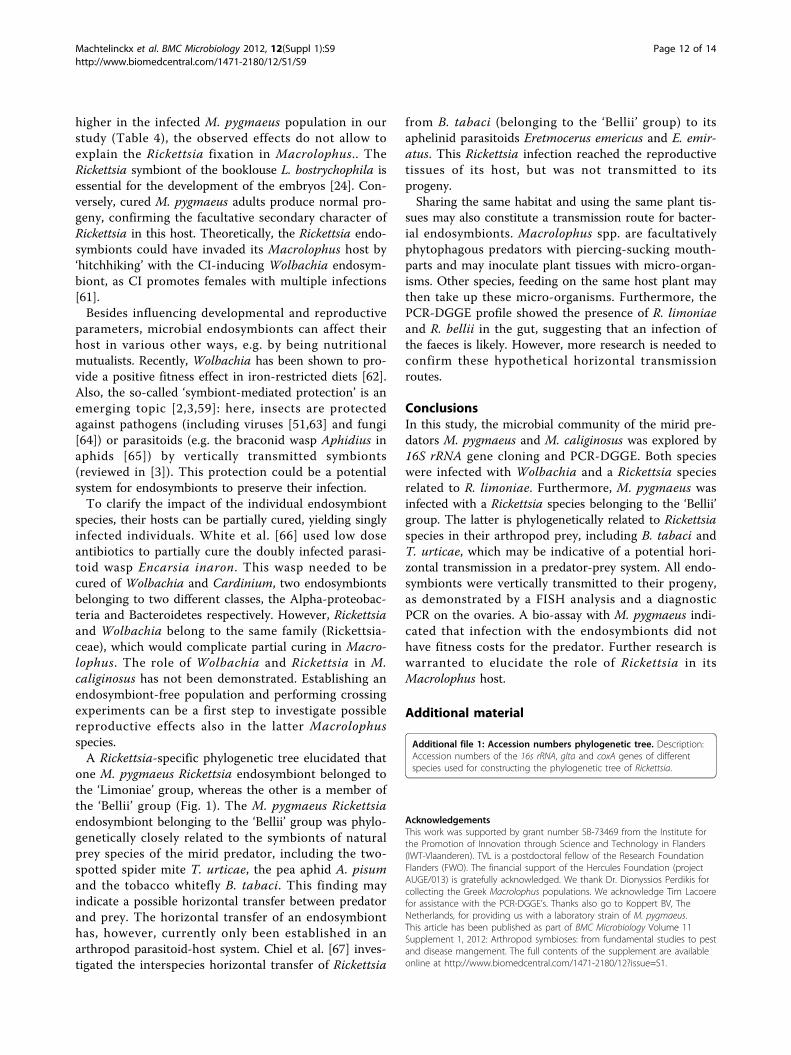
In a first experiment, nymphal development was com-pared between infected and uninfected individuals of M.pygmaeus, revealing positive effects of the infection onsome developmental traits (Table 4). Infected M. pyg-maeus males developed significantly faster than curedmales (P<0.001). Moreover, infected females were signif-icantly heavier at emergence than uninfected ones(P=0.011). In a second experiment, fecundity was com-pared between infected and uninfected M. pygmaeusfemales. Infection status had no effect on the amount ofeggs laid (P=0.575), nor on the oocyte counts of dis-sected females (P=0.069).
DiscussionIn the present study, the microbial community of var-ious populations of two predators of the mirid genusMacrolophus was investigated. The bacterial diversity of
Macrolophus spp. was explored by cloning 16S rRNAsequences and PCR-DGGE. The cloning experiment wasexecuted on the laboratory strain of M. pygmaeus,revealing the presence of bacteria from the Alpha-pro-teobacteria, Beta-proteobacteria, Gamma-proteobacteriaand Firmicutes classes (Table 3). Three bacteria -R.limoniae, R. bellii and Wolbachia- can be considered asendosymbionts. The presence of these endosymbiontswas confirmed using a PCR-DGGE profile of the hyper-variable V3 region of the 16S rRNA gene. The PCR-DGGE was carried out using a semi-nested approach, asthe bacterial primers targeting the V3-region are knownto amplify eukaryotic DNA [52]. Three bands corre-sponding to these three endosymbionts recurred in allstudied M. pygmaeus populations. The DGGE-profile ofbacteria in the M. caliginosus populations were similarto those of M. pygmaeus, confirming the presence of
Figure 3 PCR-DGGE on tissues of M. pygmaeus and M. caliginosus. PCR-DGGE profiles of hypervariable 16 rRNA V3-regions of adults, ovariesand guts of the laboratory strains of M. pygmaeus and M. caliginosus. A: M. pygmaeus adults, B: M. pygmaeus ovaries, C: M. pygmaeus guts, D: M.caliginosus adults, E: M. caliginosus ovaries, F: M. caliginosus guts, G: cured M. pygmaeus adults. Numbers correspond to PCR-DGGE amplicons thatwere excised from the gel, cloned and sequenced (Table 3).
Machtelinckx et al. BMC Microbiology 2012, 12(Suppl 1):S9http://www.biomedcentral.com/1471-2180/12/S1/S9
Page 9 of 14
Wolbachia and the Rickettsia strain from the ‘Limoniae’group, but the bellii-like Rickettsia was not found (Fig.2). A PCR using specific primers for each endosymbiontconfirmed this result.The bands with lower density present in some popu-
lations corresponded to the Gamma-proteobacteriaand Firmicutes. Most of these bands were attributed toSerratia species of the Enterobacteriaceae family,which have been found in the gut of various insectorders, including Hymenoptera, Lepidoptera, Neurop-tera and Hemiptera [53-56]. One band however (Fig. 2,no. 7), has been amplified in five wild Macrolophuspopulations. This band corresponded to an unculturedGamma-proteobacterium, the role of which isunknown. The low bacterial diversity in the gut of M.pygmaeus may be attributed to its natural diet. A morediverse bacterial community is mostly detected ininsects that consume nutritionally poor diets [57],
whereas the main food of Macrolophus bugs consistsof nutrient-rich arthropod prey. Also, the microbialdiversity of the investigated Macrolophus spp. mayhave been underestimated by the dominance of theendosymbionts in its host. Samples of the wild Macro-lophus populations were collected in ethanol andDNA-extraction was performed on whole adults; gutdissections were thus only feasible for the two labora-tory reared populations. The faint bands in the DGGE-profile of the wild populations of Macrolophus mayoriginate from prey remnants in the gut. A PCR-DGGE profile of the gut of the laboratory populationsof M. pymaeus and M. caliginosus established the pre-sence of the Gamma-proteobacteria and the Rickettsiaendosymbionts in M. pygmaeus (Fig. 3), whereas thegut of M. caliginosus was only infected by R. limoniae.In both species, Wolbachia was virtually absent in thegastro-intestinal tract.
Figure 4 FISH of infected and uninfected M. pygmaeus ovarioles (60 x objective). All images were acquired using identical settings andthe contrast has been adapted equally. A: Maximum intensity projection of 20 confocal sections of an infected M. pygmaeus ovariole, B: Opticalsection of an infected M. pygmaeus ovariole, C: Optical section of a cured M. pygmaeus ovariole. 1: Bright field channel, 2: Rickettsia Cy3 channel,3: Wolbachia Cy5 channel, 4: overlay of Rickettsia and Wolbachia channel. Green: Rickettsia, Red: Wolbachia.
Machtelinckx et al. BMC Microbiology 2012, 12(Suppl 1):S9http://www.biomedcentral.com/1471-2180/12/S1/S9
Page 10 of 14
The DGGE profile of the ovaries only indicated aninfection by the Wolbachia and Rickettsia endosym-bionts, suggesting that no other bacteria infected thereproductive tissues. A diagnostic PCR on adults andovaries of M. pygmaeus and M. caliginosus confirmedthat all individuals are multiple infected and that theendosymbionts are vertically transmitted, implying thatthe infections are fixed. A FISH analysis confirmed highdensities of both Wolbachia and Rickettsia in the ovar-ioles of M. pygmaeus (Fig. 4 and 5), suggesting a highrate of vertical transmission to the progeny [58].Wolbachia is the only endosymbiont infecting the stu-
died Macrolophus spp. which is known to cause CI inits insect host [7]. As there is no evidence in the litera-ture that Rickettsia is involved in causing CI effects ininsects, the strong CI observed in M. pygmaeus [24] is
more likely related to the presence of Wolbachia ratherthan the Rickettsia species. The impact of the Rickettsiaspecies on the biology of Macrolophus bugs is as yetunclear. A bio-assay was performed to examine differ-ences in development and fecundity between an endo-symbiont-infected and a cured population of M.pygmaeus. In accordance with the findings of Chiel el al.[59] on the tobacco whitefly B. tabaci, nymphal develop-ment of infected individuals was faster (albeit in the cur-rent study only for males), but fecundity was notaffected. On the other hand, Himler et al. [60] demon-strated the rapid spread and fixation of a southwestAmerican whitefly population infected with Rickettsiabellii. This population dominated all other populationsby large fitness advantages and a higher proportion offemales. Although the proportion of females was also
Figure 5 Volume rendered view of an infected ovariole, showing the colocalization of Rickettsia (green) and Wolbachia (red). Thepicture was made in NIS-viewer (Nikon Instruments Inc., Badhoevedorp, The Netherlands) based on 21 confocal slices. Scale bar = 10µm.
Table 4 Nymphal developmental time, adult weight, sex ratio, number of eggs laid in the first week and oocytecounts of infected and uninfected M. pygmaeus.
Cross Developmental time (days) Adult weight (mg) Sex ratio (♂ :♀)
No. of eggs laid Weighted sum ofoocytes
Males (n) Females (n) Males (n) Females (n)
I♂ x I♀ 17.61 ± 0.13 a(28)
18.04 ± 0.20 a(23)
0.82 ± 0.02 a(28)
1.31 ± 0.02 a(23)
1 : 0.8 12.33 ± 1.60 a(30)
15.02 ± 0.97 a (30)
U♂ xU♀
18.54 ± 0,19 b(26)
18.60 ± 0.30 a(15)
0.83 ± 0.02 a(26)
1.19 ± 0.04 b(15)
1 : 0.6 10.96 ± 1.20 a(22)
12.44 ± 0.94 a (28)
Mean values (±SE) within a column followed by the same letter are not significantly different (P>0.05, One-Way ANOVA or Mann-Whitney U test)
Machtelinckx et al. BMC Microbiology 2012, 12(Suppl 1):S9http://www.biomedcentral.com/1471-2180/12/S1/S9
Page 11 of 14
higher in the infected M. pygmaeus population in ourstudy (Table 4), the observed effects do not allow toexplain the Rickettsia fixation in Macrolophus.. TheRickettsia symbiont of the booklouse L. bostrychophila isessential for the development of the embryos [24]. Con-versely, cured M. pygmaeus adults produce normal pro-geny, confirming the facultative secondary character ofRickettsia in this host. Theoretically, the Rickettsia endo-symbionts could have invaded its Macrolophus host by‘hitchhiking’ with the CI-inducing Wolbachia endosym-biont, as CI promotes females with multiple infections[61].Besides influencing developmental and reproductive
parameters, microbial endosymbionts can affect theirhost in various other ways, e.g. by being nutritionalmutualists. Recently, Wolbachia has been shown to pro-vide a positive fitness effect in iron-restricted diets [62].Also, the so-called ‘symbiont-mediated protection’ is anemerging topic [2,3,59]: here, insects are protectedagainst pathogens (including viruses [51,63] and fungi[64]) or parasitoids (e.g. the braconid wasp Aphidius inaphids [65]) by vertically transmitted symbionts(reviewed in [3]). This protection could be a potentialsystem for endosymbionts to preserve their infection.To clarify the impact of the individual endosymbiont
species, their hosts can be partially cured, yielding singlyinfected individuals. White et al. [66] used low doseantibiotics to partially cure the doubly infected parasi-toid wasp Encarsia inaron. This wasp needed to becured of Wolbachia and Cardinium, two endosymbiontsbelonging to two different classes, the Alpha-proteobac-teria and Bacteroidetes respectively. However, Rickettsiaand Wolbachia belong to the same family (Rickettsia-ceae), which would complicate partial curing in Macro-lophus. The role of Wolbachia and Rickettsia in M.caliginosus has not been demonstrated. Establishing anendosymbiont-free population and performing crossingexperiments can be a first step to investigate possiblereproductive effects also in the latter Macrolophusspecies.A Rickettsia-specific phylogenetic tree elucidated that
one M. pygmaeus Rickettsia endosymbiont belonged tothe ‘Limoniae’ group, whereas the other is a member ofthe ‘Bellii’ group (Fig. 1). The M. pygmaeus Rickettsiaendosymbiont belonging to the ‘Bellii’ group was phylo-genetically closely related to the symbionts of naturalprey species of the mirid predator, including the two-spotted spider mite T. urticae, the pea aphid A. pisumand the tobacco whitefly B. tabaci. This finding mayindicate a possible horizontal transfer between predatorand prey. The horizontal transfer of an endosymbionthas, however, currently only been established in anarthropod parasitoid-host system. Chiel et al. [67] inves-tigated the interspecies horizontal transfer of Rickettsia
from B. tabaci (belonging to the ‘Bellii’ group) to itsaphelinid parasitoids Eretmocerus emericus and E. emir-atus. This Rickettsia infection reached the reproductivetissues of its host, but was not transmitted to itsprogeny.Sharing the same habitat and using the same plant tis-
sues may also constitute a transmission route for bacter-ial endosymbionts. Macrolophus spp. are facultativelyphytophagous predators with piercing-sucking mouth-parts and may inoculate plant tissues with micro-organ-isms. Other species, feeding on the same host plant maythen take up these micro-organisms. Furthermore, thePCR-DGGE profile showed the presence of R. limoniaeand R. bellii in the gut, suggesting that an infection ofthe faeces is likely. However, more research is needed toconfirm these hypothetical horizontal transmissionroutes.
ConclusionsIn this study, the microbial community of the mirid pre-dators M. pygmaeus and M. caliginosus was explored by16S rRNA gene cloning and PCR-DGGE. Both specieswere infected with Wolbachia and a Rickettsia speciesrelated to R. limoniae. Furthermore, M. pygmaeus wasinfected with a Rickettsia species belonging to the ‘Bellii’group. The latter is phylogenetically related to Rickettsiaspecies in their arthropod prey, including B. tabaci andT. urticae, which may be indicative of a potential hori-zontal transmission in a predator-prey system. All endo-symbionts were vertically transmitted to their progeny,as demonstrated by a FISH analysis and a diagnosticPCR on the ovaries. A bio-assay with M. pygmaeus indi-cated that infection with the endosymbionts did nothave fitness costs for the predator. Further research iswarranted to elucidate the role of Rickettsia in itsMacrolophus host.
Additional material
Additional file 1: Accession numbers phylogenetic tree. Description:Accession numbers of the 16s rRNA, glta and coxA genes of differentspecies used for constructing the phylogenetic tree of Rickettsia.
AcknowledgementsThis work was supported by grant number SB-73469 from the Institute forthe Promotion of Innovation through Science and Technology in Flanders(IWT-Vlaanderen). TVL is a postdoctoral fellow of the Research FoundationFlanders (FWO). The financial support of the Hercules Foundation (projectAUGE/013) is gratefully acknowledged. We thank Dr. Dionyssios Perdikis forcollecting the Greek Macrolophus populations. We acknowledge Tim Lacoerefor assistance with the PCR-DGGE’s. Thanks also go to Koppert BV, TheNetherlands, for providing us with a laboratory strain of M. pygmaeus.This article has been published as part of BMC Microbiology Volume 11Supplement 1, 2012: Arthropod symbioses: from fundamental studies to pestand disease mangement. The full contents of the supplement are availableonline at http://www.biomedcentral.com/1471-2180/12?issue=S1.
Machtelinckx et al. BMC Microbiology 2012, 12(Suppl 1):S9http://www.biomedcentral.com/1471-2180/12/S1/S9
Page 12 of 14
Author details1Laboratory of Agrozoology, Department of Crop Protection, Faculty ofBioscience Engineering, Ghent University, Ghent, Belgium. 2Laboratory ofMicrobial Ecology and Technology (LabMET), Faculty of BioscienceEngineering, Ghent University, Gent, Belgium. 3Laboratory of Bio-imagingand Cytometry, Department of Molecular Biotechnology, Faculty ofBioscience Engineering, Ghent University, Gent, Belgium. 4Center for Nano-and Biophotonics (NB-Photonics), Ghent University, Gent, Belgium. 5InstitutoMurciano de Investigación y Desarrollo Agrario y Alimentario (IMIDA),Departamento de Biotecnología y Protección de Cultivos, La Alberca, Murcia,Spain. 6AGRIS Sardegna - DIRVE, Cagliari, Italy. 7Laboratory of AppliedMolecular Genetics, Department of Molecular Biotechnology, Faculty ofBioscience Engineering, Ghent University, Ghent, Belgium.
Authors’ contributionsTM performed the experiments and wrote the manuscript. TM, TVL and PDCdesigned the experiments. TVDW and NB helped with the PCR-DGGEexperiments. JAS and MN collected Macrolophus bugs in Spain and Italy,respectively. WDV helped with the FISH experiments. TVL, TVDW, GG andPDC revised the manuscript. All authors read and approved the finalmanuscript.
Competing interestsThe authors declare that they have no competing interests.
Published: 18 January 2012
References1. Douglas AE: Nutritional interactions in insect-microbial symbioses: aphids
and their symbiotic bacteria Buchnera. Annu Rev Entomol 1998, 43:17-37.2. Gross R, Vavre F, Heddi A, Hurst GDD, Zchori-Fein E, Bourtzis K: Immunity
and symbiosis. Molecular Microbiology 2009, 73(5):751-759.3. Brownlie JC, Johnson KN: Symbiont-mediated protection in insect hosts.
Trends in Microbiology 2009, 17(8):348-354.4. Werren JH: Biology of Wolbachia. Annu Rev Entomol 1997, 42:587-609.5. Werren JH, O’Neill SL: The evolution of heritable symbionts. In Influential
Passengers: Inherited Microorganisms and Arthropod Reproduction. New York:Oxford University Press;O’Neill SL, Hoffmann AA, Werren JH 1997:1-41.
6. Hilgenboecker K, Hammerstein P, Schlattmann P, Telschow A, Werren JH:How many species are infected with Wolbachia? A statistical analysis ofcurrent data. FEMS Microbiol Lett 2008, 281(2):215-220.
7. Stouthamer R, Breeuwer JAJ, Hurst GDD: Wolbachia pipientis: microbialmanipulator of arthropod reproduction. Annu Rev Microbiol 1999, 53:71-102.
8. Stevens L, Giordano R, Fialho RF: Male-killing, nematode infections,bacteriophage infection, and virulence of cytoplasmic bacteria in thegenus Wolbachia. Annu Rev Ecol Syst 2001, 32:519-545.
9. Stouthamer R, Luck RF, Hamilton WD: Antibiotics cause parthenogeneticTrichogramma (Hymenoptera/Trichogrammatidae) to revert to sex. ProcNatl Acad Sci U S A 1990, 87(7):2424-2427.
10. Rousset F, Bouchon D, Pintureau B, Juchault P, Solignac M: Wolbachiaendosymbionts responsible for various alterations of sexuality inarthropods. Proc Biol Sci 1992, 250(1328):91-98.
11. Dedeine F, Vavre F, Fleury F, Loppin B, Hochberg ME, Bouletreau M:Removing symbiotic Wolbachia bacteria specifically inhibits oogenesis ina parasitic wasp. Proc Natl Acad Sci U S A 2001, 98(11):6247-6252.
12. Zchori-Fein E, Perlman SJ: Distribution of the bacterial symbiontCardinium in arthropods. Mol Ecol 2004, 13(7):2009-2016.
13. Zchori-Fein E, Perlman SJ, Kelly SE, Katzir N, Hunter MS: Characterization ofa ‘Bacteroidetes’ symbiont in Encarsia wasps (Hymenoptera: Aphelinidae):proposal of ‘Candidatus Cardinium hertigii’. Int J Syst Evol Microbiol 2004,54:961-968.
14. Gotoh T, Noda H, Ito S: Cardinium symbionts cause cytoplasmicincompatibility in spider mites. Heredity 2007, 98(1):13-20.
15. Skaljac M, Zanic K, Ban SG, Kontsedalov S, Ghanim M: Co-infection andlocalization of secondary symbionts in two whitefly species. BMCMicrobiol 2010, 10:15.
16. Perlman SJ, Hunter MS, Zchori-Fein E: The emerging diversity of Rickettsia.Proc Biol Sci 2006, 273(1598):2097-2106.
17. Davis MJ, Ying Z, Brunner BR, Pantoja A, Ferwerda FH: Rickettsial relativeassociated with papaya bunchy top disease. Curr Microbiol 1998,36(2):80-84.
18. Weinert LA, Werren JH, Aebi A, Stone GN, Jiggins FM: Evolution anddiversity of Rickettsia bacteria. BMC Biol 2009, 7:15.
19. Werren JH, Hurst GDD, Zhang W, Breeuwer JAJ, Stouthamer R,Majerus MEN: Rickettsial relative associated with male killing in theladybird beetle (Adalia bipunctata). J Bacteriol 1994, 176(2):388-394.
20. Majerus MEN, Hinrich J, Schulenburg GVD, Zakharov IA: Multiple causes ofmale-killing in a single sample of the two-spot ladybird, Adaliabipunctata (Coleoptera: Coccinellidae) from Moscow. Heredity 2000,84(5):605-609.
21. Lawson ET, Mousseau TA, Klaper R, Hunter MD, Werren JH: Rickettsiaassociated with male-killing in a buprestid beetle. Heredity 2001,86:497-505.
22. Hagimori T, Abe Y, Date S, Miura K: The first finding of a Rickettsiabacterium associated with parthenogenesis induction among insects.Curr Microbiol 2006, 52(2):97-101.
23. Giorgini M, Bernardo U, Monti MM, Nappo AG, Gebiola M: Rickettsiasymbionts cause parthenogenetic reproduction in the parasitoid waspPnigalio soemius (Hymenoptera: Eulophidae). Appl Environ Microbiol 2010,76(8):2589-2599.
24. Perotti MA, Clarke HK, Turner BD, Braig HR: Rickettsia as obligate andmycetomic bacteria. Faseb J 2006, 20(13):2372-+.
25. Floate KD, Kyei-Poku GK, Coghlin PC: Overview and relevance ofWolbachia bacteria in biocontrol research. Biocontrol Science andTechnology 2006, 16(8):767-788.
26. Schaefer CW, Panizzi AR: Heteroptera of Economic Importance. BocaRaton, USA: CRC Press; 2000.
27. Perdikis D, Lykouressis D: Effects of various items, host plants, andtemperatures on the development and survival of Macrolophuspygmaeus Rambur (Hemiptera: Miridae). Biol Control 2000, 17(1):55-60.
28. Vandekerkhove B, Van Baal E, Bolckmans K, De Clercq P: Effect of diet andmating status on ovarian development and oviposition in thepolyphagous predator Macrolophus caliginosus (Heteroptera: Miridae).Biol Control 2006, 39(3):532-538.
29. Machtelinckx T, Van Leeuwen T, Vanholme B, Gehesquiere B, Dermauw W,Vandekerkhove B, Gheysen G, De Clercq P: Wolbachia induces strongcytoplasmic incompatibility in the predatory bug Macrolophuspygmaeus. Insect Mol Biol 2009, 18(3):373-381.
30. Muyzer G, Dewaal EC, Uitterlinden AG: Profiling of complex microbialpopulations by denaturing gradient gel electrophoresis analysis ofpolymerase chain reaction amplified-genes coding for 16S rRNA. ApplEnviron Microbiol 1993, 59(3):695-700.
31. Torsvik V, Daae FL, Sandaa RA, Ovreas L: Novel techniques for analysingmicrobial diversity in natural and perturbed environments. Journal ofBiotechnology 1998, 64(1):53-62.
32. Marzorati M, Alma A, Sacchi L, Pajoro M, Palermo S, Brusetti L, Raddadi N,Balloi A, Tedeschi R, Clementi E, et al: A novel Bacteroidetes symbiont islocalized in Scaphoideus titanus, the insect vector of Flavescence Doréein Vitis vinifera. Appl Environ Microbiol 2006, 72(2):1467-1475.
33. Gottlieb Y, Ghanim M, Chiel E, Gerling D, Portnoy V, Steinberg S, Tzuri G,Horowitz AR, Belausov E, Mozes-Daube N, et al: Identification andlocalization of a Rickettsia sp in Bemisia tabaci (Homoptera: Aleyrodidae).Appl Environ Microbiol 2006, 72(5):3646-3652.
34. Zouache K, Voronin D, Tran-Van V, Mavingui P: Composition of bacterialcommunities associated with natural and laboratory populations ofAsobara tabida infected with Wolbachia. Appl Environ Microbiol 2009,75(11):3755-3764.
35. Martinez-Cascales JI, Cenis JL, Cassis G, Sanchez JA: Species identity ofMacrolophus melanotoma (Costa 1853) and Macrolophus pygmaeus(Rambur 1839) (Insecta: Heteroptera: Miridae) based on morphologicaland molecular data and bionomic implications. Insect Syst Evol 2006,37(4):385-404.
36. Rozen S, Skaletsky H: Primer3 on the WWW for general users and forbiologist programmers. In Bioinformatics Methods and Protocols: Methods inMolecular Biology. NJ: Humana Press;Krawetz S, Misener S. Totowa2000:365-386.
37. Altschul SF, Madden TL, Schaffer AA, Zhang JH, Zhang Z, Miller W,Lipman DJ: Gapped BLAST and PSI-BLAST: a new generation of proteindatabase search programs. Nucleic Acids Res 1997, 25(17):3389-3402.
38. Hall TA: BioEdit: a user-friendly biological sequence alignment editor andanalysis program for Windows 95/98/NT. Nucleic Acids Symposium Series1999, 41:95-98.
Machtelinckx et al. BMC Microbiology 2012, 12(Suppl 1):S9http://www.biomedcentral.com/1471-2180/12/S1/S9
Page 13 of 14

39. Huelsenbeck JP, Ronquist F: MRBAYES: Bayesian inference of phylogenetictrees. Bioinformatics 2001, 17(8):754-755.
40. Nylander JAA: MrModeltest v2. Program distributed by the author.Evolutionary Biology Centre, Uppsala University. 2004.
41. Page RD: TreeView: an application to display phylogenetic trees onpersonal computers. Comput Appl Biosci 1996, 12(4):357-358.
42. Janse I, Bok J, Zwart G: A simple remedy against artifactual double bandsin denaturing gradient gel electrophoresis. Journal of MicrobiologicalMethods 2004, 57(2):279-281.
43. Weisburg WG, Barns SM, Pelletier DA, Lane DJ: 16S Ribosomal DNAamplification for phylogenetic study. J Bacteriol 1991, 173(2):697-703.
44. Frank JA, Reich CI, Sharma S, Weisbaum JS, Wilson BA, Olsen GJ: Criticalevaluation of two primers commonly used for amplification of bacterial16S rRNA genes. Appl Environ Microbiol 2008, 74(8):2461-2470.
45. Crotti E, Damiani C, Pajoro M, Gonella E, Rizzi A, Ricci I, Negri I, Scuppa P,Rossi P, Ballarini P, et al: Asaia, a versatile acetic acid bacterial symbiont,capable of cross-colonizing insects of phylogenetically distant generaand orders. Environ Microbiol 2009, 11(12):3252-3264.
46. Heddi A, Grenier AM, Khatchadourian C, Charles H, Nardon P: Fourintracellular genomes direct weevil biology: Nuclear, mitochondrial,principal endosymbiont, and Wolbachia. Proc Natl Acad Sci U S A 1999,96(12):6814-6819.
47. Moreira LA, Iturbe-Ormaetxe I, Jeffery JA, Lu GJ, Pyke AT, Hedges LM,Rocha BC, Hall-Mendelin S, Day A, Riegler M, et al: A Wolbachia symbiontin Aedes aegypti limits infection with Dengue, Chikungunya, andPlasmodium. Cell 2009, 139(7):1268-1278.
48. Vandekerkhove B, Parmentier L, Van Stappen G, Grenier S, Febvay G, Rey M,De Clercq P: Artemia cysts as an alternative food for the predatory bugMacrolophus pygmaeus. J Appl Entomol 2009, 133(2):133-142.
49. SPSS: User’s Guide, version 17.0. Chicago, IL: SPSS Inc; 2008.50. Lykouressis D, Giatropoulos A, Perdikis D, Favas C: Assessing the suitability
of noncultivated plants and associated insect prey as food sources forthe omnivorous predator Macrolophus pygmaeus (Hemiptera: Miridae).Biol Control 2008, 44(2):142-148.
51. Perdikis D, Favas C, Lykouressis D, Fantinou A: Ecological relationshipsbetween non-cultivated plants and insect predators in agroecosystems:the case of Dittrichia viscosa (Asteraceae) and Macrolophus melanotoma(Hemiptera : Miridae). Acta Oecologica-International Journal of Ecology 2007,31(3):299-306.
52. Lopez I, Ruiz-Larrea F, Cocolin L, Orr E, Phister T, Marshall M,VanderGheynst J, Mills DA: Design and evaluation of PCR primers foranalysis of bacterial populations in wine by denaturing gradient gelelectrophoresis. Appl Environ Microbiol 2003, 69(11):6801-6807.
53. Graham RI, Zahner V, Lucarotti CJ: An intracellular symbiont and othermicrobiota associated with field-collected populations of sawflies(Hymenoptera : Symphyta). Can J Microbiol 2008, 54(9):758-768.
54. Broderick NA, Raffa KF, Goodman RM, Handelsman J: Census of thebacterial community of the gypsy moth larval midgut by using culturingand culture-independent methods. Appl Environ Microbiol 2004,70(1):293-300.
55. Dunn AK, Stabb EV: Culture-independent characterization of themicrobiota of the ant lion Myrmeleon mobilis (Neuroptera :Myrmeleontidae). Appl Environ Microbiol 2005, 71(12):8784-8794.
56. Oliver KM, Moran NA, Hunter MS: Costs and benefits of a superinfectionof facultative symbionts in aphids. Proc Biol Sci 2006,273(1591):1273-1280.
57. Cohen AC: Microbes in the diet setting. In Insect diets: science andtechnology. Boca Raton: CRC Press;Cohen AC 2004:225-248.
58. Ferree PM, Frydman HM, Li JM, Cao J, Wieschaus E, Sullivan W: Wolbachiautilizes host microtubules and Dynein for anterior localization in theDrosophila oocyte. PLoS Pathog 2005, 1(2):e14.
59. Chiel E, Inbar M, Mozes-Daube N, White JA, Hunter MS, Zchori-Fein E:Assessments of fitness effects by the facultative symbiont Rickettsia inthe sweetpotato whitefly (Hemiptera: Aleyrodidae). Ann Entomol Soc Am2009, 102(3):413-418.
60. Himler AG, Adachi-Hagimori T, Bergen JE, Kozuch A, Kelly SE, Tabashnik BE,Chiel E, Duckworth VE, Dennehy TJ, Zchori-Fein E, et al: Rapid spread of abacterial symbiont in an invasive whitefly is driven by fitness benefitsand female bias. Science 2011, 332(6026):254-256.
61. Vautrin E, Vavre F: Interactions between vertically transmitted symbionts:cooperation or conflict? Trends in Microbiology 2009, 17(3):95-99.
62. Brownlie JC, Cass BN, Riegler M, Witsenburg JJ, Iturbe-Ormaetxe I,McGraw EA, O’Neill SL: Evidence for metabolic provisioning by acommon invertebrate endosymbiont, Wolbachia pipientis, during periodsof nutritional stress. PLoS Pathog 2009, 5(4):e1000368.
63. Teixeira L, Ferreira A, Ashburner M: The bacterial symbiont Wolbachiainduces resistance to RNA viral infections in Drosophila melanogaster.PLoS Biol 2008, 6(12):e2.
64. Scarborough CL, Ferrari J, Godfray HCJ: Aphid protected from pathogenby endosymbiont. Science 2005, 310(5755):1781-1781.
65. Moran NA, Dunbar HE: Sexual acquisition of beneficial symbionts inaphids. Proc Natl Acad Sci U S A 2006, 103(34):12803-12806.
66. White JA, Kelly SE, Perlman SJ, Hunter MS: Cytoplasmic incompatibility inthe parasitic wasp Encarsia inaron: disentangling the roles of Cardiniumand Wolbachia symbionts. Heredity 2009, 102(5):483-489.
67. Chiel E, Zchori-Fein E, Inbar M, Gottlieb Y, Adachi-Hagimori T, Kelly SE,Asplen MK, Hunter MS: Almost there: transmission routes of bacterialsymbionts between trophic levels. PLoS ONE 2009, 4(3):e4767.
68. Simon C, Frati F, Beckenbach A, Crespi B, Liu H, Flook P: Evolution,weighting, and phylogenetic utility of mitochondrial gene sequencesand a compilation of conserved polymerase chain reaction primers. AnnEntomol Soc Am 1994, 87(6):651-701.
69. Relman DA, Schmidt TM, Macdermott RP, Falkow S: Identification of theuncultured Bacillus of Whipple’s disease. New England Journal of Medicine1992, 327(5):293-301.
70. Zhou W, Rousset F, O’Neill S: Phylogeny and PCR-based classification ofWolbachia strains using wsp gene sequences. Proc Biol Sci 1998,265(1395):509-515.
doi:10.1186/1471-2180-12-S1-S9Cite this article as: Machtelinckx et al.: Microbial community ofpredatory bugs of the genus Macrolophus (Hemiptera: Miridae). BMCMicrobiology 2012 12(Suppl 1):S9.
Submit your next manuscript to BioMed Centraland take full advantage of:
• Convenient online submission
• Thorough peer review
• No space constraints or color figure charges
• Immediate publication on acceptance
• Inclusion in PubMed, CAS, Scopus and Google Scholar
• Research which is freely available for redistribution
Submit your manuscript at www.biomedcentral.com/submit
Machtelinckx et al. BMC Microbiology 2012, 12(Suppl 1):S9http://www.biomedcentral.com/1471-2180/12/S1/S9
Page 14 of 14