research open access renylaima capensis, a brachylaimid trematode
TRANSCRIPT

Sirgel et al. Parasites & Vectors 2012, 5:169http://www.parasitesandvectors.com/content/5/1/169
RESEARCH Open Access
Life cycle of Renylaima capensis, a brachylaimidtrematode of shrews and slugs in South Africa:two-host and three-host transmission modalitiessuggested by epizootiology and DNA sequencingWilhelm F Sirgel1, Patricio Artigas2, M Dolores Bargues2 and Santiago Mas-Coma2*
Abstract
Background: The life cycle of the brachylaimid trematode species Renylaima capensis, infecting the urinary systemof the shrew Myosorex varius (Mammalia: Soricidae: Crocidosoricinae) in the Hottentots Holland Nature Reserve,South Africa, has been elucidated by a study of its larval stages, epizootiological data in local snails and mammalsduring a 34-year period, and its verification with mtDNA sequencing.
Methods: Parasites obtained from dissected animals were mounted in microscope slides for the parasitologicalstudy and measured according to standardized methods. The mitochondrial DNA cox1 gene was sequenced by thedideoxy chain-termination method.
Results: The slugs Ariostralis nebulosa and Ariopelta capensis (Gastropoda: Arionidae) act as specific first and secondintermediate hosts, respectively. Branched sporocysts massively develop in A. nebulosa. Intrasporocystic maturecercariae show differentiated gonads, male terminal duct, ventral genital pore, and usually no tail, opposite toBrachylaimidae in which mature cercariae show a germinal primordium and small tail. Unencysted metacercariae,usually brevicaudate, infect the kidney of A. capensis and differ from mature cercariae by only a slightly greater size.The final microhabitats are the kidneys and ureters of the shrews, kidney pelvis and calyces in light infections andalso kidney medulla and cortex in heavy infections. Sporocysts, cercariae, metacercariae and adults proved tobelong to R. capensis by analysis of a 437-bp-long cox1 fragment, which was identical except for three mutations inmetacercariae, of which only one silent. Epizootiological studies showed usual sporocyst infection in A. nebulosaand very rare metacercarial infection in A. capensis, which does not agree with high prevalences and intensities inthe shrews.
Conclusions: The presence of monotesticular adult forms and larval prevalences and intensities observed suggestthat R. capensis may use two transmission strategies, a two-host life cycle by predation of A. nebulosa harbouringintrasporocystic cercariae may be the normal pattern, whereas a second mollusc host is just starting to beintroduced. In shrews, a tissue-traversing, intraorganic migration followed by an interorganic migration to reach andpenetrate the outer surface of either of both kidneys should occur. For first slug infection, the fluke takes advantageof the phenomenon that M. varius always urinate during defaecation. Consequently, in Brachylaimidae, the secondintermediate mollusc host should evolutionarily be seen as a last addition to the cycle and their present adult stagemicrohabitat restricted to digestive tract and related organs as a loss of the tissue-traversing capacity of themetacercaria.
* Correspondence: [email protected] de Parasitología, Facultad de Farmacia, Universidad deValencia, Av. Vicente Andrés Estellés s/n, 46100 Burjassot - Valencia, SpainFull list of author information is available at the end of the article
© 2012 Sirgel et al.; licensee BioMed Central Ltd. This is an Open Access article distributed under the terms of the CreativeCommons Attribution License (http://creativecommons.org/licenses/by/2.0), which permits unrestricted use, distribution, andreproduction in any medium, provided the original work is properly cited.

Sirgel et al. Parasites & Vectors 2012, 5:169 Page 2 of 18http://www.parasitesandvectors.com/content/5/1/169
BackgroundBrachylaimidae are an interesting family of trematodesdue to their numerous species as well as their worldwidepresence and frequency. Brachylaimids are also of med-ical importance as they cause diseases in humans, whichcan even result in mortality [1,2]. In the veterinary field,they affect domestic animals [3], poultry [4-8] and wildgame birds [9,10]. They have also proved to be useful asbiological tags in studies of postfire ecosystem regener-ation processes [11,12]. Additionally, Brachylaimidae arebiologically peculiar. While the great majority of digen-ean trematodes follow an aquatic life cycle [13], brachy-laimids are one of the very few trematode groups (theonly other one is Dicrocoeliidae) that have succeeded incolonizing the terrestrial milieu, in some instances evenvery arid, xerophilic habitats [14].Brachylaimid species of the three subfamilies Brachyla-
minae, Ityogoniminae and Panopistinae [15] share thesame three-host life cycle pattern including (i) a terres-trial gastropod snail (Pulmonata: Stylommatophora) asfirst intermediate host, (ii) an additional mollusc individ-ual belonging to another (or sometimes the same) ter-restrial gastropod species as second intermediate host,and (iii) an endothermic vertebrate as definitive host[13,14,16-18].The first snail host is very specific, as brachylaimid
species use only one or a very small number of closelyrelated mollusc species inhabiting the local area[14,16,17]. This mollusc becomes infected by eatingembryonated fluke eggs shed in faeces by the definitivehost. The first sporocystogenous sporocyst generationdevelops in the hepatopancreas [19-22]. This earlymother sporocyst phase constitutes the only plathyhel-minth stage known to develop inside a host cell [23].There are no rediae in brachylaimids, so that cercariaeare directly produced inside second sporocyst genera-tions. These cercariogenous sporocysts develop primarilyin the hepatopancreas, although other secondary loca-tions become invaded by these sporocysts in massiveinfections [24], and show a very characteristic branchedmorphology [25]. Cercariae are brevicaudate (similar tothe microcercous type of aquatic cercariae but lacking astylet) and are shed by the first intermediate snail hostunder rainy conditions or high humidity, so that eitherwater from rain or snail individuals cohabiting underclose contact (as under big stones) ensure the survival ofthe free and shortliving cercaria thus enabling transmis-sion to a second intermediate snail host [26-28].The second snail host appears to be less specific. It usu-
ally belongs to another terrestrial snail species sharing thesame habitat with the first one. Sometimes it may even beother individuals of the first host species. Cercariae pene-trate through orifices of the body and migrate to the intra-molluscan microhabitat where they will develop into an
unencysted metacercarial stage. The microhabitat of meta-cercariae appears to be very specific [14,16,17]. The loca-tion of metacercariae is the kidney for most of thebrachylaimid species, although pericardial cavity andrarely pedal glands are microhabitats for a few species.Definitive hosts of brachylaimids are homeothermic
mammals and birds normally feeding on snails or acci-dentally ingesting them with other food (i.e., in pigs)[15]. Many brachylaimid species show a clearly restrictedspectrum of closely related definitive host species (orsometimes even a single one), suggesting a phylogeneticspecificity, as is the case of species infecting mammals ofthe Suborder Soricomorpha (shrews Soricidae and molesTalpidae). Within mammals, insectivores, rodents andmarsupials are the main definitive hosts. Artiodactyls(pigs Suidae), carnivores and lagomorphs are only sec-ondarily captured hosts.In Brachylaimidae, definitive hosts other than mam-
mals and birds are very rare. Another subfamily Zeyla-nurotrematinae, suggestedly close to Panopistinae, hasbeen proposed within Brachylaimidae to include flukesinfecting reptiles and amphibians [29]. Even family rank(Zeylanurotrematidae) has recently been proposed forthem [30], although the systematic position of theseflukes has remained incertae sedis in the most recent re-view of the trematode group [31]. Another rare, mono-testicular species, Parabrachylaima euglandensis, whoseadult stage progenetically develops in terrestrial snails ofLousiana, USA [32], has also recently been retainedwithin Brachylaimidae [33].The adult stage of brachylaimids is intestinal, includ-
ing species which are haematophagous [16]. Only a veryfew species present an adult stage infecting other partsof the digestive tract or organs directly related or con-nected to the digestive tract: Brachylaima oesophageiand B. fulvus in the oesophagus and stomach [34,35],Scaphiostomum species in ducts of liver and pancreas[36], and Dollfusinus frontalis in nasal and frontalsinuses [14,37]. Exceptions are the lumen of the kidneysac as final microhabitat for Parabrachylaima [32], andthe urinary system for species of Zeylanurotrema[29,38], although a species of the latter, Z. sphenomorphi,has been described from the host’s intestine [30].Recently, a new curious brachylaimid species was
described based on adult trematode specimens found inthe kidneys and ureters of the forest shrew Myosorex var-ius (Smuts, 1832) (Insectivora: Soricidae: Crocidosorici-nae) from a restricted, very damp area of the HottentotsHolland Mountain range, near Cape town, South Africa.Peculiar characteristics such as the absence of a cirruspouch and cirrus, the presence of a genital atrium thatcan be evaginated to produce a prominent ventral exten-tion of the body, and a surprising 11.1% of monotesticularforms due to the lack of an anterior testis, as well as the

Sirgel et al. Parasites & Vectors 2012, 5:169 Page 3 of 18http://www.parasitesandvectors.com/content/5/1/169
fact that the urinary system of a mammal host is an extra-ordinary microhabitat for a trematode, indicated that thisdigenean represents a new genus and species for whichthe name Renylaima capensis was proposed [39]. Thepresent article aims to describe the elucidation of its lifecycle and transmission by means of the parasitologicalstudy of its larval stages, its epizootiological data in localsnail and shrew hosts, and its verification with mtDNAmarker sequencing.
MethodsHost materialsSince the year 1978, shrews have been trapped(Figure 1A) and land snails hand collected (Figure 1B,C) from a moist habitat (34° 02′ 48.500 S; 18° 59′ 11.600
E) of the Hottentots Holland Nature Reserve, about60 km to the east of Cape town and thus in the mostsouthern part of South Africa in what is now called theWestern Province of the Republic of South Africa.Trematode adults and larval stages were collected afterhost dissection under a stereomicroscope.All the infected slugs and shrews were collected in an
area limited to about 1000 m2 near the top end of a nar-row ravine called Landdroskloof, on the western slopesof the Hottentots Holland Mountains. The locality is at
Figure 1 Environment and hosts of Renylaima capensis. A) drawing sh[70]); B) slug specimen of the species Ariostralis nebulosa (Gastropoda: Arion(Gastropoda: Arionidae: Ariopeltinae); D, E) general views of the area near tslopes of the Hottentots Holland Mountains, where the life cycle of the nesouthward facing cliff which shades off the sun for the major part of eachenclosing the habitat on its northern side (D) are approximally 35 m high.
an altitude of 1350 m above sea level and situated at thefoot of a high southward facing cliff which shades off thesun for the major part of each day. This area is normallycovered by mist accompanied by rain or drizzle for atleast a few hours of most days of the year, ensuring cooland damp conditions. The vegetation consists of a densegrowth of Cyperaceae, Restionaceae, fungi and moss ofthe genus Lophocolea (Figure 1D, E).
Ethical statementBoth definitive shrew hosts and intermediate snail hostswere collected in a protected area of the Hottentots Hol-land Nature Reserve after obtaining of the appropriateofficial permissions from Cape Nature to collect in thearea under their jurisdiction. Restriction rules on limitedspecimen number collection per year were followed. Forthis reason, a very long period of 34 years was requireduntil sufficient numbers of host specimens had been col-lected, so that significant conclusions could be made.Due to the restriction reasons, no laboratory adaptationof the hosts involved could be assayed and an indirectstrategy by means of epizootiological data analysis andDNA marker sequencing of the different adult and larvalstages has been used to elucidate the life cycle and trans-mission modalities of the brachylaimid in question.
owing the forest shrew Myosorex varius (Insectivora: Soricidae) (fromidae: Ariopeltinae); C) slug specimen of the species Ariopelta capensishe top end of a narrow ravine called Landdroskloof, on the westernw brachylaimid develops; this area is situated at the foot of a highday (1350 m altitude); south facing cliffs bordering and partlyScale bars: A= 5 cm; B= 20 mm; C= 50 mm.

Sirgel et al. Parasites & Vectors 2012, 5:169 Page 4 of 18http://www.parasitesandvectors.com/content/5/1/169
Animal ethics guidelines were strictly adhered to in themanagement of the animals.
Parasitological techniquesLarval stages were obtained from snail hosts. They wereinitially shortly studied alive in a thin layer of water in aPetri dish under the microscope, and immediately fixedin alcohol 70%. Parts of the extensively ramified sporo-cysts were dissected out from snail tissues. A whole gen-eral description was made from non-mounted snail partsobtained after dissection and a detailed higher resolutionanalysis of terminal and subterminal sporocyst branchescould be performed from fragments cut, stained withGrenacher’s borax carmine and mounted in Canada bal-sam to be studied in microscopic slide preparations. Ter-minology and measurements of sporocysts follow thestandards previously proposed for brachylaimids [25].Fully developed, mature cercariae, obtained by dissection
of terminal sporocyst branches, as well as metacercariae,were also stained with Grenacher’s borax carmine andmounted in Canada balsam to be studied as whole mounts.Adult stages obtained from shrew kidneys and ureters
were fixed in Bouin’s fluid between slide and cover glassto obtain flattened specimens, and afterwards stainedwith Grenacher’s borax carmine and mounted in Canadabalsam for microscope studies as whole mounts. Otheradult specimens were fixed in alcohol 70% for DNA ex-traction and sequencing purposes.Measurementes were made following the standardized
method proposed for brachylaimid trematodes [40] andare given in micrometres as ranges with the mean inparentheses. Drawings were made with the aid of a cam-era lucida.
DNA marker sequencingA fragment of the cytochrome c oxidase subunit I (cox1)coding gene of the mitochondrial DNA was used as a bar-code. This mitochondrial gene was selected according toits genetic characteristics of relatively fast evolution andverified intraspecific marker usefulness in invertebrates[41,42]. DNA was extracted from the alcohol-fixed sporo-cysts, cercariae, metacercariae from snails and adults fromshrews. Total DNA was isolated according to the phenol-chloroform extraction and ethanol precipitation method.The procedure steps were performed according to meth-ods outlined previously [43]. The pellet was dried andresuspended in 30 μl sterile TE buffer (pH 8.0). This sus-pension was stored at –20°C until use.The mitochondrial DNA marker selected was PCR
amplified independently for each specimen and each PCRproduct was sequenced for a bona-fide haplotype charac-terisation. The cox1 gene fragment was amplified usingspecific primers already used in trematodes [44,45].Amplifications were generated in a Mastercycle epgradient
(Eppendorf, Hamburg, Germany), using specific PCR con-ditions as previously described [44,45]. Ten μl of eachPCR product were checked by staining with ethidiumbromide on 1% NusieveW GTG agarose (FMC) gel electro-phoresis, using the Molecular Weight Marker VI (Boeh-ringer Mannheim) at 0.1 μg DNA/μl as control.Primers and nucleotides were removed from PCR pro-
ducts by purification on Wizard™ PCR Preps DNA Puri-fication System (Promega, Madison, WI, USA) accordingto the manufacturer’s protocol and suspended in 50 μlof 10 mM TE buffer (pH 7.6). The final DNA concentra-tion was determined by measuring the absorbance at260 and 280 nm.DNA sequencing was performed on both strands by the
dideoxy chain-termination method [46]. It was carried outwith the Taq dye-terminator chemistry kit for ABI 3730DNA Analyzer (Applied Biosystems, Foster City, CA,USA), using PCR primers. Sequences were compared byalignment using CLUSTAL-W version 1.8 [47]. Homolo-gies were analysed using the BLASTN programme fromthe National Center for Biotechnology information web-site (http://www.ncbi.nlm.nih.gov/BLAST).The codes for the sequences obtained follow the
standard nomenclature previously proposed for othertrematodes and snails [48,49]. It shall be noted thathaplotype codes are only definitive in the case ofcomplete sequences. When dealing with fragments or in-complete sequences, haplotype codes are provisional.Sequence data of cox1 nucleotides and COX1 amino
acids reported in this article are available in the EMBLdatabase under the accession numbers noted below.
ResultsIntermediate snail hostsOnly five species of pulmonates occur in the small areawhere the fluke species was found. Three are small snailspecies of Trachycystis (Endodontidae), a genus widelyspread throughout South Africa with numerous species:T. leucocarina, T. contrasta and T. cosmia. The remainingtwo are the slugs Ariostralis nebulosa and Ariopeltacapensis (Arionidae: Ariopeltinae) (Figure 1B, C) [50].Both slug species have been observed to be eaten by theshrew Myosorex varius.Amongst these five pulmonates, sporocysts of the
branched brachylaimed type (Figures 2A-D, 3A-I) contain-ing cercariae (Figures 4A, 5A-D) were only found in Arios-tralis nebulosa. Unencysted metacercaria (Figures 4B,5E-H) resembling those previously described for variousBrachylaima species were only found in Ariopeltacapensis.
SporocystsNon-cercariogenous mother sporocysts, similar to thosedescribed in other brachylaimid species [20-23,51] were

Figure 2 Sporocysts of Renylaima capensis in the slug species Ariostralis nebulosa. A) massive infection in the first intermediate host; B)ramified sporocyst showing terminal branches; C, D) dilated long branches including very numerous cercariae (note absence of secondarybudding in the branches).
Sirgel et al. Parasites & Vectors 2012, 5:169 Page 5 of 18http://www.parasitesandvectors.com/content/5/1/169
never found in the slug specimens dissected. However,the existence of a first generation of non-cercariogenoussporocysts of a short duration cannot be excluded.Branched, cercariogenous sporocysts of the brachylai-
mid type (Figure 2A-D) were frequently found in theslug Ariostralis nebulosa. These sporocysts wereattached to the hepatopancreas, body wall, pallial floorand even to the genital system. The lumen of thebranched tubular structures of the sporocysts containgerm balls of various degrees of development, develop-ing cercariae and fully mature cercariae (Figure 3A-E,G). Those closest to the terminalia of the sporocystbranches were continuously retracting and extendingtheir bodies. These extensions caused a stretching of theapices of the branches. Although no cercariae wereobserved to escape from the sporocysts, the presence ofa birth pore, clearly visible at the apices of several ter-minal branches, suggest that they may be able to do so.When comparing these sporocysts to cercariogenous
sporocysts of other brachylaimids [14,16,17], two aspectsshould be highlighted. First, there is no ejection chamberat the tip of a terminal branch basal to the birth pore(Figure 3A-E); in R. capensis, the maximum width of theterminal part of the branches varies pronouncedly be-tween 160.8 μm and 482.5 μm (mean 304.3 μm), with aninner continuous lumen without constrictions, and the tipof the branches shows a width of 80.4-126.3 μm (mean105.6 μm) at the level of the birth pore. Secondly, lateralbudding at the surface of these terminal branches appearsonly very rarely (Figure 3F-I), so that such terminalbranches are relatively very long. No birth pore could beobserved in these small lateral budding branches, which
suggests that it may gradually develop with branchgrowth.
CercariaeLiving cercariae extracted from the sporocysts vigorouslyextend and retract their bodies resulting in their lengthvarying between less than 230 μm and up to 900 μm, withtheir shape also changing pronouncedly (Figure 5A). Oncefixed, the body of mature cercariae is 330.0-436.6 μm longand 139.0-20.6.8 μm at maximum width. The measure-ments of mature cercariae are noted in Table 1.These cercariae are of the cercariaeum type (brevicau-
date cercariae), similar to the so-called microcercous typein aquatic cercariae, but lacking a stylet (Figure 4A). Itsmorphology follows the general pattern of brachylaimidcercariae, with well developed caeca up to almost the pos-terior end of the body and excretory canals of the stenos-toma type.There are, however, several species-specific features,
such as the very fine spinulation regularly distributed allover the body almost up to the posterior end of thebody, the postequatorial location of the acetabulum, asucker ratio of less than 1 compared to 1 in the adultstage [39], a prepharynx sometimes present (Figure 4A),and a very small tail not always present (Figures 4A, 5B),even in intrasporocystic mature cercariae (Figure 3C).But the most exceptional characteristic of the maturecercariae of R. capensis concerns the genital structures,located in the postacetabular region between the caecalends and at a short distance from the acetabulum of22.9-34.4 μm (mean 26.7 μm). This genital complexappears already divided in mature cercariae inside the
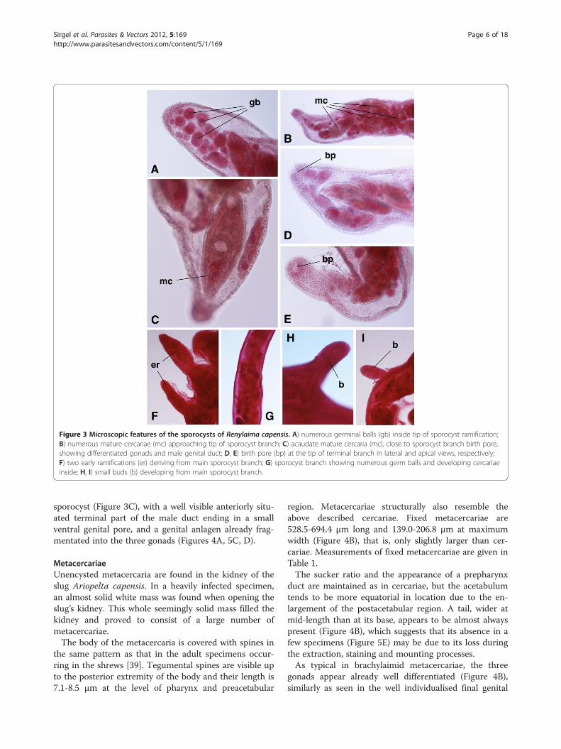
Figure 3 Microscopic features of the sporocysts of Renylaima capensis. A) numerous germinal balls (gb) inside tip of sporocyst ramification;B) numerous mature cercariae (mc) approaching tip of sporocyst branch; C) acaudate mature cercaria (mc), close to sporocyst branch birth pore,showing differentiated gonads and male genital duct; D, E) birth pore (bp) at the tip of terminal branch in lateral and apical views, respectively;F) two early ramifications (er) deriving from main sporocyst branch; G) sporocyst branch showing numerous germ balls and developing cercariaeinside; H, I) small buds (b) developing from main sporocyst branch.
Sirgel et al. Parasites & Vectors 2012, 5:169 Page 6 of 18http://www.parasitesandvectors.com/content/5/1/169
sporocyst (Figure 3C), with a well visible anteriorly situ-ated terminal part of the male duct ending in a smallventral genital pore, and a genital anlagen already frag-mentated into the three gonads (Figures 4A, 5C, D).
MetacercariaeUnencysted metacercaria are found in the kidney of theslug Ariopelta capensis. In a heavily infected specimen,an almost solid white mass was found when opening theslug’s kidney. This whole seemingly solid mass filled thekidney and proved to consist of a large number ofmetacercariae.The body of the metacercaria is covered with spines in
the same pattern as that in the adult specimens occur-ring in the shrews [39]. Tegumental spines are visible upto the posterior extremity of the body and their length is7.1-8.5 μm at the level of pharynx and preacetabular
region. Metacercariae structurally also resemble theabove described cercariae. Fixed metacercariae are528.5-694.4 μm long and 139.0-206.8 μm at maximumwidth (Figure 4B), that is, only slightly larger than cer-cariae. Measurements of fixed metacercariae are given inTable 1.The sucker ratio and the appearance of a prepharynx
duct are maintained as in cercariae, but the acetabulumtends to be more equatorial in location due to the en-largement of the postacetabular region. A tail, wider atmid-length than at its base, appears to be almost alwayspresent (Figure 4B), which suggests that its absence in afew specimens (Figure 5E) may be due to its loss duringthe extraction, staining and mounting processes.As typical in brachylaimid metacercariae, the three
gonads appear already well differentiated (Figure 4B),similarly as seen in the well individualised final genital

Figure 4 Cercaria and metacercaria of Renylaima capensis. A)caudate mature cercaria found inside a sporocyst infecting Ariostralisnebulosa in ventral view, showing prepharynx, differentiation ofgonads, male terminal duct and ventral genital pore; B) caudatemetacercaria from the kidney of Ariopelta capensis in ventral view,showing pronounced tegumental spinulation, gonads, male terminalduct and ventral genital pore. Scale bars: A = 100 μm; B = 150 μm.
Sirgel et al. Parasites & Vectors 2012, 5:169 Page 7 of 18http://www.parasitesandvectors.com/content/5/1/169
ducts of both male and female systems. The genital poreis clearly visible in a non-protruding atrium, located ven-trally (Figures 4B, 5H), in a location similar to the one inthe adult stage [39]. This larval form indicates that thenormal brachylaimid life cycle with two intermediatepulmonate hosts also applies here.
Definitive mammal hostAdults of R. capensis were very often found in shrews ofthe species Myosorex varius (Figure 1A). It seems evi-dent that the brachylaimid adults hosted by the shrewsdevelop from the aforementioned larval stages har-boured by these slugs. This is moreover supported bythe fact that adult trematodes were never found in anyof the other vertebrates living at this locality. Theseother vertebrates are two rodents, the vegetarian Otomysirroratus and the Cape spiny mouse Acomys subspinosus,and two amphibians, the toad Breviceps acutirostris andthe frog Arthroleptella landdrosia. No slug-eating birdswere ever observed in the locality. The very dense andimpenetrable vegetation would make such a predationalmost impossible in any case.
Final microhabitat and intraorganic migrationThe final microhabitats of R. capensis are the kidneysand ureters of the shrews, kidney pelvis and calyces inlight infections and also kidney medulla and cortex inheavy infections. In heavy infections both kidneys and
ureters can be infected and in one such a case up to 43individuals were recorded from only one of the ureterswhile its corresponding kidney contained more than 30individuals.A morphological study of specimens found in shrew
kidneys showed that 11.1% of the flukes were monotesti-cular forms only having a single postovarian testis.The microhabitat of this brachylaimid inside the urin-
ary system implies a transmission route through eggshedding with urine. Interestingly, fluke eggs fitting thecharacteristics (yellow-brown, symmetrical, oval, opercu-late and containing well developed embryos) and mea-surements (22.6-34.0 μm long and 14.7-20.6 μm wide) ofR. capensis eggs were found on shrew’s droppings.Unfortunately, migrating flukes were not found in
shrew dissections. However, a young individual foundattached to the outer surface of the shrews kidney, andother young individuals in some cases seen just underthe surface of the kidney, are worth mentioning. Add-itionally, very young and small individuals, resemblingthe metacercaria found in the slug Ariopelta capensis,were also found amongst fully developed individuals inthe kidneys and ureters. They present different stages ofdevelopment. The smallest individual of these only mea-sured 930 μm in length. The three primordial gonadsand vitelline glands can be clearly distinguished and adeveloping uterus can be observed as a small looped butempty duct in the area posterior to the acetabulum inthese small individuals.
EpizootiologyHabitat, rainfall and humidityRainfall data covering a period of several years (1978–2011) were obtained from the meteorological station ofthe rain gauge at Disavlei (34° 00′ 32.80¨ S, 19° 00′51.95¨ E), the closest one to the locality where R.capensis is found (Table 2). These data show that theprecipitation in this area is very high throughout theyear. The precipitation for the years 1994 to 2007shows a relatively dry period in spite of two or threeyears of normal rainfall.In this area, winter rains from May up to September-
October are normal. Thus, most of the precipitationmeasured in the summer period (from October to April)is from mist, which is brought in to the mountain topsby the South-Eastern wind. The locality of R. capensisbeing at the top end of a ravine that faces in a SouthWestern direction also regularly receives mist in sum-mer via the South Western wind. Therefore, the trans-mission area of this brachylaimid presumedly has aslightly higher precipitation than the closely located areaat Disavlei, as it receives mist from two wind directions.To understand the rainfall data, it should also be consid-
ered that although 2008 and 2009 were relatively good

Figure 5 Cercariae and metacercariae of Renylaima capensis. A) photomicrographs showing living cercariae from sporocysts infectingAriostralis nebulosa; note extension (left) and contraction (right) capacities in the same acaudate cercaria and small developing immature cercaria(centre) with caeca not yet reaching final part of body and terminal tail; B) acaudate mature cercaria from inside a sporocyst in ventral view; C)mature cercaria from a sporocyst showing absence of postacetabular rounded undifferentiated genital primordium typical of brachylaimids, butevident differentiation of genital structures; D) mature cercaria from a sporocyst showing ventral genital pore (gp); E) acaudate metacercaria fromAriopelta capensis in ventral view; F) long prepharynx (pp) in specimen of metacercaria in ventral view; G) tegumental spinulation in ametacercaria in ventral view; H) ventral genital pore (gp) in caudate metacercaria in ventral view.
Table 1 Morphometric characteristics of cercariae and metacercariae of Renylaima capensis
Larval stage Cercariae Metacercariae
Intermediate slug host species Ariostralis nebulosa Ariopelta capensis
No. larvae studied n= 10 n=10
E.V. X SD E.V. X SD
Length 330.0-436.6 392.9 40.4 528.5-694.4 604.6 52.1
Maximum width 114.9-166.3 142.0 18.6 139.0-206.8 176.7 20.0
Oral sucker (OS) 68.9-85.9/62.9-80.4 76.5/67.5 6.4/5.7 82.6-108.4/76.9-103.4 92.4/87.7 9.3/8.0
Ventral sucker (VS) 80.4-94.9/68.9-85.9 86.5/78.1 6.3/5.2 96.9-131.3/91.2-119.9 115.7/106.5 13.0/12.6
Sucker ratio (OS/VS) 0.62-0.85 0.73 0.07 0.57-0.83 0.68 0.08
Prepharynx (length) 0.0-12.2 5.4 5.5 0.0-37.0 16.3 11.4
Pharynx (length/width) 34.4-39.9/26.4-34.4 36.6/32.3 2.7/3.2 39.9-57.4/31.3-45.9 45.0/38.6 5.4/4.3
Anterior testis (T1) 22.0-28.6/34.0-34.8 33.5/26.8 2.1/5.6 25.6-57.0/18.0-37.0 40.3/29.6 10.7/6.5
Posterior testis (T2) 22.9-34.4/20.0-28.6 28.9/23.9 4.6/3.0 34.2-48.4/22.8-37.0 39.5/30.0 5.0/4.9
Ovary 22.0-28.6/17.0-22.9 25.2/20.4 2.9/2.7 25.6-31.3/19.9-25.6 28.9/23.2 2.1/2.1
Distance OS-VS 85.9-137.8 112.3 21.2 126.3-245.1 177.1 35.0
Distance VS-T1 17.5-40.0 28.6 8.0 37.0-71.2 53.8 13.3
Tail (length/width) 22.9-35.4/25.9-28.4 30.9/26.9 6.9/1.3 28.5-37.0/27.1-37.0 33.1/30.8 2.7/3.4
Meaurements in μm. E.V. = extreme values; X=mean values; SD= standard deviation.
Sirgel et al. Parasites & Vectors 2012, 5:169 Page 8 of 18http://www.parasitesandvectors.com/content/5/1/169

Table 2 Precipitation (in mm) measured in the meteorological station of the rain gauge at Disavlei (34° 00′ 32.80¨ S,19° 00′ 51.95¨ E), the closest one to the locality where Renylaima capensis is found, during the year period of1978–2011
Year January February March April May June July August September October November December TOTAL
1978 210,8 75,3 57,4 308,9 169,2 109,8 201,7 865,6 430,2 300,4 165,0 289,6 3184
1979 90,8 87,6 126,5 123,0 275,4 695,0 254,7 288,5 209,6 410,2 123,0 19,7 2704
1980 163,5 130,9 41,8 436,9 375,7 419,5 83,2 368,9 162,7 186,5 288,6 236,0 2894
1981 333,0 33,5 153,3 204,0 65,5 322,8 504,2 704,5 457,5 93,5 247,4 130,0 3249
1982 183,0 40,1 103,5 323,0 208,0 280,7 348,0 230,9 221,7 273,6 171,9 226,2 2611
1983 57,8 191,8 129,8 57,5 828,4 860,0 362,1 176,0 381,0 85,5 77,3 70,4 3278
1984 63,0 66,0 173,8 167,0 723,2 89,5 716,8 221,0 420,0 301,7 79,2 307,5 3329
1985 182,5 167,3 340,9 219,5 236,4 580,0 403,9 382,8 316,5 144,5 172,0 100,0 3246
1986 123,5 118,4 130,5 423,0 278,9 568,0 460,0 733,9 247,0 81,0 109,9 66,5 3341
1987 191,5 90,0 84,7 198,6 436,5 458,9 485,5 374,9 355,0 115,5 102,0 227,5 3121
1988 17,3 25,0 70,0 351,2 234,6 361,9 437,2 504,5 ? 225,9 80,9 109,0 2418
1989 5,5 88,8 259,0 348,3 312,7 369,0 420,7 401,7 688,2 273,5 184,9 51,0 3403
1990 75,0 167,9 64,5 737,8 158,6 312,4 868,3 452,3 183,6 62,0 138,0 89,2 3310
1991 85,0 57,9 62,8 131,0 490,4 605,5 919,4 69,8 411,0 219,1 54,7 43,5 3150
1992 33,8 122,9 148,3 376,5 482,0 847,0 451,3 362,5 358,7 431,3 147,5 56,9 3819
1993 39,7 103,0 19,5 908,2 573,0 602,4 881,0 293,0 82,5 51,5 89,0 118,0 3761
1994 108,4 55,9 33,0 200,6 195,2 870,0 281,6 209,5 224,0 121,4 90,0 86,9 2477
1995 110,4 25,0 87,0 158,2 306,0 390,0 680,0 408,4 138,5 350,0 45,4 263,0 2962
1996 20,5 136,7 76,0 166,4 166,0 470,0 410,0 0,0 841,1 539,0 430,0 213,0 3469
1997 135,0 21,5 45,0 164,0 225,0 687,0 151,8 415,0 300,0 207,0 427,0 0,0 2778
1998 206,0 43,0 128,0 246,0 625,0 0,0 790,0 270,0 180,0 0,0 370,0 315,0 3173
1999 9,0 18,0 17,0 187,0 280,0 300,0 436,0 200,0 0,0 180,0 123,0 31,0 1781
2000 105,0 94,0 96,0 57,0 0,0 695,0 240,0 0,0 800,0 107,0 57,0 95,0 2346
2001 24,0 18,5 7,0 0,0 820,0 100,0 0,0 0,0 0,0 1040,0 0,0 93,0 2103
2002 360,0 57,0 0,0 0,0 720,0 558,0 0,0 720,0 0,0 506,0 100,0 75,0 3096
2003 71,0 65,0 280,0 160,0 0,0 280,0 0,0 840,0 0,0 460,0 8,0 0,0 2164
2004 15,0 330,0 0,0 540,0 45,0 0,0 0,0 0,0 840,0 228,0 0,0 0,0 1998
2005 210,0 164,5 0,0 0,0 0,0 991,0 328,0 520,0 0,0 0,0 0,0 0,0 2214
2006 58,0 0,0 58,0 23,5 51,0 334,0 297,0 400,0 0,0 228,0 140,0 100,0 1690
2007 8,0 12,9 174,5 169,0 280,0 612,0 640,1 580,0 68,0 0,0 640,0 0,0 3184
2008 230,0 0,0 182,0 0,0 513,0 467,0 638,0 310,0 612,0 52,0 365,0 60,0 3429
2009 ? ?* 39,0 160,0 863,0 623,5 248,0 620,0 304,0 ? ? ? 3554
2010 14,5 68,0 280,0 110,0 655,5 304,0 230,0 271,0 175,0 205,2 156,0 106,4 2576
2011 21,0 23,5 3,0 212,8 180,0 761,0 87,0 324,0 186,0 140,0 190,0 106,4 2235
? =measurement could not be performed in this month because the gauge was broken.* = a fire occurred in early February 2009 which pronouncedly damaged the vegetation in the habitat of R. capensis.TOTAL = total rainfall for each year; a graphic comparison of yearly rainfall data available since 1945 from that meteorological station shows that the period1999–2006 was the most dry period.
Sirgel et al. Parasites & Vectors 2012, 5:169 Page 9 of 18http://www.parasitesandvectors.com/content/5/1/169
rainfall years, after a long dry period, a fire that occurredin early February 2009 damaged the vegetation in thehabitat of R. capensis. This fire had a significant influenceas it burnt away much of the very thick Restionaceaeavegetation and especially the rotting undergrowth at thebases of these plants. The latter when intact, acts as an
efficient sponge to retain the water and keep the area verymoist. It is also the nesting habitat for the slug Ariopeltacapensis. After the fire it was mostly burnt away and muchof the water simply flowed away with the effect that thehabitat was drier than normal. The previous dry periodobviously did put stress on the vegetation so that more

Sirgel et al. Parasites & Vectors 2012, 5:169 Page 10 of 18http://www.parasitesandvectors.com/content/5/1/169
dry dead plant material accumulated. Consequently, whenthe fire occurred it could burn with more intensity.
Prevalences and intensities in the slugsAriostralis nebulosa is readily found throughout thewhole year. During the years 1982–2011, a total of 38specimens of A. nebulosa were infected with sporocystsamong the 263 specimens collected (Table 3). Theaforementioned perturbation of the habitat of R. capen-sis by the fire, which occurred in February 2009, shouldbe taken into account. Before that fire, from 198 A.nebulosa, 34 were infected by sporocysts (mean 17.2%),with high prevalences in given years, whereas after thefire infection disappeared up to 2011 when this infec-tion rate seemed to recover. A seasonal analysis of databefore the fire indicate that sporocyst infection ismainly detected from late autumn (April) to earlyspring (September) and only sporadically in summertime (late October to March). This suggests a seasonal-ity in the transmission dynamics of the parasite.Whether sporocysts are so pathogenic that infectedslugs are not able to survive until the summer or areperhaps more easily predatable remains a question forfuture research.Contrary to A. nebulosa, A. capensis is mostly found
during the hotter months, the October-March period(nests of eggs of this slug species were found in January,that is, in midsummer). During the years 1978–2011,
Table 3 Prevalences of infection of the first intermediateslug species Ariostralis nebulosa by cercariogenoussporocysts of Renylaima capensis according to years andmonths
Year ofcollection
Month (and day)of collection
No. of slugscollected
No. infectedwith sporocysts
1982 April 23 10 4 (40%)
July 19 4 1 (25%)
1983 March 2 2 0 (0%)
April 23 4 1 (25%)
2004 October 19 4 0 (0%)
1983-2006period
March-September 127 26 (20.5%)
October-February 47 2 (4%)
2009 in February, a fire burnt a large part of the habitat
2009 May 9 3 0 (0%)
2010 February 10 0 –
April 26 21 0 (0%)
October 11 5 0 (0%)
2011 February 22 15 1 (6.6%)
May 11 21 3 (14%)
July 14 7 0 (0%)
Total 263 38 (14.4%)
only 3 specimens of A. capensis were infected with meta-cercariae among the 77 specimens collected (Table 4).Before the fire, only two (collected in April and May)from 49 specimens carried metacercariae (mean 4.0%),surprisingly lower than the prevalence of sporocysts inA. nebulosa. The third specimen infected by metacercar-iae, collected in 2011 (after fire) was also found in May.This suggests a concentration of second intermediateslug infection by metacercariae in the colder months.Additionally, the pronouncedly lower number ofA. capensis collected indicates lower population densitiesof the second intermediate slug when compared to thefirst intermediate one. With regard to infection intensity,while only one metacercaria was found in one A. capen-sis, the other two specimens showed relatively massiveinfections.
Prevalences and intensities in the shrewsThe shrew species Myosorex varius appears to be activemainly from October to May. During the years 1983–2010, a total of 21 specimens of this shrew species wereinfected with adults of R. capensis among a total of 29specimens collected, giving a prevalence of 72.4% duringthis long period (Table 5). In many years, all specimenscaptured proved to be infected, and when not all wereinfected, yearly prevalences found were usually veryhigh. Additionally, intensities also proved to usually bevery high, sometimes even higher than 50 adult flukesper shrew, with an apparent decline after the fire oc-curred in February 2009.
Mitochondrial DNA cox1 sequencesDNA sequences obtained for the mtDNA cox1 genefragment were of a length of 437 nucleotides. All sporo-cysts, cercariae and adults showed the same sequence,with a composition of 64.75% AT. However, the meta-cercariae available for sequencing, coming from onlyone Ariopelta capensis slug individual, showed threemutations (6.86% divergence) when compared to thesequence of the other developmental stages: (i) C inmetacercariae and G in the other stages in position 80,(ii) T/G respectively in position 118, and (iii) C/T re-spectively in position 373. The AT content of the meta-cercarial sequence was also 64.75%.The provisional haplotypes R.cap-cox1a and R.cap-
cox1b have been ascribed to the sequences found insporocysts, cercariae and adults on one side, and meta-cercariae on the other side, respectively. Their respectiveaccession number codes are [EMBL: HE663453] and[EMBL: HE663454].When using the BLASTN programme, homologies
were found with several cox1 fragments of other digen-ean species of which the same or very similar fragmentis available in databases. The species that provides the
Table 4 Prevalences and intensities of infection of the second intermediate slug species Ariopelta capensis bymetacercariae of Renylaima capensis according to years and months
Year of collection Month (and day)of collection
No. of slugscollected
No. infected withmetacercariae
No. of metacercariaeper slug
1978 May 18 4 1 (25%) 1
1982 January 8 3 0 (0%) -
February 1 0 (0%) -
April 23 4 0 (0%) -
July 19 0 – -
1983 March 2 5 0 (0%) -
April 23 4 1 (25%) >70
1984-1985 month? 12 0 (0%) -
June 16 0 – -
1986 May 10 5 0 (0%) -
1994 July 17 0 – -
1997 October 10 2 0 (0%) -
November 1-7 7 0 (0%) -
2004 October 19 0 – -
December 16 0 – -
2005 April 28 0 – -
2006 March 14 0 – -
May 13 2 0 (0%) -
November 30 0 – -
2009 in February, a fire burnt a large part of the habitat
2009 May 9 3 0 (0%) -
2010 February 10 0 – -
April 26 3 0 (0%) -
October 11 6 0 (0%) -
2011 February 22 0 – -
May 11 12 1 (8.3%) > > 100
July14 4 (young) 0 (0%)
Total 77 3 (3.9%)
Sirgel et al. Parasites & Vectors 2012, 5:169 Page 11 of 18http://www.parasitesandvectors.com/content/5/1/169
highest query coverage (94-96%) in a total score of 231–259 bp compared is Schistosoma japonicum. A specificsearch using the nucleotide database of the GenBankshowed that very few sequences of brachylaimids areavailable. Among them, the only cox1 sequence availableis the 897-bp-long fragment of this gene from Glaphyr-ostomum sp. [GenBank: FJ713138] [52]. In a comparisonbetween Renylaima and Glaphyrostomum haplotypes, bymeans of a 420–11 (= 409) bp-long alignment, the resultwas congruent despite the numerous mutations that ap-pear, with a total of 78 variable positions (19.07% diver-gence) (Figure 6).The resulting protein fragments from R. capensis are
both composed of 145 aminoacids and are available inthe EMBL database under the same accession numbers.An alignment comparison between them shows that of
the three nucleotide mutations two give rise to aminoacid changes whilst the third one is silent.
DiscussionLife cycleA total of 34 years work in the small area where R.capensis occurs, leads to several conclusions: (i) thisfluke species appears to be specific for M. varius, as nobrachylaimid has ever been found in any of the othervertebrates inhabiting the same area; (ii) the slugAriostralis nebulosa is the first intermediate host of thisfluke species, as branched, cercariogenous sporocysts re-sembling those typical of brachylaimids [14,16,24,25]were frequently found but only in this slug among thefive terrestrial mollusc species inhabiting the area; thismeans that this fluke is highly specific at first

Table 5 Prevalences and intensities of infection of the definitive host species Myosorex varius by adults of Renylaimacapensis according to years and months
Year of collection Month (and day)of collection
No. of shrews collected(20 traps set)
No. of shrewsinfected
Intensity(no. of flukes/shrew)
1983 April 4 1 1 (100%) >50
June 16 0 – -
1986 May 10 3 3 (100%) >50 +37 + >50
1994 July 17 0 – -
1997 October 10 1 1 (100%) >50
November (1–7) 2 2 (100%) >50 + >50
2004 October 19 0 – -
December 16 3 1 (33%) 45 + 0+ 0
2005 April 28 4 3 (75%) 33 + 16+ 13+ 0
2006 March 14 3 3 (100%) 9 + 1 + 5
May 13 4 3 (75%) 11 +7+ 24+ 0
November 30 2 1 (50%) 57 + 0
2009 in February, a fire burnt a large part of the habitat
2009 May 9 0 – -
2010 February 10 2 2 (100%) 16 + 6
April 26 4 1 (25%) 2 + 0 + 0 + 0
Total 29 21 (72.4%) >527
Sirgel et al. Parasites & Vectors 2012, 5:169 Page 12 of 18http://www.parasitesandvectors.com/content/5/1/169
intermediate host level; (iii) Ariopelta capensis, the onlyother slug species present in the same area, appears tobe the only second intermediate host species, as brachy-laimid metacercariae resembling immature juvenilesfound in the shrews were only found in this mollusc. Al-though no intracellular early mother sporocyst stagewithin hepatopancreatic cells [23] was observed nor anysporocystogenous first generation sporocysts [19-22], theexistence of such stages cannot be a priori denied.The sequencing results verify that the sporocysts, cer-
cariae, metacercariae and adult stages belong to thesame species and confirm this life cycle. The mere three
Figure 6 Nucleotide alignment of the mitochondrial DNA cox1 gene ssporocysts, cercariae and adults of Renylaima capensis; Line 2: R.cap-cox1bcapensis; Line 3: last part of the 897-bp-long fragment from Glaphyrostomu90 positions per line. Three mutation differences between the two haploty
mutations found in the metacercariae infecting anAriopeltis capensis slug individual may be considered asintraspecific genetic variability within the local area,according to the well known evolutionary rates of themtDNA cox1 gene in invertebrates and its usefulness forinter- and intrapopulation diversity studies in general[41]. This agrees with the three-host life cycle pattern ofBrachylaimidae. The observation that the shrew M. variuspreys on both species of slugs supports this view.However, results of the several years of field work indi-
cate that prevalence and intensity data do not appropri-ately fit the above statement:
equences. Line 1: R.cap-cox1a haplotype sequence obtained fromhaplotype sequence obtained from metacercariae of Renylaimam sp. [GenBank: FJ713138] [52]. Nucleotides distributed according topes of R. capensis marked in green.

Sirgel et al. Parasites & Vectors 2012, 5:169 Page 13 of 18http://www.parasitesandvectors.com/content/5/1/169
a) in brachylaimid species in general, the prevalencesof the first intermediate snail host by cercariogenoussporocysts are always pronouncedly low [14,16,17],whereas in the case of R. capensis the prevalence ispronouncedly higher, mainly in autumn-spring;
b) in brachylaimids, prevalences and intensities of thesecond intermediate snail host by metacercariae areusually very high, even up to 100%, and highinfection burdens of up to more than 100metacercariae occur in the main second snail host[14,16,17]; on the contrary, in R. capensis, theprevalence is strikingly low; this may be explained bythe second intermediate slug host not being readilyavailable in winter and thus allowing secondintermediate slug infections only very rarely;
c) the high prevalences and intensities of R. capensis inthe shrews do not correspond with the very lowprevalences (and sometimes apparently alsointensities) of metacercariae in slugs, nor with thesecond intermediate slug population densities in thearea;
d) the adult stage does not show an egg productioncapacity higher than that of other Brachylaimaspecies (similar body size, similar size of the gonads,similar uterus extent); the fluke prevalence in theshrews (72.4%) is, however, higher than thoseusually found for intestinal Brachylaima species intheir definitive host species: for instance, 56.7-64.3%in one host species and 21.4-60.0% in another hostspecies for B. ruminae [18], or 38.5-66.7% for B.mascomai [53]; moreover, M. varius shrew densitiesare not as high as to argue that a higher eggdissemination ability could be the cause for higherinfection rates of the first intermediate slug species.
From the point of view of the r/K selection concept,ecological studies on the compared fitness of the lifecycle strategies of Brachylaimidae in general show thatthese trematodes follow a compensation pattern withinthe different transmission phases [18]. According tosuch a rule, the surprising prevalence and intensity dataat the levels of first and second intermediate hosts,markedly opposite to what is found in other brachylai-mids, can be understood, although the route of trans-mission may still remain an open question.The question immediately arises about the possibility
of direct definitive host infection by ingestion of maturecercariae still inside sporocysts carried by the first inter-mediate mollusc host. Although definitive host infectionby mature cercariae resulting in the development of ma-ture adults, without being exposed to metacercariae,could not be proven, observations in three brachylaimidspecies suggest that this could be possible. In the speciesZ. spearei, juvenile specimens presenting a cercarial tail
were found in the urinary bladder of the toad Bufomarinus and it was concluded that this may be indica-tive of the possibility that the cercaria in the first inter-mediate host could be directly infective to the final host.This theory is further supported by the statement that inthe smallest of these individuals the genital system wasonly represented by a single primordium [29]. In Brachy-laima ruminae, such a cercarial tail was also found inseveral juvenile specimens from the intestine of gardendoormice Eliomys quercinus (Valero and Mas-Coma, un-published data). In D. frontalis, a cercarial tail was foundin a juvenile fluke [37] as well as in three gravid flukespecimens (Valero and Mas-Coma, unpublished data)from the nasal sinuses of the same garden dormousespecies. However, a cercarial tail such as this appears tobe sometimes retained for a period after having pene-trated the second intermediate snail host [54], so that itcannot be ruled out that tailed adults may also derivefrom tailed metacercariae infecting a second intermedi-ate snail host. The numerous tailed metacercariae foundin one Ariopelta capensis indicate that this phenomenonmay also take place in R. capensis.Moreover, the presence in mature cercariae of
R. capensis of a genital complex already divided, withvisible terminal male duct ending in a small ventral geni-tal pore, and a genital anlagen already forming the threegonads, should be highlighted. Such a precociousness isnot typical of Brachylaimidae. In species of this family,only a rounded, postacetabular, undifferentiated genitalprimordium appears in the mature cercariae [14,16,17].This suggests that mature cercariae may develop suffi-ciently inside the sporocyst as to become infective forthe definitive host (i.e., do not need further genital mat-uration in a second intermediate host). Additionally, thevery small increase in fluke size during their transmis-sion through the three hosts (length of mature cercariaeinside the sporocyst, metacercariae in slug kidney andsmallest adult in the shrew of 330–436 μm, 528–694 μm, and 930 μm, respectively), suggests that adevelpment transit in a second intermediate host maynot be necessary. In other terms, a larger size develop-ment of metacercariae in the second intermediate slughost could become an impediment for a subsequent suc-cessful intraorganic migration within the shrew.The capacity of self-infection of the first intermediate
mollusc host individual with metacercariae shed by thesporocysts harboured by the same molluscan individualalso implies the reduction of the life cycle from a three-host pattern to a two-host pattern. Such a capacity is ap-parently related to an adaptation strategy of the charac-teristics of both intermediate mollusc host and habitatenvironment [18]. Such self-infection seems to be pre-vented by a kind of premunition in species such asB. ruminae, which is consequently an obligatory three-

Sirgel et al. Parasites & Vectors 2012, 5:169 Page 14 of 18http://www.parasitesandvectors.com/content/5/1/169
host brachylaimid [16]. In the species D. frontalis inha-biting dry habitats, 90.6-100% of the snails bearing spor-ocysts also harbour metacercariae in the pericardiumwhile only 19.9-40.0% of the snails, of the same species,lacking sporocysts carry metacercariae [14]. Similarly, ina P. pericardicum inhabiting wet habitats, the relativefigures are 50.0% and 39.2% [17].In another brachylaimid, Serpentinotrema laruei (= Post-
harmostomum laruei; = P. helicis) [19-21,55-57], the dis-covery of a precociously developed metacercaria within asporocyst [58] also indicates the secondary possibility for atwo-host life cycle. In Brachylaimidae, however, the genusParabrachylaima presents a two-host life cycle, as theadult stage fully develops to maturity and egg layingwithin a terrestrial snail [32]. Progenesis within a formersecond intermediate snail host has been the interpretationused to justify its inclusion into the subfamily Brachylaimi-nae [33]. All in all, Brachylaimidae appear to be a versatilegroup where the elimination of one or another of the typ-ical hosts in the life cycle might be possible. It thus seemsas although a triheteroxenous life cycle is indicated for thenew South African fluke species, the possibility of the sec-ond intermediate host being eliminated in current trans-mission cannot be ruled out.A reduction from a three-host life cycle to a two-host
life cycle has been described in species in other familiesincluded within Brachylaimoidea, namely Hasstilesiidaefrom mammals, and Leucochloridiidae and Leucochlori-diomorphidae from birds. In all of them, metacercariaedevelop in branched sporocysts infecting snails (terres-trial snails in hasstilesiids and leucochloridiids, andaquatic snails in leucochloridiomorphids) [13,15,59-61].Leucochloridiidae and Leucochloridiomorphidae seemto be morphologically and biologically distant from theSouth African fluke, but hasstilesiid species of the genusStrzeleckia from the intestine of marsupials appear to benot as distant morphologically [62]. Another genus,Michajlovia, comprises parasites which infect the intes-tine of passerine birds and whose life cycle is still un-known. Michajlovia adults present a ventral genitalatrium just posterior to the gonads or in the region ofthe posterior testis. Michajlovia is included in Brachylai-moidea as incertae sedis although close to Leucochlori-diidae and Leucochloridiomorphidae [31,63], and alsoshows several similarities with the South African fluke.Except for the extent of vitellaria, the morphologicalsimilarities of Michajlovia with the panopistine genusDasyurotrema, whose type species D. mascomai infectsthe alimentary tract and associated organs of marsupials[64], are evident.
Fluke transmission, monotesticular forms and their originThe not uncommon monotesticular forms of R. capensisadults merit an additional analysis to elucidate fluke
transmision. In these curious adults, the single postovar-ian testis is relatively much larger and elongated thaneither the anterior or posterior testes of the bitesticularspecimens [39].The phenomenon of neoteny, as previously defined
[65], seemingly applies to these individuals seeing thatthe testis matures in the adult without the larval genitalanlagen having separated into two independent testesbeforehand. This fits the concept of cercariae having thecapacity to infect the definitive host, as the genital prim-ordium in brachylaimid cercariae does not differentiateinto the three separate gonads prior to the developmentof the mature metacercaria within the second intermedi-ate snail host [14,16,17]. Monotesticular forms ofR. capensis thus also support a direct transmission fromfirst intermediate snail host to definitive host, as suchforms may derive from intrasporocystic cercariae alreadyinfective but with division of the genital primordium stillnot complete.Monotesticular specimens have also been found, but
rarely, in other brachylaimid species. Interestingly, mod-ern brachylaimid species such as Brachylaima ruminae, itis always the posterior testis that is absent in the monotes-ticular forms. Contrarily, in presumably archaic brachylai-mids such as Ityogonimus ocreatus from moles (Talpidaeinsectivores) and Dollfusinus frontalis from hedgehogs(Erinaceidae insectivores) and dormice (Glirimorphrodents), it is always the anterior testis which is absent.Noteworthy is that certain adult specimens of D. frontalisshow a rare triangular arragement of the gonads in whichthe ovary appears anterior to both testes (Valero and Mas-Coma, unpublished data), thus remimiscent of the gonadarrangement typical of Zeylanurotrema.A strong argument to support a direct cercarial origin
for monotesticular specimens is found in the monorchidspecies Parabrachylaima euglandensis. In this peculiarbrachylaimine, the progenetic adult stage fully developsto maturity and egg laying in a terrestrial snail, andshows only one postovarian, sacculate testis with a pairof anterior projections from which the two respectivevasa efferentia arise [32]. Additional to its peculiar lifecycle, the following features of Parabrachylaima, how-ever, rules out a close relationship with the SouthAfrican fluke: (i) both suckers close together in the an-terior part of the body; (ii) caeca unequal, with rightcaecum terminating in middle third of body; (iii) verylong excretory vesicle reaching anteriorly to level of acet-abulum; and (iv) cirrus pouch present and including apoorly developed cirrus.If a definitive-host-infection capacity is accepted for
intrasporocystic mature cercariae of R. capensis, massiveinfections of the shrews by adult flukes are easily explained(one or a very few sporocyst-carrying slugs ingested wouldbe sufficient), but on the contrary a question mark is posed

Sirgel et al. Parasites & Vectors 2012, 5:169 Page 15 of 18http://www.parasitesandvectors.com/content/5/1/169
by the shrews infected by only a few adult flukes. Such lowinfections could be the consequence of (i) shrews swallow-ing sporocysts including only a few mature cercariae at thatmoment (i.e., all other intrasporocystic cercariae presentwere still immature at that moment, not able to migrate in-side the shrew’s body and hence eliminated digestively afteringestion), (ii) most cercariae may get lost in the definitivehost infection, most probably expelled by the shrew withtheir faeces; thus, the massive sporocyst infection of thefirst intermediate slug host and the very long sporocystbranches filled by mature cercariae could be interpreted asa fluke strategy to mitigate the great losses; (iii) a crowdingeffect not allowing the penetration into the kidney andureters by all migrating cercariae because of lack of micro-habitat space, (iv) massive infections becoming too patho-genic for shrews which would thus be quickly eliminatedfrom natural populations, and of course also (v) ingestionof slugs only carrying a few metacercariae. In addition to allthis, an influence of innate and cellular immunity on estab-lishment of infection may also play a role in such individualcases.
Mitochondrial DNA cox1 gene sequence variability andtransmission modalitiesIt is evident that the number of slugs and shrews, fromwhich respective larval stages and fluke adults have beenmolecularly analysed, is insufficient to conclude signifi-cant results. Unfortunately conservation restriction lawswill never allow the collection of a sufficient host num-ber, mainly of Ariopelta capensis, to obtain sufficientamount of metacercariae from different slug individuals,to perform the necessary study, given the pronouncedlylow metacercarial prevalences in this slug species.The characteristics of the three mutations found in the
metacercariae are surprising. In mtDNA coding genes,when intraspecific mutations appear they are mostly si-lent and only a small percentage give rise to amino acidchanges [41]. In R. capensis, among three mutations onlyone is silent. Whether such a rare genetic characteristicmay be interpreted as two different biological strains ofR. capensis coexisting in the study area may alwaysremain a question mark. But there is the temptation tosuggest that there coexists a majority of haplotypeR.cap-cox1a strain following a two-host life cycle modal-ity with a less frequent R.cap-cox1b strain following athree-host life cycle modality. Such a parallel circulationof the two biological strains could be perhaps related toclimatic seasonality, due to Ariopelta capensis not beingreadily available in winter.
Intraorganic migration, adult microhabitat and eggsheddingThe occurrence of R. capensis in the urinary system of amammal host is a unique phenomenon for the family
Brachylaimidae. Within Brachylaimoidea, only the speciesof Zeylanurotrema parasitising amphibians and reptilesshow a similar microhabitat: Z. lyriocephali in the urinarybladder of the agamid lizard Lyriocephalus scutatus in SriLanka [38] and Z. spearei in the urinary bladder of thecane toad Bufo marinus [29]. However, there are funda-mental differences between the urinary system of mam-mals and that of amphibians and reptiles. In mammals,the urinary system is isolated from the alimentary tract, sothat a renal helminth following an oral way of infectionunavoidably has to traverse tissues, then find the kidneyand actively penetrate it. On the contrary, in lizards theurinary system opens into the cloaca forming a direct con-nection along which the parasite can migrate between thetwo systems. Similarly, in amphibians the urinary bladderis practically merely an evagination of the alimentary tract.Furthermore, the distinct anatomy of Zeylanurotremahaving a very anterior, postbifurcal acetabulum, pretesticu-lar, lobed ovary, opposite testes and terminal genital porerules out any close relationships with R. capensis.Within Brachylaimoidea, another additional exception
presenting a similar microhabitat, although inside a de-finitive snail host, is the “progenetic” adult stage ofParabrachylaima euglandensis, which also develops inthe lumen of the kidney sac [32].That an active, tissue-traversing, intraorganic migra-
tion within the shrew takes place during its infection byR. capensis, is the only way to understand the observa-tion of both (i) a young individual attached to the outersurface of the shrews kidney, and (ii) other young indivi-duals in some cases seen just under the surface of thekidney which may be interpreted as immature flukes justpenetrated and on their way to the deeper areas of thekidney and the ureters.Concerning the intraorganic route followed by the in-
fective stage to the kidney, the knowledge about such alife cycle phase in other renal helminths suggest that aprobable intermediate migratory step through the livercould be envisaged. This is the case in the nematodeDioctophyme renale, a renal parasite of carnivores [66],as well as, interestingly, that of another trematode fromEuropean insectivores (shrews and moles) but alsoarchaic glirimorph rodents (dormice), Nephrotrematruncatum [67,68]. In both cases, the infective stagecrosses the intestinal wall and migrates through the gen-eral body cavity to penetrate the right hepatic lobeswhich cover the right kidney before penetrating the lat-ter. Such a liver phase appears to be important from thetrophic point of view [66]. Contrary to N. truncatumwhere the adult stage only infects the right kidney, inR. capensis the adult stages infect both kidneys andureters without any apparent lateral preference [39].This poses the question whether R. capensis migratesthrought the choledoc duct up to the biliary ducts to

Sirgel et al. Parasites & Vectors 2012, 5:169 Page 16 of 18http://www.parasitesandvectors.com/content/5/1/169
finally reach both kidneys after crossing the distal paren-chyma of the liver lobes. Such hypothesis is supportedby the hepatic duct microhabitat of species of anotherclose brachylaimid genus, namely Scaphiostomum [36],although a more simple migration only including intes-tinal wall crossing and direct migration through the gen-eral body cavity to enter the two kidneys of the shrew bypenetrating their surface can a priori not be denied.Should one or another intraorganic migration route be
followed in the definitive host, the infecting stage wouldevidently need the capacity to traverse host tissues. Thisis a feature worth emphasizing, because there are noother species of Brachylaimidae known to have acquiredsuch a capacity to reach their final microhabitat. InBrachylaimidae, the adult stage of the great majority ofspecies develops in the digestive tract (mainly intestine,rarely oesophagous and stomach). Only very few speciespresent other microhabitats (Scaphiostomum in ducts ofthe liver and pancreas; Dollfusinus in nasal and frontalsinuses), but these are all directly connected to thedigestive tract [14,16]. As already mentioned, the sameapplies to Zeylanurotrema, where both the reptilian andamphibian urinary bladders are part of the alimentarycanal. In Brachylaimidae in general, it thus is the cer-caria and not the metacercaria that presents a high trop-ism and migration capacity, a fact reflected by theexistence of two postacetabular lateral aggregations oflarge penetration glands in cercariae. Nothing of thiskind appears in the metacercariae [14,16,17]. This alsosupports a direct definitive-host-infection capacity byintrasporocystic cercariae and, thus, definitive host infec-tion by predation of the first intermediate slug host inthe case of R. capensis.Owing to the adult stage microhabitats of kidneys and
ureters, eggs are shed by the shrews by urinating.A priori this poses the problem of understanding howthe first intermediate slug host may become infected byingesting the fluke eggs dispersed throughout the exter-nal milieu. In other brachylaimids, eggs are in all casesshed with faeces and the fluke transmission is assuredbecause of snails being attracted by the deposited stoolmaterials, with many terrestrial gastropods showing cop-rophagous trends. Unfortunately, to our knowledge,nothing is known about potential snail attraction byurine. However, the shrew M. varius always urinate dur-ing the process of defaecation, therefore concomitantlydepositing eggs with faecal material [69]. This fact hasbeen corroborated by the finding of eggs of R. capensison the shrew’s droppings.
ConclusionsCharacteristics of the morphology of the branched sporo-cysts, brevicaudate cercariae, unencysted metacercariaeand life cycle pattern including two terrestrial slugs as first
and second intermediate hosts, support the assignment ofR. capensis to the family Brachylaimidae and subfamilyBrachylaiminae, as previously proposed [39].Epizootiological data, several morphological features,
comparisons with other archaic and modern brachylai-mids, and intraspecific mtDNA cox1 gene sequence vari-ability suggest that R. capensis may use two transmissionstrategies in the study area of the Hottentots HollandNature Reserve, South Africa. All indications are thatR. capensis presents a very primitive condition, wherethe two-host life cycle is the normal modality, while theintroduction of a second intermediate mollusc host isjust in its early stages.The kidney and ureter of the shrew, being the final
microhabitat in R. capensis, is a unique feature in bra-chylaimids infecting mammals. This implies that theinfecting stage must have the capacity to traverse thefinal host tissues in order to intra- and interorganicallymigrate from the digestive system to the urinary system.Such a requirement also supports a possible direct infec-tion of the shrew by cercariae. Further support for thisview is found in the observation that the shrew indeeddoes prey on the sporocyst and cercariae hosting slugA. nebulosa.The three-host life cycle modality typical of brachylai-
mids thus seems to only be an additional, secondary andnon-obligatory option for R. capensis, and even thenonly if its metacercariae, at least the young ones, stillkeep their capacity for such a complex tissue-traversingintraorganic migration. Consequently, in Brachylaimidae,the second intermediate mollusc host should evolution-arily be seen as a last addition to the life cycle. Further-more, the present restriction of the adult stage to themicrohabitat of the digestive tract and related organs ofthe final host must be regarded to result from a loss ofthe tissue-traversing capacity by the metacercarial stage.
Competing interestsThe authors declare that they have no competing interests.
Authors’ contributionsWFS carried out the field collections and dissections of shrews and slugs,contributed the epizootiological studies and analyses, performed the firstlarval stage studies, and wrote an initial draft of the manuscript. PA carriedout the DNA sequencing processes. MDB designed the sequencing study,analysed the sequences, and helped to draft the molecular part of themanuscript. SMC mounted and studied the larval stages, analysed the results,performed the literature review, made the drawings and photographcompositions, and wrote the final manuscript. All authors read and approvedthe final manuscript.
AcknowledgementsThanks are due to Cape Nature for cooperation and permission to collect inthe area under their jurisdiction. Spanish collaboration within the frame ofthe “Red de Investigación de Centros de Enfermedades Tropicales” (RICET,Project No. ISCIII-RETIC RD06/0021/0017) of the Programme of RedesTemáticas de Investigación Cooperativa, FIS, Ministry of Health, Madrid. Theauthors greatly acknowledge Mr. Richard Thompson (South Africa) for hisassistance with computer work.

Sirgel et al. Parasites & Vectors 2012, 5:169 Page 17 of 18http://www.parasitesandvectors.com/content/5/1/169
Author details1Department of Botany and Zoology, University of Stellenbosch, Private BagX1, Matieland 7602, South Africa. 2Departamento de Parasitología, Facultadde Farmacia, Universidad de Valencia, Av. Vicente Andrés Estellés s/n, 46100Burjassot - Valencia, Spain.
Received: 23 February 2012 Accepted: 19 July 2012Published: 13 August 2012
References1. Butcher AR, Talbot GA, Norton RE, Kirk MD, Cribb TH, Forsyth JRL, Knight B,
Cameron AS: Locally acquired Brachylaima sp. (Digenea: Brachylaimidae)intestinal fluke infection in two South Australian infants. Med J Australia1996, 164:475–478.
2. Butcher AR, Grove DI: Description of the life-cycle stages of Brachylaimacribbi n. sp. (Digenea: Brachylaimidae) derived from eggs recovered fromhuman faeces in Australia. Syst Parasitol 2001, 49:211–221.
3. Balozet L: Brachylaemus suis Mihi, 1936, trématode de l’intestin du porc.Rôle pathogène et cycle évolutif. Arch Inst Pasteur Tunis 1937, 26:36–67.
4. Harkema R: A new species of Brachylaemus from the barred owl. Parasitol1939, 25:227.
5. Alicata JE: The life cycle of Postharmostomum gallinum, the cecal fluke ofpoultry. J Parasitol 1940, 26:135–143.
6. Hodasi JK: Digenetic trematodes from the domestic fowl in Ghana.J Helminthol 1967, 41:329–336.
7. Barus V, Rysavy B, Groschaft J: The helminths of the turkey (Meleagrisgallopavo f. dom.) in Cuba. Helminthologia 1969, 10:347–360.
8. de Faria Duarte MJ: O ciclo evolutivo de Postharmostomum gallinumWitenberg, 1923, no Estado do Rio de Janeiro, Brasil (Trematoda,Brachylaemidae). Rev Brasil Biol 1980, 40:793–809.
9. Joyeux C, Baer JG, Timon-David J: Recherches sur les trématodes du genreBrachylaemus Dujardin (syn. Harmostomum Braun). Bull Biol FranceBelgique 1934, 68:385–418. 14 pl.
10. Gvozdev EV: [New trematodes of gallinaceous birds of Kazakhsthan].Trudy Institut Zoologischeski Akademii Nauk Kazakhsthan SSSR 1953,1:175–181. in Russian.
11. Feliu C, Fons R, Mas-Coma S, Galan-Puchades MT, Fuentes M, Blasco S,Grabulosa I: The helminth parasites as markers on the dynamics ofmicromammals recolonisation after fire. In Fire in MediterraneanEcosystems. Edited by Trabaud L, Prodon R. Brussels-Luxembourg:Ecosystems Research Report Series, Environmental Research Programme,Commission of the European Communities; 1993:271–279. ISBN 5.
12. Galan-Puchades MT, Fuentes MV, Cerezuela AM, Fons R, Mas-Coma S: Aproposed methodology for the use of helminth parasites as biologicaltags in the study of postfire ecosystem regeneration processes. Vie Milieu1999, 49:45–50.
13. Yamaguti S: A Synoptical Review of Life Histories of Digenetic Trematodes ofVertebrates. Tokyo: Keigaku Publishing Co; 1975:1–590. + 219 pl.
14. Mas-Coma S, Montoliu I: The life cycle of Dollfusinus frontalis, abrachylaimid trematode of small mammals (Insectivora and Rodentia).Int J Parasitol 1987, 17:1063–1079.
15. Mas-Coma S, Gallego J: Algunas consideraciones sistemáticas sobre lasfamilias Brachylaemidae Joyeux y Foley, 1930 yLeucochloridiomorphidae Travassos y Kohn, 1966 (Trematoda:Brachylaemoidea). Rev Iber Parasitol 1975, 35:339–354.
16. Mas-Coma S, Montoliu I: The life cycle of Brachylaima ruminae n. sp.(Trematoda: Brachylaimidae), a parasite of rodents. Z Parasitenkd 1986,72:739–753.
17. Mas-Coma S, Montoliu I: Life cycle of Pseudoleucochloridium pericardicumn. sp. (Trematoda: Brachylaimidae), a parasite of shrews (Insectivora:Soricidae) in the Oriental Pyrenees. Res Rev Parasitol 1995, 55:155–171.
18. Mas-Coma S, Bargues MD, Gracenea M, Montoliu I: Las estrategiasetoecológicas generales y específicas en el ciclo biológico de losDigénidos Brachylaimidae Joyeux et Foley, 1930 (Trematoda:Brachylaimoidea) y el concepto de selección r/K. In Mamíferos yHelmintos. Volumen Homenaje al Prof. Dr. Dr. Herman Kahmann en su 81Aniversario, Mas-Coma S, Bargues MD, Gracenea M, Montoliu I. Edited bySans-Coma V, Mas-Coma S, Gosálbez J. Barcelona: Ketres Editora SA;1987:253–317.
19. Robinson EJ Jr: The life history of Postharmostomum helicis (Leidy, 1847)n. comb. (Trematoda: Brachylaemidae). J Parasitol 1949, 35:531–533.
20. Ulmer MJ: Sporocyst generations of Postharmostomum laruei Mc Intosh,1934 (Trematoda: Brachylaemidae). J Parasitol 1949, 35(6):22.
21. Ulmer MJ: Postharmostomum helicis (Leidy, 1847) Robinson 1949,(Trematoda), its life history and a revision of the subfamilyBrachylaeminae. Parts I and II. Trans Amer Micr Soc 1951, 70:189–238.and 319–347.
22. Villella JB: The life history of Brachylaima rhomboideum (Sinitsin, 1931)(Trematoda: Brachylaematidae). Diss Abstr Int 1954, 14:745–746.
23. Bargues MD, Mas-Coma S: Intracellular development in Digeneansporocyst early stages. Res Rev Parasitol 1991, 51:111–124.
24. Bargues MD, Mas-Coma S, Garulo R, Montoliu I: Localización y extensiónde los esporocistos cercariógenos de Dollfusinus frontalis Biocca etFerretti, 1958 (Trematoda: Brachylaimidae) en el molusco primerhospedador intermediario. In In memoriam al Profesor Doctor D. Franciscode Paula Martínez Gómez. Edited by Hernández Rodriguez S. Córdoba:Servicio de Publicaciones, Universidad de Córdoba; 1992:481–495.
25. Bargues MD, Mas-Coma S: Metodología de estudio morfofuncional deesporocistos cercariógenos de Digénidos Brachylaimidae. Rev IberParasitol 1990, 1990(50):241–257.
26. Mas-Coma S, Gracenea M, Montoliu I, Bargues MD: Característicascronobiológicas de la emisión de cercarias de especies deBrachylaimidae Joyeux et Foley, 1930 (Trematoda: Brachylaimoidea). InMamíferos y Helmintos. Volumen Homenaje al Prof. Dr. Dr. Herman Kahmannen su 81 Aniversario. Edited by Sans-Coma V, Mas-Coma S, Gosálbez J.Barcelona: Ketres Editora SA; 1987:319–329.
27. Mas-Coma S, Bargues MD, Gracenea M: La dinámica de la produccióncercariana en Digénidos Brachylaimidae Joyeux et Foley, 1930(Trematoda: Brachylaimoidea): hipótesis de funcionamiento. In Mamíferosy Helmintos. Volumen Homenaje al Prof. Dr. Dr. Herman Kahmann en su 81Aniversario. Edited by Sans-Coma V, Mas-Coma S, Gosálbez J. Barcelona:Ketres Editora SA; 1987:331–338.
28. Gracenea M, Montoliu I, Mas-Coma S: Chronobiology and parasites:acrophases in the cercarian emergence of a brachylaimid trematode(Digenea) and ethology of its target hosts. In Chronobiology &Chronomedicine. Basic Research and Applications. Edited by Diez-Noguera A,Cambras T. Frankfurt am Main: Verlag Peter Lang GmbH; 1992:91–96.
29. Cribb TH, Barton PD: Zeylanurotrema spearei sp. n. (Digenea:Brachylaimidae) from the cane toad, Bufo marinus, in Australia. ZoolScripta 1991, 20:207–213.
30. Bursey CR, Goldberg SR, Kraus F: Endoparasites in Sphenomorphusjobiensis (Sauria: Scincidae) from Papua New Guinea with description ofthree new species. J Parasitol 2005, 91:1385–1394.
31. Pojmanska T: Superfamily Brachylaimoidae Joyeux & Foley, 1930. In Keysto the Trematoda. Edited by Gibson DI, Jones A, Bray RA. Oxon and London:CABI Publishing and The Natural History Museum; 2002:31–36. ISBN 1.
32. Lotz JM, Corkum KC: Parabrachylaima euglandensis gen. et sp. n.(Trematoda: Brachylaimidae) from the terrestrial snail, Euglandina rosea(Ferussac). J Parasitol 1975, 61:870–872.
33. Pojmanska T: Family Brachylaimidae Joyeux & Foley, 1930. In Keys to theTrematoda. Edited by Gibson DI, Jones A, Bray RA. Oxon and London: CABIPublishing and The Natural History Museum; 2002:37–43. ISBN 1.
34. Lewis JW: Studies on the life history of Brachylaemus oesophageiShaldybin, 1953 (Digenea: Brachylaemidae). J Helminthol 1969, 43:79–98.
35. Jourdane J: Helminthes parasites des micromammifères des PyréneesOrientales. II. Les Plathelminthes de Soricinae. Ann Parasitol Hum Comp1971, 46:553–573.
36. Mas-Coma S, Esteban JG, Valero MA: The genus Scaphiostomum Braun,1901 (Trematoda: Brachylaimidae): a systematic review and descriptionof Scaphiostomum palaearcticum n. sp. Syst Parasitol 1986, 8:141–150.
37. Mas-Coma S, Kahmann H: Zur Bionomie von Dollfusinus frontalis Biocca etFerretti, 1958 (Trematoda, Brachylaemidae), Schmarotzer im Sinusfrontalis und Cavum nasi von kleinen Säugetieren (Insectivora,Rodentia). Acta Parasitol Polon 1978, 25:135–147. + I pl.
38. Crusz H, Sanmugasunderam V: Parasites of the relict fauna of Ceylon. IV.Zeylanurotrema lyriocephali gen. et sp. nov. and other trematodes frommountain lizards and a rodent. Ann Parasitol Hum Comp 1973, 48:797–810.
39. Sirgel WF, Mas-Coma S: Renylaima capensis n. gen., n. sp. (Trematoda:Brachylaimidae) from the urinary system of the shrew Myosorex variusSmuts, 1832 (Insectivora: Soricidae). Parasitol Res 2010, 106:1443–1453.
40. Mas-Coma S, Montoliu I, Valero MA: Méthodologie d’étudemorphométrique de la variabilité intraspécifique chez les Digènes de la

Sirgel et al. Parasites & Vectors 2012, 5:169 Page 18 of 18http://www.parasitesandvectors.com/content/5/1/169
famille Brachylaimidae Joyeux et Foley, 1930. Bull Soc Neuchâtel Sci Nat1984, 107:185–195.
41. Mas-Coma S, Bargues MD: Populations, hybrids and the systematicconcepts of species and subspecies in Chagas disease triatomine vectorsinferred from nuclear ribosomal and mitochondrial DNA. Acta Trop 2009,110:112–136.
42. Bargues MD, Artigas P, Khoubbane M, Flores R, Glöer P, Rojas-Garcia R,Ashrafi K, Falkner G, Mas-Coma S: Lymnaea schirazensis, an overlookedsnail distorting fascioliasis data: Genotype, phenotype, ecology,worldwide spread, susceptibility, applicability. PLoS One 2011, 6(9):e24567.33 pp. + 3 Suppl. Tables + 5 Suppl. Figures.
43. Bargues MD, Artigas P, Mera y Sierra R, Pointier JP, Mas-Coma S:Characterisation of Lymnaea cubensis, L. viatrix and L. neotropica n. sp.,the main vectors of Fasciola hepatica in Latin America, by analysis oftheir ribosomal and mitochondrial DNA. Ann Trop Med Parasitol 2007,101:621–641.
44. Lee SU, Huh S, Sohn WM: Molecular phylogenic location of the Plagiorchismuris (Digenea, Plagiorchiidae) based on sequences of partial 28S rDNAand mitochondrial cytochrome c oxidase subunit I. Kor J Parasitol 2004,42:71–75.
45. Lee SU, Huh S, Sohn WM, Chai JY: Sequence comparisons of 28Sribosomal DNA and mitochondrial cytochrome c oxidase subunit I ofMetagonimus yokogawai, M. takahashii and M. miyatai. Kor J Parasitol2004, 42:129–135.
46. Sanger F, Nicklen S, Coulson AR: DNA sequencing with chain-terminatinginhibitors. Proc Natn Acad Sc USA 1977, 74:5463–5467.
47. Thompson JD, Higgins DG, Gibson TJ: CLUSTAL W: improving thesensitivity and progressive multiple sequence alignment throughsequence weighting, positions-specific gap penalties and weight matrixchoice. Nucl Acids Res 1994, 22:4673–4680.
48. Bargues MD, Mas-Coma S: Reviewing lymnaeid vectors of fascioliasis byribosomal DNA sequence analyses. J Helminthol 2005, 79:257–267.
49. Mas-Coma S, Valero MA, Bargues MD: Fasciola, lymnaeids and humanfascioliasis, with a global overview on disease transmission,epidemiology, evolutionary genetics, molecular epidemiology andcontrol. Adv Parasitol 2009, 69:41–146.
50. Sirgel WF: A new subfamily of Arionidae (Mollusca, Pulmonata). Ann NatalMus 1985, 26:471–487.
51. Villella JB: The life history of Ectosiphonus rhomboideus Sinitsin, 1931(Trematoda: Brachylaematidae) from the Short-tailed shrew Blarinabrevicauda. J Parasitol 1953, 39:21.
52. Zhao C, Onuma M, Asakawa M, Nagamine T, Kuwana T: Preliminary studieson developing a nested PCR assay for molecular diagnosis andidentification of nematode (Heterakis isolonche) and trematode(Glaphyrostomum sp.) in Okinawa rail (Gallirallus okinawae). Vet Parasitol2009, 163:156–160.
53. Gracenea M, Gonzalez Moreno O: Life cycle of Brachylaima mascomai n.sp. (Trematoda: Brachylaimidae), a parasite of rats in the Llobregat delta(Spain). J Parasitol 2002, 88:124–133.
54. Timon-David J: Développement expérimental, formes larvaires et cyclevital de Dollfusinus frontalis Biocca et Ferretti, 1958 (Trematoda, Digenea,Leucochloridiidae), parasite des sinus frontaux du hérisson. Ann ParasitolHum Comp 1965, 40:265–284.
55. Mc Intosh A: Two new species of trematodes, Scaphiostomumpancreaticum n. sp. and Postharmostomum laruei n. sp. from thechipmunk. Proc Helminthol Soc Wash 1934, 1:2–4.
56. Ulmer MJ: Life cycle of Postharmostomum laruei Mc Intosh, 1934(Trematoda: Brachylaemidae). Science 1949, 109:13–14.
57. Travassos L, Kohn A: Notas sobre Brachylaemoidea (Trematoda). Atas SocBiol Rio de Janeiro 1964, 8:45–48.
58. Ulmer MJ: A precociously developed brachylaemid metacercaria within asporocyst. J Parasitol 1950, 36(6, 58):28.
59. Pojmanska T: Family Hasstilesiidae Hall, 1916. In Keys to the Trematoda.Edited by Gibson DI, Jones A, Bray RA. Oxon and London: CABI Publishingand The Natural History Museum; 2002:45–46. ISBN 1.
60. Pojmanska T: Family Leucochloridiidae Poche, 1907. In Keys to theTrematoda. Edited by Gibson DI, Jones A, Bray RA. Oxon and London: CABIPublishing and The Natural History Museum; 2002:47–51. ISBN 1.
61. Pojmanska T: Family Leucochloridiomorphidae Yamaguti, 1958. In Keys tothe Trematoda. Edited by Gibson DI, Jones A, Bray RA. Oxon and London:CABI Publishing and The Natural History Museum; 2002:53–55. ISBN 1.
62. Cribb TH, Spratt DM: Strzeleckia major n. g., n. sp. and S. minor n. sp.(Digenea: Hasstilesiidae) from Antechinus swainsonii (Marsupioalia:Dasyuridae) in Austrralia. Syst Parasitol 1991, 19:73–79.
63. Pojmanska T: Michajlovia migrata gen. n., sp. n. (Trematoda,Brachylaimata) – the morphology of the adult. Acta Parasitol Polon 1973,21:9–20.
64. Cribb TH: The Brachylaimidae (Trematoda: Digenea) of Australian nativemammals and birds, including descriptions of Dasyurotrema n. g. andfour new species of Brachylaima. Syst Parasitol 1992, 22:45–72.
65. Valero MA, De Renzi M, Mas-Coma S: Ontogenetic trajectories: a newapproach in the study of parasite development, with special referenceto Digenea. Res Rev Parasitol 1991, 51:125–138.
66. Mace TF, Anderson RC: Development of the giant kidney worm,Dioctophyme renale (Goeze, 1782) (Nematoda: Dioctophymatoidea).Can J Zool 1975, 53:1552–1568.
67. Jourdane J: Découverte de l’hôte vecteur de Nephrotrema truncatum(Leuckart, 1842) (Trematoda) et mise en évidence d’une phasehépatique au cours de la migration du parasite chez l’hôte définitif.Compt Rend Acad Sci Paris, Sér. D 1974, 78:1533–1536.
68. Mas-Coma S: Eliomys quercinus Linnaeus, 1766 (Rodentia: Gliridae), nuevohuésped de Nephrotrema truncatum (Leuckart, 1842) (Trematoda:Troglotrematidae). Circ Farm 1978, 36:149–154.
69. Skinner JD, Smithers RHN: The Mammals of the Southern African Subregion.Pretoria: University of Pretoria; 1990:1–771.
70. Roberts A: The Mammals of South Africa. First Edition. Distributed by CentralNews Agency of South Africa. Cape Town: Published by the trustees of TheMammals of South Africa Book Fund; 1951.
doi:10.1186/1756-3305-5-169Cite this article as: Sirgel et al.: Life cycle of Renylaima capensis, abrachylaimid trematode of shrews and slugs in South Africa: two-hostand three-host transmission modalities suggested by epizootiology andDNA sequencing. Parasites & Vectors 2012 5:169.
Submit your next manuscript to BioMed Centraland take full advantage of:
• Convenient online submission
• Thorough peer review
• No space constraints or color figure charges
• Immediate publication on acceptance
• Inclusion in PubMed, CAS, Scopus and Google Scholar
• Research which is freely available for redistribution
Submit your manuscript at www.biomedcentral.com/submit