researcharticle … · researcharticle newinsightsintothephylogenyandgene...
TRANSCRIPT
RESEARCH ARTICLE
New Insights into the Phylogeny and GeneContext Analysis of Binder of Sperm Proteins(BSPs)Edith Serrano1☯, Ana B. Martínez2,3☯, Diana Arruga1, Rosaura Pérez-Pé1, Álvaro Sánchez-Ferrer2,3*, Teresa Muiño-Blanco1, José A. Cebrián-Pérez1*
1 Departamento de Bioquímica y Biología Molecular y Celular—Instituto Universitario de Investigación enCiencias Ambientales de Aragón (IUCA), Facultad de Veterinaria, Universidad de Zaragoza, 50013,Zaragoza, Spain, 2 Department of Biochemistry and Molecular Biology-A, Faculty of Biology, RegionalCampus of International Excellence “Campus Mare Nostrum”, University of Murcia, Campus Espinardo,E-30100, Murcia, Spain, 3 Murcia Biomedical Research Institute (IMIB-Arrixaca), 30120, Murcia, Spain
☯ These authors contributed equally to this work.* [email protected] (ASF); [email protected] (JACP)
AbstractSeminal plasma (SP) proteins support the survival of spermatozoa acting not only at the
plasma membrane but also by inhibition of capacitation, resulting in higher fertilizing ability.
Among SP proteins, BSP (binder of sperm) proteins are the most studied, since they may
be useful for the improvement of semen diluents, storage and subsequent fertilization
results. However, an updated and detailed phylogenetic analysis of the BSP protein super-
family has not been carried out with all the sequences described in the main databases.
The update view shows for the first time an equally distributed number of sequences
between the three families: BSP, and their homologs 1 (BSPH1) and 2 (BSPH2). The BSP
family is divided in four subfamilies, BSP1 subfamily being the predominant, followed by
subfamilies BSP3, BSP5 and BSP2. BSPH proteins were found among placental mammals
(Eutheria) belonging to the orders Proboscidea, Primates, Lagomorpha, Rodentia, Chirop-
tera, Perissodactyla and Cetartiodactyla. However, BSPH2 proteins were also found in the
Scandentia order and Metatheria clade. This phylogenetic analysis, when combined with a
gene context analysis, showed a completely new evolutionary scenario for the BSP super-
family of proteins with three defined different gene patterns, one for BSPs, one for BSPH1/
BSPH2/ELSPBP1 and another one for BSPH1/BSPH2 without ELSPBP1. In addition, the
study has permitted to define concise conserved blocks for each family (BSP, BSPH1 and
BSPH2), which could be used for a more reliable assignment for the incoming sequences,
for data curation of current databases, and for cloning new BSPs, as the one described in
this paper, ram seminal vesicle 20 kDa protein (RSVP20,Ovis aries BSP5b).
PLOS ONE | DOI:10.1371/journal.pone.0137008 September 2, 2015 1 / 19
OPEN ACCESS
Citation: Serrano E, Martínez AB, Arruga D, Pérez-Pé R, Sánchez-Ferrer Á, Muiño-Blanco T, et al.(2015) New Insights into the Phylogeny and GeneContext Analysis of Binder of Sperm Proteins (BSPs).PLoS ONE 10(9): e0137008. doi:10.1371/journal.pone.0137008
Editor: Vimal Selvaraj, Cornell University, UNITEDSTATES
Received: April 17, 2015
Accepted: August 11, 2015
Published: September 2, 2015
Copyright: © 2015 Serrano et al. This is an openaccess article distributed under the terms of theCreative Commons Attribution License, which permitsunrestricted use, distribution, and reproduction in anymedium, provided the original author and source arecredited.
Data Availability Statement: All relevant data arewithin the paper.
Funding: This work was supported by grantsMINECO-FEDER (AGL2011–25850 and AGL 2013-43328-P), Dirección General de Aragón (A- 26FSE),and MINECO-FEDER (BIO2010-22225-C02-01 andBIO2013-45336-R). E.S. was financed by the FPIfellowship BES-2012-053094, and A.B.M. is holder ofa predoctoral research contract associated withBIO2013-45336-R.
IntroductionMammalian spermatozoa require extensive sperm plasma membrane remodelling during epi-didymal transit (epididymal maturation) and in the female reproductive tract (capacitation) toacquire their ability to fertilize [1,2]. Seminal plasma (SP) proteins have been recently shown toparticipate actively in both processes, not only in the survival of the spermatozoa but alsoinhibiting the capacitation. This combined effect results in higher fertilizing ability [3]. AmongSP proteins, BSP (binder of sperm) proteins are the most studied, since they could represent upto 60% of total SP proteins in bovine [4,5]. The common characteristic of these BSP proteins isthe presence of two fibronectin type II domains (FN2 domain), which confer them many bind-ing properties, such as attachment to glycosaminoglycans [6–8], choline phospholipids [9],high and low-density lipoproteins [10,11] and gelatin [8,12]. Homologs of these proteins havebeen recently characterized in mouse and human [7,8], and named accordingly as mouse BSPhomolog 1–3 (BSPH1-3) and human BSP homolog 1 (BSPH1).
Despite of their relevance in the capacitation process, only eighteen sequences have been pre-viously compared to carry out their phylogenetic analysis [13,14]. These analyses showed thatFn2 domains found in BSP-related proteins have special features that distinguish them fromnon-BSP-related proteins and can be used to identify new BSP protein-related sequences. It hasalso been revealed that all BSP proteins can be grouped into three subfamilies: BSPH4, BSPH5and BSPH6 whose names were later changed to BSP, BSPH1 and BSPH2 [14]. The objective ofthe present study was to present an updated comprehensive phylogenetic and gene context analy-sis in order to discover new putative BSP proteins, like ram seminal vesicle 20 kDa protein(RSVP20). These two analyses have shown a completely new evolutionary scenario for the BSPsuperfamily of proteins, different from that proposed earlier [14]. In addition, the study has per-mitted to define concise conserved blocks for each family (BSP, BSPH1 and BSPH2), which couldbe used for a more reliable assignment of the incoming sequences and data curation of currentdatabases. Furthermore, the above in silico studies have been validated by cloning and expressionthe gene corresponding toOvis aries RSVP20, since no previous BSP5 protein has been cloned.
Results
Phylogenetic analysisThe phylogenetic study was carried out using the unique 64 sequences found in the UniProt,Ensembl and NCBI databases. The tree obtained (Fig 1 and S1 Fig) shows an equally distributednumber of sequences between the 3 families (BSP, BSPH1 and BSPH2). However, in BSP family,the predominant is BSP1 subfamily, followed by subfamilies BSP3 and BSP5. Of note are theonly two equine sequences (Q70GG5 and F6XU34) found in the BSP2 subfamily (Fig 1, yellowgreen), which has evolved in parallel to the other two BSP1 sequences described in Equus caballus(Table 1, Fig 1). The BSP1 group (Fig 1, olive green) is basically formed by BSP proteins fromleporidae (UniProt codes: G1U8W1 and G1U2M8), equidae (UniProt code: Q70GG5), suidae(UniProt code: P80964) and bovidae (UniProt codes: P02784 and B7VBV2), whereas the BSP3group (Fig 1, camouflage green) is restricted to 4 bovinae sequences and a newO. aries BSP (Uni-Prot code: UPI00029D7739). BSP5 clade (Fig 1, light green) is formed only by sequences of thebovinae and caprinae subfamilies, in which ram RSVP20 and RSVP22 [15] are located close toBos taurus BSP5 (UniProt code: P81019), a new B. taurus BSP5 (UniProt code: L8HUS6) andalso a newOvis aries uncharacterized protein (UniProt code: W5PFH1), giving rise to a newdefined clade, compared with previously described trees [13,14].
Updated information about BSPH1 and BSPH2 subfamilies is also provided in Fig 1. Bothtypes of BSPH proteins were found among placental mammals (Eutheria) belonging to the
Phylogeny of Binder of Sperm Proteins
PLOS ONE | DOI:10.1371/journal.pone.0137008 September 2, 2015 2 / 19
Competing Interests: The authors have declaredthat no competing interests exist.
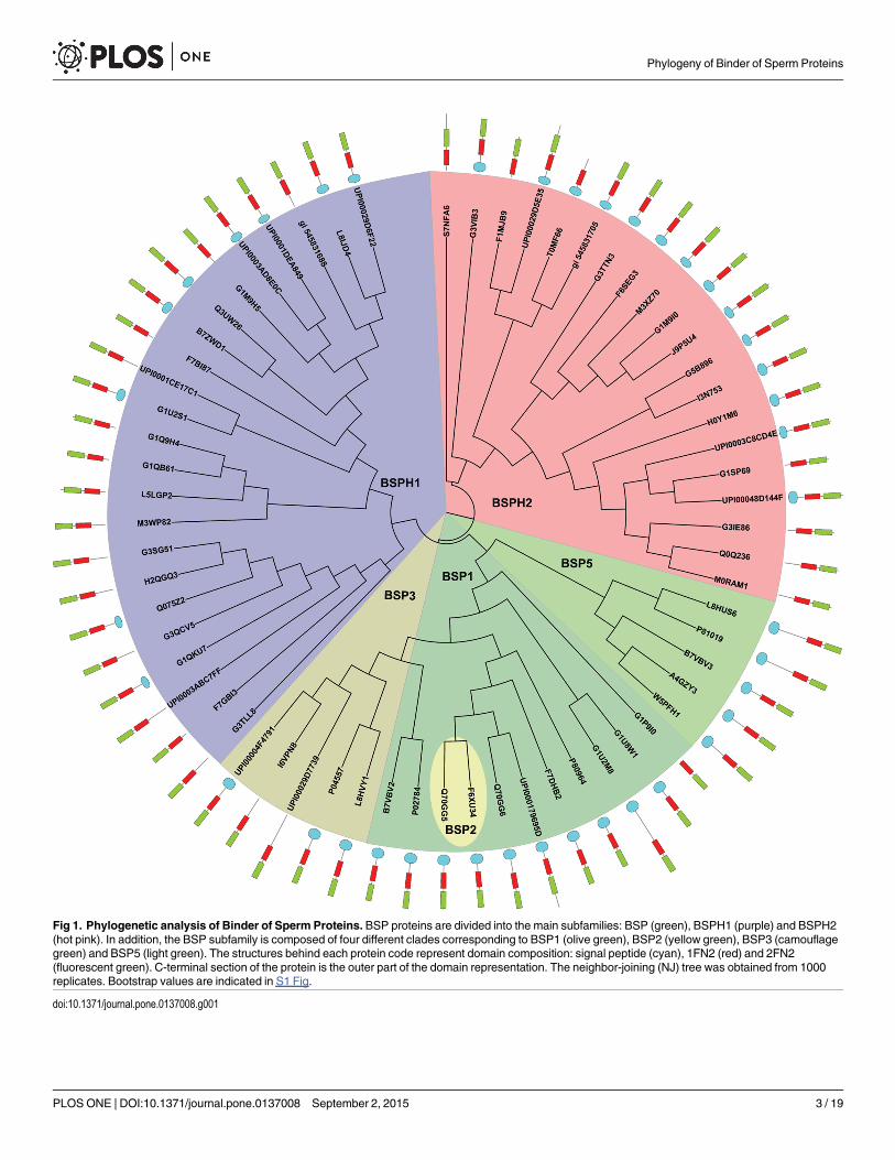
Fig 1. Phylogenetic analysis of Binder of Sperm Proteins. BSP proteins are divided into the main subfamilies: BSP (green), BSPH1 (purple) and BSPH2(hot pink). In addition, the BSP subfamily is composed of four different clades corresponding to BSP1 (olive green), BSP2 (yellow green), BSP3 (camouflagegreen) and BSP5 (light green). The structures behind each protein code represent domain composition: signal peptide (cyan), 1FN2 (red) and 2FN2(fluorescent green). C-terminal section of the protein is the outer part of the domain representation. The neighbor-joining (NJ) tree was obtained from 1000replicates. Bootstrap values are indicated in S1 Fig.
doi:10.1371/journal.pone.0137008.g001
Phylogeny of Binder of Sperm Proteins
PLOS ONE | DOI:10.1371/journal.pone.0137008 September 2, 2015 3 / 19
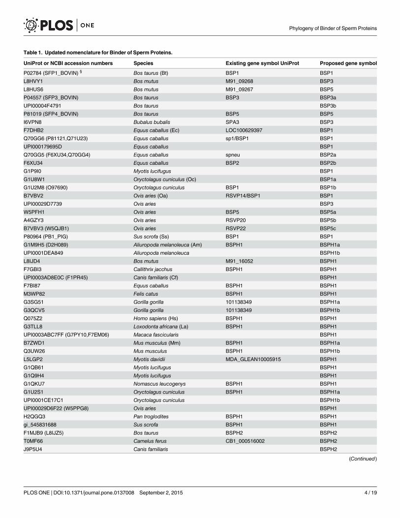
Table 1. Updated nomenclature for Binder of Sperm Proteins.
UniProt or NCBI accession numbers Species Existing gene symbol UniProt Proposed gene symbol
P02784 (SFP1_BOVIN) § Bos taurus (Bt) BSP1 BSP1
L8HVY1 Bos mutus M91_09268 BSP3
L8HUS6 Bos mutus M91_09267 BSP5
P04557 (SFP3_BOVIN) Bos taurus BSP3 BSP3a
UPI00004F4791 Bos taurus BSP3b
P81019 (SFP4_BOVIN) Bos taurus BSP5 BSP5
I6VPN8 Bubalus bubalis SPA3 BSP3
F7DHB2 Equus caballus (Ec) LOC100629397 BSP1
Q70GG6 (P81121,Q71U23) Equus caballus sp1/BSP1 BSP1
UPI000179695D Equus caballus BSP1
Q70GG5 (F6XU34,Q70GG4) Equus caballus spneu BSP2a
F6XU34 Equus caballus BSP2 BSP2b
G1P9I0 Myotis lucifugus BSP1
G1U8W1 Oryctolagus cuniculus (Oc) BSP1a
G1U2M8 (O97690) Oryctolagus cuniculus BSP1 BSP1b
B7VBV2 Ovis aries (Oa) RSVP14/BSP1 BSP1
UPI00029D7739 Ovis aries BSP3
W5PFH1 Ovis aries BSP5 BSP5a
A4GZY3 Ovis aries RSVP20 BSP5b
B7VBV3 (W5QJB1) Ovis aries RSVP22 BSP5c
P80964 (PB1_PIG) Sus scrofa (Ss) BSP1 BSP1
G1M9H5 (D2H089) Aliuropoda melanoleuca (Am) BSPH1 BSPH1a
UPI0001DEA849 Aliuropoda melanoleuca BSPH1b
L8IJD4 Bos mutus M91_16052 BSPH1
F7GBI3 Callithrix jacchus BSPH1 BSPH1
UPI0003AD8E0C (F1PR45) Canis familiaris (Cf) BSPH1
F7BI87 Equus caballus BSPH1 BSPH1
M3WP82 Felis catus BSPH1 BSPH1
G3SG51 Gorilla gorilla 101138349 BSPH1a
G3QCV5 Gorilla gorilla 101138349 BSPH1b
Q075Z2 Homo sapiens (Hs) BSPH1 BSPH1
G3TLL8 Loxodonta africana (La) BSPH1 BSPH1
UPI0003ABC7FF (G7PY10,F7EM06) Macaca fascicularis BSPH1
B7ZWD1 Mus musculus (Mm) BSPH1 BSPH1a
Q3UW26 Mus musculus BSPH1 BSPH1b
L5LGP2 Myotis davidii MDA_GLEAN10005915 BSPH1
G1QB61 Myotis lucifugus BSPH1
G1Q9H4 Myotis lucifugus BSPH1
G1QKU7 Nomascus leucogenys BSPH1 BSPH1
G1U2S1 Oryctolagus cuniculus BSPH1 BSPH1a
UPI0001CE17C1 Oryctolagus cuniculus BSPH1b
UPI00029D6F22 (W5PPG8) Ovis aries BSPH1
H2QGQ3 Pan troglodites BSPH1 BSPH1
gi_545831688 Sus scrofa BSPH1 BSPH1
F1MJB9 (L8IJZ5) Bos taurus BSPH2 BSPH2
T0MF66 Camelus ferus CB1_000516002 BSPH2
J9P5U4 Canis familiaris BSPH2
(Continued)
Phylogeny of Binder of Sperm Proteins
PLOS ONE | DOI:10.1371/journal.pone.0137008 September 2, 2015 4 / 19
orders Proboscidea, Primates, Lagomorpha, Rodentia, Chiroptera, Perissodactyla, Artiodactylaand Cetacea. However, BSPH2 proteins were also found in the Chinese tree shrew (Tupaia chi-nensis, UniProt code: UPI0003C8CD4E) belonging to the Scandentia Order and in themetatherian Tasmanian devil (UniProt code: G3VIB3), belonging to the Scandentia andDasyuromorphia orders, respectively. It is also noteworthy that two new sequences corre-sponding to O. aries BSPH1 (Uniprot code: UPI00029D6F22) and BSPH2 (UniProt code:UPI00029D5E35) were found close to their corresponding bovinae homologues (Uniprotcodes: L8IJD4 and F1MJB9, respectively).
Analysis of conserved sequence blocksIn order to fully understand the results described in Fig 1, a detailed study of the conservedsequence blocks was carried out using WebLogo3 [16] and ESPript [17] representations of thethree different subfamilies (BSP, BSPH1 and BSPH2), and illustrated in base of RSVP20sequence, corresponding to a non characterized BSP5 protein (S2 Fig). Nine conserved blockswere found (Fig 2), corresponding to two tandem FN2 domains with four blocks each and alinker block in between. Surprisingly, the C-ter FN2 (2FN2) domain is four amino acids longerthan N-ter FN2 (1FN2) (Fig 2).
The first block of the 1FN2 domain (Block I) has a consensus sequence C V/A FPFxY (Fig2A). This block includes the first conserved cysteine (C1 in Fig 2A, C63 in RSVP20, S2 Fig)involved in one of the two conserved disulfide bonds (C1-C25, Fig 2A), and the first β strand(F5-Y7 in Fig 2A, F67-Y69 in RSVP20, S2 Fig) of the double stranded antiparallel β sheet (β1-β2) (Fig 3A, right cyan and tan β-strands, respectively). Curiously, β2 (positions 10–12 in Fig2A, R72-Y74 in RSVP20, S2 Fig; Fig 3A right, tan β-strand) does not define a clear conservedblock either in BSPs or in BSPHs (Fig 2A). Block II (C T/I/V xxxS) is located the interveningβ2-β3 loop (positions 15–20 in Fig 2A, C77-S82 in RSVP20, S2 Fig: Fig 3A right, red loop) andits last two amino acids form (positions 19–20 in Fig 2A, N81-S82 in RSVP20) together withthe Block I conserved Y (Y7 in Fig 2A, Y69 in RSVP20, S2 Fig), one of the walls of the
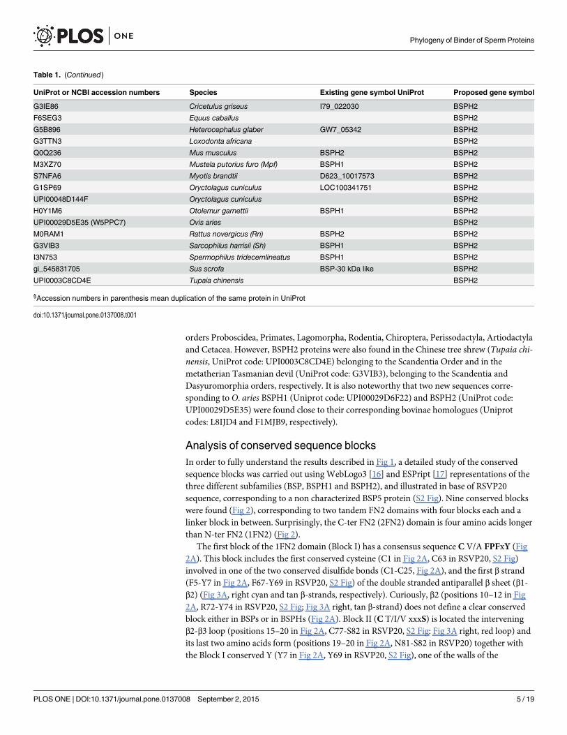
Table 1. (Continued)
UniProt or NCBI accession numbers Species Existing gene symbol UniProt Proposed gene symbol
G3IE86 Cricetulus griseus I79_022030 BSPH2
F6SEG3 Equus caballus BSPH2
G5B896 Heterocephalus glaber GW7_05342 BSPH2
G3TTN3 Loxodonta africana BSPH2
Q0Q236 Mus musculus BSPH2 BSPH2
M3XZ70 Mustela putorius furo (Mpf) BSPH1 BSPH2
S7NFA6 Myotis brandtii D623_10017573 BSPH2
G1SP69 Oryctolagus cuniculus LOC100341751 BSPH2
UPI00048D144F Oryctolagus cuniculus BSPH2
H0Y1M6 Otolemur garnettii BSPH1 BSPH2
UPI00029D5E35 (W5PPC7) Ovis aries BSPH2
M0RAM1 Rattus novergicus (Rn) BSPH2 BSPH2
G3VIB3 Sarcophilus harrisii (Sh) BSPH1 BSPH2
I3N753 Spermophilus tridecemlineatus BSPH1 BSPH2
gi_545831705 Sus scrofa BSP-30 kDa like BSPH2
UPI0003C8CD4E Tupaia chinensis BSPH2
§Accession numbers in parenthesis mean duplication of the same protein in UniProt
doi:10.1371/journal.pone.0137008.t001
Phylogeny of Binder of Sperm Proteins
PLOS ONE | DOI:10.1371/journal.pone.0137008 September 2, 2015 5 / 19
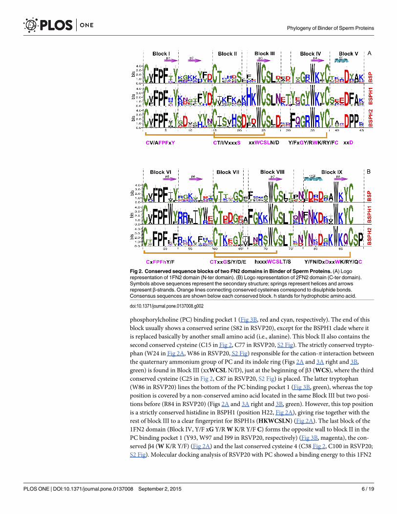
phosphorylcholine (PC) binding pocket 1 (Fig 3B, red and cyan, respectively). The end of thisblock usually shows a conserved serine (S82 in RSVP20), except for the BSPH1 clade where itis replaced basically by another small amino acid (i.e., alanine). This block II also contains thesecond conserved cysteine (C15 in Fig 2, C77 in RSVP20, S2 Fig). The strictly conserved trypto-phan (W24 in Fig 2A, W86 in RSVP20, S2 Fig) responsible for the cation-π interaction betweenthe quaternary ammonium group of PC and its indole ring (Figs 2A and 3A right and 3B,green) is found in Block III (xxWCSL N/D), just at the beginning of β3 (WCS), where the thirdconserved cysteine (C25 in Fig 2, C87 in RSVP20, S2 Fig) is placed. The latter tryptophan(W86 in RSVP20) lines the bottom of the PC binding pocket 1 (Fig 3B, green), whereas the topposition is covered by a non-conserved amino acid located in the same Block III but two posi-tions before (R84 in RSVP20) (Figs 2A and 3A right and 3B, green). However, this top positionis a strictly conserved histidine in BSPH1 (position H22, Fig 2A), giving rise together with therest of block III to a clear fingerprint for BSPH1s (HKWCSLN) (Fig 2A). The last block of the1FN2 domain (Block IV, Y/F xG Y/RW K/R Y/F C) forms the opposite wall to block II in thePC binding pocket 1 (Y93, W97 and I99 in RSVP20, respectively) (Fig 3B, magenta), the con-served β4 (W K/R Y/F) (Fig 2A) and the last conserved cysteine 4 (C38 Fig 2, C100 in RSVP20;S2 Fig). Molecular docking analysis of RSVP20 with PC showed a binding energy to this 1FN2
Fig 2. Conserved sequence blocks of two FN2 domains in Binder of Sperm Proteins. (A) Logorepresentation of 1FN2 domain (N-ter domain). (B) Logo representation of 2FN2 domain (C-ter domain).Symbols above sequences represent the secondary structure; springs represent helices and arrowsrepresent β-strands. Orange lines connecting conserved cysteines correspond to disulphide bonds.Consensus sequences are shown below each conserved block. h stands for hydrophobic amino acid.
doi:10.1371/journal.pone.0137008.g002
Phylogeny of Binder of Sperm Proteins
PLOS ONE | DOI:10.1371/journal.pone.0137008 September 2, 2015 6 / 19
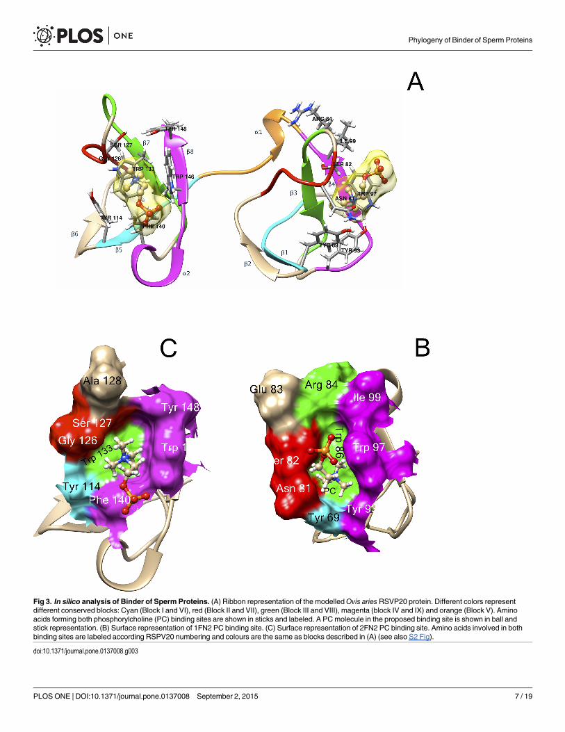
Fig 3. In silico analysis of Binder of Sperm Proteins. (A) Ribbon representation of the modelledOvis ariesRSVP20 protein. Different colors representdifferent conserved blocks: Cyan (Block I and VI), red (Block II and VII), green (Block III and VIII), magenta (block IV and IX) and orange (Block V). Aminoacids forming both phosphorylcholine (PC) binding sites are shown in sticks and labeled. A PCmolecule in the proposed binding site is shown in ball andstick representation. (B) Surface representation of 1FN2 PC binding site. (C) Surface representation of 2FN2 PC binding site. Amino acids involved in bothbinding sites are labeled according RSPV20 numbering and colours are the same as blocks described in (A) (see also S2 Fig).
doi:10.1371/journal.pone.0137008.g003
Phylogeny of Binder of Sperm Proteins
PLOS ONE | DOI:10.1371/journal.pone.0137008 September 2, 2015 7 / 19
domain binding pocket of -5.99 kcal/mol, which is in the range of previously described valuesof murine BSPH1 and BSPH2 (both with a value of -3.8 kcal/mol) [8]. The interdomain orlinker segment (Block V, Fig 3A, orange) shows only a clear conserved aspartic at the end of α1(xxD, D42 in Fig 2A, D104 in RSVP20, S2 Fig). However, this domain displays a clear finger-print for BSPH2s (DPP) together with Block IV (FxGRWRYC). This Block V has beendescribed as being involved in the dimerization process together with edges of the β2-β3 loop[18].
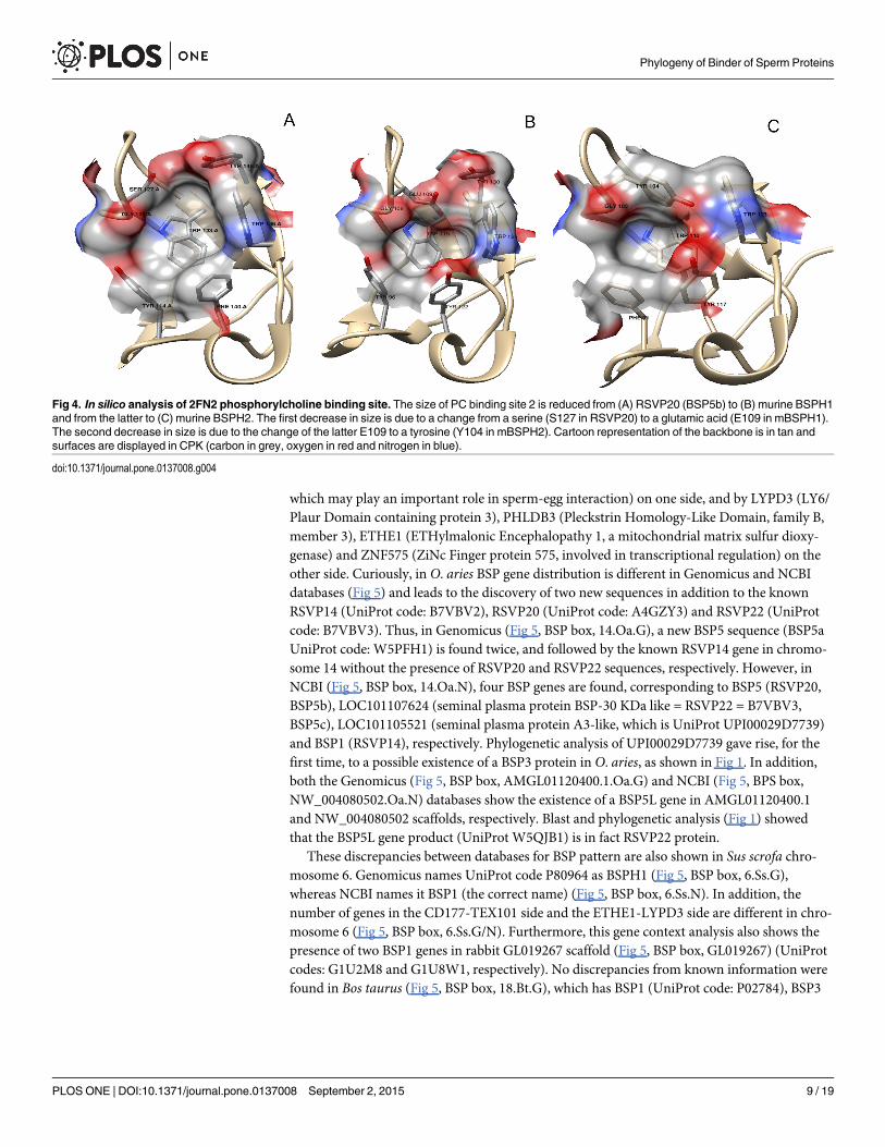
Block VI (CxFPFh Y/F, where h stands for hydrophobic amino acid) at the 2FN2 domain issimilar to that of Block I (Fig 3A left, cyan). However, this latter block is clearly another finger-print in BSPH1 proteins (ChFPFWY), where a strictly conserved tryptophan in the middle ofβ5 appeared, giving rise to a clear hydrophobic tract (V/A FPFW) (Fig 2B). The last hydropho-bic amino acid of Block VI (position 52 in Fig 2B, and Y114 in RSVP20; S2 Fig; Fig 3C, cyan)and the last two amino acids of Block VII (GTxxG S/Y/D/E) (positions 64–65 in Fig 2B, G126and S127 in RSVP20, S2 Fig; Fig 3C, red) form one of the walls of the PC binding pocket 2. Thebottom of this pocket 2 is occupied by the completely conserved tryptophan (W71 in Fig 2B,W133 in RSVP20, S2 Fig; Fig 3C, green) of Block VIII (hxxxWCSL T/S). This latter block isalso another fingerprint for the BSPH1 family (FGKKWCSLT). Two differences are clearlyshown when this block VIII is compared with the homolog block (Block III) in the 1FN2domain. The first is related with the size, as Block VIII is two amino acids longer than Block III(Fig 2). The second is related with the fact that only one amino acid (W71 in Fig 2B, W133 inRSVP20, S2 Fig) of this Block VIII is involved in the PC binding pocket 2 structure (Fig 3C,green), whereas two amino acids (position 22 andW24 in Fig 2A, R84 and W86 in RSVP20, S2Fig) of Block III are associated with the PC binding site 1 structure (Fig 3B, green). Finally,Block IX (Y/F N/D xDxxW K/R Y/Q C) at C-ter delimits the opposite wall to Block VI and VIIat the PC binding pocket 2 (Fig 3C, magenta). In fact, the wall is formed by the first amino acidof α2 (Y/F N/D xD; position 78 in Fig 2B, F140 in RSVP20, S2 Fig), and the first and the lastamino acid of β8 (W K/R Y/Q; W84 and position 86 in Fig 2B, W146 and Y148 in RSVP20, S2Fig, respectively) (Fig 3C, magenta). This Block IX also differs in size from the homolog in1FN2, this block again being two amino acids longer (Fig 2). It is noteworthy that this Block IXis an unquestionable BSPH2 fingerprint (YNxDxKWKQC), which has to be supplementedwith two strictly conserved amino acids (serine and proline, SP) at the end of the 2FN2 motif(Fig 2B). This conserved C-ter extension after 2FN2 is shown neither in BSPs nor BSPH1s (Fig2B). The 2FN2 pocket appeared to bind PC with an affinity of -5.29 kcal/mol, which is in therange of murine BSPH1 (-3.5 kcal/mol) but higher than that described for murine BSPH2 (-2.8kcal/mol) [8]. These docking results could be explained by the decreasing size of the PC bind-ing pocket 2 from BSPs (i.e., RSVP20) to mBSPH1 and from mBSPH1 to mBSPH2 (Fig 4, S2Fig triangle). The first decrease in size is due to a change from a serine (S127 in RSVP20) to aglutamic acid (E109 in mBSPH1) (Fig 4A and 4B, S2 Fig triangle). The second is due to thechange of the latter glutamic acid (E109 in mBSPH1) for a tyrosine (Y104 in mBSPH2), whoseelectronic density connects with that of Y117, closing the binding site (Fig 4B and 4C, S2 Figtriangle).
Gene context analysisTo better understand the role of the BSP family, a gene context analysis was carried out todetermine its potential operonic associations, using the Genomicus and NCBI databases (Fig5). The BSP subfamily, to which RSVP20 belongs, shows a regular pattern in which BSP pro-teins are flanked on one side by the gene cluster formed by CD177 (GPI-linked surface protein)and TEX101 (Testis EXpressed 101 protein, found in epididymal sperm plasma membrane and
Phylogeny of Binder of Sperm Proteins
PLOS ONE | DOI:10.1371/journal.pone.0137008 September 2, 2015 8 / 19
which may play an important role in sperm-egg interaction) on one side, and by LYPD3 (LY6/Plaur Domain containing protein 3), PHLDB3 (Pleckstrin Homology-Like Domain, family B,member 3), ETHE1 (ETHylmalonic Encephalopathy 1, a mitochondrial matrix sulfur dioxy-genase) and ZNF575 (ZiNc Finger protein 575, involved in transcriptional regulation) on theother side. Curiously, in O. aries BSP gene distribution is different in Genomicus and NCBIdatabases (Fig 5) and leads to the discovery of two new sequences in addition to the knownRSVP14 (UniProt code: B7VBV2), RSVP20 (UniProt code: A4GZY3) and RSVP22 (UniProtcode: B7VBV3). Thus, in Genomicus (Fig 5, BSP box, 14.Oa.G), a new BSP5 sequence (BSP5aUniProt code: W5PFH1) is found twice, and followed by the known RSVP14 gene in chromo-some 14 without the presence of RSVP20 and RSVP22 sequences, respectively. However, inNCBI (Fig 5, BSP box, 14.Oa.N), four BSP genes are found, corresponding to BSP5 (RSVP20,BSP5b), LOC101107624 (seminal plasma protein BSP-30 KDa like = RSVP22 = B7VBV3,BSP5c), LOC101105521 (seminal plasma protein A3-like, which is UniProt UPI00029D7739)and BSP1 (RSVP14), respectively. Phylogenetic analysis of UPI00029D7739 gave rise, for thefirst time, to a possible existence of a BSP3 protein in O. aries, as shown in Fig 1. In addition,both the Genomicus (Fig 5, BSP box, AMGL01120400.1.Oa.G) and NCBI (Fig 5, BPS box,NW_004080502.Oa.N) databases show the existence of a BSP5L gene in AMGL01120400.1and NW_004080502 scaffolds, respectively. Blast and phylogenetic analysis (Fig 1) showedthat the BSP5L gene product (UniProt W5QJB1) is in fact RSVP22 protein.
These discrepancies between databases for BSP pattern are also shown in Sus scrofa chro-mosome 6. Genomicus names UniProt code P80964 as BSPH1 (Fig 5, BSP box, 6.Ss.G),whereas NCBI names it BSP1 (the correct name) (Fig 5, BSP box, 6.Ss.N). In addition, thenumber of genes in the CD177-TEX101 side and the ETHE1-LYPD3 side are different in chro-mosome 6 (Fig 5, BSP box, 6.Ss.G/N). Furthermore, this gene context analysis also shows thepresence of two BSP1 genes in rabbit GL019267 scaffold (Fig 5, BSP box, GL019267) (UniProtcodes: G1U2M8 and G1U8W1, respectively). No discrepancies from known information werefound in Bos taurus (Fig 5, BSP box, 18.Bt.G), which has BSP1 (UniProt code: P02784), BSP3
Fig 4. In silico analysis of 2FN2 phosphorylcholine binding site. The size of PC binding site 2 is reduced from (A) RSVP20 (BSP5b) to (B) murine BSPH1and from the latter to (C) murine BSPH2. The first decrease in size is due to a change from a serine (S127 in RSVP20) to a glutamic acid (E109 in mBSPH1).The second decrease in size is due to the change of the latter E109 to a tyrosine (Y104 in mBSPH2). Cartoon representation of the backbone is in tan andsurfaces are displayed in CPK (carbon in grey, oxygen in red and nitrogen in blue).
doi:10.1371/journal.pone.0137008.g004
Phylogeny of Binder of Sperm Proteins
PLOS ONE | DOI:10.1371/journal.pone.0137008 September 2, 2015 9 / 19
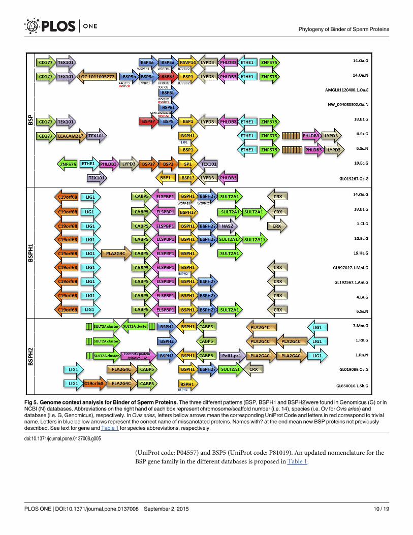
(UniProt code: P04557) and BSP5 (UniProt code: P81019). An updated nomenclature for theBSP gene family in the different databases is proposed in Table 1.
Fig 5. Genome context analysis for Binder of Sperm Proteins. The three different patterns (BSP, BSPH1 and BSPH2)were found in Genomicus (G) or inNCBI (N) databases. Abbreviations on the right hand of each box represent chromosome/scaffold number (i.e. 14), species (i.e. Ov forOvis aries) anddatabase (i.e. G, Genomicus), respectively. InOvis aries, letters bellow arrows mean the corresponding UniProt Code and letters in red correspond to trivialname. Letters in blue bellow arrows represent the correct name of missanotated proteins. Names with? at the end mean new BSP proteins not previouslydescribed. See text for gene and Table 1 for species abbreviations, respectively.
doi:10.1371/journal.pone.0137008.g005
Phylogeny of Binder of Sperm Proteins
PLOS ONE | DOI:10.1371/journal.pone.0137008 September 2, 2015 10 / 19
A second gene context group was found to be associated with BSPH1 genes (Fig 5, BSPH1box). In this group, BSPH1 is flanked on one side by C19orf68, LIG1 (ATP-dependent DNALIGase I), CABP5 (Calcium Binding Protein 5) and ELSPBP1 (EpididymaL Sperm BindingProtein 1), and on the other side by BSPH2, SULT2A1 (SULfoTransferase 2A1) and CRX(Cone-Rod homeoboX) (Fig 5, BSPH1 box). This pattern is also associated with O. ariesBSPHs, with BSPH1 (UniProt code: W5PPG8) and BSPH2 (UniProt code: W5PPC7) beingdescribed for the first time in chromosome 14 (Fig 5, BSPH1 box, 14.Oa.G). New BSPH2s werealso found in this gene context in dog (UniProt code: J9P5V4), horse (UniProt code: F6SEG3),giant panda (UniProt code: G1M910), African bush elephant (UniProt code: G3TTN3) andpig (NCBI code: gi_545831705) (Fig 5, BSPH1 box, 1.Cf.G, 10.Ec.G, GL192567.1.Am.G, 4.La.Gand 6.Ss.N, respectively). Interestingly, a misannotation was found in ferret (Mustela putoriusfuro) GL897027.1 scaffold (Fig 5, BSPH1 box, GL897027.1.Mpf.G), where M3XZ70 isdescribed as BSPH1, when in fact, it is a BSPH2 (Figs 1 and 5). Moreover, this ferret BSPH2does not fulfill in the third group of the gene neighborhood pattern described by some BSPH2s(Fig 5, BSPH2 box), in which BSPH2/BSPH1 are not related with ELSPBP1 and a new geneappears (PLA2G4C, PhosphoLipase A2 Group IVC) not found in the above-described pat-terns. This pattern is present in mouse, rabbit, Tasmanian devil and rat. In the latter, new dis-crepancies between NCBI and Genomicus were found (Fig 5, BSPH2 box, 1.Rn.G and 1.Rn.N,respectively), focused on the absence of BSPH1 (UniProt code: B7ZWD1) in Genomicus. Inaddition, Genomicus ascribed G3VIB3 in Tasmanian devil GL850016.1 scaffold to a BSPH1(Fig 5, BSPH2 box, GL8500161.Sh.G), when in fact it is a BSPH2. These new and misannotatedsequences are also compiled in Table 1.
Expression of RSVP20 and binding of the recombinant protein tospermatozoaIn order to validate the above in silico studies, the gene corresponding to Ovis aries RSVP20was cloned and expressed in E. coli, since no previous BSP5 protein has been cloned, probablygiven their tendency to aggregate as inclusion bodies (IB). Using yeast SUMO (a small ubiqui-tin-like modifier) as a tag, the RSVP20 expression degree was high. Following IB solubilisation,RSVP20 was refolded on column and purified by affinity chromatography. SDS-PAGE analysis(S3 Fig) revealed the presence of RSVP20 in the flow-through (FT), where a high urea concen-tration occurs, and in two elution fractions. In the first elution fraction (E1), which corre-sponds to elution with 100 mM imidazole, RSVP20 was observed with other smaller bands.However, these bands were not present in the E2 fraction (elution with 250 mM imidazole),giving rise to purer fraction.
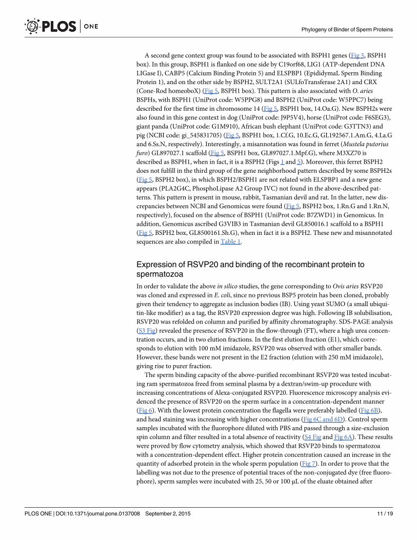
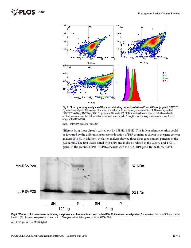
The sperm binding capacity of the above-purified recombinant RSVP20 was tested incubat-ing ram spermatozoa freed from seminal plasma by a dextran/swim-up procedure withincreasing concentrations of Alexa-conjugated RSVP20. Fluorescence microscopy analysis evi-denced the presence of RSVP20 on the sperm surface in a concentration-dependent manner(Fig 6). With the lowest protein concentration the flagella were preferably labelled (Fig 6B),and head staining was increasing with higher concentrations (Fig 6C and 6D). Control spermsamples incubated with the fluorophore diluted with PBS and passed through a size-exclusionspin column and filter resulted in a total absence of reactivity (S4 Fig and Fig 6A). These resultswere proved by flow cytometry analysis, which showed that RSVP20 binds to spermatozoawith a concentration-dependent effect. Higher protein concentration caused an increase in thequantity of adsorbed protein in the whole sperm population (Fig 7). In order to prove that thelabelling was not due to the presence of potential traces of the non-conjugated dye (free fluoro-phore), sperm samples were incubated with 25, 50 or 100 μL of the eluate obtained after
Phylogeny of Binder of Sperm Proteins
PLOS ONE | DOI:10.1371/journal.pone.0137008 September 2, 2015 11 / 19
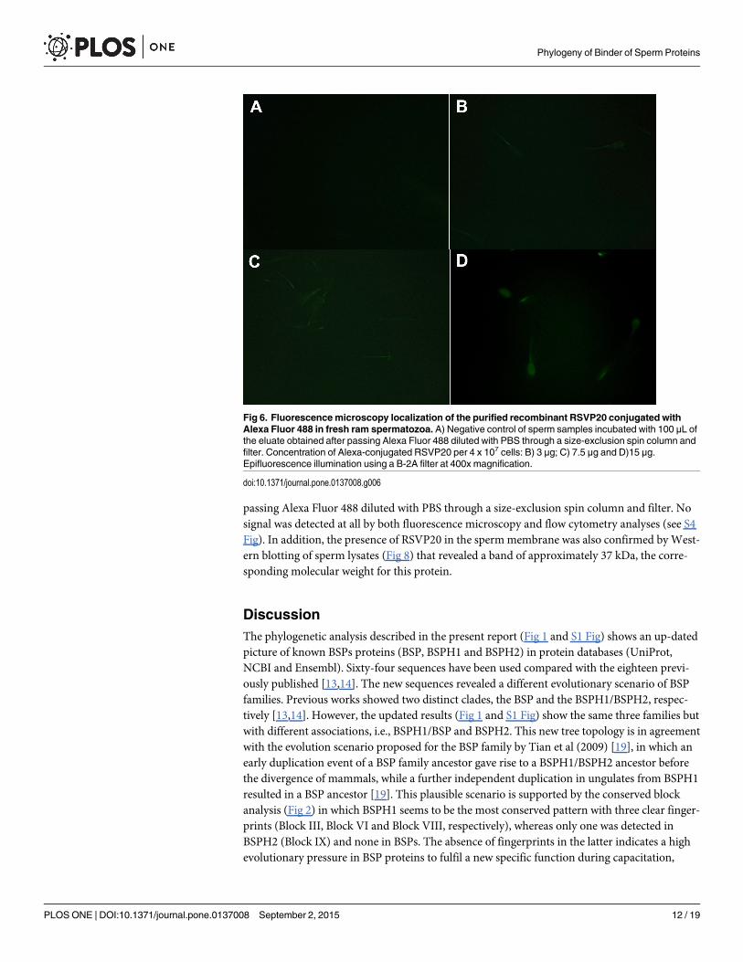
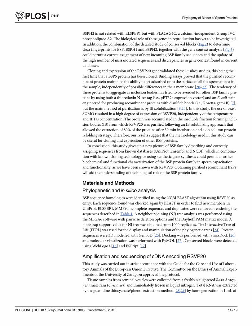
passing Alexa Fluor 488 diluted with PBS through a size-exclusion spin column and filter. Nosignal was detected at all by both fluorescence microscopy and flow cytometry analyses (see S4Fig). In addition, the presence of RSVP20 in the spermmembrane was also confirmed byWest-ern blotting of sperm lysates (Fig 8) that revealed a band of approximately 37 kDa, the corre-sponding molecular weight for this protein.
DiscussionThe phylogenetic analysis described in the present report (Fig 1 and S1 Fig) shows an up-datedpicture of known BSPs proteins (BSP, BSPH1 and BSPH2) in protein databases (UniProt,NCBI and Ensembl). Sixty-four sequences have been used compared with the eighteen previ-ously published [13,14]. The new sequences revealed a different evolutionary scenario of BSPfamilies. Previous works showed two distinct clades, the BSP and the BSPH1/BSPH2, respec-tively [13,14]. However, the updated results (Fig 1 and S1 Fig) show the same three families butwith different associations, i.e., BSPH1/BSP and BSPH2. This new tree topology is in agreementwith the evolution scenario proposed for the BSP family by Tian et al (2009) [19], in which anearly duplication event of a BSP family ancestor gave rise to a BSPH1/BSPH2 ancestor beforethe divergence of mammals, while a further independent duplication in ungulates from BSPH1resulted in a BSP ancestor [19]. This plausible scenario is supported by the conserved blockanalysis (Fig 2) in which BSPH1 seems to be the most conserved pattern with three clear finger-prints (Block III, Block VI and Block VIII, respectively), whereas only one was detected inBSPH2 (Block IX) and none in BSPs. The absence of fingerprints in the latter indicates a highevolutionary pressure in BSP proteins to fulfil a new specific function during capacitation,
Fig 6. Fluorescencemicroscopy localization of the purified recombinant RSVP20 conjugated withAlexa Fluor 488 in fresh ram spermatozoa. A) Negative control of sperm samples incubated with 100 μL ofthe eluate obtained after passing Alexa Fluor 488 diluted with PBS through a size-exclusion spin column andfilter. Concentration of Alexa-conjugated RSVP20 per 4 x 10⁷ cells: B) 3 μg; C) 7.5 μg and D)15 μg.Epifluorescence illumination using a B-2A filter at 400x magnification.
doi:10.1371/journal.pone.0137008.g006
Phylogeny of Binder of Sperm Proteins
PLOS ONE | DOI:10.1371/journal.pone.0137008 September 2, 2015 12 / 19
different from those already carried out by BSPH1/BSPH2. This independent evolution couldbe favoured by the different chromosome location of BSP proteins as shown in the gene contextanalysis (Fig 5). In addition, the latter analysis showed three clear gene context patterns in theBSP family. The first is associated with BSPs and is closely related to the CD177 and TEX101genes. In the second, BSPH1/BSPH2 coexists with the ELSPBP1 gene. In the third, BSPH1/
Fig 7. Flow cytometry analysis of the sperm binding capacity of Alexa Fluor 488-conjugated RSVP20.Cytometry analysis of the effect of sperm incubation with increasing concentration of Alexa-conjugatedRSVP20: A) 3 μg; B) 7.5 μg; C) 15 μg per 4 x 10⁷ cells. D) Plots showing the number of cells linked withprotein (events) and the different fluorescence intensity (FL1 Log) for increasing concentrations of Alexa-conjugated RSVP20.
doi:10.1371/journal.pone.0137008.g007
Fig 8. Western blot membrane indicating the presence of recombinant and native RSVP20 in ram sperm lysates. Supernatant fraction (SN) and pelletfraction (P) of sperm samples incubated with (100 ug) or without (0 ug) recombinant RSVP20.
doi:10.1371/journal.pone.0137008.g008
Phylogeny of Binder of Sperm Proteins
PLOS ONE | DOI:10.1371/journal.pone.0137008 September 2, 2015 13 / 19
BSPH2 is not related with ELSPBP1 but with PLA2AG4C, a calcium-independent Group IVCphospholipase A2. The biological role of these genes in reproduction has yet to be investigated.In addition, the combination of the detailed study of conserved blocks (Fig 2) to determineclear fingerprints for BSP, BSPH1 and BSPH2, together with the gene context analysis (Fig 5)could permit a correct assignment of new incoming BSP family sequences and the update ofthe high number of missanotated sequences and discrepancies in gene context found in currentdatabases.
Cloning and expression of the RSVP20 gene validated these in silico studies, this being thefirst time that a BSP5 protein has been cloned. Binding assays proved that the purified recom-binant protein maintains the ability to get adsorbed onto the surface of all the spermatozoa inthe sample, independently of possible differences in their membrane [20–22]. The tendency ofthese proteins to aggregate as inclusion bodies has tried to be avoided for other BSP family pro-teins by using both a thioredoxin N-ter tag (i.e., pET32a expression vector) and an E. coli stainengineered for producing recombinant proteins with disulfide bonds (i.e., Rosetta-gami B) [7],but the main method of purifcation is by IB solubilization [8,23]. In this study, the use of yeastSUMO resulted in a high degree of expression of RSVP20, independently of the temperatureand IPTG concentration. The protein was accumulated in the insoluble fraction forming inclu-sion bodies (IB) from which RSVP20 was purified following an IB solubilizing approach thatallowed the extraction of 80% of the proteins after 30 min incubation and a on column proteinrefolding strategy. Therefore, our results suggest that the methodology used in this study canbe useful for cloning and expression of other BSP proteins.
In conclusion, this study gives up a new picture of BSP family describing and correctlyassigning sequences from known databases (UniProt, Ensembl and NCBI), which in combina-tion with known cloning technology or using synthetic gene synthesis could permit a furtherbiochemical and functional characterization of the BSP protein family in sperm capacitationand functionality, as we have been shown with RSVP20. Obtaining purified recombinant BSPswill aid the understanding of the biological role of the BSP protein family.
Materials and Methods
Phylogenetic and in silico analysisBSP sequence homologies were identified using the NCBI BLAST algorithm using RSVP20 asentry. Each sequence found was checked again by BLAST in order to find new members inUniProt. ELSPBP1, MMP9, incomplete sequences and duplicates were removed, rendering thesequences described in Table 1. A neighbour-joining (NJ) tree analysis was performed usingthe MEGA6 software with pairwise deletion options and the Dayhoff PAMmatrix model. Abootstrap support value for NJ tree was obtained from 1000 replicates. The Interactive Tree ofLife (iTOL) was used for the display and manipulation of the phylogenetic trees [24]. Proteinsequences were 3D modelled with Geno3D [25]. Docking was performed with SwissDock [26]and molecular visualization was performed with PyMOL [27]. Conserved blocks were detectedusing WebLogo3 [16] and ESPript [17].
Amplification and sequencing of cDNA encoding RSVP20This study was carried out in strict accordance with the Guide for the Care and Use of Labora-tory Animals of the European Union Directive. The Committee on the Ethics of Animal Exper-iments of the University of Zaragoza approved the protocol.
Tissue samples from seminal vesicles were collected from a freshly slaughtered Rasa Arago-nesamale ram (Ovis aries) and immediately frozen in liquid nitrogen. Total RNA was extractedby the guanidine thiocyanate/phenol extraction method [28,29] by homogenization in 1 mL of
Phylogeny of Binder of Sperm Proteins
PLOS ONE | DOI:10.1371/journal.pone.0137008 September 2, 2015 14 / 19
TRI reagent (Sigma-Aldrich) per 200 mg of tissue. RNA concentration was measured in a Nano-Drop ND-100 Spectrophotometer (Wilmington, DE, USA). 500 ng of total RNA was reversetranscribed using poly (dT) primers and the SuperScript III RT enzyme (Invitrogen, CA, USA).
Degenerate PCR primers (S1 Table) for RSVP20 were designed according to the primarysequence of the protein [30] in the 5’ region. Primer RSVP20-5’ was based on amino acids resi-dues 1–6. The 3’ primer was Oligo (dT)20. PCR was performed with 2 μL of cDNA. Using theseprimers, PCR (35 cycles) were carried out on reverse-transcribed RNA from ram seminal vesi-cles. Cycling conditions consisted of 45 s at 94°C, 1 min and 30 s at 53°C, and 3 min at 72°C. A1 min denaturation step at 94°C preceded cycling; at the end, a final 10 min extension at 72°Cwas performed. PCR products were separated on 2% agarose gel in 1x Tris-borate-EDTA(TBE) buffer containing 0.5 μL/mL ethidium bromide and were visualized under ultraviolet(UV) light. Molecular size was estimated by using GeneRuler 1kb plus (Thermo Scientific).PCR products were gel-purified using a GeneJet gel extraction kit (Thermo Scientific), in accor-dance with the manufacturer’s instructions and sequenced on an ABI Prism 3730 sequencer(Applied Biosystems, Foster City, CA, USA).
Cloning of cDNA sequences into E. coliRSVP20 was cloned into pE-SUMO3 plasmid, which contained both a His-tag and a SUMO-tag,using primers 20BbsI-Fw and 20XbaI-Rv (S1 Table). The plasmid pE-SUMO3-RSVP20 wastransformed into Origami B (DE3)pLysS competent cells. Transformed bacteria were grown over-night on LB-agar plates containing 50 mg/L ampicillin and 15 mg/L kanamycin. Selected colonieswere grown in 5 mL LB media with 50 mg/L ampicillin and 15 mg/L kanamycin to an OD600 of0.6–0.8. Protein expression was induced by adding isopropyl-β-D-thiogalactoside (IPTG) to finalconcentrations of 0.5 and 1 mM. Inductions were performed at 37°C for 8 h and 20°C for 16 h.One mL culture samples were collected before and after the IPTG induction. E. coli cells were har-vested from the samples by centrifugation at 6800 x g for 2 min at 4°C, resuspended in lysis buffer(50 mM Tris-HCl, 300 mMNaCl, 10% B-PER, lysozyme 1mg/mL, pH 8.0) and lysed by sonica-tion. The lysate was centrifuged for 5 min at 6500 x g and the resulting pellet was resuspended inlysis buffer. Both the pellet and the supernatant were checked for expression.
Nickel affinity chromatography and refolding of RSVP20Purification of RSVP20 was carried out from inclusion bodies (IB) using nickel affinity chro-matography. E. coli cells were grown in 1 L of TB (Terrific broth) medium to an OD600 of4.0. Protein expression was induced with 0.5 mM of IPTG for 16 h at 20°C. Afterwards, thecells were harvested by centrifugation at 6500 x g for 15 min at 4°C and the cell pellets wereresuspended in lysis buffer and lysed by sonication. The lysate was centrifuged for 15 min at6500 x g and the pellet was washed twice, first with lysis buffer and then with sample buffer(20 mM Tris, 500 mM NaCl, 5 mM imidazole pH 7.5) containing 2 M urea. The final pelletwas resuspended in denaturing buffer (sample buffer with 8 M urea and 10 mM β-mercap-toethanol). Soluble and insoluble fractions were separated by centrifugation at 40000 x g for40 minutes and the soluble fraction was loaded into a His-TrapFF column (GE Healthcare)equilibrated with denaturing buffer at a flow rate of 1 mL/min. The column was then washedwith 5 bed volumes of denaturing buffer and 5 volumes of washing buffer (20 mM Tris,500 mM NaCl, 80 mM imidazole, 8 M urea, 10 mM β-mercaptoethanol, pH 7.5). Refoldingof the bound protein was performed with an on-column decreasing linear gradient of 7 mMurea/min from denaturing buffer to sample buffer. The refolded proteins were eluted succes-sively with elution buffer containing two different imidazole concentrations (20 mM Tris,500 mM NaCl pH 7.5, with 100 mM and 250 mM imidazole, respectively).
Phylogeny of Binder of Sperm Proteins
PLOS ONE | DOI:10.1371/journal.pone.0137008 September 2, 2015 15 / 19
Fluorescent labeling and binding of RSVP20 to spermatozoaRecombinant RSVP20 was conjugated with Alexa Fluor using Alexa Fluor 488 Microscale Pro-tein Labeling Kit (Life Technologies) following the manufacturer’s instructions. Briefly, 100 μgof protein (1 μg/μL) were incubated with the dye for 15 minutes. Afterwards, non-conjugateddye was removed using the size-exclusion spin columns (Bio-Gel P-6) and the filters providedwith the kit.
Final protein concentration was calculated using the formula:
Protein concentration ðMÞ ¼ ½A280 � ðA494 � 0:11Þ� � dilution factor45; 380
where 45,380 is the theoretical molar extinction coefficient (ε) in cm–1 M–1 of RSVP20 at280 nm and 0.11 is a correction factor for the fluorophore’s contribution to the absorbance at280 nm. The obtained protein concentration was 0.15 mg/mL.
Fresh ram semen was collected from 4 mature Rasa aragonesa rams using an artificialvagina. The rams belonged to the National Association of Rasa Aragonesa Breeding (ANGRA)and were 2 to 4 years old. They were kept at the Experimental Farm of the University of Zara-goza (Spain) under uniform nutritional conditions in compliance with the requirements of theEuropean Union Directive for Scientific Procedures. A seminal plasma–free sperm populationwas obtained by a dextran/swim-up procedure (García-López et al, 1996) performed at 37°Cusing a medium without Ca2Cl and NaHCO3 (Pérez-Pé et al, 2002). Sperm concentration wascalculated in duplicate with a Neubauer’s chamber (Marienfeld, Germany). 4 x 10⁷ cells in afinal volume of 500 μL completed with PBS were used for all the experiments.
The binding of different concentrations of Alexa conjugated RSVP20 (3 μg, 7.5 μg and15 μg) was detected by fluorescence microscopy using a Nikon Eclipse E400 microscope(Nikon, Tokyo, Japan) with a B-2A filter (excitation 450–490 nm) at 400x magnification, andflow cytometry using an equipment (Beckman Coulter FC 500, IZASA, Barcelona) with a CXPsoftware, equipped with two lasers of excitation (Argon ion laser 488 nm and solid state laser633 nm) and five filters of absorbance (FL1-525, FL2-575, FL3-610, FL4-675, and FL5-755;65 nm each band pass filter). 4 x 10⁷ spermatozoa were incubated at room temperature (RT) indarkness for 15 min with different concentrations of RSVP20 (final volume 500 μL), thenwashed with 500 μL PBS at 600 xg for 5 min. The final pellet was resuspended in 500 μL PBSand samples were mounted onto microscope slides or analyzed by flow cytometry. At a mini-mum, 20,000 events were counted in all cytometry experiments. The sperm population wasgated for further analysis on the basis of its specific forward (FS) and side scatter (SS) proper-ties; other non-sperm events were excluded. A flow rate stabilized at 200–300 cells per secondwas used. Monitored parameters were FS log, SS log, and FL1 (Alexa Fluor 488).
In order to prove that the labelling was not due to the presence of potential traces of thenon-conjugated dye (free fluorophore), the content of another fluorophore vial was dilutedwith 100 μL PBS instead of the protein sample, and passed through a new size-exclusion spincolumn and filter provided with the kit. Sperm samples were incubated with the same volumesof this eluate as those used in the binding protein assays (20, 50 and 100 μL), and fluorescencemicroscopy and flow cytometry analyses were carried out.
To study the binding of RSVP20 to sperm membranes by Western blot, 4 x 10⁷ cells freedfrom seminal plasma were incubated 15 min at RT in the presence or absence of 100 μg ofRSVP20 in a final volume of 500 μL. Following incubation, the unbound protein was removedby centrifuging at 600 xg for 8 min. Then, the supernatant was removed and the pellet waswashed with 300 μL of PBS and centrifuged again. The pellet was resuspended with 100 μL ofPBS and 100 μL of extraction buffer (125 mM Tris-HCl, 4% SDS) and, after incubation at
Phylogeny of Binder of Sperm Proteins
PLOS ONE | DOI:10.1371/journal.pone.0137008 September 2, 2015 16 / 19
100°C in a sand bath for 5 min, it was centrifuged again at 12,000 xg for 5 min. A mix of 10%of a protease and phosphatase inhibitor cocktail (Sigma-Aldrich), 10% β-mercaptoethanol,20% glycerol, and 0.02% bromophenol blue were added to all recovered fractions, and the sam-ples were analyzed by SDS-PAGE and Western blot.
SDS-PAGE was performed in 14% polyacrylamide gel using a Mini protean III system (Bio-Rad, Hercules, CA, USA). Electrophoresis was performed for 90 min at 130 V at 4°C. A mix-ture of pre-stained protein standards (Bio-Rad, Hercules, CA, USA) was used as a marker. Theproteins were transferred to a polyvinylidene difluoride (PVDF) membrane using the Trans-blot Turbo (Bio-Rad, Hercules, CA, USA). The transference was performed for 10 min at 2.5A-25 V and the membrane was air dried for 15 min. Non-specific sites on the membrane wereblocked for 1 h with 5% BSA in PBS. RSVP20 was detected by incubating overnight at 4°C withspecific rabbit generated antibodies [31] diluted 1:60,000 in PBS with 1% BSA and 1% Tween.After exhaustive washing, the membranes were incubated with a secondary anti-rabbit Dylight680 conjugated (Thermo Scientific, Madrid, Spain) diluted 1:15,000 for 1 h at RT. After wash-ing, the membrane was scanned using Odyssey Clx (Li-Cor Biosciences, Lincoln, NE, USA).
Supporting InformationS1 Fig. NJ phylogenetic tree of Binder of Sperm Proteins. The numbers indicate the NJ boot-strap values for 1000 replicates (see Material and Method for details). Only bootstrap valueslarger than 50% are shown and bold numbers indicate the three main families. UniProt andNCBI codes are listed in Table 1.(TIF)
S2 Fig. Amino acid sequence of RSVP20. ESPript output obtained with the modeled RVSP20protein retrieved from Uniprot database and later aligned with murine BSPH1 and murineBSPH2 using CLUSTAL-W. Residues strictly conserved are in red. Symbols above blocks ofsequences represent the secondary structure, springs represent helices and arrows represent β-strands. The signal peptide is in a box. The disulfide bonds in each FN2 domain are indicatedwith italic numbers, 1–2 for 1FN2 and 3–4 for 2FN2 domains.(TIF)
S3 Fig. SDS-PAGE analysis of recombinant RSVP20 purification and on-column refoldingprocess. SN, supernatant after incubation with 8 M urea and 10 mM β-mercaptoethanol; FT,flow through onto a nickel affinity chromatography; W, wash; E1, elution with 100 mM imid-azole; E2, elution with 100 mM imidazole.(TIF)
S4 Fig. Fluorescence microscopy localization (A, B, C, D) and Flow cytometry analysis (E,F, G, H) of sperm samples incubated with 0 μL (A, E), 25 μL (B, F), 50 μL (C, G) or 100 μL(D, H) of the eluate obtained after passing Alexa Fluor 488 diluted with PBS through asize-exclusion spin column and filter. Epifluorescence illumination using a B-2A filter at400x magnification. Flow cytometry plots showing the number of cells (events) and the differ-ent fluorescence intensity (FL1 Log) for increasing concentrations of Alexa-conjugatedRSVP20.(TIF)
S1 Table. Oligonucleotide sequences used for PCR amplifications and sequencing.(DOCX)
Phylogeny of Binder of Sperm Proteins
PLOS ONE | DOI:10.1371/journal.pone.0137008 September 2, 2015 17 / 19
Author ContributionsConceived and designed the experiments: ES ABM DA RPP ASF TMB JACP. Performed theexperiments: ES ABM DA RPP ASF TMB JACP. Analyzed the data: ES ABM DA RPP ASFTMB JACP. Contributed reagents/materials/analysis tools: ES ABM DA RPP ASF TMB JACP.Wrote the paper: ES ABM DA RPP ASF TMB JACP.
References1. Austin CR (1985) Sperm maturation in the male and female genital tracts. In: Monroy A, editor. Biol Fer-
til. New York: Academic Press. pp. 121–.
2. Yanagimachi R (1994) Fertility of mammalian spermatozoa: its development and relativity. Zygote 3:371–372.
3. Mendoza N, Casao A, Perez-Pe R, Cebrian-Perez JA, Muino-Blanco T (2013) New Insights into theMechanisms of Ram Sperm Protection by Seminal Plasma Proteins. Biol Reprod 88.
4. Manjunath P, Baillargeon L, Marcel YL, Seidah NG, Chretien M, Chapdelaine A (1988) Diversity ofnovel proteins of gonadal fluids. In: McKerns KW, Chretien M, editors. Mol Biol Brain End Sys. NewYork: Plenum Press. pp. 259–273.
5. Nauc V, Manjunath P (2000) Radioimmunoassays for bull seminal plasma proteins (BSP-A1/-A2, BSP-A3, and BSP-30-kilodaltons), and their quantification in seminal plasma and sperm. Biol Reprod 63:1058–1066. PMID: 10993827
6. Therien I, Bergeron A, Bousquet D, Manjunath P (2005) Isolation and characterization of glycosamino-glycans from bovine follicular fluid and their effect on sperm capacitation. Mol Reprod Dev 71: 97–106.PMID: 15736127
7. Lefebvre J, Boileau G, Manjunath P (2009) Recombinant expression and affinity purification of a novelepididymal human sperm-binding protein, BSPH1. Mol Hum Reprod 15: 105–114. doi: 10.1093/molehr/gan077 PMID: 19091820
8. Plante G, Fan JJ, Manjunath P (2014) Murine Binder of SPerm Homolog 2 (BSPH2): The Black Sheepof the BSP Superfamily. Biol Reprod 90.
9. Desnoyers L, Manjunath P (1992) Major proteins of bovine seminal plasma exhibit novel interactionswith phospholipid. J Biol Chem 267: 10149–10155. PMID: 1577785
10. Manjunath P, Marcel YL, Uma J, Seidah NG, Chretien M, Chapdelaine A (1989) Apolipoprotein A-Ibinds to a family of bovine seminal plasma proteins. J Biol Chem 264: 16853–16857. PMID: 2506184
11. Manjunath P, Nauc V, Bergeron A, Menard M (2002) Major proteins of bovine seminal plasma bind tothe low-density lipoprotein fraction of hen's egg yolk. Biol Reprod 67: 1250–1258. PMID: 12297543
12. Manjunath P, SairamMR, Uma J (1987) Purification of four gelatin-binding proteins from bovine semi-nal plasma by affinity chromatography. Biosci Rep 7: 231–238. PMID: 3663888
13. Fan J, Lefebvre J, Manjunath P (2006) Bovine seminal plasma proteins and their relatives: A newexpanding superfamily in mammals. Gene 375: 63–74. PMID: 16678981
14. Manjunath P, Lefebvre J, Jois PS, Fan J, Wright MW (2009) New nomenclature for mammalian BSPgenes. Biol Reprod 80: 394–397. doi: 10.1095/biolreprod.108.074088 PMID: 18923155
15. Fernandez-Juan M, Gallego M, Barrios B, Osada J, Cebrian-Perez JA, Muiño-Blanco T (2006) Immu-nohistochemical localization of sperm-preserving proteins in the ram reproductive tract. J Androl 27:588–595. PMID: 16582412
16. Crooks GE, Hon G, Chandonia J-M, Brenner SE (2004) WebLogo: a sequence logo generator.Genome Research 14: 1188–1190. PMID: 15173120
17. Gouet P, Courcelle E, Stuart DI, Métoz F (1999) ESPript: analysis of multiple sequence alignments inPostScript. Bioinformatics (Oxford, England) 15: 305–308.
18. Wah DA, Fernández-Tornero C, Sanz L, Romero A, Calvete JJ (2002) Sperm coating mechanism fromthe 1.8 A crystal structure of PDC-109-phosphorylcholine complex. Structure (London, England: 1993)10: 505–514.
19. Tian X, Pascal G, Fouchécourt S, Pontarotti P, Monget P (2009) Gene birth, death, and divergence: thedifferent scenarios of reproduction-related gene evolution. Biol Reprod 80: 616–621. doi: 10.1095/biolreprod.108.073684 PMID: 19129511
20. Pérez-Pé R, Cebrián-Pérez JA, Muiño-Blanco T (2001) Semen plasma proteins prevent cold-shockmembrane damage to ram spermatozoa. Theriogenology 56: 425–434. PMID: 11516122
Phylogeny of Binder of Sperm Proteins
PLOS ONE | DOI:10.1371/journal.pone.0137008 September 2, 2015 18 / 19
21. Pérez-Pé R, Grasa P, Fernandez-Juan M, Peleato ML, Cebrián-Pérez JA, Muiño-Blanco T (2002)Seminal plasma proteins reduce protein tyrosine phosphorylation in the plasmamembrane of cold-shocked ram spermatozoa. Mol Reprod Dev 61: 226–233. PMID: 11803559
22. Pérez-Pé R, Muiño-Blanco T, Cebrián-Pérez JA (2001) Sperm washing method alters the ability ofseminal plasma proteins to revert the cold-shock damage on ram spermmembrane. Int J Androl 24:352–359. PMID: 11737416
23. Plante G, Therien I, Manjunath P (2012) Characterization of recombinant murine binder of sperm pro-tein homolog 1 and its role in capacitation. Biol Reprod 87: 20, 21–11. doi: 10.1095/biolreprod.111.096644 PMID: 22539676
24. Letunic I, Bork P (2011) Interactive Tree Of Life v2: online annotation and display of phylogenetic treesmade easy. Nucleic Acids Research 39: W475–478. doi: 10.1093/nar/gkr201 PMID: 21470960
25. Combet C, Jambon M, Deléage G, Geourjon C (2002) Geno3D: automatic comparative molecularmodelling of protein. Bioinformatics (Oxford, England) 18: 213–214.
26. Grosdidier A, Zoete V, Michielin O (2011) SwissDock, a protein-small molecule docking web servicebased on EADock DSS. Nucleic Acids Research 39: W270–277. doi: 10.1093/nar/gkr366 PMID:21624888
27. DeLanoWL (2002) PyMOLmolecular graphics system. Available at: http://www.pymol.org.
28. Chomczynski P, Sacchi N (1987) Single-step method of RNA isolation by acid guanidinium thiocya-nate-phenol-chloroform extraction. Anal Biochem 162: 156–159. PMID: 2440339
29. Chomczynski P, Sacchi N (2006) The single-step method of RNA isolation by acid guanidinium thiocya-nate-phenol-chloroform extraction: twenty-something years on. Nat Protoc 1: 581–585. PMID:17406285
30. Barrios B, Fernandez-Juan M, Muino-Blanco T, Cebrian-Perez JA (2005) Immunocytochemical locali-zation and biochemical characterization of two seminal plasma proteins that protect ram spermatozoaagainst cold shock. J Androl 26: 539–549. PMID: 15955894
31. Serrano E, Perez-Pe R, Calleja L, Guillen N, Casao A, Hurtado-Guerrero R, et al. (2013) Characteriza-tion of the cDNA and in vitro expression of the ram seminal plasma protein RSVP14. Gene 519: 271–278. doi: 10.1016/j.gene.2013.02.016 PMID: 23462333
Phylogeny of Binder of Sperm Proteins
PLOS ONE | DOI:10.1371/journal.pone.0137008 September 2, 2015 19 / 19