review carbonic anhydrase and acid–base regulation in fish
TRANSCRIPT
1647
IntroductionAcid–base regulation in vertebrates, including fish, is inextricablylinked to carbon dioxide (CO2) excretion through the reversiblehydration/dehydration reactions of CO2 and the acid–baseequivalents H+ and HCO3
–: CO2+H2O}H++HCO3–. In fish,
however, acid–base regulation is also coupled to ionic regulationbecause acid–base compensation relies primarily on the directtransfer of H+ and HCO3
– across the gill in exchange for Na+ andCl–, respectively. Regulation of NaCl movement across the gill, inturn, is the keystone to maintaining ionic and osmotic balance infish. A vital participant in all three processes is carbonic anhydrase(CA), the zinc metalloenzyme that catalyses the reversiblehydration/dehydration reactions of CO2 and that therefore is criticalto CO2 excretion, ionic regulation and acid–base balance. The roleplayed by CA in acid–base balance in fish often takes a backseatto its more visible roles in CO2 excretion (for reviews, see Randalland Val, 1995; Henry and Heming, 1998; Tufts and Perry, 1998;Henry and Swenson, 2000; Tufts et al., 2003; Evans et al., 2005;Esbaugh and Tufts, 2006a) and ionic regulation (for reviews, seeMaetz, 1971; Maetz and Bornancin, 1975; Haswell et al., 1980;Pelis and Renfro, 2004; Evans et al., 2005; Tresguerres et al.,2006a). Thus, the objective of this paper is to highlight thecontributions of CA to acid–base regulation in fish and, keeping inmind previous reviews of acid–base regulation in fish that havetouched upon CA (e.g. Haswell et al., 1980; Perry and Laurent,1990; Marshall and Grosell, 2006; Perry and Gilmour, 2006), thispaper will focus primarily on recent developments in ourunderstanding of the diversity and distribution of CA isoforms infish as well as on emerging models of acid–base regulation.
CA isoforms in fishTo date, 16 CA isoforms belonging to the α-CA gene family havebeen identified in mammals; this number may represent the fullcomplement of mammalian α-CAs as genomic database searches
have failed to yield additional CA sequences (Hilvo et al., 2008).The mammalian α-CAs differ in molecular sequence, kineticproperties, susceptibility to inhibitors, tissue distribution andsubcellular localization (Fig.1) (for reviews, see Sly and Hu, 1995;Chegwidden and Carter, 2000; Hewett-Emmett, 2000; Sly, 2000;Tashian et al., 2000; Schwartz, 2002; Purkerson and Schwartz,2007; Hilvo et al., 2008). Three isoforms, VIII, X and XI, lackcatalytic activity and are termed CA-related proteins (CA-RP). Atleast two CA-RPs have been identified in fish (Fig.2) (Esbaugh andTufts, 2007; Lin et al., 2008), but as in mammals, the function ofCA-RPs remains unclear (Tashian et al., 2000). The mammalianenzymes with catalytic activity can be divided on the basis ofsubcellular localization. The intracellular CAs include fivecytosolic isoforms [I, II, III, VII and XIII (Lehtonen et al., 2004)(for reviews, see Chegwidden and Carter, 2000; Hewett-Emmett,2000)] as well as the two CA V homologues (A and B) that, byvirtue of the leader sequence they possess, are targeted tomitochondria (Fujikawa-Adachi et al., 1999b). Among theextracellular CA isoforms, three groupings can be detected. CA VIis secreted (Fernley et al., 1988), CAs IX, XII and XIV areexpressed as single-pass transmembrane proteins with CA XIVnormally appearing on the apical membrane, as opposed to thebasolateral membrane for CA XII and CA IX (Opavsky et al., 1996;Türeci et al., 1998; Mori et al., 1999; Fujikawa-Adachi et al.,1999a; Schwartz et al., 2001), and CAs IV and XV are anchoredby a glycosylphosphatidylinositol (GPI) linkage to the outer leafletof the plasma membrane and therefore typically appear on theapical membrane (Zhu and Sly, 1990; Waheed et al., 1992; Hilvoet al., 2005) (for a review, see Sly, 2000).
Homology-based cloning strategies (e.g. Peterson et al., 1997)and recent mining of genomic databases for teleost fish (e.g. Lin etal., 2008) indicate that piscine orthologues exist for many of themammalian CA isoforms (Fig.2). Little is known of many of theseorthologues beyond their molecular sequence; much work remains
The Journal of Experimental Biology 212, 1647-1661Published by The Company of Biologists 2009doi:10.1242/jeb.029181
Review
Carbonic anhydrase and acid–base regulation in fish
K. M. Gilmour* and S. F. PerryDepartment of Biology and Centre for Advanced Research in Environmental Genomics, University of Ottawa, Ottawa, ON, Canada
*Author for correspondence (e-mail: [email protected])
Accepted 23 March 2009
SummaryCarbonic anhydrase (CA) is the zinc metalloenzyme that catalyses the reversible reactions of CO2 with water. CA plays a crucialrole in systemic acid–base regulation in fish by providing acid–base equivalents for exchange with the environment. Unlike air-breathing vertebrates, which frequently utilize alterations of breathing (respiratory compensation) to regulate acid–base status,acid–base balance in fish relies almost entirely upon the direct exchange of acid–base equivalents with the environment(metabolic compensation). The gill is the critical site of metabolic compensation, with the kidney playing a supporting role. At thegill, cytosolic CA catalyses the hydration of CO2 to H+ and HCO3
– for export to the water. In the kidney, cytosolic and membrane-bound CA isoforms have been implicated in HCO3
– reabsorption and urine acidification. In this review, the CA isoforms that havebeen identified to date in fish will be discussed together with their tissue localizations and roles in systemic acid–base regulation.
Key words: carbonic anhydrase, acid–base regulation, fish, gill, kidney, gut, V-type H+-ATPase, Na+/H+ exchanger, Cl–/HCO3– exchanger,
Na+/HCO3– cotransporter, CA isoforms.
THE JOURNAL OF EXPERIMENTAL BIOLOGY

1648
to characterize the individual isoforms in terms of activity, inhibitorsensitivity and tissue distribution, as well as to explore interspecificdiversity within a given isoform. Fish are notably different frommammals with respect to CA IV and cytosolic CA isoforms (Fig.2).Whereas mammals possess only one CA IV isozyme, at least someteleost fish appear to express as many as nine CA IV-like isoforms.These type IV-like CAs have been grouped into three cladesannotated as CA IV, CA 15 (CA IV-like group 1) and CA 16 (CAIV-like group 2), each containing a, b and c isoforms (Lin et al.,2008). At least two of the teleost CA IV-related isoforms, CA IVaand CA 15a, exhibit differences in tissue distribution in bothembryonic and adult zebrafish (Danio rerio) (Lin et al., 2008), afinding that imbues the diversity of teleost CA IV-like isoformswith potential functional significance (see below). The question ofwhether fish express a CA XV-like isoform remains open. The CAIV-like isoforms identified by Lin and colleagues (Lin et al., 2008)are grouped with mammalian and teleost CA IV orthologues on thebasis of sequence identity, but CA IV and XV in mammals areclosely related, sharing a common domain structure. Moreover,recent work on Pacific hagfish (Eptatretus stouti), an agnathan fish,identified both CA IV-like and CA XV-like sequences (Esbaugh etal., 2009a). A type IV-like CA isoform has also been detected indogfish (Squalus acanthias), an elasmobranch (Gilmour et al.,2007a). However, in the absence of sequence information foragnathan and elasmobranch species, whether multiple CA IV-likeisoforms exist in fish groups other than teleosts remains to bedetermined.
Phylogenetic analyses suggest that the main cytosolic CAisoforms in fish and mammals differ. The point of divergence occursafter the appearance of CA V and CA VII, which appear in both fishand mammals (Figs2 and 3). The I, II, III, XIII gene cluster emergedin mammals (or in the tetrapod line – our present knowledge of non-mammalian CA isoforms is insufficient to pinpoint the divergencemore accurately than fish versus mammals). CA II is the workhorseof this cluster, being a high activity isoform of near ubiquitousdistribution (Chegwidden and Carter, 2000) that contributes tosystemic acid–base regulation both as the main red blood cell (RBC)isoform underlying CO2 excretion (reviewed by Geers and Gros,2000; Henry and Swenson, 2000; Swenson, 2000) and as a key playerin HCO3
– reabsorption in the mammalian kidney, where it accounts
for 95% of renal total CA activity (reviewed by Swenson, 2000;Schwartz, 2002; Purkerson and Schwartz, 2007). By contrast, andkeeping in mind that the data on which to base phylogenetic analysesare very limited, fish seem to have retained the ancestral state of asingle, high activity CA isoform until the appearance of the teleosts,where a whole genome duplication of the teleost common ancestorgave rise to two closely related cytosolic isoforms differing in tissuedistribution and kinetic properties (Figs2 and 3). Our understandingof the functional significance of the two teleost isoforms rests entirelyon work carried out in two species, zebrafish (Lin et al., 2008) andrainbow trout (Oncorhynchus mykiss) (Rahim et al., 1988; Esbaughet al., 2004; Esbaugh et al., 2005), emphasizing a need for cautionin making broad generalizations. Nevertheless, the available dataindicate that mRNA for one isoform, variously termed CA II-like bor CAb, is expressed predominantly in the blood of zebrafish (Lin etal., 2008) and trout (Esbaugh et al., 2004; Esbaugh et al., 2005),whereas that of the second isoform, CA II-like a or CAc, is morewidely distributed, with high expression in the gills, lower expressionin the kidney and little or no RBC expression (Esbaugh et al., 2005;Lin et al., 2008). Correspondingly, immunohistochemicallocalization of CA in trout gills revealed the existence of two distinctisozymes, one in RBCs and the other in gill tissue (Rahim et al.,1988), with the RBC form exhibiting a higher catalytic efficiency(kcat/Km) and greater sensitivity to the endogenous CA inhibitorpresent in trout plasma (Esbaugh et al., 2005). Assuming that thispattern holds across other teleost species, with the only evidence atthe moment being that CAs cloned from blood have generallygrouped with the ‘b’ isoforms (e.g. carp, Cyprinus carpio, in Fig.3)while those cloned from gill tissue have generally grouped with the‘a’/‘c’ isoforms (e.g. Osorezan dace, Tribolodon hakonensis, inFig.3), then teleost fish differ from mammals in making use ofcytosolic CAs that have been tailored to favour CO2 excretion (thehigher activity RBC isoform) versus acid–base regulation (the slowerisoform found in gill and kidney tissue). Notably, neither of theseisoforms is closely related to mammalian CA II and regardless ofwhether the ‘a/b’ or ‘c/b’ nomenclature ultimately holds sway, fishcytosolic CA should not be termed CA II (see also Tufts et al., 2003;Esbaugh et al., 2005; Esbaugh and Tufts, 2006a; Lin et al., 2008).Evidence supporting the hypothesis that non-teleost fish express asingle cytosolic isoform (beyond CA VII) is sparse. CA has been
K. M. Gilmour and S. F. Perry
I
II
III
XIII
VA
VII
VIII
X
XI
XIV
XII
IV
XV
VI
IX
CA
CA
CA
CA
CA
CA
GPICA
GPICA
CA
TMCA
TMCA
TMCA
CA-RP
CA-RP
CA-RP
Extracellular
Az
Az
Az
Az
AzAz
Az
AzAz
Az
Az
Az
Intracellular
CA-RP
Fig. 1. A summary of the phylogenetic relationshipsamong mammalian carbonic anhydrase (CA) isoformsillustrating their grouping into extracellular, intracellularand CA-related proteins (CA-RP). In addition, thesensitivity of the various isoforms to inhibition by thesulphonamide acetazolamide is depicted together withthe domain structure of the associated genes. Thephylogenetic relationships are for mouse (Musmusculus) CA isoforms and were taken from Hilvo etal. (Hilvo et al., 2005) (please see Hilvo et al. fordetails of the phylogenetic tree). Sensitivity to inhibitionby acetazolamide was obtained from Hilvo et al. (Hilvoet al., 2008), and larger font indicates greater sensitivity(i.e. lower inhibition constant or Ki value). Domainstructures for the various CA genes are according toPurkerson and Schwartz (Purkerson and Schwartz,2007) with leader sequences indicated by arrows orlines, catalytic activity indicated by CA domain colour[high=green, moderate=blue and low=red (Hilvo et al.,2008)], and membrane linkage indicated astransmembrane (TM) or glycosylphosphatidylinositol(GPI).
THE JOURNAL OF EXPERIMENTAL BIOLOGY

1649Acid–base regulation in fish
cloned from the blood of several non-teleost fish, including gar(Lepisosteus osseus), a holostean fish (Lund et al., 2002), dogfish, anelasmobranch (Gilmour et al., 2007a), and lamprey (Petromyzonmarinus), an agnathan (Esbaugh and Tufts, 2006b), but only inlamprey was a systematic attempt made to determine whetheradditional cytosolic CA isoforms were present in other tissues(Esbaugh and Tufts, 2006b). The results of this analysis indicatedthat lamprey express only a single high-activity cytosolic CA isoformthat groups most closely with CA VII and is found in a variety oftissues including blood and gill (Esbaugh and Tufts, 2006b). By thedivergence of elasmobranchs, on the other hand, a CA isoformcloned from blood can be clearly distinguished from CA VII (Fig.3)(Gilmour et al., 2007a). Clearly, additional sequence informationtogether with functional characterization of fish cytosolic CAs isrequired to clarify these evolutionary relationships.
Acid–base regulation in fish: an overviewAcid–base compensation in fish relies on metabolic strategies inwhich acid–base equivalents are transferred between the animal andthe external environment, primarily across the gill (for reviews, see
Claiborne et al., 2002; Perry et al., 2003b; Evans et al., 2005; Perryand Gilmour, 2006). This approach contrasts with that of tetrapods,where acid–base disturbances are regulated through the adjustmentof ventilation (respiratory compensation) and/or urinary acidexcretion (metabolic compensation). Differences in compensatorystrategies between fish and mammals reflect the relatively low O2
content of the aquatic environment and resultant high convectionrequirement for O2 in fish (ventilation volume per unit O2 uptake).The high convection requirement for O2 constrains the capacity offish to reduce ventilation without impacting negatively upon O2
uptake, and at the same time sets arterial CO2 tensions to low values[~2Torr (~0.27 kPa) in fish versus ~40Torr (5.32 kPa) in humans],limiting the capacity of fish to raise pH by ‘blowing off’ CO2
through hyperventilation (see reviews by Heisler, 1986; Swenson,2000; Perry and Gilmour, 2006). When these constraints arerelieved, as for example in the air-breathing lungfish (Protopterusannectens), then respiratory compensation of an acid–basedisturbance may be employed (Gilmour et al., 2007b). The vastmajority of fish, however, regulate pH by adjusting plasma HCO3
–
levels through the differential regulation of H+ and HCO3– effluxes
mCA ImCA III
mCA XIIImCA II
ZebrafishZebrafish CA IICA II--like a like a Trout Trout CAcCAc
Pufferfish CA IICA II--like a like a Medaka CA IICA II--like a like a
Pufferfish CA II-like b Zebrafish CA IICA II--like b like b
Medaka CA IICA II--like b like b Trout CAbCAb
mCA VIIZebrafish CA VII
mCAmCA VVAAmCAmCA VVBB
Zebrafish CA VCA V
mCA VIIIZebrafish CA VIII
mCA XZebrafish CA Xa
Zebrafish CA XbmCAmCA XIXI
mCAmCA IVIVZebrafish CACA IVaIVa
Zebrafish CACA IVbIVbTrout CA IVZebrafish CACA IVcIVc
Zebrafish CA 15aZebrafish CA 15c
Zebrafish CA 15bZebrafish CA 16aCA 16a
Zebrafish CA 16bCA 16bZebrafish CA 16cCA 16c
mCA VIZebrafish CA VI
mCAmCA IXIXZebrafish CA IXCA IX
mCAmCA XIIXIIZebrafish CA XIICA XII
mCAmCA XIVXIVZebrafish CA XIVCA XIV
Chlamydomonas reinhardtii CAH2Chlamydomonas reinhardtii CAH1
Neisseria gonorrhoeae CAH
54
75
59
48
77
58
55
76
89
99
91
100
56
100
67
100
100
75100
100
62
67
7299
60
86
10056
10088
42
78
100
49
97
6438
55
98
54
100100
0.1
MammalianCA I, II, III, XIII
cluster
TeleostTeleostcytosoliccytosolic CACA
CA VII
CA V CA V
TeleostTeleost RBCRBCcytosoliccytosolic CACA
CA IVCA IV
TeleostCA IV-like
TransmembraneCAsCAs
CA VI
Extracellular
Intracellular
CA-RP
PufferfishMedaka
ZebrafishMedaka
Trout
Zebrafish
ZebrafishZebrafish
Trout CA IVZebrafish
ZebrafishZebrafish
Zebrafish
TransmembraneZebrafish
Zebrafish
Zebrafish
Fig. 2. A summary of the phylogenetic relationships amongmammalian (mouse, Mus musculus; m) and fish (medakaOryzias latipes; pufferfish Tetraodon nigroviridis; rainbowtrout Oncorhynchus mykiss; zebrafish Danio rerio) CAisoforms. The original consensus tree was created by Lin etal. (Lin et al., 2008), which should be consulted for details oftree construction and bootstrap values. The original tree hasbeen modified for this figure to emphasize the relationshipsbetween mammalian (represented by mouse) and fishisoforms and to emphasize clusters of isoforms that formgroups (varying font colours have been used to highlight themembers of a group). Mouse CA XI is highlighted because afish equivalent has not been identified. CA-RP, carbonicanhydrase-related protein.
THE JOURNAL OF EXPERIMENTAL BIOLOGY

1650
across the gill, effluxes that are coupled to the influx of,respectively, Na+ and Cl– (Goss et al., 1992b; Claiborne et al., 2002;Perry et al., 2003a; Perry et al., 2003b; Evans et al., 2005).Compensation for a systemic acidosis is achieved by increasingbranchial net acid excretion, through increased H+ excretion and/ordecreased HCO3
– excretion, so as to accumulate HCO3– ions.
Individual species vary in their use of enhanced proton extrusion,reduced HCO3
– loss or both. For example, whereas rainbow troutprimarily reduce the rate of Cl–/HCO3
– exchange to regulate pHduring a hypercapnic acidosis, eel (Anguilla rostrata) rely entirelyon the stimulation of H+ loss in exchange for Na+ uptake (Hydeand Perry, 1989) and brown bullhead (Ameiurus nebulosus) employboth mechanisms (Goss et al., 1992a). Intriguingly, in some casesthe mechanisms employed depend on whether the acidosis was ofmetabolic or respiratory origin. Rainbow trout, for example,respond to a hypercapnic acidosis by reducing the rate ofCl–/HCO3
– exchange (Hyde and Perry, 1989) but increase branchialproton extrusion when infused with HCl (Goss and Wood, 1991).Regardless of the mechanism employed, HCO3
– accumulationwould be short-circuited by urinary HCO3
– loss in the absence ofrenal adjustments to enhance the reabsorption of filtered HCO3
–
(Perry and Fryer, 1997; Perry et al., 2003b; Perry and Gilmour,
2006). It is therefore not surprising that the responses of the kidney,at least in freshwater teleosts, are both significant and influencedby the type of acid–base challenge (Wood et al., 1999); the apparentinsensitivity of the kidney to acid–base challenges in marine fish(Swenson, 2003) remains puzzling (see below). The kidney alsoplays a role in compensation for a systemic alkalosis, whichinvolves lowering plasma HCO3
– levels by increasing bothbranchial and renal base excretion (e.g. Goss and Wood, 1991;Goss and Perry, 1994; Wood et al., 1999).
Despite our basic understanding of how fish respond toacid–base challenges, the cellular and molecular mechanismsunderlying the transfer of acid–base equivalents at the gill andkidney remain uncertain. There is widespread agreement thatHCO3
– and proton excretion at the gill occur in exchange for,respectively, Cl– and Na+ uptake (Claiborne et al., 2002; Marshall,2002; Hirose et al., 2003; Perry et al., 2003b; Evans et al., 2005),and since most fish, marine or freshwater, regulate internal NaCllevels at the gill (by excreting NaCl in a marine environment oractively taking up NaCl in fresh water), acid–base and ionicregulation in fish are tightly coupled. Marine elasmobranchs are anexception in this regard because the rectal gland is used to excreteNaCl (Shuttleworth, 1988) but acid–base disturbances are regulatedat the gill (see below). CA plays a key role in both processes byproviding the acid–base equivalents that function as counter-ionsfor NaCl movement and that are excreted to maintain or restoreacid–base balance. Recent work on CA has provided insight intoits importance in acid–base regulation at both the gill and kidney.
CA in branchial acid–base regulationHigh levels of CA activity have been found in the gills of all fishspecies examined (reviewed by Perry and Laurent, 1990; Henry andSwenson, 2000) but as Henry and Swenson point out, thephysiological role of branchial CA cannot be inferred only from itspresence or activity in the gill (Henry and Swenson, 2000). CA iswidely acknowledged as contributing to both ion movement andacid–base regulation, but experimental evidence in support of thesefunctions is relatively scarce. Because the CA isoform complementof the gill seems to differ among fish groups, the role of CA inbranchial acid–base regulation will be considered on a group-specific basis.
Teleost fishBased on assessments of CA activity in gill homogenates, the gillhas long been known as a CA-rich tissue (Maren, 1967). First usingHansson’s technique, a histochemical approach employing a cobaltphosphate/cobalt sulphate vital stain (e.g. Dimberg et al., 1981;Conley and Mallatt, 1988; Flügel et al., 1991), and more recentlyimmunohistochemical approaches with homologous antibodies(e.g. Rahim et al., 1988; Sender et al., 1999; Hirata et al., 2003;Georgalis et al., 2006b), in situ hybridization (Hirata et al., 2003;Georgalis et al., 2006b; Lin et al., 2008) and cell subtype isolationin conjunction with real-time RT-PCR (Nawata et al., 2007), CAhas been localized to most cell types in the teleost fish gill includingmitochondrion-rich (MR) cells, pavement cells, mucus cells andeven neuroepithelial cells (Z. Qin, J. Lewis and S. F. Perry,unpublished observations). However, characterization of the CAisoform complement as well as the cellular and subcellulardistribution of branchial CA remains a work in progress, not leastbecause recent work has emphasized both the diversity of ionocytespresent in the branchial epithelium and interspecific variation inthis diversity (for reviews, see Hirose et al., 2003; Evans et al.,2005; Hwang and Lee, 2007) [as well as the review by Hwang in
K. M. Gilmour and S. F. Perry
Drosophila
Dogfish (blood)
Human IIRat IIMouse IIHuman XIII
Rat XIIIMouse XIII
Human IIIRat III
Mouse IIIHuman IRat IMouse I
Trout CAcCAcTilapiaOsorezanOsorezan dace dace CAcCAc
Zebrafish CA llll--like alike aTrout CAbCAbSalmon CA
Zebrafish CA II--like blike bCarp CAbCAb
Gar (blood)
Dogfish VIIZebrafish VIIXenopus VII
Human VIIRat VII
Mouse VII
Lamprey (blood)
0.1
87
64100
97
60
49
100100
100100
10094
100100
99
56
100
100
87
76
51
Trout Salmon CA
Zebrafish CA IICarp
Trout Tilapia
Zebrafish CA
Dogfish (blood)
Gar (blood)
Lamprey (blood)
Fig. 3. A summary of the phylogenetic relationships among mammalian(human Homo sapiens; mouse Mus musculus; rat Rattus norvegicus) andfish (carp Cyprinus carpio; dogfish Squalus acanthias; gar Lepisosteusosseus; lamprey Petromyzon marinus; Osorezan dace Tribolodonhakonensis; rainbow trout Oncorhynchus mykiss; salmon Salmo salar;tilapia Oreochromis mossambicus; zebrafish Danio rerio) cytosolic CAisoforms. Redrawn from Gilmour et al. (Gilmour et al., 2007a), whichshould be consulted for details of tree construction and bootstrap values.
THE JOURNAL OF EXPERIMENTAL BIOLOGY

1651Acid–base regulation in fish
this issue (Hwang, 2009)]. A further complication amongeuryhaline teleosts is intraspecific variation in ionocyte diversitywith environmental salinity (Hirose et al., 2003; Hwang and Lee,2007), although ionocyte heterogeneity in marine teleosts hasreceived far less attention than is the case for freshwater species(see Hwang, 2009). Because studies of branchial CA expression ineuryhaline/seawater teleost fish have tended to focus more onwhether salinity changes affect CA expression or abundance (e.g.Dimberg et al., 1981; Kültz et al., 1992; Sender et al., 1999; Scottet al., 2005; Scott et al., 2008) than on the cellular distribution ofcytosolic CA, our knowledge of CA isoform distribution in the gillsof marine and euryhaline teleosts is very limited. Hence, thefollowing discussion will focus on freshwater species, which havebeen more thoroughly investigated to date.
Among freshwater teleosts, the gills of rainbow trout (Georgaliset al., 2006b), zebrafish (Lin et al., 2008) and Osorezan dace (Hirataet al., 2003) have been found to express the teleost general cytosolicCA isoform (i.e. the ‘a’/‘c’ isoform; see above), but only inzebrafish has the absence of other intracellular isoforms, CA V andCA VII, been confirmed (Lin et al., 2008). In rainbow trout(Fig.4A), cytosolic CA is present in both pavement and MR cells(Rahim et al., 1988; Georgalis et al., 2006b; Nawata et al., 2007)with a primarily apical localization (Rahim et al., 1988). Twopopulations of MR cells exist in rainbow trout and can bedistinguished on the basis of whether peanut lectin agglutinin(PNA) binding sites are present on the apical membranes (Goss etal., 2001; Galvez et al., 2002). Both cell types are enriched inNa+,K+-ATPase (NKA), but only the PNA+ MR cells also exhibit
the extensive three-dimensional tubular network of basolateralmembranes that typifies the classical freshwater ‘chloride cell’(Perry, 1997). PNA+ MR cells are postulated to be responsible forbase excretion (coupled to Cl– uptake), whereas acid excretion(coupled to Na+ uptake) is proposed to occur via PNA– MR cells(reviewed by Perry and Gilmour, 2006). Cytosolic CA is presumedto be present in both MR cell subtypes (Fig.4), but this assumptionremains to be confirmed experimentally. In zebrafish (Fig.4B), atleast three ionocyte subtypes probably occur (Hwang and Lee,2007), with cytosolic CA expression having been localized to onlyone of the three, the proton pump-rich (HR) cell (Esaki et al., 2007;Lin et al., 2008). Zebrafish HR cells express V-type H+-ATPasehighly in the apical membrane (Lin et al., 2006) and are responsiblefor Na+ uptake and H+ extrusion (Lin et al., 2006; Horng et al.,2007; Esaki et al., 2007), thereby playing a role in ion uptake andacid–base balance similar to that proposed for the rainbow troutPNA– MR cells (Fig.4). A second ionocyte in the zebrafishbranchial epithelium, the NaR cell, is mitochondrion rich with highNKA expression localized to tubular enfoldings of the basolateralmembrane (Lin et al., 2006). The NaR cell corresponds to theclassical freshwater chloride cell and is analogous in many respectsto the trout PNA+ MR cell, but lacks detectable cytosolic CAexpression and, unlike the HR cell, does not bind the lectinconcanavalin A (Esaki et al., 2007; Lin et al., 2008). Relative toNaR cells, zebrafish HR cells are poorer in mitochondria and NKAbut enriched in proton pump (Lin et al., 2006; Esaki et al., 2007).The same statement holds true for trout PNA– MR cells relative toPNA+ MR cells (Galvez et al., 2002). Despite the similarities,
Acid-secreting cell Base-secreting cell
PNA– MR cell PNA+ MR cellTrout
HR cellZebrafish
A
B
Na+
CAc
H+
Na+
H+
CO2
CO2
H+
Na+
K+
Na+
Cl–
NHE
H+
H+Cl–
HCO3–
K+
CO2
CO2
Na+
CA II-like a
CA 15a
Water
Blood
Gillepithelium
Water
Blood
Gillepithelium
HCO3–
NBC
HCO3–
CAc
HCO3–
Na+CO2 Na+HCO3–
NHE
NBC
Na+
CO2
H+Na+
K+HCO3–
CO2H+
HCO3–
H+
Fig. 4. Schematic representations of cell types in the (A) trout and(B) zebrafish branchial epithelium that are thought to be responsiblefor the excretion of acid–base equivalents (see Perry and Gilmour,2006; Hwang and Lee, 2007) [see also Ivanis et al. (Ivanis et al.,2008b) for data that necessitate a re-evaluation of the trout model].Electroneutral exchangers are drawn as open circles while ATPasesare drawn as filled circles. The extensive basolateral tubular networkof the trout PNA+ MR cell is indicated by the irregular basolateralmembrane; the basolateral membranes of the trout PNA– MR celland zebrafish HR cell probably possess some infoldings but not tothe extent observed in the trout PNA+ MR cell. The zebrafish HR cellis less enriched in Na+,K+-ATPase than is another ionocyte in thebranchial epithelium and this difference is indicated by the greying ofthe basolateral Na+,K+-ATPase (see text). PNA, peanut lectinagglutinin; MR, mitochondrion rich; HR, V-type H+-ATPase-rich;NHE, Na+/H+ exchanger; NBC, Na+/HCO3
– cotransporter; CAc, troutgeneral cytosolic carbonic anhydrase isoform; CA II-like a, zebrafishgeneral cytosolic carbonic anhydrase isoform; CA 15a, zebrafish CAIV-like isoform a.
THE JOURNAL OF EXPERIMENTAL BIOLOGY

1652
however, several key differences in transporter protein make-updistinguish trout from zebrafish ionocyte complements [discussedby Hwang in this issue (Hwang, 2009)], not least of which is theapparent difference in cytosolic CA expression. Branchial ionocyteheterogeneity has also been explored in tilapia (Oreochromismossambicus) (Hwang and Lee, 2007; Hiroi et al., 2008; Hwang,2009), but not as yet with respect to CA expression.
Most biochemical and physiological evidence suggests thatbranchial CA in teleost fish is restricted to the intracellularenvironment. Differential centrifugation of gill homogenates toisolate subcellular fractions has indicated that the vast majority ofCA activity is associated with the cytoplasmic fraction (Henry etal., 1988; Henry et al., 1993; Henry et al., 1997; Gervais and Tufts,1998; Sender et al., 1999; Gilmour et al., 2001; Gilmour et al.,2002; Tufts et al., 2002). The low level of CA activity detected inmembrane fractions has usually (e.g. Gervais and Tufts, 1998;Gilmour et al., 2001; Gilmour et al., 2002), but not always (e.g.Tufts et al., 2002), failed to pass tests considered diagnostic ofmembrane-associated CA, such as resistance to SDS (Waheed etal., 1996) or release of CA IV from its membrane anchorage bytreatment with phosphatidylinositol-specific phospholipase C (PI-PLC) (Zhu and Sly, 1990). Physiological experiments making useof a stopflow approach have also supported the idea that branchialCA in teleost fish is exclusively intracellular. In the stopflowapproach, a change in pH upon halting the flow of a solution istaken to be indicative of CO2 reactions that are out of equilibriumbecause of insufficient access to CA activity (Gilmour, 1998b).Blood leaving the gills of rainbow trout exhibits a disequilibriumpH that can be eliminated by the addition of exogenous CA to theplasma, indicating that plasma CO2 reactions do not have access toCA activity during passage through the gills (Gilmour et al., 1994)(reviewed by Gilmour, 1998a) (see also Randall, 1982). Similarly,use of the stopflow approach suggested that CO2 reactions in waterpassing across the gills of rainbow trout do not have access to CAactivity (Heming, 1986; Perry et al., 1999) [the conflicting resultsof a separate stopflow study on rainbow trout (Wright et al., 1986)have been attributed to methodological considerations (Henry andHeming, 1998)]. Given this context, it is interesting that Lin andcolleagues (Lin et al., 2008) recently localized mRNA expressionfor the CA IV-like isoform, CA 15a, to the HR cells of the zebrafishgill. This finding is not, however, necessarily inconsistent with thedata of other studies. In particular, it is unclear whether thebiochemical and physiological approaches used in previous studieswould be sufficiently sensitive to detect CA activity that isassociated with a specific and relatively uncommon cell type withinthe branchial epithelium, as is postulated for zebrafish HR cell CA15a. The recent work of Lin and colleagues (Lin et al., 2008) onzebrafish branchial CA emphasizes a pressing need to re-evaluatethe rainbow trout model in greater detail with respect to the cellulardistribution of CA isoforms.
Does branchial CA contribute to acid–base regulation in teleostfish? Surprisingly few studies have attempted to address thisquestion directly. More have instead focused on the question ofwhether branchial CA contributes to ionic regulation, findingsupport for such a role by using acetazolamide or other permeantCA inhibitors to demonstrate a significant reduction in Na+
[goldfish, Carassius auratus (Maetz, 1956; Maetz and Garcia-Romeu, 1964); rainbow trout (Kerstetter et al., 1970; Payan et al.,1975); zebrafish (Boisen et al., 2003; Esaki et al., 2007)] or Cl–
uptake [goldfish (Maetz and Garcia-Romeu, 1964); tilapia (Changand Hwang, 2004); zebrafish (Boisen et al., 2003)] when CA isinhibited (for reviews, see Perry and Laurent, 1990; Evans et al.,
2005; Tresguerres et al., 2006a). Somewhat unexpectedly,elimination of cytosolic CA or CA 15a activity from HR cells usingthe antisense oligonucleotide morpholino approach (to selectivelyknockdown translation of the CA isoform of interest) increasedrather than decreased Na+ influx in zebrafish larvae (Lin et al.,2008). These apparently anomalous results probably reflectcompensatory mechanisms initiated in the developing embryo inthe absence of cytosolic CA or CA 15a activity; for example,morphants exhibited increased expression of the Na+/H+ exchangerNHE3 (Lin et al., 2008). Additional support for the role of branchialCA in ion uptake has come from the observation that CA mRNAexpression [cytosolic CA (Craig et al., 2007); CA 15a (Lin et al.,2008)] as well as cytosolic CA protein levels (Perry and Laurent,1990; Chang and Hwang, 2004) increased in soft, low Na+ or lowCl– water, conditions under which ion uptake mechanisms must beenhanced if ion balance is to be maintained.
Given that cytosolic CA contributes to ion uptake by catalysingCO2 hydration to provide H+ and HCO3
– as counter-ions for Na+
and Cl– uptake, respectively, it is reasonable to assume that CA willalso contribute to acid–base regulation, although the experimentaldata to support this assumption are somewhat meager. Using an invivo approach in rainbow trout, Georgalis and colleagues(Georgalis et al., 2006b) demonstrated a significant reduction inbranchial net acid excretion following treatment with the permeantCA inhibitor acetazolamide, a finding that clearly implicatesbranchial CA in acid–base regulation. Moreover, in keeping withthe role of enhanced branchial net acid excretion duringcompensation for a systemic acidosis (see above), the effect ofacetazolamide treatment on branchial net acid excretion wasstronger in trout exposed to environmental hypercapnia (Georgaliset al., 2006b). CA has also been implicated in net acid excretion byzebrafish embryos and larvae. Using a non-invasive H+-selectivemicroelectrode to probe H+ activity at the surface of zebrafishembryos/larvae, Lin and colleagues (Lin et al., 2008) were able tomeasure pH gradients indicative of proton extrusion by the embryoor larva. Proton extrusion was significantly reduced in morphantsin which cytosolic CA translation was knocked down, as expectedbased on the proposed role of cytosolic CA in providing H+ forexport from the cell (Fig.4B). Proton extrusion was significantlyenhanced at 24h in morphants in which CA 15a translation wasknocked down, but impaired by 96h. Increased proton extrusion isin agreement with the model in which apically localized CA 15acatalyses the conversion of extruded H+ to CO2, thereby reducingexternal H+ activity (Fig.4B); the subsequent reversal of this effectby 96h was attributed to compensatory changes in the expressionof other H+ and/or Na+ transport mechanisms (Lin et al., 2008).Assessment of CA mRNA expression and/or protein duringacid–base challenges has provided some additional support for therole of branchial CA in acid–base regulation in teleost fish. Forexample, branchial cytosolic CA mRNA expression was increasedin rainbow trout exposed to hypercapnia (Georgalis et al., 2006b)and Osorezan dace exposed to acidic water (Hirata et al., 2003),while branchial CA 15a mRNA expression was increased inzebrafish exposed to acidic pH (Lin et al., 2008). Georgalis andcolleagues (Georgalis et al., 2006b) also detected an increase inbranchial cytosolic CA protein levels, but, interestingly, notbranchial CA activity, in hypercapnic rainbow trout. Interpretationof the data for rainbow trout is complicated by the likelihood thatCA is involved in both acid excretion by PNA– MR cells and baseexcretion by PNA+ MR cells (Fig.4); both cell subtypes will bepresent in gill homogenates but are expected to respond toacid–base challenges in opposite fashions. Taken as a whole, the
K. M. Gilmour and S. F. Perry
THE JOURNAL OF EXPERIMENTAL BIOLOGY
1653Acid–base regulation in fish
data indicate that acid–base fluxes are affected by loss of branchialCA activity and that regulation of branchial CA expression issensitive to acid–base challenges, findings that strongly endorse therole of branchial CA in acid–base regulation in freshwater teleostfish.
Electroneutral exchanges of Na+/H+ and Cl–/HCO3– for the
purposes of acid–base regulation are also postulated to occur at themarine teleost fish gill, despite the fact that such exchanges arelikely to augment the NaCl burden faced by these hypo-osmoticregulators (Claiborne et al., 2002; Evans et al., 2005). A role forCA in providing H+ and HCO3
– ions for such exchanges isexpected, although no significant impact of acetazolamidetreatment on net acid excretion was detected in mudskipper(Periophthalmodon schlosseri) (Wilson et al., 2000a). The cellularlocation(s) of the exchange mechanisms and associated CA as wellas the contribution of CA to acid–base regulation requireinvestigation.
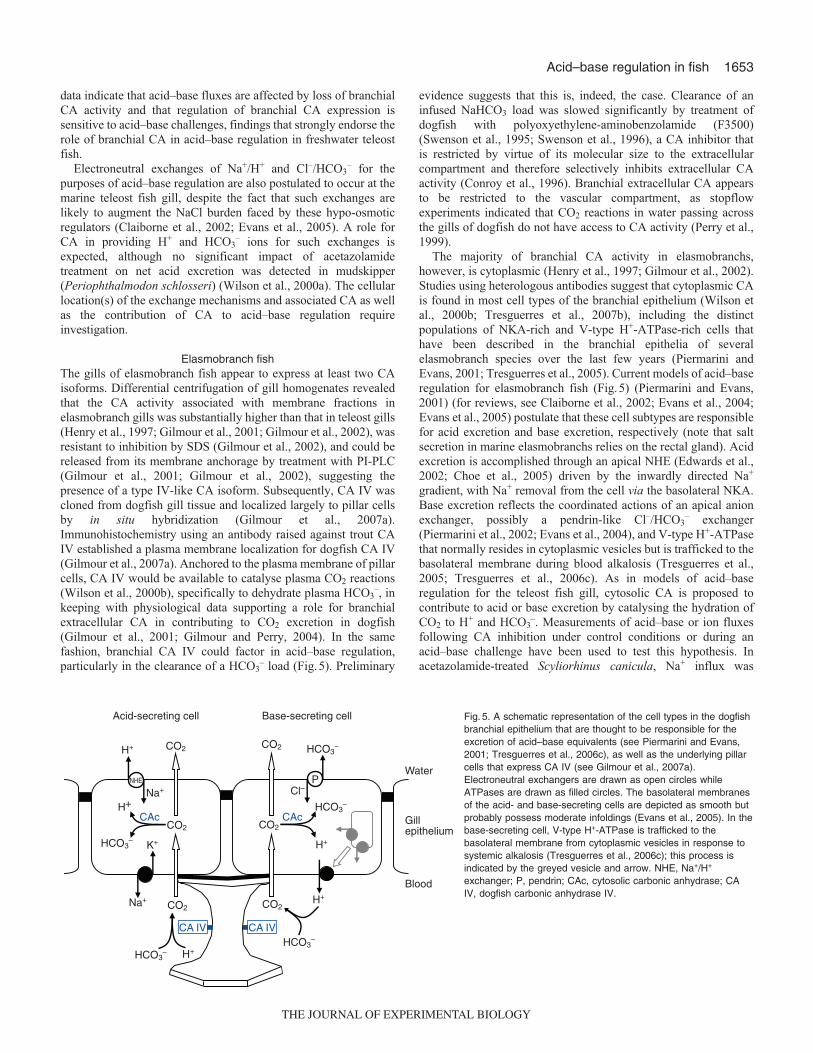
Elasmobranch fishThe gills of elasmobranch fish appear to express at least two CAisoforms. Differential centrifugation of gill homogenates revealedthat the CA activity associated with membrane fractions inelasmobranch gills was substantially higher than that in teleost gills(Henry et al., 1997; Gilmour et al., 2001; Gilmour et al., 2002), wasresistant to inhibition by SDS (Gilmour et al., 2002), and could bereleased from its membrane anchorage by treatment with PI-PLC(Gilmour et al., 2001; Gilmour et al., 2002), suggesting thepresence of a type IV-like CA isoform. Subsequently, CA IV wascloned from dogfish gill tissue and localized largely to pillar cellsby in situ hybridization (Gilmour et al., 2007a).Immunohistochemistry using an antibody raised against trout CAIV established a plasma membrane localization for dogfish CA IV(Gilmour et al., 2007a). Anchored to the plasma membrane of pillarcells, CA IV would be available to catalyse plasma CO2 reactions(Wilson et al., 2000b), specifically to dehydrate plasma HCO3
–, inkeeping with physiological data supporting a role for branchialextracellular CA in contributing to CO2 excretion in dogfish(Gilmour et al., 2001; Gilmour and Perry, 2004). In the samefashion, branchial CA IV could factor in acid–base regulation,particularly in the clearance of a HCO3
– load (Fig.5). Preliminary
evidence suggests that this is, indeed, the case. Clearance of aninfused NaHCO3 load was slowed significantly by treatment ofdogfish with polyoxyethylene-aminobenzolamide (F3500)(Swenson et al., 1995; Swenson et al., 1996), a CA inhibitor thatis restricted by virtue of its molecular size to the extracellularcompartment and therefore selectively inhibits extracellular CAactivity (Conroy et al., 1996). Branchial extracellular CA appearsto be restricted to the vascular compartment, as stopflowexperiments indicated that CO2 reactions in water passing acrossthe gills of dogfish do not have access to CA activity (Perry et al.,1999).
The majority of branchial CA activity in elasmobranchs,however, is cytoplasmic (Henry et al., 1997; Gilmour et al., 2002).Studies using heterologous antibodies suggest that cytoplasmic CAis found in most cell types of the branchial epithelium (Wilson etal., 2000b; Tresguerres et al., 2007b), including the distinctpopulations of NKA-rich and V-type H+-ATPase-rich cells thathave been described in the branchial epithelia of severalelasmobranch species over the last few years (Piermarini andEvans, 2001; Tresguerres et al., 2005). Current models of acid–baseregulation for elasmobranch fish (Fig.5) (Piermarini and Evans,2001) (for reviews, see Claiborne et al., 2002; Evans et al., 2004;Evans et al., 2005) postulate that these cell subtypes are responsiblefor acid excretion and base excretion, respectively (note that saltsecretion in marine elasmobranchs relies on the rectal gland). Acidexcretion is accomplished through an apical NHE (Edwards et al.,2002; Choe et al., 2005) driven by the inwardly directed Na+
gradient, with Na+ removal from the cell via the basolateral NKA.Base excretion reflects the coordinated actions of an apical anionexchanger, possibly a pendrin-like Cl–/HCO3
– exchanger(Piermarini et al., 2002; Evans et al., 2004), and V-type H+-ATPasethat normally resides in cytoplasmic vesicles but is trafficked to thebasolateral membrane during blood alkalosis (Tresguerres et al.,2005; Tresguerres et al., 2006c). As in models of acid–baseregulation for the teleost fish gill, cytosolic CA is proposed tocontribute to acid or base excretion by catalysing the hydration ofCO2 to H+ and HCO3
–. Measurements of acid–base or ion fluxesfollowing CA inhibition under control conditions or during anacid–base challenge have been used to test this hypothesis. Inacetazolamide-treated Scyliorhinus canicula, Na+ influx was
Acid-secreting cell Base-secreting cell
Cl–
Water
Blood
Gillepithelium
P
CA IV CA IV
CAc CAcH+
Na+
CO2H+
K+
NHE
HCO3–
CO2
Na+
H+HCO3–
CO2H+
HCO3–
CO2
H+
HCO3–
CO2
HCO3–CO2
Fig. 5. A schematic representation of the cell types in the dogfishbranchial epithelium that are thought to be responsible for theexcretion of acid–base equivalents (see Piermarini and Evans,2001; Tresguerres et al., 2006c), as well as the underlying pillarcells that express CA IV (see Gilmour et al., 2007a).Electroneutral exchangers are drawn as open circles whileATPases are drawn as filled circles. The basolateral membranesof the acid- and base-secreting cells are depicted as smooth butprobably possess moderate infoldings (Evans et al., 2005). In thebase-secreting cell, V-type H+-ATPase is trafficked to thebasolateral membrane from cytoplasmic vesicles in response tosystemic alkalosis (Tresguerres et al., 2006c); this process isindicated by the greyed vesicle and arrow. NHE, Na+/H+
exchanger; P, pendrin; CAc, cytosolic carbonic anhydrase; CAIV, dogfish carbonic anhydrase IV.
THE JOURNAL OF EXPERIMENTAL BIOLOGY

1654
significantly reduced, supporting a role for branchial CA in acidexcretion coupled to Na+ uptake (Payan and Maetz, 1973). Usingchanges in plasma HCO3
– concentrations during or followinginfusion of a NaHCO3 load as an index of HCO3
– excretion,inhibition of branchial CA was found to attenuate recovery from ametabolic alkalosis (Hodler et al., 1955; Swenson and Maren, 1987;Tresguerres et al., 2007b). Moreover, the HCO3
– load was clearedmore slowly during inhibition of both intracellular and extracellularbranchial CA than when extracellular CA alone was selectivelyinhibited, an observation that intimates distinct yet additivefunctions for cytosolic and extracellular CA in base excretion(Swenson et al., 1995; Swenson et al., 1996). Extracellular CAcould, for example, facilitate entry of HCO3
– into gill epithelialcells by catalysing its conversion to the more diffusible CO2.Recently, an additional role was proposed for CA in acid–baseregulation in elasmobranchs. Base-infused dogfish treated withacetazolamide failed to exhibit basolateral membrane localizationof H+-ATPase, implicating CA in the mechanism that mediates H+-ATPase translocation during a systemic alkalosis (Tresguerres etal., 2007b).
Agnathan fishThe lampreys include a number of anadromous species in whichboth marine and freshwater life stages occur. The ionoregulatoryand osmoregulatory problems faced by these species are similar tothose encountered by marine or freshwater teleost fish, andbranchial mechanisms analogous to those of teleost fish are thoughtto be involved in maintaining ionic and osmotic balance, eventhough the cellular composition of the branchial epithelium inlamprey differs from that of teleosts (Karnaky, 1998; Bartels andPotter, 2004; Evans et al., 2005). Branchial CA in lamprey isthought to be exclusively cytoplasmic; there is no evidence to datefor membrane-associated or extracellular branchial CA (Henry etal., 1993; Tufts and Perry, 1998). Unlike the situation in teleost fish,where distinct blood and gill cytosolic CA isoforms appear to exist(see above), only one cytosolic CA has been detected in lamprey(Esbaugh and Tufts, 2006b). Recent work indicates that thelamprey cytosolic CA is a high activity isoform that is unusuallyresistant to inhibition by acetazolamide (Esbaugh and Tufts,2006b). Using heterologous antibodies, CA has been localized inthe gills of two lamprey species (Geotria australis and P. marinus)to a population of H+-ATPase-enriched MR cells (Choe et al., 2004;Reis-Santos et al., 2008) where it is presumed to catalyse thehydration of CO2 to supply the proton pump with H+ in a model ofacid–base regulation similar to that of teleost or elasmobranch fish(Choe et al., 2004). Experimental data in support of this model,including the involvement of CA, are needed, however.
The gills of hagfish, which are stenohaline marineosmoconformers, contain MR cells that have been proposed tofunction in acid–base regulation as there is little requirement forionic or osmotic regulation (Mallatt et al., 1987). Acid–baseregulation in hagfish, as in other fish, appears to involve branchialNa+/H+ and Cl–/HCO3
– exchanges (Evans, 1984), and several of thetransporters that have been implicated in driving these exchangesin teleost and elasmobranch fish, including NHE, V-type H+-ATPase and NKA, have been detected in the hagfish gill usingheterologous antibodies (Tresguerres et al., 2006b), real-time PCR(Edwards et al., 2001) or histochemical approaches (Mallatt et al.,1987). Interestingly and unlike the models that have beendeveloped for teleosts, elasmobranchs and lamprey, all threetransporters appear to be expressed in a single cell type that ispresumed to be the MR cell (Tresguerres et al., 2006b). The
generation of appropriate responses to acidosis versus alkalosiswould, under these circumstances, probably involve trafficking oftransporters between cytoplasmic vesicles and the apical orbasolateral membrane (Tresguerres et al., 2006b), a possibility thatis supported by changes in the membrane abundance of thesetransporters in acid- or base-infused hagfish (Parks et al., 2007;Tresguerres et al., 2007a). In the emerging hagfish model, cytosolicCA would play its usual role of catalysing CO2 hydration to providethe acid–base equivalents for exchange. Biochemical analysis ofhagfish gill homogenates revealed that most CA activity wascytoplasmic (Esbaugh et al., 2009a), and CA has been localized tohagfish branchial MR cells by histochemistry (Mallatt et al., 1987),although no isoform information is yet available for hagfishcytosolic CA. Recently, two extracellular CA isoforms were clonedfrom hagfish tissues and localized to gill tissue by real-time PCR(Esbaugh et al., 2009a). The mRNA expression of a CA XV-likeisoform was widespread and included the gill. By contrast, mRNAfor a CA IV-like isoform was expressed only in the gill, where itsexpression was 6-fold higher than that of the CA XV-like isoform(Esbaugh et al., 2009a). Although the cellular location andphysiological function of these isoforms remain to be determined,it is tempting to speculate that one or both may contribute to CO2
excretion and acid–base regulation in fashions similar to that ofpillar cell CA IV in the elasmbranch gill (see above). In this regard,it is notable that CA was localized to hagfish gill pillar cells usingHansson’s (histochemical) technique (Mallatt et al., 1987).
CA in renal acid–base regulationLocalization of CA in the kidney
There are inherent difficulties associated with specificallymeasuring renal CA activities and mRNA/protein expression thathave led to considerable confusion as to the presence or absence ofCA in the fish kidney. Because the fish kidney containshaematopoietic cells and an extensive blood supply, it has beenchallenging to distinguish CA activity that is strictly associatedwith the renal nephrons from other tissues enriched with CA.Although urine acid–base status in freshwater fish is influenced byin vivo administration of CA inhibitors (Hodler et al., 1955; Maren,1967; Nishimura, 1977; Georgalis et al., 2006a), similar treatmenthas no effect in marine fish (Hodler et al., 1955; Maren et al., 1992;Swenson, 2003). Indeed, until the finding of CA-mediated SO4
2–
excretion (Renfro et al., 1999), it was believed that the kidney ofmarine fish lacked CA activity entirely. Although relatively fewspecies have been examined, the available data derived from studiesusing isolated tubules or employing immunocytochemistry suggestthat CA is present in the kidneys of both marine and freshwater fish(Renfro et al., 1999; Pelis et al., 2003; Pelis and Renfro, 2004;Georgalis et al., 2006a; Gilmour et al., 2007a).
The predominant CA isoform in trout kidney is the generalcytosolic variant (tCAc) (Esbaugh et al., 2005). Although previousstudies have reported the presence of ‘CA II’ in fish kidney [e.g.Osorezan dace (Hirata et al., 2003); winter flounder (Pleuronectesamericanus) (Pelis et al., 2003)], this renal CA is probably moreappropriately termed cytosolic CA (‘c’ or ‘a’ nomenclature; seeabove). In trout and flounder, cytosolic CA is found withinproximal tubules where it immunolocalizes to the cytosol and brushborder membranes (Pelis and Renfro, 2004; Georgalis et al., 2006a)(Fig.6). In trout kidney, the distal tubule appears to be a moreimportant location of renal CAc expression (Georgalis et al.,2006a). On the basis of the different migration patterns of purifiedcytosolic CA and crude protein on non-denaturing gels, Pelis andcolleagues (Pelis et al., 2003) have suggested that a sub-fraction of
K. M. Gilmour and S. F. Perry
THE JOURNAL OF EXPERIMENTAL BIOLOGY

1655Acid–base regulation in fish
CA in flounder proximal tubules is directly associated withmembranes. Additionally, two membrane-specific CA isoformshave been identified in the fish kidney, CA IV in rainbow trout(Georgalis et al., 2006a) and CA IX in zebrafish (Esbaugh et al.,2009b). In trout, CA IV is localized specifically to the apical andbasolateral membranes of proximal tubule cells (Georgalis et al.,2006a) (Fig.6).
Role of the kidney in acid–base regulationAlthough a fair bit of interspecific variability exists, the freshwaterfish kidney generally plays a significant role in regulating systemicacid–base disorders (Wood and Caldwell, 1978; Kobayashi andWood, 1980; Cameron, 1980; McDonald and Wood, 1981;Cameron and Kormanik, 1982; Wheatly et al., 1984; Perry et al.,1987; Wood, 1988; Ferguson and Boutilier, 1989; Curtis andWood, 1992; Maren et al., 1992). A few studies (Wood andCaldwell, 1978; McDonald and Wood, 1981) have reported thatchanges in renal net H+ excretion could account for 100% ofacid–base compensation, but it is generally accepted that the kidneycontributes about 5–30% to altered rates of whole body H+
excretion during systemic pH changes. Thus, the major route ofregulated H+ secretion during acid–base disorders is via the gill (seeabove). The majority of studies on renal acid–base regulation havebeen conducted on freshwater species that produce large volumesof dilute urine. Marine fish (teleosts and elasmobranchs) producesmall quantities of urine containing elevated levels of Ca2+ andMg2+ salts. The low urine volumes coupled with an apparentrequirement to acidify the urine to prevent the formation of Ca2+
and Mg2+ precipitates are likely key factors constraining theparticipation of the marine kidney in acid–base balance (Hickmanand Trump, 1969). McDonald and colleagues (McDonald et al.,1982) reported an insignificant contribution of the kidney of marinelemon sole (Parophrys vetulus) to regulating metabolic acidosis.The kidney of marine sculpin (Myoxocephalus octodecimspinosus),while contributing significantly to the regulation of metabolic
acidosis, is not involved in regulating metabolic alkalosis followinginfused loads of NaHCO3 (Maren et al., 1992). Interestingly, theimposition of a NaHCO3 load in sculpin was associated with amarked alkalization of the urine without any detected increase inurinary precipitates (Maren et al., 1992). Thus, the requirement toproduce acidic urine to prevent the formation of precipitates maynot be as strict as originally thought. Unlike in teleosts, the kidneyof elasmobranchs appears to produce urine of fixed acidityregardless of the prevailing blood acid–base status (Hodler et al.,1955; Cross et al., 1969; Swenson and Maren, 1986).
As noted above, fish generally regulate blood pH exclusivelythrough metabolic processes. For example, during respiratoryacidosis, blood pH is regulated at constant PCO2 by a gradualaccumulation of plasma HCO3
– (Heisler, 1984) achieved largelythrough altered rates of branchial ion exchange. Such a strategy canbe effective only if the filtered HCO3
– is reabsorbed, a processrequiring increased renal H+ secretion. Thus, the successfulmetabolic compensation of respiratory acidosis can only beachieved if renal H+ secretion is increased to match the increasingfiltered HCO3
– load (Wood and Jackson, 1980). Depending on thespecies and the nature of the respiratory disturbance, plasma HCO3
–
levels may exceed 70mmol l–1 in partially compensated animals(McKenzie et al., 2003), resulting in a massive increase in filteredHCO3
– load that necessitates an equally massive increase in H+
secretion.
Renal H+ secretion and HCO3– reabsorption
The predominant site of renal H+ secretion and HCO3– reabsorption
in teleost fish, as in the mammalian nephron, is thought to be theproximal tubule; the putative mechanisms are depicted in Fig.7.The addition of H+ to the filtrate is achieved by at least twomechanisms, electroneutral exchange with Na+ or active H+
pumping (Perry et al., 2003a; Perry et al., 2003b; Perry andGilmour, 2006). Available evidence suggests that electroneutralNa+/H+ exchange is accomplished by NHE3 (SLC9A3), a member
Control Acetazolamide
Net
aci
d flu
x (µ
mol
kg–1
h–1
)
–80
–60
–40
–20
0
20
Control F3500–80
–60
–40
–20
0
20
A
C D
B
*
*
Fig. 6. Immunolocalization of (A) cytosolic carbonicanhydrase (CAc) and (C) CA IV in renal proximaltubules of rainbow trout (Oncorhynchus mykiss); ineach micrograph CA is depicted by greenfluorescence, Na+,K+-ATPase by red fluorescence andnuclei by blue fluorescence. Inhibition of (B) total CAactivity with acetazolamide or (D) only extracellularCA activity with F3500 significantly reduced neturinary acid excretion (negative values indicate netbase loss; asterisks indicate statistically significantdifferences between control and treatment values).Data were obtained from Georgalis et al. (Georgalis etal., 2006a).
THE JOURNAL OF EXPERIMENTAL BIOLOGY

1656
of the NHE (SLC9) multi-gene family (Hirata et al., 2003; Ivaniset al., 2008a) while active H+ secretion is driven by the V-type H+-ATPase (Perry and Fryer, 1997; Perry et al., 2000; Perry et al.,2003a; Hirata et al., 2003). The mRNA levels for both the V-ATPase and NHE3 genes are increased during acidificationinduced by hypercapnia (Perry et al., 2003a; Ivanis et al., 2008a)or exposure to water of low pH (Hirata et al., 2003). The H+ ionsfuelling the V-ATPase and NHE are derived from the hydration ofintracellular CO2 in the presence of cytosolic CA. Once in thefiltrate, the H+ combines with filtered HCO3
– to form CO2, areaction catalysed by membrane-associated CA IV (Georgalis et al.,2006a) which is oriented to face the extracellular compartment(Fig.7). The CO2 then diffuses into the tubule and is hydrated inthe presence of CA to re-fuel the apical membrane H+ secretorymechanisms and to provide HCO3
– to be ‘reabsorbed’ across thebasolateral membranes via the Na+/HCO3
– co-transporter isoform1 (NBC1) (Hirata et al., 2003; Perry et al., 2003a). In trout, thelevels of renal NBC1 mRNA are increased during hypercapnicacidosis, presumably to match the increased rates of HCO3
–
reabsorption (Perry et al., 2003a).The most direct evidence for an important role of renal CA in
H+ secretion and HCO3– reabsorption comes from in vivo studies
employing selective CA inhibitors under resting conditions andperiods of acid–base disturbance. Thus, the inhibition of total CAactivity in rainbow trout using acetazolamide caused a significantdecrease in renal net acid excretion (see Fig.6) that was associatedwith a rise in urine pH (Georgalis et al., 2006a). In that same study,the inhibition of total renal CA activity during compensatedhypercapnic acidosis was associated with marked increases inurinary losses of Na+ and HCO3
– (Georgalis et al., 2006a). The factthat levels of HCO3
– in the urine increased from approximately 7to 30mmol l–1 after CA inhibition in hypercapnic fish clearlyreveals the critical roles of the kidney (and renal CA) in preventingthe loss of HCO3
– that has accumulated largely by adjustment ofbranchial ion transfers (see above). The use of a membrane-permeant inhibitor such as acetazolamide does not permit an
assessment of the relative roles of the cytosolic and extracellularisoforms of renal CA. Georgalis and colleagues (Georgalis et al.,2006a), however, used F3500 (Conroy et al., 1996) to selectivelyinhibit renal extracellular CA in trout. Although too large to crosscell membranes, the relatively low molecular mass (3500Da) ofF3500 allows it to be filtered, thereby providing it with access toluminal extracellular CA. Using this approach, it was demonstratedthat renal luminal extracellular CA was playing a significant rolein the reabsorption of filtered HCO3
– in hypercapnic trout althoughto a lesser extent than the cytosolic isoform (Georgalis et al.,2006a). The proposed role of luminal extracellular CA in thedehydration of filtrate HCO3
– is depicted in Fig.7. Based onimmunocytochemical data, the CA IV isoform is implicated incatalysing the dehydration of filtrate HCO3
– but it is uncertainwhether other membrane-associated isoforms are also involved(e.g. CA XII or XIV).
The relatively few studies that have examined the effects ofacid–base disturbances on renal CA expression have yieldedconflicting or ambiguous data. In rainbow trout, respiratoryacidosis was accompanied by increasing mRNA and proteinexpression of CAc; CA IV was unaffected (Georgalis et al., 2006a).Similarly, Hirata and colleagues (Hirata et al., 2003) demonstratedincreased renal expression of cytosolic CA in Osorezan daceexposed to acidic water. Lungfish, while exhibiting a significantrenal component to the compensation of metabolic acidosis oralkalosis, showed no changes in renal CA mRNA expression(Gilmour et al., 2007b). The absence of any change in renal mRNAlevels for cytosolic CA during metabolic acidosis is surprisinggiven that similar treatment in mammals causes a marked increasedin the expression of CA II (Tsuruoka et al., 1998). Severalexplanations can be offered for the lack of an effect in the lungfish:the sampling time may have been inappropriate thus preventingdetection of a transient change in mRNA expression; or CA wasbeing modified post-translationally. Alternatively, it is conceivablethat CA activity was already sufficient to cope with the imposedacid–base disturbances. Because it is relatively easy to measure CAactivity in tissues (Henry, 1981) concomitantly with estimatingprotein (western blotting) and mRNA levels (real-time PCR),researchers are encouraged to measure all three parameters to allowa complete assessment of the underlying mechanisms contributingto changes in CA activity. For tissues such as kidney, however,such an integrated approach is not always feasible because of thedifficulty in eliminating the contribution of haematopoietic cells orRBCs to measurements of CA activity or protein (unless isoform-specific antibodies are used).
The important role of CA in promoting renal H+ secretion andHCO3
– reabsorption in freshwater teleosts (i.e. rainbow trout)contrasts with the apparent absence of this role for CA in the kidneyof marine fish (Hodler et al., 1955; Swenson and Maren, 1986;Maren et al., 1992; Swenson, 2003). Particularly striking is themarine elasmobranch (Squalus acanthias) that has long beenthought to lack renal CA but nevertheless displays a remarkablecapacity to reabsorb HCO3
– from the renal filtrate. Indeed, it hasbeen documented that even during periods of metabolic alkalosiswhen plasma HCO3
– levels increase to >30mmol l–1, urinary lossof HCO3
– remains minimal and constant (Swenson and Maren,1986). In both dogfish (Swenson and Maren, 1986) and long-horned sculpin (Hodler et al., 1955; Maren et al., 1992), CAinhibition is without effect on urine acid–base status. In the lightof these observations and additional in situ measurements of renalacidification in the marine skate Raja erinacea, a novel mechanismof HCO3
– reabsorption was proposed for elasmobranchs involving
K. M. Gilmour and S. F. Perry
CAc CA IV
LumenBlood Tubule
CO2
Na+NHE
NBC
HCO3–H+
H+
H+
Na+
HCO3– HCO3
–
CO2
Fig. 7. A schematic representation of HCO3– reabsorption by the teleost
kidney proximal tubule. Luminal CA isoform IV (and/or another membrane-associated isoform) catalyses the combination of filtered HCO3
– with H+
provided by Na+/H+ exchange (NHE) or H+-ATPase (ATPases are drawnas filled circles). The CO2 formed by the dehydration reaction diffuses intothe cytosol where cytoplasmic CA (CAc) catalyses its hydration to HCO3
–
and H+. The HCO3– exits across the serosal membrane via the Na+/HCO3
–
cotransporter isoform 1 (NBC1) and the H+ refuels the NHE or H+-ATPase.
THE JOURNAL OF EXPERIMENTAL BIOLOGY

1657Acid–base regulation in fish
the direct transfer of ionic HCO3– from filtrate to lumen (Deetjen
and Maren, 1974). Years later, Swenson and colleagues (Swensonet al., 1994) proposed a mechanism for CA-independent urinaryacidification involving the secretion of H+ via a luminal H+,K+-ATPase. Although these findings led to, or supported, acceptanceof the hypothesis that marine fish lack renal CA, this conclusion isno longer believed given the physiological evidence for cytosolicCA activity in proximal tubules of winter flounder (Renfro et al.,1999; Pelis et al., 2003; Pelis and Renfro, 2004) and the finding ofmembrane-associated CA IV mRNA in dogfish kidney (Gilmouret al., 2007a). While playing a role in renal SO4
2– excretion, thepotential involvement of CA in renal acidification and HCO3
–
reabsorption may be constrained by other factors such as theabsence of suitable ion transporters (e.g. NHE or V-ATPase) on theluminal membrane. Considering that only a few (three) specieshave been examined and given the claims of radical differences inthe involvement of renal CA in freshwater and marine fish, weencourage a re-examination of the involvement of CA in urinaryacidification and HCO3
– reabsorption in marine fish species.
CA in piscine-specific acid–base regulatory mechanismsThus far, this review has focused on the role of CA in the keytissues (gill and kidney) that contribute to the regulation of systemicacid–base status. Additionally, however, CA has been implicatedin several other structures exhibiting physiological properties thatare unique to certain fish. These include secretion of NaCl by theelasmobranch rectal gland, secretion of H+ by the gas gland of theteleost swimbladder, acidification by the pseudobranch of thearterial blood supplying the eye, and the addition of HCO3
– to gutfluid by intestinal epithelial cells [please refer to the review byGrosell et al. in this issue (Grosell et al., 2009)].
Elasmobranch rectal glandThe rectal gland of elasmobranchs is a sausage-shaped structurethat is capable of secreting into the posterior intestine a solution ofroughly 500mmol l–1 NaCl (Burger and Hess, 1960). The volumeof rectal gland secretion produced is highly variable but is clearlyincreased by blood volume expansion associated withfeeding/ingestion of seawater (Evans, 1993). While it has long beenheld that the rectal gland plays an important role in extra-renal saltexcretion (reviewed by Shuttleworth, 1988), studies that havephysically removed or ablated the gland have documentedrelatively minor (Evans et al., 1982; Evans, 1993) or no (Chan etal., 1967; Wilson et al., 2002; Wilson and Laurent, 2002)significant impact on plasma ion levels. Obviously, therefore, otherroutes of net salt excretion exist in elasmobranchs. The two sitesthat first spring to mind are the kidney and the gill. As discussedby Evans et al. (Evans et al., 2005), because marine elasmobranchsare slightly hyperosmotic, they gain water by osmosis that isbalanced by equivalent volumes of urine production, therebyproviding a route of salt efflux normally not present in most marinespecies. Although the gill is generally discounted as a site of netNaCl excretion in elasmobranchs (Shuttleworth, 1988), its potentialinvolvement in extrarenal salt excretion should probably bereconsidered.
CA is abundant in the rectal gland (Lacy, 1983) where it playsan important role in fluid secretion; in vivo administration of CAinhibitors markedly reduced the rates of glandular secretion indogfish (Swenson and Maren, 1984; Shuttleworth et al., 2006). Ithas been suggested that the role of CA in rectal gland secretion isto facilitate removal of metabolically produced CO2 so as tomaintain an appropriate intracellular acid–base status (Swenson
and Maren, 1984). Two mechanisms of CA action have beenproposed; Swenson and Maren (Swenson and Maren, 1984)suggested that cytosolic CA catalyses the inter-conversion ofHCO3
– to CO2 while Shuttleworth and colleagues (Shuttleworth etal., 2006) concluded that a more likely scenario was the catalysedhydration of CO2 by membrane-associated CA in the extracellularcompartment. In both models, CA contributes to CO2 removal fromcells by enhancing CO2 diffusion gradients. Future studies shouldincorporate specific permeant and impermeant inhibitors todifferentiate the potential roles of cytosolic versus extracellular(membrane-associated) CA.
Rectal gland fluid output is directly related to blood pH, risingwith alkalosis and falling with acidosis (Swenson and Maren, 1984;Shuttleworth et al., 2006; Wood et al., 2007). Because blood pH indogfish increases after eating [alkaline tide (Wood et al., 2005)],there would be an associated post-prandial increase in rectal glandsecretion to aid in the excretion of the additional salt and watergained during ingestion. Similarly, a reduction in blood pHassociated with exhaustive exercise would act as a cue to inhibitglandular secretion and thus help to retain fluid during periods ofincreased fluid entry into muscle (Shuttleworth et al., 2006; Woodet al., 2007).
The swimbladderInflation of the swimbladder in deep sea fish relies on the secretionof O2 to generate exceedingly high partial pressures of O2 (Pelster,2004). The O2 is derived from haemoglobin via the Root effectfollowing acidification of the blood by the swimbladder gas gland.In addition to CO2, a variety of acidic metabolites contribute to theacidification of the blood in the gas gland including lactic acid andH+ originating from Na+/H+ exchange and V-type H+-ATPaseactivities (Pelster, 1995). A role for CA in gas gland acid secretionwas first suggested by the results of experiments showing that gasdeposition was slowed by CA inhibition (Fange, 1953). Subsequentexperiments have shown that CA inhibition reduces lactate releaseby eel swimbladder in vitro (Kutchai, 1971) or acid secretion fromisolated gas gland cells (Pelster, 1995). Because acid secretion fromisolated gas gland cells was inhibited by membrane-permeant and-impermeant inhibitors, it was proposed that membrane-associated(extracellular) CA IV was involved (Pelster, 1995). The existenceof membrane-associated CA (proposed to be CA IV) on the blood-facing plasma membranes and capillary endothelium was laterconfirmed using histochemistry (Wurtz et al., 1999). Futureresearch should evaluate CA isoforms as well as the relativeinvolvement of membrane-associated and cytosolic CA inswimbladder inflation.
The pseudobranchAlthough the physiological function of the pseudobranch has longbeen debated (Laurent and Rouzeau, 1972; Laurent and Dunel-Erb,1984; Bridges et al., 1998), there is accumulating evidence that oneof its roles is to assist the O2-concentrating mechanisms occurringwithin the choroid rete of the eye of certain marine teleosts. In thesefish, the retina is thick and non-vascularized, thereby impeding thedelivery of O2 to the retinal tissue. To meet the high metabolicdemand of the retinal pigmented epithelium, extremely high partialpressures of O2 are generated (reported to exceed 1atm, ~101kPa)to facilitate diffusion. As in the swimbladder (Pelster, 2004), themechanism thought to underlie the O2-concentrating mechanism isthe Root effect (for reviews, see Brittain, 1987; Pelster and Weber,1991; Berenbrink, 2007) whereby acidification of the bloodentering the rete causes O2 to be unloaded from haemoglobin for
THE JOURNAL OF EXPERIMENTAL BIOLOGY
1658
delivery to the eye. The most compelling empirical evidence insupport of a role for the Root effect is the observation that O2
secretion into the ocular fluid of an in vitro blood-perfused rainbowtrout eye preparation occurs only when blood exhibiting a Rooteffect is used as the perfusion fluid (i.e. trout, but not human blood)(Waser and Heisler, 2005). Although blood can be acidified in thechoroid rete as well as in the eye itself, it has been proposed thatthe pseudobranch plays a crucial role in the pre-conditioning ofblood prior to its entry into the rete (Bridges et al., 1998). Theaddition of CO2 and H+ to the blood flowing within thepseudobranch is thought to acidify the RBCs so as to approach thethreshold for the onset of the Root effect. Such pre-conditioningwould allow maximal release of O2 from haemoglobin withminimal acidification (Bridges et al., 1998). A minimally acidifiedenvironment is important because unlike the swimbladder, whichcan tolerate extreme acidification (pH 6.5), the retinal tissue isintolerant of comparable reductions in pH. The mechanism ofacidification of the blood within the pseudobranch involves thedirect excretion of CO2 and secretion of H+ via Na+/H+ exchangeand V-type H+-ATPase (Kern et al., 2002). The importance of CAin sustaining these acidifying processes and allowing O2
concentration within the eye was convincingly demonstrated(Fairbanks et al., 1969; Fairbanks et al., 1974) although to datethere are no data on the identification of the participating isoforms.It also should be recognized that the pseudobranch is but one ofseveral sites participating in CA-mediated acidification of pre-occular blood.
ConclusionsThe above discussion emphasizes the point that the diversity of CAisoforms and functions in fish is at least as great as that in tetrapods,if not more so owing to the variation among fish species and groups,yet our knowledge of CA isoform diversity, distribution, functionand regulation in fish lags well behind that for mammals.Particularly critical is a need for more genomic information thatcan be used to identify CA isoforms and clarify the evolutionaryrelationships with respect to CA between fish and tetrapods. Bettertools, including fish species- and isoform-specific CA antibodies,are required to describe the cellular localization of CA in thetissues, gill and kidney, that are primarily responsible for acid–baseregulation in fish. Physiological data are needed to speak to thefunctional significance of CA in acid–base processes. Finally, theregulation of CA expression and activity warrant investigation,together with the potential regulatory roles of CA. Accomplishingthese goals for even a single fish species is a challenge but one thatneeds to be addressed if an integrated picture of the role of CA inacid–base regulation in fish is to be achieved.
Original research by the authors was supported by Discovery and Research Toolsand Instruments grants from the Natural Sciences and Engineering ResearchCouncil of Canada. Thanks are extended to the Company of Biologists andJournal of Experimental Biology for the invitation to participate in the DiscussionMeeting on ‘Epithelial solute transporter and acid–base regulation’.
ReferencesBartels, H. and Potter, I. C. (2004). Cellular composition and ultrastructure of the gill
epithelium of larval and adult lampreys: implications for osmoregulation in fresh andseawater. J. Exp. Biol. 207, 3447-3462.
Berenbrink, M. (2007). Historical reconstructions of evolving physiological complexity:O2 secretion in the eye and swimbladder of fishes. J. Exp. Biol. 210, 1641-1652.
Boisen, A. M. Z., Amstrup, J., Novak, I. and Grosell, M. (2003). Sodium andchloride transport in soft water and hard water acclimated zebrafish (Danio rerio).Biochim. Biophys. Acta 1618, 207-218.
Bridges, C. R., Berenbrink, M., Muller, R. and Waser, W. (1998). Physiology andbiochemistry of the pseudobranch: an unanswered question? Comp. Biochem.Physiol. A 119, 67-77.
Brittain, T. (1987). The root effect. Comp. Biochem. Physiol. B 86, 473-481.
Burger, J. W. and Hess, W. N. (1960). Function of the rectal gland in the spinydogfish. Science 131, 670-671.
Cameron, J. N. (1980). Body fluid pools, kidney function, and acid-base regulation inthe freshwater catfish Ictalurus punctatus. J. Exp. Biol. 86, 171-185.
Cameron, J. N. and Kormanik, G. A. (1982). The acid-base responses of gills andkidneys to infused acid and base loads in the channel catfish, Ictalurus punctatus. J.Exp. Biol. 99, 143-160.
Chan, D. K. O., Phillips, J. G. and Jones, I. C. (1967). Studies on electrolytechanges in lip-shark Hemiscyllium plagiosum (Bennett) with special reference tohormonal influence on rectal gland. Comp. Biochem. Physiol. 23, 185-198.
Chang, I. C. and Hwang, P. P. (2004). Cl– uptake mechanism in freshwater-adaptedtilapia (Oreochromis mossambicus). Physiol. Biochem. Zool. 77, 406-414.
Chegwidden, W. R. and Carter, N. D. (2000). Introduction to the carbonicanhydrases. EXS 90, 13-28.
Choe, K. P., O’Brien, S., Evans, D. H., Toop, T. and Edwards, S. L. (2004).Immunolocalization of Na+/K+-ATPase, carbonic anhydrase II, and vacuolar H+-ATPase in the gills of freshwater adult lampreys, Geotria australis. J. Exp. Zool.301A, 654-665.
Choe, K. P., Kato, A., Hirose, S., Plata, C., Sindié, A., Romero, M. F., Claiborne, J.B. and Evans, D. H. (2005). NHE3 in an ancestral vertebrate: primary sequence,distribution, localization, and function in gills. Am. J. Physiol. 289, R1520-R1534.
Claiborne, J. B., Edwards, S. L. and Morrison-Shetlar, A. I. (2002). Acid-baseregulation in fishes: cellular and molecular mechanisms. J. Exp. Zool. 293, 302-319.
Conley, D. M. and Mallatt, J. (1988). Histochemical localization of Na+-K+ ATPaseand carbonic anhydrase activity in gills of 17 fish species. Can. J. Zool. 66, 2398-2405.
Conroy, C. W., Wynns, G. C. and Maren, T. H. (1996). Synthesis and properties oftwo new membrane-impermeant high-molecular-weight carbonic anhydraseinhibitors. Bioorg. Chem. 24, 262-272.
Craig, P. M., Wood, C. M. and McLelland, G. B. (2007). Gill membrane remodelingwith soft-water acclimation in zebrafish (Danio rerio). Physiol. Genomics 30, 53-60.
Cross, C. E., Packer, B. S., Linta, J. M., Murdaugh, H. V., Jr and Robin, E. D.(1969). H+ buffering and excretion in response to acute hypercapnia in the dogfishSqualus acanthias. Am. J. Physiol. 216, 440-452.
Curtis, B. J. and Wood, C. M. (1992). Kidney and urinary bladder responses offreshwater rainbow trout to isosmotic NaCl and NaHCO3 infusion. J. Exp. Biol. 173,181-203.
Deetjen, P. and Maren, T. (1974). The dissociation between renal HCO3– reabsorption
and H+ secretion in the skate, Raja erinacea. Pflugers Arch. 346, 25-30.Dimberg, K., Höglund, L. B., Knutsson, P. G. and Ridderstråle, Y. (1981).
Histochemical localization of carbonic anhydrase in gill lamellae from young salmon(Salmo salar L.) adapted to fresh and salt water. Acta Physiol. Scand. 112, 218-220.
Edwards, S. L., Claiborne, J. B., Morrison-Shetlar, A. and Toop, T. (2001).Expression of Na+/H+ exchanger mRNA in the gills of the Atlantic hagfish (Myxineglutinosa) in response to metabolic acidosis. Comp. Biochem. Physiol. A 130, 81-91.
Edwards, S. L., Donald, J. A., Toop, T., Donowitz, M. and Tse, C.-M. (2002).Immunolocalisation of sodium/proton exchanger-like proteins in the gills ofelasmobranchs. Comp. Biochem. Physiol. A 131, 257-265.
Esaki, M., Hoshijima, K., Kobayashi, S., Fukuda, H., Kawakami, K. and Hirose, S.(2007). Visualization in zebrafish larvae of Na+ uptake in mitochondria-rich cellswhose differentiation is dependent on foxi3a. Am. J. Physiol. 292, R470-R480.
Esbaugh, A. and Tufts, B. L. (2006a). The structure and function of carbonicanhydrase isozymes in the respiratory system of vertebrates. Respir. Physiol.Neurobiol. 154, 185-198.
Esbaugh, A. and Tufts, B. L. (2006b). Tribute to R. G. Boutilier: evidence of a highactivity carbonic anhydrase isozyme in the red blood cells of an ancient vertebrate,the sea lamprey Petromyzon marinus. J. Exp. Biol. 209, 1169-1178.
Esbaugh, A. and Tufts, B. L. (2007). Evidence for a carbonic anhydrase-relatedprotein in the brain of rainbow trout (Oncorhynchus mykiss). Comp. Biochem.Physiol. D 2, 287-294.
Esbaugh, A., Lund, S. G. and Tufts, B. L. (2004). Comparative physiology andmolecular analysis of carbonic anhydrase from the red blood cells of teleost fish. J.Comp. Physiol. B 174, 429-438.
Esbaugh, A., Perry, S. F., Bayaa, M., Georgalis, T., Nickerson, J. G., Tufts, B. L.and Gilmour, K. M. (2005). Cytoplasmic carbonic anhydrase isozymes in rainbowtrout Oncorhynchus mykiss: comparative physiology and molecular evolution. J. Exp.Biol. 208, 1951-1961.
Esbaugh, A. J., Gilmour, K. M. and Perry, S. F. (2009a). Membrane-associatedcarbonic anhydrase in the respiratory system of the Pacific hagfish (Eptatretusstouti). Respir. Physiol. Neurobiol. 166, 107-116.
Esbaugh, A. J., Perry, S. F. and Gilmour, K. M. (2009b). Hypoxia-inducible carbonicanhydrase IX expression is insufficient to alleviate intracellular metabolic acidosis inthe muscle of zebrafish Danio rerio. Am. J. Physiol. 296, R150-R160.
Evans, D. H. (1984). Gill Na+/H+ and Cl–/HCO3– exchange systems evolved before the
vertebrates entered fresh water. J. Exp. Biol. 113, 465-469.Evans, D. H. (1993). Osmotic and ionic regulation. In The Physiology of Fishes (ed. D.
H. Evans), pp. 315-341. Boca Raton, FL: CRC Press.Evans, D. H., Oikari, A., Kormanik, G. A. and Mansberger, L. (1982).
Osmoregulation by the prenatal spiny dogfish, Squalus acanthias. J. Exp. Biol. 101,295-305.
Evans, D. H., Piermarini, P. M. and Choe, K. P. (2004). Homeostasis:Osmoregulation, pH regulation, and nitrogen excretion. In Biology of Sharks andTheir Relatives (ed. J. C. Carrier, J. A. Musick and M. R. Heithaus), pp. 247-268.Boca Raton, FL: CRC Press.
Evans, D. H., Piermarini, P. M. and Choe, K. P. (2005). The multifunctional fish gill:dominant site of gas exchange, osmoregulation, acid-base regulation, and excretionof nitrogenous waste. Physiol. Rev. 85, 97-177.
Fairbanks, M. B., Hoffert, J. R. and Fromm, P. O. (1969). The dependence of theoxygen-concentrating mechanism of the teleost eye (Salmo gairdneri) on theenzyme carbonic anhydrase. J. Gen. Physiol. 54, 203-211.
K. M. Gilmour and S. F. Perry
THE JOURNAL OF EXPERIMENTAL BIOLOGY

1659Acid–base regulation in fish
Fairbanks, M. B., Hoffert, J. R. and Fromm, P. O. (1974). Short circuiting the ocularoxygen concentrating mechanism in the teleost Salmo gairdneri using carbonicanhydrase inhibitors. J. Gen. Physiol. 64, 263-273.
Fange, R. (1953). The mechanisms of gas transport in the euphysoclist swimbladder.Acta Physiol. Scand. Suppl. 30, 1-133.
Ferguson, R. A. and Boutilier, R. G. (1989). Metabolic-membrane coupling in redblood cells of trout: the effects of anoxia and adrenergic stimulation. J. Exp. Biol.143, 149-164.
Fernley, R. T., Wright, R. D. and Coghlan, J. P. (1988). Complete amino acidsequence of ovine salivary carbonic anhydrase. Biochemistry 27, 2815-2820.
Flügel, C., Lütjen-Drecoll, E. and Zadunaisky, J. A. (1991). Histochemicaldemonstration of carbonic anhydrase in gills and opercular epithelium of seawater-and freshwater-adapted killyfish (Fundulus heteroclitus). Acta Histochem. 91, 67-75.
Fujikawa-Adachi, K., Nishimori, I., Taguchi, T. and Onishi, S. (1999a). Humancarbonic anhydrase XIV (CA14): cDNA cloning, mRNA expression, and mapping tochromosome 1. Genomics 61, 74-81.
Fujikawa-Adachi, K., Nishimori, I., Taguchi, T. and Onishi, S. (1999b). Humanmitochondrial carbonic anhydrase VB: cDNA cloning, mRNA expression, subcellularlocalization, and mapping to chromosome X. J. Biol. Chem. 274, 21228-21233.
Galvez, F., Reid, S. D., Hawkings, G. S. and Goss, G. G. (2002). Isolation andcharacterization of mitochondria-rich cell types from the gill of freshwater rainbowtrout. Am. J. Physiol. 282, R658-R668.
Geers, C. and Gros, G. (2000). Carbon dioxide transport and carbonic anhydrase inblood and muscle. Physiol. Rev. 80, 681-715.
Georgalis, T., Gilmour, K. M., Yorston, J. and Perry, S. F. (2006a). The roles ofcytosolic and membrane bound carbonic anhydrase in the renal control of acid-basebalance in rainbow trout Oncorhynchus mykiss. Am. J. Physiol. 291, F407-F421.
Georgalis, T., Perry, S. F. and Gilmour, K. M. (2006b). The role of branchial carbonicanhydrase in acid-base regulation in rainbow trout (Oncorhynchus mykiss). J. Exp.Biol. 209, 518-530.
Gervais, M. R. and Tufts, B. L. (1998). Evidence for membrane-bound carbonicanhydrase in the air bladder of bowfin (Amia calva), a primitive air-breathing fish. J.Exp. Biol. 201, 2205-2212.
Gilmour, K. M. (1998a). Causes and consequences of acid-base disequilibria. In FishPhysiology Volume 17: Fish Respiration (ed. S. F. Perry and B. L. Tufts), pp. 321-348. San Diego, CA: Academic Press.
Gilmour, K. M. (1998b). The disequilibrium pH: a tool for the localization of carbonicanhydrase. Comp. Biochem. Physiol. 119A, 243-254.
Gilmour, K. M. and Perry, S. F. (2004). Branchial membrane-associated carbonicanhydrase activity maintains CO2 excretion in severely anemic dogfish. Am. J.Physiol. 286, R1138-R1148.
Gilmour, K. M., Randall, D. J. and Perry, S. F. (1994). Acid-base disequilibrium inthe arterial blood of rainbow trout. Respir. Physiol. 96, 259-272.
Gilmour, K. M., Perry, S. F., Bernier, N. J., Henry, R. P. and Wood, C. M. (2001).Extracellular carbonic anhydrase in dogfish, Squalus acanthias: a role in CO2
excretion. Physiol. Biochem. Zool. 74, 477-492.Gilmour, K. M., Shah, B. and Szebedinszky, C. (2002). An investigation of carbonic
anhydrase activity in the gills and blood plasma of brown bullhead (Ameiurusnebulosus), longnose skate (Raja rhina), and spotted ratfish (Hydrolagus colliei). J.Comp. Physiol. B 172, 77-86.
Gilmour, K. M., Bayaa, M., Kenney, L., McNeill, B. and Perry, S. F. (2007a). TypeIV carbonic anhydrase is present in the gills of spiny dogfish (Squalus acanthias).Am. J. Physiol. 292, R556-R567.
Gilmour, K. M., Euverman, R., Esbaugh, A. J., Kenney, L., Chew, S. F., Ip, Y. K.and Perry, S. F. (2007b). Mechanisms of acid-base regulation in the African lungfishProtopterus annectens. J. Exp. Biol. 210, 1944-1959.
Goss, G. G. and Perry, S. F. (1994). Different mechanisms of acid-base regulation inrainbow trout (Oncorhynchus mykiss) and American eel (Anguilla rostrata) duringNaHCO3 infusion. Physiol. Zool. 67, 381-406.
Goss, G. G. and Wood, C. M. (1991). Two-substrate kinetic analysis: a novelapproach linking ion and acid-base transport at the gills of freshwater trout,Oncorhynchus mykiss. J. Comp. Physiol. B 161, 635-646.
Goss, G. G., Laurent, P. and Perry, S. F. (1992a). Evidence for a morphologicalcomponent in acid-base regulation during environmental hypercapnia in the brownbullhead (Ictalurus nebulosus). Cell Tissue Res. 268, 539-552.
Goss, G. G., Perry, S. F., Wood, C. M. and Laurent, P. (1992b). Mechanisms of ionand acid-base regulation at the gills of freshwater fish. J. Exp. Zool. 263, 143-159.
Goss, G. G., Adamia, S. and Galvez, F. (2001). Peanut lectin binds to asubpopulation of mitochondria-rich cells in the rainbow trout gill epithelium. Am. J.Physiol. 281, R1718-R1725.
Grosell, M., Mager, E., Williams, C. and Taylor, J. R. (2009). High rates of HCO3–
secretion and Cl– absorption against adverse gradients in the marine teleostintestine: the involvement of an electrogenic anion exchanger and H+-pumpmetabolon? J. Exp. Biol. 212, 1684-1696.
Haswell, M. S., Randall, D. J. and Perry, S. F. (1980). Fish gill carbonic anhydrase:acid-base regulation or salt transport? Am. J. Physiol. 238, R240-R245.
Heisler, N. (1984). Acid-base regulation in fishes. In Fish Physiology, vol. 10A (ed. W.S. Hoar and D. J. Randall), pp. 315-401. New York: Academic Press.
Heisler, N. (1986). Comparative aspects of acid-base regulation. In Acid-baseRegulation in Animals (ed. N. Heisler), pp. 397-449. Amsterdam: Elsevier.
Heming, T. A. (1986). Carbon dioxide excretion and ammonia toxicity in fishes: isthere a relationship? In Problems of Aquatic Toxicology, Biotesting, and WaterQuality Management: Proceedings of USA-USSR Symposium, Borok, JaroslavlOblast, July 30-August 1, 1984 (ed. R. C. Ryans), pp. 84-94. Athens, GA: USEnvironmental Protection Agency.
Henry, R. P. (1981). Techniques for measuring carbonic anhydrase activity in vitro: theelectrometric delta pH and pH stat assays. In The Carbonic Anhydrases: CellularPhysiology and Molecular Genetics (ed. S. J. Dodgson, R. E. Tashian, G. Gros andN. D. Carter), pp. 119-126. New York: Plenum.
Henry, R. P. and Heming, T. A. (1998). Carbonic anhydrase and respiratory gasexchange. In Fish Physiology Volume 17: Fish Respiration (ed. S. F. Perry and B. L.Tufts), pp. 75-111. San Diego, CA: Academic Press.
Henry, R. P. and Swenson, E. R. (2000). The distribution and physiologicalsignificance of carbonic anhydrase in vertebrate gas exchange organs. Respir.Physiol. 121, 1-12.
Henry, R. P., Smatresk, N. J. and Cameron, J. N. (1988). The distribution ofbranchial carbonic anhydrase and the effects of gill and erythrocyte carbonicanhydrase inhibition in the channel catfish Ictalurus punctatus. J. Exp. Biol. 134,201-218.
Henry, R. P., Tufts, B. L. and Boutilier, R. G. (1993). The distribution of carbonicanhydrase type I and II isozymes in lamprey and trout: possible co-evolution witherythrocyte chloride/bicarbonate exchange. J. Comp. Physiol. B 163, 380-388.
Henry, R. P., Gilmour, K. M., Wood, C. M. and Perry, S. F. (1997). Extracellularcarbonic anhydrase activity and carbonic anhydrase inhibitors in the circulatorysystem of fish. Physiol. Zool. 70, 650-659.
Hewett-Emmett, D. (2000). Evolution and distribution of the carbonic anhydrase genefamilies. EXS 90, 29-76.
Hickman, C. P. and Trump, B. F. (1969). The kidney. In Fish Physiology, vol. 1 (ed.W. S. Hoar and D. J. Randall), pp. 91-239. New York: Academic Press.
Hilvo, M., Tolvanen, M., Clark, A., Shen, B., Shah, G. N., Waheed, A., Halmi, P.,Hänninen, M., Hämäläinen, J. M., Vihinen, M. et al. (2005). Characterization of CAXV, a new GPI-anchored form of carbonic anhydrase. Biochem. J. 392, 83-92.
Hilvo, M., Innocenti, A., Monti, S. M., De Simone, G., Supuran, C. T. and Parkkila,S. (2008). Recent advances in research on the most novel carbonic anhydrases, CAXIII and XV. Curr. Pharmaceut. Des. 14, 672-678.
Hirata, T., Kaneko, T., Ono, T., Nakazato, T., Furukawa, N., Hasegawa, S.,Wakabayashi, S., Shigekawa, M., Chang, M. H., Romero, M. F. et al. (2003).Mechanism of acid adaptation of a fish living in a pH 3.5 lake. Am. J. Physiol. 284,R1199-R1212.
Hiroi, J., Yasumasu, S., McCormick, S. D., Hwang, P. P. and Kaneko, T. (2008).Evidence for an apical Na-Cl cotransporter involved in ion uptake in a teleost fish. J.Exp. Biol. 211, 2584-2599.
Hirose, S., Kaneko, T., Naito, N. and Takei, Y. (2003). Molecular biology of majorcomponents of chloride cells. Comp. Biochem. Physiol. B 136, 593-620.
Hodler, J., Heinemann, H. O., Fishman, A. P. and Smith, H. W. (1955). Urine pHand carbonic anhydrase activity in the marine dogfish. Am. J. Physiol. 183, 155-162.
Horng, J. L., Lin, L. Y., Huang, C. Y., Katoh, F., Kaneko, T. and Hwang, P. P.(2007). Knockdown of V-ATPase subunit A (atp6v1a) impairs acid secretion and ionbalance in zebrafish (Danio rerio). Am. J. Physiol. 292, R2068-R2076.
Hwang, P.-P. (2009). Ion uptake and acid secretion in zebrafish (Danio rerio). J. Exp.Biol. 212, 1745-1752.
Hwang, P. P. and Lee, T. H. (2007). New insights into fish ion regulation andmitochondrion-fich cells. Comp. Biochem. Physiol. A 148, 479-497.
Hyde, D. A. and Perry, S. F. (1989). Differential approaches to blood acid-baseregulation during exposure to prolonged hypercapnia in two freshwater teleosts: therainbow trout (Salmo gairdneri) and the American eel (Anguilla rostrata). Physiol.Zool. 62, 1164-1186.
Ivanis, G., Braun, M. and Perry, S. F. (2008a). Renal expression and localization ofSLC9A3 sodium hydrogen exchanger (NHE3) and its possible role in acid-baseregulation in freshwater rainbow trout (Oncorhynchus mykiss). Am. J. Physiol. 295,R971-R978.
Ivanis, G., Esbaugh, A. and Perry, S. F. (2008b). Branchial expression andlocalization of SLC9A2 and SLC9A3 sodium/hydrogen exchangers and their possiblerole in acid-base regulation in freshwater rainbow trout (Oncorhynchus mykiss). J.Exp. Biol. 211, 2467-2477.
Karnaky, K. J., Jr (1998). Osmotic and ionic regulation. In The Physiology of Fishes,2nd edn (ed. D. H. Evans), pp. 157-176. Boca Raton, FL: CRC Press.
Kern, G., Bosch, S. T., Unterhuber, E. and Pelster, B. (2002). Mechanisms of acidsecretion in pseudobranch cells of rainbow trout (Oncorhynchus mykiss). J. Exp.Biol. 205, 2943-2954.
Kerstetter, T. H., Kirschner, L. B. and Rafuse, D. D. (1970). On the mechanisms ofsodium ion transport by the irrigated gills of rainbow trout (Salmo gairdneri). J. Gen.Physiol. 56, 342-359.
Kobayashi, K. A. and Wood, C. M. (1980). The response of the kidney of thefreshwater rainbow trout to true metabolic acidosis. J. Exp. Biol. 84, 227-244.
Kültz, D., Bastrop, R., Jürss, K. and Siebers, D. (1992). Mitochondria-rich (MR) cellsand the activities of the Na+/K+-ATPase and carbonic anhydrase in the gill andopercular epithelium of Oreochromis mossambicus adapted to various salinities.Comp. Biochem. Physiol. 102B, 293-301.
Kutchai, H. (1971). Role of carbonic anhydrase in lactate secretion by swimbladder.Comp. Biochem. Physiol. 39, 357-359.
Lacy, E. R. (1983). Carbonic anhydrase localization in the elasmobranch rectal gland.J. Exp. Zool. 226, 163-169.
Laurent, P. and Dunel-Erb, S. (1984). The pseudobranch: morphology and function.In Fish Physiology Volume 10B: Gills: Ion and Water Transfer (ed. W. S. Hoar andD. J. Randall), pp. 285-323. San Diego, CA: Academic Press.
Laurent, P. and Rouzeau, J. D. (1972). Afferent neural activity from pseudobranch ofthe teleosts. Effects of Po2, pH, osmotic pressure and Na+ ions. Respir. Physiol. 14,307-3331.
Lehtonen, J., Shen, B., Vihinen, M., Casini, A., Scozzafava, A., Supuran, C. T.,Parkkila, A. K., Saarnio, J., Kivelä, A. J., Waheed, A. et al. (2004).Characterization of CA XIII, a novel member of the carbonic anhydrase isozymefamily. J. Biol. Chem. 279, 2719-2727.
Lin, L. Y., Horng, J. L., Kunkel, J. G. and Hwang, P. P. (2006). Proton pump-richcell secretes acid in skin of zebrafish larvae. Am. J. Physiol. 290, C371-C378.
Lin, T. Y., Liao, B. K., Horng, J. L., Yah, J. J., Hsiao, C. D. and Hwang, P. P.(2008). Carbonic anhydrase 2-like a and 15a are involved in acid-base regulationand Na+ uptake in zebrafish H+-ATPase-rich cells. Am. J. Physiol. 294, C1250-C1260.
THE JOURNAL OF EXPERIMENTAL BIOLOGY
1660
Lund, S. G., Dyment, P., Gervais, M. R., Moyes, C. D. and Tufts, B. L. (2002).Characterization of erythrocyte carbonic anhydrase in an ancient fish, the longnosegar (Lepisosteus osseus). J. Comp. Physiol. B 172, 467-476.
Maetz, J. (1956). Les échanges de sodium chez le poisson Carassius auratus L.Action d’un inhibiteur de l’anydrase carbonique. J. Physiol. Paris 48, 1085-1099.
Maetz, J. (1971). Fish gills: mechanisms of salt transfer in fresh water and sea water.Philos. Trans. R. Soc. Lond. B Biol. Sci. 262, 209-249.
Maetz, J. and Bornancin, M. (1975). Biochemical and biophysical aspects of saltexcretion by chloride cells in teleosts. Fortschr. Zool. 23, 322-362.
Maetz, J. and Garcia-Romeu, F. (1964). The mechanism of sodium and chlorideuptake by the gills of a fresh-water fish, Carassius auratus II. Evidence for NH4
+/Na+
and HCO3–/Cl– exchanges. J. Gen. Physiol. 47, 1209-1227.
Mallatt, J., Conley, D. M. and Ridgway, R. L. (1987). Why do hagfish have gill“chloride cells” when they need not regulate plasma NaCl concentration? Can. J.Zool. 65, 1956-1965.
Maren, T. H. (1967). Carbonic anhydrase: chemistry, physiology and inhibition.Physiol. Rev. 47, 598-781.
Maren, T. H., Fine, A., Swenson, E. R. and Rothman, D. (1992). Renal acid-basephysiology in marine teleost, the long-horned sculpin (Myoxocephalusoctodecimspinosus). Am. J. Physiol. 263, F49-F55.
Marshall, W. S. (2002). Na+, Cl–, Ca2+ and Zn2+ transport by fish gills: Retrospectivereview and prospective synthesis. J. Exp. Zool. 293, 264-283.
Marshall, W. S. and Grosell, M. (2006). Ion transport, osmoregulation, and acid-basebalance. In The Physiology of Fishes, 3rd edn (ed. D. H. Evans and J. B. Claiborne),pp. 177-230. Boca Raton, FL: CRC Press.
McDonald, D. G. and Wood, C. M. (1981). Branchial and renal acid and ion fluxes inthe rainbow trout, Salmo gairdneri, at low environmental pH. J. Exp. Biol. 93, 101-118.
McDonald, D. G., Walker, R. L., Wilkers, P. R. H. and Wood, C. M. (1982). H+
excretion in the marine teleost, Parophrys vetulus. J. Exp. Biol. 98, 403-414.McKenzie, D. J., Piccolella, M., Dalla Valle, A. Z., Taylor, E. W., Bolis, C. L. and
Steffensen, J. F. (2003). Tolerance of chronic hypercapnia by the European eelAnguilla anguilla. J. Exp. Biol. 206, 1717-1726.
Mori, K., Ogawa, Y., Ebihara, K., Tamura, N., Tashiro, K., Kuwahara, T.,Mukoyama, M., Sugawara, A., Ozaki, S., Tanaka, I. et al. (1999). Isolation andcharacterization of CA XIV, a novel membrane-bound carbonic anhydrase frommouse kidney. J. Biol. Chem. 274, 15701-15705.
Nawata, C. M., Hung, C. C. Y., Tsui, T. K. N., Wilson, J. M., Wright, P. A. andWood, C. M. (2007). Ammonia excretion in rainbow trout (Oncorhynchus mykiss):evidence for Rh glycoprotein and H+-ATPase involvement. Physiol. Genomics 31,463-474.
Nishimura, H. (1977). Renal responses to diuretic drugs in freshwater catfish Ictaluruspunctatus. Am. J. Physiol. 232, F278-F285.
Opavsky, R., Pastoreková, S., Zelník, V., Gibadulinová, A., Stanbridge, E. J.,Závada, J., Kettmann, R. and Pastorek, J. (1996). Human MN/CA9 gene, a novelmember of the carbonic anhydrase family: Structure and exon to protein domainrelationships. Genomics 33, 480-487.
Parks, S. K., Tresguerres, M. and Goss, G. G. (2007). Blood and gill responses toHCl infusions in the Pacific hagfish (Eptatretus stoutii). Can. J. Zool. 85, 855-862.
Payan, P. and Maetz, J. (1973). Branchial sodium transport mechanisms inScyliorhinus canicula: evidence for Na+/NH4
+ exchanges and for a role of carbonicanhydrase. J. Exp. Biol. 58, 487-502.
Payan, P., Matty, A. J. and Maetz, J. (1975). A study of the sodium pump in theperfused head preparation of the trout Salmo gairdneri in freshwater. J. Comp.Physiol. 104, 33-48.
Pelis, R. M. and Renfro, J. L. (2004). Role of tubular secretion and carbonicanhdyrase in vertebrate renal sulfate excretion. Am. J. Physiol. 287, R491-R501.
Pelis, R. M., Goldmeyer, J. E., Crivello, J. and Renfro, J. L. (2003). Cortisol alterscarbonic anhydrase-mediated renal sulfate secretion. Am. J. Physiol. 285, R1430-R1438.
Pelster, B. (1995). Mechanisms of acid release in isolated gas gland cells of theEuropean eel Anguilla anguilla. Am. J. Physiol. 269, R793-R799.
Pelster, B. (2004). pH regulation and swimbladder function in fish. Respir. Physiol.Neurobiol. 144, 179-190.
Pelster, B. and Weber, R. E. (1991). The physiology of the root effect. In Advances inComparative and Environmental Physiology, vol. 8 (ed. R. Gilles, R. E. Weber andC. P. Mangum), pp. 51-77. Berlin: Springer-Verlag.
Perry, S. F. (1997). The chloride cell: structure and function in the gills of freshwaterfishes. Annu. Rev. Physiol. 59, 325-347.
Perry, S. F. and Fryer, J. N. (1997). Proton pumps in the fish gill and kidney. FishPhysiol. Biochem. 17, 363-369.
Perry, S. F. and Gilmour, K. M. (2006). Acid-base balance and CO2 excretion in fish:unanswered questions and emerging models. Respir. Physiol. Neurobiol. 154, 199-215.
Perry, S. F. and Laurent, P. (1990). The role of carbonic anhydrase in carbon dioxideexcretion, acid-base balance and ionic regulation in aquatic gill breathers. In AnimalNutrition and Transport Processes 2: Transport, Respiration and Excretion:Comparative and Environmental Aspects (ed. J. P. Truchot and B. Lahlou), pp. 39-57. Basel: Karger.
Perry, S. F., Malone, S. and Ewing, D. (1987). Hypercapnic acidosis in rainbow trout(Salmo gairdneri). II. Renal ionic fluxes. Can. J. Zool. 65, 896-902.
Perry, S. F., Gilmour, K. M., Bernier, N. J. and Wood, C. M. (1999). Does gillboundary layer carbonic anhydrase contribute to carbon dioxide excretion: acomparison between dogfish (Squalus acanthias) and rainbow trout (Oncorhynchusmykiss). J. Exp. Biol. 202, 749-756.
Perry, S. F., Beyers, M. L. and Johnson, D. J. (2000). Cloning and molecularcharacterisation of the trout (Oncorhynchus mykiss) vacuolar H+-ATPase B subunit.J. Exp. Biol. 203, 459-470.
Perry, S. F., Furimsky, M., Bayaa, M., Georgalis, T., Shahsavarani, A., Nickerson,J. G. and Moon, T. W. (2003a). Integrated responses of Na+/HCO3
– cotransporters
and V-type H+-ATPases in the fish gill and kidney during respiratory acidosis.Biochim. Biophys. Acta 1618, 175-184.
Perry, S. F., Shahsavarani, A., Georgalis, T., Bayaa, M., Furimsky, M. andThomas, S. (2003b). Channels, pumps, and exchangers in the gill and kidney offreshwater fishes: their role in ionic and acid-base regulation. J. Exp. Zool. 300A, 53-62.
Peterson, R. E., Tu, C. and Linser, P. J. (1997). Isolation and characterization of acarbonic anhydrase homologue from the zebrafish (Danio rerio). J. Mol. Evol. 44,432-439.
Piermarini, P. M. and Evans, D. H. (2001). Immunochemical analysis of the vacuolarproton-ATPase B-subunit in the gills of a euryhaline stingray (Dasyatis sabina):effects of salinity and relation to Na+/K+-ATPase. J. Exp. Biol. 204, 3251-3259.
Piermarini, P. M., Verlander, J. W., Royauz, I. E. and Evans, D. H. (2002). Pendrinimmunoreactivity in the gill epithelium of a euryhaline elasmobranch. Am. J. Physiol.283, R983-R992.
Purkerson, J. M. and Schwartz, G. J. (2007). The role of carbonic anhydrases inrenal physiology. Kidney Int. 71, 103-115.
Rahim, S. M., Delaunoy, J. P. and Laurent, P. (1988). Identification andimmunocytochemical localization of two different carbonic anhydrase isoenzymes inteleostean fish erythrocytes and gill epithelia. Histochemie 89, 451-459.
Randall, D. J. (1982). The control of respiration and circulation in fish during exerciseand hypoxia. J. Exp. Biol. 100, 275-288.
Randall, D. J. and Val, A. L. (1995). The role of carbonic anhydrase in aquatic gasexchange. In Advances in Comparative and Environmental Physiology, vol. 21 (ed.N. Heisler), pp. 25-39. Berlin: Springer-Verlag.
Reis-Santos, P., McCormick, S. D. and Wilson, J. M. (2008). Ionoregulatorychanges during metamorphosis and salinity exposure of juvenile sea lamprey(Petromyzon marinus L.). J. Exp. Biol. 211, 978-988.
Renfro, J. L., Maren, T. H., Zeien, C. and Swenson, E. R. (1999). Renal sulfatesecretion is carbonic anhydrase dependent in a marine teleost, Pleuronectesamericanus. Am. J. Physiol. 276, F288-F294.
Schwartz, G. J. (2002). Physiology and molecular biology of renal carbonic anhydrase.J. Nephrol. 15, S61-S74.
Schwartz, G. J., Kittelberger, A. M., Watkins, R. H. and O’Reilly, M. A. (2001).Carbonic anhydrase XII mRNA encodes a hydratase that is differentially expressedalong the rabbit nephron. Am. J. Physiol. 284, F399-F410.
Scott, G. R., Claiborne, J. B., Edwards, S. L., Schulte, P. M. and Wood, C. M.(2005). Gene expression after freshwater transfer in gills and opercular epithelia ofkillifish: insight into divergent mechanisms of ion transport. J. Exp. Biol. 208, 2719-2729.
Scott, G. R., Baker, D. W., Schulte, P. M. and Wood, C. M. (2008). Physiologicaland molecular mechanisms of osmoregulatory plasticity in killifish after seawatertransfer. J. Exp. Biol. 211, 2450-2459.
Sender, S., Böttcher, K., Cetin, Y. and Gros, G. (1999). Carbonic anhydrase in thegills of seawater- and freshwater-acclimated flounders Platichthys flesus: purification,characterization, and immunohistochemical localization. J. Histochem. Cytochem. 47,43-50.
Shuttleworth, T. J. (1988). Salt and water balance: extrarenal mechanisms. InPhysiology of Elasmobranch Fishes (ed. T. J. Shuttleworth), pp. 171-199. Berlin:Springer-Verlag.
Shuttleworth, T. J., Thompson, J., Munger, R. S. and Wood, C. M. (2006). A criticalanalysis of carbonic anhydrase function, respiratory gas exchange, and the acid-base control of secretion in the rectal gland of Squalus acanthias. J. Exp. Biol. 209,4701-4716.
Sly, W. S. (2000). The membrane carbonic anhydrases: from CO2 transport to tumormarkers. EXS 90, 95-104.
Sly, W. S. and Hu, P. Y. (1995). Human carbonic anhydrases and carbonic anhydrasedeficiencies. Annu. Rev. Biochem. 64, 375-401.
Swenson, E. R. (2000). Respiratory and renal roles of carbonic anhydrase in gasexchange and acid-base regulation. EXS 90, 281-341.
Swenson, E. R. (2003). A comparative approach to carbonic anhydrase: the work ofThomas H. Maren. Comp. Biochem. Physiol. A 136, 229-241.
Swenson, E. R. and Maren, T. H. (1984). Effects of acidosis and carbonic anhydraseinhibition in the elasmobranch rectal gland. Am. J. Physiol. 247, F86-F92.
Swenson, E. R. and Maren, T. H. (1986). Dissociation of CO2 hydration and renalacid secretion in the dogfish, squalus acanthias. Am. J. Physiol. 250, F288-F293.
Swenson, E. R. and Maren, T. H. (1987). Roles of gill and red cell carbonicanhydrase in elasmobranch HCO3
– and CO2 excretion. Am. J. Physiol. 253, R450-R458.
Swenson, E. R., Fine, A. D., Maren, T. H., Reale, E., Lacy, E. R. and Smolka, A. J.(1994). Physiological and immunocytochemical evidence for a putative H-K-ATPasein elasmobranch renal acid secretion. Am. J. Physiol. Renal Physiol. 267, F639-F645.
Swenson, E. R., Lippincott, L. and Maren, T. H. (1995). Effect of gill membrane-bound carbonic anhydrase inhibition on branchial bicarbonate excretion in thedogfish shark, Squalus acanthias. Bull. MDI Biol. Lab. 34, 94-95.
Swenson, E. R., Taschner, B. C. and Maren, T. H. (1996). Effect of membrane-bound carbonic anhydrase (CA) inhibition on bicarbonate excretion in the shark,Squalus acanthias. Bull. MDI Biol. Lab. 35, 47.
Tashian, R. E., Hewett-Emmett, D., Carter, N. and Bergenhem, N. C. H. (2000).Carbonic anhydrase (CA)-related proteins (CA-RPs), and transmembrane proteinswith CA or CA-RP domains. In The Carbonic Anhydrases: New Horizons (ed. W. R.Chegwidden, N. Carter and Y. H. Edwards), pp. 105-120. Basel, Switzerland:Birkhäuser Verlag.
Tresguerres, M., Katoh, F., Fenton, H., Jasinska, E. and Goss, G. G. (2005).Regulation of branchial V-H+-ATPase, Na+/K+-ATPase and NHE2 in response to acidand base infusions in the Pacific spiny dogfish (Squalus acanthias). J. Exp. Biol.208, 345-354.
K. M. Gilmour and S. F. Perry
THE JOURNAL OF EXPERIMENTAL BIOLOGY

1661Acid–base regulation in fish
Tresguerres, M., Katoh, F., Orr, E., Parks, S. K. and Goss, G. G. (2006a). Chlorideuptake and base secretion in freshwater fish: a transepithelial ion-transportmetabolon? Physiol. Biochem. Zool. 79, 981-996.
Tresguerres, M., Parks, S. K. and Goss, G. G. (2006b). V-H+-ATPase, Na+/K+-ATPase and NHE2 immunoreactivity in the gill epithelium of the Pacific hagfish(Eptatretus stoutii). Comp. Biochem. Physiol. A 145, 312-321.
Tresguerres, M., Parks, S. K., Katoh, F. and Goss, G. G. (2006c). Microtubule-dependent relocation of branchial V-H+-ATPase to the basolateral membrane in thePacific spiny dogfish (Squalus acanthias): a role in base secretion. J. Exp. Biol. 209,599-609.
Tresguerres, M., Parks, S. K. and Goss, G. G. (2007a). Recovery from bloodalkalosis in the Pacific hagfish (Eptatretus stoutii): involvement of gill V-H+-ATPaseand Na+/K+-ATPase. Comp. Biochem. Physiol. A 148, 133-141.
Tresguerres, M., Parks, S. K., Wood, C. M. and Goss, G. G. (2007b). V-H+-ATPasetranslocation during blood alkalosis in dogfish gills: interaction with carbonicanhydrase and involvement in the postfeeding alkaline tide. Am. J. Physiol. 292,R2012-R2019.
Tsuruoka, S., Kittelberger, A. M. and Schwartz, G. J. (1998). Carbonic anhydrase IIand IV mRNA in rabbit nephron segments: stimulation during metabolic acidosis.Am. J. Physiol. 274, F259-F267.
Tufts, B. L. and Perry, S. F. (1998). Carbon dioxide transport and excretion. In FishPhysiology Volume 17: Fish Respiration (ed. S. F. Perry and B. L. Tufts), pp. 229-281. San Diego, CA: Academic Press.
Tufts, B. L., Gervais, M. R., Staebler, M. and Weaver, J. (2002). Subcellulardistribution and characterization of gill carbonic anhydrase and evidence for aplasma carbonic anhydrase inhibitor in Antarctic fish. J. Comp. Physiol. B 172, 287-295.
Tufts, B. L., Esbaugh, A. and Lund, S. G. (2003). Comparative physiology andmolecular evolution of carbonic anhydrase in the erythrocytes of early vertebrates.Comp. Biochem. Physiol. A 136, 259-269.
Türeci, Ö., Sahin, U., Vollmar, E., Siemer, S., Gõttert, E., Seitz, G., Parkkila, A. K.,Shah, G. N., Grubb, J., Pfreundschuh, M. et al. (1998). Human carbonicanhydrase XII: cDNA cloning, expression, and chromosomal localization of acarbonic anhydrase gene that is overexpressed in some renal cell cancers. Proc.Natl. Acad. Sci. USA 95, 7608-7613.
Waheed, A., Zhu, X. L. and Sly, W. S. (1992). Membrane-associated carbonicanhydrase from rat lung. J. Biol. Chem. 267, 3308-3311.
Waheed, A., Okuyama, T., Heyduk, T. and Sly, W. S. (1996). Carbonic anhydraseIV: purification of a secretory form of the recombinant human enzyme andidentification of the positions and importance of its disulfide bonds. Arch. Biochem.Biophys. 333, 432-438.
Waser, W. and Heisler, N. (2005). Oxygen delivery to the fish eye: root effect ascrucial factor for elevated retinal PO2. J. Exp. Biol. 208, 4035-4047.
Wheatly, M. G., Hobe, H. and Wood, C. M. (1984). The mechanisms of acid-baseand ionoregulation in the freshwater rainbow trout during environmental hyperoxiaand subsequent normoxia. II. The role of the kidney. Respir. Physiol. 55, 155-173.
Wilson, J. M. and Laurent, P. (2002). Fish gill morphology: inside out. J. Exp. Zool.293, 192-213.
Wilson, J. M., Randall, D. J., Donowitz, M., Vogl, A. W. and Ip, A. K. Y. (2000a).Immunolocalization of ion-transport proteins to branchial epithelium mitochondria-richcells in the mudskipper (Periophthalmodon schlosseri). J. Exp. Biol. 203, 2297-2310.
Wilson, J. M., Randall, D. J., Vogl, A. W., Harris, J., Sly, W. S. and Iwama, G. K.(2000b). Branchial carbonic anhydrase is present in the dogfish, Squalus acanthias.Fish Physiol. Biochem. 22, 329-336.
Wilson, J. M., Morgan, J. D., Vogl, A. W. and Randall, D. J. (2002). Branchialmitochondria-rich cells in the dogfish Squalus acanthias. Comp Biochem. Physiol. A132, 365-374.
Wood, C. M. (1988). Acid-base and ionic exchanges at gills and kidney afterexhaustive exercise in the rainbow trout. J. Exp. Biol. 136, 461-481.
Wood, C. M. and Caldwell, F. H. (1978). Renal regulation of acid-base balance in afreshwater fish. J. Exp. Zool. 205, 301-307.
Wood, C. M. and Jackson, E. B. (1980). Blood acid-base regulation duringenvironmental hyperoxia in the rainbow trout (Salmo gairdneri). Respir. Physiol. 42,351-372.
Wood, C. M., Milligan, C. L. and Walsh, P. J. (1999). Renal responses of trout tochronic respiratory and metabolic acidoses and metabolic alkalosis. Am. J. Physiol.277, R482-R492.
Wood, C. M., Kajimura, M., Mommsen, T. P. and Walsh, P. J. (2005). Alkaline tideand nitrogen conservation after feeding in an elasmobranch (Squalus acanthias). J.Exp. Biol. 208, 2693-2705.
Wood, C. M., Munger, R. S., Thompson, J. and Shuttleworth, T. J. (2007). Controlof rectal gland secretion by blood acid-base status in the intact dogfish shark(Squalus acanthias). Respir. Physiol. Neurobiol. 156, 220-228.
Wright, P. A., Heming, T. A. and Randall, D. J. (1986). Downstream pH changes inwater flowing over the gills of rainbow trout. J. Exp. Biol. 126, 499-512.
Wurtz, J., Salvenmoser, W. and Pelster, B. (1999). Localization of carbonicanhydrase in swimbladder of European eel (Anguilla anguilla) and perch (Percafluviatilis). Acta Physiol. Scand. 165, 219-224.
Zhu, X. L. and Sly, W. S. (1990). Carbonic anhydrase IV from human lung:purification, characterization, and comparison with membrane carbonic anhydrasefrom human kidney. J. Biol. Chem. 265, 8795-8801.
THE JOURNAL OF EXPERIMENTAL BIOLOGY