review open access …
TRANSCRIPT

JOURNAL OF HEMATOLOGY& ONCOLOGY
Shenoy et al. Journal of Hematology & Oncology (2015) 8:88 DOI 10.1186/s13045-015-0180-y
REVIEW Open Access
Role of DNAmethylation in renal cell carcinomaNiraj Shenoy1*†, Nishanth Vallumsetla1†, Yiyu Zou1, Jose Nahun Galeas1, Makardhwaj Shrivastava1, Caroline Hu1,Katalin Susztak2 and Amit Verma1*
Abstract
Alterations in DNA methylation are seen in cancers and have also been examined in clear cell renal cell carcinoma(ccRCC). Numerous tumor suppressor genes have been reported to be partially or completely silenced due tohypermethylation of their promoters in single-locus studies, and the use of hypomethylating agents has beenshown to restore the expression of many of these genes in vitro. In particular, members of the Wnt and TGF-betapathways, pro-apoptotic genes such as APAF-1 and negative cell-cycle regulators such as KILLIN have been shownto be epigenetically silenced in numerous studies in ccRCC. Recently, TCGA analysis of a large cohort of ccRCCsamples demonstrated that aberrant hypermethylation correlated with the stage and grade in kidney cancer. Ourgenome-wide studies also revealed aberrant widespread hypermethylation that affected regulatory regions of thekidney genome in ccRCC. We also observed that aberrant enhancer hypermethylation was predictive of adverseprognosis in ccRCC. Recent discovery of mutations affecting epigenetic regulators reinforces the importance ofthese changes in the pathophysiology of ccRCC and points to the potential of epigenetic modulators in thetreatment of this malignancy.
Keywords: DNA methylation, Epigenetics, Kidney cancer
IntroductionRenal cell carcinoma (RCC) is the most common ma-lignant neoplasm arising in the kidney. These cancersencompass a heterogeneous group of tumors and areclassified on the basis of morphology and histology intoclear cell carcinomas (60 % of cases), papillary tumors,chromophobic tumors, oncocytomas, and collecting orBellini duct tumors. The number of cases of RCC con-tinues to rise annually and is usually more common inmales (2:1) with peak incidence between 50–70 years ofage. RCC is usually sporadic, but some familial formshave been reported like the one associated with vonHippel-Lindau (VHL) syndrome.Epigenetics is the study of heritable changes in gene
expression without alteration of the DNA sequence.Epigenetic changes could involve chemical modificationof DNA bases via methylation of cytosines, or histoneprotein modification, or regulation by non-coding RNAs.DNA methylation involves the addition of a methyl groupat the 5-carbon position of the cytosine ring at CpG
* Correspondence: [email protected]; [email protected]†Equal contributors1Albert Einstein College of Medicine, 1300 Morris Park Avenue, Bronx, NY10467, USAFull list of author information is available at the end of the article
© 2015 Shenoy et al. This is an Open Access a(http://creativecommons.org/licenses/by/4.0),provided the original work is properly creditedcreativecommons.org/publicdomain/zero/1.0/
dinucleotides within a promoter and/or enhancer and is acommon reversible epigenetic alteration that has been re-ported in many cancers. Methylation is facilitated by DNAmethyltransferase (DNMT) enzymes. Hypermethylation ofpromoter or enhancer CpG regions can result in inactiva-tion of important tumor suppressor genes whereas hypo-methylation of genomic DNA has been associated withchromosomal instability and tumorigenesis. The role ofDNA methylation in RCC has been an active area of re-search over the past decade, and the various genes thatare epigenetically altered in this cancer by DNA methy-lation have been reviewed here to give us an insightinto the numerous pathways that can be influenced byhypomethylating agents.
SFRP1 geneIt is a tumor suppressor gene encoding secreted frizzled-related protein 1 (SFRP1), a WnT antagonist controllingcell growth and neovascularization. Epigenetic silencingof SFRP1 gene occurs in premalignant tissues associatedwith chronic inflammation or in human cancer [1]. Thetumorigenic role of SFRP1 in RCC cell lines has beendemonstrated by functional analyses [2]. The evaluationof the methylation status of SFRP1 gene in clear cell
rticle distributed under the terms of the Creative Commons Attribution Licensewhich permits unrestricted use, distribution, and reproduction in any medium,. The Creative Commons Public Domain Dedication waiver (http://) applies to the data made available in this article, unless otherwise stated.

Shenoy et al. Journal of Hematology & Oncology (2015) 8:88 Page 2 of 13
RCC (ccRCC) from 10 patient-paired tissue samples usingmethylation-specific PCR (MSP) revealed hypermethyla-tion in 8 of 10 tumor samples and 1 of 10 normal samples.Four ccRCC cell lines A498, 786-O, UMRC3, and ACHNrevealed CGI methylation providing additional evidence ofthe role of methylation in the loss of SFRP1 expression inccRCC [3].
Rap1GAP geneIt encodes for Rap1Gap protein that inactivates Rap,which is a protein involved in cancer growth, invasion,and metastasis [4]. Rap1GAP protein is a member of theRap GTPase-activating protein (GAP) family, which hasbeen documented to regulate integrin-mediated celladhesion pathways [5]. Rap1Gap promoter was foundto be hypermethylated in RCC cell lines—SN12C andCaki-1—and treatment with decitabine (5-aza-2′-deox-ycytidine) of SN12C cells significantly increased therelative expression of Rap1Gap. Treatment with decita-bine restoring the expression of Rap1GAP showed asignificant reduction in the invasiveness of SN12C andCaki1 of matrigel matrices, suggesting a role in cancercell invasion. However, Rap1Gap was shown to have noimpact on cancer cell proliferation in either of the twocell lines [6].
KILLIN geneKILLIN gene is involved in cell-cycle arrest and is regu-lated by p53. It encodes for a small nuclear DNA-binding protein that inhibits DNA synthesis in vitro andcauses the S-phase arrest [7]. In a study conducted todetermine the methylation and expression of KILLIN ingermline, somatic, and RCC cell lines, 23 out of 41ccRCC patients (56 %) exhibited germline methylationof KILLIN promoter whereas none of the 50 controlsshowed methylation (p < 0.0001). Somatic hypermethyla-tion examined in 19 primary ccRCC tumor specimensand 1 metastatic RCC specimen revealed that 19 (95 %)had hypermethylation of KILLIN promoter region. In apanel of eight RCC cell lines (RC-6, RC-9, RC-13, RC-45, 786-O, ACHN, CAKI-1, and CAKI-2) and one nor-mal kidney cell line HEK293T examined for KILLINpromoter methylation, 100 % of the RCC cell linesshowed methylation in at least one of the four CpG re-gions identified whereas HEK293T was completelyunmethylated. All the eight RCC cell lines had signifi-cant downregulation of KILLIN expression. The use ofaza on seven of the eight RCC cell lines resulted in in-creased expression of KILLIN in six of the seven RCCcell lines [8].
WIF-1 geneWnt (Wingless type) inhibitor factor-1 is a secretedglycoprotein antagonist that binds to Wnt protein. Wnt
proteins are known to have a role in cell proliferationand oncogenesis [9]. Wnt antagonists are divided intotwo classes, namely secreted frizzled-related protein(SFRP) class and Dickkopf (Dkk) class. The SFRP familyincludes the SFRP-1, SFRP-2, SFRP-3, SFRP-4, SFRP-5,WIF-1, and Cerberus. These bind directly to Wnt andinhibit the signaling of the Wnt pathway whereas theDkk family proteins bind to a component of the Wnt re-ceptor complex to inhibit the Wnt pathway. The WIF-1gene has been reported to have a tumor suppressor rolein various cancers. A study on the role of WIF-1 as atumor suppressor gene and the methylation status of itspromoter in RCC was done using Caki-2, ACHN, andA498 RCC cell lines and a tissue microarray of 24ccRCCs and matched normal adjacent tissues. Immuno-staining for WIF-1 in normal specimens was signifi-cantly (p < 0.0001) higher compared with that in ccRCCspecimens. The promoter region was found to bedensely methylated in the three RCC cell lines andunmethylated in normal kidney samples. Treatment ofthe cell lines with a demethylating agent restored thegene expression significantly. WIF-1 transfection intoRCC cell lines significantly reduced RCC cell viability,increased apoptotic cell fraction, and suppressed colonyformation [10].
DKK1 geneIt encodes for a protein called Dickkopf-related protein1 which is a member of the Dickkopf family. DKK1 is adirect inhibitor of Wnt with a role in the suppression ofhuman cancers. A study on the DKK1 methylation andexpression in RCC cell lines A498 and 769-P, normalkidney cell line HK2, and RCC patient samples revealedhypermethylation of DKK1 promoter in the RCC celllines and no methylation in the normal kidney cell line.DKK1 mRNA expression was lower in A498 and 769-Pcompared to that in HK2. Treatment of the RCC celllines with azacytidine resulted in a significant increase inthe expression of DKK1 and an increased percentage ofapoptotic cells. Immunohistochemical study on 50 ccRCCtumor samples with corresponding 50 adjacent normaltissues revealed significantly lower DKK1 expression inRCC tissues. Transfection of DKK1 into RCC cells re-sulted in growth inhibition, increased apoptosis, anddecreased invasion ability [11].
SFRP2 geneIt is a tumor suppressor gene acting as a Wnt antagonistlocated at 4q31.3. Kawamoto et al. [12] studied epigeneticmechanisms of DNA methylation and histone modifica-tions of the silencing of SFRP2 in RCC. They examined 20samples from newly diagnosed RCC patients who under-went radical nephrectomy, human kidney cell line HK-2,and RCC cell lines Caki-1, Caki-2, A-498, and ACHN. By

Shenoy et al. Journal of Hematology & Oncology (2015) 8:88 Page 3 of 13
immunostaining, they noted that SFRP2 expression wasstrongly positive in 70 % of the normal kidney specimenswhereas majority of the RCC specimens were negative.Using real-time quantitative reverse transcription (RT)-PCR, they noted that SFRP2 was expressed at a signifi-cantly higher level in HK-2 cells compared to the RCC celllines (A-498 and ACHN showed no SFRP2 expressionwhereas Caki-1 and Caki-2 showed a low expression).Using bisulfite genomic sequencing, it was observed thatHK-2 had no methylation of the promoter region ofSFRP2 whereas Caki-2, A-498, and ACHN cell lines dis-played dense methylation and Caki-1 had a lower degreeof methylation compared to other RCC cell lines. Treat-ment with DAC restored mRNA and protein expressionsof SFRP2 in all RCC cell lines.
SFRP5 geneThis gene encodes for the protein-secreted frizzled-related protein-5(SFRP-5), a member of the SFRP familyof glycoproteins that have been identified as negativemodulators of the Wnt signal transduction pathway.SFRP1 and SFRP2 have been shown to be epigeneticallyregulated in RCC [3, 13, 14]. A study on the role ofSFRP-5 and its epigenetic regulation in RCC cell linesCaki1, Caki2, ACHN, A498, and a tissue microarray con-sisting of 12 ccRCCs and matched adjacent tissues re-vealed dense methylation of SFRP-5 promoter in allRCC cell lines and ccRCC tissue samples whereas thesame region in normal kidney tissue was mostlyunmethylated. The expression of SFRP-5 mRNA wasfound to be significantly downregulated in RCC cell linescompared to normal kidney tissue (p < 0.01). Treatmentwith azacytidine restored the SFRP-5 mRNA expressionsuggesting its downregulation by methylation. Transfec-tion of A498 cells with SFRP-5 resulted in reduced an-chorage independent colony formation efficacy, invasiveability, and also increased apoptotic cell fraction [15].
miR-9-1 and miR-9-3 genesThe genes miR-9-1, miR-9-2, and miR-9-3 are locatedon chromosomes 1, 5, and 15, respectively. These aremicroRNA genes encoding hsa-miR-9, a microRNA, astheir product. MicroRNAs are small, non-coding RNAswhich control expression by targeted suppression ofgene transcription and translation. MicroRNA geneshave been shown to have a role in cancer [16, 17] andcan be epigenetically modified [18]. A study on themethylation status of miR-9-1 and miR-9-3 in 32 pairsof ccRCC tumor and adjacent normal tissue samplesrevealed significantly higher methylation levels for both(p = 0.00021 for miR-9-1 and p = 0.000074 for miR-9-3)in the tumor samples compared to their adjacent normaltissues. The methylation level was found to be nearly 2-fold higher for miR-9-1 and over 4-fold higher for miR-9-
3. They also found a significant trend of increased methy-lation in tumor samples of higher clinical stages for bothmiR-9-1 (p = 0.024) and miR-9-3 (p = 0.040). The role ofthe methylation status of miR-9 genes in cases with meta-static recurrence was then analyzed based on follow-up in-formation of 59 ccRCC patients, of which 21 developedmetastatic recurrence. These 21 patients had a significantlyhigher methylation level in their primary tumors comparedto those with no recurrence. There was significant greater-than-30-month-reductions in recurrence-free survivaltimes for patients with high miR-9-1 and miR-9-3 methyla-tion levels (p = 0.034 and p = 0.007, respectively) [19].
GREM1 geneThe gene encodes for the protein, Gremlin 1, an inhibitorin the TGF-beta signaling pathway. GREM 1 is a secretedglycoprotein, which is an antagonist to bone morpho-genetic proteins (BMP) 2, 4, and 7. This antagonistic ac-tion prevents the ligands from binding to their receptors,thereby inhibiting downstream TGF-β signaling [20]. Astudy investigating the methylation status of GREM1 pro-moter CpG islands in four RCC cell lines—SKRC1,SKRC10, SKRC52, and SKRC59; 150 tumor samples frompatients with sporadic ccRCC revealed heavy methylationin the cell lines and tumor samples. There was little or noGREM1 mRNA expression in the cell lines, and treatmentwith 5′-aza-2-deoxycytidine induced mRNA expression inall four cell lines [21].
BTG3 geneThis gene is located at chromosome 21q.21.1 and encodesfor a protein of the BTG/Tob family, a family of struc-turally related proteins, which appear to have anti-proliferative activity. BTG3 has been shown to be acandidate tumor suppressor gene through its inhibitoryactivity on E2F1 causing negative regulation of cellcycle [22]. The methylation status of BTG3 in RCC wasexamined using human RCC cell lines A498, ACHN,HEK-293, normal kidney cell line HK-2, and 20 patient-matched tumor and adjacent normal tissue paired samples.The promoter region in RCC cell lines was completelymethylated compared to absence of methylation in HK-2cell line. The tumor samples were hypermethylatedcompared to their normal tissue samples. Real-timequantitative PCR (qPCR) analysis for expression levelsshowed relatively low levels of BTG3 expression intumor samples and RCC cell lines of A498, ACHN, andHEK-293 compared to normal tissue samples and HK-2cells. BTG3 expression was significantly increased inRCC cell lines on treatment with 5′-aza [23].
XAF-1 geneXIAP-associated factor-1 is a gene located at chr 17p13.1that encodes for a protein, which is an antagonist of

Shenoy et al. Journal of Hematology & Oncology (2015) 8:88 Page 4 of 13
inhibitor of apoptosis (IAP) protein family. IAP proteinsinhibit caspases which are usually active during apoptosis,and by inhibiting these IAPs, the protein encoded by theXAF-1 gene helps prevent uncontrolled growth in cancers.It is an interferon (IFN)-stimulated gene inducing highlevels of XAF-1 protein in cells which are sensitive to pro-apoptotic effects of IFN-β [24]. Pre-treatment with 5-aza-dC has been shown to increase apoptosis induction withIFN in RCC cell lines, ACHN, and SK-RC-45 [25]. Of thethree RCC cell lines studied—ACHN, SK-RC-45, and SK-RC-29, ACHN had the highest increase in XAF1 expres-sion on treatment with 5-aza-dC. Transfection of siRNAagainst XAF1 was shown to reduce IFN-induced apoptosisin cells pretreated with 5-aza-dC, confirming XAF-1 gene’srole in apoptosis.
APAF-1 geneThe apoptotic protease activating factor-1 (APAF-1) geneis a p53-targeted gene located at 12q23 involved in theapoptosis regulatory network. It is involved in the activa-tion of caspase 9 which stimulates the caspase cascadeleading to apoptosis. Inactivation of APAF-1 may result infailure to initiate apoptosis resulting in uncontrolled cellgrowth. Two studies have shown that the methylation fre-quency of APAF-1 gene in the tumor samples is signifi-cantly higher compared to their paired normal kidneysamples and the expression of APAF-1 is significantlylower in the tumor samples [26, 27]. In the second study,two RCC cell lines (A498, ClearCa-5) were treated withsequential 5-aza-2′-deoxycytidine and significant growthinhibition was observed from the fifth day after the initialtreatment. Also, the level of APAF-1 mRNA in the twoRCC cell lines (A498, ClearCa-5) significantly increasedafter treatment with 5-aza-2′-deoxycytidine compared topre-treatment levels, suggesting that methylation indeedinhibits the gene.
DAPK-1 geneDeath associated protein kinase 1 gene encodes acalmodulin-dependent serine/threonine kinase involved inthe cell signaling pathways of survival, apoptosis, and au-tophagy. It is a candidate tumor suppressor gene, whichacts as a mediator of IFN-γ-induced apoptosis and its lossof expression has been linked to several tumor types. It isreported to counteract oncogene-induced transformationby activating a p19ARF/p53-dependent apoptotic check-point, which is designed to eliminate pre-cancerous cellsduring cancer development [28]. Christoph et al. [27] intheir study on methylation of DAPK-1 and APAF-1 andeffects of demethylating agents in kidney cancer examined80 RCC patient samples of clear cell tumors and 20 nor-mal kidney samples from other patients. They observedthat methylation frequency of DAPK-1 was 33 % in RCCsamples and 5 % in normal kidney samples.
Nbk/Bik geneThe natural born killer gene is located at 22q13.31 andencodes for a tissue-specific BH3-only protein withpro-apoptotic properties. The pro-apoptotic activityhas been reported to be dependent on Bax, a memberof the Bcl-2 family [29]. In a study investigating thelink between inactivation of Nbk gene and RCC, non-malignant renal tissue was observed to have signifi-cantly higher protein expression of Nbk gene on immuno-histochemistry compared to weak or absent expression ofNbk in 57 ccRCC samples studied. Western blot analysesshowed similar results for Nbk protein expression in RCCcell lines. RT-PCR revealed absence of Nbk mRNA in 9 of10 RCC cell lines studied. Treatment of the cell lines with5′-aza-2-dC resulted in increase in the expression of NbkmRNA and protein except in one. These results indicatethe role of Nbk as a tumor suppressor gene in RCC whichis inactivated by promoter methylation.
HOXB13 geneThe homeobox 13 gene located at chr 17q21.2 encodes atranscription factor that belongs to the homeobox genefamily. Homeobox is a DNA sequence found withingenes that are responsible for pattern of anatomical de-velopment. They encode for homeodomains which whenexpressed as proteins can bind to DNA and switch oncascades of other genes. HOXB13 gene has been identi-fied to be methylated at a higher rate in primary renaltumors [30]. Seventeen (30 %) of 56 primary RCC tumorsamples examined showed tumor-specific methylationof HOXB13 and none in the adjacent normal kidneytissue samples. Fifteen RCC cell lines were also studied,and 11 (73 %) of 15 showed aberrant methylation ofHOXB13. Almost complete methylation of HOXB13was detected in four cell lines while partial methylationwas observed in seven others The expression ofHOXB13 mRNA in the RCC cell lines was studiedusing RT-PCR, and the four cell lines which showedcomplete methylation revealed absence of the expres-sion of HOXB13. Three of the four RCC cell lines weretreated with 5-aza and TSA separately, and 5-aza alonewas reported to have induced recovery of the HOXB13expression. Transfection of KC-12 cell line with FLAG-tagged HOXB13 was done, and the re-expression ofHOXB13 was reported to have shown significantly re-duced number of colonies indicating its inhibitory rolein colony formation. UMRC-6 RCC cells transfectedwith FLAG-HOXB13 showed increased caspase-3 activ-ity, increased apoptotic activity with nuclear condensa-tion, and cell death. HOXB13 methylation was alsonoted to have significant correlation to tumor grade,stage, size, and microvessel invasion. These results indi-cate that methylation plays an important role in theepigenetic silencing of HOXB13.

Shenoy et al. Journal of Hematology & Oncology (2015) 8:88 Page 5 of 13
DAL1/4.1B geneDifferentially expressed in adenocarcinoma of the lung(DAL1) gene located at chr 18p11.32 encodes for an actin-binding protein which has been reported to bind to tumorsuppressor in lung cancer 1 (TSLC1) protein through its4.1-binding motif [31]. Ten of 19 (53 %) RCC cell lines ex-amined in a study lacked DAL1 mRNA expression [32].Nineteen surgically resected ccRCC samples with non-cancerous renal tissues from the same patients were thenanalyzed, and 12 of 19 (63 %) of the primary ccRCC sam-ples showed absent or reduced expression of DAL1whereas the normal kidney samples showed significant ex-pression. The methylation status of DAL1 was assessed inthe 19 RCC cell lines of which 9 showed strong promoterhypermethylation. These results correlated with expres-sion studies where hypermethylation was associated withsuppressed gene expression. Caki-1, Caki-2, and KMRC-3cell lines were treated with 5-aza-2-dC. Caki-2 andKMRC-3 cell lines, which had hypermethylated DAL1promoter, showed re-expression of DAL1 on treatmentwhereas Caki-1 cell line, which lacked methylation, didnot show any re-expression. These results suggest a pro-moter methylation causal role in DAL1 epigenetic silen-cing. DAL1 promoter methylation was also found to be anindependent prognostic factor for metastatic recurrence inccRCC patients.
SPINT2/HAI2 geneSerine peptidase inhibitor, Kunitz type 2 gene (SPINT2 isalso known as Bikunin), located at 19q13.1 encodes for atransmembrane protein, which inhibits various serine pro-teases. The SPINT2 protein inhibits hepatocyte growthfactor (HGF) activator, preventing the formation of activeHGF. Active HGF has the important role of activatingMET which in turn activates Ras/MAPK and PI3K/AKTeffector pathways that promote RCC growth and me-tastasis [33]. Morris et al. [34] investigated the role ofepigenetic silencing of SPINT2 in RCC. Eleven RCC-derived cell lines were subjected to treatment with 5-azafor 5 days, and 5 of 11 (SKRC39, UMRC2, 786-O, Caki-1,and KTCL26) had upregulation of SPINT2 expression.Dense promoter methylation of SPINT2 was detected inUMRC2 and SKRC39 RCC cell lines, which had showncomplete silencing of SPINT2 on RT-PCR. Thirty-four of102 RCC samples studied and only 2 of 38 normal kidneysamples showed promoter region methylation. Transfec-tion of SPINT2 gene in UMRC2 and SKRC39 cell linesresulted in reduced colony formation in in vitro colony as-says. These results suggest an important role of methyla-tion in the silencing of SPINT2 gene in RCC.
Gamma (γ)-catenin/JUP geneGamma-catenin, also known as junction plakoglobin orJUP, is a cell adhesion protein and a member of the
catenin family encoded by the gene located at chr 17q21.It is a common constituent of desmosomes and inter-mediate junctions, which are cell structures specializedfor cell-to-cell adhesion. Loss of function of cell adhesionproteins like gamma-catenin has been reported to disruptintercellular interaction between tumor cells correlatingwith poorer prognosis in cancers [35]. The adverse effecton prognosis of silencing γ-catenin gene expression hasbeen reported in thyroid and lung cancers [36, 37]. Therole of DNA methylation as a mechanism of silencing theγ-catenin gene in RCC was investigated in a study with 54patient samples of primary RCC and matched normalkidney samples and 3 RCC cell lines A498, Caki-1, andCaki-2. The study revealed strong expression in normalkidney samples and weak expression in the majority ofRCC tissues. The levels of mRNA transcripts were alsosignificantly lower in RCC samples compared to normalkidney samples (p < 0.05). Treatment with 5-aza-dC ofthe three RCC cell lines (A498, Caki-1 and Caki-2)showed increased levels of γ-catenin mRNA expressionafter treatment compared to untreated controls.
TCF 21 geneTranscription factor 21 (TCF21) has been identified as acandidate tumor suppressor at 6q23-q24 that is epige-netically inactivated in many cancers [38]. The methyla-tion status of TCF21 has been found to be very high inclear cell RCC samples compared to normal adjacent tis-sue samples. Treatment with DNA demethylating agent5′-azacytidine restored part of the TCF21 expression in786-O cell lines [39]. In another study, a significant in-verse correlation was found between TCF21 methylationand expression levels in untreated renal cancer cell lines.Treatment with 1 μM or 5 μM 5-aza-dC resulted in in-creased expression of the gene [40].
PCDH17 genePCDH17 encodes a protocadherin which is believed tohave tumor-suppressing functions due to its cell adhe-sion, signal transduction, and growth control functionseven though its exact mechanism has not been clearlydefined. PCDH17 promoter region was found to behypermethylated in 48 % of BlCa, 61 % of RCT, and60 % of Pca samples [40].
HIC1 geneThis gene encodes the hypermethylated-in-cancer 1protein which functions as a transcriptional repressor.The gene has been shown to participate in complexregulatory loops regulating p53 and E2 transcriptionand factor 1 (E2F1) DNA damage responses [41–43].Loss of HIC1 function has been associated with occur-rence of various tumors. A study on the role of CpG islandmethylation of HIC1 in RCC using bisulfite conversion
Shenoy et al. Journal of Hematology & Oncology (2015) 8:88 Page 6 of 13
and pyrosequencing in 98 tumor and 70 normal adjacenttissue specimens identified HIC1 hypermethylation in tu-mors as an independent predictor of reduced recurrence-free survival [44].
LRRC3B geneLeucine-rich repeat-containing 3B (LRRC3B) is an evolu-tionarily, highly conserved leucine-rich repeat-containingprotein, but its biological significance is largely unknown.It was suggested that human genome has more than 2000LRR-containing proteins and they participate in manyimportant processes, including plant and animal im-munity, hormone–receptor interactions, cell adhesion,signal transduction, regulation of gene expression, andapoptosis. A number of microarray expression profilingstudies have shown that LRRC3B is downregulated invarious cancers suggesting that LRRC3B is involved incarcinogenesis [45]. In a study on its role in RCC, 57 %of the renal cancer samples studied showed hypermethyla-tion and/or deletion. Expression analysis of LRRC3B usingRT-qPCR revealed that it was almost unchanged in stagesI and II of RCC but decreased 2- to 6-fold in stage III.The gene was also shown to exhibit strong cell growthinhibiting activity in colony formation experimentsin vitro, further suggesting its role as a tumor suppres-sor gene [46].
GATA5 geneGATA5 belongs to a family of zinc-finger transcriptionfactors (GATA1–6) showing high affinity to the consen-sus DNA sequence (A/T)GATA(A/G) [47] and is atumor suppressor gene downregulated in various can-cers. A study on the CGI methylation level of GATA5gene in RCC cell lines RCC-GS, 786-O, A498, ACHN,RCC-HS, as well as primary renal proximal tubular epi-thelial cells (RPTEC) using combined bisulphite restric-tion analysis (COBRA) revealed that the methylationlevel was 77 % in 786-O, 53 % in RCC-GS, 89 % inRCC-HS, 99 % in RCC-MF, 99 % in A498, 12 % inACHN, and 2 % in RPTEC. Increased GATA5 CGImethylation in the ccRCC group correlated with metas-tasis (p = 0.005) and decreased progression-free survival(p = 0.005) [48].
RASSF1 geneIt encodes for the protein Ras-association-domain-con-taining protein 1 and is a tumor suppressor gene locatedat 3p21. The RASSF1 gene has several major isoformsbecause of alternative splicing and promoter usage, butepigenetic silencing of the longer isoform, RASSF1A, isspecifically associated with cancer [49].RASSF1A is functionally involved in cell-cycle control,
microtubule stabilization, cellular adhesion, motility, aswell as apoptosis [50]. In one study, the median normalized
index of methylation of RASSF1A promoter was signifi-cantly higher in papillary RCC samples compared to theirnormal adjacent tissue samples (2.11 vs 0.61, p < 0.001)[51]. Another study assessed the relation between methyla-tion levels of RASSF1A and prognosis in 179 patients whounderwent radical or partial nephrectomy for ccRCC. Thelevel of methylation of RASSF1A promoter was found tobe more in patients with stage III or IV RCC compared topatients with stage I or II (p = 0.043). Higher methylationlevel was independently associated with a poor prognosisfollowing multivariate analysis (p = 0.0053) [52]. In anotherstudy, RASSF1A was observed to have significantly higherpercentage of methylation in papillary RCC compared toother types of RCC [53].
UNC5C geneThe gene product UNC5C belongs to the UNC5H func-tional dependence receptor family, acts as one of theNetrin-1 receptors and has the ability to induce apop-tosis in the absence of the Netrin-1 ligand [54, 55].UNC5C is expressed in the proximal tubule of the kid-ney where most of the RCCs originate [56]. RT-PCRstudy of five RCC cell lines 786-O, Os-RC-2, A498,ACHN, and Caki-1 revealed weak expression of UNC5Cin Caki-1 cell line and undetectably low expression inthe other four. Two out of five RCC cell lines—786-Oand Os-RC2, were found to have hypermethylatedUNC5C. Treatment of the two cell lines with DNA de-methylation agent aza resulted in a significant increasein the expression of UNC5C. Forty-four primary RCCsamples paired with their normal adjacent tissues werethen studied, and all RCC samples were found to havedownregulated UNC5C expression compared to theiradjacent normal tissues. Of the 44, 12 showed hyperme-thylation of UNC5C promoter (27.3 %) in comparison tono methylation in their corresponding normal tissues.
KRT19KRT19 gene located at 17q21.2, encodes for the proteincytokeratin (CK) 19 [57]. It is a potential tumor suppres-sor that negatively regulates Akt signaling [58]. In nor-mal kidney tissue, CK19 is expressed by distal tubulesand collecting duct cells. A study was conducted onRCC cell lines 769-P, 786-O, Caki-1, Caki-2, A498, andACHN and 112 RCC patient samples for methylationand expression levels of KRT19. The promoter methyla-tion and mRNA relative expression levels showed a sta-tistically significant (p < 0.001) inverse relation in 769-P,Caki-1, Caki-2, A498, and ACHN. The RCC cell line786-O showed low levels of both methylation and ex-pression. Treatment with 5 μm azacytidine of four RCCcell lines—769-P, Caki-1, Caki-2, and A498, was done(786-O and ACHN had low levels of methylation andwere therefore excluded) and except for Caki-2, KRT19

Shenoy et al. Journal of Hematology & Oncology (2015) 8:88 Page 7 of 13
promoter demethylation in three cell lines resulted in in-creased mRNA expression. Caki-2 had the lowestmethylation levels among the four cell lines to start with.In the tissue samples, 23 out of 112 (9/52 ccRCC, 6/22pRCC, 2/22 chrRCC, 6/16 oncocytoma) showed hyper-methylation of KRT19 [59].
GATA3 geneTrans-acting T cell-specific transcription factor GATA-3 isa protein encoded by this gene. GATA3 protein has a rolein regulating Th2 development and function and is select-ively expressed during human kidney embryogenesis [60].It regulates the expression of TβRIII (a receptor for TGF-β signaling pathway that contributes to inhibition of cellproliferation) by acting on the proximal promoter regionof the TβRIII gene. GATA3 expression has been shown tobe downregulated in all stages of ccRCC in two studies[61, 62], and reduced expression of GATA3 results in con-comitant downregulation of TβRIII protein expression.GATA3 gene promoter has been found to be stronglymethylated in tumor samples and treatment of an RCCcell line, and UMRC2 with 5′-aza resulted in dramatic in-crease in GATA 3 mRNA expression [61].
TIMP3 geneTissue inhibitor of metalloproteinase 3 is a member ofendogenous matrix metalloproteinase (MMP) inhibitors[63] and is a physiological antagonist for VEGFR-2.VEGF signaling is highly enhanced in most ccRCC tu-mors. A study on 105 patients who underwent radical orpartial nephrectomy for ccRCC revealed that, comparedto the normal renal tissue, 100 out of 105 of them hadreduced TIMP3 expression [64]. Another study reportedthat 4 out of 12 (33 %) RCC cell lines, 28 out of 36(78 %) tumor samples, and 0 out of 27 normal tissuesamples studied had hypermethylation of TIMP3 genepromoter. The methylation was found to be similar inall grades of renal cell carcinomas suggesting that thisepigenetic change might occur during early stages ofcancer [65].
TU3A geneTU3A is a candidate tumor suppressor gene located inchromosome 3p21.1. The role of methylation in silen-cing of TU3A expression in human cancers like renal,bladder, cervical, breast, hepatic, ovarian, and testicularcancer cells was studied by Awakura et al. They exam-ined the RCC cell lines of Caki-1, ACHN, and NC65 andfound that ACHN had low expression of TU3A whilethe other two did not show any expression. The treat-ment with 5′-aza restored the expression of TU3AmRNA expression in all the three cell lines, and expressionwas even more prominent when 5′aza and Trichostatin A(histone deacetylase inhibitor) were used in combination.
They also found the hypermethylation of TU3A promoterto be significantly associated with advanced tumor stageand poor disease-specific survival [66].
FHIT geneFragile histidine triad protein (FHIT) is an enzyme that isencoded by the FHIT gene located at 3p14.2. The gene isreported to act as a tumor suppressor gene by growth in-hibition and apoptosis induction in various cancers. Therole of DNA promoter hypermethylation in the expressionof FHIT in clear cell RCC was studied by Kvasha et al. [67]They conducted methylation-specific PCR for 22 pairedclear cell renal cell carcinoma and non-malignant renal tis-sues and observed hypermethylation in 12 of 22 (54.5 %) ofthe ccRCC samples and almost none in the non-malignanttissues. They also noted that frequency of hypermethyla-tion was significantly higher in patients older than 50 yearsthan in patients younger than 50 (p = 0.027). Theyobserved significantly decreased expression of FHIT intumor samples with methylated FHIT gene usingsemi-quantitative RT-PCR whereas the unmethylatedsamples showed FHIT expression at high levels.
DLK1 geneThe delta-like 1 homolog gene located at chr 14q32 is animprinted gene that is paternally expressed and encodesfor a transmembrane protein involved in differentiation ofseveral cell types including adipocytes. Kawakami et al.[68] noted the common deletion at 14q32 in RCCs andhypothesized DLK1 involvement in RCC tumorigenesis.Fifteen RCC cell lines and a series of 50 cases of surgicallyresected primary RCC tissues (clear cell, non-papillaryRCCs) and their adjacent normal kidney tissues were usedin this study. They noted loss of expression of DLK1 in 39out of 50 (78 %) primary RCC tissues whereas expressionof DLK1 was maintained in every normal kidney tissue.Only 1 (NC65) of 15 RCC cell lines was noted to haveexpression. Using immunohistochemical analysis ofDLK1 protein, they reported that it was most expressedin the renal tubular cells, which is the site of origin ofRCCs. Transfection of DLK-1 resulted in increasedapoptosis in all four transfected RCC cell lines com-pared to controls. A significant decrease in subcutane-ous tumor growth in nude mice was observed afterrestoration of DLK1 in comparison to control cell linessuggesting tumor suppressive activity of DLK1 bothin vitro and in vivo. DLK1 and GLT2 were previouslyreported to be reciprocally imprinted genes [69], and itwas further confirmed in this study. The methylation ofthe CpG2 island of GLT2 gene was found to corres-pond with the expression of DLK1 gene. Treatmentwith 5′aza resulted in the recovery of DLK1 expressionassociated with demethylation of CpG2 region of GLT2in cell lines.

Shenoy et al. Journal of Hematology & Oncology (2015) 8:88 Page 8 of 13
Connexin 32 geneThe Cx32 gene located at chr Xq13.1 encodes for a gapjunction protein, which forms a part of gap junctionchannels that facilitate the transfer of ions and smallmolecules between cells. Patients undergoing long-termhemodialysis have the risk of developing acquired cystickidney disease (ACKD), and these cysts develop fromthe progenitor cell of RCC, the proximal tubular cell.RCC tends to arise from the papillary adenomas thattend to arise from the cysts of ACKD. Connexin 32 hasbeen reported to have a tumor-suppressive role againstrenal cell carcinoma in hemodialysis patients [70, 71]. Ina study investigating the methylation status of Cx32promoter and its role as tumor suppressor gene in RCCoccurring in hemodialysis patients, 19 tumors and 19matched non-cancerous tissues of kidneys fromhemodialysis patients along with 10 matched pairs of RCCtumors and normal tissues from general patients who didnot have any hemodialysis were studied. It was observedthat 15 of 19 non-cancerous tissues from the hemodialysispatients were methylated and all of the 10 normal tissuesof kidneys from the general patients were unmethylatedwhich was a significant difference (p = 0.0001). Themethylation status correlated with the expression of thegene. The mean duration of hemodialysis in patients withpositive methylation status was 51.2 months whereas itwas 33.6 months in patients with negative methylationstatus for Cx32. It was suggested that longer durationof hemodialysis induces methylation of Cx32.
ECAD geneECAD, also known as E Cadherin or CDH1 or Cadherin1 located at 16q22.1 is from the cadherin superfamily.The gene encodes for a calcium-dependent cell-cell ad-hesion glycoprotein. Loss of the gene function has beenstudied in various cancers and is thought to contributeto the progression of cancer in the later stages by in-creased proliferation, invasion, and/or metastasis. Agood correlation has been observed between ECAD genepromoter methylation and tumor grade with grade 3RCC tumors having 100 % methylation, grade 2 having57 %, and grade 1 having 57 % methylation [72]. In thesame study, four RCC cell lines 769-P, 786-O, Caki-1,and Caki-2 were reported to have a higher degree ofmethylation at most of the CpG sites of the ECAD pro-moter region compared to normal kidney cells whichdid not have any methylation. Treatment with 5-aza for3 days resulted in increased ECAD mRNA expression inall four RCC cell lines.
HOXA5Homeobox A5 is a part of the homeobox genes found incluster, named as A, B, C, and D on four separate chromo-somes. This gene is part of the A cluster on chromosome
7 and codes for a DNA-binding transcription factor whichcontrols gene expression, morphogenesis, and differenti-ation. The protein upregulates p53, a tumor suppressorgene, and its epigenetic silencing by methylation can thusplay a significant role in tumorigenesis. A study on 62clear cell RCC cases with paired tumor and normal adja-cent tissue revealed that the methylation status of HOXA5had a significant relationship with Fuhrman’s nucleargrade (p = 0.041). The high methylation status group hadhigh Fuhrman’s nuclear grade. The HOXA5 gene had amethylation rate of 51.6 % in cancer tissue and 21 % innormal tissue. However, there was no significant relation-ship between the HOXA5 methylation status and patientsurvival [73].
MSH2It is located on chromosome 2 and encodes for DNAmismatch repair protein Msh2. Msh2 protein is involvedin many different forms of DNA repair, including tran-scription coupled repair, homologous recombination,and base excision repair. Yoo et al. [73] identified MSH2along with HOXA5 as a hypermethylated gene. AtMSH2, the methylation rate was 54.8 % in cancer tissueand 26.1 % in normal tissue. There was no statisticallysignificant relationship between the MSH2 methylationstatus and patient survival, similar to HOXA5. Furtherstudies with demethylating drugs on RCC samples andcell lines could be done to identify the role of epigeneticsilencing on MSH2.
VHL geneVon Hippel-Lindau tumor suppressor gene inactivationby promoter region hypermethylation was first reportedin 1998. The VHL promoter region was reported to behypermethylated in 11 % of the primary RCCs (11/99),and 10 of these tumors showed no evidence of concomi-tant VHL gene mutation suggesting that the two weremutually exclusive [74]. Similarly, epigenetic silencing ofVHL gene was reported in 7 % of clear cell RCC tumorsby the Cancer Genome Atlas Research Network groupin 2013 [75].
UQCRH geneUQCRH is a tumor suppressor gene encoding a mito-chondrial hinge protein. It was found to be hyper-methylated and suppressed in 36 % of the clear cellRCC tumors by the Cancer Atlas Research Networkgroup [75].
DACH1It is a tumor suppressor that inhibits cyclin D1 expres-sion and is known to be suppressed in RCC by hyperme-thylation. Treatment with decitabine has been shown torestore the expression of DACH1 [76].

Table 1 List of genes known to be modified by the methylation status in renal cell cancer (references in the text)
Gene Function Promoter methylationin renal cancer tissuecompared to normaltissue
Gene expression in renalcancer tissue comparedto normal tissue
Effect of hypomethylatingagent on gene expression
TCF-21 Tumor suppressor gene involved inspecification of differentiation
↑↑ ↓ ↑
PCDH17 Tumor suppressor gene involved incalcium-dependent cell adhesion
↑↑ ↓ ?
LRRC3B Tumor suppressor gene involved in celladhesion and apoptosis
↑↑ ↓ ?
SFRP1 Wnt antagonist tumor suppressor ↑↑↑ ↓↓ ?
RAP1GAP Tumor suppressor (inactivates Rap-mediated invasion and metastasis)
↑↑ ↓ ↑
RASSF1 Tumor suppressor (cell-cycle control,microtubule stabilization)
↑↑↑ ↓↓ ↑↑
UNC5C Tumor suppressor (induction of apoptosis) ↑↑ ↓↓ ↑↑
KILLIN Tumor suppressor (cell-cycle arrest,regulated by p53)
↑↑↑ ↓↓ ↑↑
KRT19 Organization of myofibrils and maintainingstructural integrity of epithelial cells
↑↑ ↓↓ ↑↑
HOXA5 Tumor suppressor (upregulates p53) ↑ ? ?
MSH-2 DNA mismatch repair ↑ ↓ ↑
miR-9-1 and miR-9-3 Targeted suppression of gene transcriptionand translation
↑ ↓ ?
DKK Tumor suppressor Wnt direct inhibitor ↑↑ ↓ ↑ (Transfection of geneinhibits cell proliferationand invasion, andincreases apoptosis)
SFRP5 Tumor suppressor Wnt antagonist ↑↑↑ ↓↓ ↑↑ (Transfection of geneinhibits anchorageindependent colonyformation ability andinvasion and increasesapoptosis)
GATA-3 TBRIII signaling. inihibitor of cellproliferation
↑↑↑ ↓↓ ↑↑
TIMP-3 VEGF-3 antagonist, matrixmetalloproteinase inhibitor
↑↑ ↓↓ ?
GREM-1 gene TGF-β signaling inhibition ↑↑ ↓↓ ↑↑
WIF-1 Wnt antagonist ↑↑↑ ↓↓ ↑↑ (Transfection of genereduced cell viability,suppressed colonyformation, andincreased apoptosis)
UCHL1 Ubiquitination of proteins ↑↑ ↓ ↑
BTG3 E2F1 inhibition → negative regulation ofcell cycle
↑↑↑ ↓↓ ↑↑
TU3A Tumor suppressor, mechanism unknown ↑↑ ↓ ↑
14-3-3 sigma Causes G2 phase block for DNA repair ↑ ↓ ↑
p16 Cyclin kinase-dependent inhibition →causes cell-cycle arrest
↑ ↓ ?
SFRP2 Wnt antagonist ↑↑↑ ↓↓ ↑↑
FHIT Tumor suppressor, induction of apoptosis ↑↑ ↓↓ ?
XAF-1 Pro-apoptotic gene, inhibitor of “inhibitionof apoptosis” protein
↑↑ ↓ ↑
Shenoy et al. Journal of Hematology & Oncology (2015) 8:88 Page 9 of 13
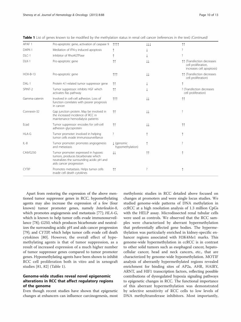
Table 1 List of genes known to be modified by the methylation status in renal cell cancer (references in the text) (Continued)
APAF 1 Pro-apoptotic gene, activation of caspase 9 ↑↑↑↑ ↓↓↓ ↑↑
DAPK-1 Mediation of IFN-γ induced apoptosis ↑ ↓ ?
DLC-1 Inhibitor of RhoAGTPase ↑ ↓ ?
DLK-1 Pro-apoptotic gene ↑↑ ↓↓ ↑↑ (Transfection decreasescell proliferation,increases cell apoptosis)
HOX-B-13 Pro-apoptotic gene ↑↑↑ ↓↓ ↑↑ (Transfection decreasescell proliferation)
DAL-1 Protein 4.1-related tumor suppressor gene ↑↑ ↓ ↑
SPINT-2 Tumor suppressor: inhibits HGF whichactivates Ras pathway
↑↑ ↓ ? (Transfection decreasescell proliferation)
Gamma-catenin Involved in cell-cell adhesion. Loss offunction correlates with poorer prognosisin cancer
↑↑↑ ↓↓ ↑↑
Connexin-32 Gap junction protein. May be involved inthe increased incidence of RCC inmaintenance hemodialysis patients
↑↑ ↓↓ ?
Ecad Tumor suppressor: encodes for cell-celladhesion glycoprotein
↑↑ ↓↓ ↑↑
HLA-G Tumor promoter: involved in helpingtumor cells evade immunosurveillance
? ↑ ↑
IL-8 Tumor promoter: promotes angiogenesisand metastasis
↓ (genomichypomethylation)
↑ ?
CA9/G250 Tumor promoter: expressed in hypoxictumors, produces bicarbonate whichneutralizes the surrounding acidic pH andaids cancer progression
↓↓ ↑↑ ↑
CYTIP Promotes metastasis. Helps tumor cellsevade cell death cytokines
↑↑ ? ↑
Shenoy et al. Journal of Hematology & Oncology (2015) 8:88 Page 10 of 13
Apart from restoring the expression of the above men-tioned tumor suppressor genes in RCC, hypomethylatingagents may also increase the expression of a few (fourknown) tumor promoter genes, namely Interleukin-8,which promotes angiogenesis and metastasis [77]; HLA-G,which is known to help tumor cells evade immunosurveil-lance [78]; G250, which produces bicarbonate and neutral-izes the surrounding acidic pH and aids cancer progression[79]; and CYTIP, which helps tumor cells evade cell deathcytokines [80]. However, the overall effect of hypo-methylating agents is that of tumor suppression, as aresult of increased expression of a much higher numberof tumor suppressor genes compared to tumor promotergenes. Hypomethylating agents have been shown to inhibitRCC cell proliferation both in vitro and in xenograftstudies [81, 82] (Table 1).
Genome-wide studies reveal novel epigenomicalterations in RCC that affect regulatory regionsof the genomeEven though recent studies have shown that epigeneticchanges at enhancers can influence carcinogenesis, most
methylomic studies in RCC detailed above focused onchanges at promoters and were single locus studies. Westudied genome-wide patterns of DNA methylation inccRCC at a high resolution analysis of 1.3 million CpGswith the HELP assay. Microdissected renal tubular cellswere used as controls. We observed that the RCC sam-ples were characterized by aberrant hypermethylationthat preferentially affected gene bodies. The hyperme-thylation was particularly enriched in kidney-specific en-hancer regions associated with H3K4Me1 marks. Thisgenome-wide hypermethylation in ccRCC is in contrastto other solid tumors such as esophageal cancer, hepato-cellular cancer, head and neck cancers, etc., that arecharacterized by genome-wide hypomethylation. MOTIFanalysis of aberrantly hypermethylated regions revealedenrichment for binding sites of AP2a, AHR, HAIRY,ARNT, and HIF1 transcription factors, reflecting possiblecontributions of dysregulated hypoxia signaling pathwaysto epigenetic changes in RCC. The functional importanceof this aberrant hypermethylation was demonstratedby selective sensitivity of RCC cells to low levels ofDNA methyltransferase inhibitors. Most importantly,

Shenoy et al. Journal of Hematology & Oncology (2015) 8:88 Page 11 of 13
methylation of enhancers was predictive of adverseprognosis in a large cohort of ccRCC samples [83].The Cancer Genome Atlas Research Network group
also reported that increasing promoter hypermethylationfrequency correlated with higher stage and grade in clearcell RCC [75]. Apart from discovering hypermethylationof VHL and UQCRH tumor suppressor genes in someccRCC samples (as described above), they also made aninteresting evaluation of the global consequence of mu-tations in specific epigenetic modifiers. Inactivating mu-tations of the SetD2 gene occur in a few cases of ccRCCand was found to be associated with increased loss ofDNA methylation. SetD2 is a H3K36 methyltransferase.H3K36 trimethylation has been suggested to be involvedin the maintenance of the heterochromatin state. DNAmethyltransferase 3A (DNMT3A) binds to trimethylatedH3K36 and methylates nearby DNA resulting in tran-scriptional silencing. Consequently, it was thought thatreduction in trimethylated H3K36 levels through SetD2inactivation could lead to regional loss of DNA methyla-tion [75]. It would be interesting to study the effect ofhypomethylating agents in SetD2 mutant and wild-typeccRCC to determine if inactivating mutations of SetD2can be indicators of poor response to hypomethylatingagents.
ConclusionsDNA methylation plays an important role in the develop-ment and progression of renal cell carcinoma. Epigeneticsilencing of numerous tumor suppressor genes caused byhypermethylation of enhancer and promoter CpGs leads toincreased tumor cell proliferation, invasion, and metastasis.In particular, activation of Wnt pathway by suppression ofWnt antagonists, namely SFRP1, SFRP2, SFRP5, and WIF-1 appears to play a very prominent role in the proliferationof many RCC cell lines and patient tumor samples—as de-termined by a higher percentage of methylation in cancertissue compared to normal tissue, very low expressioncompared to normal tissue, significant inhibition of prolif-eration, and increase in apoptosis caused by transfection ofthe Wnt antagonist genes. Similarly, TGF-β pathwayactivation by promoter methylation-induced inhibitionof pathway inhibitory genes GATA-3, GREM-1, andSMAD-6 plays a significant role in many RCC celllines. Silencing of pro-apoptotic genes such as APAF-1and negative regulators of cell cycle such as KILLINand RASSF1 also appear to play an important role.Hypomethylating agents have shown inhibition of
renal carcinoma cell proliferation both in vitro and inxenograft studies by the re-expression of numeroustumor suppressor genes silenced by enhancer/promoterhypermethylation [81, 82]. Despite the possibility of sim-ultaneous increased expression of some tumor promotergenes, the overall effect of hypomethylating agents on
RCC is that of tumor suppression due to increased ex-pression of a much higher number of tumor suppressorgenes. The genome-wide hypermethylation in RCC is incontrast to other solid tumors which demonstrate wide-spread hypomethylation, thereby making hypomethylat-ing agents and other epigenetic modulators potentialtherapeutic agents in RCC, along with emerging targetedtherapies [84–86].
Competing interestsThe authors declare that they have no competing interests.
Authors’ contributionsNS wrote and edited the article. NV wrote and edited the article. YZ, JNG,MS, CH, and KS performed the literature search and contributed to the text.AV wrote and edited the article. All authors read and approved the finalmanuscript.
Author details1Albert Einstein College of Medicine, 1300 Morris Park Avenue, Bronx, NY10467, USA. 2University of Pennsylvania Perelman School of Medicine,Philadelphia, PA, USA.
Received: 5 June 2015 Accepted: 26 June 2015
References1. Katoh M, Katoh M. WNT signaling pathway and stem cell signaling network.
Clin Cancer Res. 2007;13(14):4042–5.2. Morris MR et al. Identification of candidate tumour suppressor
genes frequently methylated in renal cell carcinoma. Oncogene.2010;29(14):2104–17.
3. Gumz ML et al. Secreted frizzled-related protein 1 loss contributes to tumorphenotype of clear cell renal cell carcinoma. Clin Cancer Res.2007;13(16):4740–9.
4. Gloerich M, Bos JL. Regulating Rap small G-proteins in time and space.Trends Cell Biol. 2011;21(10):615–23.
5. Gloerich M, Bos JL. Epac: defining a new mechanism for cAMP action. AnnuRev Pharmacol Toxicol. 2010;50:355–75.
6. Kim WJ, Gersey Z, Daaka Y. Rap1GAP regulates renal cell carcinomainvasion. Cancer Lett. 2012;320(1):65–71.
7. Cho YJ, Liang P. Killin is a p53-regulated nuclear inhibitor of DNA synthesis.Proc Natl Acad Sci USA. 2008;105(14):5396–401.
8. Bennett KL et al. Germline and somatic DNA methylation and epigeneticregulation of KILLIN in renal cell carcinoma. Genes Chromosomes Cancer.2011;50(8):654–61.
9. Polakis P. Wnt signaling and cancer. Genes Dev. 2000;14(15):1837–51.10. Kawakami K et al. Functional significance of Wnt inhibitory factor-1 gene in
kidney cancer. Cancer Res. 2009;69(22):8603–10.11. Hirata H et al. Wnt antagonist DKK1 acts as a tumor suppressor gene that
induces apoptosis and inhibits proliferation in human renal cell carcinoma.Int J Cancer. 2011;128(8):1793–803.
12. Kawamoto K et al. DNA methylation and histone modifications causesilencing of Wnt antagonist gene in human renal cell carcinoma cell lines.Int J Cancer. 2008;123(3):535–42.
13. Awakura Y et al. Methylation-associated silencing of SFRP1 in renal cellcarcinoma. Oncol Rep. 2008;20(5):1257–63.
14. Dahl E et al. Frequent loss of SFRP1 expression in multiple human solidtumours: association with aberrant promoter methylation in renal cellcarcinoma. Oncogene. 2007;26(38):5680–91.
15. Kawakami K et al. Secreted frizzled-related protein-5 is epigeneticallydownregulated and functions as a tumor suppressor in kidney cancer.Int J Cancer. 2011;128(3):541–50.
16. Garzon R, Calin GA, Croce CM. MicroRNAs in cancer. Annu Rev Med.2009;60:167–79.
17. Wang WT, Chen YQ. Circulating miRNAs in cancer: from detection totherapy. J Hematol Oncol. 2014;7(1):86.
18. Lujambio A et al. A microRNA DNA methylation signature for human cancermetastasis. Proc Natl Acad Sci USA. 2008;105(36):13556–61.

Shenoy et al. Journal of Hematology & Oncology (2015) 8:88 Page 12 of 13
19. Hildebrandt MAT et al. Hsa-miR-9 methylation status is associated withcancer development and metastatic recurrence in patients with clearcell renal cell carcinoma. Oncogene. 2010;29(42):5724–8.
20. Brazil DP et al. BMP signalling: agony and antagony in the family. TrendsCell Biol. 2015;25(5):249–64.
21. van Vlodrop IJ et al. Prognostic significance of Gremlin1 (GREM1) promoterCpG island hypermethylation in clear cell renal cell carcinoma. Am J Pathol.2010;176(2):575–84.
22. Ou YH et al. The candidate tumor suppressor BTG3 is a transcriptionaltarget of p53 that inhibits E2F1. EMBO J. 2007;26(17):3968–80.
23. Majid S et al. BTG3 tumor suppressor gene promoter demethylation,histone modification and cell cycle arrest by genistein in renal cancer.Carcinogenesis. 2009;30(4):662–70.
24. Leaman DW et al. Identification of X-linked inhibitor of apoptosis-associatedfactor-1 as an interferon-stimulated gene that augments TRAIL Apo2L-induced apoptosis. J Biol Chem. 2002;277(32):28504–11.
25. Reu FJ et al. Overcoming resistance to interferon-induced apoptosis ofrenal carcinoma and melanoma cells by DNA demethylation. J Clin Oncol.2006;24(23):3771–9.
26. Ahmad ST et al. Methylation of the APAF-1 and DAPK-1 promoter regioncorrelates with progression of renal cell carcinoma in North Indianpopulation. Tumor Biol. 2012;33(2):395–402.
27. Christoph F et al. Methylation of tumour suppressor genes APAF-1 andDAPK-1 and in vitro effects of demethylating agents in bladder and kidneycancer. Br J Cancer. 2006;95(12):1701–7.
28. Raveh T et al. DAP kinase activates a p19ARF/p53-mediated apoptoticcheckpoint to suppress oncogenic transformation. Nat Cell Biol.2001;3(1):1–7.
29. Gillissen B et al. Induction of cell death by the BH3-only Bcl-2 homologNbk/Bik is mediated by an entirely Bax-dependent mitochondrial pathway.EMBO J. 2003;22(14):3580–90.
30. Okuda H et al. Epigenetic inactivation of the candidate tumorsuppressor gene HOXB13 in human renal cell carcinoma. Oncogene.2006;25(12):1733–42.
31. Tran YK et al. A novel member of the NF2/ERM/4.1 superfamily with growthsuppressing properties in lung cancer. Cancer Res. 1999;59(1):35–43.
32. Yamada D et al. Promoter hypermethylation of the potential tumorsuppressor DAL-1/4.1B gene in renal clear cell carcinoma. Int J Cancer.2006;118(4):916–23.
33. Banumathy G, Cairns P. Signaling pathways in renal cell carcinoma. CancerBiol Ther. 2010;10(7):658–64.
34. Morris MR et al. Tumor suppressor activity and epigenetic inactivation ofhepatocyte growth factor activator inhibitor type 2/SPINT2 in papillary andclear cell renal cell carcinoma. Cancer Res. 2005;65(11):4598–606.
35. Syrigos KN et al. Altered gamma-catenin expression correlates with poorsurvival in patients with bladder cancer. J Urol. 1998;160(5):1889–93.
36. Cerrato A et al. Beta- and gamma-catenin expression in thyroid carcinomas.J Pathol. 1998;185(3):267–72.
37. Pantel K et al. Reduced expression of plakoglobin indicates an unfavorableprognosis in subsets of patients with non-small-cell lung cancer. J ClinOncol. 1998;16(4):1407–13.
38. Smith LT et al. Epigenetic regulation of the tumor suppressor gene TCF21on 6q23-q24 in lung and head and neck cancer. Proc Natl Acad Sci USA.2006;103(4):982–7.
39. Ye YW et al. Down-regulation of TCF21 is associated with poor survival inclear cell renal cell carcinoma. Neoplasma. 2012;59(6):599–605.
40. Costa VL et al. TCF21 and PCDH17 methylation: an innovative panel ofbiomarkers for a simultaneous detection of urological cancers. Epigenetics.2011;6(9):1120–30.
41. Chen WY et al. Tumor suppressor HIC1 directly regulates SIRT1 to modulatep53-dependent DNA-damage responses. Cell. 2005;123(3):437–48.
42. Wang CG et al. Interactions between E2F1 and SirT1 regulate apoptoticresponse to DNA damage. Nat Cell Biol. 2006;8(9):1025–U109.
43. Jenal M et al. The tumor suppressor gene hypermethylated in cancer 1 istranscriptionally regulated by E2F1. Mol Cancer Res. 2009;7(6):916–22.
44. Eggers H et al. Prognostic and diagnostic relevance of hypermethylated incancer 1 (HIC1) CpG island methylation in renal cell carcinoma. Int J Oncol.2012;40(5):1650–8.
45. Kim M et al. LRRC3B, encoding a leucine-rich repeat-containing protein,is a putative tumor suppressor gene in gastric cancer. Cancer Res.2008;68(17):7147–55.
46. Haraldson K et al. LRRC3B gene is frequently epigenetically inactivated inseveral epithelial malignancies and inhibits cell growth and replication.Biochimie. 2012;94(5):1151–7.
47. Merika M, Orkin SH. DNA-binding specificity of GATA family transcriptionfactors. Mol Cell Biol. 1993;13(7):3999–4010.
48. Peters I et al. GATA5 CpG island methylation in renal cell cancer: apotential biomarker for metastasis and disease progression. BJU Int.2012;110(2 Pt 2):E144–52.
49. Morrissey C et al. Epigenetic inactivation of the RASSF1A 3p21.3 tumorsuppressor gene in both clear cell and papillary renal cell carcinoma.Cancer Res. 2001;61(19):7277–81.
50. Peters I et al. RASSF1A promoter methylation and expression analysis innormal and neoplastic kidney indicates a role in early tumorigenesis. MolCancer. 2007;6:49.
51. Ellinger J et al. DNA hypermethylation in papillary renal cell carcinoma.BJU Int. 2011;107(4):664–9.
52. Kawai Y et al. Methylation level of the RASSF1A promoter is anindependent prognostic factor for clear-cell renal cell carcinoma.Ann Oncol. 2010;21(8):1612–7.
53. Costa VL et al. Quantitative promoter methylation analysis of multiplecancer-related genes in renal cell tumors. BMC Cancer. 2007;7:133.
54. Arakawa H. Netrin-1 and its receptors in tumorigenesis. Nat Rev Cancer.2004;4(12):978–87.
55. Cirulli V, Yebra M. Netrins: beyond the brain. Nat Rev Mol Cell Biol.2007;8(4):296–306.
56. Lv D et al. Genetic and epigenetic control of UNC5C expression in humanrenal cell carcinoma. Eur J Cancer. 2011;47(13):2068–76.
57. Bader BL, Jahn L, Franke WW. Low level expression of cytokeratins 8, 18 and19 in vascular smooth muscle cells of human umbilical cord and in culturedcells derived therefrom, with an analysis of the chromosomal locuscontaining the cytokeratin 19 gene. Eur J Cell Biol. 1988;47(2):300–19.
58. Ju JH et al. Regulation of cell proliferation and migration by keratin19-induced nuclear import of early growth response-1 in breast cancer cells.Clin Cancer Res. 2013;19(16):4335–46.
59. Cao Y et al. Expression of MUC1, Thomsen-Friedenreich-related antigens,and cytokeratin 19 in human renal cell carcinomas and tubular clear celllesions. Virchows Arch. 2000;436(2):119–26.
60. Labastie MC et al. The Gata-3 gene is expressed during human kidneyembryogenesis. Kidney Int. 1995;47(6):1597–603.
61. Cooper SJ et al. Loss of type III transforming growth factor-beta receptorexpression is due to methylation silencing of the transcription factorGATA3 in renal cell carcinoma. Oncogene. 2010;29(20):2905–15.
62. Tavares TS et al. Gene microarray analysis of human renal cell carcinoma:the effects of HDAC inhibition and retinoid treatment. Cancer Biol Ther.2008;7(10):1607–18.
63. Qi JH et al. A novel function for tissue inhibitor of metalloproteinases-3(TIMP3): inhibition of angiogenesis by blockage of VEGF binding to VEGFreceptor-2. Nat Med. 2003;9(4):407–15.
64. Masson D et al. Loss of expression of TIMP3 in clear cell renal cellcarcinoma. Eur J Cancer. 2010;46(8):1430–7.
65. Bachman KE et al. Methylation-associated silencing of the tissue inhibitor ofmetalloproteinase-3 gene suggest a suppressor role in kidney, brain, andother human cancers. Cancer Res. 1999;59(4):798–802.
66. Awakura Y et al. Methylation-associated silencing of TU3A in human cancers.Int J Oncol. 2008;33(4):893–9.
67. Kvasha S et al. Hypermethylation of the 5′CpG island of the FHIT gene inclear cell renal carcinomas. Cancer Lett. 2008;265(2):250–7.
68. Kawakami T et al. Imprinted DLK1 is a putative tumor suppressor gene andinactivated by epimutation at the region upstream of GTL2 in human renalcell carcinoma. Hum Mol Genet. 2006;15(6):821–30.
69. Schmidt JV et al. The Dlk1 and Gtl2 genes are linked and reciprocallyimprinted. Genes Dev. 2000;14(16):1997–2002.
70. Yano T et al. Tumor-suppressive effect of connexin 32 in renal cell carcinomafrom maintenance hemodialysis patients. Kidney Int. 2003;63(1):381–1.
71. Yano T et al. Hypermethylation of the CpG island of connexin 32, a candiatetumor suppressor gene in renal cell carcinomas from hemodialysis patients.Cancer Lett. 2004;208(2):137–42.
72. Nojima D et al. CpG methylation of promoter region inactivates E-cadheringene in renal cell carcinoma. Mol Carcinog. 2001;32(1):19–27.
73. Yoo KH et al. Epigenetic inactivation of HOXA5 and MSH2 gene in clear cellrenal cell carcinoma. Pathol Int. 2010;60(10):661–6.

Shenoy et al. Journal of Hematology & Oncology (2015) 8:88 Page 13 of 13
74. Clifford SC et al. Inactivation of the von Hippel-Lindau (VHL) tumour suppressorgene and allelic losses at chromosome arm 3p in primary renal cell carcinoma:evidence for a VHL-independent pathway in clear cell renal tumourigenesis.Genes Chromosomes Cancer. 1998;22(3):200–9.
75. Cancer Genome Atlas Research Network. Comprehensive molecularcharacterization of clear cell renal cell carcinoma. Nature. 2013;499(7456):43–9.
76. Chu Q et al. DACH1 inhibits cyclin D1 expression, cellular proliferation andtumor growth of renal cancer cells. J Hematol Oncol. 2014;7(1):73.
77. Yoo KH, Park YK, Chang SG. DNA hypomethylation of interleukin 8 in clearcell renal cell carcinoma. Oncol Lett. 2013;5(1):39–42.
78. Dunker K et al. Expression and regulation of non-classical HLA-G in renal cellcarcinoma. Tissue Antigens. 2008;72(2):137–48.
79. Cho M et al. Activation of the MN/CA9 gene is associated withhypomethylation in human renal cell carcinoma cell lines. Mol Carcinog.2000;27(3):184–9.
80. Vanharanta S et al. Epigenetic expansion of VHL-HIF signal output drivesmultiorgan metastasis in renal cancer. Nat Med. 2013;19(1):50–6.
81. Hagiwara H et al. 5-Aza-2′-deoxycytidine suppresses human renal carcinomacell growth in a xenograft model via up-regulation of the connexin 32gene. Br J Pharmacol. 2008;153(7):1373–81.
82. Ricketts CJ et al. Methylation profiling and evaluation of demethylatingtherapy in renal cell carcinoma. Clin Epigenetics. 2013;5(1):16.
83. Hu CY et al. Kidney cancer is characterized by aberrant methylation oftissue-specific enhancers that are prognostic for overall survival. Clin CancerRes. 2014;20(16):4349–60.
84. Xu KY, Wu S. Update on the treatment of metastatic clear cell and non-clearcell renal cell carcinoma. Biomark Res. 2015;3:5.
85. Parikh K et al. Selective inhibitors of nuclear export (SINE)—a novel class ofanti-cancer agents. J Hematol Oncol. 2014;7:78.
86. Smith AD, Roda D, Yap TA. Strategies for modern biomarker and drugdevelopment in oncology. J Hematol Oncol. 2014;7(1):70.
Submit your next manuscript to BioMed Centraland take full advantage of:
• Convenient online submission
• Thorough peer review
• No space constraints or color figure charges
• Immediate publication on acceptance
• Inclusion in PubMed, CAS, Scopus and Google Scholar
• Research which is freely available for redistribution
Submit your manuscript at www.biomedcentral.com/submit