revised guidelines for good practice in ivf laboratories...
TRANSCRIPT

1
ESHRE Guideline Group on good practice in IVF labs
December 2015
Revised guidelines for good practice in IVF laboratories (2015)
Guideline of the European Society of Human Reproduction and Embryology

2
Disclaimer
The European Society of Human Reproduction and Embryology (hereinafter referred to as 'ESHRE') developed the current clinical practice guideline, to provide clinical recommendations to improve the quality of healthcare delivery within the European field of human reproduction and embryology. This guideline represents the views of ESHRE, which were achieved after careful consideration of the scientific evidence available at the time of preparation. In the absence of scientific evidence on certain aspects, a consensus between the relevant ESHRE stakeholders has been obtained.
The aim of clinical practice guidelines is to aid healthcare professionals in everyday clinical decisions about appropriate and effective care of their patients.
However, adherence to these clinical practice guidelines does not guarantee a successful or specific outcome, nor does it establish a standard of care. Clinical practice guidelines do not override the healthcare professional's clinical judgment in diagnosis and treatment of particular patients. Ultimately, healthcare professionals must make their own clinical decisions on a case-by-case basis, using their clinical judgment, knowledge, and expertise, and taking into account the condition, circumstances, and wishes of the individual patient, in consultation with that patient and/or the guardian or carer.
ESHRE makes no warranty, express or implied, regarding the clinical practice guidelines and specifically excludes any warranties of merchantability and fitness for a particular use or purpose. ESHRE shall not be liable for direct, indirect, special, incidental, or consequential damages related to the use of the information contained herein. While ESHRE makes every effort to compile accurate information and to keep it up-to-date, it cannot, however, guarantee the correctness, completeness, and accuracy of the guideline in every respect. In any event, these clinical practice guidelines do not necessarily represent the views of all clinicians that are member of ESHRE.
The information provided in this document does not constitute business, medical or other professional advice, and is subject to change.

3
Introduction
In line with the ESHRE scope, this document represents an updated version of the guidelines published in 2008
(Magli, et al., 2008). The aim is to provide a wider coverage of key aspects of the IVF laboratory, to give continuous
support to laboratory specialists and consequently contribute to improving IVF patient care.
1 Staffing and direction
Personnel are one of the most important parts of an IVF laboratory. The number of laboratory staff should reflect
the number of cycles performed per year. As an approximate guide, clinics that perform up to 150 retrievals
and/or cryopreservation cycles per year should have always a minimum of two qualified clinical embryologists.
This initial number will increase depending not only on the number of treatments, but also on the complexity of
the procedures, techniques and tasks undertaken within the laboratory. Other duties such as administration,
training, education, quality management and communication also need consideration.
Appropriate human resources should provide an adequate climate to perform all laboratory tasks in a timely
manner, to ensure patient safety and quality care. Sufficient qualified personnel should be available to provide
back-up for the laboratory staff.
The hierarchical laboratory organisation depends on staff size. Larger facilities can delegate responsibilities to
different staff levels, e.g. supervisors, clinical embryologists, laboratory technicians and administrative personnel.
1.1 Laboratory director
The laboratory should be directed by a person with officially recognised qualifications and expertise in clinical
embryology and biological/medical sciences. In accordance with the results of the ESHRE survey on the education
and professional status of clinical embryologists (Kovacic, et al., 2015), this would include a higher academic
degree (MD, MSc, PhD) with a minimum of 6 years of documented human embryology experience, and preferably
attainment of the ESHRE senior clinical embryologist certification or similar.
Laboratory directors should be able to evaluate and interpret the significance of medical and laboratory findings,
and communicate them to laboratory staff members, clinical colleagues, patients and the public. They should
proactively seek clinical and scientific updates, promote science and participate in clinical studies and research,
where possible.
Laboratory director responsibilities include ensuring:
1.1.1 Selection and implementation of the most adequate materials and procedures to reach the highest
standards in clinical IVF.
1.1.2 Safe and appropriate laboratory facilities and equipment according to European and/or national
regulations.
1.1.3 Implementation of a quality management system (QMS).
1.1.4 Implementation of a laboratory risk management and prevention policy.

4
1.1.5 Sufficient laboratory staff members with the appropriate skills.
1.1.6 A comprehensive orientation and introduction programme for all new staff members.
1.1.7 Management of laboratory staff training and continual scientific and biomedical education.
1.1.8 Implementation and review of key performance indicators (KPIs) for all laboratory procedures for quality
control and quality assurance purposes.
1.1.9 Reporting of clinical data and adverse events according to European and/or national regulations.
1.1.10 Approval of research projects by competent authorities.
1.2 Laboratory supervisors
Some laboratories may require additional managerial positions. These require specific qualifications, e.g. at least
a BSc in biomedical sciences, 3 years of documented human embryology experience and preferably attainment
of the ESHRE clinical embryologist certification or similar.
Laboratory supervisor responsibilities include ensuring:
1.2.1 Efficient organisation of daily work of their areas of responsibility
1.2.2 Effective communication with laboratory staff and clinical colleagues.
1.2.3 Continuous improvement where possible.
1.2.4 Structured training of staff members and students.
1.3 Clinical embryologists
Clinical embryologists represent the first line of participation in daily clinical practice. These positions require at
least a BSc in biomedical sciences. New staff should follow a structured training programme supervised by
experienced clinical embryologists.
Clinical embryologists with 3 years’ experience should endeavour to apply for the ESHRE clinical embryologist
certification, whereas those with higher degrees and 6 years’ experience should endeavour to apply for the ESHRE
senior clinical embryologist certification.
Clinical embryologist responsibilities include:
1.3.1 Execution of standard operating procedures (SOPs).
1.3.2 Participation in daily practice, communication and organisation.
1.3.3 Contribution to laboratory clinical decisions.
1.3.4 Training of staff members and students.

5
2 Quality management
According to the European directives and recommendations (European Commission, 2006a, c, 2012; Council of
Europe, 2013), working in compliance with a QMS is mandatory. The requirements cover the organisation,
management, personnel, equipment and materials, facilities/premises, documentation, records and quality
review. This includes:
- defining responsibilities and ensuring all personnel are qualified and competent;
- having validated, written instructions for each process (SOP), including management of adverse
events;
- ensuring full traceability of cells and tissues, materials, equipment and personnel involved in
specific laboratory activities, with records maintained accordingly;
- confirming that all media/reagents/disposables are tested for quality using appropriate assays
whenever possible;
- ensuring proper and periodic equipment maintenance, service and calibration;
- verifying conformance to specifications;
- taking corrective action to keep procedures under conformity;
- reviewing performance to ensure continuous and systematic QMS improvement;
- providing risk assessment analysis for all laboratory activities.
2.1 It is recommended that a clinical embryologist is made responsible for quality management within the
laboratory.
2.2 Written, authorised, signed and up-to-date SOPs should exist for all processes in order to optimise
outcomes.
2.3 The QMS must include provision for unique identification of patients and their reproductive cells and
tissues, while retaining patient confidentiality.
2.4 All relevant data concerning laboratory work must be recorded in a database that allows KPI extraction
and statistical analysis. Corrections, either written or electronic, should be traceable. Data should include:
- Morphological characteristics of gametes and embryos.
- Detailed information of the procedures, including timing and staff involved.
- All information needed to comply with the requirements of national and international data
registries.
2.5 Every relevant communication with the patient should be recorded in the patient’s files.
2.6 Taking into account the high degree of attention needed during laboratory work, distractions should be
minimised.
2.7 Proactive risk assessments should be made and preventive actions taken to minimise non-conformities.

6
2.8 A documentation system should be in place for dealing with non-compliances, emergencies, errors,
adverse events and complaints. Corrective and preventive actions, implementation dates and assessments
of their effectiveness should be documented. Under certain circumstances, a period of follow-up may be
advisable to ensure the adequacy of actions. Non-compliances should be discussed regularly and reviewed
at least annually.
2.9 KPIs should be objective and relevant, regularly checked and discussed, and communicated to all staff.
KPIs can be based on a reference patient group with good prognosis, as well as on the whole patient
population. Appropriate statistics can be used to account for patient variation and the number of previous
treatment cycles patients may have already undertaken.
2.10 Critical performance levels should be defined for each KPI with reference to national data and European
registry data collected by the European IVF-monitoring programme for ESHRE. If necessary, appropriate
action should be taken.
2.11 In addition to laboratory and clinical performance, operator performance should be checked regularly to
ensure competence, compliance and consistency, via direct observation of procedural skills (DOPS) and/or
individual KPIs. If necessary, retraining should be implemented.
2.12 Participation in Internal Quality Control (IQC) and External Quality Assurance (EQA) programmes, either
commercial or in collaboration with other laboratories, is highly recommended. QC records should be
maintained and reviewed, including documentation of results and any corrective action.
2.13 The laboratory’s QMS should be systematically reviewed annually to ensure continuous improvement of
the entire process by identifying current challenges, problems, errors or improvements.
2.14 An audit system, both internal and external, must be in place. An independent, competent auditor should
verify compliance of all procedures with SOPs and requirements. Any findings, corrective actions and their
effectiveness must be documented.

7
3 Laboratory safety
3.1 Laboratory design
The IVF laboratory must have adequate functionalities to minimise any damaging effects upon reproductive cells,
and ensure good laboratory practice. The laboratory should be adjacent to the operating room where clinical
procedures are performed.
When commissioning the IVF laboratory, the most recent developments in facilities, equipment and procedures
should be considered. Attention should be given to operator comfort to provide a safe working environment that
minimises the risk of distraction, fatigue and thereby making a mistake. Taking into account local, national and
European occupational health and safety requirements, considerations should include bench height, adjustable
chairs, adequate work space per person, microscope eye height, efficient use of space and surfaces, sufficient
environmental lighting and air-conditioning with controlled humidity and temperature.
More specifically:
3.1.1 Materials used in laboratory construction, painting, flooring and furniture should be appropriate for
clean room standards, minimising Volatile Organic Compounds (VOC) release and embryo toxicity.
3.1.2 Laboratory design should ensure optimal workflow over minimal distances while handling reproductive
cells during all treatment phases.
3.1.3 Laboratory access should be restricted to authorised personnel.
3.1.4 A system for clean access of personnel and materials to the laboratory is highly recommended.
3.1.5 Rooms for changing clothes should be separate from the laboratory.
3.1.6 Hand-washing facilities should be placed outside the laboratory.
3.1.7 Separate office space for administrative work should be available outside the laboratory.
3.1.8 A separate laboratory with a safety fume hood should be provided for analyses using fixatives and other
toxic reagents.
3.1.9 The area for cleaning and sterilisation of materials, if present, should be separate from the laboratory.
3.2 Laboratory air quality
3.2.1 To optimise environmental conditions, laboratory air should be subjected to high-efficiency particulate
air (HEPA) and VOC control.
3.2.2 Positive pressure is recommended to minimise air contamination.
3.2.3 Procedures involving gamete or embryo manipulation should be performed in a controlled environment.
Background and processing air quality should comply with European and national guidelines, and should
be regularly monitored.
3.2.4 According to the European Union Tissues and Cells Directive (EUTCD), tissue and cell processing must be
performed in a Good Manufacturing Practice (GMP) Grade A environment with a background of at least
GMP Grade D. However, if it is detrimental or not feasible to carry out a specific procedure in a Grade A
environment, it can be performed in at least a Grade D environment.

8
3.3 Laboratory equipment
3.3.1 The laboratory should contain all essential items required for IVF, in a number appropriate to the
workload.
3.3.2 The incubator number is critical and should be based on the number of cycles and embryo culture
duration. Gametes and embryos should be conveniently distributed across incubators to minimise door
openings.
3.3.3 Equipment must be adequate for optimal laboratory work, easy to disinfect and kept clean to avoid
contamination.
3.3.4 All equipment must be validated as fit for its purpose, and performance verified by calibrated
instruments. Equipment should preferably be CE-marked.
3.3.5 Gas cylinders should be located outside the laboratory. There should be an automatic change-over
system and sufficient cylinders stocked for immediate replacement. High-purity gas and inline HEPA and
VOC filters are highly recommended.
3.3.6 Equipment validation, calibration, maintenance and repair must be documented and records retained.
3.3.7 Heating devices should be installed to maintain the temperature of media and reproductive cells during
handling.
3.3.8 Accepted ranges of use for all measured parameters should be determined and recorded. If
measurements are out of range, corrections should be made and their effectiveness verified.
3.3.9 For every item of equipment, the instruction manual, and simplified instructions where needed, should
be available.
3.3.10 Malfunctioning equipment should be labelled as “out-of-use” to avoid its use by mistake.
3.3.11 Critical items of equipment, including incubators and cryostorage units, should be continuously
monitored and equipped with alarm systems.
3.3.12 An automatic emergency backup power system must be in place for all critical equipment.
3.4 Cryopreservation facilities and material
3.4.1 Cryopreservation facilities should be rationally and safely located outside but close to the laboratory and,
for safety reasons, with visible access to the interior (e.g. via a window, camera).
3.4.2 Adequate ventilation and low oxygen alarms should be installed. Personal low oxygen alarms are
recommended, as additional security measure.
3.4.3 Cryostorage units should be continuously monitored and equipped with alarm systems, detecting any
out of range temperature and/or levels of liquid nitrogen (LN2).
3.4.4 Protection devices (e.g. glasses, face shield, cryogloves, apron, footwear) should be used during LN2
handling.
3.4.5 All staff dealing with LN2 should be trained in safety aspects of its use.
3.5 Infectious agents
All assisted reproductive technologies (ART) involve handling of biological material, and pose a potential hazard
9
of transmitting diseases to personnel and to other patients’ biological material (cross-contamination).
3.5.1 Procedures to ensure personnel safety and prevent cross-contamination should be established, taking
European and national safety regulations into consideration. Therefore:
- Vaccination of all personnel against hepatitis B or other viral diseases, for which a vaccine is
available, is recommended.
- Patients must be screened for infectious diseases according to national and international
regulations.
- Staff must be informed when a viral-positive patient is to be treated and be aware of the risks
of handling infected biological material.
- SOPs should be in place to manage eventualities where infection might take place, e.g. needle-
stick injuries.
3.5.2 To ensure adequate safety measures, the treatment of viral-positive patients should be only performed
in IVF laboratories with dedicated areas and equipment. Alternatively, such patient treatments could
be allocated to specific time slots provided processing of their biological materials is followed by a
thorough disinfection of the allocated areas and equipment.
3.5.3 Whenever biological material is imported into the IVF laboratory from another clinic, full screening
results should be obtained in advance. If any transported material is viral-positive, a dedicated dry
shipper may be needed, depending on European and national regulations.
3.6 Protective measures
All body fluids (blood, follicular fluid, semen, etc.) should be treated as potentially contaminated.
Protective measures for laboratory staff to ensure aseptic conditions for tissue, gametes and embryos include:
- Strict adherence to staff hygiene regulations and aseptic techniques.
- Use of protective laboratory clothing, preferably with low particle-shedding.
- Use of hairnets and non-toxic, non-powdered gloves and masks where appropriate.
- Use of appropriate vertical laminar flow benches for handling biological material.
- Use of mechanical pipetting devices.
- Disposal of single-use consumables immediately into proper waste containers. Potentially
infectious materials must be disposed of in a manner that protects laboratory workers and other
staff from exposure. Viral-positive waste segregated into a separate bin, labelled and disposed
of according to biosafety policies.
- Needles, glassware and other sharps should be handled with extreme caution and discarded
into sharps containers.
- Disinfectants with proven compatibility and efficacy for an IVF laboratory should be used.
- Food, gum, drinks and tobacco are strictly forbidden.
- Use of cosmetics should be minimised and perfumes should be avoided.
- Staff should be appropriately attired to diminish possible sources of contamination.

10
4 Identification of patients and traceability of their reproductive cells
Identification of patients and traceability of their reproductive cells are crucial aspects of ART treatments. Each
IVF laboratory must have an effective and accurate system to uniquely identify, trace and locate reproductive
cells during each procedural step. A proper identification system should ensure that the main characteristics of
patients (or donors) and their tissues and cells, together with relevant data regarding products and materials
coming into contact with them, are available at all times.
4.1 Proper training in traceability procedures for all laboratory staff is mandatory.
4.2 Before commencing any procedure, the laboratory must be provided with each patient’s unique
identification code which has to clearly and easily refer to the patient’s documentation. Each treatment
cycle must be assigned a unique code.
4.3 Corresponding consent forms, clinical data and serological examinations undertaken by patients/donors
prior to admission to the treatment should be available to the laboratory staff.
4.4 Rules concerning the correct identification and processing of reproductive cells must be established in the
laboratory by a system of codes and checks including:
- Direct verification of patient identity and correspondence with their assigned unique
identification code is required at every critical step. Patients should be directly asked to give
their own identifying information (at least full name and date of birth) before procurement or
artificial insemination / embryo transfer.
- All devices containing biological material must be clearly and permanently labelled with the
unique patient identification code and date of treatment.
- Biological material from different patients must not be processed in the same working area at
the same time.
- Incubators and cryostorage systems should be organised to ensure easy access and
identification of the biological materials therein.
- During critical steps (such as first identification of cells and tissues, each time biological material
is moved from one container to another and at final destination e.g. embryo transfer,
cryocontainer), double-checks by a second person (witness) and/or an electronic identification
system is strongly advised.
- Products and materials used with biological materials must be traceable.
- The date and time of each manipulation and identity of all operators and witnesses must be
documented throughout the treatment. These records should be kept for a specified period of
time according to European and/or national legislation.
- Gametes and embryos from non-partner donation may require specific coding for those
countries that are regulated according to European Commission Directives (European
Commission, 2006c) (additional Directive anticipated in 2016).
11
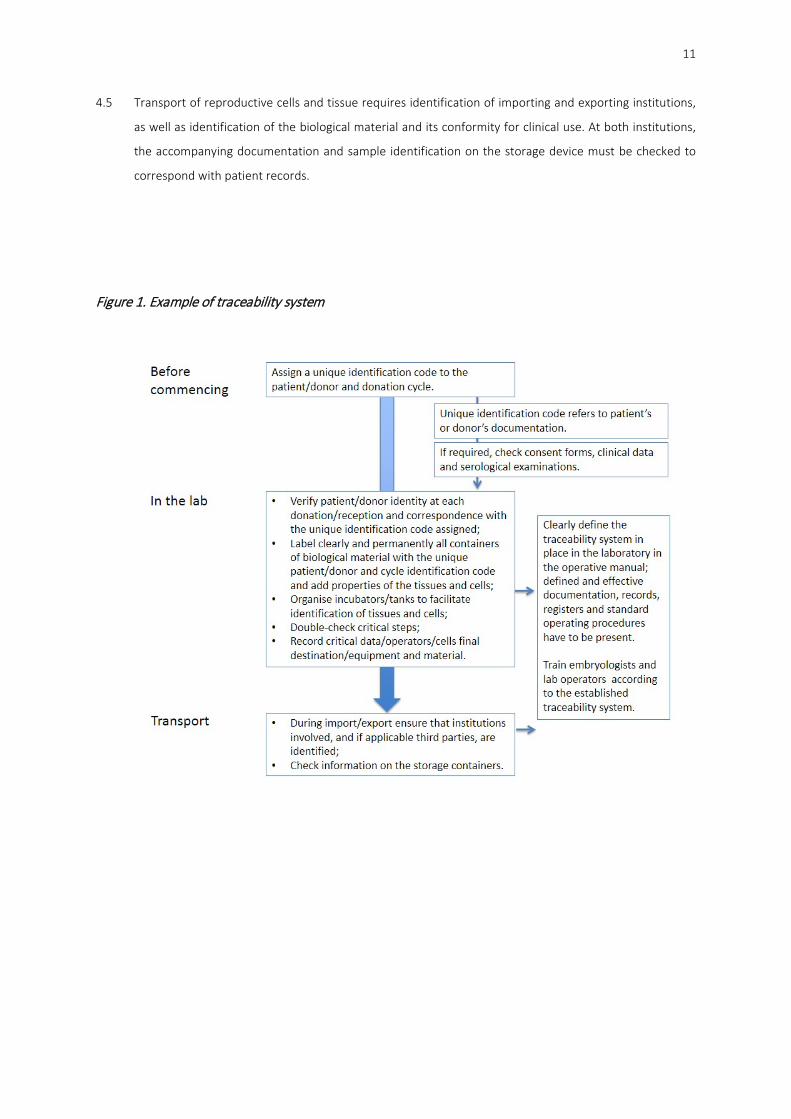
4.5 Transport of reproductive cells and tissue requires identification of importing and exporting institutions,
as well as identification of the biological material and its conformity for clinical use. At both institutions,
the accompanying documentation and sample identification on the storage device must be checked to
correspond with patient records.
Figure 1. Example of traceability system

12
5 Consumables
Specifications of critical reagents and materials should be in compliance with European and/or national
regulations.
5.1 All consumables and media should be fit for their purpose, of embryo culture grade quality and preferably
CE-marked. Use of quality controlled media, oil and disposables is recommended. If appropriate quality
control testing for IVF purposes is not provided, this must be performed by the laboratory itself or by a
designated company. In addition, packaging integrity and appropriate delivery conditions should be
checked. Documentation of quality control testing must be supplied for any commercial media and this
must correspond with the delivered batch.
5.2 Sterile single-use disposable consumables should be used.
5.3 Reagents, media and consumables should always be used prior to the manufacturer’s expiry date.
5.4 Size of the bottles and other packaging must be appropriate to minimise openings and time between first
and last use.
5.5 Appropriate refrigeration facilities must be available for storage of media and reagents. The correct
temperature during their shipment to the clinic should be verified. Repeated shifts of temperature should
be avoided while handling in the laboratory.
5.6 Patient or donor serum and follicular fluid should not be used as a protein supplement. Commercial
suppliers of human serum albumin or media containing a serum-derived protein source should provide
evidence of screening according to European and/or national regulations.
5.7 An appropriate stock management system for media, oil and consumables, including the batch number,
date of entry and expiration date should be available.
5.8 Risk assessments should be performed to ensure all consumables and media are easily identified to avoid
any misuse.

13
6 Handling of biological material
6.1 Handling of biological material should be easy, simple and effective and should preferably be performed
in laminar flow hoods equipped with heating stages and pre-warmed heating blocks, using aseptic
techniques at all times.
6.2 Measures must be taken to ensure that oocytes and embryos are always maintained at the appropriate
temperature, pH and osmolality during culture and handling. Exposure to light, toxic substances or harmful
radiation should be minimised.
6.3 Buffered media (HEPES, MOPS or similar) should be kept in atmospheric air, whereas bicarbonate-buffered
media should be kept in 5-7% CO2.
6.4 In-house-made or sterilised devices for handling human gametes and embryos should be avoided.
Pipetting devices (handling or denudation pipettes) should be used for one procedure only.
6.5 Traceability should be confirmed at all times (see Section 4 and Figure 1).

14
7 Oocyte retrieval
Oocyte retrieval is a particularly sensitive procedure and special attention should be given to temperature and pH
as well as efficient and quick handling.
7.1 An identity check before the oocyte retrieval is mandatory.
7.2 The time between oocyte retrieval and culture of washed oocytes should be minimal. Prolonged oocyte
exposure to follicular fluid is not recommended.
7.3 Appropriate equipment must be in place to maintain oocytes close to 37°C. Flushing medium, collection
tubes and dishes for identifying oocytes should be pre-warmed.
7.4 Follicular aspirates should be checked for the presence of oocytes using a stereomicroscope and heated
stage, usually at 8-60x magnification. Exposure of oocytes to light should be minimised.
7.5 Timing of retrieval, number of collected oocytes and the operator should be documented.

15
8 Sperm preparation
Before starting a treatment cycle, at least one diagnostic semen analysis should be performed according to the
protocols described in the World Health Organization (WHO) manual (World Health Organization, 2010). In
addition, a test sperm preparation may be advisable in order to propose the most adequate insemination
technique (IVF/ICSI). Patients should be given clear instructions regarding the collection of the sperm sample
(hygiene, sexual abstinence, timing etc.). A frozen back-up sample should be requested if sperm collection
difficulty on the day of oocyte retrieval is anticipated.
Sperm preparation aims to:
- eliminate seminal plasma, debris and contaminants;
- concentrate progressively motile sperm;
- select against morphologically abnormal sperm.
8.1 Semen samples should be collected into sterile, plastic containers (tissue grade, sperm-toxicity tested).
The use of spermicidal condoms, creams or lubricants must be avoided. The container should be clearly
labelled and correct identification should be confirmed by the patient. Collection should be preferably
performed in a room near to the laboratory. After collection, the sample should be delivered to the
laboratory as soon as possible avoiding extreme temperatures (< 20°C and > 37°C). Sperm analysis and
preparation should start within 1 h of collection. Prolonged sperm exposure to seminal plasma is not
recommended.
8.2 Records should be kept of the type of container used, the time and place of collection and the time interval
between collection and analysis/preparation. The use of medication, fever during the previous months
and completeness of the ejaculate collection should be documented.
8.3 The following data on sperm preparation should be documented:
- sample origin (ejaculate/epididymal/testicular, donor/partner, fresh/frozen);
- preparation method;
- pre- and post-preparation sperm parameters and any dilution carried out;
8.4 An appropriate sperm preparation method should be chosen according to the characteristics and origin of
individual samples. The swim-up technique and discontinuous density-gradient centrifugation are most
frequently used and widely accepted.
8.5 In case of azoospermia on the day of oocyte retrieval and in the absence of a back-up sample, alternative
sperm retrieval procedures or oocyte cryopreservation should be considered.
8.6 For patients diagnosed with blood-borne viruses, extensive semen preparation by density-gradient
centrifugation followed by swim-up is recommended. Depending on the serological status, it is
recommended to freeze the prepared sperm suspension and to test its viral load before release. Only viral-
free suspensions should be used for ART.

16
9 Insemination of oocytes
Oocytes can be inseminated by conventional IVF or by ICSI. The insemination/injection time should be decided
based on the number of hours elapsed from ovulation trigger and/or oocyte retrieval, also keeping in mind that
fertilisation will need to be checked 16-18 h later.
9.1 Conventional IVF
9.1.1 The number of progressively motile sperm used for insemination must be sufficient to optimise the
chance of normal fertilisation. Typically, a progressively motile sperm concentration ranging between
0.1 and 0.5x10⁶/ml is used.
9.1.2 The final sperm suspension should be in a medium compatible with oocyte culture. The fertilisation
medium should contain glucose to allow for appropriate sperm function.
9.1.3 A double-check of identity of gametes at the time of insemination procedure is mandatory.
9.1.4 Records should be kept of the time of insemination, the operator and the concentration of progressively
motile sperm used.
9.1.5 Co-incubation of cumulus oocyte complexes and sperm is usually performed overnight, although a
shorter period may be sufficient.
9.2 ICSI procedure
9.2.1 Preparation of oocytes for ICSI.
When removing cumulus cells from oocytes, hyaluronidase concentration and exposure should be kept
to a minimum. In order to prevent oocyte damage, pipettes with appropriate lumen size should be used
and vigorous pipetting avoided. After denudation, oocytes should be thoroughly washed to remove
traces of hyaluronidase. The maturation stage of the oocytes should be recorded. Current evidence does
not suggest that denudation should be performed at a specific time between oocyte recovery and ICSI.
However, since denuded oocytes are more vulnerable to pH changes, the timing of denudation should
be kept close to the timing of injection.
9.2.2 The injection procedure
Records should be kept of the injection time (start and end of the procedure) and the performing
operator. The duration of sperm identification and immobilisation followed by injection should be
minimised. The number of oocytes transferred to the injection dish should relate to operator’s skills and
sperm quality. During ICSI, the following points are important:
- Only mature oocytes should be injected.
- Oocyte morphology should be recorded. Giant oocytes or oocytes with a very large polar body
should not be injected.
- Morphologically normal, motile sperm should be selected.
- Tail membrane breakage should be posterior to the midpiece, and performed immediately
before the injection of each individual oocyte.

17
- Polar body should be away from the injection site.
- Oolemma rupture should be assured prior to sperm injection.
- Appropriate temperature and pH should be maintained during injection.
Viscous substances such as polyvinylpyrrolidone (PVP) can be used to facilitate sperm manipulation.
In case of only immotile sperm cells, a non-invasive vitality test can be used to select viable sperm for
injection. After injection, oocytes should be washed prior to culture.
9.2.3 A double-check of identity of gametes before starting injection is mandatory.

18
10 Scoring for fertilisation
10.1 All inseminated or injected oocytes should be examined for the presence of pronuclei (PN) and polar
bodies at 16-18 h post insemination. For conventional IVF, cumulus cells must be removed and normally
fertilised (2PN) oocytes transferred into new dishes containing pre-equilibrated culture medium.
10.2 Fertilisation assessment should be performed under high magnification (at least 200x), using an inverted
microscope equipped with Hoffman or equivalent optics (or a suitable time-lapse microscopy device), in
order to verify PN number and morphology.
10.3 Embryos derived from ≥ 3PN oocytes should never be transferred or cryopreserved. Even if no transferable
embryos derived from 2PN oocytes are available, the use of embryos derived from 1PN oocytes or oocytes
showing no PN is not recommended.

19
11 Embryo culture and transfer
In order to optimise embryo development, fluctuations of culture conditions should be minimised. Precautions
must be taken to maintain adequate conditions of pH and temperature to protect embryo homeostasis during
culture and handling.
11.1 Different approaches or culture systems can be used in order to optimise embryo development.
11.1.1 A culture medium designed for embryo development should be used, e.g. sequential or single-step
media.
11.1.2 The type and number of incubators should be appropriate to the workload.
11.1.3 Oil overlay minimises changes to temperature, pH and osmolality.
11.1.4 For traceability purposes, single embryo culture is advisable.
11.1.5 For blastocyst culture, a low oxygen concentration should be used.
11.2 Embryo scoring should be performed at high magnification (at least 200x, preferably 400x) using an
inverted microscope with Hoffman or equivalent optics. Evaluation of cleavage-stage embryos should
include cell number, size and symmetry, percentage of fragmentation, granulation, vacuoles and nuclear
status (e.g. multinucleation). Blastocyst scoring should include expansion grade, blastocoel cavity size and
morphology of the inner cell mass (ICM) and trophectoderm (TE). Assessment should be performed at
standardised times post insemination. Embryo development can also be assessed using time-lapse
imaging, allowing a more precise evaluation of the timing of consecutive events while not interfering with
the embryo culture environment.
11.3 Embryo quality assessment records should include the operator(s), date and time of assessment and
embryo morphological characteristics.
11.4 Embryo selection for transfer is primarily based on developmental stage and morphological aspects. Other
selection parameters, such as time-lapse kinetics, may be considered.
11.5 Single embryo transfer is recommended to avoid multiple pregnancies. The decision on the number of
embryos to transfer should be based on embryo quality and stage of development, female age, ovarian
response and rank of treatment. It is advisable not to transfer more than two embryos.
11.6 Supernumerary embryos may be cryopreserved, donated to research or discarded, according to their
quality, patient wishes and national legislation.
11.7 For the transfer procedure, the patient records should include:
- date and time of embryo transfer;
- name of the operator;
- name of the practitioner performing the transfer;
- number, developmental stage and quality of embryo(s) at the time of transfer;
- type of catheter used;

20
- fate of supernumerary embryos;
- details about the procedure, e.g. presence of blood, retained embryo(s).
11.8 If the laboratory is some distance from the embryo transfer room, arrangements should be made to
maintain temperature and pH whilst transporting embryos.
11.9 A double identity check of the patient, the patient file and the culture dish(es) is mandatory immediately
before the transfer.

21
12 Cryopreservation
Cryopreservation can be performed for gametes, embryos and tissues.
12.1 Facilities should be available to cryopreserve and store biological material.
12.2 Different cryopreservation approaches, including slow freezing and vitrification, can be used according to
the type of biological material.
12.2.1 For sperm, slow freezing is still the method of choice, but rapid cooling is a possible alternative.
12.2.2 For oocytes, vitrification has been reported to be highly successful and is recommended.
12.2.3 For cleavage-stage embryos and blastocysts, high success rates have been reported when using
vitrification. However, for pronuclear and cleavage-stage embryos, good results can also be
obtained using slow-freezing methods.
12.2.4 For tissues, the method of choice is slow freezing, but vitrification of ovarian tissue is an option.
12.3 In order to minimise any risk of transmission of infection via LN2:
12.3.1 Contamination of the external surface of cryo-devices should be avoided when loading them with
samples.
12.3.2 Safety issues have been raised regarding direct contact of the biological material with the LN2;
however, at this point closed devices cannot be favoured over open devices. Laboratories should
make decisions based upon their results, risk analysis and regulations in place.
12.3.3 Specimens from sero-positive patients should be stored in high-security closed devices. Dedicated
vapour phase tanks are recommended.
12.4 At cryopreservation, documentation on biological material should include:
- Labelling of devices;
- Cryopreservation method;
- Date and time of cryopreservation;
- Operator;
- Embryo quality and stage of development;
- Number of oocytes or embryos per device;
- Number of devices stored per patient;
- Location of stored samples (tank, canister).
12.5 Cryo-devices must be clearly and permanently labelled with reference to patient details, treatment
number and/or a unique identification code.
12.6 A periodic inventory of the contents of the cryobank is recommended, including cross-referencing
contents with storage records.
12.7 At thawing, documentation on biological material should include:
- Thawing method;

22
- Date and time of thawing;
- Operator;
- Post-thawing sample quality.
12.8 A double-check of patient identity is recommended in the following steps: transfer of samples into labelled
cryo-dish, loading of the labelled device, deposition in the cryobank, removal from the cryobank.
12.9 During storage and handling of cryopreserved material, care should be taken to maintain adequate and
safe conditions. Temperatures should never rise above -130°C.

23
13 Emergency plan
As a part of the clinic’s general emergency plan, all IVF laboratories should develop and implement an emergency
plan with specific procedures in case of an exceptional failure of infrastructure and facilities, either of natural or
human origin.
Emergency planning aims to describe the actions to be taken for (in order of importance):
- safety of personnel and patients;
- protection of all fresh and cryopreserved human material;
- limitation of damage to equipment and medical records.
13.1 The following factors should be considered:
13.1.1 Communication measures in emergency situation: contacts (responsible persons, technical services,
contact numbers) should be clear for all personnel.
13.1.2 Facilities:
- Electricity: loss of electrical power should be compensated by generators or uninterrupted
power supply (UPS) systems.
- LN2: in case of failure of automatic supply lines, tanks should be filled manually. A fully filled
reserve LN2 tank should be available.
13.1.3 Equipment:
- In case of power failure, critical equipment should be prioritised.
- A second item of critical equipment should be available if the first item fails. All reserve
equipment should be fully validated and ready for use.
- Freezer (-20°C) and refrigerator: back-up cooled freezers and refrigerators should be available.
- Cryopreservation vessels: it may be necessary to move tanks to another location.
13.1.4 Medical records: records to identify the ownership of human tissue should be kept on a secure web
server.
13.2 Regular revision of the emergency plan is necessary.
13.3 Third-party arrangements should be in place with another IVF laboratory for emergency transfer of
gametes and embryos (fresh and cryopreserved).

24
References
Alpha Scientists in Reproductive Medicine. The Alpha consensus meeting on cryopreservation key performance indicators and
benchmarks: proceedings of an expert meeting. Reproductive BioMedicine Online 2012;25: 146-167.
Asociación Española de Normalización y Certificación. UNE 179007:2013 - Servicios sanitarios Sistemas de gestión de la calidad
para laboratorios de reproducción asistida (Health services Systems of quality management for assisted reproduction
laboratories). 2013.
Council of Europe. Guide to the quality and safety of tissues and cells for human application. 1st edn. 2013.
European Commission. 32006L0017: Commission Directive 2006/17/EC of 8 February 2006 implementing Directive
2004/23/EC of the European Parliament and of the Council as regards certain technical requirements for the donation,
procurement and testing of human tissues and cells (Text with EEA relevance). Official Journal of the European Union 2006a.
European Commission. 32006L0086: Commission Directive 2006/86/EC of 24 October 2006 implementing Directive
2004/23/EC of the European Parliament and of the Council as regards traceability requirements, notification of serious adverse
reactions and events and certain technical requirements for the coding, processing, preservation, storage and distribution of
human tissues and cells (Text with EEA relevance). Official Journal of the European Union 2006c.
European Commission. 32012L0039: Commission Directive 2012/39/EU of 26 November 2012 amending Directive 2006/17/EC
as regards certain technical requirements for the testing of human tissues and cells Text with EEA relevance. Official Journal
of the European Union 2012.
The Istanbul consensus workshop on embryo assessment: proceedings of an expert meeting. Hum Reprod 2011;26: 1270-
1283.
Kovacic B, Plas C, Woodward BJ, Verheyen G, Prados FJ, Hreinsson J, De Los Santos MJ, Magli MC, Lundin K, Plancha CE. The
educational and professional status of clinical embryology and clinical embryologists in Europe. Hum Reprod 2015;30: 1755-
1762.
Magli MC, Van den Abbeel E, Lundin K, Royere D, Van der Elst J, Gianaroli L, Committee of the Special Interest Group on
Embryology. Revised guidelines for good practice in IVF laboratories. Hum Reprod 2008;23: 1253-1262.
World Health Organization. WHO Laboratory Manual for the Examination and Processing of Human Semen. 2010.
Zegers-Hochschild F, Adamson GD, de Mouzon J, Ishihara O, Mansour R, Nygren K, Sullivan E, van der Poel S, International
Committee for Monitoring Assisted Reproductive Technology, World Health Organization. The International Committee for
Monitoring Assisted Reproductive Technology (ICMART) and the World Health Organization (WHO) Revised Glossary on ART
Terminology, 2009. Hum Reprod 2009;24: 2683-2687.

25
ANNEX 1 : Methodology
The steering committee of the ESHRE SIG Embryology decided there was a need for an update of the “Revised
guidelines for good practice in IVF laboratories” (Magli, et al., 2008). After approval of the ESHRE Executive
Committee, a guideline development group (GDG) consisting of 10 embryologists was composed (see annex 2).
During a first meeting, a six-step procedure was set-up for the development of the current document.
1. Formulations of comments to the Revised guidelines for good practice in IVF laboratories (Magli,
et al., 2008)
The GDG members were asked to formulate comments to the 2008 document, including which
recommendations needed to be rewritten, deleted or added.
2. Rewriting of the different subsections
Based on the comments formulated by all the GDG members, every section was rewritten by an assigned
GDG member.
3. Formulations of comments to the rewritten subsections
All GDG members were asked to formulate comments to each of the rewritten sections. These
comments were taken into account during the discussion of the recommendations.
4. Discussion and reformulation of the recommendations until consensus
Two 2-day meetings were organised in which the GDG members discussed and reformulated each
individual recommendation until consensus within the group was reached. After the meetings, the whole
document was revised by the GDG members focussing on clarity, consistency and completeness of the
sections.
5. Review by the ESHRE membership
The draft version of the guideline was published on the ESHRE website between 9 September and 21
October 2015. Members of the ESHRE SIG Embryology were invited to formulate comments to the
document. Twelve reviewers, listed in Annex 3, submitted 106 comments, of which 11 were compliments
to the guideline or statements of agreement with the content; 95 comments were formulated requesting
a change in the guideline, of which 42 were considered valid and resulted in a modification in the
guideline text. The remaining 53 comments were assessed, but did not result in a change in the guideline.
A reply to the reviewer was formulated. The details on the review are summarised in a review report
which is available at the ESHRE website.
6. Publication of the guidelines
The guidelines will be published on the ESHRE pages of Human Reproduction. The guidelines and
accompanying documents will also be published on the ESHRE website.
As evidence on most of the issues is scarce, a formal assessment of any scientific evidence was not performed.
The GDG have taken into account the recommendations published in the EUTCD (European Commission, 2006a,
c, 2012) and recent documents, manuals and consensus papers (Magli, et al., 2008; Zegers-Hochschild, et al.,

26
2009; World Health Organization, 2010; The Istanbul consensus workshop on embryo assessment: proceedings
of an expert meeting, 2011; Alpha Scientists in Reproductive Medicine, 2012; Asociación Española de
Normalización y Certificación, 2013; Council of Europe, 2013; Kovacic, et al., 2015)

27
ANNEX 2 : ESHRE Guideline Group on Good practice in IVF labs
The Revised guidelines for good practice in IVF laboratories (2015) were developed, as described in annex 1 by a
guideline development group (GDG) of 10 embryologists, representing different European countries, settings and
levels of expertise.
Chair of the guideline group
Maria Jose de los Santos IVI Valencia (Spain)
Members of the guideline group
Susanna Apter Fertilitetscentrum Stockholm (Sweden)
Giovanni Coticchio Biogenesi, Reproductive Medicine Centre (Italy)
Sophie Debrock Leuven University Hospital (Belgium)
Kersti Lundin Sahlgrenska University Hospital Göteborg (Sweden)
Carlos E. Plancha Inst. Histology and Developmental Biology, Faculty of Medicine of Lisbon
(Portugal)
Fernando Prados Hospital HM Montepríncipe, Madrid (Spain)
Laura Rienzi Genera c/o Clinica Valle Giulia, Roma (Italy)
Greta Verheyen UZ Brussels (Belgium)
Bryan Woodward IVF Consultancy Services (UK)
Methodological support
Nathalie Vermeulen European Society of Human Reproduction and Embryology

28
Annex 3: Reviewers of the guideline draft
As mentioned in the methodology, the guideline draft was open for review for 6 weeks, between 9 September
and 21 October 2015. All reviewers, their comments and the reply of the guideline group are summarized in the
review report, which is published on the ESHRE website as supporting documentation to the guideline. The
reviewers are listed here.
Reviewer Country Organisation
Pavel trávník Czech Republic REPROMEDA s.r.o.
Gianluca di luigi Italy University of L'Aquila, MeSVA Department
Asina Bayram Abu Dhabi IVI GCC Fertility LLC
Michael Scholtes Germany IVF center Dusseldorf
Eliezer Girsh Israel Barzilai Univ.Med.Centre, Ashkelon. Israel
Julius Hreinsson Sweden IVF Sweden
Mario Sousa Portugal Institute of Biomedical Sciences Abel Salazar, University of
Porto
Fang Ma and Qianhong Ma China West China Medical Center of Sichuan University, China
Sofia Johansson Cyprus ISIS CLINIC
Rugescu Ioana Adina Romania AER Embryologists Association
Montse Boada Spain ASEBIR
Verena Nordhoff Germany QM Study Group and the Steering Board of the German
Society of Human Reproductive Biology (AGRBM):

29

30
Copyright © European Society of Human Reproduction and Embryology - All rights reserved
The content of these ESHRE guidelines has been published for personal and educational use only. No
commercial use is authorised. No part of the ESHRE guidelines may be translated or reproduced in any form
without prior written permission of the ESHRE communications manager.