revision del dengue
TRANSCRIPT

8/12/2019 Revision del Dengue
http://slidepdf.com/reader/full/revision-del-dengue 1/21
Dengue viruses an overview
Anne Tuiskunen Back, PhD1,2,3* and A ˚ ke Lundkvist,Professor 4,1,2
1Department of Microbiology, Tumor and Cell Biology, Karolinska Institutet, Stockholm, Sweden; 2SwedishInstitute for Communicable Disease Control, Solna, Sweden; 3Swedish International DevelopmentCooperation Agency, Unit for Research Cooperation, Stockholm, Sweden; 4IMBIM, BMC, UppsalaUniversity, Uppsala, Sweden
Dengue viruses (DENVs) cause the most common arthropod-borne viral disease in man with 50 100 million
infections per year. Because of the lack of a vaccine and antiviral drugs, the sole measure of control is limiting
the Aedes mosquito vectors. DENV infection can be asymptomatic or a self-limited, acute febrile disease
ranging in severity. The classical form of dengue fever (DF) is characterized by high fever, headache, stomach
ache, rash, myalgia, and arthralgia. Severe dengue, dengue hemorrhagic fever (DHF), and dengue shock
syndrome (DSS) are accompanied by thrombocytopenia, vascular leakage, and hypotension. DSS, which can
be fatal, is characterized by systemic shock. Despite intensive research, the underlying mechanisms causing
severe dengue is still not well understood partly due to the lack of appropriate animal models of infection and
disease. However, even though it is clear that both viral and host factors play important roles in the course of
infection, a fundamental knowledge gap still remains to be filled regarding host cell tropism, crucial host
immune response mechanisms, and viral markers for virulence.
Keywords: dengue virus; dengue fever; dengue hemorrhagic fever; dengue shock syndrome; flavi virus; vector-borne virus;
arbovirus
Received: 5 October 2012; Revised: 11 July 2013; Accepted: 16 July 2013; Published: 30 August 2013
Dengue is an acute febrile disease caused by themosquito-borne dengue viruses (DENVs), con-
sisting of four serotypes (DENV 1 to 4), that are
members of the flavi viridae family, genus flavivirus (1).
All four DENV serotypes have emerged from sylvatic
strains in the forests of South-East Asia (2).
DENV is presently the most common cause of arbo-
viral disease globally, and all four serotypes of DENV
can be found worldwide. More than 100 countries are
endemic, primarily affecting 2.5 billion inhabitants in
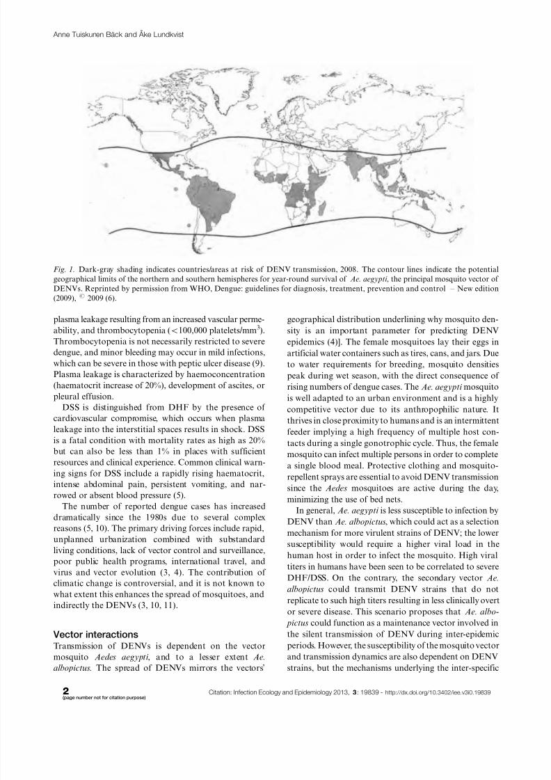
the tropical and subtropical regions (Fig. 1) as well as 120
million travelers to these regions every year (3). The World
Health Organization (WHO) estimates an annual inci-dence of approximately 100 million infections, with ap-
proximately 500,000 people with dengue hemorrhagic
fever (DHF) requiring hospitalization, a large proportion
being children. DHF may develop into dengue shock
syndrome (DSS) whereof the mortality rate is approxi-
mately 1 2.5%. Successful treatment of patientswith DHF
and DSS is labor intensive and expensive, but without
proper treatment, fatality rates may exceed 20% (4).
The four DENV serotypes can cause a wide range of
diseases in humans even though DENV infections may
also be asymptomatic. The diseases range in severity from
undifferentiated acute febrile illness, classical denguefever (DF), to the life-threatening conditions DHF/DSS
(5). Dengue illness was previously categorized on a I IV
grade scale, but a simplified categorization for dengue
case classification has been proposed by WHO’s Special
Program for Research and Training in Tropical Diseases
(TDR) in 2009 where DHF and DSS cases are grouped
together as ‘severe dengue’ (group C) to avoid false-
negative DHF/DSS diagnosis (6).
After an incubation period of 3 15 days (usually 5 to
8), classical DF begins with an abrupt onset of high
fever. During the febrile phase, dehydration may cause
neurological disturbances and febrile seizures in youngchildren (6). The condition is self-limiting through debili-
tating illness with headache, retro-orbital pain, myalgia,
arthralgia, petechiae rash, and leucopenia. A macular-
papular recovery rash appears 3 5 days after the onset of
fever, and it usually starts on the trunk before spreading
peripherally (7). DF is sometimes referred to as ‘break
bone fever’ due to its incapacitating symptoms with
severe muscle and joint pain (8); or ‘seven-day fever’
since the symptoms usually persist for 7 days.
Early symptoms of DF and DHF are indistinguishable,
but DHF is associated with hemorrhagic manifestations,
(page number not for citation purpose)
REVIEW ARTICLE
InfectionEcologyand Epidemiology 2013.# 2013Anne TuiskunenBa ¨ ckand A ˚ ke Lundk vist.This is an OpenAccessarticle distributed under theterms of theCreative Commons
Attribution-Noncommercial 3.0 Unported License (http://creativecommons.org/licenses/by-nc/3.0/), permitting all non-commercial use, distribution, and reproduction in any
medium, provided the original work is properly cited.
1
Citation: Infection Ecology and Epidemiology 2013, 3: 19839 - http://dx.doi.org/10.3402/iee.v3i0.19839
8/12/2019 Revision del Dengue
http://slidepdf.com/reader/full/revision-del-dengue 2/21
plasma leakage resulting from an increased vascular perme-
ability, and thrombocytopenia (B100,000 platelets/mm3).
Thrombocytopenia is not necessarily restricted to severe
dengue, and minor bleeding may occur in mild infections,
which can be severe in those with peptic ulcer disease (9).
Plasma leakage is characterized by haemoconcentration
(haematocrit increase of 20%), development of ascites, or
pleural effusion.
DSS is distinguished from DHF by the presence of cardiovascular compromise, which occurs when plasma
leakage into the interstitial spaces results in shock. DSS
is a fatal condition with mortality rates as high as 20%
but can also be less than 1% in places with sufficient
resources and clinical experience. Common clinical warn-
ing signs for DSS include a rapidly rising haematocrit,
intense abdominal pain, persistent vomiting, and nar-
rowed or absent blood pressure (5).
The number of reported dengue cases has increased
dramatically since the 1980s due to several complex
reasons (5, 10). The primary driving forces include rapid,
unplanned urbanization combined with substandard
living conditions, lack of vector control and surveillance,
poor public health programs, international travel, and
virus and vector evolution (3, 4). The contribution of
climatic change is controversial, and it is not known to
what extent this enhances the spread of mosquitoes, and
indirectly the DENVs (3, 10, 11).
Vector interactionsTransmission of DENVs is dependent on the vector
mosquito Aedes aegypti , and to a lesser extent Ae.
albopictus. The spread of DENVs mirrors the vectors’
geographical distribution underlining why mosquito den-
sity is an important parameter for predicting DENV
epidemics (4)]. The female mosquitoes lay their eggs in
artificial water containers such as tires, cans, and jars. Due
to water requirements for breeding, mosquito densities
peak during wet season, with the direct consequence of
rising numbers of dengue cases. The Ae. aegypti mosquito
is well adapted to an urban environment and is a highly
competitive vector due to its anthropophilic nature. Itthrives in close proximity to humans and is an intermittent
feeder implying a high frequency of multiple host con-
tacts during a single gonotrophic cycle. Thus, the female
mosquito can infect multiple persons in order to complete
a single blood meal. Protective clothing and mosquito-
repellent sprays are essential to avoid DENV transmission
since the Aedes mosquitoes are active during the day,
minimizing the use of bed nets.
In general, Ae. aegypti is less susceptible to infection by
DENV than Ae. albopictus, which could act as a selection
mechanism for more virulent strains of DENV; the lower
susceptibility would require a higher viral load in thehuman host in order to infect the mosquito. High viral
titers in humans have been seen to be correlated to severe
DHF/DSS. On the contrary, the secondary vector Ae.
albopictus could transmit DENV strains that do not
replicate to such high titers resulting in less clinically overt
or severe disease. This scenario proposes that Ae. albo-
pictus could function as a maintenance vector involved in
the silent transmission of DENV during inter-epidemic
periods. However, the susceptibility of the mosquito vector
and transmission dynamics are also dependent on DENV
strains, but the mechanisms underlying the inter-specific
Fig. 1. Dark-gray shading indicates countries/areas at risk of DENV transmission, 2008. The contour lines indicate the potentialgeographical limits of the northern and southern hemispheres for year-round survival of Ae. aegypti , the principal mosquito vector of
DENVs. Reprinted by permission from WHO, Dengue: guidelines for diagnosis, treatment, prevention and control New edition
(2009), # 2009 (6).
Anne Tuiskunen Ba ¨ ck and A ˚ ke Lundkvist
2(page number not for citation purpose)
Citation: Infection Ecology and Epidemiology 2013, 3: 19839 - http://dx.doi.org/10.3402/iee.v3i0.19839

8/12/2019 Revision del Dengue
http://slidepdf.com/reader/full/revision-del-dengue 3/21
and inter-strain differences in vector susceptibility to
DENV infection remains to be determined.
Once ingested by the mosquito, the DENV establishes
a productive infection in the mosquito midgut, where-
from the virus disseminates and replicates in other tissues.
In order to be transmitted to a human (or non-human
primate [NHP]) host during the next blood meal, theDENV must ultimately infect the salivary glands and
be shed in the saliva. Vector competence is genetically
determined, and genetic traits influencing both midgut
infection and escape barriers have been mapped to
various loci on the Ae. aegypti chromosomes (12).
Mosquito cell lines have been generated for studies of
mosquito-borne viruses and can be used for field isola-
tion of such viruses. Ae. albopictus cells have been used
for isolation and identification of DENVs from a
patient’s blood (13, 14). Arboviral infection of mosquito
cell culture yields high concentrations of virus and is
characterized by a persistent infection as most mosquito
cells are not killed by the infection (15).
In the continuing search for an effective vaccine
and anti-dengue drugs; two measures to prevent DENV
transmission are to reduce the vector population and to
educate people in affected areas about basic protection
measures. Anti-vector control programs include rigorous
surveillance, spraying pesticides, genetically modified
mosquitoes, minimizing potential breeding sites, and pro-
moting decent housing and infrastructure. Personal pro-
tection includes protective clothing and anti-insecticide
sprays.
Rapid, unplanned growth of urban centers in South-
East Asian and South American countries combinedwith inadequate water supply and sewerage systems have
dramatic consequences on the transmission of DENV
(16, 17).
The DENVs
Replication cycle
DENV is an enveloped, single-stranded positive-sense
RNA virus. The RNA genome consists of approximately
10,700 nucleotides and encodes a 3,411 amino acids
long precursor polyprotein containing three structural
proteins (capsid [C], precursor membrane [prM], andenvelope [E]) and seven non-structural (NS) proteins
(NS1, NS2A, NS2B, NS3, NS4A, NS4B, and NS5). The
structural proteins are components of the mature virus
particle whereas the NS proteins are expressed only in the
infected cell and are not packaged to detectable levels
into mature particles. The structural proteins are not
involved in replication of the viral genome (18 20).
The open reading frame is flanked by two untranslated
regions (5’ and 3’ UTR) of approximately 95 135 and
114 650 nucleotides, respectively. The 5’-end contains
a type I cap, similar to cellular mRNA, and the viral
RNA (vRNA) is translated by a cap-dependent initiation
scanning the 5’-UTR. The 3’-end lacks a poly(A) tail but
ends in a conserved stem-loop (SL) structure. Both the
5’- and 3’-UTRs are required for efficient translation
and replication (21, 22). The UTRs have characteristic
secondary structures that confer distinct functions and
show high sequence conservation among different DENVserotypes. The 5’-UTR contains a large stem-loop (SLA)
that is proposed to act as the promoter for the viral
RNA-dependent RNA polymerase (RdRp) NS5 (23).
Both the 5’- and the 3’-UTRs contain complementary
Upstream AUG Regions (UAR) and cyclization se-
quences (CS) that hybridize in order to mediate genome
cyclization and RNA synthesis (21).
The various steps in the flavivirus life cycle include
1) virions binding to cell-surface attachment molecules
and receptors, and are internalized through endocytosis.
2) Due to the low pH of the endosome, viral glycopro-
teins mediate fusion of viral and cellular membranes,
allowing disassembly of the virion and release of vRNA
into the cytoplasm. 3) vRNA is translated into a poly-
protein that is processed by viral and cellular proteases,
and 4) the viral NS proteins replicate the genome RNA.
5) Virus assembly occurs at the endoplasmic reticulum
(ER) membrane, where C protein and vRNA are en-
veloped by the ER membrane and glycoproteins to form
immature virus particles. 6) Immature virus particles are
transported through the secretory pathway, and in the
acidic environment of the trans-Golgi network (TGN),
furin-mediated cleavage of prM drives maturation of the
virus. 7) Mature virus is released from the cell.
Virus entry
Viral entry into the host cell is mediated by receptor-
mediated endocytosis through an as yet unidentified cell-
surface receptor. Candidate cellular receptors required
for viral entry are various glycoproteins (i.e. heparin
sulfates), dendritic cell-specific intercellular adhesion
molecule-3-grabbing non-integrin (DC-SIGN), or a man-
nose receptor (24 26). The human C-type lectin-like mole-
cule CLEC5A has been suggested to act as a critical
macrophage receptor for DENV and has been described
as a proinflammatory receptor for DENV that contri-
butes to lethal disease in mice (27, 28). There is a generalconsensus that the viral E glycoprotein affects host cell
receptor binding, viral entry, and is a major target for
humoral immunity. The E protein is composed of three
domains: domain I, domain II harboring the fusion
peptide at its distal tip, and domain III responsible for
receptor-binding activity. In the mature state, E exists
as a homodimer with the fusion peptide inaccessible.
Low-pH induced trimerization exposes the hydrophobic
fusion peptide in a manner consistent with membrane
fusion mediated by class II fusion protein (29). Mutations
of residues constituting the ligand pocket at the interface
Dengue viruses an overview
Citation: Infection Ecology and Epidemiology 2013, 3: 19839 - http://dx.doi.org/10.3402/iee.v3i0.19839 3(page number not for citation purpose)
8/12/2019 Revision del Dengue
http://slidepdf.com/reader/full/revision-del-dengue 4/21
of domain I and II alter the required pH threshold and
affect virulence (30). There are two potential asparagine
(N)-linked glycosylation sites at positions Asn-67 and
Asn-153, whereof the former is unique for DENVs and
the latter is conserved in most flaviviruses (31). The
glycosylation pattern differs according to DENV sero-
type and even among different strains, as well as the cellsin which the virus is propagated. The degree and position
of N-linked glycans affect the antigenic properties of
DENV (32 34).
Upon internalization, the acidic pH in the endosome
triggers a conformational change in the E protein me-
diating membrane fusion. The viral nucleocapsid is
released into the cytoplasm whereupon the virus uncoats
and releases the genome (31). The input positive-strand
vRNA is translated into a single polyprotein, which is
cleaved into the individual structural and NS proteins.
The input strand translation is followed by a switch from
translation to synthesis of a negative-strand intermediate,which serves as a template for new positive-strand vRNA.
Multiple rounds of translation produce high levels of
viral proteins that together with vRNA are assembled
into progeny virions (35).
DENV infection induces intracellular membrane
alterations in the cytosol forming vesicle packets (VPs)
or smooth membrane structures (SMS) where the viral
replication complex (RC) accumulates (36, 37). The
induction of membrane structures may serve as a scaffold
for anchoring the viral RC. The C-terminal regions of C,
prM, and E contain hydrophobic amino acids that serve
as signal sequences for insertion of the remaining protein
into the ER membrane (38). An ER signal peptidase
together with the viral NS2B-NS3 protease cleaves the
structural proteins and NS1 protein into individual
membrane-bound proteins (39 41). The NS3 protein
acts, together with its cofactor NS2B, as the viral serine
protease needed for polyprotein-processing through its
N-terminal end (42, 43). This heterodimer protein com-
plex cleaves on the cytoplasmic side of the ER membrane
at the junctions between NS2A-NS2B, NS2B-NS3, NS3-
NS4A, and NS4B-NS5, as well as on the internal sites
within C, NS2A, NS3, and NS4A.
The C-terminal end of the NS3 protein has three
enzymatic properties: a 5’ RNA-triphosphatase (RTP), anucleoside triphosphatase (NTPase), and a helicase. NS3
forms a complex with NS5 and assists in viral replication
through unwinding of RNA and dephosphorylation prior
to 5’-end capping. The remaining NS proteins are cleaved
by the viral serine protease NS3 that requires NS2B as a
cofactor for catalytic activity (43). However, a host cell
signal peptidase mediates post-translational modifica-
tions on the NS4A-4B proteins (44).
The NS1 is a glycoprotein with two glycosylation sites
that are conserved among flaviviruses. It is synthesized in
the ER as a hydrophilic monomer but exists as a more
hydrophobic homodimer. The NS1 dimer is transported
to the Golgi apparatus where it undergoes carbohydrate
trimming (45). The role of NS1 in virus replication is
unknown but is believed to facilitate viral infection and
DENV pathogenesis (46). NS1 is in addition secreted
from infected cells (sNS1) and has been shown to be
immunologically important (47, 48). Antibodies raisedagainst sNS1 proteins have been proposed to cause
endothelial dysfunction due to cross-reactivity to host
proteins and endothelial cells (49). Data indicate that
sNS1 could be an important modulator of the comple-
ment pathway and is proposed to protect DENV from
complement-dependent neutralization in solution (47).
The small hydrophobic proteins NS2A, NS4A, and
NS4B are less well characterized. Recent findings propose
an inhibitory role in interferon (IFN)-mediated signal
transduction. Their hydrophobic nature potentially im-
plicates them in proper localization of viral proteins and
vRNA during replication and virion assembly. Formation
of DENV-induced cytoplasmic membrane structures are
believed to be an arrangement of the NS4A protein.
The largest NS protein encoded in the DENV genome
is the NS5 protein, approximately 103 kDa big. The
NS5 protein has three major functional domains: the
N-terminal S-adenosyl methionine methyltransferase
(MTase), the nuclear localization sequences (NLS), and
the RdRp activity in its C-terminal domain. The MTase
spans amino acid residues 1 to 239 and is responsible for
guanine N-7 and ribose 2’-O-methylations required for
the capping of the DENV genome. The cap structure is
recognized by the host cell translational machinery. The
NLS (residues 320 405) interacts with the NS3 viralhelicase and is recognized by cellular factors, allowing
protein transport to the nucleus. The NS5 polymerase
domain RdRp (residue 273 900) is responsible for
synthesizing new vRNA genomes (43).
Prior to secretion of new viral particles, the third
structural protein (pr)M is processed into the mature
M protein in the TGN by furin host protease (50). It is
believed that prM protects the E proteins from pH-
induced reorganization and premature fusion during
secretion; hence, the maturation event is necessary for
infectivity (51 53).
Laboratory diagnosis
Since laboratory-based dengue diagnosis is often unavail-
able at the time of care, the preliminary diagnosis relies
on a combination of travel history and clinical symptoms.
Travel history provides key information that can rule
out other potentially life-threatening diseases since the
incubation period of DENV is less than 2 weeks (54).
A confirmed diagnosis for a DENV infection is estab-
lished by culture of the virus, polymerase-chain reaction
(PCR), or serologic assays. There are, however, limita-
tions with each test, and detection is based on different
Anne Tuiskunen Ba ¨ ck and A ˚ ke Lundkvist
4(page number not for citation purpose)
Citation: Infection Ecology and Epidemiology 2013, 3: 19839 - http://dx.doi.org/10.3402/iee.v3i0.19839

8/12/2019 Revision del Dengue
http://slidepdf.com/reader/full/revision-del-dengue 5/21
virological markers, namely infectious virus, vRNA, and
DENV-specific antibodies, respectively.
Culturing the virus requires an acute patient serum
with sufficient levels of virus, and the period when DENV
can be successfully isolated in patient serum is short.
Viremia peaks before the onset of symptoms, hence
virus levels might drop significantly once the patientseeks medical care. Furthermore, rising levels of antibody
interfere with virus culture already within a day or two
after the subsidence of fever. Apart from sample collec-
tion limitations, practical considerations limit the use of
this method. Culture of the virus is both time- and labor
intensive; infectious patient material must be kept cold,
and a bio-safety level 3 laboratory is required, necessitat-
ing professional training of the personnel. These require-
ments limit the use of this diagnostic tool, especially in
rural areas (4).
The Ae. albopictus cell line C6/36 (CRL 1660, ATCC)
is commonly used to isolate DENVs from patient mate-rial. Specimens that may be suitable for virus isolation
include acute phase serum; plasma or washed buffy
coat from the patient; autopsy tissues from fatal cases,
especially liver, spleen, lymph nodes, and thymus; and
mosquitoes collected in nature (55). Detection of vRNA
from serum, plasma, or cells with PCR is based on
DENV-specific oligonucleotide primers, and is fast and
robust, although sensitive only in the very early stages of
disease (54). PCR is particularly useful in situations when
virus culture has not been successful but nevertheless
depends on sample collection during the symptomatic
phase.
The third laboratory diagnostic option is not based
on direct detection but on the presence of anti-DENV
antibodies. Thus, it is not hindered by the limitations
of virus culture and PCR, and the timing of sample
collection can be more flexible. The acute anti-DENV
IgM antibody response lasts for a couple of weeks after
infection and the IgG antibodies for several years. The
immunoglobulins (Ig) are not easily inactivated and do
not have the same strict requirements for low temperature
as infectious virus specimen. The assay techniques are
relatively simple and there are commercial diagnostic kits
available, whereof the assays based on IgM detection are
the most commonly used in routine diagnostics (4). Themajor drawback with serological tests is the considerable
risk for false-positive results due to potential cross-
reactivity with other flaviviruses, for example, vaccination
against Yellow fever virus (YFV) (56).
Due to the drawbacks of serological methods to
reliably diagnose acute infections, alternative methods
based on the detection of the viral NS1 protein have been
developed. NS1 can be found both membrane-associated
inside the host cell and in a soluble, secreted form. The
amount of secreted NS1 in patient serum correlates with
viremia and DENV pathogenesis (46, 57 60), and the
NS1 protein is detectable in serum by enzyme-linked
immunosorbent assay (ELISA) from the first day of
fever up to 9 days post-infection (46, 61 63). NS1-based
ELISAs have become an important diagnostic tool for
acute samples in which IgM is not detectable and where
PCR is not available. Several commercial NS1 antigen
kits are available and are widely used in endemic as wellas non-endemic countries. The sensitivity varies from
63% to 94% (58, 62, 64, 65), and it depends on sample
time-point, DENV serotype, and if it is a primary or
secondary DENV infection (66).
Vaccines
Unlike flaviviruses such as YFV, Japanese encephalitis
virus (JEV), and Tick-borne encephalitis virus (TBEV),
no licensed vaccine exists for dengue. Vaccination must
protect against all four serotypes without predisposing
for antibody-dependent enhancement (ADE) and has
proven difficult to design. Nearly 80 years of vaccine-related research and development have passed, and over
25 unique DENV vaccine candidates have been tested in
clinical trials during the past decade.
To be safe, a dengue vaccine must be functionally
tetravalent, eliciting simultaneous protection against all
four DENV serotypes. Hence, vaccination cannot pro-
ceed in an analogous sequential manner, and herein lies
the greatest obstacle (67 69). Live attenuated vaccines
can induce durable humoral and cellular immune re-
sponses that mimic natural infection (70). However,
the viral replication must be discrete to preclude the
development of significant illness. A reasonable range of
viremia for a live attenuated vaccine is believed to be
approximately 101-102 infectious units/mL (71) compared
to high levels of viremia upon natural infection that can
be 105-107 infectious units/mL (59).
It is expected that a live attenuated vaccine would
be successful and require only a single dose since the
vaccine against YFV is based on a live attenuated virus.
However, it is more likely that booster immunizations will
be required based on results from clinical trials using
tetravalent formulations of live vaccine candidates aimed
at eliciting neutralizing antibodies (72 75) The obvious
challenge is when and how to boost; infectivity and
immunogenicity in NHP models have not always clearlypredicted the outcome of human trials (76, 77). Vaccina-
tion compliance may also be lower with a multi-dose
vaccination strategy, especially in regions where resources
are scarce, and at the same time where the need for a
vaccine often is the most acute.
Currently, there are several dengue vaccine candidates
at different stages of preclinical or clinical development.
The most advanced clinical development stage is a
candidate developed by Sanofi Pasteur (CYD-TDV),
which is under evaluation in phase II and phase III
clinical studies. Phase III efficacy studies of CYD-TDV
Dengue viruses an overview
Citation: Infection Ecology and Epidemiology 2013, 3: 19839 - http://dx.doi.org/10.3402/iee.v3i0.19839 5(page number not for citation purpose)
8/12/2019 Revision del Dengue
http://slidepdf.com/reader/full/revision-del-dengue 6/21
are currently underway in 31,000 children and adoles-
cents in 10 countries in Asia and Latin America. These
multi-center studies in a variety of epidemiological settings
will be important to obtain data regarding efficacy and
safety, and will shed further light on the relationship
between vaccine-induced immune responses and protec-
tion against clinical dengue disease (73).There are in addition other live-attenuated, subunit,
and DNA vaccine candidates at earlier stages of clinical
development. Other technological approaches include
viral-vectored and virus-like particle vaccines, which
currently are being tested in preclinical studies. It is
hoped that clinical trials evaluating novel recombinant
subunit proteins, DNA, and vectored vaccines would be
initiated in the coming years. These approaches could be
part of a prime-boost strategy, or stand-alone (78). The
use of different types of vaccines depends on the purpose
of vaccination and target group reflecting the disease
setting. In endemic areas, there is an urgent need for
routine immunization against dengue for infants and
young children aged 1 3 years. A dengue vaccine would
be coordinated with current childhood immunization
schedules. Due to the socioeconomic status of many
endemic countries, this type of vaccine ought to be
inexpensive. In contrast, a protective vaccine for interna-
tional travel, seasonal work personnel, and military staff
that visit or work in DENV endemic areas are more
tolerant to increased cost. Vaccination in this case will
need to be rapid.
Hence, the different requirements for a dengue vaccine
vary according to target group and their specific needs
(life-long immunity or temporal protection), and efficientantiviral drugs would be a useful complement for pro-
tection and/or treatment. In addition, antiviral drugs
would be more potent in an outbreak situation than a
vaccine when there is no time to complete a multi-dose
immunization schedule spanning 6 months or more.
Treatment and therapeutic approaches
Currently, vector control, regarded as both expensive
and ineffective, is the only method for disease prevention
(79, 80). In the absence of available vaccines and antiviral
drugs against DENV infection, specific treatment for
dengue patients consist primarily of supportive careincluding bed rest, antipyretics, and analgesics. Urgent
resuscitation with intravenous fluids to replace lost
intravascular volume in DSS patients is a prerequisite;
Ringer’s lactate has been shown to be effective in moder-
ately severe dengue, and starch or dextran have been
suggested for more severe cases (81). Aspirin and other
salicylates should be avoided due to plasma leakage (6).
The design of novel therapeutic approaches for dengue
disease has focused on the various stages of the viral
replication cycle. The conformational changes of the E
protein and its interaction with prM or M have been a
major interest. These transition states present opportu-
nities for antiviral targeting of the entry, assembly, or
maturation steps of the virus life cycle. Antiviral peptides
have been designed and tested for blocking of both
DENV and West Nile virus (WNV) entry with positive
results, indicating that antiviral peptides could be a
promising form of DENV therapy. Targeting of maturevirus entry into host cells is an extremely promising
candidate since delivery of target compounds into the
host cell during stages of fusion and maturation is
significantly more challenging.
Another approach to inhibit the structural changes of
the E-prM protein interactions has been to synthesize
peptides mimicking the pr peptide of the M protein,
thereby preventing membrane fusion and release of newly
synthesized virions. The viral protease is another inter-
esting target for antiviral discovery, since proteases are
common to most viruses and generally important for
efficient replication. Protease inhibitors for hepatitis Cvirus (HCV) may eventually be further developed to
inhibit the DENV protease NS2B-NS3.
Nucleoside analogues are usually prodrugs that need to
be converted to their antiviral nucleotide metabolite
forms. Ribavirin (1-b-d-ribofuranosyl-1H -1, 2, 4-triazole-
3-carboxamide) possesses broad spectrum antiviral activ-
ity and is used in combination with IFN to combat HCV
infection. Ribavirin depletes the nucleotide pool and
thereby indirectly affects capping and polymerase activ-
ities of both cellular and viral proteins. In addition,
ribavirin causes a more error-prone replication of several
viral genomes. Despite successful in vi vo results with
several RNA viruses, ribavirin has a cytostatic effect in
DENV-infected cells and has not been effective in animal
models.
Nucleic acid based therapies offer various alternatives.
RNA interference (RNAi) is thought to protect the host
from viral infections by degrading the extraneous genetic
material such as vRNA. It has been used in therapeutic
approaches for several infectious diseases, tumors, and
metabolic disorders. Small interfering RNA (siRNA)
treatment reduces viral load of WNV in mice, but there
are several obstacles yet to overcome; the RNA of
flaviviruses are resistant to RNAi since replication occurs
in reorganized ER membrane packets. In addition, HCVreplication was found to be stimulated by the RNAi
machinery.
Another nucleic acid based antiviral approach is
antisense DNA or RNA decoys, for example, phosphor-
odiamidate morpholino oligomers (PMOs). These com-
pounds act by forming a stable, sequence-specific duplex
with RNA, thereby blocking access to target RNA by
biomolecules required for replication. PMOs targeting
the translation initiation site of DENV RNA, the 3’
UTR, 5’SL, and 3’CS were effective in reducing the viral
load in various cell lines. These compounds meet most
Anne Tuiskunen Ba ¨ ck and A ˚ ke Lundkvist
6(page number not for citation purpose)
Citation: Infection Ecology and Epidemiology 2013, 3: 19839 - http://dx.doi.org/10.3402/iee.v3i0.19839
8/12/2019 Revision del Dengue
http://slidepdf.com/reader/full/revision-del-dengue 7/21
of the requirements for an anti-DENV therapeutic;
non-toxic, cheap, easy to administer, stable for months
at variable temperatures, but remain to be tested in
animal models.
Sulfated polysaccharides have been investigated for
anti-DENV activity, although inconsistency in the activ-
ity results indicates that they need to be further testedboth in vitro and in vi vo (24).
The processing of N -linked oligosaccharides in the
ER is important for viral glycoprotein maturation, and
inhibition of glucosidase-mediated trimming affects the
replication cycle of several enveloped viruses. DENV
production was inhibited in mouse neuronal cells by
two ER a-glucosidase inhibitors, castanospermine (CST)
and deoxynojirimycin. CST was effective against all four
serotypes in human hepatoma cells and prevented mor-
tality in DENV-2 infected mice. This effect was restricted
to DENV, not being observed against other flaviviruses
such as WNV and YFV. A third ER a-glucosidase in-
hibitor, N-nonyl-deoxynojirimycin, inhibits DENV-2 in-
fection in BHK-cells. These results with a-glucosidase
inhibitors are encouraging and should be investigated
further in vi vo.
Nitric oxide (NO) is generated by macrophages, mono-
cytes, dendritic cells (DCs), and neutrophils; the same
cells that are supposed to be the main sites of replica-
tion for DENV. In vitro assays have revealed that NO
specifically affects the viral RdRp activity, suggesting
possible viral targets of NO during DENV infection (82).
Hence, there are multiple options for designing novel
therapeutics for dengue disease. However, the main
concern with most therapeutic approaches is that theyare not validated for inhibitory effects on all four DENV
serotypes. In addition, several studies have not been
examined in an animal model, and several reported
antivirals have been tested at only one time point, pre-
or post-infection in tissue culture systems, and therefore
need to be subjected to more diverse regimes, and
different cell types.
Risk factors for severe dengueDENV pathogenesis remains a challenging jigsaw puzzle
with many pieces missing to understand the complex
interplay of viral and host factors. Despite intensiveresearch, it is not well understood. The severity of DENV
infection is modulated by multiple risk factors such as
age (83, 84), the genetic background of the host (85, 86),
viral serotype (83, 87) and genotype (88, 89), and
secondary DENV infection by a heterologous serotype
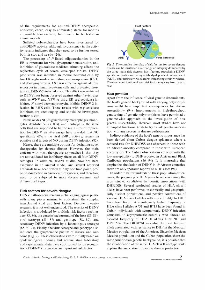
(85, 90 93). Finally, the virus serotype and genotype also
influence the symptomatic picture of disease and out-
come (Fig. 2). These observations were initially based on
epidemiological findings, but accumulating laboratory
and experimental data have contributed to the recogni-
tion of DENV virulence as an important risk factor.
Host genetics
Apart from the influence of viral genetic determinants,
the host’s genetic background with varying polymorph-isms might have important consequences for disease
susceptibility (94). Improvements in high-throughput
genotyping of genetic polymorphisms have permitted a
genome-wide approach to the investigation of host
genetic susceptibility. However, most studies have not
attempted functional trials to try to link genetic associa-
tion with any process in disease pathogenesis.
Indirect evidence of the host’s genetic importance has
been derived from Cuban dengue epidemics where a
reduced risk for DHF/DSS was observed in those with
an African ancestry compared to those with European
ancestry (3). The Cuban observations coincide with thelow susceptibility to DHF reported in African and Black
Caribbean populations (86, 94). It is interesting that
despite the circulation of DENV in 19 African countries,
there are only sporadic reports of DHF cases (94).
In order to better understand these population differ-
ences, the polymorphic HLA genes have been among the
most studied candidates for genetic associations with
DHF/DSS. Several serological studies of HLA class I
alleles have been performed in ethnically and geographi-
cally distinct populations, and positive correlations of
various HLA class I alleles with susceptibility to DHF
have been found. A significantly higher frequency of HLA class I alleles A*31 and B*15 have been found in
Cuban individuals with symptomatic DENV infection
compared to asymptomatic controls, who showed an
elevated frequency of HLA II alleles DRB1*07 and
DRB1*04. The DRB1*04 was also the most frequent
allele associated with resistance to DHF in the Mexican
Mestizo populations of the Americas. Since the Mexican
Mestizo population and the Cuban population share the
same Amerindian genetic background, it is possible that
the identification of the same HLA class II allotype could
explain the association to dengue disease protection.
Fig. 2. The complex interplay of risk factors for severe dengue
disease can be illustrated as a triangular interplay dominated by
the three main risk factors: host factors, preexisting DENV-
specific antibodies mediating antibody-dependent enhancement
(ADE), and intrinsic virus features influencing strain virulence.
The exact contribution of each risk factor may vary from case to
case.
Dengue viruses an overview
Citation: Infection Ecology and Epidemiology 2013, 3: 19839 - http://dx.doi.org/10.3402/iee.v3i0.19839 7(page number not for citation purpose)
8/12/2019 Revision del Dengue
http://slidepdf.com/reader/full/revision-del-dengue 8/21
A case-control study in ethnic Thai cases also reported
the association of HLA class I alleles (A2, A*0207, B46,
B51) with different clinical outcomes. A similar study
in a Vietnamese population confirmed the association
with polymorphism of the HLA class I loci and DHF
suslceptibility. The same study also found that poly-
morphisms in the HLA-DRB1 allele are not associatedwith DHF susceptibility, highlighting the findings in the
Amerindian populations.
The number of studies on polymorphisms within genes
other than the HLA loci remains low. Variants of the
vitamin D receptor and the FcgRIIA gene are associated
with resistance to severe dengue. In addition, an allelic
variant of the DC-SIGN1 coding gene CD209 is believed
to protect against DHF (94).
Host health and age
An increased association between severe dengue and
bronchial asthma, diabetes mellitus, peptic ulcers, andsickle cell anaemia has been observed (94, 95). However,
the impact of dengue on chronic diseases and other
pathogens needs to be further investigated.
Primary infections are supposed to cause mild disease
in children, compared to secondary infections that tend
to lead to severe dengue. In South-East Asia, DHF/DSS
is predominantly an illness in children. The greater
relative prevalence of DSS in children relative to adults
is believed to be due to the intrinsically more permeable
vascular endothelium in children (94). There is no clear
consensus; studies conducted in South American coun-
tries have reported similar (84) as well as contradictory
results indicating that adults are the most affected (94).
Autoimmune responses in dengue virus infection
Anti-DENV antibodies can cross-react to host proteins
and endothelial cells, and this could enhance the en-
dothelial dysfunction observed in DHF/DSS. Antibodies
against the viral surface E protein cross-react with
plasminogen and have been associated with bleeding in
acute DENV infection, and anti-DENV NS1 antibodies
cross-react with host proteins and endothelial cells (49,
94). In addition, immune activation markers (e.g. IL-6,
IL-8, TNFa, IFNg, and complement components 3A and
5A) together with altered platelet, DC, monocyte, and T-cell functions suggest that immune responses to various
DENV components could contribute to autoimmune
processes resulting in DHF/DSS (94).
Antibody-dependent enhancement
A secondary infection by a heterologous DENV serotype
is an important risk factor for developing DHF/DSS.
The explanation lies within the cross-reactive antibodies
raised after a primary DENV infection (90, 92, 94).
Serotype-specific antibodies confer life-long immunity
to the homologous serotype, whereas cross-protection
against heterologous serotypes last for 3 4 months.
Beyond this time period of cross-protection, the preexist-
ing antibodies of sub-neutralizing concentration will
instead cross-react with the heterologous virus facilitating
viral infection of FcgR-bearing cells. This phenomenon
is known as ADE (94). The limited cross-protection
between the four DENV serotypes has allowed them tocoexist in the same or overlapping geographical areas.
Thus, their antigenic uniqueness has implied an evolu-
tionary advantage (96).
Low-affinity/Low-affinity, sub-neutralizing antibodies
and DENV form virus-antibody immune complexes that
bind to Fcg-receptors on monocytes. The net result will
be a larger number of infected cells compared to the
primary infection when there were no cross-reactive
antibodies present, or compared to earlier after the
primary infection when antibody levels are high enough
to achieve neutralization of the heterologous virus.
Hence, the viral biomass will be larger during a secondaryDENV infection compared to during a primary DENV
infection.
In vitro studies indicate that non-neutralizing antibo-
dies against the viral prM protein can potentially mediate
ADE. These anti-prM antibodies are in addition non-
neutralizing even at high concentrations (97, 98). The
proposed hypothesis for prM-mediated ADE is based on
the fact that the viral prM protein needs to be cleaved
to render the virus infectious. Hence, immature virus
particles that would otherwise be non- or less-infectious
are rendered infectious in combination with anti-prM
antibodies that mediate ADE to infect new host cells (97).
The time interval between heterotypic DENV infec-
tions is another parameter influencing the magnitude of
ADE; a longer interval between heterologous DENV
infections causes higher DHF/DSS ratios (85). The dif-
ferences in DENV genotype could influence the patho-
genic consequences, but a contributing risk factor is the
progressive loss of heterotypic neutralizing antibodies
(99). The time effect of DENV-specific antibodies can be
seen in DENV-immune mothers and their infants. Before
the age of 3 4 months, the maternally derived DENV-
specific antibodies confer protection against a DENV
infection. However, primary infections in infants aged
between 4 and 12 months of age run a higher risk of developing severe dengue due to maternally derived non-
neutralizing antibodies. The risk of severe dengue de-
creases after the age of 1 year as the concentration of
cross-reactive antibodies declines (94, 100, 101).
A higher viral burden elicits a greater host inflamma-
tory response and increased plasma levels of proinflam-
matory cytokines. Secondary DENV infections and
severe disease in DHF/DSS patients have elevated serum
levels of IL-2, IL-6, IL-8, IL-10, IL-13, IL-18, IFNg,
TNFa, and MCP-1 (102 108). Thus, an increased in-
fected cell mass would stimulate T-cell and cytokine
Anne Tuiskunen Ba ¨ ck and A ˚ ke Lundkvist
8(page number not for citation purpose)
Citation: Infection Ecology and Epidemiology 2013, 3: 19839 - http://dx.doi.org/10.3402/iee.v3i0.19839
8/12/2019 Revision del Dengue
http://slidepdf.com/reader/full/revision-del-dengue 9/21
responses that are proportional to the antigenic stimulus.
This hypothesis is consistent with the observations that a
high initial viremia or high NS1 concentrations in blood
during secondary infections are associated with DHF/
DSS (46, 59, 109, 110).
Accumulating evidence questions whether ADE of
infection alone is sufficient to explain DHF/DSS (59).Severe dengue with plasma leakage can occur in primary
infection without ADE. In addition, by the time plasma
leakage occurs, viral titers are several logs below peak
levels, and there are patients with high viral titers that
do not develop plasma leakage (59, 109, 111). Thus,
increased viremia alone is not the direct cause of plasma
leakage and other mechanisms are involved in the cytokine
storm. Furthermore, ADE is not a useful correlate of
disease risk (112, 113).
Dengue virus virulence
ADE has dominated as the explanatory model for severedengue disease in secondary infections. However, evi-
dence for ADE in humans is indirect and controversial
results against ADE exist (112, 114). Many parts of the
world have become hyperendemic, implying that all four
serotypes of DENV co-circulate in the same country, with
the consequence that secondary infections are common
scenarios. Epidemiological data also indicate that not all
secondary infections cause DHF/DSS, and that there are
even cases of tertiary and quaternary DENV infections
(115). Studies from Thailand report that 0.08% 0.8% of
dengue hospitalizations may be caused by tertiary and
quaternary DENV infections (67). In Cuba, 17.5% of the
total DHF dengue cases were caused by third or fourth
infections (116).
The four DENV serotypes 1 4 diverge at 30% across
the polyprotein (117), but each serotype also consists
of phylogenetically distinct ‘subtypes’ or ‘genotypes’,
which have different geographical distributions (94).
The hypothesis that some DENV genotypes have greater
virulence and epidemic potential than others was intro-
duced during the 1970s around the same time that the
ADE phenomenon was coined (118 120). However, in
contrast to the ADE hypothesis, experimental evidence
for increased virulence was for long absent and, therefore,
primarily based on epidemiological observations. Recentwork has shed light on this question and confirmed
what Rosen, et al. proposed almost four decades ago
(118, 119).
There have been specific geographic examples of the
appearance of DENV genotypes correlating to DHF/
DSS epidemics. The appearance of a South-East Asian
DENV-2 strain in the Americas in 1981 resulted in the
sudden emergence of DHF/DSS cases. It turned out that
DENV-2 could be subdivided into a variety of genotypes,
minimally Asian and American. The Asian genotype is
more virulent and more likely to result in DHF/DSS than
the American genotype even after a secondary infection
(121, 122). Viruses to the South-East Asian DENV-2
lineage replicate to higher titers in human DCs than
American genotype viruses. It was also seen that the
South-East Asian genotype infects and disseminates to
the head tissue of Ae. aegypti mosquitoes more rapidly
and in a greater proportion compared to the Americangenotype viruses (121).
The emergence of group B subtype III DENV-3 strain
in Sri Lanka in 1989 is another example of clade
replacement correlating with an increase in DHF/DSS
(88, 123, 124). As for the South-East Asian DENV-2
strain, the invasive DENV-3 strain replicated to higher
levels in mosquitoes and disseminated to the head tissue
more readily than the displaced, native DENV-3 strain
from Sri Lanka (125). Both traits likely enhanced the
capacity to spread and displace endemic strains.
Based on the examples given, one hypothetical me-
chanism for increased virulence suggests that highly
pathogenic DENV strains have been selected for en-
hanced ability to replicate in key human targets, such as
macrophages and DCs (94, 126). Thus, virulent DENVs
would produce more viruses per cell, resulting in higher
viremia and inflammatory response, than with a low
pathogenic strain (94, 127).
The second hypothesis for increased virulence proposes
that strains associated with severe DHF/DSS better
escape neutralization by the presence of serotype cross-
reactive antibodies in the semi-immune host compared to
strains associated with DF (128). Enhancement of virus
replication following heterologous infection may favor
coexistence of multiple serotypes. If such enhancementalso results in increased transmission, DENVs from
different serotypes would benefit from prior and con-
current circulation of several serotypes in the same
location (96).
It is still not known if the tendency of certain geno-
types to cause severe disease results from greater intrinsic
virulence, or if greater virulence is a result of enhanced
infectivity in the presence of heterologous antibodies, or a
combination of the two. Determining whether DENVs
differ in virulence, as well as identifying the genetic basis
of such differences, is of fundamental importance.
Immune responses and dengue viruspathogenesisDENV infection is a systemic and dynamic disease with
a wide clinical spectrum. Gross pathological findings
in cases of DHF or DSS include hemorrhages in the
skin, subcutaneous tissues, gastrointestinal tract, and
heart (129). Hemorrhage, dilatation and congestion of
vessels, and edema of arterial walls are commonly found,
and hemorrhagic manifestations in other organs com-
bined with fluid accumulations in body cavities may be
substantial (130, 131).
Dengue viruses an overview
Citation: Infection Ecology and Epidemiology 2013, 3: 19839 - http://dx.doi.org/10.3402/iee.v3i0.19839 9(page number not for citation purpose)

8/12/2019 Revision del Dengue
http://slidepdf.com/reader/full/revision-del-dengue 10/21
However, the underlying mechanisms of vascular
leakage and hemorrhage are not well characterized.
Elevated plasma levels of pro-inflammatory and vasoactive
cytokines before and at the time of plasma leakage in
patients with DHF suggest that excessive cytokine pro-
duction (a ‘cytokine storm’) induce vascular permeability.
Available data propose that the outcome of a DENVinfection depends on a balance between favorable and
unfavorable immune responses; the former providing
control of viral replication, whereas the latter enhancing
inflammatory and vascular permeability. The lack of
reliable immunological markers for either protective or
pathological immune responses to DENV and the lack of
a suitable animal model for dengue disease hamper the
understanding of dengue pathogenesis. Insights into the
immune response against DENV infection rely primarily
on clinical and epidemiological studies.
Tropism Identification of the primary target cells of DENV
replication has proven to be extremely difficult. Existing
data are based on virus detection by immunohistochemical
(IHC) analysis with antibodies against viral structural
proteins, or by in situ hybridization to the positive-strand
vRNA. However, it is difficult to prove direct infection of
specific target cells by these methods as a positive signal
could be due to virus endocytosed or phagocytosed by
uninfected cells. Detection of negative-strand vRNA and/
or DENV NS proteins would provide much stronger
evidence of active DENV replication.
After inoculation by an infected mosquito, the initial
round of viral replication is believed to occur in the sub-
dermal Langerhans DCs (132 135). These infected cells
become activated and migrate to draining lymph nodes
(136). The activated DCs elicit a robust IFNa/b and
TNFa response together with a strong pro-inflammatory
response to limit contiguous spread (133). Viral replica-
tion continues in still undefined cells in the lymph node.
There is a general consensus that candidate cell types
belong to the macrophage-monocyte lineage. Autopsies
and human biopsies confirm that cells from the mono-
nuclear phagocyte linage probably are the primary targets
of DENV infection following initial dissemination from
the local skin site. Infiltrating mononuclear cells in af-fected tissues have been shown to contain DENV antigen
(137, 138), and DENVs can occasionally be isolated from
peripheral blood leukocyte fractions (139). Similar ob-
servations have been made in rhesus macaques where
DENV was recovered from leukocyte-rich tissues such as
regional lymph nodes, systemic lymphatic tissues, and
disseminated skin sites.
Infection is amplified within the lymph nodes and
viremia can be detected when the infectious virus enters
the circulation via the efferent lymphatic system and
thoracic duct. Circulating monocytes in the blood are
believed to be infected due to the viremia facilitating
spread to secondary visceral organs where macrophages
within the spleen, liver, and bone marrow are infected
(140 145).
There has been limited and inconsistent dissemination
to solid organs (146); DENV antigen has been detected in
lymphocytes (140, 147), hepatocytes (147 150), endothe-lium (140, 147, 151, 152), and cerebral neurons and
astrocytes (147, 152). There are in addition other studies
with contradicting results where the same tissues have
been examined without any detected DENV antigen (140,
150, 151).
A further controversy surrounds the role of endothelial
cells as the target for DENV infection. Severe dengue
disease is characterized by systemic endothelial dysfunc-
tion accompanied by vascular leakage, even though de-
structive vascular lesions are generally absent in fatal
cases (83). Primary human endothelial cells and human
endothelial cell lines are permissive for DENV infection
(47, 153), but endothelial infection, however, does not
seem to be required for severe pathologic changes in
individual tissues (154). Their contribution in vi vo
remains to be established.
The presence of DENV antigens in various organs and
cell types suggest that the host receptor(s) is broadly
distributed. Host receptors for DENV are believed to
include mannose binding protein, heparan sulfate, chon-
droitin sulfate, and DC-SIGN (26, 155, 156). Following
DENV infection natural antibodies (IgM), complement,
and possibly NK cells control the initial levels of viremia
and to certain extent tissue dissemination. Upon recogni-
tion by cytotoxic T lymphocytes, infected cells are targetedby the cellular immune system (discussed below).
The humoral immune response
The humoral immune response is hypothesized to be vital
for controlling DENV infection and dissemination, and
infection with one serotype provides long-lasting protec-
tion to that specific serotype (homotypic immunity).
Subsequent infection by another serotype results in
short-lived protection (heterotypic immunity), and may
eventually be harmful and increase the risk of severe
dengue disease. The transient nature of heterotypic
immunity is believed to be due to cross-reactive viral Eprotein specific antibodies which are protective above a
certain concentration threshold (157).
The principal targets of the antibody response to
DENV infection in humans are the prM, the E structural
proteins, and the NS1 protein. Weak antibody responses
to other NS proteins, for example, NS3 and NS5, have
also been detected (158, 159). Neutralizing antibodies
are directed against the viral E protein and inhibit
viral attachment, internalization, and replication within
cells. There are multiple epitopes residing within each
of the three E domains (160, 161), but not all are equally
Anne Tuiskunen Ba ¨ ck and A ˚ ke Lundkvist
10(page number not for citation purpose)
Citation: Infection Ecology and Epidemiology 2013, 3: 19839 - http://dx.doi.org/10.3402/iee.v3i0.19839
8/12/2019 Revision del Dengue
http://slidepdf.com/reader/full/revision-del-dengue 11/21
accessible for antibody binding due to the dimeric
conformation of the E protein on the virion surface,
and its tight packing in the mature form (162 164).
Domain III of the E protein, which contains the
putative host receptor-binding site, is the most variable
in amino acid sequence between serotypes. As a result,
antibodies specific for this domain show the greatestdegree of serotype specificity (165). However, mutations
in domain III of the E protein are common for escaping
neutralizing antibody (166, 167). Loss of an effective
neutralizing antibody response due to sequence variation
has also been detected for the C and NS2B proteins (168,
169).
Antibodies against DENV may also bind to comple-
ment proteins and promote their activation. Anti-prM
and/or E protein antibody mediated complement fixa-
tion to virions can inhibit viral infection (170). As for
other host immune responses to dengue, complement in-
volvement may also be pathological. Complement activa-
tion is a feature of severe dengue and is temporally
related to plasma leakage. This suggests that complement
activation constitutes a major factor in the pathogenesis
of dengue hemorrhagic shock (171, 172). Increased
complement activation at endothelial cell surfaces could
contribute to the vascular leakage, and the viral protein
NS1 is proposed to be a modulator of the complement
pathway. By promoting efficient degradation of C4
to C4b, NS1 may protect DENV from complement-
dependent neutralization in solution (47).
The cellular immune response
In addition to the humoral immune response, cellular
immune responses are also crucial in dengue pathogen-
esis. The DENV can infect both CD4 T-cells and
CD8 T-cells (173), and similar to DENV-specific anti-
bodies, the cellular immune responses can be either
protective or harmfully reactive. DENV-specific T-cells
respond with a diverse set of effector functions, including
proliferation, target cell lysis, and the production of a
range of cytokines. CD4 T-cells produce IFNg, TNFa,
TNFb, interleukin (IL)-2, and CC-chemokine ligand 4
(CCL4; also known as MIP1b) which may contribute to
pathogenesis (174). The production of T helper type-2
cytokines, such as IL-4, is less common (175 178). Inuncomplicated DENV infections, relatively more CD8
T-cells are present resulting in lower levels of IFNg and
TNFa (179). CD8 T-cell clones specific for DENV
partially protect mice from lethal DENV challenge (180).
The role of T-regulatory cells is unclear in dengue, but
there is a study suggesting they are functional and expand
in acute DENV infection (181).
Following primary infection, both serotype-specific and
serotype cross-reactive memory T-cells are generated.
Upon secondary exposure, both the protective and
cross-reactive memory T-lymphocytes are activated and
the non-protective memory T-cells will augment infection
(182). Activated memory T-cells recognize both conserved
and altered peptide ligand epitopes. The antigen sequence
differences depend on the specific DENV epitope but
will nevertheless affect the quality of the effector T-
lymphocyte response. This in turn modifies the immuno-
logical repertoire and is suggested to be involved in thedevelopment of plasma leakage (183). A full agonist pep-
tide will induce a full range of T-cell responses including
production of multiple cytokines (e.g. IFNg, TNF, and
CCL4) and lysis of the infected cell. A partial agonist
peptide, that is, one which varies at one residue, will cause
cross-reactivity in memory T-cells and induce a skewed
functional response, involving production of some cyto-
kines but little of other cytokines and inefficient cell
lysis. Thus, because of sequence diversity between DENV
serotypes, the memory T-cells (and B cells) that are re-
activated during a secondary DENV infection may not
have optimal avidity for the epitopes of the new infecting
virus. The ‘memory’ of the primary DENV infection alters
the immune response to the secondary infection influen-
cing the clinical outcome. There is a correlation between
the level of T-cell responses and disease severity (184, 185).
The phenomenon of low affinity for the current infecting
serotype but a high affinity for a past infection with
a different serotype is referred to as Original Antigenic
Sin, and is the net effect of an altered balance between
a protective and pathological outcome (185, 186). The
pattern of antibody/T-cell responses in secondary DENV
infections is also influenced by the sequence and interval
between DENV infections (67, 116, 187 190). As for the
ADE scenario, memory T-cell responses exhibiting sero-type cross-reactive proliferative activity decades after the
primary infection could potentially alter the balance from
a protective immune response toward an improper and
non-protective immune response. Interestingly, most of
the identified CD4 and CD8 T-cell epitopes reside
in the NS3 protein, which represents only 20% of the
DENV amino acid coding sequence (179).
Cytokines in dengue pathogenesis
Viral recognition by the host cell occurs immediately upon
virus entry to raise an appropriate antiviral response. Two
main families of pathogen recognition receptors mediateDENV sensing; the extracellular/endosomal toll-like re-
ceptors (TLRs) (191, 192) and the cytoplasmic receptor
family of DExD/H box RNA helicases (e.g. retinoic acid
inducible gene 1 [RIG-1] and melanoma differentiation-
associated gene-5 [MDA5]) (193). Binding to a TLR leads
to activation of two families of transcriptional factors:
the interferon regulatory factors (IRFs) and the nucleic
factor-kappa B (NF-kB). These signaling cascades acti-
vate production of IFNa/b and proinflammatory cyto-
kines that stimulate maturation of DCs and elicits an
antiviral response (133, 194).
Dengue viruses an overview
Citation: Infection Ecology and Epidemiology 2013, 3: 19839 - http://dx.doi.org/10.3402/iee.v3i0.19839 11(page number not for citation purpose)

8/12/2019 Revision del Dengue
http://slidepdf.com/reader/full/revision-del-dengue 12/21
DENV is believed to primarily infect cells of the DC/
macrophage/monocyte lineage via receptor-mediated en-
docytosis and/or enhanced uptake via antibody virus
complexes attached to Fc-gamma receptors (FcgR) (195).
The exact mechanisms behind DHF/DSS are not under-
stood, but the consensus is that infected cells and
activated endothelial cells produce TNFa (196, 197),and NO (198, 199), increasing vascular wall permeability
(200). The coincidence of severe disease manifestations
with abatement of fever and virus control suggests that
the symptoms may be a consequence of the immune
response against the virus rather than virus-induced
cytopathology. Consistent with this hypothesis is the
increased levels of many different cytokines that have
been observed in DENV infection (201). Elevated serum
levels of cytokines and chemokines include IL-2 (202
204), IL-6 (205, 206), IL-8 (207), IL-10 (109, 202), IL-13,
IL-18 (105), IFNg (102, 103), TNFa (102, 205, 208, 209),
and monocyte chemotactic protein-1 (MCP-1) (210).Furthermore, these cytokines are associated with second-
ary infections and severe dengue implying immunopatho-
genesis. However, it is not fully understood how these
cytokines cause malfunction of vascular endothelial cells
leading to plasma leakage. A Th1-type response is linked
to recovery from acute infection, whereas a Th2-type
response is associated with exacerbation of infection and
a poor clinical outcome. Patients with non-severe dengue
predominantly have a Th1-type response. Cross-regula-
tion of Th1 and Th2 is primarily mediated by IL-10 and
IFNg, respectively (211). In addition, activated macro-
phages recruit CD4 T-cells that produce human
cytotoxic factor (hCF), which in turn induces a cytokine
cascade that leads to a Th1-type or Th2-type response.
Levels of hCF can be elevated in severe dengue cases and
hCF autoantibodies protect against severe disease (201).
As the severity of the illness increases, the response shifts
to a Th2-type response, characterized by secretion of
IL-4, IL-5, IL-6, IL-10, and IL-13. Infections primarily
eliciting a humoral immune response induce a higher
expression of Th2-related cytokines (201, 211).
Pathological features of hemorrhagic dengue are
increased capillary permeability in the absence of mor-
phological damage to the capillary endothelium, altered
number and functions of leukocytes, increased haemato-crit and thrombocytopenia. Thrombocytopenia is accom-
panied by plasma leakage and deregulated coagulation,
and the latter is likely to be mediated by cytokines, for
example, TNFa (212)]. Increased levels of IL-6 and IL-8
are associated with deregulated coagulation and fibrino-
lysis in dengue (213, 214). Thus, it is believed that indirect
effects of virus infection render the vascular wall perme-
able. Secreted TNFa from activated, infected cells
promotes increased endothelial permeability and in-
creases the expression of adhesion molecules on endo-
thelial cells (215 218), whereas increased IL-10 levels
correlate to reduced levels of platelets and reduced
platelet function (109, 219). This could contribute to
the development of bleeding complications. Extensive
plasma leakage into various serous cavities of the body,
including the pleural, pericardial, and peritoneal cavities,
may result in profound shock.
The immunopathogenesis of dengue has to a largeextent been correlative in nature describing temporal
associations between cytokine concentrations and the
clinical events of plasma leakage. There is a need to
identify causal immunopathogenic mechanisms compared
to the abundance of descriptive studies of dengue patho-
genesis. It is worth noting that other infectious diseases
and inflammatory disorders with elevated cytokine levels
are not accompanied by increased vascular permeability
as seen in severe dengue. Thus, the challenge is to identify
the key elements of the host immune response that are
causally linked to papillary permeability from those that
constitute the normal host immune response.
Animal models of dengue virus infection anddiseaseThe pathogenesis of DENV infections is, despite intensive
research, not well understood. Many fundamental ques-
tions in dengue pathogenesis are difficult to address due
to the lack of appropriate animal models of infection and
disease. No non-human species naturally exhibits the
more severe forms of dengue disease that mimic human
DF, DHF, and DSS, and this has hampered the devel-
opment of a suitable animal model.
Non-human primates
NHPs are the only vertebrates apart from humans known
to be naturally infected by DENV. However, the viral
strains isolated from NHPs are genetically distinct from
those infecting humans indicating that these transmission
cycles diverged a long time ago (2). In addition, NHPs do
not develop hemorrhagic fever, but only a mild infection.
Animal models of DENV infection were however initially
limited to NHP despite the fact that NHPs only develop
mild viremia without severe clinical symptoms. Attemptsto induce dengue-like disease in other animals failed and
the suboptimal NHP models remained the only available
model for a long time. Studies in rhesus macaques
revealed no abnormalities in haematocrit or prothrombin
time, and only a minority of animals displayed a limited
decrease in platelet counts. Thus, DENV infection in
macaques has been unable to offer any major insights
into the molecular mechanisms of DENV pathogenesis.
In contrast, NHPs are widely used in vaccine testing as
they are capable of developing neutralizing antibodies in
response to DENV infection (220).
Anne Tuiskunen Ba ¨ ck and A ˚ ke Lundkvist
12(page number not for citation purpose)
Citation: Infection Ecology and Epidemiology 2013, 3: 19839 - http://dx.doi.org/10.3402/iee.v3i0.19839
8/12/2019 Revision del Dengue
http://slidepdf.com/reader/full/revision-del-dengue 13/21
Mouse models
Rodents are susceptible to DENV infection although they
do not exhibit disease similar to that in human DENV
infections. To compensate for these limitations, murine
models have relied on mouse-adapted DENVs that
appear to be attenuated with respect to human infection.
New mouse models have been developed to render themice more susceptible to infection but none have been
able to mimic DENV-specific immune responses.
Mouse models based on intracranial inoculation of
DENV have for long been used in parallel to studies in
NHPs. Mice usually exhibit a neurovirulent phenotype
quite unlike human dengue disease, and succumb to
intracranial infection. Multiple efforts have generated a
diverse set of mouse models for DENV infection, each
with distinct advantages and disadvantages (220). The
various approaches can be divided into four groups:
1) Immunocompetent mouse models, 2) Severe combined
immunodeficient (SCID)-tumor transplant mouse mod-els, 3) Humanized mouse models, and 4) Interferon-
deficient mouse models. Consequently, the various
models have contributed in different ways to under-
standing mechanisms underlying DENV pathogenesis
and immunity, and in the development of antiviral drugs
and vaccines.
The unimpaired nature of the immune system of
immunocompetent mice renders them poorly susceptible
for DENV infection. Despite the lack of clinical symp-
toms, these models have been used extensively to study
other aspects of DENV pathogenesis (212, 221 225). In
addition, immunocompetent mouse models of DENVinfection have been popular for drug and vaccine devel-
opment studies since the intact immune system is
valuable for assessing vaccine immunogenicity. Clinical
and neurological disease can be induced by high-dose
infection and/or intracranial injection, as well as mouse-
adapted DENV strains that render mice more susceptible
to DENV infection (226, 227). However, the relevance of
observations made with manipulated DENV strains
should be interpreted with caution regarding their
relevance to wild-type infections.
Measurable viremia is a desired trait when studying
DENV infection, and a common strategy to render themouse more susceptible to DENV infection is to manip-
ulate the mouse in one or several ways. Transplanted
human tumor mass that provides a replication site for
infectious DENVs have been applied in SCID mice. Since
SCID mice lack an adaptive immune system, DENV
infection has been successfully established within the
transplanted cells and even some dengue disease features
have been reproduced (228 230). However, it is unclear
how any insights into pathogenesis might apply to human
pathogenesis since viral replication is restricted primarily
to the transplanted cells.
An improved strategy based on the SCID-tumor model
is ‘humanized’ SCID mice that are irradiated to destroy
the haematopoietic progenitors in the bone marrow, prior
to transplantation with human CD34 haematopoietic
stem cells. The result is an adaptive immune system
consisting exclusively of human cells, with certain parts
of the innate system being humanized as well (231, 232).The advantage with this model is the increased suscept-
ibility to clinical DENV isolates without adaptation to
mice, and the mice display some signs of human disease.
The exact cell types in which DENV replicates in
humanized mice remains to be identified. In addition,
the difficulties with the humanized mouse model are
multiple, such as the genetic variation in stem cell donors,
the hardship to generate sufficient numbers of humanized
mice, variability in the degree of human cell engraftment,
and radiation sensitivity of SCID-mice (233). A limitation
of crucial importance is the lack of lymph nodes that
support a human immune system (234 236), as well as thelack of a robust immune response in these humanized
mice; both fluctuations in the elicited immune response as
well as poor reproducibility (237, 238). Human-specific
cytokines required for human cell development and sur-
vival are not successfully generated, and there is a low and
variable level of T-cell-dependent antibody responses
(239, 240). Thus, humanized mouse models are time-
consuming and labor-intensive and, therefore, not opti-
mal for most drug and vaccine development purposes.
These new models may eventually have the potential to
answer important and fundamental questions concerning
infection of human cells in vi vo, but there are still majorissues to be resolved regarding stem cell development
in order to obtain a functional and intact adaptive
immune system.
Another way to increase the susceptibility of mice
to DENV infection is to generate mice deficient in the
IFN pathway (241, 242). The IFN system is a potent
suppressor of DENV replication, and mice lacking the
IFN-a/b and g receptors (AG129 mice) can be lethally
challenged without intracranial inoculation (241). The
AG129 mouse strain exhibits viral replication in relevant
peripheral cell types such as macrophages and DCs in
spleen and lymph node (243), hepatocytes, and myeloidcells in the bone marrow (154). A variety of DENV
isolates have been used and the clinical symptoms are
either neurological disease or a hemorrhagic shock-
like vascular permeability syndrome depending on the
virus isolate and route of inoculation (241, 243 248).
Consequently, AG129 mice have been used for numerous
studies, including tropism, pathogenesis, immune protec-
tion and enhancement, as well as for antiviral drug
testing and vaccine development (241 243, 246, 247, 249,
250). However, the genetically deficient background does
not make the AG129 mouse suitable for genetic studies
Dengue viruses an overview
Citation: Infection Ecology and Epidemiology 2013, 3: 19839 - http://dx.doi.org/10.3402/iee.v3i0.19839 13(page number not for citation purpose)

8/12/2019 Revision del Dengue
http://slidepdf.com/reader/full/revision-del-dengue 14/21
since it would be time-consuming to introduce a third
knock-out gene.
ConclusionsThe DENVs are old viruses that have re-emerged during
the latter half of the 20th century. Regarded as a tropical
fever disease affecting more than two thirds of the world’spopulation, dengue is also the main cause after malaria
of tropical fever among travelers and ranks as the most
important mosquito-borne viral disease in the world. The
lack of potent antiviral drugs and an effective vaccine
results in 500,000 individuals, mainly children, being
hospitalized with severe dengue every year and causes
tremendous economic losses for both households and
whole nations. The pathogenesis of the DENVs are not
well understood, partly due to the absence of good
animal models. Effective vector control measures are
the sole weapon against dengue today, while we are
hoping for improved diagnostics, clinical treatment, andan effective vaccine.
Acknowledgements
This review is based on the doctoral thesis ‘Characterization of
dengue virus isolates from patients experiencing dengue fever,
dengue hemorrhagic fever, and dengue shock syndrome’, by Anne
Tuiskunen 2011, published by the Karolinska Institutet, ISBN 978-
91-7457-450-0.
Conflict of interest and fundingThe authors have no conflict of interest.
References
1. Westaway EG, Brinton MA, Gaidamovich S, Horzinek MC,
Igarashi A, Kaariainen L, et al. Flaviviridae. Intervirology
1985; 24: 183 92.
2. Wang E, Ni H, Xu R, Barrett AD, Watowich SJ,
Gubler DJ, et al. Evolutionary relationships of endemic/
epidemic and sylvatic dengue viruses. J Virol 2000; 74: 3227 34.
3. Guzman MG, Kouri G. Dengue: an update. Lancet Infect Dis
2002; 2: 33 42.
4. WHO. Dengue haemorrhagic fever. Diagnosis, treatment,
prevention and control. Geneva: WHO; 1997, pp. 12 23.
5. Gubler DJ. Epidemic dengue/dengue hemorrhagic fever as a
public health, social and economic problem in the 21st century.Trends Microbiol 2002; 10: 100 3.
6. WHO. Dengue: guidelines for diagnosis, treatment, prevention
and control New ed. Geneva: World Health Organization;
2009.
7. Henchal EA, Putnak JR. The dengue viruses. Clin Microbiol
Rev 1990; 3: 376 96.
8. Rush B. An account of the bilious remitting fever, as it
appeared in Philadelphia in the summer and autumn of the
year 1780. In: Garrison-Morton, ed. Medical inquiries and
observations. Philadelphia: Pritchard & Hall; 1789, p. 104.
9. Tsai CJ, Kuo CH, Chen PC, Changcheng CS. Upper gastro-
intestinal bleeding in dengue fever. Am J Gastroenterol 1991;
86: 33 5.
10. Thomas SJ, Strickman D, Vaughn DW. Dengue epidemiology:
virus epidemiology, ecology, and emergence. Adv Virus Res
2003; 61: 235 89.
11. Tun-Lin W, Burkot TR, Kay BH. Effects of temperature and
larval diet on development rates and survival of the dengue
vector Aedes aegypti in north Queensland, Australia. Med Vet
Entomol 2000; 14: 31 7.
12. Ooi E-E, Gubler DJ. Dengue virus-mosquito interactions.In: Hanley KA, Weaver SC, eds. Frontiers in dengue virus
research. Norfolk, UK: Caister Academic Press; 2010, pp.
143 56.
13. Singh KR, Paul SD. Isolation of dengue viruses in Aedes
albopictus cell cultures. Bull World Health Organ 1969; 40:
982 3.
14. Pavri KM, Ghosh SN. Complement-fixation tests for simulta-
neous isolation and identification of dengue viruses, using
tissue cultures. Bull World Health Organ 1969; 40: 984 6.
15. Stalder J, Reigel F, Flaviano A, Koblet H. Infection of the
Aedes albopictus cell clone C6/36 with Semliki Forest virus.
Experientia 1981; 37: 1229.
16. McCall P, Kittayapong P (2006): Control of dengue vectors:
tools and strategies. Report of the Scientific Working Group on
Dengue. Geneva: World Health Organization. (TDR/SWG/08).17. McCall PJ, Lenhart A. Dengue control. Lancet Infect Dis
2008; 8: 7 9.
18. Pang X, Zhang M, Dayton AI. Development of dengue virus
replicons expressing HIV-1 gp120 and other heterologous
genes: a potential future tool for dual vaccination against
dengue virus and HIV. BMC Microbiol 2001; 1: 28.
19. Pang X, Zhang M, Dayton AI. Development of Dengue virus
type 2 replicons capable of prolonged expression in host cells.
BMC Microbiol 2001; 1: 18.
20. Alvarez DE, Lodeiro MF, Filomatori CV, Fucito S,
Mondotte JA, Gamarnik AV, et al. Structural and functional
analysis of dengue virus RNA. Novartis Found Symp 2006;
277: 120 32; discussion 132 5, 251 3.
21. Gamarnik A. Role of the dengue virus 5’ and 3’ untranslated
regions in viral replication. In: Hanley KA, Weaver SC, eds.Frontiers in dengue virus research. Norfolk, UK: Caister
Academic Press; 2010, pp. 55 78.
22. Padmanabhan R, Strongin AY. Translation and processing of
the dengue virus polyprotein. In: Hanley KA, Weaver SC, eds.
Frontiers in dengue virus research. Norfolk, UK: Caister
Academic Press; 2010, pp. 13 34.
23. Yu L, Nomaguchi M, Padmanabhan R, Markoff L. Specific
requirements for elements of the 5’ and 3’ terminal regions in
flavivirus RNA synthesis and viral replication. Virology 2008;
374: 170 85.
24. Chen Y, Maguire T, Hileman RE, Fromm JR, Esko JD,
Linhardt RJ, et al. Dengue virus infectivity depends on
envelope protein binding to target cell heparan sulfate. Nat
Med 1997; 3: 866 71.
25. Tassaneetrithep B, Burgess TH, Granelli-Piperno A,Trumpfheller C, Finke J, Sun W, et al. DC-SIGN (CD209)
mediates dengue virus infection of human dendritic cells. J Exp
Med 2003; 197: 823 9.
26. Miller JL, de Wet BJ, Martinez-Pomares L, Radcliffe CM,
Dwek RA, Rudd PM, et al. The mannose receptor mediates
dengue virus infection of macrophages. PLoS Pathog 2008; 4:
e17.
27. Chen ST, Lin YL, Huang MT, Wu MF, Cheng SC,
Lei HY, et al. CLEC5A is critical for dengue-virus-induced
lethal disease. Nature 2008; 453: 672 6.
28. Watson AA, Lebedev AA, Hall BA, Fenton-May AE,
Vagin AA, Dejnirattisai W, et al. Structural flexibility of
the macrophage dengue virus receptor CLEC5A: implications
Anne Tuiskunen Ba ¨ ck and A ˚ ke Lundkvist
14(page number not for citation purpose)
Citation: Infection Ecology and Epidemiology 2013, 3: 19839 - http://dx.doi.org/10.3402/iee.v3i0.19839

8/12/2019 Revision del Dengue
http://slidepdf.com/reader/full/revision-del-dengue 15/21
for ligand binding and signaling. J Biol Chem 2011. 286:
24208 24218.
29. Modis Y, Ogata S, Clements D, Harrison SC. Structure of the
dengue virus envelope protein after membrane fusion. Nature
2004; 427: 313 9.
30. Rey FA, Heinz FX, Mandl C, Kunz C, Harrison SC. The
envelope glycoprotein from tick-borne encephalitis virus at 2 A
resolution. Nature 1995; 375: 291 8.31. Heinz FX, Allison SL. Flavivirus structure and membrane
fusion. Adv Virus Res 2003; 59: 63 97.
32. Johnson AJ, Guirakhoo F, Roehrig JT. The envelope glyco-
proteins of dengue 1 and dengue 2 viruses grown in mosquito
cells differ in their utilization of potential glycosylation sites.
Virology 1994; 203: 241 9.
33. Lee E, Weir RC, Dalgarno L. Changes in the dengue virus
major envelope protein on passaging and their localization on
the three-dimensional structure of the protein. Virology 1997;
232: 281 90.
34. Navarro-Sanchez E, Altmeyer R, Amara A, Schwartz O,
Fieschi F, Virelizier JL, et al. Dendritic-cell-specific ICAM3-
grabbing non-integrin is essential for the productive infection
of human dendritic cells by mosquito-cell-derived dengue
viruses. EMBO Rep 2003; 4: 723 8.35. Clyde K, Kyle JL, Harris E. Recent advances in deciphering
viral and host determinants of dengue virus replication and
pathogenesis. J Virol 2006; 80: 11418 31.
36. Mackenzie JM, Jones MK, Young PR. Immunolocalization of
the dengue virus nonstructural glycoprotein NS1 suggests a
role in viral RNA replication. Virology 1996; 220: 232 40.
37. Miller S, Sparacio S, Bartenschlager R. Subcellular localization
and membrane topology of the dengue virus type 2 Non-
structural protein 4B. J Biol Chem 2006; 281: 8854 63.
38. Salonen A, Ahola T, Kaariainen L. Viral RNA replication in
association with cellular membranes. Curr Top Microbiol
Immunol 2005; 285: 139 73.
39. Lobigs M. Flavivirus premembrane protein cleavage and spike
heterodimer secretion require the function of the viral protei-
nase NS3. Proc Natl Acad Sci U S A 1993; 90: 6218 22.
40. Yamshchikov VF, Compans RW. Regulation of the late events
in flavivirus protein processing and maturation. Virology 1993;
192: 38 51.
41. Pethel M, Falgout B, Lai CJ. Mutational analysis of the
octapeptide sequence motif at the NS1-NS2A cleavage junction
of dengue type 4 virus. J Virol 1992; 66: 7225 31.
42. Bazan JF, Fletterick RJ. Detection of a trypsin-like serine
protease domain in flaviviruses and pestiviruses. Virology 1989;
171: 637 9.
43. Miller S, Romero-Brey I, Bartenschlager R. The dengue virus
replication complex. In: Hanley KA, Weaver SC, eds. Frontiers
in dengue virus research. Norfolk, UK: Caister Academic
Press; 2010, pp. 35 54.
44. Preugschat F, Strauss JH. Processing of nonstructural proteinsNS4A and NS4B of dengue 2 virus in vitro and in vivo.
Virology 1991; 185: 689 97.
45. Winkler G, Maxwell SE, Ruemmler C, Stollar V. Newly
synthesized dengue-2 virus nonstructural protein NS1 is a
soluble protein but becomes partially hydrophobic and mem-
brane-associated after dimerization. Virology 1989; 171: 302 5.
46. Libraty DH, Young PR, Pickering D, Endy TP,
Kalayanarooj S, Green S, et al. High circulating levels of the
dengue virus nonstructural protein NS1 early in dengue illness
correlate with the development of dengue hemorrhagic fever.
J Infect Dis 2002; 186: 1165 8.
47. Avirutnan P, Fuchs A, Hauhart RE, Somnuke P, Youn S,
Diamond MS, et al. Antagonism of the complement compo-
nent C4 by flavivirus nonstructural protein NS1. J Exp Med
2010; 207: 793 806.
48. Schlesinger JJ, Brandriss MW, Walsh EE. Protection of mice
against dengue 2 virus encephalitis by immunization with the
dengue 2 virus non-structural glycoprotein NS1. J Gen Virol
1987; 68: 853 7.
49. Lin CF, Wan SW, Cheng HJ, Lei HY, Lin YS. Autoimmune
pathogenesis in dengue virus infection. Viral Immunol 2006;19: 127 32.
50. Stadler K, Allison SL, Schalich J, Heinz FX. Proteolytic
activation of tick-borne encephalitis virus by furin. J Virol
1997; 71: 8475 81.
51. Guirakhoo F, Heinz FX, Mandl CW, Holzmann H, Kunz C.
Fusion activity of flaviviruses: comparison of mature and
immature (prM-containing) tick-borne encephalitis virions. J
Gen Virol 1991; 72: 1323 9.
52. Guirakhoo F, Bolin RA, Roehrig JT. The Murray Valley
encephalitis virus prM protein confers acid resistance to virus
particles and alters the expression of epitopes within the R2
domain of E glycoprotein. Virology 1992; 191: 921 31.
53. Zhang Y, Corver J, Chipman PR, Zhang W, Pletnev SV,
Sedlak D, et al. Structures of immature flavivirus particles.
EMBO J 2003; 22: 2604 13.54. Rigau-Perez JG, Clark GG, Gubler DJ, Reiter P, Sanders EJ,
Vorndam AV, et al. Dengue and dengue haemorrhagic fever.
Lancet 1998; 352: 971 7.
55. WHO. Prevention and control of dengue and dengue haemor-
rhagic fever. Comprehensive Guidelines. Vol. 29. New Delhi:
World Health Organization, Regional Publication; 1999.
56. Schwartz E, Mileguir F, Grossman Z, Mendelson E. Evalua-
tion of ELISA-based sero-diagnosis of dengue fever in
travelers. J Clin Virol 2000; 19: 169 73.
57. Wang WK, Chen HL, Yang CF, Hsieh SC, Juan CC,
Chang SM, et al. Slower rates of clearance of viral load and
virus-containing immune complexes in patients with dengue
hemorrhagic fever. Clin Infect Dis 2006; 43: 1023 30.
58. Hang VT, Nguyet NM, Trung DT, Tricou V, Yoksan S, Dung
NM, et al. Diagnostic accuracy of NS1 ELISA and lateral flow
rapid tests for dengue sensitivity, specificity and relationship to
viraemia and antibody responses. PLoS Negl Trop Dis 2009; 3:
e360.
59. Vaughn DW, Green S, Kalayanarooj S, Innis BL,
Nimmannitya S, Suntayakorn S, et al. Dengue viremia titer,
antibody response pattern, and virus serotype correlate with
disease severity. J Infect Dis 2000; 181: 2 9.
60. Young PR, Hilditch PA, Bletchly C, Halloran W. An antigen
capture enzyme-linked immunosorbent assay reveals high
levels of the dengue virus protein NS1 in the sera of infected
patients. J Clin Microbiol 2000; 38: 1053 7.
61. Alcon S, Talarmin A, Debruyne M, Falconar A, Deubel V,
Flamand M. Enzyme-linked immunosorbent assay specific to
dengue virus type 1 nonstructural protein NS1 reveals circula-tion of the antigen in the blood during the acute phase of
disease in patients experiencing primary or secondary infec-
tions. J Clin Microbiol 2002; 40: 376 81.
62. Lapphra K, Sangcharaswichai A, Chokephaibulkit K,
Tiengrim S, Piriyakarnsakul W, Chakorn T, et al. Evaluation
of an NS1 antigen detection for diagnosis of acute dengue
infection in patients with acute febrile illness. Diagn Microbiol
Infect Dis 2008; 60: 387 91.
63. Schilling S, Ludolfs D, Van An L, Schmitz H. Laboratory
diagnosis of primary and secondary dengue infection. J Clin
Virol 2004; 31: 179 84.
64. Dussart P, Petit L, Labeau B, Bremand L, Leduc A, Moua D,
et al. Evaluation of two new commercial tests for the diagnosis
Dengue viruses an overview
Citation: Infection Ecology and Epidemiology 2013, 3: 19839 - http://dx.doi.org/10.3402/iee.v3i0.19839 15(page number not for citation purpose)
8/12/2019 Revision del Dengue
http://slidepdf.com/reader/full/revision-del-dengue 16/21
of acute dengue virus infection using NS1 antigen detection in
human serum. PLoS Negl Trop Dis 2008; 2: e280.
65. Phuong HL, Thai KT, Nga TT, Giao PT, Hung le Q, Binh TQ,
et al. Detection of dengue nonstructural 1 (NS1) protein in
Vietnamese patients with fever. Diagn Microbiol Infect Dis
2009; 63: 372 8.
66. Duong V, Ly S, Lorn Try P, Tuiskunen A, Ong S, Chroeung N,
et al. Clinical and virological factors influencing the perfor-mance of a NS1 antigen-capture assay and potential use as a
marker of dengue disease severity. PLoS Negl Trop Dis 2011; 5:
e1244.
67. Gibbons RV, Kalanarooj S, Jarman RG, Nisalak A, Vaughn
DW, Endy TP, et al. Analysis of repeat hospital admissions for
dengue to estimate the frequency of third or fourth dengue
infections resulting in admissions and dengue hemorrhagic
fever, and serotype sequences. Am J Trop Med Hyg 2007; 77:
910 3.
68. Raviprakash K, Apt D, Brinkman A, Skinner C, Yang S,
Dawes G, et al. A chimeric tetravalent dengue DNA vaccine
elicits neutralizing antibody to all four virus serotypes in rhesus
macaques. Virology 2006; 353: 166 73.
69. Konishi E, Kosugi S, Imoto J. Dengue tetravalent DNA vaccine
inducing neutralizing antibody and anamnestic responses tofour serotypes in mice. Vaccine 2006; 24: 2200 7.
70. Wisseman CL, Jr, Sweet BH, Rosenzweig EC, Eylar OR.
Attenuated living type I dengue vaccines. Am J Trop Med Hyg
1963; 12: 620 3.
71. Guirakhoo F, Kitchener S, Morrison D, Forrat R, McCarthy
K, Nichols R, et al. Live attenuated chimeric yellow fever
dengue type 2 (ChimeriVax-DEN2) vaccine: Phase I clinical
trial for safety and immunogenicity: effect of yellow fever pre-
immunity in induction of cross neutralizing antibody responses
to all 4 dengue serotypes. Hum Vaccin 2006; 2: 60 7.
72. Kanesa-Thasan N, Edelman R, Tacket CO, Wasserman SS,
Vaughn DW, Coster TS, et al. Phase 1 studies of Walter Reed
Army Institute of Research candidate attenuated dengue
vaccines: selection of safe and immunogenic monovalentvaccines. Am J Trop Med Hyg 2003; 69(6 Suppl): 17 23.
73. Sabchareon A, Lang J, Chanthavanich P, Yoksan S, Forrat R,
Attanath P, et al. Safety and immunogenicity of tetravalent
live-attenuated dengue vaccines in Thai adult volunteers: role
of serotype concentration, ratio, and multiple doses. Am J Trop
Med Hyg 2002; 66: 264 72.
74. Edelman R, Tacket CO, Wasserman SS, Vaughn DW, Eckels
KH, Dubois DR, et al. A live attenuated dengue-1 vaccine
candidate (45AZ5) passaged in primary dog kidney cell culture
is attenuated and immunogenic for humans. J Infect Dis 1994;
170: 1448 55.
75. Sun W, Edelman R, Kanesa-Thasan N, Eckels KH, Putnak JR,
King AD, et al. Vaccination of human volunteers with
monovalent and tetravalent live-attenuated dengue vaccine
candidates. Am J Trop Med Hyg 2003; 69(6 Suppl): 24 31.76. Men R, Bray M, Clark D, Chanock RM, Lai CJ. Dengue type
4 virus mutants containing deletions in the 3’ noncoding region
of the RNA genome: analysis of growth restriction in cell
culture and altered viremia pattern and immunogenicity in
rhesus monkeys. J Virol 1996; 70: 3930 7.
77. Guirakhoo F, Arroyo J, Pugachev KV, Miller C, Zhang ZX,
Weltzin R, et al. Construction, safety, and immunogenicity in
nonhuman primates of a chimeric yellow fever-dengue virus
tetravalent vaccine. J Virol 2001; 75: 7290 304.
78. Durbin AP, Whitehead SS. Next-generation dengue vaccines:
novel strategies currently under development. Viruses 2011; 3:
1800 14.
79. Gubler DJ. The global pandemic of dengue/dengue haemor-
rhagic fever: current status and prospects for the future. Ann
Acad Med Singap 1998; 27: 227 34.
80. Halstead SB, Deen J. The future of dengue vaccines. Lancet
2002; 360: 1243 5.
81. Wills BA, Nguyen MD, Ha TL, Dong TH, Tran TN,
Le TT, et al. Comparison of three fluid solutions for resus-
citation in dengue shock syndrome. N Engl J Med 2005; 353:877 89.
82. LaBauve ME, Kuhn RJ. Novel therapeutic approaches for
dengue disease. In: Hanley KA, Weaver SC, eds. Frontiers in
dengue virus research. Norfolk, UK: Caister Academic Press;
2010, pp. 239 64.
83. Gubler DJ. Dengue and dengue hemorrhagic fever. Clin
Microbiol Rev 1998; 11: 480 96.
84. Guzman MG, Kouri G, Bravo J, Valdes L, Vazquez S, Halstead
SB. Effect of age on outcome of secondary dengue 2 infections.
Int J Infect Dis 2002; 6: 118 24.
85. Guzman MG, Kouri G, Valdes L, Bravo J, Vazquez S, Halstead
SB. Enhanced severity of secondary dengue-2 infections: death
rates in 1981 and 1997 Cuban outbreaks. Rev Panam Salud
Publica 2002; 11: 223 7.
86. Halstead SB, Streit TG, Lafontant JG, Putvatana R, Russell K,Sun W, et al. Haiti: absence of dengue hemorrhagic fever
despite hyperendemic dengue virus transmission. Am J Trop
Med Hyg 2001; 65: 180 3.
87. Balmaseda A, Hammond SN, Perez L, Tellez Y, Saborio SI,
Mercado JC, et al. Serotype-specific differences in clinical
manifestations of dengue. Am J Trop Med Hyg 2006; 74:
449 56.
88. Messer WB, Gubler DJ, Harris E, Sivananthan K, de Silva
AM. Emergence and global spread of a dengue serotype 3,
subtype III virus. Emerg Infect Dis 2003; 9: 800 9.
89. Rico-Hesse R, Harrison LM, Salas RA, Tovar D, Nisalak A,
Ramos C, et al. Origins of dengue type 2 viruses associated
with increased pathogenicity in the Americas. Virology 1997;
230: 244 51.
90. Burke DS, Nisalak A, Johnson DE, Scott RM. A prospectivestudy of dengue infections in Bangkok. Am J Trop Med Hyg
1988; 38: 172 80.
91. Halstead SB, Nimmannitya S, Cohen SN. Observations related
to pathogenesis of dengue hemorrhagic fever. IV. Relation of
disease severity to antibody response and virus recovered. Yale
J Biol Med 1970; 42: 311 28.
92. Sangkawibha N, Rojanasuphot S, Ahandrik S, Viriyapongse S,
Jatanasen S, Salitul V, et al. Risk factors in dengue shock
syndrome: a prospective epidemiologic study in Rayong,
Thailand. I. The outbreak 1980. Am J Epidemiol 1984; 120:
653 69.
93. Thein S, Aung MM, Shwe TN, Aye M, Zaw A, Aye K, et al.
Risk factors in dengue shock syndrome. Am J Trop Med Hyg
1997; 56: 566 72.
94. Guzman MG, Sierra B, Kouri G, Farrar J, Simmons C. Hostand virus determinants of susceptibility and dengue disease
severity. In: Hanley KA, Weaver SC, eds. Frontiers in dengue
virus research. Norfolk, UK: Caister Academic Press; 2010, pp.
79 102.
95. Bravo JR, Guzman MG, Kouri GP. Why dengue haemorrhagic
fever in Cuba? 1. Individual risk factors for dengue haemor-
rhagic fever/dengue shock syndrome (DHF/DSS). Trans R Soc
Trop Med Hyg 1987; 81: 816 20.
96. Ferguson NM, Donnelly CA, Anderson RM. Transmission
dynamics and epidemiology of dengue: insights from age-
stratified sero-prevalence surveys. Philos Trans R Soc Lond B
Biol Sci 1999; 354: 757 68.
Anne Tuiskunen Ba ¨ ck and A ˚ ke Lundkvist
16(page number not for citation purpose)
Citation: Infection Ecology and Epidemiology 2013, 3: 19839 - http://dx.doi.org/10.3402/iee.v3i0.19839
8/12/2019 Revision del Dengue
http://slidepdf.com/reader/full/revision-del-dengue 17/21
97. Dejnirattisai W, Jumnainsong A, Onsirisakul N, Fitton P,
Vasanawathana S, Limpitikul W, et al. Cross-reacting anti-
bodies enhance dengue virus infection in humans. Science
2010; 328: 745 8.
98. Rodenhuis-Zybert IA, van der Schaar HM, da Silva Voorham
JM, van der Ende-Metselaar H, Lei HY, Wilschut J, et al.
Immature dengue virus: a veiled pathogen? PLoS Pathog 2010;
6: e1000718.99. Guzman MG, Alvarez M, Rodriguez-Roche R, Bernardo L,
Montes T, Vazquez S, et al. Neutralizing antibodies after
infection with dengue 1 virus. Emerg Infect Dis 2007; 13:
282 6.
100. Chau TN, Hieu NT, Anders KL, Wolbers M, Lien le B, Hieu
LT, et al. Dengue virus infections and maternal antibody decay
in a prospective birth cohort study of Vietnamese infants. J
Infect Dis 2009; 200: 1893 900.
101. Pengsaa K, Luxemburger C, Sabchareon A, Limkittikul K,
Yoksan S, Chambonneau L, et al. Dengue virus infections in
the first 2 years of life and the kinetics of transplacentally
transferred dengue neutralizing antibodies in thai children.
J Infect Dis 2006; 194: 1570 6.
102. Azeredo EL, Zagne SM, Santiago MA, Gouvea AS, Santana
AA, Neves-Souza PC, et al. Characterisation of lymphocyteresponse and cytokine patterns in patients with dengue fever.
Immunobiology 2001; 204: 494 507.
103. Chakravarti A, Kumaria R. Circulating levels of tumour
necrosis factor-alpha & interferon-gamma in patients with
dengue & dengue haemorrhagic fever during an outbreak.
Indian J Med Res 2006; 123: 25 30.
104. Nguyen TH, Lei HY, Nguyen TL, Lin YS, Huang KJ, Le BL,
et al. Dengue hemorrhagic fever in infants: a study of clinical
and cytokine profiles. J Infect Dis 2004; 189: 221 32.
105. Mustafa AS, Elbishbishi EA, Agarwal R, Chaturvedi UC.
Elevated levels of interleukin-13 and IL-18 in patients with
dengue hemorrhagic fever. FEMS Immunol Med Microbiol
2001; 30: 229 33.
106. Perez AB, Garcia G, Sierra B, Alvarez M, Vazquez S,
Cabrera MV, et al. IL-10 levels in dengue patients: somefindings from the exceptional epidemiological conditions in
Cuba. J Med Virol 2004; 73: 230 4.
107. Pinto LM, Oliveira SA, Braga EL, Nogueira RM, Kubelka CF.
Increased pro-inflammatory cytokines (TNF-alpha and IL-6)
and anti-inflammatory compounds (sTNFRp55 and sTNFR
p75) in Brazilian patients during exanthematic dengue fever.
Mem Inst Oswaldo Cruz 1999; 94: 387 94.
108. Yang KD, Wang CL, Shaio MF. Production of cytokines and
platelet activating factor in secondary dengue virus infections.
J Infect Dis 1995; 172: 604 5.
109. Libraty DH, Endy TP, Houng HS, Green S, Kalayanarooj S,
Suntayakorn S, et al. Differing influences of virus burden and
immune activation on disease severity in secondary dengue-3
virus infections. J Infect Dis 2002; 185: 1213 21.
110. Wang WK, Chao DY, Kao CL, Wu HC, Liu YC, Li CM, et al.High levels of plasma dengue viral load during defervescence
in patients with dengue hemorrhagic fever: implications for
pathogenesis. Virology 2003; 305: 330 8.
111. Vaughn DW, Green S, Kalayanarooj S, Innis BL,
Nimmannitya S, Suntayakorn S, et al. Dengue in the early
febrile phase: viremia and antibody responses. J Infect Dis
1997; 176: 322 30.
112. Laoprasopwattana K, Libraty DH, Endy TP, Nisalak A,
Chunsuttiwat S, Vaughn DW, et al. Dengue virus (DV)
enhancing antibody activity in preillness plasma does not
predict subsequent disease severity or viremia in secondary DV
infection. J Infect Dis 2005; 192: 510 9.
113. Libraty DH, Acosta LP, Tallo V, Segubre-Mercado E, Bautista
A, Potts JA, et al. A prospective nested case-control study
of dengue in infants: rethinking and refining the antibody-
dependent enhancement dengue hemorrhagic fever model.
PLoS Med 2009; 6: 1000171.
114. Endy TP, Nisalak A, Chunsuttitwat S, Vaughn DW, Green S,
Ennis FA, et al. Relationship of preexisting dengue virus (DV)
neutralizing antibody levels to viremia and severity of diseasein a prospective cohort study of DV infection in Thailand.
J Infect Dis 2004; 189: 990 1000.
115. Halstead SB. Dengue. Lancet 2007; 370: 1644 52.
116. Alvarez M, Rodriguez-Roche R, Bernardo L, Vazquez S,
Morier L, Gonzalez D, et al. Dengue hemorrhagic fever caused
by sequential dengue 1 3 virus infections over a long time
interval: havana epidemic, 2001 2002. Am J Trop Med Hyg
2006; 75: 1113 7.
117. Westaway EG, Blok J. Taxonomy an evolutionary relationships
of flaviviruses. In: Gubler DJ, Kuno G, eds. Dengue an dengue
hemorrhagic fever. New York: CABI; 1997, pp. 147 74.
118. Barnes WJ, Rosen L. Fatal hemorrhagic disease and shock
associated with primary dengue infection on a Pacific island.
Am J Trop Med Hyg 1974; 23: 495 506.
119. Rosen L. The emperor’s new lothes revisited, or reflections onthe pathogenesis of dengue hemorrhagic fever. Am J Trop Med
Hyg 1977; 26: 337 43.
120. Gubler DJ, Suharyono W, Lubis I, Eram S, Gunarso S.
Epidemic dengue 3 in central Java, associated with low viremia
in man. Am J Trop Med Hyg 1981; 30: 1094 9.
121. Anderson JR, Rico-Hesse R. Aedes aegypti vectorial capacity
is determined by the infecting genotype of dengue virus. Am J
Trop Med Hyg 2006; 75: 886 92.
122. Rico-Hesse R. Molecular evolution and distribution of dengue
viruses type 1 and 2 in nature. Virology 1990; 174: 479 93.
123. Messer WB, Vitarana UT, Sivananthan K, Elvtigala J,
Preethimala LD, Ramesh R, et al. Epidemiology of dengue
in Sri Lanka before and after the emergence of epidemic
dengue hemorrhagic fever. Am J Trop Med Hyg 2002; 66:
765 73.124. Kanakaratne N, Wahala WM, Messer WB, Tissera HA,
Shahani A, Abeysinghe N, et al. Severe dengue epidemics in
Sri Lanka, 2003 2006. Emerg Infect Dis 2009; 15: 192 9.
125. Hanley KA, Nelson JT, Schirtzinger EE, Whitehead SS,
Hanson CT. Superior infectivity for mosquito vectors con-
tributes to competitive displacement among strains of dengue
virus. BMC Ecol 2008; 8: 1.
126. Vasilakis N, Shell EJ, Fokam EB, Mason PW, Hanley KA,
Estes DM, et al. Potential of ancestral sylvatic dengue-2 viruses
to re-emerge. Virology 2007; 358: 402 12.
127. Leitmeyer KC, Vaughn DW, Watts DM, Salas R, Villalobos I,
de C, et al. Dengue virus structural differences that correlate
with pathogenesis. J Virol 1999; 73: 4738 47.
128. Kochel TJ, Watts DM, Gozalo AS, Ewing DF, Porter KR,
Russell KL. Cross-serotype neutralization of dengue virus inAotus nancymae monkeys. J Infect Dis 2005; 191: 1000 4.
129. Bhamarapravati N. Hemostatic defects in dengue hemorrhagic
fever. Rev Infect Dis 1989; 11(Suppl 4): S826 9.
130. Hotta S. Propagation of dengue virus in tissue culture. Acta
Trop 1969; 16: 108 50.
131. Bhamarapravati N, Toochinda P, Boonyapaknavik V. Pathol-
ogy of Thailand haemorrhagic fever: a study of 100 autopsy
cases. Am J Trop Med Parasitol 1967; 61: 500 10.
132. Ho LJ, Wang JJ, Shaio MF, Kao CL, Chang DM,
Han SW, et al. Infection of human dendritic cells by dengue
virus causes cell maturation and cytokine production. J
Immunol 2001; 166: 1499 506.
Dengue viruses an overview
Citation: Infection Ecology and Epidemiology 2013, 3: 19839 - http://dx.doi.org/10.3402/iee.v3i0.19839 17(page number not for citation purpose)

8/12/2019 Revision del Dengue
http://slidepdf.com/reader/full/revision-del-dengue 18/21
133. Libraty DH, Pichyangkul S, Ajariyakhajorn C, Endy TP,
Ennis FA. Human dendritic cells are activated by dengue virus
infection: enhancement by gamma interferon and implications
for disease pathogenesis. J Virol 2001; 75: 3501 8.
134. Marovich M, Grouard-Vogel G, Louder M, Eller M, Sun W,
Wu SJ, et al. Human dendritic cells as targets of dengue virus
infection. J Investig Dermatol Symp Proc 2001; 6: 219 24.
135. Wu SJ, Grouard-Vogel G, Sun W, Mascola JR, Brachtel E,Putvatana R, et al. Human skin Langerhans cells are targets of
dengue virus infection. Nat Med 2000; 6: 816 20.
136. Johnston LJ, Halliday GM, King NJ. Langerhans cells migrate
to local lymph nodes following cutaneous infection with an
arbovirus. J Invest Dermatol 2000; 114: 560 8.
137. Boonpucknavig S, Boonpucknavig V, Bhamarapravati N,
Nimmannitya S. Immunofluorescence study of skin rash in
patients with dengue hemorrhagic fever. Arch Pathol Lab Med
1979; 103(9): 463 6.
138. Sahaphong S, Riengrojpitak S, Bhamarapravati N,
Chirachariyavej T. Electron microscopic study of the vascu-
lar endothelial cell in dengue hemorrhagic fever. Southeast
Asian J Trop Med Public Health 1980; 11: 194 204.
139. Scott RM, Nisalak A, Cheamudon U, Seridhoranakul S,
Nimmannitya S. Isolation of dengue viruses from peripheralblood leukocytes of patients with hemorrhagic fever. J Infect
Dis 1980; 141: 1 6.
140. Jessie K, Fong MY, Devi S, Lam SK, Wong KT. Localization
of dengue virus in naturally infected human tissues, by
immunohistochemistry and in situ hybridization. J Infect Dis
2004; 189: 1411 8.
141. Diamond MS, Shrestha B, Marri A, Mahan D, Engle M. B
cells and antibody play critical roles in the immediate defense
of disseminated infection by West Nile encephalitis virus. J
Virol 2003; 77: 2578 86.
142. Solomon T, Vaughn DW. Pathogenesis and clinical features
of Japanese encephalitis and West Nile virus infections. Curr
Top Microbiol Immunol 2002; 267: 171 94.
143. Xiao SY, Guzman H, Zhang H, Travassos da Rosa AP, Tesh
RB. West Nile virus infection in the golden hamster (Mesocri-
cetus auratus): a model for West Nile encephalitis. Emerg
Infect Dis 2001; 7: 714 21.
144. Xiao SY, Zhang H, Guzman H, Tesh RB. Experimental yellow
fever virus infection in the golden hamster (Mesocricetus
auratus). II. Pathology. J Infect Dis 2001; 183: 1437 44.
145. Durbin AP, Vargas MJ, Wanionek K, Hammond SN, Gordon
A, Rocha C, et al. Phenotyping of peripheral blood mono-
nuclear cells during acute dengue illness demonstrates infection
and increased activation of monocytes in severe cases com-
pared to classic dengue fever. Virology 2008; 376: 429 35.
146. Marchette NJ, Halstead SB, Falkler WA, Jr, Stenhouse A,
Nash D. Studies on the pathogenesis of dengue infection
in monkeys. 3. Sequential distribution of virus in primary and
heterologous infections. J Infect Dis 1973; 128: 23 30.147. Bhoopat L, Bhamarapravati N, Attasiri C, Yoksarn S,
Chaiwun B, Khunamornpong S, et al. Immunohistochemical
characterization of a new monoclonal antibody reactive
with dengue virus-infected cells in frozen tissue using immu-
noperoxidase technique. Asian Pac J Allergy Immunol 1996;
14: 107 13.
148. Couvelard A, Marianneau P, Bedel C, Drouet MT, Vachon F,
Henin D, et al. Report of a fatal case of dengue infection with
hepatitis: demonstration of dengue antigens in hepatocytes and
liver apoptosis. Hum Pathol 1999; 30: 1106 10.
149. Huerre MR, Lan NT, Marianneau P, Hue NB, Khun H, Hung
NT, et al. Liver histopathology and biological correlates in five
cases of fatal dengue fever in Vietnamese children. Virchows
Arch 2001; 438: 107 15.
150. Miagostovich MP, Ramos RG, Nicol AF, Nogueira RM,
Cuzzi-Maya T, Oliveira AV, et al. Retrospective study on
dengue fatal cases. Clin Neuropathol 1997; 16: 204 8.
151. Hall WC, Crowell TP, Watts DM, Barros VL, Kruger H,
Pinheiro F, et al. Demonstration of yellow fever and dengue
antigens in formalin-fixed paraffin-embedded human liver byimmunohistochemical analysis. Am J Trop Med Hyg 1991; 45:
408 17.
152. Ramos C, Sanchez G, Pando RH, Baquera J, Hernandez D,
Mota J, et al. Dengue virus in the brain of a fatal case of
hemorrhagic dengue fever. J Neurovirol 1998; 4: 465 8.
153. Andrews BS, Theofilopoulos AN, Peters CJ, Loskutoff DJ,
Brandt WE, Dixon FJ, et al. Replication of dengue and junin
viruses in cultured rabbit and human endothelial cells. Infect
Immun 1978; 20: 776 81.
154. Balsitis SJ, Coloma J, Castro G, Alava A, Flores D, McKerrow
JH, et al. Tropism of dengue virus in mice and humans defined
by viral nonstructural protein 3-specific immunostaining. Am J
Trop Med Hyg 2009; 80: 416 24.
155. Wang L, Chen RF, Liu JW, Lee IK, Lee CP, Kuo HC, et al.
DC-SIGN (CD209) Promoter -336 A/G polymorphism isassociated with dengue hemorrhagic fever and correlated to
DC-SIGN expression and immune augmentation. PLoS Negl
Trop Dis 2011; 5: e934.
156. Avirutnan P, Zhang L, Punyadee N, Manuyakorn A,
Puttikhunt C, Kasinrerk W, et al. Secreted NS1 of dengue
virus attaches to the surface of cells via interactions with
heparan sulfate and chondroitin sulfate E. PLoS Pathog 2007;
3: e183.
157. Whitehorn J, Simmons CP. The pathogenesis of dengue.
Vaccine; 2011, 29: 7221 8.
158. Valdes K, Alvarez M, Pupo M, Vazquez S, Rodriguez R,
Guzman MG. Human dengue antibodies against structural
and nonstructural proteins. Clin Diagn Lab Immunol 2000; 7:
856 7.
159. Churdboonchart V, Bhamarapravati N, Peampramprecha S,Sirinavin S. Antibodies against dengue viral proteins in pri-
mary and secondary dengue hemorrhagic fever. Am J Trop
Med Hyg 1991; 44: 481 93.
160. Roehrig JT, Bolin RA, Kelly RG. Monoclonal antibody
mapping of the envelope glycoprotein of the dengue 2 virus,
Jamaica. Virology 1998; 246: 317 28.
161. Sukupolvi-Petty S, Austin SK, Engle M, Brien JD, Dowd KA,
Williams KL, et al. Structure and function analysis of
therapeutic monoclonal antibodies against dengue virus type
2. J Virol 2010; 84: 9227 39.
162. Lok SM, Kostyuchenko V, Nybakken GE, Holdaway HA,
Battisti AJ, Sukupolvi-Petty S, et al. Binding of a neutralizing
antibody to dengue virus alters the arrangement of surface
glycoproteins. Nat Struct Mol Biol 2008; 15: 312 7.
163. Kaufmann B, Nybakken GE, Chipman PR, Zhang W,Diamond MS, Fremont DH, et al. West Nile virus in complex
with the Fab fragment of a neutralizing monoclonal antibody.
Proc Natl Acad Sci U S A 2006; 103: 12400 4.
164. Cherrier MV, Kaufmann B, Nybakken GE, Lok SM, Warren
JT, Chen BR, et al. Structural basis for the preferential
recognition of immature flaviviruses by a fusion-loop antibody.
EMBO J 2009; 28: 3269 76.
165. Lai CY, Tsai WY, Lin SR, Kao CL, Hu HP, King CC, et al.
Antibodies to envelope glycoprotein of dengue virus during the
natural course of infection are predominantly cross-reactive
and recognize epitopes containing highly conserved residues at
the fusion loop of domain II. J Virol 2008; 82: 6631 43.
Anne Tuiskunen Ba ¨ ck and A ˚ ke Lundkvist
18(page number not for citation purpose)
Citation: Infection Ecology and Epidemiology 2013, 3: 19839 - http://dx.doi.org/10.3402/iee.v3i0.19839
8/12/2019 Revision del Dengue
http://slidepdf.com/reader/full/revision-del-dengue 19/21
166. Lin B, Parrish CR, Murray JM, Wright PJ. Localization of a
neutralizing epitope on the envelope protein of dengue virus
type 2. Virology 1994; 202: 885 90.
167. Lok SM, Ng ML, Aaskov J. Amino acid and phenotypic
changes in dengue 2 virus associated with escape from
neutralisation by IgM antibody. J Med Virol 2001; 65: 315 23.
168. Wang WK, Lin SR, Lee CM, King CC, Chang SC. Dengue
type 3 virus in plasma is a population of closely relatedgenomes: quasispecies. J Virol 2002; 76: 4662 5.
169. Wang WK, Sung TL, Lee CN, Lin TY, King CC. Sequence
diversity of the capsid gene and the nonstructural gene NS2B
of dengue-3 virus in vivo. Virology 2002; 303: 181 91.
170. Mehlhop E, Ansarah-Sobrinho C, Johnson S, Engle M,
Fremont DH, Pierson TC, et al. Complement protein C1q
inhibits antibody-dependent enhancement of flavivirus infec-
tion in an IgG subclass-specific manner. Cell Host Microbe
2007; 2: 417 26.
171. Bokisch VA, Top FH, Jr, Russell PK, Dixon FJ, Muller-
Eberhard HJ. The potential pathogenic role of complement in
dengue hemorrhagic shock syndrome. N Engl J Med 1973; 289:
996 1000.
172. Malasit P. Complement and dengue haemorrhagic fever/shock
syndrome. Southeast Asian J Trop Med Public Health 1987;18: 316 20.
173. Mentor NA, Kurane I. Dengue virus infection of human T
lymphocytes. Acta Virol 1997; 41: 175 6.
174. Gagnon SJ, Ennis FA, Rothman AL. Bystander target cell
lysis and cytokine production by dengue virus-specific human
CD4() cytotoxic T-lymphocyte clones. J Virol 1999; 73:
3623 9.
175. Mangada MM, Ennis FA, Rothman AL. Quantitation of
dengue virus specific CD4 T cells by intracellular cytokine
staining. J Immunol Methods 2004; 284: 89 97.
176. Bashyam HS, Green S, Rothman AL. Dengue virus-reactive
CD8 T cells display quantitative and qualitative differences
in their response to variant epitopes of heterologous viral
serotypes. J Immunol 2006; 176: 2817 24.
177. Imrie A, Meeks J, Gurary A, Sukhbataar M, Kitsutani P,
Effler P, et al. Differential functional avidity of dengue virus-
specific T-cell clones for variant peptides representing hetero-
logous and previously encountered serotypes. J Virol 2007; 81:
10081 91.
178. Dong T, Moran E, Vinh Chau N, Simmons C, Luhn K, Peng
Y, et al. High pro-inflammatory cytokine secretion and loss of
high avidity cross-reactive cytotoxic T-cells during the course
of secondary dengue virus infection. PLoS One 2007; 2: e1192.
179. Duangchinda T, Dejnirattisai W, Vasanawathana S, Limpitikul
W, Tangthawornchaikul N, Malasit P, et al. Immunodominant
T-cell responses to dengue virus NS3 are associated with DHF.
Proc Natl Acad Sci U S A 2010; 107: 16922 7.
180. An J, Zhou DS, Zhang JL, Morida H, Wang JL, Yasui K.
Dengue-specific CD8 T cells have both protective andpathogenic roles in dengue virus infection. Immunol Lett
2004; 95: 167 74.
181. Luhn K, Simmons CP, Moran E, Dung NT, Chau TN, Quyen
NT, et al. Increased frequencies of CD4 CD25(high)
regulatory T cells in acute dengue infection. J Exp Med 2007;
204: 979 85.
182. Kurane I, Innis BL, Nimmannitya S, Nisalak A, Rothman AL,
Livingston PG, et al. Human immune responses to dengue
viruses. Southeast Asian J Trop Med Public Health 1990; 21:
658 62.
183. Rothman AL, Ennis FA. Immunopathogenesis of dengue
hemorrhagic fever. Virology 1999; 257: 1 6.
184. Zivna I, Green S, Vaughn DW, Kalayanarooj S, Stephens HA,
Chandanayingyong D, et al. T cell responses to an HLA-B*07-
restricted epitope on the dengue NS3 protein correlate with
disease severity. J Immunol 2002; 168: 5959 65.
185. Mongkolsapaya J, Dejnirattisai W, Xu XN, Vasanawathana S,
Tangthawornchaikul N, Chairunsri A, et al. Original antigenic
sin and apoptosis in the pathogenesis of dengue hemorrhagic
fever. Nat Med 2003; 9: 921 7.186. Mangada MM, Rothman AL. Altered cytokine responses
of dengue-specific CD4 T cells to heterologous serotypes.
J Immunol 2005; 175: 2676 83.
187. Valdes L, Guzman MG, Kouri G, Delgado J, Carbonell I,
Cabrera MV, et al. Epidemiology of dengue and hemorrhagic
dengue in Santiago, Cuba 1997. Rev Panam Salud Publica
1999; 6: 16 25.
188. Halstead SB, Rojanasuphot S, Sangkawibha N. Original
antigenic sin in dengue. Am J Trop Med Hyg 1983; 32: 154 6.
189. Guzman MG, Alvarez M, Rodriguez R, Rosario D, Vazquez S,
Vald S, et al. Fatal dengue hemorrhagic fever in Cuba, 1997.
Int J Infect Dis 1999; 3: 130 5.
190. Pelaez O, Guzman MG, Kouri G, Perez R, San Martin JL,
Vazquez S, et al. Dengue 3 epidemic, Havana, 2001. Emerg
Infect Dis 2004; 10: 719 22.191. Akira S, Takeda K. Toll-like receptor signalling. Nat Rev
Immunol 2004; 4: 499 511.
192. Bowie AG, Haga IR. The role of toll-like receptors in the host
response to viruses. Mol Immunol 2005; 42: 859 67.
193. Meylan E, Tschopp J. Toll-like receptors and RNA helicases:
two parallel ways to trigger antiviral responses. Mol Cell 2006;
22: 561 9.
194. Severa M, Fitzgerald KA. TLR-mediated activation of type I
IFN during antiviral immune responses: fighting the battle to
win the war. Curr Top Microbiol Immunol 2007; 316: 167 92.
195. Anderson R. Manipulation of cell surface macromolecules by
flaviviruses. Adv Virus Res 2003; 59: 229 74.
196. Carr JM, Hocking H, Bunting K, Wright PJ, Davidson A,
Gamble J, et al. Supernatants from dengue virus type-2
infected macrophages induce permeability changes in endothe-
lial cell monolayers. J Med Virol 2003; 69: 521 8.
197. Espina LM, Valero NJ, Hernandez JM, Mosquera JA.
Increased apoptosis and expression of tumor necrosis factor-
alpha caused by infection of cultured human monocytes with
dengue virus. Am J Trop Med Hyg 2003; 68: 48 53.
198. Charnsilpa W, Takhampunya R, Endy TP, Mammen MP, Jr,
Libraty DH, and Ubol S. Nitric oxide radical suppresses
replication of wild-type dengue 2 viruses in vitro. J Med Virol
2005; 77: 89 95.
199. Neves-Souza PC, Azeredo EL, Zagne SM, Valls-de-Souza R,
Reis SR, Cerqueira DI, et al. Inducible nitric oxide synthase
(iNOS) expression in monocytes during acute Dengue Fever
in patients and during in vitro infection. BMC Infect Dis 2005;
5: 64.200. Borish LC, Steinke JW. 2. Cytokines and chemokines. J Allergy
Clin Immunol 2003; 111(2 Suppl): S460 75.
201. Basu A, Chaturvedi UC. Vascular endothelium: the battlefield
of dengue viruses. FEMS Immunol Med Microbiol 2008; 53:
287 99.
202. Green S, Vaughn DW, Kalayanarooj S, Nimmannitya S,
Suntayakorn S, Nisalak A, et al. Elevated plasma interleukin-
10 levels in acute dengue correlate with disease severity. J Med
Virol 1999; 59: 329 34.
203. Kurane I, Innis BL, Nimmannitya S, Nisalak A, Meager A,
Janus J, et al. Activation of T lymphocytes in dengue virus
infections. High levels of soluble interleukin 2 receptor, soluble
Dengue viruses an overview
Citation: Infection Ecology and Epidemiology 2013, 3: 19839 - http://dx.doi.org/10.3402/iee.v3i0.19839 19(page number not for citation purpose)
8/12/2019 Revision del Dengue
http://slidepdf.com/reader/full/revision-del-dengue 20/21
CD4, soluble CD8, interleukin 2, and interferon-gamma in
sera of children with dengue. J Clin Invest 1991; 88: 1473 80.
204. Hober D, Poli L, Roblin B, Gestas P, Chungue E,
Granic G, et al. Serum levels of tumor necrosis factor-alpha
(TNF-alpha), interleukin-6 (IL-6), and interleukin-1 beta
(IL-1 beta) in dengue-infected patients. Am J Trop Med Hyg
1993; 48: 324 31.
205. Iyngkaran N, Yadav M, Sinniah M. Augmented inflammatorycytokines in primary dengue infection progressing to shock.
Singap Med J 1995; 36: 218 21.
206. Juffrie M, Meer GM, Hack CE, Haasnoot K, Sutaryo AJ,
Veerman, et al. Inflammatory mediators in dengue virus
infection in children: interleukin-6 and its relation to C-reactive
protein and secretory phospholipase A2. Am J Trop Med Hyg
2001; 65: 70 5.
207. Talavera D, Castillo AM, Dominguez MC, Gutierrez AE,
Meza I. IL8 release, tight junction and cytoskeleton dynamic
reorganization conducive to permeability increase are induced
by dengue virus infection of microvascular endothelial mono-
layers. J Gen Virol 2004; 85: 1801 13.
208. Hober D, Shen L, Benyoucef S, De Groote D, Deubel V,
Wattre P. Enhanced TNF alpha production by monocytic-like
cells exposed to dengue virus antigens. Immunol Lett 1996; 53:115 20.
209. Bethell DB, Flobbe K, Cao XT, Day NP, Pham TP, Buurman
WA, et al. Pathophysiologic and prognostic role of cytokines in
dengue hemorrhagic fever. J Infect Dis 1998; 177: 778 82.
210. Sierra B, Perez AB, Vogt K, Garcia G, Schmolke K, Aguirre E,
et al. MCP-1 and MIP-1alpha expression in a model resem-
bling early immune response to dengue. Cytokine 2010; 52:
175 83.
211. Mosmann TR, Sad S. The expanding universe of T-cell
subsets: Th1, Th2 and more. Immunol Today 1996; 17: 138 46.
212. Chen HC, Hofman FM, Kung JT, Lin YD, Wu-Hsieh BA.
Both virus and tumor necrosis factor alpha are critical for
endothelium damage in a mouse model of dengue virus-
induced hemorrhage. J Virol 2007; 81: 5518 26.
213. Lei HY, Yeh TM, Liu HS, Lin YS, Chen SH, Liu CC.
Immunopathogenesis of dengue virus infection. J Biomed Sci
2001; 8: 377 88.
214. Martina BE, Koraka P, Osterhaus AD. Dengue virus patho-
genesis: an integrated view. Clin Microbiol Rev 2009; 22:
564 81.
215. Kallmann BA, Hummel V, Lindenlaub T, Ruprecht K, Toyka
KV, Rieckmann P. Cytokine-induced modulation of cellular
adhesion to human cerebral endothelial cells is mediated by
soluble vascular cell adhesion molecule-1. Brain 2000; 123:
687 97.
216. Madan B, Singh I, Kumar A, Prasad AK, Raj HG, Parmar VS,
et al. Xanthones as inhibitors of microsomal lipid peroxida-
tion and TNF-alpha induced ICAM-1 expression on human
umbilical vein endothelial cells (HUVECs). Bioorg Med Chem2002; 10: 3431 6.
217. Dagia NM, Goetz DJ. A proteasome inhibitor reduces con-
current, sequential, and long-term IL-1 beta- and TNF-alpha-
induced ECAM expression and adhesion. Am J Physiol Cell
Physiol 2003; 285: C813 22.
218. Javaid K, Rahman A, Anwar KN, Frey RS, Minshall RD,
Malik AB. Tumor necrosis factor-alpha induces early-onset
endothelial adhesivity by protein kinase Czeta-dependent
activation of intercellular adhesion molecule-1. Circ Res
2003; 92: 1089 97.
219. Anderson R, Wang S, Osiowy C, Issekutz AC. Activation of
endothelial cells via antibody-enhanced dengue virus infection
of peripheral blood monocytes. J Virol 1997; 71: 4226 32.
220. Weaver KA, H.a.S.C, eds. Frontiers in dengue virus research.
Chapter 6. Norfolk, UK: Caister Academic Press; 2010.
221. Tuiskunen A, Monteil V, Plumet S, Boubis L, Wahlstrom M,
Duong V, et al. Phenotypic and genotypic characterization of
dengue virus isolates differentiates dengue fever and dengue
hemorrhagic fever from dengue shock syndrome. Arch Virol
2011; 156: 2023 32.
222. Barreto DF, Takiya CM, Schatzmayr HG, Nogueira RM,Farias-Filho Jda C, Barth OM. Histopathological and ultra-
structural aspects of mice lungs experimentally infected with
dengue virus serotype 2. Mem Inst Oswaldo Cruz 2007; 102:
175 82.
223. Paes MV, Pinhao AT, Barreto DF, Costa SM, Oliveira MP,
Nogueira AC, et al. Liver injury and viremia in mice infected
with dengue-2 virus. Virology 2005; 338: 236 46.
224. Huang KJ, Li SY, Chen SC, Liu HS, Lin YS, Yeh TM, et al.
Manifestation of thrombocytopenia in dengue-2-virus-infected
mice. J Gen Virol 2000; 81: 2177 82.
225. Tuiskunen A, Wahlstrom M, Bergstrom J, Buchy P,
Leparc-Goffart I, Lundkvist A. Phenotypic characterization
of patient dengue virus isolates in BALB/c mice differentiates
dengue fever and dengue hemorrhagic fever from dengue shock
syndrome. Virol J 2011; 8: 398.226. Atrasheuskaya A, Petzelbauer P, Fredeking TM, Ignatyev G.
Anti-TNF antibody treatment reduces mortality in experimen-
tal dengue virus infection. FEMS Immunol Med Microbiol
2003; 35: 33 42.
227. Zulueta A, Martin J, Hermida L, Alvarez M, Valdes I, Prado I,
et al. Amino acid changes in the recombinant dengue 3
envelope domain III determine its antigenicity and immuno-
genicity in mice. Virus Res 2006; 121: 65 73.
228. An J, Kimura-Kuroda J, Hirabayashi Y, Yasui K. Development
of a novel mouse model for dengue virus infection. Virology
1999; 263: 70 7.
229. Lin YL, Liao CL, Chen LK, Yeh CT, Liu CI, Ma SH, et al.
Study of dengue virus infection in SCID mice engrafted with
human K562 cells. J Virol 1998; 72: 9729 37.
230. Blaney JE, Jr, Johnson DH, Manipon GG, Firestone CY,Hanson CT, Murphy BR, et al. Genetic basis of attenuation of
dengue virus type 4 small plaque mutants with restricted
replication in suckling mice and in SCID mice transplanted
with human liver cells. Virology 2002; 300: 125 39.
231. Bente DA, Melkus MW, Garcia JV, Rico-Hesse R.
Dengue fever in humanized NOD/SCID mice. J Virol 2005;
79: 13797 9.
232. Kuruvilla JG, Troyer RM, Devi S, Akkina R. Dengue virus
infection and immune response in humanized RAG2(-/-)
gamma(c)(-/-) (RAG-hu) mice. Virology 2007; 369: 143 52.
233. Shultz LD, Schweitzer PA, Christianson SW, Gott B,
Schweitzer IB, Tennent B, et al. Multiple defects in innate
and adaptive immunologic function in NOD/LtSz-scid mice.
J Immunol 1995; 154: 180 91.
234. Cao X, Shores EW, Hu-Li J, Anver MR, Kelsall BL, RussellSM, et al. Defective lymphoid development in mice lacking
expression of the common cytokine receptor gamma chain.
Immunity 1995; 2: 223 38.
235. DiSanto JP, Muller W, Guy-Grand D, Fischer A, Rajewsky K.
Lymphoid development in mice with a targeted deletion of the
interleukin 2 receptor gamma chain. Proc Natl Acad Sci U S A
1995; 92: 377 81.
236. Ohbo K, Suda T, Hashiyama M, Mantani A, Ikebe M,
Miyakawa K, et al. Modulation of hematopoiesis in mice
with a truncated mutant of the interleukin-2 receptor gamma
chain. Blood 1996; 87: 956 67.
237. Ifversen P, Borrebaeck CA. SCID-hu-PBL: a model for
making human antibodies? Semin Immunol 1996; 8: 243 8.
Anne Tuiskunen Ba ¨ ck and A ˚ ke Lundkvist
20(page number not for citation purpose)
Citation: Infection Ecology and Epidemiology 2013, 3: 19839 - http://dx.doi.org/10.3402/iee.v3i0.19839

8/12/2019 Revision del Dengue
http://slidepdf.com/reader/full/revision-del-dengue 21/21
238. Murphy WJ, Taub DD, Longo DL. The huPBL-SCID mouse
as a means to examine human immune function in vivo. Semin
Immunol 1996; 8: 233 41.
239. Shultz LD, Lyons BL, Burzenski LM, Gott B, Chen X,
Chaleff S, et al. Human lymphoid and myeloid cell develop-
ment in NOD/LtSz-scid IL2R gamma null mice engrafted with
mobilized human hemopoietic stem cells. J Immunol 2005; 174:
6477
89.240. Ishikawa F, Yasukawa M, Lyons B, Yoshida S, Miyamoto T,
Yoshimoto G, et al. Development of functional human blood
and immune systems in NOD/SCID/IL2 receptor {gamma}
chain(null) mice. Blood 2005; 106: 1565 73.
241. Johnson AJ, Roehrig JT. New mouse model for dengue virus
vaccine testing. J Virol 1999; 73: 783 6.
242. Shresta S, Kyle JL, Snider HM, Basavapatna M, Beatty PR,
Harris E. Interferon-dependent immunity is essential for
resistance to primary dengue virus infection in mice, whereas
T- and B-cell-dependent immunity are less critical. J Virol
2004; 78: 2701 10.
243. Kyle JL, Beatty PR, Harris E. Dengue virus infects macro-
phages and dendritic cells in a mouse model of infection.
J Infect Dis 2007; 195: 1808 17.
244. Huang CY, Butrapet S, Tsuchiya KR, Bhamarapravati N,Gubler DJ, Kinney RM. Dengue 2 PDK-53 virus as a chimeric
carrier for tetravalent dengue vaccine development. J Virol
2003; 77: 11436 47.
245. Lee E, Wright PJ, Davidson A, Lobigs M. Virulence attenua-
tion of dengue virus due to augmented glycosaminoglycan-
binding affinity and restriction in extraneural dissemination. J
Gen Virol 2006; 87: 2791 801.
246. Schul W, Liu W, Xu HY, Flamand M, Vasudevan SG. A
dengue fever viremia model in mice shows reduction in viral
replication and suppression of the inflammatory response after
treatment with antiviral drugs. J Infect Dis 2007; 195: 665 74.
247. Shresta S, Sharar KL, Prigozhin DM, Beatty PR, Harris E.
Murine model for dengue virus-induced lethal disease with
increased vascular permeability. J Virol 2006; 80: 10208 17.
248. Stein DA, Huang CY, Silengo S, Amantana A, Crumley S,Blouch RE, et al. Treatment of AG129 mice with antisense
morpholino oligomers increases survival time following chal-
lenge with dengue 2 virus. J Antimicrob Chemother 2008; 62:
555 65.
249. Kyle JL, Balsitis SJ, Zhang L, Beatty PR, Harris E. Antibodies
play a greater role than immune cells in heterologous protec-
tion against secondary dengue virus infection in a mouse
model. Virology 2008; 380: 296 303.
250. Prestwood TR, Prigozhin DM, Sharar KL, Zellweger RM,
Shresta S. A mouse-passaged dengue virus strain with reduced
affinity for heparan sulfate causes severe disease in mice by
establishing increased systemic viral loads. J Virol 2008; 82:
8411 21.
*Anne Tuiskunen Back
Swedish International Development Cooperation Agency
Research Cooperation Unit
Valhallavagen 199, SE-105 25 Sweden
Phone: 46 8 698 54 92
Fax: 46 8 698 56 13
Email: [email protected]
Dengue viruses an overview