ribosomal protein genes are overexpressed colorectal
TRANSCRIPT

Vol. 11, No. 8MOLECULAR AND CELLULAR BIOLOGY, Aug. 1991, p. 3842-38490270-7306/91/083842-08$02.00/0Copyright © 1991, American Society for Microbiology
Ribosomal Protein Genes Are Overexpressed in Colorectal Cancer:Isolation of a cDNA Clone Encoding the Human
S3 Ribosomal ProteinKAY POGUE-GEILE,l JOHN R. GEISER,lt MIN SHU,' CARLA MILLER,lt
IRA G. WOOL,2 ARNOLD I. MEISLER,3 AND JAMES M. PIPAS1*
Department of Biological Sciences, University of Pittsburgh, Pittsburgh, Pennsylvania 152601; Department ofBiochemistry and Molecular Biology, University of Chicago, Chicago, Illinois 606372; and Department of Medicine and
VA Medical Center, University of Pittsburgh School of Medicine, Pittsburgh, Pennsylvania 152403
Received 4 January 1991/Accepted 3 May 1991
We have isolated a cDNA clone encoding the human S3 ribosomal protein from a normal human colon cDNAlibrary. The clone was identified as one of many that detected genes whose level of expression was increased inadenocarcinoma of the colon relative to normal colonic mucosa. Increased levels of the S3 transcript werepresent in the tumors of all eight patients examined. Moreover, the S3 mRNA was also more abundant in 7 of10 adenomatous polyps, the presumed precursor of carcinoma. Additional studies demonstrated that increasedlevels of mRNAs encoding several other ribosomal proteins, including S6, S8, S12, L5, and P0, were presentin colorectal tumors and polyps. These results suggest that there is increased synthesis of ribosomes incolorectal tumors and that this increase is an early event in colon neoplasia.
Tumorigenesis of the colon is thought to be a complexmultistep process involving multiple molecular events thatare seen as changes in gene structure, expression, andactivity (24, 25). Some of these changes are important to thedevelopment and progression of the disease in that theydirectly contribute to the neoplastic or invasive behavior ofthe tumor. On the other hand, many of these changes may bethe secondary result of tumorigenesis. Both types of eventsare of interest in that the former provide information con-cerning the molecular mechanism(s) leading to cancer, whilethe latter may serve as markers for specific steps occurringin this process.Aside from its clinical importance, colorectal cancer offers
a unique opportunity to study molecular events associatedwith the onset and progression of neoplasia. The diseaseoccurs as several distinct stages that can be defined histo-pathologically, including benign tubular, tubulovillous, andvillous adenomatous polyps, and different stages and pathol-ogies of invasive adenocarcinomas. Most often the initialtreatment is surgical removal of the tumor and neighboringtissue. This procedure provides from the same individualnormal and tumor tissue that has not been exposed to theeffects of ionizing radiation or chemotherapeutic drugs.A number of laboratories have looked for changes in gene
structure, expression, or activity that are important to thedevelopment of neoplasia in the colon. These studies haveled to the observations that (i) the K-ras gene is frequentlymutated at position 12 or 13 in adenocarcinomas and in largeadenomatous polyps (8, 27, 35, 60); (ii) the p53 gene, locatedon chromosome 17, frequently undergoes a reduction tohemizygosity with mutation of the remaining allele in carci-nomas (45, 60); (iii) a gene termed DCC shows an allelic lossin 71% of carcinomas (23), and other loci are also frequently
* Corresponding author.t Present address: Department of Biochemistry, University of
Washington, Seattle, WA 98195.t Present address: Department of Microbiology and Immunology,
University of Maryland School of Medicine, Baltimore, MD 21201.
altered (25, 44); (iv) allelic losses on chromosome 5 areassociated with familial polyposis coli, a genetic syndromethat predisposes affected individuals to the development ofcolorectal cancer (5, 39, 55); (v) members of the myc genefamily are overexpressed in a majority of adenocarcinomasand adenomatous polyps (22, 26, 54); (vi) the c-erbB-2 geneproduct is overexpressed in carcinomas (19); and (vii) ade-nocarcinomas, large adenomatous polyps, and colon tumorcell lines have increased c-src kinase activity (6, 7, 9, 10, 51).We have screened cDNA libraries constructed by using
mRNA isolated from adenocarcinomas of the colon and fromnormal colonic mucosa to search for genes whose level ofexpression changes during tumorigenesis. In this report, wedescribe the isolation of a complete cDNA clone for thehuman S3 ribosomal protein from such a screen. The expres-sion of this gene, as well as those encoding other ribosomalproteins, is increased in colorectal tumors and polyps.
MATERIALS AND METHODS
Plasmids. The ,B-actin probe (pBSactin) contains the full-length human cDNA clone of ,B-actin described by Ponte etal. (49) subcloned into pBluescript (Stratagene). Construc-tion of plasmids pS6- 4, pS8-14, pS12-6, pL5-6-4, pL26-19,pL35-a, and pPO-3 have been described (12-14, 40, 47, 50,57, 58). pGPDH contains the full-length cDNA clone of therat glyceraldehyde-3-phosphate dehydrogenase gene (28).
Sequencing and DNA sequence analysis. Restriction endo-nuclease fragments were subcloned into M1310 and -11 (42)and sequenced by the dideoxy sequencing method, using aUnited States Biochemical Corp. Sequenase kit (52). DNAsequences were compared with those in the GenBank database, and amino acid sequences generated by open readingframes (ORFs) were compared with sequences in the NBRF/PIR and Swiss-Prot data bases, using the FastA-Mail pro-gram (48) on Bionet.
Preparation of RNA. RNA used for the preparation of thecDNA library was prepared by the method of Chirgwin et al.(16) and was selected twice on oligo(dT)-cellulose (21). The
3842
Dow
nloa
ded
from
http
s://j
ourn
als.
asm
.org
/jour
nal/m
cb o
n 26
Jan
uary
202
2 by
1.3
6.17
6.15
9.
RIBOSOMAL PROTEIN GENES IN COLORECTAL CANCER 3843
RNA that appears on Northern (RNA) blots was preparedeither as described above or by the procedure of Chomczyn-ski and Sacchi (17), with additional modifications by Cinna/Biotech. Briefly, tissue samples were homogenized in 20 mlof RNAzol (Cinna/Biotech) per g of tissue with a Polytron.Next, 1/10 volume chloroform was added, and the mixturewas vortexed and left on ice for at least 15 min. Aftercentrifugation for 15 min at 12,000 rpm in a Sorvall SS34rotor, two more phenol-chloroform extractions and onechloroform extraction were done. The poly(A)+ RNA thatappears on Northern blots was selected once on oligo(dT)-cellulose.cDNA library construction. The plasmid library was pre-
pared essentially by the method of Gubler and Hoffman (30).Four micrograms of poly(A)+ RNA was primed with oligo(dT)-cellulose and reverse transcribed with Moloney reversetranscriptase obtained from Bethesda Research Laborato-ries (BRL) as described by the manufacturer, using the BRLcDNA synthesis kit (catalog no. 8267SA). The double-stranded cDNA was tailed with oligo(dC) by using terminaldeoxynucleotidyltransferase (38) and cloned into oligo(dG)-tailed pUC9 (Pharmacia).
Screening of cDNA libraries. Competent Escherichia coliDH5a cells, library efficiency from BRL, were transformedwith the pUC9 clones. Colonies were picked and put ontofresh plates, which were incubated overnight. A nitrocellu-lose filter was applied to the plates, and the colonies weretransferred to an ampicillin plate and incubated for severalmore hours. Colonies were then transferred to a chloram-phenicol plate and incubated overnight. These filters wereprobed with a single-stranded cDNA derived from normalcolonic mucosal RNA, using 200 to 500 ng of poly(A)+ RNAfor the first-strand reaction. The probe was treated with 50mM sodium hydroxide at 65°C for 1 h to digest the RNA andthen added to the hybridization buffer (0.725 M sodiumchloride, 0.15 M sodium phosphate, pH 7.2, 1 mM EDTA,1% sodium dodecyl sulfate [SDS], 100 ,ug of single-strandedDNA per ml) and hybridized for 16 to 35 h at 65°C. Clonesthat did not hybridize to the mucosa probe were rescreenedby- preparing DNA and spotting it onto duplicate nitrocellu-lose filters, which were hybridized to either a mucosa or acarcinoma-derived cDNA probe. Interesting clones werelabeled by using the BRL nick translation kit and hybridizedto Northern blots containing RNA from colon carcinoma andmucosa from the same individual.To obtain a full-length clone of plA-4, a human colon
Agtll cDNA library (HL1034b) from Clontech Laboratorieswas screened. Filter preparation, hybridization, and washeswere performed as described by Maniatis et al. (41). Approx-imately 100 ng of plA-4 insert was labeled by randompriming, using the oligolabeling kit from Boehringer Mann-heim.
RESULTS
Isolation clone pl9E. To detect genes that were overex-pressed in colorectal tumors relative to normal colonicmucosa, we prepared a cDNA library from a moderatelywell differentiated adenocarcinoma. A library composed of470,000 transformants per ,ug of cDNA was screened withcDNA probes derived from mRNA isolated from the adeno-carcinoma from which the library was prepared and fromhistologically normal colonic mucosa directly adjacent to thetumor. Approximately 600 transformants were screened, ofwhich 11 showed differential hybridization by dot blot anal-ysis. These were used as probes in subsequent Northern blot
analyses. One of these clones, plA-4, was characterizedfurther.
Clone plA-4 contains a 360-bp insert that detects a 1-kbmRNA on Northern blots. This transcript was about 10-foldmore abundant in total RNA derived from the tumor than inRNA from the normal mucosa. To obtain a full-length cloneof the message that plA-4 detects, a commercially preparedAgtll cDNA library from Clontech Laboratories was probedwith the plA-4 insert. Positive hybridizing plaques werepicked, amplified, and analyzed by restriction enzyme diges-tion and hybridization. A clone with an 852-bp insert wasobtained and subcloned into pUC19, and the resulting plas-mid was termed pl9E. When pl9E was used to probe aNorthern blot of normal mucosa and tumor RNA, it detecteda transcript of the same size and abundance as detected byplA-4.pl9E encodes ribosomal protein S3. Analysis of the se-
quence of nucleotides in plA-4 revealed a 362-bp insertcontaining a continuous ORF of 120 codons. There was nopoly(A) stretch or obvious polyadenylation signal. The DNAsequence of nucleotides in clone pl9E revealed a 826-bpinsert (Fig. 1). Nucleotides 6 to 334 of the pl9E insert wereidentical to those of the insert present in plA-4. The se-quences differed at two positions near the 5' ends and atthree positions near the 3' end of plA-4. The sequence of apolymerase chain reaction-generated fragment correspond-ing to the 3' end of plA-4 derived from a primary coloncarcinoma matched the sequence of pl9E. Therefore, weassume the differences present in plA-4 were due to cloningartifacts or sequencing errors. From the size of the mRNAdetected by pl9E in Northern blots, the insert present in thisplasmid must be close to a full-length cDNA.The pl9E insert contained a continuous ORF of 243
codons terminated by a TAA stop codon. There were eightnucleotides at the 5' end of the clone before the apparentinitiation codon of the ORF and 88 additional nucleotidesfollowing the termination codon. A potential poly(A) addi-tion signal (AATAAA) is located at nucleotides 798 to 803,58 nucleotides beyond the stop codon. The methioninecodon at nucleotide 9 is in a strong initiation context (37).The predicted protein of 243 amino acids has no obviousstructural motifs, and secondary structure predictions usingChou and Fasman (18) analysis predict a protein withapproximately 40% a-helical and 40% 1-sheet content. Thecarboxy-terminal 40 residues tend to be hydrophobic, andthis region of the molecule is predicted to exist largely asrandom coil.Comparison of the putative 19E-encoded protein sequence
with sequences in the NBRF/PIR and Swiss-Prot data baseson Bionet revealed a match of 61 of 63 consecutive aminoacids with a Xenopus laevis ribosomal protein termed Si byAmaldi et al. (1). The homology between the Xenopus S1ribosomal protein and the translated ORF of pl9E is be-tween amino acids 19 and 79 in the Xenopus Si sequence andbetween amino acids 146 and 208 in the pl9E insert. Theremainder of these two proteins show little or no sequencesimilarity. However, comparison of the translated pl9EORF with the rat ribosomal protein S3 (11) revealed thatthey were identical in 242 of 243 positions. On the basis ofthis close identity, we conclude that the pl9E insert encodesthe human S3 ribosomal protein.
Colorectal tumors and polyps contain increased levels of S3mRNA. To determine whether the increased level of S3mRNA observed in the differential screen of the cDNAlibrary is common in colorectal cancer, we performed aNorthern blot analysis on poly(A)+ RNA derived from the
VOL . 1 l, 1991
Dow
nloa
ded
from
http
s://j
ourn
als.
asm
.org
/jour
nal/m
cb o
n 26
Jan
uary
202
2 by
1.3
6.17
6.15
9.

3844 POGUE-GEILE ET AL.
CGGGAAAG
ATG GCA GTG CAA ATA TCC AAG AGG AGG AAG TTT GTC OCT OAT GGC ATC TTC AAA GCT GAA 68MET ALA VAL GLN ILE SER LYS LYS ARG LYS PHE VAL ALA ASP GLY ILE PHE LYS ALA GLU 20
CTG AT GAG TTT CTT ACT CO ACT OTOAkAT GOC TA TCT GOGTT_GA_ OTG COA 128LEU ASN GLU PHE LEU THR ARG GLU LEU ALA GLU ASP GLY TYR SER GLY VAL GLU VAL ARG 40
GTT ACA CCA AMCAG ACAA ATC- ATT ATC TA GCC ACC AGA ACACG A GTT CTT GOT 188VAL THR PRO THR ARG THR GLU ILE ILE ILE LEU ALA THR ARG THR GLN ASN VAL LEU GLY 60
GAG AAG GGC CGO COO ATT COO GAA CTG ACT OCT GTA OTT CAG AAG AGO TTT GGC TTT CCA 248GLU LYS GLY ARG ARG ILE ARG GLU LEU THR ALA VAL VAL GLN LYS ARG PHE GLY PHE PRO 80
GAG GGC AGT GTA GAO CTT TAT GCT GAA AAG GTG GCC ACT AGA GOT CTG TGT GCC ATT GCC 308GLU GLY SER VAL GLU LEU TYR ALA GLU LYS VAL ALA THR ARG GLY LEU CYS ALA ILE ALA 100
ICAG GCA GAG TCT CTO COT C AAA CTC CTA GGA GGG CTT GCT GTG CGG AGG GCC TGC TAT 368GLN ALA GLU SER LEU ARG TYR LYS LEU LEU GLY GLY LEU ALA VAL ARG ARG ALA CYS TYR 120
GGT GTG CTG CGG TTC ATC ATG GAG AGT GGG GCC AAA GGC TGC GAG GTT GTG GTG TCT GGG 428GLY VAL LEU ARG PHE ILE MET GLU SER GLY ALA LYS GLY CYS GLU VAL VAL VAL SER GLY 140
AAA CTC CGA GGA CAG AGG GCT AAA TCC ATG AAG TTT GTG GAT GGC CTG ATG ATC CAC AGC 488LYS LEU ARG GLY GLN ARG ALA LYS SER MET LYS PHE VAL ASP GLY LEU MET ILE HIS SER 160
GGA GAC CCT GTT AAC TAC TAC GTT GAC ACT GCT GTG CGC CAC GTG TTG CTC AGA CAG GGT 548GLY ASP PRO VAL ASN TYR TYR VAL ASP THR ALA VAL ARG HIS VAL LEU LEU ARG GLN GLY 180
GTG CTG GGC ATC AAG GTG AAG ATC ATG CTGVAL LEU GLY ILE LYS VAL LYS ILE MET LEU
CCC TGG GAC CCA ACTPRO TRP ASP PRO THR
SER
GGT AAG ATT GGC CCT 608GLY LYS ILE GLY PRO 200
AAG AAG CCC CTG CCT GAC CAC GTG AGC ATT GTG GAA CCC AAA GAT GAG ATA CTG CCC ACC 668LYS LYS PRO LEU PRO ASP HIS VAL SER ILE VAL GLU PRO LYS ASP GLU ILE LEU PRO THR 220
ACC CCC ATC TCA GAA CAG AAG GGT GGG AAG CCA GAG CCG CCT GCC ATG CCC CAG CCA GTC 728THR PRO ILE SER GLU GLN LYS GLY GLY LYS PRO GLU PRO PRO ALA MET PRO GLN PRO VAL 240
CCC ACA GCA TAA CAGGGTCTCCTTGGCAGCTGTATTCTGGAGTCTGGATGTTGCTCTCTAAAGACCTTTAATAAA 803PRO THR ALA 243
ATTTTGTACAAAGGCGGGAATTC 826FIG. 1. Sequence of 19e. The sequence of nucleotides in 19e is shown with the longest ORF. The nucleotides that are identical in 1A-4
and 19e are underlined. The amino acid that differs in 19e and rat ribosomal protein S3 is shown below the amino acid sequence.
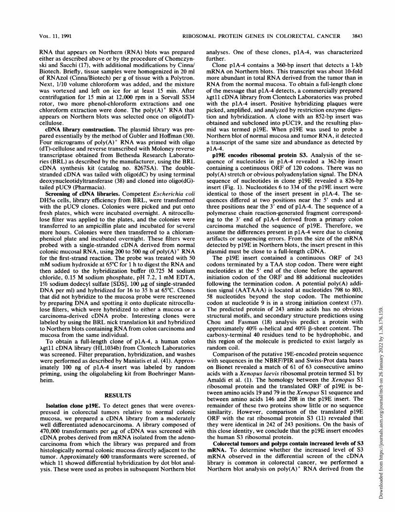
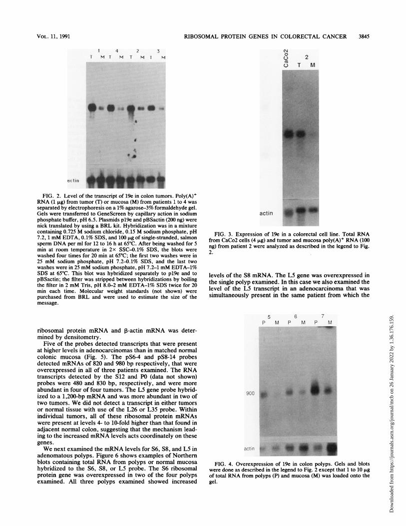
adenocarcinomas and normal mucosa of eight patients.Examples of these Northern blots are shown in Fig. 2. TheNorthern blots were also probed with the ,B-actin gene tocorrect for variability in RNA loading. The relative increasein RNA abundance for a typical tumor mucosa pair wasdetermined by densitometry. All of these tumors except onecontained 5- to 10-fold more S3 mRNA than was present inhistologically normal mucosa directly adjacent to the tumor.The S3 mRNA was overexpressed about twofold in the othertumor. We conclude that increased levels of the S3 mRNA isa common occurrence in carcinoma of the colon. We alsoexamined the levels of S3 mRNA in established cell linesderived from colorectal tumors. Figure 3 shows that theCaCo2 line expresses a greater amount of S3 mRNA than isfound in a primary tumor, a level about 20-fold higher thanthat found in normal colonic mucosa. Similar results wereobtained with the HT29 and LIM1863 cell lines (data notshown).Adenocarcinomas of the colon are thought to arise from
benign neoplastic growths called adenomatous polyps. Pol-yps are generally much smaller than tumors, and thus it is
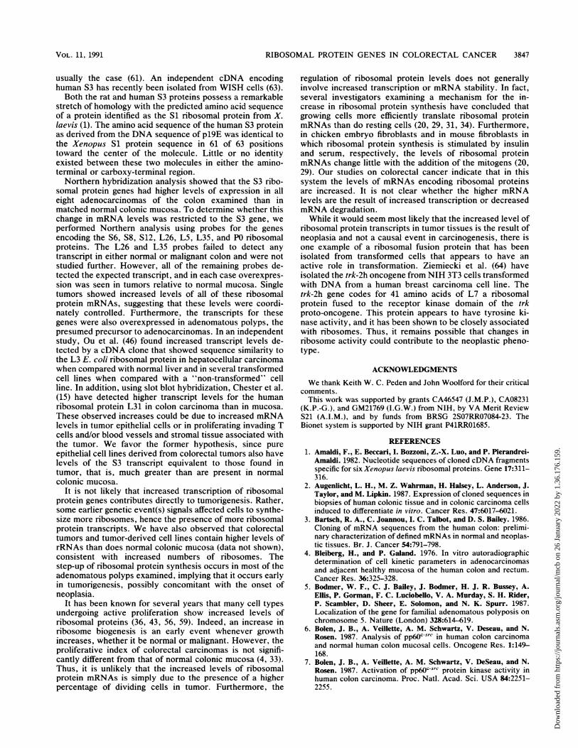
more difficult to obtain large quantities of RNA. For thisreason, we isolated total RNA from 10 adenomatous polypsand from normal mucosa and examined the levels of S3mRNA by Northern hybridization, again normalizing for theamount of 1-actin mRNA present in each preparation.Examples of these results are shown in Fig. 4. Of the 10polyps examined, 7 showed increased levels of the S3transcript. In one patient from whom RNA from normalmucosa, adenomatous polyp, and carcinoma was available,the amount of S3 transcript in the polyp was approximatelyequivalent to the amount of S3 transcript in the tumor (datanot shown). Thus, whatever the event(s) that leads to theincreased abundance of the mRNA for this ribosomal pro-tein, it precedes the onset of frank malignancy.
Overexpression of several ribosomal protein genes in colontumors and polyps. To determine whether the genes for otherribosomal proteins were also overexpressed in tumors andpolyps, we used rat probes to examine the mRNA levels forS6, S8, S12, L5, L26, L35, and P0. Examples of these resultsare shown in Fig. 5 and 6. Again the relative levels of
MOL. CELL. BIOL.
Dow
nloa
ded
from
http
s://j
ourn
als.
asm
.org
/jour
nal/m
cb o
n 26
Jan
uary
202
2 by
1.3
6.17
6.15
9.
RIBOSOMAL PROTEIN GENES IN COLORECTAL CANCER
1 4 2 3T H H T H T H
** " ..
.a
actin
FIG. 2. Level of the transcript of 19e in colon tumors. Poly(A)+RNA (1 FLg) from tumor (T) or mucosa (M) from patients 1 to 4 wasseparated by electrophoresis on a 1% agarose-3% formaldehyde gel.Gels were transferred to GeneScreen by capillary action in sodiumphosphate buffer, pH 6.5. Plasmids pl9e and pBSactin (200 ng) werenick translated by using a BRL kit. Hybridization was in a mixturecontaining 0.725 M sodium chloride, 0.15 M sodium phosphate, pH7.2, 1 mM EDTA, 0.1% SDS, and 100 itg of single-stranded, salmonsperm DNA per ml for 12 to 16 h at 65°C. After being washed for 5min at room temperature in 2x SSC-0.1% SDS, the blots werewashed four times for 20 min at 65°C; the first two washes were in25 mM sodium phosphate, pH 7.2-0.1% SDS, and the last twowashes were in 25 mM sodium phosphate, pH 7.2-1 mM EDTA-1%SDS at 65°C. This blot was hybridized separately to pl9e and topBSactin; the filter was stripped between hybridizations by boilingthe filter in 2 mM Tris, pH 8.0-2 mM EDTA-1% SDS twice for 20min each time. Molecular weight standards (not shown) werepurchased from BRL and were used to estimate the size of themessage.
ribosomal protein mRNA and ,-actin mRNA was deter-mined by densitometry.
Five of the probes detected transcripts that were presentat higher levels in adenocarcinomas than in matched normalcolonic mucosa (Fig. 5). The pS64 and pS8-14 probesdetected mRNAs of 820 and 980 bp respectively, that wereoverexpressed in all of three patients examined. The RNAtranscripts detected by the S12 and P0 (data not shown)probes were 480 and 830 bp, respectively, and were moreabundant in four of four tumors. The L5 gene probe hybrid-ized to a 1,200-bp mRNA and was more abundant in two oftwo tumors. We did not detect a transcript in either tumorsor normal tissue with use of the L26 or L35 probe. Withinindividual tumors, all of these ribosomal protein mRNAswere present at levels 4- to 10-fold higher than that found inadjacent normal colon, suggesting that the mechanism lead-ing to the increased mRNA levels acts coordinately on thesegenes.We next examined the mRNA levels for S6, S8, and L5 in
adenomatous polyps. Figure 6 shows examples of Northernblots containing total RNA from polyps or normal mucosahybridized to the S6, S8, or L5 probe. The S6 ribosomalprotein gene was overexpressed in two of the four polypsexamined. All three polyps examined showed increased
actin _ _
FIG. 3. Expression of 19e in a colorectal cell line. Total RNAfrom CaCo2 cells (4 jig) and tumor and mucosa poly(A)+ RNA (100ng) from patient 2 were analyzed as described in the legend to Fig.2.
levels of the S8 mRNA. The L5 gene was overexpressed inthe single polyp examined. In this case we also examined thelevel of the L5 transcript in an adenocarcinoma that wassimultaneously present in the same patient from which the
5 6 7P M P M P M
900* *1000
actin ~4 9 0
FIG. 4. Overexpression of 19e in colon polyps. Gels and blotswere done as described in the legend to Fig. 2 except that 1 to 10 jigof total RNA from polyps (P) and mucosa (M) was loaded onto thegel.
N0o 2o T M
VOL. 11, 1991 3845
Dow
nloa
ded
from
http
s://j
ourn
als.
asm
.org
/jour
nal/m
cb o
n 26
Jan
uary
202
2 by
1.3
6.17
6.15
9.

3846 POGUE-GEILE ET AL.
I 2 3S6 T M T NA T M SB g ~ MsO - M -; Mw T FM1 polyp was obtained as well as in histologically normal
mucosa that was either directly adjacent to or distant fromthe tumor. The L5 mRNA was present at increased levels inboth the tumor and the polyp.
DISCUSSION
U---,
4 2 3S 1 2 M T ht T M T l
a a " a to
ac tir _ _ ;
FIG. 5. Overexpression of S6, S8, S12,teins in colon tumors. Gels, transfer, and hyas described in the legend to Fig. 2 except thybridization was 55°C. Estimated sizes oftides) are shown at the left of each blot.
8P M P
Plus/minus screening of cDNA libraries has been used to0* , identify genes whose mRNA levels change as a result of
differentiation or in response to treatment with growthfactors. Screening cDNA libraries for differential expressionof genes in familial polyposis, colonic adenocarcinoma, andnormal colonic mucosa has been used to identify the genesthat undergo changes in expression during the developmentof colon cancer. Studies in which a large number of genes
** - * "have been screened show that approximately 2 to 7% of theclones examined show altered expression in the carcinomaor in familial polyposis in comparison with normal mucosa(2, 3). With this approach, changes in expression of a
L5 T M T IA laminin-binding protein (62), the ribosomal protein L31 (15),cytochrome c oxidase (32), and type I and type II keratingenes (53) have been observed. In this study, we prepared acDNA library by using mRNA isolated from an adenocarci-noma of the colon and used differential screening with cDNAprobes prepared from the carcinoma and from adjacent
ff pw normal mucosa to detect genes whose level of expressionW changes during tumorigenesis. We anticipated that some of
the products of the genes detected by such a screen would beinvolved in determining the biological properties of thetumor, while many would be genes whose transcript levelschange as a result of tumorigenesis.We were able to isolkte a cDNA clone (pl9E) encoding the
human S3 ribosomal protein because the mRNA is moreabundant in adenocarcinomas than in normal colonic epithe-lium. We identified the protein encoded in the cDNA by its
and L5 ribosomal pro- similarity to the rat S3 amino acid sequence (11). The,bridizations were done deduced rat and human amino acid sequences differ at onlythat the temperature of 1 of 243 positions. The rat and human ORFs are 90%f messages (in nucleo- identical. Thus, the coding sequences for this protein have
been highly conserved between rats and humans, as is
9 5 6 7P M P M P M
1 0T A 0 P
L5
S6 at
S8
a actin
GPDH a 44FIG. 6. Overexpression of S6, S8, and L5 in colon polyps. Gels, transfers, and hybridizations were done as described in the legend to Fig.
5 except that the amount of total RNA loaded varied between 1 and 10 p.g. Both pGPDH and pBSactin were used as probes.
MOL. CELL. BIOL.
Dow
nloa
ded
from
http
s://j
ourn
als.
asm
.org
/jour
nal/m
cb o
n 26
Jan
uary
202
2 by
1.3
6.17
6.15
9.
RIBOSOMAL PROTEIN GENES IN COLORECTAL CANCER
usually the case (61). An independent cDNA encodinghuman S3 has recently been isolated from WISH cells (63).Both the rat and human S3 proteins possess a remarkable
stretch of homology with the predicted amino acid sequenceof a protein identified as the Si ribosomal protein from X.laevis (1). The amino acid sequence of the human S3 proteinas derived from the DNA sequence of p19E was identical tothe Xenopus Si protein sequence in 61 of 63 positionstoward the center of the molecule. Little or no identityexisted between these two molecules in either the amino-terminal or carboxy-terminal region.Northern hybridization analysis showed that the S3 ribo-
somal protein genes had higher levels of expression in alleight adenocarcinomas of the colon examined than inmatched normal colonic mucosa. To determine whether thischange in mRNA levels was restricted to the S3 gene, weperformed Northern analysis using probes for the genesencoding the S6, S8, S12, L26, L5, L35, and PO ribosomalproteins. The L26 and L35 probes failed to detect anytranscript in either normal or malignant colon and were notstudied further. However, all of the remaining probes de-tected the expected transcript, and in each case overexpres-sion was seen in tumors relative to normal mucosa. Singletumors showed increased levels of all of these ribosomalprotein mRNAs, suggesting that these levels were coordi-nately controlled. Furthermore, the transcripts for thesegenes were also overexpressed in adenomatous polyps, thepresumed precursor to adenocarcinomas. In an independentstudy, Ou et al. (46) found increased transcript levels de-tected by a cDNA clone that showed sequence similarity tothe L3 E. coli ribosomal protein in hepatocellular carcinomawhen compared with normal liver and in several transformedcell lines when compared with a "non-transformed" cellline. In addition, using slot blot hybridization, Chester et al.(15) have detected higher transcript levels for the humanribosomal protein L31 in colon carcinoma than in mucosa.These observed increases could be due to increased mRNAlevels in tumor epithelial cells or in proliferating invading Tcells and/or blood vessels and stromal tissue associated withthe tumor. We favor the former hypothesis, since pureepithelial cell lines derived from colorectal tumors also havelevels of the S3 transcript equivalent to those found intumor, that is, much greater than are present in normalcolonic mucosa.
It is not likely that increased transcription of ribosomalprotein genes contributes directly to tumorigenesis. Rather,some earlier genetic event(s) signals affected cells to synthe-size more ribosomes, hence the presence of more ribosomalprotein transcripts. We have also observed that colorectaltumors and tumor-derived cell lines contain higher levels ofrRNAs than does normal colonic mucosa (data not shown),consistent with increased numbers of ribosomes. Thestep-up of ribosomal protein synthesis occurs in most of theadenomatous polyps examined, implying that it occurs earlyin tumorigenesis, possibly concomitant with the onset ofneoplasia.
It has been known for several years that many cell typesundergoing active proliferation show increased levels ofribosomal proteins (36, 43, 56, 59). Indeed, an increase inribosome biogenesis is an early event whenever growthincreases, whether it be normal or malignant. However, theproliferative index of colorectal carcinomas is not signifi-cantly different from that of normal colonic mucosa (4, 33).Thus, it is unlikely that the increased levels of ribosomalprotein mRNAs is simply due to the presence of a higherpercentage of dividing cells in tumor. Furthermore, the
regulation of ribosomal protein levels does not generallyinvolve increased transcription or mRNA stability. In fact,several investigators examining a mechanism for the in-crease in ribosomal protein synthesis have concluded thatgrowing cells more efficiently translate ribosomal proteinmRNAs than do resting cells (20, 29, 31, 34). Furthermore,in chicken embryo fibroblasts and in mouse fibroblasts inwhich ribosomal protein synthesis is stimulated by insulinand serum, respectively, the levels of ribosomal proteinmRNAs change little with the addition of the mitogens (20,29). Our studies on colorectal cancer indicate that in thissystem the levels of mRNAs encoding ribosomal proteinsare increased. It is not clear whether the higher mRNAlevels are the result of increased transcription or decreasedmRNA degradation.While it would seem most likely that the increased level of
ribosomal protein transcripts in tumor tissues is the result ofneoplasia and not a causal event in carcinogenesis, there isone example of a ribosomal fusion protein that has beenisolated from transformed cells that appears to have anactive role in transformation. Ziemiecki et al. (64) haveisolated the trk-2h oncogene from NIH 3T3 cells transformedwith DNA from a human breast carcinoma cell line. Thetrk-2h gene codes for 41 amino acids of L7 a ribosomalprotein fused to the receptor kinase domain of the trkproto-oncogene. This protein appears to have tyrosine ki-nase activity, and it has been shown to be closely associatedwith ribosomes. Thus, it remains possible that changes inribosome activity could contribute to the neoplastic pheno-type.
ACKNOWLEDGMENTS
We thank Keith W. C. Peden and John Woolford for their criticalcomments.
This work was supported by grants CA46547 (J.M.P.), CA08231(K.P.-G.), and GM21769 (I.G.W.) from NIH, by VA Merit ReviewS21 (A.I.M.), and by funds from BRSG 2S07RR07084-23. TheBionet system is supported by NIH grant P41RR01685.
REFERENCES1. Amaldi, F., E. Beccari, I. Bozzoni, Z.-X. Luo, and P. Pierandrei-
Amaldi. 1982. Nucleotide sequences of cloned cDNA fragmentsspecific for six Xenopus laevis ribosomal proteins. Gene 17:311-316.
2. Augenlicht, L. H., M. Z. Wahrman, H. Halsey, L. Anderson, J.Taylor, and M. Lipkin. 1987. Expression of cloned sequences inbiopsies of human colonic tissue and in colonic carcinoma cellsinduced to differentiate in vitro. Cancer Res. 47:6017-6021.
3. Bartsch, R. A., C. Joannou, I. C. Talbot, and D. S. Bailey. 1986.Cloning of mRNA sequences from the human colon: prelimi-nary characterization of defined mRNAs in normal and neoplas-tic tissues. Br. J. Cancer 54:791-798.
4. Bleiberg, H., and P. Galand. 1976. In vitro autoradiographicdetermination of cell kinetic parameters in adenocarcinomasand adjacent healthy mucosa of the human colon and rectum.Cancer Res. 36:325-328.
5. Bodmer, W. F., C. J. Bailey, J. Bodmer, H. J. R. Bussey, A.Ellis, P. Gorman, F. C. Luciobello, V. A. Murday, S. H. Rider,P. Scambler, D. Sheer, E. Solomon, and N. K. Spurr. 1987.Localization of the gene for familial adenomatous polyposis onchromosome 5. Nature (London) 328:614-619.
6. Bolen, J. B., A. Veillette, A. M. Schwartz, V. Deseau, and N.Rosen. 1987. Analysis of pp60csrc in human colon carcinomaand normal human colon mucosal cells. Oncogene Res. 1:149-168.
7. Bolen, J. B., A. Veillette, A. M. Schwartz, V. DeSeau, and N.Rosen. 1987. Activation of pp60c-rc protein kinase activity inhuman colon carcinoma. Proc. Natl. Acad. Sci. USA 84:2251-2255.
VOL . 1 l, 1991 3847
Dow
nloa
ded
from
http
s://j
ourn
als.
asm
.org
/jour
nal/m
cb o
n 26
Jan
uary
202
2 by
1.3
6.17
6.15
9.
3848 POGUE-GEILE ET AL.
8. Bos, J. L., E. R. Fearon, S. R. Hamilton, M. Verlaan-de Vries,J. H. van Boom, A. J. van der Eb, and B. Vogelstein. 1987.Prevalence of ras gene mutations in human colorectal cancers.
Nature (London) 327:293-297.9. Cartwright, C. A., M. P. Kamps, A. I. Meisler, J. M. Pipas, and
W. Eckhart. 1989. pp6O-src activation in human colon carci-noma. J. Clin. Invest. 83:2025-2033.
10. Cartwright, C. A., A. I. Meisler, and W. Eckhart. 1990. Activa-tion of the pp60csrc protein kinase is an early event in coloniccarcinogenesis. Proc. Natl. Acad. Sci. USA 87:558-562.
11. Chan, Y.-L., K. R. G. Devi, J. Olvera, and I. G. Wool. 1990. Theprimary structure of rat ribosomal protein S3. Arch. Biochem.Biophys. 283:546-550.
12. Chan, Y.-L., A. Lin, J. McNally, and I. G. Wool. 1987. Theprimary structure of rat ribosomal protein L5. J. Biol. Chem.262:12879-12886.
13. Chan, Y.-L., A. Lin, V. Paz, and I. G. Wool. 1987. The primarystructure of rat ribosomal protein S8. Nucleic Acids Res.15:9451-9459.
14. Chan, Y.-L., and I. G. Wool. 1988. The primary structure of ratribosomal protein S6. J. Biol. Chem. 263:2891-2896.
15. Chester, K. A., L. Robson, R. H. J. Regent, 1. C. Talbot, J. H.Pringle, L. Primrose, A. J. S. Macpherson, G. Boxer, P. South-all, and A. D. B. Malcolm. 1989. Identification of a humanribosomal protein mRNA with increased expression in colorec-tal tumours. Biochim. Biophys. Acta 1009:297-300.
16. Chirgwin, J. M., A. E. Przybyla, R. J. MacDonald, and W. J.Rutter. 1979. Isolation of biologically active ribonucleic acidfrom sources enriched in ribonuclease. Biochemistry 18:5294-5299.
17. Chomczynski, P., and N. Sacchi. 1987. Single-step method ofRNA isolation by acid guanidinium thiocyanate-phenol-chloro-form extraction. Anal Biochem. 162:156-159.
18. Chou, P. Y., and G. D. Fasman. 1978. Prediction of thesecondary structure of proteins from their amino acid sequence.Adv. Enzymol. 47:45-148.
19. D'Emilia, J., K. Bulovas, K. D'Ercole, B. Wolf, G. Steele, andI. C. Summerhayes. 1989. Expression of the c-erbB-2 gene
product (p185) at different stages of neoplastic progression inthe colon. Oncogene 4:1233-1239.
20. DePhilip, R. M., W. A. Rudert, and I. Lieberman. 1980. Prefer-ential stimulation of ribosomal protein synthesis by insulin andin the absence of ribosomal and messenger ribonucleic acidformation. Biochemistry 19:1662-1669.
21. Edmonds, M., M. H. Vaughn, Jr., and H. Nakazato. 1971.Polyadenylic acid sequences in the heterogeneous nuclear RNAand rapidly-labeled polyribosomal RNA of HeLa cells: possibleevidence for a precursor relationship. Proc. Natl. Acad. Sci.USA 68:1336.
22. Erisman, M. D., P. G. Rothberg, R. E. Diehl, C. C. Morse, J. M.Spandorfer, and S. M. Astrin. 1985. Deregulation of c-myc gene
expression in human colon carcinoma is not accompanied byamplification or rearrangement of the gene. Mol. Cell. Biol.5:1969-1976.
23. Fearon, E. R., K. R. Cho, J. M. Nigro, S. E. Kern, J. W. Simons,J. M. Ruppert, S. R. Hamilton, A. C. Preisinger, G. Thomas,K. W. Kinzler, and B. Vogelstein. 1990. Identification of achromosome 18q gene that is altered in colorectal cancers.Science 247:49-56.
24. Fearon, E. R., and B. Vogelstein. 1990. A genetic model forcolorectal tumorigenesis. Cell 61:759-767.
25. Feinberg, A. P., D. J. Law, D. Lefrancois, 0. Delattre, and G.Thomas. 1989. A multistep genetic model of human colorectalcarcinogenesis. Cancer Cells 7:245-248.
26. Finley, G. G., N. T. Schulz, S. A. Hill, J. R. Geiser, J. M. Pipas,and A. I. Meisler. 1989. Expression of the myc gene family indifferent stages of human colorectal cancer. Oncogene 4:963-971.
27. Forrester, K., C. Almoguera, K. Han, W. E. Grizzle, and M.Perucho. 1987. Detection of high incidence of K-ras oncogenesduring human colon tumorigenesis. Nature (London) 327:298-303.
28. Fort, P., L. Marty, M. Piechaczyk, S. El Sabrouty, C. Dani, P.
Jeanteur, and J. M. Blanchard. 1985. Various rat adult tissuesexpress only one major mRNA species from the glyceralde-hyde-3-phosphate-dehydrogenase multigenic family. NucleicAcids Res. 13:1431-1441.
29. Geyer, P. K., 0. Meyuhas, R. P. Perry, and L. F. Johnson. 1982.Regulation of ribosomal protein mRNA content and translationin growth-stimulated mouse fibroblasts. Mol. Cell. Biol. 2:685-693.
30. Gubler, U., and B. J. Hoffman. 1983. A simple and very efficientmethod for generating cDNA libraries. Gene 25:263-269.
31. Hammond, M. L., and L. H. Bowman. ±988. Insulin stimulatesthe translation of ribosomal proteins and the transcription ofrDNA in mouse myoblasts. J. Biol. Chem. 263:17785-17791.
32. Heerdt, B. G., H. K. Halsey, M. Lipkin, and L. H. Augenlicht.1990. Expression of mitochondrial cytochrome c oxidase inhuman colonic cell differentiation, transformation, and risk forcolonic cancer. Cancer Res. 50:1596-1600.
33. Hoffman, J., and J. Post. 1967. In vivo studies ofDNA synthesisin human normal and tumor cells. Cancer Res. 27:898-902.
34. Ignotz, G. G., S. Hokari, R. M. DePhilip, K. Tsukada, and I.Lieberman. 1981. Lodish model and regulation of ribosomalprotein synthesis by insulin-deficient chick embryo fibroblasts.Biochemistry 20:2550-2558.
35. Kerr, I. B., V. A. Murday, L. R. Hiorns, H. J. R. Bussey, andW. F. Bodmer. 1989. Prevalence of Ki-ras mutation and chro-mosome 5 allele loss in colorectal carcinomas arising in cases offamilial adenomatous polyposis. Cancer Cells 7:241-244.
36. Kief, D. R., and J. R. Warner. 1981. Coordinate control ofsyntheses of ribosomal ribonucleic acid and ribosomal proteinsduring nutritional shift-up in Saccharomyces cerevisiae. Mol.Cell. Biol. 1:1007-1015.
37. Kozak, M. 1986. Point mutations define a sequence flanking theAUG initiator codon that modulates translation by eukaryoticribosomes. Cell 44:283-292.
38. Kraus, J. P., C. L. Williamson, F. A. Firgaira, T. L. Yang-Feng,M. Munke, U. Francke, and L. E. Rosenberg. 1986. Cloning andscreening with nanogram amounts of immunopurified mRNAs:cDNA cloning and chromosomal mapping of cystathionineB-synthase and the B subunit of propionyl-CoA carboxylase.Proc. Natl. Acad. Sci. USA 83:2047-2051.
39. Leppert, M., M. Dobbs, P. Scambler, P. O'Connell, Y. Naka-mura, D. Stauffer, S. Woodward, R. Burt, J. Hughes, E.Gardner, M. Lathrop, J. Wasmuth, J.-M. Lalouel, and R. White.1987. The gene for familial polyposis coli maps to the long armof chromosome 5. Science. 238:1411-1413.
40. Lin, A., Y.-L. Chan, R. Jones, and I. G. Wool. 1987. Theprimary structure of rat ribosomal protein S12. J. Biol. Chem.262:14343-14351.
41. Maniatis, T., E. E. Fritsch, and J. Sambrook. 1982. Molecularcloning: a laboratory manual. Cold Spring Harbor Laboratory,Cold Spring Harbor, N.Y.
42. Messing, J. 1983. New M13 vectors for cloning. MethodsEnzymol. 101:20-78.
43. Meyuhas, 0. 1984. Ribosomal protein gene expression in pro-liferating and nonproliferating cells, p. 243-267. In G. S. Steinand J. L. Stein (ed.), Recombinant DNA and cell proliferation.Academic Press, Inc., New York.
44. Monpezat, J.-P., 0. Delattre, A. Bernard, D. Grunwald, Y.Remvikos, M. Muleris, R. J. Salmon, G. Frelat, B. Dutrillaux,and G. Thomas. 1988. Loss of alleles on chromosome 18 and onthe short arm of chromosome 17 in polyploid colorectal carci-nomas. Int. J. Cancer 41:404-408.
45. Nigro, J. M., S. J. Baker, A. C. Preisinger, J. M. Jessup, R.Hostetter, K. Clearly, S. H. Bigner, N. Davidson, S. Baylin, P.Devilee, T. Glover, F. S. Collins, A. Weston, R. Modali, C. C.Harris, and B. Vogelstein. 1989. Mutations in the p53 gene occurin diverse human tumour types. Nature (London) 342:705-708.
46. Ou, J.-h., T. S. B. Yin, Y.-F. Wang, W. K. Kam, and W. J.Rutter. 1987. Cloning and characterization of a human ribo-somal protein gene with enhanced expression in fetal andneoplastic cells. Nucleic Acids Res. 15:8919-8934.
47. Paz, V., J. Olvera, Y.-L. Chan, and I. G. Wool. 1989. Theprimary structure of rat ribosomal protein L26. FEBS Lett.
MOL. CELL. BIOL.
Dow
nloa
ded
from
http
s://j
ourn
als.
asm
.org
/jour
nal/m
cb o
n 26
Jan
uary
202
2 by
1.3
6.17
6.15
9.
RIBOSOMAL PROTEIN GENES IN COLORECTAL CANCER 3849
251:89-93.48. Pearson, W. R., and D. J. Lipman. 1988. Improved tools for
biological sequence comparison. Proc. Natl. Acad. Sci. USA85:2444 2448.
49. Ponte, P., S. Y. Ng, J. Engel, P. Gunning, and L. Kedes. 1984.Evolutionary conservation in the untranslated regions of actinmRNAs: DNA sequence of a human beta-actin cDNA. NucleicAcids Res. 12:1687-1696.
50. Rich, B. E., and J. A. Steitz. 1987. Human acidic ribosomalphosphoproteins P0, P1, and P2: analysis of cDNA clones, invitro synthesis, and assembly. Mol. Cell. Biol. 7:4065-4074.
51. Rosen, N., 0. Sartor, F. M. Foss, A. VeiHette, and J. B. Bolen.1989. Altered expression of src-related tyrosine kinases inhuman colon cancer. Cancer Cells 7:161-166.
52. Sanger, F., S. Nicklen, and A. R. Coulson. 1977. DNA sequenc-ing with chain-terminating inhibitors. Proc. Natl. Acad. Sci.USA 74:5463-5467.
53. Schweinfest, C. W., K. W. Henderson, J.-R. Gu, S. D. Kottari-dis, S. Besbeas, E. Panotopoulou, and T. S. Papas. 1990. Sub-traction hybridization cDNA libraries from colon carcinoma andhepatic cancer. Genet. Annal. Tech. Appl. 7:64-70.
54. Sikora, K., S. Chan, G. Evan, H. Gabra, N. Markham, J.Stewart, and J. Watson. 1987. c-Myc oncogene expression incolorectal cancer. Cancer 59:1289-1295.
55. Solomon, E. 1990. Colorectal cancer genes. Nature (London)343:412-414.
56. Stanners, C. P., M. E. Adams, J. L. Harkins, and J. W. Polard.1979. Transformed cells have lost control of ribosome numberthrough their growth cycle. J. Cell. Physiol. 100:127-138.
57. Suzuki, K., J. Olvera, and I. G. Wool. 1990. The primary
structure of rat ribosomal protein L35. Biochem. Biophys. Res.Commun. 167:1377-1382.
58. Tanaka, T., K. Wakasugi, Y. Kuwano, K. Ishikawa, and K.Ogota. 1986. Nucleotide sequence of cloned cDNA specific forrat ribosomal protein L35a. Eur. J. Biochem. 154:523-527.
59. Tushinski, R. J., and J. R. Warner. 1982. Ribosomal proteinsare synthesized preferentially in cells commencing growth. J.Cell. Physiol. 112:128-135.
60. Vogelstein, B., E. R. Fearon, B. A. Stanley, S. R. Hamilton, S. E.Kern, A. C. Preisinger, M. Leppert, Y. Nakamura, R. White,A. M. M. Smits, and J. L. Bos. 1988. Genetic alterations duringcolorectal-tumor development. N. EngI. J. Med. 319:525-532.
61. Wool, I. G., Y. Endo, Y. L. Chan, and A. Gluck. 1990.Structure, function and evolution of mammalian ribosomes, p.203-214. In W. E. Hill, A. Dahlberg, R. A. Garrett, P. B.Moore, D. Schlessinger, and J. R. Warner (ed.), The ribosome:structure, function, and evolution. American Society for Micro-biology, Washington, D.C.
62. Yow, H., J. M. Wong, H. S. Chen, C. Lee, G. D. Steele, andL. B. Chen. 1988. Increased mRNA expression of a laminin-binding protein in human colon carcinoma: complete sequenceof a full-length cDNA encoding the protein. Proc. Natl. Acad.Sci. USA 85:6394-6398.
63. Zhang, X. T., Y.-M. Tan, and Y. H. Tan. 1990. Isolation of acDNA encoding human 40S ribosomal protein s3. Nucleic AcidsRes. 18:6689.
64. Ziemiecki, A., R. G. Muller, F. Xiao-Chang, N. E. Hynes, and S.Kozma. 1990. Oncogenic activation of the human trk proto-oncogene by recombination with the ribosomal large subunitprotein L7a. EMBO J. 9:191-196.
VOL . 1 l, 1991
Dow
nloa
ded
from
http
s://j
ourn
als.
asm
.org
/jour
nal/m
cb o
n 26
Jan
uary
202
2 by
1.3
6.17
6.15
9.