rna recombination between persisting pestivirus and a ...jvi.asm.org/content/75/14/6256.full.pdf ·...
TRANSCRIPT

JOURNAL OF VIROLOGY,0022-538X/01/$04.0010 DOI: 10.1128/JVI.75.14.6256–6264.2001
July 2001, p. 6256–6264 Vol. 75, No. 14
Copyright © 2001, American Society for Microbiology. All Rights Reserved.
RNA Recombination between Persisting Pestivirus and a VaccineStrain: Generation of Cytopathogenic Virus and
Induction of Lethal DiseasePAUL BECHER,* MICHAELA ORLICH, AND HEINZ-JURGEN THIEL
Institut fur Virologie (FB Veterinarmedizin), Justus-Liebig-Universitat, D-35392 Giessen, Germany
Received 11 December 2000/Accepted 9 April 2001
Molecular analysis of a cytopathogenic (cp) bovine viral diarrhea virus (BVDV) isolate (1741) obtained froma case of mucosal disease (MD) led to the identification of five different viral subgenomic RNAs in addition toa noncytopathogenic (noncp) strain (NCP 1741). For each of the subgenomes, a large internal deletion wasfound together with an inserted sequence encoding part of ribosomal protein S27a fused to an N-terminallytruncated ubiquitin monomer. Surprisingly, the two cellular insertions together with flanking viral sequencesencoding parts of NS3 and NS4B are >99% identical to the previously described sequence of BVDV vaccinestrain RIT (P. Becher, M. Orlich, and H.-J. Thiel, J. Virol. 72:8697–8704, 1998), while the remainder of thesubgenomes is derived from the genome of NCP 1741. Further analyses including molecular cloning andnucleotide sequencing of the recombination partners revealed that both homologous and nonhomologous RNArecombination contributed to the generation of the viral subgenomes. Interestingly, for another cp BVDVisolate (CP 4584) from an independent case of MD, again an insertion of a RIT-derived sequence element wasdetected. In contrast to CP 1741, for CP 4584 a duplication of the genomic region encoding NS3 and parts ofNS4A and NS4B was found. Transfection of bovine cells with RNA transcribed from a chimeric cDNA constructshowed that the RIT-derived insertion together with the CP 4584-specific duplication of viral sequencesrepresents the genetic basis of cytopathogenicity of CP 4584. Remarkably, passages of the recovered cp virusin cell culture led to emergence of noncp BVDV and a number of viral subgenomes whose genome organizationwas similar to that in BVDV 1741.
The genera Pestivirus, Flavivirus, and Hepacivirus constitutethe family Flaviviridae. The genus Pestivirus is represented bythe species Bovine viral diarrhea virus 1 (BVDV-1), BVDV-2,Classical swine fever virus (CSFV), and Border disease virus(18). Pestiviruses have a positive-sense single-strandedRNA genome of about 12.3 kb in length with one large openreading frame (ORF) flanked by 59 and 39 nontranslated re-gions (NTR) (see references 26 and 33 for reviews). This ORFencodes a polyprotein of approximately 3,900 amino acids (aa)which is co- and posttranslationally processed by viral andcellular proteases, leading to the mature viral proteins. Thefirst third of the ORF encodes an autoprotease and four struc-tural proteins, while the 39 part of the RNA genome codes forthe other nonstructural (NS) proteins (see references 26 and33 for reviews). Based on the effects in tissue culture, twobiotypes, cytopathogenic (cp) and noncytopathogenic (noncp),are distinguished (17, 20).
BVDV represents one of the most important pathogens ofcattle, causing significant economical losses worldwide (1).Horizontal BVDV infection can have different consequences,such as abortion, diarrhea, hemorrhagic syndrome, and, mostfrequently, inapparent courses (see references 1 and 33 forreviews). Diaplacental infection with noncp BVDV can resultin the birth of persistently infected animals with an acquiredimmunotolerance to the original BVDV strain. Such persis-
tently infected animals may come down with mucosal disease(MD). In addition to the persisting noncp BVDV, a cp BVDVcan always be isolated from animals with MD (12, 26).
Molecular characterization of several BVDV pairs stronglysuggested that the cp viruses can evolve from the respectivenoncp viruses by nonhomologous RNA recombination (seereference 26 for a review). For the cp viruses, various genomicalterations were identified, including insertions of cellular se-quences, frequently together with large duplications of viralsequences, and genomic rearrangements with large duplica-tions and deletions (2, 4, 8, 23, 26, 30). One important differ-ence between cp and noncp BVDV is the expression of NS3,which is colinear to the C-terminal part of NS2-3. While NS2-3is expressed in both cp and noncp BVDV-infected cells, NS3 isfound exclusively after infection with cp BVDV. Accordingly,NS3 is regarded as the marker protein for cp BVDV strains.
In this paper, we report the identification of BVDV vaccinestrain RIT-derived insertions in the genomes of two cp BVDVisolates obtained from independent cases of MD. The resultsof this study, including the molecular characterization of theputative recombination partners, strongly suggest that homol-ogous and nonhomologous RNA recombination between per-sisting noncp BVDV and BVDV vaccine strain RIT can beresponsible for induction of fatal MD.
MATERIALS AND METHODS
Cells and viruses. Madin-Darby bovine kidney (MDBK) cells were obtainedfrom the American Type Culture Collection (Manassas, Va.). Cells were grownin Dulbecco’s modified Eagle’s medium supplemented with 10% horse serum.The cp BVDV isolates 1741 and 4584 were isolated from cattle in Germany in1996 that came down with MD. BVDV strains CP7 and NCP7 as well as BVDV
* Corresponding author. Mailing address: Institut fur Virologie (FBVeterinarmedizin), Justus-Liebig-Universitat Giessen, Frankfurter Str.107, D-35392 Giessen, Germany. Phone: 49 641 99 38376. Fax: 49 64199 38359. E-mail: [email protected].
6256
on May 11, 2018 by guest
http://jvi.asm.org/
Dow
nloaded from

vaccine strain RIT 4350 (Pfizer, Karlsruhe, Germany) have been described pre-viously (8, 12, 21, 24). Comparative sequence analyses indicate that all virusisolates included in this study are BVDV-1 strains.
Infection of cells. Supernatants and lysates of infected cells were combined andused for infection of MDBK cells. Material for infection was prepared by freez-ing and thawing cultures 48 h postinfection and stored at 270°C. Infection withnoncp BVDV was detected by immunofluorescence (IF) with monoclonal anti-body 8.12.7 (directed against NS3), kindly provided by E. J. Dubovi (CornellUniversity, Ithaca, N.Y.).
RNA preparation, gel electrophoresis, and Northern (RNA) hybridization.Preparation of RNA, gel electrophoresis, radioactive labeling of the probes,hybridization, and posthybridization washes were performed as described previ-ously (3). A 2.5-kb NotI-NsiI fragment from the cDNA clone pCP7-5A (7)encompassing the NS3 gene of BVDV CP7 was used as a probe.
Oligonucleotides. If not otherwise indicated, numbering of nucleotidesthroughout this work refers to the genomic sequence of BVDV SD-1, the firstcompletely sequenced noncp BVDV strain (13). Oligonucleotides were pur-chased from MWG Biotech GmbH (Ebersberg, Germany). Oligonucleotides OlBVDV 7100 (nucleotides [nt] 7313 to 7335; sense), Ol NS3R (nt 5326 to 5343;antisense), Ol 100 (nt 107 to 127; sense), Ol 1400R (nt 1430 to 1448; antisense),Ol Rit-ubi4 (corresponding to part of the ubiquitin coding sequence; sense), andOl Rit-4AR (nt 7417 to 7434; antisense) have been described previously (5, 6, 8).Primers Ol 5150 (59-AGRGGGCCWGCCGTGTG-39; R 5 A or G, W 5 A orT; corresponding to nt 5150 to 5166; sense), Ol 7400R (59-AGTCTCYTTCCCCTCAGTTC-39; Y 5 C or T; corresponding to nt 7359 to 7378; antisense), OlNS3 (59-CTGGCAGTWGACCTCCTAG-39; W 5 A or T; corresponding to nt7109 to 7127; sense), and Ol 8600R (59-GCYTTCATCTCATARCCRCA-39;Y 5 C or T, R 5 A or G; corresponding to nt 8609 to 8628; antisense) weredesigned by using published sequences of BVDV-1 strains NADL, SD-1, Osloss,and CP7. The sequences of the following primers were derived from the obtainedsequences of CP 1741 and CP 4584: Ol 1741-AgeIR (59-CCAACCGGTTTCCAATCCCCTCCTCACCTTTAGCAATGCTG-39, antisense) and Ol 4584-MluI(59-TTACGCGTCCGGGGGATTGGATTTC-39, sense).
RT-PCR and molecular cloning. Reverse transcription-PCR (RT-PCR) ofapproximately 500 ng of heat-denatured RNA was carried out as describedpreviously (5). The cDNA fragments obtained after RT-PCR were separated byagarose gel electrophoresis and purified by using a QIAEX DNA purification kit(Qiagen, Hilden, Germany). The respective cDNA fragments were cloned byusing a TA cloning kit (Invitrogen, De Schelp, The Netherlands).
For NCP 1741, part of the 59 NTR together with the genomic region encodingNpro, C, and part of Ems was amplified by RT-PCR with primers Ol 100 and Ol1400R. For NCP 1741 and NCP 4584, the genomic region encoding NS3, NS4A,NS4B, and part of NS5A was amplified by using primer pairs Ol 5150-Ol 7400Rand Ol NS3-Ol 8600R, respectively. For amplification of the genomic regionsencoding NS3, NS4A, and part of NS4B of CP 1741 and CP 4584, primers OlRit-ubi4 and Ol Rit-4AR were used.
Nucleotide sequencing and sequence analysis. Nucleotide sequences weredetermined by cycle sequencing with a Thermo Sequenase kit (AmershamBuchler, Braunschweig, Germany) and Li-Cor 4000 L DNA sequencer (MWGBiotech). All sequences were determined by sequencing both complementarystrands of at least three independent cDNA clones. Computer analysis of se-quence data was performed with HUSAR (DKFZ, Heidelberg, Germany), whichprovides the Genetics Computer Group software package (14).
Construction of BVDV full-length cDNA clone. The noncp BVDV full-lengthcDNA clones pNCP7-5A and pNCP7-5A-(AgeI-) have been described previ-ously (2). These plasmids are derivatives of the cp BVDV infectious full-lengthcDNA clone pCP7-5A (7). Numbering of nucleotides throughout this paragraphrefers to the NCP7-5A sequence. Construction of p7/4584 was based on pNCP7-5A-(AgeI-), which differs from pNCP7-5A by the absence of the single AgeI site(nt 5309 to 5314). A CP 4584-specific MluI/AgeI fragment was obtained by PCRwith primer Ol 4584-MluI (encompassing an MluI site which corresponds to nt7434 to 7439) and primer Ol 1741-AgeIR, using the CP 4584 -derived cDNA astemplate, and cloned into pCR2.1. The resulting plasmid was termed p4584-MluI. Next, the SacI (nt 5842 to 5847)/MluI (nt 7434 to 7439) fragment ofpNCP7-5A was cloned into p4584-MluI precut with SacI (located in thepolylinker) and MluI. Addition of the pNCP7-5A-derived AgeI/SalI (nt 7716 to7721) fragment completed the cloning of the SacI/SalI fragment, which was thenintroduced into pNCP7-5A-(AgeI-) precut with SacI and SalI. The genomeorganization of the resulting chimeric cDNA construct p7/4584, the genomicregion derived from CP 4584, and positions of the AgeI and MluI sites areindicated in Fig. 4A. According to comparative analysis of the NS3 codingsequences, CP 4584 and NCP7 are about 94% identical.
In vitro transcription and transfection of RNA. In vitro synthesis of RNA andtransfection of MDBK cells were carried out as described elsewhere (7). About2 mg of RNA was used for each transfection. For electroporation (one pulse, 950mF and 180 V), a Gene Pulser II (Bio-Rad, Munich, Germany) was used.
Plaque assay. MDBK cells were infected with 10-fold serial dilutions of su-pernatants from transfected cells. After incubation at 37°C for 4 h, the attachedcells were overlaid with semisolid medium containing 0.6% low-melting-pointagarose (Gibco-BRL) and 5% horse serum. After 4 days of incubation at 37°C,the agarose overlays were removed, and the cells were washed with phosphate-buffered saline and then fixed with acetone-methanol (1:1) for 1 h at 220°C.Immunostaining of cells using the BVDV E2-specific monoclonal antibody D5(kindly provided by E. Weiland, Tubingen, Germany) was carried out as de-scribed elsewhere (7).
Determination of growth kinetics. MDBK cells (106 in a six-well dish) wereinfected with transcript-derived virus at a multiplicity of infection (MOI) of 0.05.After adsorption for 1 h at 37°C, the cells were washed six times with phosphate-buffered saline, overlaid with medium containing 10% horse serum, and thenincubated for 5 days. After the indicated time intervals, aliquots (200 ml) of thecell culture supernatant were removed and used for titration on MDBK cells.The viral yields were determined as the titer of 50% tissue culture infectiousdoses (TCID50) per milliliter.
Analysis of viral RNA synthesis by Northern blotting. MDBK cells (106) wereinfected with transcript-derived virus at an MOI of 0.05 and processed parallel tocells used for determination of the growth kinetics. After 1 and 2 days ofincubation at 37°C, RNA was prepared using an RNeasy total RNA kit (Qiagen).Five micrograms of glyoxylated RNA was subjected to Northern blot analysis.The viral RNAs were detected by autoradiography, and the intensity of bandswas determined with a phosphorimager (Fujik BAS 1000; Fuji).
Nucleotide sequence accession numbers. Sequence data from this study havebeen deposited in the EMBL and GenBank data libraries and assigned accessionnumbers AF321450 to AF321458.
RESULTS
Characterization of BVDV 1741 by hybridization. BVDV1741 was obtained from a bovine that died of MD, the fatalform of a BVDV infection. In MDBK cells, this virus causes acytopathic effect. Limiting dilution of BVDV 1741 from thethird cell passage allowed isolation of an accompanying noncpvirus, termed NCP 1741. However, several attempts failed tobiologically clone the cp virus by plaque purification. In thetext that follows, cp BVDV 1741 is termed CP 1741.
Previous studies have demonstrated that most cp BVDVisolates contain RNA genomes significantly larger or shorterthan those of noncp BVDV; the alterations are due to largeduplications of viral sequences or deletions, respectively (2, 4,8, 23, 26). To investigate whether similar genomic differencesare present in CP 1741, a Northern blot analysis was per-formed. Total RNA from MDBK cells infected with either CP1741 or NCP 1741 was hybridized with a BVDV-specific cDNAprobe (Fig. 1A). Viral genomic RNA with a size of about 12.3kb was detected in cells infected with CP 1741 and NCP 1741.In addition, subgenomic RNA with a size of about 8 kb wasidentified after infection with CP 1741 but not NCP 1741.Accordingly, cytopathogenicity of CP 1741 correlates with thepresence of viral subgenomic RNA.
RT-PCR analysis. So far, subgenomic RNAs have been de-scribed for two cp BVDV isolates (19, 32) as well as for fourclosely related cp BVDV isolates obtained during an outbreakof MD on a single farm (4). A common feature of thesesubgenomes is the deletion of all or almost all of the genomicregion encoding the structural proteins, as well as the non-structural proteins p7 and NS2. To determine the deletionwhich led to the subgenomic RNA of CP 1741, we performedan RT-PCR analysis using antisense primer Ol NS3R, locatedin the NS3 coding region, and sense primer Ol 100, located in
VOL. 75, 2001 RECOMBINATION BETWEEN PERSISTING AND VACCINE BVDV STRAINS 6257
on May 11, 2018 by guest
http://jvi.asm.org/
Dow
nloaded from

the 59 NTR. Interestingly, five specific products ranging in sizefrom 0.9 to 1.7 kb were generated (data not shown). Changingthe conditions of RT-PCR to include elongation times of up to300 s did not result in detection of additional cDNA fragments.Under the chosen conditions, cDNA to be obtained from viralRNA without deletion with an expected size of about 5.2 kbwas not amplified. We did not investigate whether this varietyof cDNA fragments changed with culture conditions.
Genome organization of the subgenomes and identificationof CP RIT-derived sequences. For further characterization, theamplified cDNA fragments were cloned and subjected to nu-cleotide sequence analysis. To allow comparison with the cor-responding sequences of NCP 1741, part of the 59 NTR to-gether with the genomic regions encoding Npro and C as well asNS3, NS4A, NS4B, and part of NS5A of NCP 1741 wereamplified by RT-PCR, cloned, and sequenced. Analysis of theCP 1741-derived cDNA fragments resulted in identification offive subgenomes differing in size, termed CP 1741-A, -B, -C,-D, and -E. To our knowledge, there is only one other reporton cp BVDV isolates consisting of such a variety of viralsubgenomes in addition to noncp BVDV (4). In contrast, allother described cp BVDV isolates comprise only one cp (sub)genome. Comparative sequence analyses revealed that each ofthe subgenomes carries two cellular insertions which encodepart of ribosomal protein S27a (S27ap) fused to a truncatedubiquitin monomer lacking the N-terminal 3 aa (ubip). Inter-estingly, these two cellular insertions have been previouslyfound in the genome of BVDV CP RIT, a temperature-sensi-tive strain widely used for vaccination (8). Moreover, furtheranalyses of the CP 1741 sequences demonstrated that the twocellular insertions as well as flanking viral sequences encodingpart of NS4B and the N-terminal region of NS3 are .99%identical to the CP RIT sequence, while the identity between
these viral sequences flanking the insertions and the corre-sponding sequences of NCP 1741 is ,93% (Fig. 1B). In con-trast, the region upstream of the RIT-specific sequence ele-ment is .99% identical to the corresponding sequence of NCP1741 but ,91% identical to the CP RIT sequence. Theseresults strongly suggest that the CP 1741 subgenomes weregenerated by RNA recombination between NCP 1741 andvaccine strain CP RIT.
The genome organizations of the five identified subgenomesof CP 1741 are similar to each other. The following featuresare common to all subgenomes described here: (i) deletion ofeither all or most of the genomic region encoding the struc-tural proteins, p7, and NS2 and (ii) presence of the samehost-specific insertion directly upstream of the NS3 gene. Re-markably, all deletions and insertions occurred in frame. Sub-genomes CP 1741-A, -B, -C, and -D each carry an additionalNCP 1741-derived viral insertion encoding various C-terminalparts of NS4B together with 3 aa of the NS5A N terminusdirectly upstream of the RIT-specific sequence. This duplica-tion of viral sequences is not present in subgenome CP 1741-E,which also lacks 9 nt from the 59 end of the S27ap codingregion compared to the other subgenomes and CP Rit (Fig.1B).
Comparison of NS3 coding sequences. Our analysis of theCP 1741 subgenomic RNAs indicated that the 59 regions of thesubgenomes were derived from NCP 1741, while the two cel-lular insertions together with flanking viral sequences wereobtained from CP RIT. On the basis of these data, however, wedid not know whether the entire 39 region or only a part of itwas derived from CP RIT. To address this question, thegenomic region encoding NS3, NS4A, and part of NS4B of CP1741 was amplified by RT-PCR (using a sense primer encom-passing part of the ubiquitin coding sequence and an antisense
FIG. 1. Analysis of BVDV 1741. (A) Northern blot analysis of total RNA from MDBK cells infected with CP 1741, NCP 1741, and noninfectedMDBK cells (n.i.). RNA was separated by denaturing agarose gel electrophoresis, blotted onto a nylon membrane and hybridized with a 2.5-kbNotI-NsiI fragment from the cDNA clone pCP7-5A (7). Numbers refer to RNA ladder sizes. Migration positions of the viral genomic andsubgenomic RNAs are marked with arrows. (B) Genome organization of NCP 1741 and the CP 1741 subgenomic RNAs. All subgenomic RNAscontain an insertion of CP RIT-derived sequences which encode part of S27a (S27ap) fused to an N-terminally truncated ubiquitin (ubip) (blackbox) as well as flanking viral sequences (gray boxes). The deletions are indicated by dashed lines. The lengths of the bars are not drawn to scale.The underlined parts of the (sub)genomes have been sequenced. With respect to the analyzed regions, all subgenomes maintain one large ORF.
6258 BECHER ET AL. J. VIROL.
on May 11, 2018 by guest
http://jvi.asm.org/
Dow
nloaded from

primer corresponding to the NS4B coding region), cloned, andsubjected to nucleotide sequence analysis. Interestingly, the 59terminal 280 nt of the NS3 gene of CP 1741 were .99.5%identical to the sequence of CP RIT but ,92% identical to theNCP 1741 sequence. Analysis of the remaining part of the NS3gene of CP 1741 revealed only 93% identity to CP RIT, while99.5% of nucleotides were identical to the sequence of NCP1741. Moreover, RT-PCR using an antisense primer corre-sponding to nt 553 to 572 of the NS3 gene and a set of senseprimers each spanning the internal deletion of the individualsubgenomes was performed. Sequence analysis of the obtainedcDNA fragments demonstrated for each of the subgenomesthat the genomic region encompassing the 59-terminal 280 ntof the NS3 gene was derived from CP RIT, while the remainingpart of the NS3 coding region was derived from NCP 1741.Interestingly, the junction of the NS3 coding sequences oc-curred without deletion or insertion of nucleotides at the crossover site and thus resulted from precise homologous recombi-nation. In contrast, the internal deletions and the 59 junctionbetween the NCP 1741-specific sequences and the CP RIT-derived sequences (see above) are due to nonhomologous re-combination. Taken together, the results indicate that bothnonhomologous and homologous recombination contributedto the generation of the viral subgenomes. Comparative anal-ysis of the recombination junctions in the subgenomes with thecorresponding regions of the recombination partners revealedshort regions of sequence similarity at several cross over sites(data not shown), suggesting that base pairing between thenascent RNA and the acceptor template may facilitate tem-plate switching of the RNA-dependent RNA polymerase andthereby contributes to nonhomologous RNA recombination.The presence of such regions of sequence similarity at crossover sites has recently been reported for another set of BVDVsubgenomes (4).
Characterization of BVDV 4584. BVDV 4584 was also iso-lated from an animal that died of MD. After two passages intissue culture cells, cp and noncp BVDV were biologicallycloned and termed CP 4584 and NCP 4584, respectively.
Northern blot analysis revealed that the genome of CP 4584 isabout 15.2 kb long, or about 2.9 kb longer than the genomes ofNCP 4584 and other noncp BVDV strains (Fig. 2A). Molecu-lar cloning of parts of the genome of CP 4584 by RT-PCRusing primers Ol 7100 and Ol NS3R and subsequent nucleo-tide sequencing indicated the presence of duplicated viral se-quences; these encode NS3, NS4A, and aa 1 to 29 and 52 to132 of NS4B. In addition, two cellular insertions encodingparts of ribosomal protein S27a and ubiquitin were found (Fig.2B). Comparative analyses of the genomic region encodingNS3, NS4A, and part of NS4B of CP 4584, NCP 4584, andvaccine strain RIT demonstrated that the part of the CP 4584sequence which encodes the two cellular insertions (S27ap-ubip) as well as flanking viral sequences encoding aa 52 to 132of NS4B (NS4Bp) and aa 1 to 371 of NS3 (NS3p) are .99%identical to the published sequence of BVDV RIT (8), whileNS4Bp and NS3p are only 93 and 95%, respectively, identicalto the NCP 4584 sequence. In contrast, the remaining part ofthe determined CP 4584 sequence encoding NS4A, aa 1 to 29of NS4B, as well as aa 372 to 683 of NS3 is .99% identical tothe sequence of NCP 4584 but ,95% identical to the RITsequence (Fig. 2B). The results of our analysis strongly suggestthat CP 4584 evolved by RNA recombination between NCP4584 and BVDV vaccine strain RIT. RNA recombination be-tween the NS3 coding sequences of BVDV RIT and NCP 4584occurred without deletion or insertion of nucleotides and istherefore homologous in nature, while recombination betweenthe NS4B coding sequences of these strains resulted from anonhomologous reaction. It should be noted that the RIT-derived insertion of CP 4584 differs from that identified in thesubgenomes of CP 1741 with respect to the 59 and 39 bordersof the insertions (Fig. 3).
Infectious BVDV cDNA clone with S27ap and ubip codinginsertions and duplicated NS3 gene. The subgenomes of CP1741 and the genome of CP 4584 were partially sequenced.Accordingly, it cannot be excluded that other genetic changesare present in these viral (sub)genomes in addition to theidentified insertions and deletions or duplications. To investi-
FIG. 2. Analysis of cp and noncp BVDV 4584. (A) Northern blot analysis of total RNA from MDBK cells infected with BVDV CP 4584, NCP4584, and noninfected MDBK cells (n.i.). RNA ladder sizes are indicated on the left. Migration positions of the viral genomic RNAs are markedwith arrows. (B) Genome organization of NCP 4584 and CP 4584. The genome of CP 4584 contains a duplication of viral sequences encoding NS3,NS4A, and part of NS4B together with an insertion of CP RIT-derived sequences which encode S27ap and ubip (black box) as well as flanking viralsequences (NS4Bp, NS3p; gray boxes). The lengths of the bars are not drawn to scale. The underlined parts of the genomes have been sequenced.
VOL. 75, 2001 RECOMBINATION BETWEEN PERSISTING AND VACCINE BVDV STRAINS 6259
on May 11, 2018 by guest
http://jvi.asm.org/
Dow
nloaded from
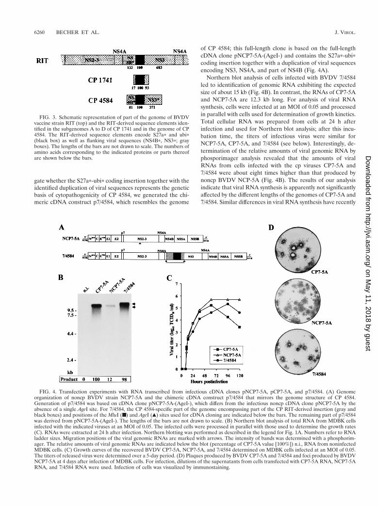
gate whether the S27ap-ubip coding insertion together with theidentified duplication of viral sequences represents the geneticbasis of cytopathogenicity of CP 4584, we generated the chi-meric cDNA construct p7/4584, which resembles the genome
of CP 4584; this full-length clone is based on the full-lengthcDNA clone pNCP7-5A-(AgeI-) and contains the S27ap-ubipcoding insertion together with a duplication of viral sequencesencoding NS3, NS4A, and part of NS4B (Fig. 4A).
Northern blot analysis of cells infected with BVDV 7/4584led to identification of genomic RNA exhibiting the expectedsize of about 15 kb (Fig. 4B). In contrast, the RNAs of CP7-5Aand NCP7-5A are 12.3 kb long. For analysis of viral RNAsynthesis, cells were infected at an MOI of 0.05 and processedin parallel with cells used for determination of growth kinetics.Total cellular RNA was prepared from cells at 24 h afterinfection and used for Northern blot analysis; after this incu-bation time, the titers of infectious virus were similar forNCP7-5A, CP7-5A, and 7/4584 (see below). Interestingly, de-termination of the relative amounts of viral genomic RNA byphosporimager analysis revealed that the amounts of viralRNAs from cells infected with the cp viruses CP7-5A and7/4584 were about eight times higher than that produced bynoncp BVDV NCP-5A (Fig. 4B). The results of our analysisindicate that viral RNA synthesis is apparently not significantlyaffected by the different lengths of the genomes of CP7-5A and7/4584. Similar differences in viral RNA synthesis have recently
FIG. 3. Schematic representation of part of the genome of BVDVvaccine strain RIT (top) and the RIT-derived sequence elements iden-tified in the subgenomes A to D of CP 1741 and in the genome of CP4584. The RIT-derived sequence elements encode S27ap and ubip(black box) as well as flanking viral sequences (NS4Bp, NS3p; grayboxes). The lengths of the bars are not drawn to scale. The numbers ofamino acids corresponding to the indicated proteins or parts thereofare shown below the bars.
FIG. 4. Transfection experiments with RNA transcribed from infectious cDNA clones pNCP7-5A, pCP7-5A, and p7/4584. (A) Genomeorganization of noncp BVDV strain NCP7-5A and the chimeric cDNA construct p7/4584 that mirrors the genome structure of CP 4584.Generation of p7/4584 was based on cDNA clone pNCP7-5A-(AgeI-), which differs from the infectious noncp cDNA clone pNCP7-5A by theabsence of a single AgeI site. For 7/4584, the CP 4584-specific part of the genome encompassing part of the CP RIT-derived insertion (gray andblack boxes) and positions of the MluI (■) and AgeI (Œ) sites used for cDNA cloning are indicated below the bars. The remaining part of p7/4584was derived from pNCP7-5A-(AgeI-). The lengths of the bars are not drawn to scale. (B) Northern blot analysis of total RNA from MDBK cellsinfected with the indicated viruses at an MOI of 0.05. The infected cells were processed in parallel with those used to determine the growth rates(C). RNAs were extracted at 24 h after infection. Northern blotting was performed as described in the legend for Fig. 1A. Numbers refer to RNAladder sizes. Migration positions of the viral genomic RNAs are marked with arrows. The intensity of bands was determined with a phosphorim-ager. The relative amounts of viral genomic RNAs are indicated below the blot (percentage of CP7-5A value [100%]) n.i., RNA from noninfectedMDBK cells. (C) Growth curves of the recovered BVDV CP7-5A, NCP7-5A, and 7/4584 determined on MDBK cells infected at an MOI of 0.05.The titers of released virus were determined over a 5-day period. (D) Plaques produced by BVDV CP7-5A and 7/4584 and foci produced by BVDVNCP7-5A at 4 days after infection of MDBK cells. For infection, dilutions of the supernatants from cells transfected with CP7-5A RNA, NCP7-5ARNA, and 7/4584 RNA were used. Infection of cells was visualized by immunostaining.
6260 BECHER ET AL. J. VIROL.
on May 11, 2018 by guest
http://jvi.asm.org/
Dow
nloaded from

been reported for another isogenic BVDV pair consisting of cpand noncp BVDV derived from cDNA constructs as well asthree additional BVDV pairs (22, 35).
Cell lysis was observed 2 days after transfection of bovinecells with 7/4584 RNA. To characterize the recovered cp virus7/4584, a plaque assay was performed on MDBK cells infectedwith supernatants from transfected cells (Fig. 4D). For com-parison, plaque and focus-forming assays were also performedwith supernatants from MDBK cells transfected with CP7-5ARNA and NCP7-5A RNA, respectively. The plaques generatedby 7/4584 were slightly smaller than the ones produced byCP7-5A. As expected, no signs of cytopathology were observedin NCP7-5A-infected cells. For further characterization, thegrowth kinetics of 7/4584, CP7-5A, and NCP7-5A were deter-mined. MDBK cells were infected with supernatants from cellstransfected with the various RNAs at an MOI of 0.05. Afterincubation at 37°C, virus released into the medium was titratedover a 5-day period. The chimeric virus 7/4584 grew slightlymore slowly than the other two viruses (Fig. 4C). Each of thethree viruses reached titers of .105 TCID50/ml.
Generation of noncp BVDV and subgenomic RNAs afterpassaging of BVDV 7/4584. In a recent study, we demonstratedthat replication of a cp BVDV strain carrying a cellularNEDD8 coding insertion together with duplicated viral se-quences rapidly resulted in the emergence of noncp BVDV(2). It was therefore of interest to investigate whether passagesof 7/4584 in MDBK cells would also lead to generation ofnoncp BVDV. The possible emergence of noncp BVDV wasmonitored by IF of infected cells and Northern blot analysis. IFanalysis actually indicated the presence of noncp BVDV incells infected with 7/4584. With respect to the fifth and allsubsequent passages of 7/4584, viral RNA with a size of 12.3 kbwas detected in addition to the larger viral RNA of 7/4584 (Fig.5A). For further analysis, the emerged noncp BVDV was bi-ologically cloned using serial dilutions of supernatants fromcells infected with 7/4584. To characterize this noncp BVDV,part of the NS2-3 coding region was cloned and sequenced;within this region, BVDV strain NCP7 and the engineeredvirus NCP7-5A carry an AgeI site (corresponding to positions5309 to 5314 of the NCP7-5A sequence) which is absent in thegenome of 7/4584. Sequence analysis revealed the absence ofthe AgeI site for the emerged noncp BVDV, which allows aclear distinction from NCP7 and NCP7-5A. To address thepossibility that the emergence of noncp deletion genomes re-sulted from (i) the presence of low-level deletion templates inthe plasmid DNA used for in vitro transcription or (ii) low-level deletion transcript RNAs generated by the SP6 RNApolymerase, a plaque assay using dilutions of cells transfectedwith 7/4584 RNA was performed directly after transfection;after 5 days a single plaque was harvested from a six-well dishwith a total of seven plaques and used for further propagation.After three passages in MDBK cells noncp BVDV emergedwhich was biologically cloned and shown to contain the above-described genetic marker. Taken together, the results of ouranalyses demonstrate that the noncp BVDV evolved from7/4584 by RNA recombination during replication in tissue cul-ture cells.
Surprisingly, Northern blot analysis of viral RNA from cellsinfected with the 8th, 9th, and 10th passages of 7/4584 led toidentification of subgenomic RNAs in addition to the genomic
RNAs of 7/4584 and the emerged noncp BVDV (Fig. 5A). Forcharacterization of these subgenomes, the RNAs obtained af-ter 8 and 10 passages of 7/4584 were subjected to RT-PCRanalysis using primers Ol NS3R and Ol 100. For both RNAsobtained after 8 and 10 passages, a number of cDNA frag-ments ranging in size between 0.8 and 1.3 kb were generated.With respect to the eighth passage of 7/4584, molecular cloningand nucleotide sequencing of the obtained fragments resultedin identification of four different subgenomic RNAs (7/4584-A,-B, -C, and -D), two of which were also found after 10 passages(Fig. 5). Furthermore, two additional subgenomes (7/4584-E,and -F) were detected after 10 passages of 7/4584 in MDBKcells. The genome organizations of the 7/4584-derived subge-nomes are very similar to each other, both exhibiting (i) a largeinternal deletion of viral sequences and (ii) the presence ofS27ap-ubip coding sequences directly upstream of the NS3gene. Remarkably, all deletions resulting in generation of thesubgenomes occurred in frame. To our knowledge, this is thefirst report on the emergence of noncp BVDV and a numberof subgenomic RNAs during propagation of cp BVDV in tis-sue culture cells.
DISCUSSION
One particularly interesting aspect of pestivirology concernsthe existence of two biotypes, noncp and cp viruses. In theBVDV system, the occurrence of cp BVDV in cattle persis-tently infected (p.i.) with noncp BVDV is directly linked toinduction of lethal MD. In p.i. animals, cp BVDV can begenerated in the absence of exogenous BVDV and thus byendogenous evolution (see reference 26 for a review). More-over, superinfection of p.i. animals with cp BVDV strains mayresult in this fatal disease (10, 11, 15, 28). In this context, it hasbeen speculated that vaccination with live attenuated cpBVDV may cause MD (9, 16, 29). In the present study, two cpBVDV isolates (1741 and 4584) obtained from independentcases of MD were characterized at the molecular level. Theresults of our analyses strongly suggest that RNA recombina-tion between persisting noncp BVDV and cp BVDV vaccinestrain RIT resulted in generation of cp BVDV and induction ofMD. This conclusion is consistent with the fact that the animalshad been vaccinated with BVDV RIT prior to onset of disease.In Germany, several hundred thousand bovines are vaccinatedwith BVDV RIT per year. In this context, it is noteworthy thatmolecular analyses of three additional cp BVDV isolates fromanimals with MD housed on different farms from distinct geo-graphic areas in Germany also led to detection of BVDVRIT-derived sequences within the genomes of the viruses (P.Becher, unpublished data). The RIT-derived insertions of CP1741, CP 4584, and the other three cp BVDV isolates encom-pass sequences encoding parts of ribosomal protein S27a andubiquitin (S27ap-ubip) together with flanking viral sequencesof different lengths. Apart from the host-specific S27ap-ubipcoding sequences, only short viral sequences derived from CPRIT are present in the genomes of the recombinant cp viruses(Fig. 1B, 2B, and 3). It appears reasonable to assume that theacquired immunotolerance of the p.i. animals selected againstthe presence of larger genomic regions derived from vaccinestrain RIT with regard to both structural and nonstructural
VOL. 75, 2001 RECOMBINATION BETWEEN PERSISTING AND VACCINE BVDV STRAINS 6261
on May 11, 2018 by guest
http://jvi.asm.org/
Dow
nloaded from

proteins. To our knowledge, there is only one previous reportwhich suggested recombination between noncp BVDV and aBVDV vaccine strain and where sequences from cp BVDV-1vaccine strain NADL were found in the genome of a BVDV-2strain (31). Taking into account the latter study and our datafrom five cp BVDV strains, it can be concluded that RNArecombination between persisting noncp BVDV and cp BVDVvaccine strains can lead to fatal MD in cattle.
Molecular characterization of CP 1741 and CP 4584 re-vealed the presence of S27ap-ubip coding sequences upstreamof the genomic region encoding NS3. In a previous study, it wasdemonstrated that S27ap-ubip serves as a processing signal toyield NS3 (8). Transfection experiments with RNAs tran-scribed from infectious cDNA clone p7/4584 showed that the
insertion of S27ap-ubip coding sequences directly upstream ofthe NS3 coding region also represents the genetic basis ofcytopathogenicity of CP 4584 (Fig. 4). Furthermore, transfec-tion experiments with engineered subgenomic RNAs carryinginsertions of S27ap-ubip coding sequences revealed that thesesubgenomes are capable of autonomous replication and induc-tion of cytopathogenicity (data not shown).
Molecular characterization of noncp and cp pestiviruses hasrevealed that the genomes of cp viruses comprise a broadrange of genomic alterations which in general are not presentin the genomes of noncp viruses (26). The genetic alterationsof most cp pestiviruses resulted from RNA recombination (4,26). With respect to integration of cellular sequences as well asduplication and deletion of viral sequences, the nature of re-
FIG. 5. Emergence of noncp BVDV and subgenomic RNAs after passages of cp BVDV 7/4584 in MDBK cells. (A) Northern blot of total RNAfrom MDBK cells infected with the indicated passage number (passage 1 [P1] and P5 to P10) of 7/4584 at an MOI of about 0.1. RNAs wereextracted at 48 h after infection. Northern blotting was performed as described in the legend for Fig. 1A. Numbers refer to RNA ladder sizes.Migration positions of the genomic 7/4584 RNA (upper arrow), the genomic RNA of the emerged noncp BVDV (middle arrow), and the emergedsubgenomic RNAs (lower arrow) are indicated on the right. (B) Genome organizations of BVDV 7/4584 and the subgenomic RNAs 7/4584-A (Bto F) which evolved after 8 and 10 passages of 7/4584 in MDBK cells. The CP RIT-derived sequences are marked by gray and black boxes.Deletions are indicated by dashed lines. The lengths of the bars are not drawn to scale. The sequenced regions of the subgenomes are indicatedby a line below the bar at the bottom. With respect to the analyzed regions, all subgenomes maintain one large ORF. The genome organizationof the emerged noncp BVDV is not included.
6262 BECHER ET AL. J. VIROL.
on May 11, 2018 by guest
http://jvi.asm.org/
Dow
nloaded from

combination is nonhomologous. In contrast to other positive-stranded RNA viruses, nonhomologous RNA recombinationhas been observed in the BVDV system far more frequentlythan homologous reactions. In this context, it is noteworthythat both nonhomologous and homologous RNA recombina-tion between CP RIT and persisting BVDV contributed togeneration of BVDV CP 1741 and CP 4584.
For CP 1741, five different viral subgenomic RNAs wereidentified, each comprising a unique deletion together with aninsertion of S27ap-ubip coding sequences. Moreover, a com-mon crossover site within the NS3 gene was detected for all ofthese subgenomes. It is thus considered unlikely that the dif-ferent subgenomes were generated by independent RNA re-combination events. Alternatively, the various subgenomesmay have developed in the course of two separate recombina-tion processes. In a first step, integration of S27ap-ubip codingsequences together with flanking viral sequences derived fromCP RIT into the genome of NCP 1741 resulted in a hypothet-ical precursor virus with a duplication of viral sequences en-coding NS3, NS4A, and part of NS4B. In a second step, severaldifferent deletions resulted in emergence of the various sub-genomes. Interestingly, our analysis of 7/4584 demonstratedthat replication of a virus carrying S27ap-ubip coding se-quences together with duplicated viral sequences actually ledto generation of a number of different subgenomes (Fig. 5).The observed emergence of 7/4584-derived subgenomes sup-ports the hypothesis that the CP 1741 subgenomes evolvedfrom a common precursor with a genomic structure similar tothat of 7/4584.
Tissue culture passages of cp BVDV 7/4584 carrying a largeduplication of viral sequences resulted in the evolution ofnoncp BVDV and at least six different subgenomic RNAs (Fig.5). To our knowledge, this is the first report about emergenceof a variety of unique viral subgenomes during replication intissue culture cells. Apart from cp CSFV subgenomes gener-ated by internal deletion from the genomes of noncp CSFV(25, 27, 34), there are no reports about emergence of cp pes-tiviruses during replication of noncp pestiviruses in cell culture.Similar to the observed generation of noncp BVDV after pas-sages of cp BVDV 7/4584, a switch from cp to noncp biotypehas been demonstrated for another recently described chi-meric cp BVDV strain with duplicated viral sequences (2).There is so far no published evidence that replication of nat-urally occurring cp BVDV strains in tissue culture cells leads togeneration of noncp BVDV. In this context, it is interesting tomention that 10 passages of the biologically cloned CP 4584 inbovine cells did not result in noncp BVDV. It is thereforeremarkable that tissue culture passages of two different cpBVD viruses derived from cDNA constructs with duplicationsof viral sequences led to deletion of part of the genomes andthereby to the emergence of noncp BVDV. Moreover, it canbe speculated that evolution of noncp BVDV from cp BVDVgenomes also occurs during replication of cp BVDV in theanimal. Animal experiments, in particular with cp pestivirusesderived from cDNA constructs, will show whether noncpBVDV actually emerge during replication of cp BVDV in theanimal. Such studies will provide further insight into the fas-cinating changes from noncp to cp biotype and vice versa.
ACKNOWLEDGMENTS
We thank M. Baroth for help with cDNA cloning and nucleotidesequencing.
This study was supported by SFB 535 “Invasionsmechanismen andReplikationsstrategien von Krankheitserregern” from the DeutscheForschungsgemeinschaft.
REFERENCES
1. Baker, J. C. 1987. Bovine viral diarrhea virus: a review. J. Am. Vet. Med.Assoc. 190:1449–1458.
2. Baroth, M., M. Orlich, H.-J. Thiel, and P. Becher. 2000. Insertion of cellularNEDD8 coding sequences in a pestivirus. Virology 278:456–466.
3. Becher, P., G. Meyers, A. D. Shannon, and H.-J. Thiel. 1996. Cytopathoge-nicity of border disease virus is correlated with integration of cellular se-quences into the viral genome. J. Virol. 70:2992–2998.
4. Becher, P., M. Orlich, M. Konig, and H.-J. Thiel. 1999. NonhomologousRNA recombination in bovine viral diarrhea virus: molecular characteriza-tion of a variety of subgenomic RNAs isolated during an outbreak of fatalmucosal disease. J. Virol. 73:5646–5653.
5. Becher, P., M. Orlich, A. D. Shannon, G. Horner, M. Konig, and H.-J. Thiel.1997. Phylogenetic analysis of pestiviruses from domestic and wild rumi-nants. J. Gen. Virol. 78:1357–1366.
6. Becher, P., M. Orlich, and H.-J. Thiel. 1998. Complete genomic sequence ofborder disease virus, a pestivirus from sheep. J. Virol. 72:5165–5173.
7. Becher, P., M. Orlich, and H.-J. Thiel. 2000. Mutations in the 59 nontrans-lated region of bovine viral diarrhea virus result in altered growth charac-teristics. J. Virol. 74:7884–7894.
8. Becher, P., M. Orlich, and H.-J. Thiel. 1998. Ribosomal S27a-coding se-quences upstream of ubiquitin-coding sequences in the genome of a pesti-virus. J. Virol. 72:8697–8704.
9. Bittle, J. L., and J. A. House. 1973. Comments on bovine viral diarrheavaccine reactions. J. Am. Vet. Med. Assoc. 163:879.
10. Bolin, S. R., A. W. McClurkin, R. C. Cutlip, and M. F. Coria. 1985. Severeclinical disease induced in cattle persistently infected with noncytopatho-genic bovine viral diarrhea virus by superinfection with cytopathogenic bo-vine viral diarrhea virus. Am. J. Vet. Res. 46:573–576.
11. Brownlie, J., M. C. Clarke, and C. J. Howard. 1984. Experimental productionof fatal mucosal disease in cattle. Vet. Rec. 114:535–536.
12. Corapi, W. V., R. O. Donis, and E. J. Dubovi. 1988. Monoclonal antibodyanalyses of cytopathic and noncytopathic viruses from fatal bovine viraldiarrhea infections. J. Virol. 62:2823–2827.
13. Deng, R., and K. V. Brock. 1992. Molecular cloning and nucleotide sequenceof a pestivirus genome, noncytopathogenic bovine viral diarrhea virus strainSD-1. Virology 191:867–879.
14. Devereux, J., P. Haeberli, and O. A. Smithies. 1984. A comprehensive set ofsequence analysis programs for the VAX. Nucleic Acids Res. 12:387–395.
15. Fritzemeier, J., I. Greiser-Wilke, L. Haas, E. Pituco, V. Moennig, and B.Liess. 1995. Experimentally induced “late-onset” mucosal disease—charac-terization of the cytopathogenic viruses isolated. Vet. Microbiol. 46:285–294.
16. Fuller, D. A. 1965. When to vaccinate for IBR-VD. Mod. Vet. Pract. 46:40–43.
17. Gillespie, J. H., J. A. Baker, and K. McEntee. 1960. A cytopathogenic strainof virus diarrhea virus. Cornell Vet. 50:73–79.
18. Heinz, F. X., M. S. Collett, R. H. Purcell, E. A. Gould, C. R. Howard, M.Houghton, R. J. M. Moormann, C. M. Rice, and H.-J. Thiel. 2000. FamilyFlaviviridae, p. 859–878. In M. H. V. van Regenmortel, C. M. Fauquet,D. H. L. Bishop, E. B. Carstens, M. K. Estes, S. M. Lemon, J. Maniloff, M. A.Mayo, D. J. McGeoch, C. R. Pringle, and R. B. Wickner (ed.), Virus taxon-omy. Seventh Report of the International Committee on Taxonomy of Vi-ruses. Academic Press, San Diego, Calif.
19. Kupfermann, H., H.-J. Thiel, E. J. Dubovi, and G. Meyers. 1996. Bovine viraldiarrhea virus: characterization of a cytopathogenic defective interferingparticle with two internal deletions. J. Virol. 70:8175–8181.
20. Lee, K. M., and J. H. Gillespie. 1957. Propagation of virus diarrhea virus ofcattle in tissue culture. Am. J. Vet. Res. 18:953.
21. Lobmann, M., P. Charlier, G. Florent, and N. Zygraich. 1984. Clinicalevaluation of a temperature-sensitive bovine viral diarrhea vaccine strain.Am. J. Vet. Res. 45:2498–2503.
22. Mendez, E., N. Ruggli, M. S. Collett, and C. M. Rice. 1998. Infectious bovineviral diarrhea virus (strain NADL) RNA from stable cDNA clones: a cellularinsert determines NS3 production and viral cytopathogenicity. J. Virol. 72:4737–4745.
23. Meyers, G., D. Stoll, and M. Gunn. 1998. Insertion of a sequence encodinglight chain 3 of microtubule-associated proteins 1A and 1B in a pestivirusgenome: connection with virus cytopathogenicity and induction of lethaldisease in cattle. J. Virol. 72:4139–4148.
24. Meyers, G., N. Tautz, P. Becher, H.-J. Thiel, and B. Kummerer. 1996.Recovery of cytopathogenic and noncytopathogenic bovine viral diarrheaviruses from cDNA constructs. J. Virol. 70:8606–8613.
25. Meyers, G., and H.-J. Thiel. 1995. Cytopathogenicity of classical swine fever
VOL. 75, 2001 RECOMBINATION BETWEEN PERSISTING AND VACCINE BVDV STRAINS 6263
on May 11, 2018 by guest
http://jvi.asm.org/
Dow
nloaded from

virus caused by defective interfering particles. J. Virol. 69:3683–3689.26. Meyers, G., and H.-J. Thiel. 1996. Molecular characterization of pestiviruses.
Adv. Virus Res. 47:53–118.27. Mittelholzer, C., C. Moser, J.-D. Tratschin, and M. A. Hofmann. 1997.
Generation of cytopathogenic subgenomic RNA of classical swine fever virusin persistently infected porcine cell lines. Virus Res. 51:125–137.
28. Moennig, V., H.-R. Frey, E. Liebler, P. Polenz, and B. Liess. 1990. Repro-duction of mucosal disease with cytopathogenic bovine viral diarrhoea virusseleced in vitro. Vet. Rec. 127:200–203.
29. Peter, C. P., D. E. Tyler, and F. K. Ramsey. 1967. Characterization of acondition following vaccination with bovine virus diarrhea vaccine. J. Am.Vet. Med. Assoc. 150:46–52.
30. Qi, F., J. F. Ridpath, and E. S. Berry. 1998. Insertion of a bovine SMT3Bgene in NS4B and duplication of NS3 in a bovine viral diarrhea virus genomecorrelate with the cytopathogenicity of the virus. Virus Res. 57:1–9.
31. Ridpath, J. F., and S. R. Bolin. 1995. Delayed onset postvaccinal mucosaldisease as a result of genetic recombination between genotype 1 and geno-type 2 BVDV. Virology 212:259–262.
32. Tautz, N., H.-J. Thiel, E. J. Dubovi, and G. Meyers. 1994. Pathogenesis ofmucosal disease: a cytopathogenic pestivirus generated by internal deletion.J. Virol. 68:3289–3297.
33. Thiel, H.-J., P. G. W. Plagemann, and V. Moennig. 1996. Pestiviruses, p.1059–1073. In B. N. Fields, D. M. Knipe, and P. M. Howley (ed.), Fieldsvirology, 3rd ed., vol. 1. Lippincott-Raven Publishers, Philadelphia, Pa.
34. Tratschin, J.-D., C. Moser, N. Ruggli, and M. A. Hofmann. 1998. Classicalswine fever virus leader proteinase Npro is not required for viral replicationin cell culture. J. Virol. 72:7681–7684.
35. Vassilev, V. B., and R. O. Donis. 2000. Bovine viral diarrhea virus inducedapoptosis correlates with increased intracellular viral RNA accumulation.Virus Res. 69:95–107.
6264 BECHER ET AL. J. VIROL.
on May 11, 2018 by guest
http://jvi.asm.org/
Dow
nloaded from